Bleeding pancreatic pseudoaneurysms: management by ...
12
Bleeding pancreatic pseudoaneurysms: management by angioembolization combined with therapeutic endoscopy Taina Nyka ¨nen 1 • Marianne Udd 1 • Erno K. Peltola 2 • Ari Leppa ¨niemi 1 • Leena Kyla ¨npa ¨a ¨ 1 Received: 14 January 2016 / Accepted: 4 June 2016 / Published online: 17 June 2016 Ó Springer Science+Business Media New York 2016 Abstract Background Bleeding pancreatic pseudocysts (PPCs) are a rare but lethal complication of pancreatitis. Transcatheter arterial embolization (TAE) is the first-line treatment of acute hemorrhage, but consensus on the definitive man- agement of bleeding PPCs is lacking. The aim of this study was to evaluate the safety and efficacy of the combination of TAE and therapeutic endoscopy in the treatment of bleeding PPCs. Methods Patients with acute or chronic pancreatitis treated for bleeding PPCs in Helsinki University Hospital during 2004–2014 comprised the study group. Inpatients with acute necrotizing pancreatitis were excluded. Patients underwent TAE as the primary treatment to control the bleeding. Therapeutic endoscopy performed on an outpatient visit after TAE allowed the definitive treatment of PPCs. Results A total of 58 patients underwent TAE. Re-bleeding rate ( \ 30 days) was 15.5 %, necessitating re-embolization on seven and surgical intervention on two patients. Overall, TAE success rate was 96.6 %. Mortality rate ( \ 30 days) was 3.4 %. Of the 58, 47 patients were followed up for their PPCs in our unit. PPCs resolved spontaneously in 13 (27.1 %). The remaining 34 had an endoscopic treatment attempt with endoscopic draining performed on 32 and unsuccessful cannulation on two (5.9 %). Of the 32 patients with initially successful endoscopy, 7 (21.9 %) needed an additional drainage procedure (six non-surgical and one surgical). Overall success rate of non-surgical management was 91.5 %. Post-endoscopy mortality rate ( \ 30 days) was 2.9 %. Our follow-up continued for 15 (1–75) months. By the time of data retrieval, 35 of 58 patients had died with alcohol liver disease being the most common cause of death. Five-year survival estimate was 63 %. Conclusions Bleeding pancreatic pseudoaneurysms require non-surgical management. We need more data on the optimal timing of therapeutic endoscopy and on the role of empirical embolizations. Keywords Pancreatitis Á Pseudoaneurysm Á Pseudocyst Á Arterial embolization Á Endoscopy Approximately 5–40 % of patients with acute (AP) and chronic pancreatitis (CP) experience the formation of pancreatic pseudocysts (PPCs) [1]. Chronic inflammation and digesting pancreatic enzymes can produce aneurysms in nearby visceral arteries. Such visceral artery aneurysms (VAAs) may rupture into the PPCs, transforming them into pancreatic pseudoaneurysms (PPAs). Spontaneous PPA hemorrhage is a rare but lethal complication of pancreatitis; it occurs in 1–5 % of patients [2–4], and, if left untreated, has a mortality of up to 90 % [5]. The recommended first-line treatment for bleeding PPAs, with success rates of 67–97 % and mortality rates of 4–19 %, is transcatheter arterial embolization (TAE) [4, 6–8]. The traditional surgical treatment with vessel ligation or pancreatic resection leads to higher morbidity and mortality in patients who are often hemodynamically compromised. Thus, surgery is indicated only if TAE fails or is not feasible [5, 9]. & Taina Nyka ¨nen taina.nykanen@hus.fi 1 Gastroenterological surgery, University of Helsinki and Helsinki University Hospital, Helsinki, Finland 2 Interventional radiology, University of Helsinki and Helsinki Univeristy Hospital, Helsinki, Finland 123 Surg Endosc (2017) 31:692–703 DOI 10.1007/s00464-016-5023-6 and Other Interventional Techniques
Transcript of Bleeding pancreatic pseudoaneurysms: management by ...

Bleeding pancreatic pseudoaneurysms: managementby angioembolization combined with therapeutic endoscopy
Taina Nykanen1 • Marianne Udd1 • Erno K. Peltola2 • Ari Leppaniemi1 •
Leena Kylanpaa1
Received: 14 January 2016 / Accepted: 4 June 2016 / Published online: 17 June 2016
� Springer Science+Business Media New York 2016
Abstract
Background Bleeding pancreatic pseudocysts (PPCs) are a
rare but lethal complication of pancreatitis. Transcatheter
arterial embolization (TAE) is the first-line treatment of
acute hemorrhage, but consensus on the definitive man-
agement of bleeding PPCs is lacking. The aim of this study
was to evaluate the safety and efficacy of the combination
of TAE and therapeutic endoscopy in the treatment of
bleeding PPCs.
Methods Patients with acute or chronic pancreatitis treated
for bleeding PPCs in Helsinki University Hospital during
2004–2014 comprised the study group. Inpatients with acute
necrotizing pancreatitis were excluded. Patients underwent
TAE as the primary treatment to control the bleeding.
Therapeutic endoscopy performed on an outpatient visit after
TAE allowed the definitive treatment of PPCs.
Results A total of 58 patients underwent TAE. Re-bleeding
rate (\30 days) was 15.5 %, necessitating re-embolization
on seven and surgical intervention on two patients. Overall,
TAE success rate was 96.6 %. Mortality rate (\30 days)
was 3.4 %. Of the 58, 47 patients were followed up for
their PPCs in our unit. PPCs resolved spontaneously in 13
(27.1 %). The remaining 34 had an endoscopic treatment
attempt with endoscopic draining performed on 32 and
unsuccessful cannulation on two (5.9 %). Of the 32
patients with initially successful endoscopy, 7 (21.9 %)
needed an additional drainage procedure (six non-surgical
and one surgical). Overall success rate of non-surgical
management was 91.5 %. Post-endoscopy mortality rate
(\30 days) was 2.9 %. Our follow-up continued for 15
(1–75) months. By the time of data retrieval, 35 of 58
patients had died with alcohol liver disease being the most
common cause of death. Five-year survival estimate was
63 %.
Conclusions Bleeding pancreatic pseudoaneurysms
require non-surgical management. We need more data on
the optimal timing of therapeutic endoscopy and on the role
of empirical embolizations.
Keywords Pancreatitis � Pseudoaneurysm � Pseudocyst �Arterial embolization � Endoscopy
Approximately 5–40 % of patients with acute (AP) and
chronic pancreatitis (CP) experience the formation of
pancreatic pseudocysts (PPCs) [1]. Chronic inflammation
and digesting pancreatic enzymes can produce aneurysms
in nearby visceral arteries. Such visceral artery aneurysms
(VAAs) may rupture into the PPCs, transforming them into
pancreatic pseudoaneurysms (PPAs). Spontaneous PPA
hemorrhage is a rare but lethal complication of pancreatitis;
it occurs in 1–5 % of patients [2–4], and, if left untreated,
has a mortality of up to 90 % [5].
The recommended first-line treatment for bleeding
PPAs, with success rates of 67–97 % and mortality rates of
4–19 %, is transcatheter arterial embolization (TAE)
[4, 6–8]. The traditional surgical treatment with vessel
ligation or pancreatic resection leads to higher morbidity
and mortality in patients who are often hemodynamically
compromised. Thus, surgery is indicated only if TAE fails
or is not feasible [5, 9].
& Taina Nykanen
1 Gastroenterological surgery, University of Helsinki and
Helsinki University Hospital, Helsinki, Finland
2 Interventional radiology, University of Helsinki and Helsinki
Univeristy Hospital, Helsinki, Finland
123
Surg Endosc (2017) 31:692–703
DOI 10.1007/s00464-016-5023-6
and Other Interventional Techniques

With symptomatic or complicated PPCs, a consensus
exists as to the need for their active treatment [10–12]. The
modern treatment of choice is therapeutic endoscopy with
transpapillary or transmural stenting. Endoscopy is more
cost-effective (associated with shorter hospital stays) than
traditional surgical treatment and comparable to surgery in
efficacy [13, 14]. After embolization of bleeding PPAs,
however, consensus on the preferred treatment approach is
still lacking [5].
Even if TAE has an established role in the management
of PPA hemorrhage, as has therapeutic endoscopy in the
treatment of complicated PPCs, data on the efficacy of the
combination of these methods as a definitive treatment of
bleeding PPAs are limited; only three publications, mere
case reports, address non-surgical treatment for this clinical
entity [15–17]. Important questions such as optimal timing
of therapeutic endoscopy after TAE remain. Long-term
treatment results are also lacking.
According to our hypothesis, the non-surgical manage-
ment of bleeding PPAs with a combination of interven-
tional radiology and therapeutic endoscopy is safe and
efficient. The aim of this study is to evaluate the clinical
outcomes of this multidisciplinary two-step approach.
Patients and methods
Patient selection and data sources
Our study group comprised patients treated for bleeding PPAs
in Helsinki University Hospital (HUH) during 2004–2014.
Patients who had a bleedingVAAor PPA as a consequence of
AP or CP, and who received TAE as their primary treatment,
were included. To establish a homogenous study group, acute
necrotizing pancreatitis, no underlying pancreatitis, or surgi-
cal treatment as primary treatment resulted in exclusion. To
identify the patients, we reviewedHUH surgical interventions
database and interventional radiology unit database. ICD-10
codes K85.xx–K86.xx for AP and CP and their complications
were used in database search.We found74patients altogether,
58 of thesemeeting the inclusion criteria for the analysis of the
safety and efficacy of TAE (Fig. 1). Of the group of 58
patients, 55 had a PPC. Five of these were referred to our
hospital only for angiography and TAE, the final treatment of
PPC taking place elsewhere. Three patients did not receive
endoscopic treatment for their PPCs: Two died soon after
TAE, and one received percutaneous draining for splenic
collection. This leaves 47 patients with PPCs suitable for the
analysis of the safety and efficacy of the non-operative man-
agement of PPCs after TAE (Fig. 2).
CT and angiography findings confirmed the presence of
VAAhemorrhage or PPA.A reviewof patientmedical history
with computed tomography (CT) and endoscopic retrograde
cholangiopancreatography (ERCP) findings, and fecal elas-
tase level, whenmeasured, allowed the diagnosis ofAP orCP.
When reviewing comorbidities, we coded the patient to have
alcoholism or alcohol-induced pancreatitis only when it was
explicit from the patient history and clinical findings. Hospital
medical records provided details on patients at baseline,
admission time, imagingmethods and findings, initial therapy
and further procedures, as well as the success and complica-
tion rate of therapeutic interventions (\30 days, total). ERCP
complications defined by Cotton et al. [18] and TAE com-
plications defined by Barge et al. [19] were recorded. Col-
lected data include also time and cause of death.
Setup and technique for transcatheter arterial
embolizations
HUH receives 24/7 services from interventional radiolo-
gists that performed the intravascular treatment on VAAs
and PPAs. Depending on patient’s hemodynamics, the
embolization procedure took place in the unit of interven-
tional radiology or in a hybrid theater designed for com-
bined intravascular and open procedures, allowing general
anesthesia with invasive monitoring and conversion to
emergency surgery if necessary. Postoperative monitoring
of embolized patients continued at a surgical ward, in a
high-dependency unit, or in an intensive care unit. Re-
bleeding necessitating repeated angiography or surgery
within 30 days of embolization indicated clinical failure.
Our routine protocol in performing TAE on bleeding
PPAs and VAAs begins by gaining arterial access through
the common femoral artery. Gastroduodenal artery, splenic
artery and superior mesenteric artery are selectively
catheterized and visualized by angiography (Fig. 3). When
detecting PPA, VAA or extravasation of contrast agent as a
sign of acute bleeding, the damaged arterial segment is
embolized proximally and distally to the bleeding site. The
routine embolization method is coiling with microcatheter
and microcoils. If the patient has a history with hemorrhage
but no bleeding site detectable in angiography, the inter-
ventional radiologist and surgeon evaluate each case indi-
vidually. If a consensus on the arterial segment most likely
causing the bleeding exists, empirical embolization follows.
Setup and technique for endoscopic management
of pseudocysts
CT scan or ultrasound done on a follow-up visit, allowed
determination of any remaining PPCs. The definitive
treatment of such PPCs took place in the unit of therapeutic
endoscopy of HUH, where the number of ERCP procedures
totals 1300 per year, including the management of
approximately 60 PPCs annually. Gastrointestinal surgeons
with experience in therapeutic endoscopy performed the
Surg Endosc (2017) 31:692–703 693
123

procedures with pancreatic duct stenting, pseudocystogas-
trostomy and pseudocystoduodenostomy being the meth-
ods used. Biliary duct stenting was necessary if PPC caused
biliary obstruction. Post-endoscopy follow-up included
CT-imaging. A persisting or complicated PPC necessitat-
ing surgery indicated treatment failure.
Endoscopic treatment of PPCs took place a minimum of
2 weeks following TAE in order to avoid infection and re-
bleeding complications. We exploited transpapillary route,
if the PPC was not in immediate contact with gastric or
duodenal wall. Pancreatic duct strictures necessitated
pancreatic sphincterotomy followed by dilatation over
guidewire and insertion of 1–4 (7–10 Fr) pancreatic stents.
Aim was to at least pass the stricture with stent and, if
possible, bridge communication between pancreatic duct
and PPC. Follow-up included a CT scan scheduled
2 months later. Stents remained in place as long as the PPC
was detectable in a CT scan. Further follow-up comprised
control imaging followed by exchange or removal of stents
in 6–12 months.
We chose transmural route to drain a PPC, when CT
scan showed a PPC in immediate contact with gastric or
duodenal wall. Duodenoscopy enabled the penetration of
PPC wall with a Zimmon needle knife papillotome (Wilson
Cook Medical, Winston-Salem, NC, USA) after visualizing
the bulging site. We use endoscopic ultrasound (EUS) in
transmural drainage only when the PPC is not bulging
intraluminally [20]. In this study, we used EUS with one
patient. Routine sampling of pseudocyst contents produced
material for bacterial culture. After introducing a guidewire
(Jagwire Super Stiff; Boston Scientific Microvasive, Nat-
ick, MA, USA) and dilatation of the tract with an 8-mm
biliary balloon (MaxForce, Boston Scientific Microvasive),
the PPC received one or more Zimmon double-pigtail
(10 Fr) stents with a distance of 2 cm between the loops
(Wilson Cook Medical). If follow-up CT scan in 2 months
showed resolution of PPC, pseudocystoduodenostomies
were removed usually 6 months after the procedure.
Pseudocystogastrostomies were left in situ indefinitely as
has been our standard over the years without any problems
[21]. The optimal timing of transmural stent removal is
unclear, and some studies even show fewer PPC recur-
rences with delayed stent removal [22, 23].
Statistical analysis
IBM SPSS Statistic 22 was used for statistical analysis.
Continuous variables are expressed with median (range)
Fig. 1 Inclusion and exclusion
protocol of the study on the
safety and efficacy of
transcatheter arterial
embolizations (TAE) in the
treatment of bleeding pancreatic
pseudoaneurysms (PPAs) and
visceral artery aneurysms
(VAAs) in acute (AP) and
chronic pancreatitis (CP)
Fig. 2 Follow-up and treatment of pancreatic pseudocysts after
transcatheter arterial embolization
694 Surg Endosc (2017) 31:692–703
123

and categorical variables as percentages. Mann–Whitney
U test was utilized to compare the distributions of contin-
uous variables and Chi-square test or Fisher’s exact test to
compare categorical data between groups. Kaplan–Meier
analysis enabled the estimation of survival.
Ethical approval
The institutional ethical committee approval to conduct
retrospective research using patient files was applicable in
our study.
Results
Patients’ characteristics
All patients had their initial evaluations at the emergency
department of HUH (Tables 1, 2). One of them underwent
an exploratory laparotomy on the day of TAE in another
hospital with no bleeding site found. Of the 58 patients, 22
(37.9 %) presented with hematemesis or melena. Gas-
troscopy was done on 16 patients, five of these showing
bleeding from papilla. All but one patient received an
Fig. 3 A 52-year old man presented with melena. Gastroscopy was
negative. Top left Computed tomography (CT) showed a 3.4-cm
pancreatic pseudoaneurysm in the pancreatic head. Top right Digital
subtraction angiography (DSA) detected a pseudoaneurysm of
gastroduodenal artery (GDA). Right middle Control DSA after coiling
of GDA showed no more filling of the pseudoaneurysm. Bottom right
Endoscopic retrograde cholangiopancreatography (ERCP) with pan-
creatic duct stenting was scheduled 1 month after angioembolization.
Bottom left Control CT 2 months after pancreatic duct stenting
showed the resolution of pseudocysts
Surg Endosc (2017) 31:692–703 695
123

emergency CT scan (Table 3). If it showed a PPA or VAA
suspicious for bleeding, angiography was necessary. One
patient underwent elective ERCP for a PPC detected ear-
lier, when bleeding from papilla revealed hemorrhage from
PPA necessitating TAE. Of the 58 patients, 6 (10.3 %) had
a CT scan showing only PPC or VAA but no signs of active
bleeding. Clinical presentation and other findings directed
the decision to proceed to angiography with these patients:
Three had gastrointestinal bleeding. One had persistent
anemia and episodes of upper of abdominal pain. One had
upper abdominal pain only. A patient described earlier had
an elective ERCP for PPC that revealed bleeding from
papilla necessitating embolization.
Table 1 Patient baseline
characteristicsn = 58
Sex [n (%)]
Male 48 (82.8)
Female 10 (17.2)
Age (years) 55 (26–73)a
Comorbidity [n (%)]
Alcoholism 39 (67.2)
Cardiovascular disease 18 (31.0)
Diabetes mellitus 9 (15.5)
Clinical presentation [n (%)]
CP 46 (79.3)
CP ? PC ? PA 39 (67.2)
CP ? PC 4 (6.9)
CP ? PA 3 (5.2)
AP 12 (20.7)
AP ? PC ? PA 7 (12.1)
AP ? PC 5 (8.6)
Etiology of pancreatitis [n (%)]
Alcohol 51 (87.9)
Idiopathic 7 (12.1)
Gallstones 0 (0)
History with documented episode of AP [n (%)]
Yes 43 (74.1)
No 2 (3.4)
Unknown 13 (22.4)
Time elapsed after first episode of AP (months) 16 (0–155)a
Previous history with PC [n (%)]
Yes 53 (91.4)
No 5 (8.6)
Previous procedure prior to embolization [n (%)]
Yes 8 (13.8)
Pseudocystogastrostomy 3 (5.2)
Pseudocystoduodenostomy 1 (1.7)
ERCP ? pancreatic stent ? pseudocystogastrostomy 1 (1.7)
ERCP ? pancreatic stent 1 (1.7)
Percutaneous drainage of PC 1 (1.7)
Explorative laparotomy 1 (1.7)
No 50 (86.2)
CP chronic pancreatitis, PC pseudocyst, PA pseudoaneurysm, AP acute pancreatitis, ERCP endoscopic
retrograde cholangiopancreatographya Median (range)
696 Surg Endosc (2017) 31:692–703
123

Transcatheter arterial embolization
All 58 patients received angiography and embolization with
nine documented re-bleeds, indicating an initial angioem-
bolization success rate of 84.5 % (Table 4). The most
common bleeding sites were splenic artery explaining 30
(51.7 %) and gastroduodenal artery explaining 11 (19.0 %)
of the bleeding episodes (Table 3). Of nine suspected re-
bleeds, eight occurred within 30 days of initial embolization
with the delay to reoperation being 9 days (0–30). One
hepatic artery aneurysm had recanalized necessitating re-
embolization 41 weeks after initial embolization.
Table 2 Clinical findings on admission
n = 58
Main complaints [n (%)]
Abdominal pain 41 (70.7)
Melena 17 (29.3)
Hematemesis 12 (20.7)
Fever 6 (10.3)
Jaundice 2 (3.4)
None of the above 2 (3.4)
Main findings on clinical examination [n (%)]
Abdominal tenderness 33 (56.9)
Melena 16 (27.6)
Hemodynamic instability 10 (17.2)
Palpable abdominal mass 6 (10.3)
Hematemesis 5 (8.6)
Body temperature[ 38 �C 4 (6.8)
Jaundice 2 (3.4)
None of the above 7 (12.1)
Hemodynamic and clotting status
Systolic blood pressure (mmHg) 135 (72–216)a
Systolic blood pressure\ 100 mmHg [n (%)] 6 (10.3)
Heart rate (BPM) 88 (58–131)a
Heart rate[ 100 BPM [n (%)] 13 (22.4)
Hemoglobin [B-Hb (g/l)] 100 (31–152)a
Hemoglobin\ 100 g/l [n (%)] 28 (48.3)
Thrombocyte count, B-Trom, E9/l 303 (24–941)a
Thromboplastin time (TT%) 75 (14–513)a
Blood transfusions required prior to embolization,
units of packed RBCs [n (%)]
0 32 (55.2)
1–5 17 (29.3)
6–10 8 (13.8)
[10 1 (1.7)
BPM beats per minute, RBC red blood cella Median (range)
Table 3 Imaging methods and findings on admission
n = 58
Imaging modality [n (%)]
CT without iv contrast (renal function) 1 (1.7)
Contrast-enhanced CT (dual phase) 19 (32.8)
Abdominal CT angiography (3 phases) 32 (55.2)
Aortic CT angiography (3 phases) 5 (8.6)
CT not done 1 (1.7)
VAA found in CT or angiography [n (%)]
Yes 49 (84.5)
No 9 (15.5)
VAA diameter (cm) 2.1 (0.6–8.4)a
PC found in CT [n (%)]
Yes 55 (94.8)
No 3 (5.2)
PC location in CT [n (%)] n = 55
Head of pancreas 18 (32.7)
Head and body of pancreas 4 (7.3)
Body of pancreas 3 (5.5)
Body and tail of pancreas 13 (23.6)
Tail of pancreas 16 (29.1)
Whole pancreas 1 (1.8)
PC diameter in CT (cm) 5.9 (2.4–42)a
Hemorrhage of VAA/PC suspected in CT [n (%)] n = 57
Yes 51 (89.5)
No 6 (10.5)
Active bleeding in CT angiography [n (%)] n = 37
Yes 16 (43.2)
No 21 (56.8)
Bleeding site detected in angiography [n (%)]
Yes 50 (86.2)
No 9 (15.5)
Suspected vessel in angiography [n (%)]
Splenic artery 30 (51.7)
Gastroduodenal artery 11 (19.0)
Gastroepiploic artery 5 (8.6)
Superior mesenteric artery branches 4 (6.9)
Left gastric artery 4 (6.9)
Pancreaticoduodenal artery 2 (3.4)
Hepatic artery 2 (3.4)
Bleeding site (clinical presentation and imaging findings) [n (%)]
Gastrointestinal tract 29 (50)
Limited in PC 21 (36.2)
Intra-abdominal 7 (12.1)
Retroperitoneal 1 (1.7)
CT computed tomography, VAA visceral artery aneurysm, PC
pseudocysta Median (range)
Surg Endosc (2017) 31:692–703 697
123

Two of nine patients with suspected re-bleed underwent
a repeated angiography with no hemorrhage detectable.
Five underwent a successful re-embolization, showing that
bleeding could be controlled with embolization in 96.6 %.
Of nine patients with a clinical failure, two underwent a
laparotomy. The first was performed after a negative
angiography and empirical embolization due to suspected
re-bleeding and pancreatic necrotic infection. After
necrosectomy with hematoma evacuation from the omental
bursa, still no bleeding site was detectable. The other
Table 4 Results: transcatheter
arterial embolizationn = 58
Embolization feasibility [n (%)] 58 (100)
Active bleeding embolized 50 (86.2)
Empirical embolization with no bleeding site detected 9 (15.5)
Embolization method [n (%)]
Coiling 52 (89.7)
Stent 3 (5.2)
Particles 2 (3.4)
Plug 1 (1.7)
Clinical failure (re-bleeding\ 30 days) [n (%)] 9 (15.5)
Re-intervention, angiography (n = 7) ± embolization (n = 6) 7
Re-intervention, surgery 2
Overall angioembolization success rate 56 (96.6)
Thirty-day morbidity (overall complication rate) [n (%)] 18 (31.0)
Splenic infarction 15 (25.7)
Minor infarction in follow-up CT 8
Large infarction or necrotic collection in follow-up CT 7
Drainage of splenic necrotic collection/abscess performed 4
Pseudocyst infection 1 (1.7)
Duodenal mucosal ischemia 1 (1.7)
Colon pseudo-obstruction and necrosis of cecum 1 (1.7)
Unknown (follow-up elsewhere) 4 (6.9)
Thirty-day mortality [n (%)] 2 (3.4)
Patient dies intraoperatively (bleeding complicated by DIC) 1
Patient dies 8 days postoperatively (COPD exacerbated by pneumonia, ARDS) 1
Hospital stay
Length of stay at intensive observation unit or ICU (n = 58) (days) 0 (0–13)a
0 days [n (%)] 33 (56.9)
1–2 days 17 (29.3)
3–4 days 4 (6.9)
[5 days 4 (6.9)
Length of stay when discharged home (n = 31) (days) 3 (0–27)a
B3 days [n (%)] 18 (58.1)
4–7 days 8 (25.8)
8–14 days 3 (9.7)
[14 days 2 (6.4)
Need for blood transfusions in total, units of packed RBCs (n = 58) 4 (0–28)a
0 units [n (%)] 18 (31)
1–5 units 20 (17.2)
6–10 units 13 (22.4)
[10 units 7 (12.1)
CT computed tomography, DIC disseminated intravascular coagulopathy, COPD chronic obstructive
pulmonary disease, ARDS acute respiratory distress syndrome, ICU intensive care unit, RBC red blood cella Median (range)
698 Surg Endosc (2017) 31:692–703
123

laparotomy followed a series of therapeutic procedures:
PPC in the pancreatic tail following acute pancreatitis
received a pancreatic stent. PPC infection ensued, and a
pseudocystogastrostomy was placed. PPC hemorrhage
followed resulting in coiling of the splenic artery. Even-
tually, the infected PPA ruptured and re-bled intraperi-
toneally. A pancreatic tail resection with splenectomy was
necessary. We compared patients with re-bleeding to
patients with successful embolization and found no dif-
ference (p[ 0.05) in their baseline characteristics (age,
gender, hemodynamic status, clotting status) or CT findings
(size and location of PPC or VAA).
Bleeding was undetectable in nine (15.5 %) patients’
initial angiographies. All of these underwent an empirical
embolization in the artery suspected as the bleeder based
on CT findings. Three of such embolizations failed with the
re-bleeds occurring 0, 8 and 18 days later. The first patient
underwent an empirical embolization of gastroduodenal
artery and pancreaticoduodenal artery. A repeated
angiography showed active bleeding from superior
mesenteric artery branches that were successfully coiled.
The second patient underwent a repeated angiography for
suspected re-bleeding after empirical coiling of splenic
artery. Angiography detected no bleeding, but splenic
artery was recoiled. No further bleeding occurred in these
two patients. The third patient underwent a laparotomy
that, after necrosectomy and omental bursa hematoma
evacuation, led to no detection of bleeding site. The inci-
dence of re-bleeds was not higher in patients who received
empirical embolization (p = 0.136).
Discharge from the hospital was possible 3 (0–27) days
after embolization. Need for blood transfusions during the
hospital stay totaled 2 (0–18) units of packed red blood
cells (Table 4).
The overall complication rate was 31 % (Table 4).
Immediate procedure-related complications did not occur.
A CT-confirmed splenic infarction following splenic artery
embolization was the most common complication seen in
15 out of 30 (50 %) patients after TAE of splenic artery. In
seven of these, a large infarction or a necrosis of the whole
spleen was detectable. Splenic necrotic collections required
draining in three patients. After coiling of splenic artery,
one developed Ogilvie syndrome and necrosis of cecum
necessitating laparotomy and right hemicolectomy. One
patient complained post-embolization upper abdominal
pain. Gastroscopy showed duodenal mucosal ischemic
ulceration after embolization of gastroduodenal artery.
Follow-up gastroscopy was normal a week later.
Endoscopic treatment of pancreatic pseudocysts
Follow-up imaging performed 8 (5–44) weeks after TAE
showed spontaneous resolution of a PPC in 13 out of 47
patients (27.7 %) receiving treatment for their PPCs in our
unit (Fig. 2). The remaining 34 underwent endoscopic
treatment attempt. Five (14.7 %) received elective endo-
scopic treatment for their PPCs before the embolization.
Pre-endoscopy CT of any of these patients did not detect
the presence of VAA. Bleeding from PPC or VAA
occurred 2 (0–20) weeks after endoscopy. Out of 34
patients, 29 (85.3 %) had an elective endoscopy 4 (2–45)
weeks after TAE. One patient received pancreatic duct
stent only 45 weeks later after a delayed re-bleeding epi-
sode. Cannulation of pancreatic duct was unsuccessful on
two of 34 (5.9 %). Pulmonary carcinoma with a poor
prognosis led to a decision to refrain from further proce-
dures and follow-up with the first patient. The other suf-
fered from alcoholism. Due to his poor general health, we
scheduled follow-up without further interventions. First,
the PPCs increased in number, but ultrasound 2 years later
detected no PPCs.
Of the 32 patients, 7 (21.9 %) needed an additional
procedure after the primary endoscopy due to a persisting
PPC. Out of 24 patients with primary pancreatic duct
stenting, 3 (12.5 %) received an additional drainage pro-
cedure (one transmural, one percutaneous and one surgi-
cal). Out of 8 patients with primary transmural drainage, 4
(50 %) received additional drainage (two transpapillary
and two additional transmural). In the conversion rate, we
found a statistical difference favoring transpapillary
approach (p = 0.047), but we had no randomization and
the numbers are far too small to give any solid data on this.
Surgery was necessary on one for a bleeding complication
after series of therapeutic interventions: PPC in the pan-
creatic tail following AP received a pancreatic stent. PPC
infection ensued, and a pseudocystogastrostomy was
placed. PPC hemorrhage followed resulting in coiling of
the splenic artery. Eventually, the infected PPA ruptured
and re-bled intraperitoneally. Pancreatic tail resection with
splenectomy was necessary. After the resection, patient
needed pancreatic duct stenting due to fistula.
Control imaging scheduled 10 (3–75) weeks after suc-
cessful initial endoscopy showed complete resolution of a
PPC in 30 of 32 (93.8 %). Treatment with pancreatic stents
lasted 15 (4–41) months. Spontaneous dislocation of pan-
creatic stents occurred with two patients. Post-endoscopy
follow-up was discontinued at 15 (1–75) months. One of
the patients whose PPC did not resolve after endoscopy had
uneventful pancreatic duct stenting 16 weeks after suc-
cessful coiling of a bleeding 3.4-cm splenic PPA, but died
29 days after ERCP for an unknown cause. The other had a
bleeding complication necessitating surgery (patient
described earlier).
Complication rate after endoscopic procedures was
20.6 % (Table 5). PPC infection was the most common
complication seen in 4 (11.8 %) patients. All of these had
Surg Endosc (2017) 31:692–703 699
123
received an antibiotic prophylaxis prior to the endoscopic
procedure. Two patients recovered with intravenous
antibiotics. Two needed additional draining procedures:
The first received an additional pseudocystogastrostomy,
because the pancreatic duct stent was too short and not
draining the PPC efficiently. The other received additional
percutaneous draining. Hemorrhage during endoscopy
occurred in two patients (5.9 %). The first had a bleeding
during the insertion of pseudocystoduodenostomy neces-
sitating embolization. This complication was potentially
preventable had EUS been used. Following a series of
treatment procedures, the other had an infected PPA rup-
turing into abdominal cavity and necessitating laparotomy
(patient described earlier). Post-ERCP pancreatitis with a
mild course occurred in one (2.9 %) and resolved with
conservative measures.
Survival
Thirty-day mortality rate after embolization was 3.4 %
(n = 2). One patient developed disseminated intravascular
coagulopathy due to profuse bleeding and died after
embolization on the operating table. One patient with
chronic obstructive pulmonary disease died 8 days after
embolization for pneumonia and acute respiratory distress
syndrome. Thirty-day mortality after ERCP was 2.9 %
(n = 1) the patient dying 29 days after ERCP for an
unknown cause.
Our follow-up after the treatment procedures discon-
tinued at 15 (1–75) months. By the end of the follow-up, 8
of the 58 patients died giving an overall mortality rate of
13.8 %. By the time of data retrieval, 35 of 58 patients had
died. Kaplan–Meier analysis shows a 5-year survival esti-
mate of 63 % (Fig. 4). Alcohol liver disease was the most
common cause of death (12.1 %) followed by cancer
(5.2 %). Chronic pancreatitis was the cause of death of two
patients (3.4 %).
Discussion
Our data show an angioembolization success rate of
96.6 %. Non-surgical management of a PPC after
embolization was successful in 91.5 %, including the
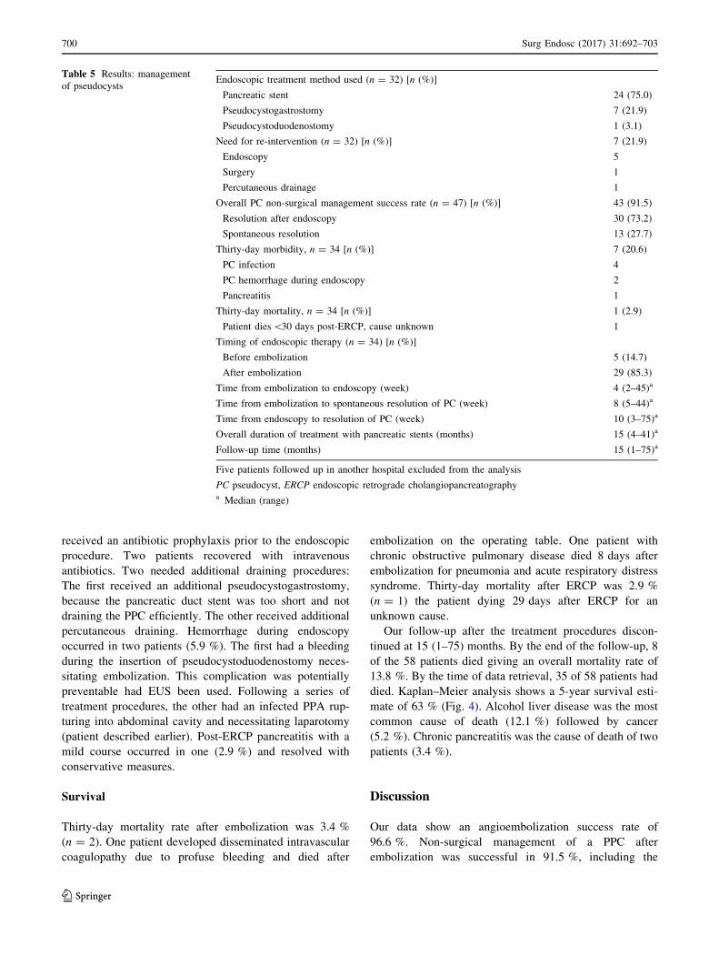
Table 5 Results: management
of pseudocystsEndoscopic treatment method used (n = 32) [n (%)]
Pancreatic stent 24 (75.0)
Pseudocystogastrostomy 7 (21.9)
Pseudocystoduodenostomy 1 (3.1)
Need for re-intervention (n = 32) [n (%)] 7 (21.9)
Endoscopy 5
Surgery 1
Percutaneous drainage 1
Overall PC non-surgical management success rate (n = 47) [n (%)] 43 (91.5)
Resolution after endoscopy 30 (73.2)
Spontaneous resolution 13 (27.7)
Thirty-day morbidity, n = 34 [n (%)] 7 (20.6)
PC infection 4
PC hemorrhage during endoscopy 2
Pancreatitis 1
Thirty-day mortality, n = 34 [n (%)] 1 (2.9)
Patient dies\30 days post-ERCP, cause unknown 1
Timing of endoscopic therapy (n = 34) [n (%)]
Before embolization 5 (14.7)
After embolization 29 (85.3)
Time from embolization to endoscopy (week) 4 (2–45)a
Time from embolization to spontaneous resolution of PC (week) 8 (5–44)a
Time from endoscopy to resolution of PC (week) 10 (3–75)a
Overall duration of treatment with pancreatic stents (months) 15 (4–41)a
Follow-up time (months) 15 (1–75)a
Five patients followed up in another hospital excluded from the analysis
PC pseudocyst, ERCP endoscopic retrograde cholangiopancreatographya Median (range)
700 Surg Endosc (2017) 31:692–703
123

endoscopically treated PPCs and the ones that resolved
spontaneously. Severe complications were rare. Only 3
(5.2 %) patients required surgery: two for a re-bleeding
episode and one for necrosis of cecum caused by post-
embolization Ogilvie’s syndrome. If left untreated, bleed-
ing PPAs and VAAs have a mortality of up to 90 % [5]. No
data on the long-term survival of these patients after the
successful treatment of hemorrhage exist. To study this, we
performed a Kaplan–Meier analysis that gave a 5-year
survival estimate of 63 % (Fig. 4). This shows that a
bleeding VAA or PPA with underlying pancreatitis is often
a late manifestation of long-term alcohol abuse and
degenerative way of life, and is, thus, associated with poor
prognosis.
In recent studies with patients recruited after year 2000,
TAE success rates reach 95–97 % [4, 6]. Our results are
consistent with these findings, supporting the current rec-
ommendations to use TAE as the first-line treatment for
bleeding PPAs and VAAs. Many earlier reports evaluating
the safety and efficacy of TAE have included a mixed
group of patients with various pancreatitis-associated
bleeding complications, showing mortality rates up to
19–21 % after TAE [3, 8]. We included patients who had
developed PPCs as a complication of AP and CP. None of
our patients had acute necrotizing pancreatitis, which most
likely explains our low 30-day mortality of 3.4 % after
TAE.
We had 52 coiling, three stenting, two particle injection
and only one plug placement procedures. Coils are safe and
easy to use, and coiling is our standard method in TAE.
Particle injections in the mesenteric arteries and celiac axis
may be risky due to ischemic complications caused by
particle spread, and in these arteries, particle injections
should be used with caution. Stents and plugs are safe to
use but stiff and inflexible and, thus, technically more
challenging to position. Stenting also allows a transarterial
approach when the visceral artery aneurysm is, e.g., in the
superior mesenteric artery that cannot be coiled. We cannot
draw any reliable conclusions on the post-embolization
complication profile of different embolization methods due
to the low numbers of stents, plugs and particle injections
used.
Current study is the largest so far reporting the outcomes
of non-surgical management of PPAs. Bhasin et al. [15]
published their results with eight patients in 2013. Case
reports from Elton et al. (1997) included three patients and
from Sayilir et al. (2011) only one patient [16, 17]. Earlier
reports by Bhasin et al., Elton et al. and Sayilir et al. have
shown promising results concerning the conservative
treatment of bleeding PPCs with combining endoscopy and
TAE [15–17]. In their study on the efficacy of TAE in the
treatment of PPAs in CP, Udd et al. [4] performed thera-
peutic endoscopy following TAE on 13 patients. None of
these needed surgery for their PPCs within a 14-month
follow-up. With larger study population and more exten-
sive follow-up, our findings strengthen the existing
evidence.
Our study bears all the known weaknesses of a retro-
spective study. In the modern era, cystoenterostomies are
the standard of care with draining PPCs in many centers
[24]. In our unit, however, we use pancreatic duct stenting
in two out of three PPC drainage procedures, and the same
trend can be seen with our study subjects with a history of
bleeding PPAs (9 out of 32 received transmural drainage).
This exception to the common practice should be kept in
mind when reading our results. It is at least to some extent
a result of the high prevalence of CP (79.3 %) in our
material, transpapillary approach allowing simultaneous
access to often-strictured pancreatic duct. When retro-
spectively analyzing the pre-ERCP CT scans, the trans-
mural approach was an alternative for only four out of the
24 patients treated primarily with a pancreatic duct stent.
With the remaining 20 patients, the PPCs lay far in the
pancreatic tail (seven patients), had pancreatic tissue in
between the gastrointestinal lumen and the PPC (five
patients), were too far from the gastric or duodenal wall
(four patients), or had vascular structures close to the PPC
(two patients), precluding the transmural approach. Two
patients had multiple PPCs that were all managed with
transpapillary stenting. It is also worth pointing out that we
excluded eight patients that received surgery as the first-
line treatment, which could affect the generalizability of
our results. A look into their patient files shows various
reasons behind the decision to proceed directly to surgery:
Angiography was negative with no bleeding site found on
Fig. 4 Kaplan–Meier curve showing survival after embolization in
patients with acute or chronic pancreatitis complicated by bleeding
pseudoaneurysms or visceral artery aneurysms
Surg Endosc (2017) 31:692–703 701
123
four of the eight. Angiography showed anomalous arteries
suspicious for malignancy with one, necessitating Whipple
procedure. Interventional radiologist was not available in
the hospital on one occasion. With two remaining cases, no
rationale for direct surgical intervention without angiog-
raphy was evident when reading the patient files.
Traditionally, negative angiography with no
detectable bleeding site has indicated surgery. No pub-
lished data solely on empirical embolizations of pancre-
atitis-associated bleeding complications exist. We
performed empirical embolization on nine patients with no
contrast extravasation or VAA detectable in angiography.
Three of these patients re-bled, after which two underwent
successful recoiling and one needed surgery. Incidence of
re-bleeds was not higher in patients who received empirical
TAE. Over the years, interventional radiologists have
become more active in performing empirical embolizations
that have become increasingly important in the treatment of
PPAs on patients with negative angiographies. Major
complications and organ ischemia (excluding splenic
infarctions) are rare but still worth taking into considera-
tion [25]. An alternative strategy for hemodynamically
stable patients could be watchful waiting with careful
monitoring and repeated angiography when re-bleeding
occurs. Comparison and further studies on these approa-
ches are necessary.
Optimal timing of therapeutic endoscopy after TAE
remains unsolved. In the three reports already mentioned,
patients received ERCP during the same hospital admission
within a few days from the embolization [15–17]. We
performed endoscopy on an outpatient visit 4 (2–47) weeks
following TAE believing that this would reduce bleeding
and infection complications. Depending on the underlying
etiology, previous publications show a spontaneous
regression of a PPC in 3–65 % of patients [26–29]. In our
study, PPCs resolved spontaneously in 27.7 %, which also
favors delayed endoscopic intervention.
Our findings show that combining TAE with therapeutic
endoscopy is a safe and efficient approach for the definitive
treatment of PPCs complicated by bleeding VAAs in AP or
CP. TAE is associated with shorter hospital stays than
surgery in the treatment of bleeding PPAs [4]. It is likely to
be more cost-effective also when combined with thera-
peutic endoscopy for definitive treatment of PPAs. PPAs in
AP and CP often appear in the context of alcoholism and
other severe comorbidities increasing the risks for surgery.
Non-surgical approach provides a less-invasive treatment
method for this moribund group of patients. Last, pro-
ceeding to surgery is still an option, if non-surgical treat-
ment fails. Prophylactic embolization is necessary for
incidental VAAs and should precede therapeutic endo-
scopy when CT detects an incidental PPC with a VAA. We
performed therapeutic endoscopy before TAE on five
patients with a history of bleeding episode. Pre-endoscopy
CT scans did not detect their VAAs. Acute bleeding
occurred during two of these endoscopies, necessitating
instant embolization.
In conclusion, bleeding PPAs require non-surgical
management. Patients with PPAs or bleeding VAAs should
receive a prompt angiography and embolization. Surgery is
necessary only if embolization is not feasible or fails.
Follow-up imaging with CT allows for determination of
any remaining PPCs after embolization. Utilizing thera-
peutic endoscopy in managing such PPCs is safe and
efficient and has a potential to save patients from the risks
of pancreatic surgery. We need more data to give a solid
recommendation on the optimal timing of the endoscopic
procedures. Further research is also necessary to clarify the
roles of empirical embolizations and watchful waiting after
negative angiography.
Acknowledgments One-month researcher’s salary paid for Taina
Nykanen by the Helsinki University Hospital Research Fund to allow
full-time research.
Compliance with ethical standards
Disclosures Marianne Udd, Erno Peltola, Ari Leppaniemi and Leena
Kylanpaa have no conflicts of interest or financial ties to disclose.
References
1. Andren-Sandberg A, Dervenis C (2004) Pancreatic pseudocysts
in the 21st century. Part I: classification, pathophysiology, ana-
tomic considerations and treatment. J Pancreas 5:8–24
2. Balthazar EJ, Fisher LA (2001) Hemorrhagic complications of
pancreatitis: radiologic evaluation with emphasis on CT imaging.
Pancreatology 1:306–313. doi:10.1159/000055829
3. Bergert H, Hinterseher I, Kersting S, Leonhardt J, Bloomenthal
A, Saeger HD (2005) Management and outcome of hemorrhage
due to arterial pseudoaneurysms in pancreatitis. Surgery
137:323–328. doi:10.1016/j.surg.2004.10.009
4. Udd M, Leppaniemi AK, Bidel S, Keto P, Roth W-D, Haapiainen
RK (2007) Treatment of bleeding pseudoaneurysms in patients
with chronic pancreatitis. World J Surg 31:504–510. doi:10.1007/
s00268-006-0209-z
5. Chiang K-C, Chen T-H, Hsu J-T (2014) Management of chronic
pancreatitis complicated with a bleeding pseudoaneurysm. WJG
20:16132–16137. doi:10.3748/wjg.v20.i43.16132
6. Kim J, Shin JH, Yoon H-K, Ko G-Y, Gwon DI, Kim E-Y, Sung
K-B (2015) Endovascular intervention for management of pan-
creatitis-related bleeding: a retrospective analysis of thirty-seven
patients at a single institution. Diagn Interv Radiol 21:140–147.
doi:10.5152/dir.2014.14085
7. Nicholson AA, Patel J, McPherson S, Shaw DR, Kessel D (2006)
Endovascular treatment of visceral aneurysms associated with
pancreatitis and a suggested classification with therapeutic
implications. J Vasc Interv Radiol 17:1279–1285. doi:10.1097/
01.RVI.0000231948.08617.04
8. Balachandra S, Siriwardena AK (2005) Systematic appraisal of
the management of the major vascular complications of pancre-
atitis. Am J Surg 190:489–495. doi:10.1016/j.amjsurg.2005.03.
009
702 Surg Endosc (2017) 31:692–703
123

9. Kirby JM, Vora P, Midia M, Rawlinson J (2007) Vascular
complications of pancreatitis: imaging and intervention. Cardio-
vasc Interv Radiol 31:957–970. doi:10.1007/s00270-007-9138-y
10. Dumonceau J-M (2013) Endoscopic management of complica-
tions of chronic pancreatitis. WJG 19:7308–7309. doi:10.3748/
wjg.v19.i42.7308
11. Tandan M (2013) Endotherapy in chronic pancreatitis. WJG
19:6156–6164. doi:10.3748/wjg.v19.i37.6156
12. Dumonceau JM, Delhaye M, Tringali A, Dominguez-Munoz J,
Poley JW, Arvanitaki M, Costamagna G, Costea F, Deviere J,
Eisendrath P, Lakhtakia S, Reddy N, Fockens P, Ponchon T,
Bruno M (2012) Endoscopic treatment of chronic pancreatitis:
European Society of Gastrointestinal Endoscopy (ESGE) Clinical
Guideline. Endoscopy 44:784–800. doi:10.1055/s-0032-1309840
13. Varadarajulu S, Bang JY, Sutton BS, Trevino JM, Christein JD,
Wilcox CM (2013) Equal efficacy of endoscopic and surgical
cystogastrostomy for pancreatic pseudocyst drainage in a ran-
domized trial. Gastroenterology 145(583–590):e1. doi:10.1053/j.
gastro.2013.05.046
14. Johnson MD, Walsh M, Henderson JM, Brown N, Ponsky J,
Dumont J, Zuccaro G, Vargo J (2009) Surgical versus nonsur-
gical management of pancreatic pseudocysts. J Clin Gastroenterol
43:586–590
15. Bhasin DK, Rana SS, Sharma V, Rao C, Gupta V, Gupta R, Kang
M, Singh K (2013) Non-surgical management of pancreatic
pseudocysts associated with arterial pseudoaneurysm. Pancre-
atology 13:250–253. doi:10.1016/j.pan.2013.02.011
16. Elton E, Howell D, Amberson S, Dykes T (1997) Combined
angiographic and endoscopic management of bleeding pancreatic
pseudoaneurysms. Gastrointest Endosc 46:544–549
17. Sayilir A, Onal IK, Beyazit Y, Surmelioglu A, Salper Okten R,
Odemis B, Parlak E, Sasmaz N (2011) A rare cause of upper
gastrointestinal bleeding: hemosuccus pancreaticus: angiographic
and endoscopic combined treatment. Surg Laparosc Endosc
Percutaneous Techn 21:e286–e287. doi:10.1097/SLE.
0b013e31822f50b6
18. Cotton PB, Lehman G, Vennes J, Geenen JE, Russel RC, Meyers
WC, Liguory C, Nickl N (1991) Endoscopic sphincterotomy
complications and their management: an attempt at consensus.
Gastrointest Endosc 37:383–393
19. Barge JU, Lopera JE (2012) Vascular complications of pancre-
atitis: role of interventional therapy. Korean J Radiol 13:S45–
S55. doi:10.3348/kjr.2012.13.S1.S45
20. Park D, Lee S, Moon SH, Choi S, Jung S, Seo D, Lee S, Kim MH
(2009) Endoscopic ultrasound-guided versus conventional
transmural drainage for pancreatic pseudocysts: a prospective
randomized trial. Endoscopy 41:842–848. doi:10.1055/s-0029-
1215133
21. Weckman L, Kylanpaa ML, Puolakkainen P, Halttunen J (2006)
Endoscopic treatment of pancreatic pseudocysts. Surg Endosc
20:603–607. doi:10.1007/s00464-005-0201-y
22. Arvanitakis M, Delhaye M, Bali MA, Matos C, De Maertelaer V,
Le Moine O, Deviere J (2007) Pancreatic-fluid collections: a
randomized controlled trial regarding stent removal after endo-
scopic transmural drainage. Gastrointest Endosc 65:609–619.
doi:10.1016/j.gie.2006.06.083
23. Cahen D, Rauws E, Fockens P, Weverling G, Huibregtse K,
Bruno M (2005) Endoscopic drainage of pancreatic pseudocysts:
long-term outcome and procedural factors associated with safe
and successful treatment. Endoscopy 37:977–983. doi:10.1055/s-
2005-870336
24. Yang D, Amin S, Gonzalez S, Mullady D, Hasak S, Gaddam S,
Edmundowicz S, Gromski M, DeWitt J, Zein MD, El M, Khashab
M, Wang A, Gaspar J, Uppal D, Nagula S, Kapadia S, Buscaglia
J, Bucobo JC, Schlachterman A, Wagh M, Draganov P, Kyu Jung
M, Stevens T, Vargo J, Khara H, Huseini M, Diehl D, Keswani R,
Law R, Komanduri S, Yachimski P, DaVee T, Prabhu A, Lapp R,
Kwon R, Watson R, Goodman A, Chhabra N, Wang W, Benias P,
Carr-Locke D, DiMaio C (2016) Transpapillary drainage has no
added benefit on treatment outcomes in patients undergoing EUS-
guided transmural drainage of pancreatic pseudocysts: a large
multicenter study. Gastrointest Endosc 83:720–729. doi:10.1016/
j.gie.2015.10.040
25. Andersson E, Ansari D, Andersson R (2010) Major haemorrhagic
complications of acute pancreatitis. Br J Surg 97:1379–1384.
doi:10.1002/bjs.7113
26. Lankisch PG, Weber-Dany B, Maisonneuve P, Lowenfels AB
(2012) Pancreatic pseudocysts: prognostic factors for their
development and their spontaneous resolution in the setting of
acute pancreatitis. Pancreatology 12:85–90. doi:10.1016/j.pan.
2012.02.007
27. Mehta R, Suvarna D, Sadasivan S, John A, Raj V, Nair P,
Balakrishnan V (2004) Natural course of asymptomatic pancre-
atic pseudocyst: a prospective study. Indian J Gastroenterol
23:140–142
28. Andren-Sandberg A, Dervenis C (2004) Pancreatic pseudocysts
in the 21st century. Part II: natural history. J Pancreas 5:64–70
29. Bradley EL, Gonzalez AC, Clements JR Jr (1976) Acute pan-
creatic pseudocysts: incidence and implications. Ann Surg
184:734–737
Surg Endosc (2017) 31:692–703 703
123