Biology and principles of periodontal wound healing/regeneration
18
Biology and principles of periodontal wound healing/regeneration G IUSEPPE P OLIMENI ,A NDREAS V. X IROPAIDIS &U LF M. E. W IKESJO ¨ The native periodontium includes cementum, a functionally oriented periodontal ligament, alveolar bone and gingiva. Pathologic and/or traumatic events may lead to the loss or damage of this anatomical structure. Since the 1970s, a number of procedures have been investigated in an attempt to restore such lost tissues. Numerous clinical trials have shown positive outcomes for various reconstructive surgical protocols. Reduced probing depths, clinical attach- ment gain, and radiographic bone fill have been reported extensively for intrabony and furcation de- fects following scaling and root planing, open flap debridement, autogenous bone grafting, implanta- tion of biomaterials including bone derivatives and bone substitutes, guided-tissue regeneration (GTR) procedures, and implantation of biologic factors, including enamel matrix proteins. Histological studies have shown that various surgical periodontal procedures can lead to differ- ent patterns of healing. Healing by formation of a long junctional epithelium (epithelial attachment) is characterized by a thin epithelium extending apically interposed between the root surface and the gingival connective tissue (4, 23). Connective tissue repair (new attachment) is represented by collagen fibers oriented parallel or perpendicular to a root surface previously exposed to periodontal disease or other- wise deprived of its periodontal attachment. In con- trast, periodontal regeneration is characterized by de novo formation of cementum, a functionally ori- ented periodontal ligament, alveolar bone, and gin- giva (restitutio ad integrum). Nevertheless, it would be naive to expect these to occur as distinctly sep- arate biologic outcomes following reconstruction of the periodontal attachment. For example, periodon- tal regeneration should be expected to include ele- ments of a new, as well as an epithelial, attachment. Predictability of outcomes following surgical pro- cedures is of fundamental importance in medicine. As periodontal-regenerative procedures are time consuming and financially demanding, there is increasing interest by clinicians to learn of factors that may influence the clinical outcome following periodontal reconstructive surgery in order to pro- vide the best possible service to patients. This goal can only be achieved if biological aspects of wound healing and regeneration are taken into considera- tion. The objectives of the present article are to provide an overview of wound healing following periodontal surgical procedures, to discuss the basic principles of periodontal regeneration, and to illus- trate the factors that influence this process. Wound healing The healing of wounds in nonoral sites has been studied in great detail (Fig. 1) (5). The general prin- ciples of healing, and the cellular and molecular events observed in nonoral sites, also apply to the healing processes that take place following perio- dontal surgery. Traumatic injury causes capillary damage and hemorrhage and, as a result, a blood clot is formed. The formation of a clot is the immediate response to any trauma. The clot has two functions: it temporarily protects the denuded tissues; and it serves as a provisional matrix for cell migration (5). The blood clot consists of all cellular components of blood (including red and white blood cells and platelets) in a matrix of fibrin, plasma fibronectin, vitronectin, and thrombosporin (24). Clot formation is followed by an early stage of inflammation. Within hours of injury, inflammatory cells (predominantly neutrophils and monocytes) 30 Periodontology 2000, Vol. 41, 2006, 30–47 Printed in Singapore. All rights reserved Copyright Ó Blackwell Munksgaard 2006 PERIODONTOLOGY 2000
Transcript of Biology and principles of periodontal wound healing/regeneration

Biology and principles ofperiodontal woundhealing/regeneration
GI U S E P P E PO L I M E N I, AN D R E A S V. XI R O P A I D I S & UL F M. E. WI K E S J O
The native periodontium includes cementum, a
functionally oriented periodontal ligament, alveolar
bone and gingiva. Pathologic and/or traumatic events
may lead to the loss or damage of this anatomical
structure. Since the 1970s, a number of procedures
have been investigated in an attempt to restore such
lost tissues. Numerous clinical trials have shown
positive outcomes for various reconstructive surgical
protocols. Reduced probing depths, clinical attach-
ment gain, and radiographic bone fill have been
reported extensively for intrabony and furcation de-
fects following scaling and root planing, open flap
debridement, autogenous bone grafting, implanta-
tion of biomaterials including bone derivatives and
bone substitutes, guided-tissue regeneration (GTR)
procedures, and implantation of biologic factors,
including enamel matrix proteins.
Histological studies have shown that various
surgical periodontal procedures can lead to differ-
ent patterns of healing. Healing by formation of a
long junctional epithelium (epithelial attachment) is
characterized by a thin epithelium extending apically
interposed between the root surface and the gingival
connective tissue (4, 23). Connective tissue repair
(new attachment) is represented by collagen fibers
oriented parallel or perpendicular to a root surface
previously exposed to periodontal disease or other-
wise deprived of its periodontal attachment. In con-
trast, periodontal regeneration is characterized by
de novo formation of cementum, a functionally ori-
ented periodontal ligament, alveolar bone, and gin-
giva (restitutio ad integrum). Nevertheless, it would
be naive to expect these to occur as distinctly sep-
arate biologic outcomes following reconstruction of
the periodontal attachment. For example, periodon-
tal regeneration should be expected to include ele-
ments of a new, as well as an epithelial, attachment.
Predictability of outcomes following surgical pro-
cedures is of fundamental importance in medicine.
As periodontal-regenerative procedures are time
consuming and financially demanding, there is
increasing interest by clinicians to learn of factors
that may influence the clinical outcome following
periodontal reconstructive surgery in order to pro-
vide the best possible service to patients. This goal
can only be achieved if biological aspects of wound
healing and regeneration are taken into considera-
tion. The objectives of the present article are to
provide an overview of wound healing following
periodontal surgical procedures, to discuss the basic
principles of periodontal regeneration, and to illus-
trate the factors that influence this process.
Wound healing
The healing of wounds in nonoral sites has been
studied in great detail (Fig. 1) (5). The general prin-
ciples of healing, and the cellular and molecular
events observed in nonoral sites, also apply to the
healing processes that take place following perio-
dontal surgery. Traumatic injury causes capillary
damage and hemorrhage and, as a result, a blood clot
is formed. The formation of a clot is the immediate
response to any trauma. The clot has two functions: it
temporarily protects the denuded tissues; and it
serves as a provisional matrix for cell migration (5).
The blood clot consists of all cellular components of
blood (including red and white blood cells and
platelets) in a matrix of fibrin, plasma fibronectin,
vitronectin, and thrombosporin (24).
Clot formation is followed by an early stage of
inflammation. Within hours of injury, inflammatory
cells (predominantly neutrophils and monocytes)
30
Periodontology 2000, Vol. 41, 2006, 30–47
Printed in Singapore. All rights reserved
Copyright � Blackwell Munksgaard 2006
PERIODONTOLOGY 2000

populate the clot. These cells cleanse the wound of
bacteria and necrotic tissue through phagocytosis
and release of enzymes and toxic oxygen products.
Within 3 days, the inflammatory reaction moves into
its late phase. Macrophages migrate into the wound
area and, in addition to wound debridement, secrete
polypeptide mediators targeting cells involved in the
wound-healing process (5, 45).
The macrophage plays an important role in the
formation of granulation tissue. Growth factors and
cytokines secreted by macrophages are involved in
the proliferation and migration of fibroblasts,
endothelial cells, and smooth muscle cells into the
wound area. The cell-rich granulation tissue next
undergoes maturation and remodeling. Fibroblasts
responsible for the replacement of the provisional
extracellular matrix produce a new collagen-rich
matrix. Approximately 1 week following wounding,
and once the collagen matrix has been synthesized,
some fibroblasts undergo transformation into myo-
fibroblasts and express a-smooth muscle actin. This
transformation and synthesis is responsible for
wound contraction. Endothelial cells, responsible for
angiogenesis, migrate into the provisional wound
matrix to form vascular tubes and loops, and as the
provisional matrix matures, the endothelial cells un-
dergo programmed cell death (apoptosis) and the
number of vascular units is reduced (1, 24).
Epithelization of the wound is initiated within hours
of injury. Epithelial cells from the basal layer prolif-
erate and migrate through the fibrin clot and eventu-
ally the breach in the epithelium is sealed. The
epithelial cells in normal gingival tissues use surface
receptors, known as integrins, to bind to laminin in the
basal lamina. In order to initiate migration, the kera-
tinocytes dissolve this attachment to start expressing
integrins suitable for the wound environment (1).
Maturation of the granulation tissue will lead to the
regeneration or repair (scar formation) of the injured
tissues. Whether the damaged tissues heal by regen-
eration or repair depends upon two crucial factors: the
availability of cell type(s) needed; and the presence or
absence of cues and signals necessary to recruit and
stimulate these cells (8). This summary represents
an oversimplified explanation of the wound-healing
process. The stages described may overlap consider-
ably and the time needed for completion of each stage
may vary, depending on local and systemic factors.
Periodontal wound healing
A more complex situation presents itself when a
mucoperiosteal flap is apposed to an instrumented
root surface deprived of its periodontal attachment.
In this case, the wound margins are not two opposing
vascular gingival margins but comprise the rigid
nonvascular mineralized tooth surface, on the one
hand, and the connective tissue and epithelium of
the gingival flap, on the other. The periodontal
wound also includes tissue resources from the
alveolar bone and the periodontal ligament. Clot
formation at the interface between the tooth and a
gingival flap is initiated as blood elements are im-
posed onto the root surface during surgery and at
wound closure in a seemingly random manner, much
like a Jackson Pollock dripped and poured style
canvas (http://www.nga.gov/feature/pollock/pollock
home.html [accessed 16 February 2006]). This
represents the very first healing event at the tooth–
gingival flap interface (i.e. the absorption and adhe-
sion of plasma proteins onto the root surface) (Fig. 2)
(49). Within minutes, a fibrin clot attached to the root
surface is developed. Within hours, one may observe
the early phase of inflammation as inflammatory
cells, predominantly neutrophils and monocytes,
accumulate on the root surface, and within 3 days
the late phase of inflammation dominates the healing
picture as macrophages migrate into the wound fol-
lowed by the formation of granulation tissue. At
7 days, a connective tissue attachment may be seen
at the root surface; however, areas of the fibrin clot in
various stages of maturation may also be observed,
depending on wound volume and tissue resources.
Earlyphase
Latephase
Fig. 1. Phases of wound healing (epidermal incisional
wounds), including an early (within hours) and a late
(within days) phase of inflammation dominated by poly-
morphonuclear neutrophils and macrophages, respect-
ively. The magnitude of wound contraction parallels the
phase of granulation tissue formation. Collagen accumu-
lation is first observed during the phase of granulation
tissue formation, continuing through the phase of matrix
formation and remodeling. Redrawn with permission
from Dr Richard AF Clark.
31
Biology and principles of periodontal wound healing/regeneration

Fig. 2. Early healing events at the tooth–gingival flap
interface. (A) Red blood cells (RBC) in a granular preci-
pitate adhering to the dentin surface, shown immediately
(10 min) upon wound closure; an artifactual split (arrows)
between the red blood cells and the dentin-adhering
precipitate verifies the absorption/adhesion of blood ele-
ments to the dentin surface (transmission electron
micrograph, ·4000). (B) Red blood cells in a fibrin network
in close proximity to the root surface observed within 1 h
of wound closure (photomicrograph ·450). (C) Early
phase of inflammation: RBC aggregates are loosely inter-
spersed in an organized fibrin network at 6 h. The fibrin
clot appears to be attached to the dentin surface.
Numerous polymorphonuclear cells are observed lining
the dentin surface (photomicrograph ·450). (D) Late
phase of inflammation, 3 days following wound closure,
showing macrophages lining the dentin surface (trans-
mission electron micrograph, ·10,500). (E) Granulation
tissue formation, including fibroblasts in the maturing
fibrin clot at the dentin surface (photomicrograph, ·450).(F) Cell-rich connective tissue closely adapted to the
dentin surface at 7 days following wound closure (pho-
tomicrograph, ·450). D, Dentin/root surface; F, fibrin; FB,
fibroblast; M, macrophage; P, plasma precipitate; PMN,
polymorphonuclear neutrophils; RBC, red blood cells;. For
detail see: Wikesjo et al. 1991 (49). These figures are
copyrighted by and modified with permission from the
American Academy of Periodontology.
32
Polimeni et al.

The studies described above investigated the
absorption, adhesion, and structural maturation of
the fibrin clot in periodontal wound healing, but have
not taken into consideration the functional integrity
of the tooth–gingival flap interface. Only a few
experimental studies have as such evaluated the
functional integrity of a maturing periodontal wound.
Hiatt et al. (11) examined the tensile strength of the
tooth–gingival flap interface following reconstructive
surgery of relatively small surgical dehiscence defects
over the maxillary canine teeth in the dog. They
found that the tensile strength increases from �200 g
at 3 days postsurgery to 340 g at 5–7 days postsur-
gery, and to >1700 g at 2 weeks postsurgery. In other
words, they found that a relatively limited perio-
dontal wound might not reach functional integrity
until 2 weeks postsurgery. These data suggest that
wound integrity during the early healing phase rests
primarily on the stabilization of the gingival flaps
offered by suturing. In consequence, the choice of
suture material, placement and removal, for perio-
dontal surgical procedures aimed at regeneration
should be dictated by such observations as should
postoperative protocols be instituted aimed at pro-
tecting the surgical site from trauma from oral hy-
giene procedures, and plaque colonization and
infection (21, 37).
As mentioned above, the regeneration of lost tis-
sues depends on the availability of the cell type(s)
needed and the presence or absence of cues and
signals necessary to recruit and stimulate these cells.
The extracellular matrix regulates how cells respond
to these signals. Stem cells responsible for regener-
ation of the periodontal tissues reside within the
periodontal ligament (8). The innate regenerative
potential of the periodontium has been investigated
extensively and clearly appears to be dependent on
wound management (see below). Current research
focuses on identifying biologic factors that favor
migration and proliferation of periodontal tissues
and to use those to alter the microenvironment of the
wound, favoring unimpeded healing and regener-
ation of the periodontium.
Biological factors at the base ofperiodontal regeneration
Melcher (25) postulated biological concepts at the
base of periodontal regeneration. Accordingly,
periodontal structures are subdivided in four
compartments (gingival corium, periodontal liga-
ment, cementum, and bone) and the nature of the new
attachment following periodontal surgery is deter-
mined by the cells repopulating the root surface.
Karring, Nyman et al. (14) corroborated these
concepts in a series of experiments. They asked: �Can
a new connective tissue attachment be established to
a root surface previously exposed to the oral envi-
ronment and implanted into bone?� (13). In a dog
model, they extracted and crown-resected perio-
dontitis-affected teeth, scaled and root planed the
portion of the root that was affected with periodon-
titis, and implanted the roots into cavities created in
the alveolar bone. While connective tissue repair did
not occur at the periodontitis-affected portion of the
roots, the portion where the periodontal ligament
was preserved showed an attachment with func-
tionally oriented periodontal fibers.
A second study answered the question: �Can a new
connective tissue attachment establish to a perio-
dontitis-affected root implanted into gingival con-
nective tissue?� (26). Using the dog model, they
implanted teeth oriented in such a way that one side
of the tooth faced alveolar bone while the other faced
the gingival connective tissue. Similarly to the previ-
ous study, no connective tissue attachment was
observed at the periodontitis-affected portion of
the roots. Conversely, the half of the root where
periodontal ligament was preserved showed a con-
nective tissue attachment.
They next asked: �Can a new connective tissue
attachment establish to root surface deprived of its
periodontal attachment giving preference to cells
from the periodontal ligament?� (27). Periodontal
fenestration defects were created at the maxillary
lateral incisors and mandibular canines in nonhu-
man primates. A Millipore filter was placed to cover
the fenestration defects with the aim of preventing
gingival connective tissue from contacting the root
surface. New cementum with a functionally oriented
periodontal ligament was observed within a 6-month
healing interval. Using the same rationale and tech-
nology, they subsequently provided the first evidence
that periodontal regeneration can be obtained at a
periodontitis-affected tooth in a human (28).
This series, and several associated studies by
Karring, Nyman et al., established that cells from
periodontal ligament have the capacity to regenerate
the periodontal attachment, while the alveolar bone
and gingival connective tissue do not possess this
ability (14). As postulated by Melcher, these findings
suggest that if preference is provided to cells origin-
ating from the periodontal ligament, periodontal
regeneration may consistently occur (25). It also
appears from these studies that occlusion of cells
33
Biology and principles of periodontal wound healing/regeneration
originating from the gingiva by means of tissue bar-
riers, also known as GTR techniques, is of paramount
importance in achieving periodontal regeneration.
Although Karring, Nyman et al. elegantly pioneered
and elucidated biological concepts at the base of
periodontal regeneration, a review of additional
studies investigating wound-healing dynamics and
maturation in periodontal defects suggest that addi-
tional factors play a role. Early observations by
Linghorne & O’Connell (22) suggest that a lack of
mechanical stability of the wound is the main factor
in the formation of a long junctional epithelium.
Hiatt et al. (11) demonstrated a fundamental role of
the root surface-adhering fibrin clot as a preventive
measure to apical migration of the gingival epithe-
lium. Polson & Proye (34), using a monkey model,
pointed to the importance of the unimpeded ab-
sorption, adhesion and maturation of a fibrin clot in
periodontal wound healing. In brief, teeth were re-
implanted after root planing of the coronal third of
the root (control) or root planing followed by root
surface demineralization with citric acid. While
healing by long junctional epithelium occurred in the
controls, the root surface-demineralized teeth ex-
hibited a �fibrin linkage� maturing into a connective
tissue attachment. Apparently, root surface demin-
eralization provided a stable anchorage of the fibrin
clot over that of the root planed-only teeth, allowing
its maturation into a connective tissue attachment.
Collectively, these studies all appear to support the
vital importance of unimpeded absorption, adhesion
and maturation of the fibrin clot for formation of a
connective tissue attachment over a long junctional
epithelium, as discussed above.
Our laboratories have developed and characterized
a preclinical model, designated as the Critical-size
Supraalveolar Periodontal Defect Model (Figs 3 and 4)
(17, 18, 44, 50). This animal model does not sponta-
neously regenerate following reconstructive surgery
without adjunctive measures. In addition, it allows
clinically relevant periodontal regeneration, induced
or supported by implanted biologics, biomaterials, or
devices over that in a surgical control. We initially
used this model to evaluate the significance of fibrin
clot absorption, adhesion and maturation to the root
surface. In a first study, root surfaces were coated
with heparin, to potentially interfere with fibrin clot
formation/absorption/adhesion. It was shown that
root surfaces coated with heparin exhibited forma-
tion of an epithelial attachment (long junctional
epithelium) whereas at control sites conditioned with
saline, the epithelium was arrested at or immediately
apical to the cemento–enamel junction (Fig. 5) (48).
Apparently, the heparin coating compromised local
fibrin clot formation/absorption/adhesion, either by
compromising the clotting cascade, or by some
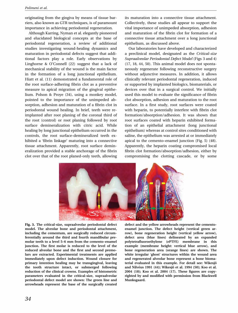
Fig. 3. The critical-size, supraalveolar periodontal defect
model. The alveolar bone and periodontal attachment,
including the cementum, are surgically reduced circum-
ferentially around the third and fourth mandibular pre-
molar teeth to a level 5–6 mm from the cemento–enamel
junction. The first molar is reduced to the level of the
reduced alveolar bone and the first and second premo-
lars are extracted. Experimental treatments are applied
immediately upon defect induction. Wound closure for
primary intention healing may be transgingival, leaving
the tooth structure intact, or submerged following
reduction of the clinical crowns. Examples of histometric
parameters evaluated in the critical-size, supraalveolar
periodontal defect model are shown: The green line and
arrowheads represent the base of the surgically created
defect and the yellow arrowheads represent the cemento-
enamel junction. The defect height (vertical green ar-
row), bone regeneration height (vertical yellow arrow),
defect area (blue lines) delineated by an expanded
polytetrafluoroethylene (ePTFE) membrane in this
example (membrane height: vertical blue arrow), and
bone regeneration area (orange lines) are shown. The
white irregular �ghost� structures within the wound area
and regenerated alveolar bone represent a bone bioma-
terial evaluated in this example. For detail see: Wikesjo
and Nilveus 1991 (44); Wikesjo et al. 1994 (50); Koo et al.
2004 (18); Koo et al. 2004 (17). These figures are copy-
righted by and modified with permission from Blackwell
Munksgaard.
34
Polimeni et al.

nonspecific surface action, or by a combination of
these effects. This single experimental manipulation
apparently prevented the maturation of the fibrin clot
into a connective tissue attachment but resulted in
epithelial migration and proliferation along the root
surface, probably as a consequence of exposure of
the compromised fibrin clot to wound-rupturing
forces acting on the gingival margins. In contrast,
when a polylactic acid implant or expanded poly-
tetrafluoroethylene (ePTFE) membranes supported
the gingival flaps in heparin-coated defects, the
tooth–gingival flap interface healed by formation of
connective tissue rather than by forming an epithelial
attachment (Fig. 6) (9, 43). In wound sites stabilized
by means of the polylactic acid implant or ePTFE
membranes, the epithelium was arrested coronally at
some distance from the implant or membrane, in
itself indicating that wound stability, and not tissue
occlusion, played a fundamental role in the outcomes
of healing. Apparently the polylactic acid implant and
the ePTFE membrane stabilized the wound, protect-
ing the compromised fragile fibrin clot from wound-
rupturing forces acting on the gingival margins.
These studies suggest that provided adequate wound
stability, periodontal wound healing may result in the
formation of a connective tissue attachment rather
than an epithelial attachment (long junctional epi-
thelium), which in turn should be considered a
consequence of wound failure. In perspective,
modification of the root surface by application of
etching and chelating agents may enhance fibrin clot
adhesion (2, 3) and promote a connective tissue
attachment (34, 47). In contrast, conditioning the
root surface with protein constructs may compro-
mise fibrin clot adhesion and, consequently, perio-
dontal regeneration (3, 46).
Altogether, the evidence suggests that wound sta-
bility is essential for the establishment of a new con-
nective tissue attachment to a root surface deprived of
its periodontal attachment, and that tissue resources
originating from the periodontal ligament represent a
single source for periodontal regeneration, providing
Fig. 4. The critical-size, supraalveolar periodontal defect
model. The photomicrograph shows a representative
section of a sham-surgery control following transgingival
wound closure and a 4-week healing interval. The green
arrowhead identifies the apical extension of the defect
(see Fig. 3) and the red arrowhead delineates the extent
of alveolar regeneration. The schematic illustration
shows healing, expressed as a percentage of the defect
height, in the critical-size, supraalveolar periodontal de-
fect model following a 4-week healing interval and
transgingival wound closure, and following an 8-week
healing interval and submerged wound closure. Note that
the epithelium is arrested at or immediately below the
cemento–enamel junction in sham-surgery control sites.
There is limited, if any, regeneration of the periodontal
attachment, as evaluated by regeneration of cementum
or a cementum-like tissue extending from the apical
extension of the defect. Bone regeneration is limited to
<25% of the defect height following a 4- or 8-week
healing interval, indicating that in control sites bone
regeneration is exhausted within 4 weeks. These char-
acteristics provide a discriminating critical-size model for
evaluation of the clinical potential of implantable/
injectable devices, biomaterials, biologics, and cell con-
structs, with or without root surface biomodifications.
Substantial regeneration in this discriminating model
warrants clinical pursuit. Limited regeneration appears
less deserving. For detail see: Wikesjo and Nilveus 1991
(44); Wikesjo et al. 1994 (50); Koo et al. 2004 (18); Koo
et al. 2004 (17). These figures are copyrighted by and
modified with permission from the American Academy of
Periodontology.
35
Biology and principles of periodontal wound healing/regeneration

cells with the ability to differentiate into cemento-
blasts, fibroblasts, and osteoblasts. Detachment of the
maturating fibrin clot from the root surface owing to a
lack of wound stabilization will inexorably compro-
mise periodontal wound healing, ultimately jeopard-
izing the regenerative process.
Clinical and biologic variablesaffecting periodontal regeneration
Kornman & Robertson (20) classified factors that may
influence the successful management of periodontal
osseous defects. Their classification includes:
• Bacterial contamination.
• Innate wound-healing potential.
• Local site characteristics.
• Surgical procedure/technique.
Cortellini & Tonetti (6) suggested decision trees,
along these lines, to provide clinicians with direction
in their treatment of periodontal intrabony defects.
Again, patient factors and defect morphology appear
to be crucial for the direction of therapy. In the fol-
lowing we use biologic observations in the Critical-
size Supraalveolar Periodontal Defect Model to
elucidate factors, including wound maturation, tissue
occlusion, primary intention healing, wound failure
and membrane exposure, defect characteristics,
space provision, and innate regenerative potential,
that clinicians may need to consider in the regener-
ative treatment of periodontal defects.
Wound maturation
Haney et al. (9) evaluated periodontal wound healing
associated with GTR membranes in supraalveolar
Fig. 5. Critical-size, 5-mm, supraalveolar periodontal
defect including coating the root surfaces with a heparin
solution immediately prior towoundclosurewith the intent
to interfere with local fibrin clot formation/absorption/
adhesion. The clinical series shows the defect, the root
surfaces isolated with a rubber dam for the heparin appli-
cation, transgingival wound closure, and healing at
4 weeks. The left photomicrographs show sites that have
received the heparin coating. The green arrowheads indi-
cate the base of the defects and the blue arrowhead the
apical termination of the epithelial attachment (long
junctional epithelium) formed in these sites. Apparently,
the heparin coating compromised the fibrin clot in the
tooth–gingival flap interface to such an extent that allowed
apical migration and proliferation of cells from the gingival
epithelium rather thanmaturation into a connective tissue
attachment. In contrast, the epithelium is arrested at the
cemento–enamel junction in control sites (right) treated
with saline, leaving the entire denuded root surface with a
new connective tissue attachment. This singular manipu-
lation aimed at interfering with coagulum formation/
absorption/adhesion points to the critical importance of
the provisionary matrix of the fibrin clot in periodontal
wound healing and ultimately periodontal regeneration.
Healing interval 4 weeks. For detail see: Wikesjo et al. 1991
(48). These figures are copyrighted by and modified with
permission from Blackwell Munksgaard.
Fig. 6. Critical-size, 5-mm, supraalveolar periodontal de-
fect including coating of the root surfaces with a heparin
solution immediately prior towoundclosurewith the intent
to interfere with local fibrin clot formation/absorption/
adhesion. The clinical series shows the defect, the root
surfaces isolated with a rubber dam for the heparin appli-
cation, placement of an expanded polytetrafluoroethylene
(ePTFE) membrane with the intent to stabilize the wound,
and transgingival wound closure. Control defects were
coatedwith heparin but did not receive ePTFEmembranes.
The photomicrographs show the epithelium arrested at the
cemento–enamel junction at some distance from the cor-
onal extension of the ePTFE membrane (blue arrow) in
heparin-coated defects. The controls (not shown) exhibited
formation of an epithelial attachment (long junctional
epithelium). Similar observations were made in heparin-
coated defects implanted with a polylactic acid block bio-
material. Collectively, these observations suggest that the
implanted device or biomaterial provided some stability to
the heparin-compromised tooth–gingival flap interface,
allowing the provisionary matrix (i.e. the fibrin clot) to
mature into a connective tissue attachment rather than
migration and proliferation of cells from the gingival epi-
thelium, resulting in formation of an epithelial attachment.
Healing interval 4 weeks. For detail see: Haney et al. 1993
(9); Wikesjo and Nilveus 1990 (43). These figures are copy-
righted by and modified with permission from the Amer-
ican Academy of Periodontology.
36
Polimeni et al.

periodontal defects and observed that most of the
space adjacent to the teeth underneath the mem-
branes filled with alveolar bone within a 4-week
healing interval (Fig. 7) (9). However, there was lim-
ited, if any, appreciable regeneration of cementum
and a functionally oriented periodontal ligament, as
evaluated by incandescent light microscopy, also
observed in subsequent studies using a 4-week
healing interval (19, 54). In contrast, evaluations of
periodontal regeneration in supraalveolar periodon-
tal defects using incandescent light microscopy and
healing intervals of 8 or 24 weeks demonstrated that
the observed bone formation is accompanied by the
regeneration of cementum and a functionally orien-
ted periodontal ligament (Fig 8–10) (15, 38, 51–53). As
experimental conditions were similar among these
studies, these observations point to the possibility of
a delayed structural maturation of the periodontal
attachment compared with that of the alveolar bone
following regenerative procedures.
Tissue occlusion
Design criteria for GTR membranes include bio-
compatibility, cell occlusion, space maintenance,
tissue integration, and ease of use (10, 36). Although
biocompatibility, space maintenance, tissue integra-
tion, and ease of use have been evaluated extensively,
the concept of tissue occlusion has received limited
attention. Karaki et al. (12) evaluated bone formation
in periodontal sites using surgically created con-
tralateral horizontal periodontal defects in the
mandibular premolar region in dogs. A tissue-
expanding gold mesh was applied on one side, while
the contralateral side served as a sham-surgery con-
trol. Compared with the surgical control, bone for-
mation was enhanced in defects receiving the gold
mesh. Evidently, osteogenesis in a periodontal envi-
ronment may proceed in the presence of space pro-
vision without strict occlusion of the gingival
connective tissues. A concept of regeneration util-
izing space provision without connective tissue
occlusion emerges from this observation. Thus, a
study was initiated to evaluate the possibility of
periodontal regeneration without gingival tissue
occlusion (52). Structurally reinforced, spaceprovi-
ding, macroporous ePTFE membranes were sur-
gically implanted into supraalveolar periodontal
defects and compared with occlusive membranes
(Fig. 10). These observations clearly demonstrate that
tissue occlusion is not an absolute requirement for
periodontal regeneration, as sites receiving the por-
ous membrane showed significant regeneration of
cementum, a functionally oriented periodontal liga-
ment and alveolar bone similar to that observed at
sites receiving the occlusive membrane. There were,
Fig. 7. Critical-size, 5-mm, supraalveolar periodontal
defect, including coating of the root surfaces with a
heparin solution immediately prior to wound closure,
with the intent to interfere with local fibrin clot forma-
tion/absorption/adhesion. The clinical series shows the
defect, the root surfaces isolated with a rubber dam for
heparin application, placement of an expanded poly-
tetrafluoroethylene (ePTFE) membrane with the intent to
stabilize the wound, and transgingival wound closure.
Control defects were coated with heparin but did not
receive ePTFE membranes. The left photomicrograph
shows a site where the ePTFE membrane allows a space
at the root surface, resulting in complete fill with newly
formed alveolar bone. The center photomicrograph
shows a membrane collapsed or compressed onto the
root surface, obstructing any regeneration of periodontal
structures. There was a significant correlation between
the space provided by the membrane and the newly
formed alveolar bone (r ¼ 0.997; P ¼ 0.002). The right
photomicrograph (yellow arrow) shows an experimental
site with wound failure, membrane exposure, infection,
inflammation and necrosis. The green arrowheads
delineate the apical extension of the defects. Healing
interval 4 weeks. For detail see: Haney et al. 1993 (9).
These figures are copyrighted by and modified with
permission from the American Academy of Period-
ontology.
37
Biology and principles of periodontal wound healing/regeneration

however, remarkable clinical differences between the
experimental conditions. Whereas all sites receiving
the porous membrane remained submerged for pri-
mary intention healing, 50% of sites receiving the
occlusive membrane exhibited wound failure and
membrane exposure. Obviously the porous mem-
brane supported flap survival, probably being less of
a challenge to the vascular support of the gingival
flaps than the occlusive membrane. The results of
this study ultimately support a concept of periodon-
tal regeneration following gingival flap surgery,
including primary intention healing and space pro-
vision without barrier membranes.
Primary intention healing vs. woundfailure and membrane exposure
Wound failure including membrane exposure is a
calamity of periodontal-regenerative therapy utilizing
GTR techniques, making the procedure unpredicta-
ble in clinical practice (35, 41). The membrane can be
difficult to submerge completely by gingival tissues at
Fig. 9. Critical-size, 5-mm, supraalveolar periodontal de-
fect implanted with an occlusive, space-providing expan-
ded polytetrafluoroethylene (ePTFE) membrane. The
high-magnification photomicrographs from the apical,
mid, and coronal aspect of the defect show regeneration of
the periodontal attachment, including cellular cementum,
a functionally oriented periodontal ligament, and alveolar
bone. Note the gradual thinning of the regenerated cel-
lular cementum in a coronal direction. Healing interval
8 weeks. For detail see: Sigurdsson et al. 1994 (38). These
figures are copyrighted by and modified with permission
from the American Academy of Periodontology.
Fig. 8. Critical-size, 5-mm, supraalveolar periodontal
defect implanted with an occlusive, space-providing
expanded polytetrafluoroethylene (ePTFE) membrane.
The green arrow points to newly regenerated bone
reaching from the apical aspect of the defect to the ce-
mento–enamel junction, the ePTFE membrane provides
a suitable space for periodontal regeneration, and the
green arrowheads delineate the apical aspect of the
supraalveolar periodontal defect. The second photomi-
crograph shows the membrane collapsed or compressed
onto the root, with minimal regeneration as a conse-
quence. The third photomicrograph shows a sham-sur-
gery control also with minimal regeneration, the muco-
gingival flap being collapsed or compressed onto the
root. Finally, the fourth photomicrograph with a yellow
arrow shows a site where the membrane has been ex-
posed to the oral cavity, resulting in infection and nec-
rosis without any regeneration of periodontal tissues.
This study points to the critical importance of primary
intention wound healing and unobstructed space provi-
sion for periodontal regeneration. Healing interval
8 weeks. For detail see: Sigurdsson et al. 1994 (38). These
figures are copyrighted by and modified with permission
from the American Academy of Periodontology.
38
Polimeni et al.

wound closure, or it may exhibit subclinical exposure
or poor flap retention, even following the best
intentions for primary intention healing, and thus
becomes exposed during the healing sequel. Clinical
experience and histologic evaluations of periodontal
wound healing in supraalveolar periodontal defects
demonstrate that GTR membranes frequently
become exposed, possibly as a consequence of
compromised nutritional support to the overlaying
gingival tissues (38, 52). Oral bacteria, provoking an
inflammatory reaction within the regenerate under-
neath the membrane, in turn colonize the exposed
sites. In early studies, animals experiencing mem-
brane exposure received systemic antimicrobial
therapy and daily rinses with a chlorhexidine gluco-
nate solution throughout the healing sequel. Al-
though this treatment maintains optimal gingival
health in nonexposed sites, exposed sites exhibit
large inflammatory infiltrates and necrotic tissues
with limited, if any, signs of periodontal regeneration
in the histological evaluation (Fig 7 and 8). In con-
trast, when GTR membranes were removed imme-
diately upon exposure followed by wound closure
over the exposed regenerate, the newly formed tis-
sues matured into alveolar bone, cementum, and a
functionally oriented periodontal ligament, even in
sites where the wound failure/membrane exposure
occurred as early as 1 week postsurgery (52). In sites
where periodontal regeneration is allowed to pro-
gress unobstructed under conditions for primary
intention healing, complete, or almost complete,
regeneration of the periodontal attachment becomes
an imminent possibility (Fig 8–10) (38, 51–53). The
clinical significance of these biologic observations
have been demonstrated in a retrospective evaluation
of GTR therapy in 38 healthy patients receiving
treatment of intrabony periodontal defects with a
defect depth averaging 6.5 ± 1.6 mm and probing
depth averaging 7.6 ± 1.5 mm (41). Probing bone
level gain in sites without membrane exposure aver-
aged 4.1 ± 2.3 mm, in contrast to 2.2 ± 2.3 mm for
sites with membrane exposure. These observations
likely apply to all membrane technologies until
shown otherwise. The observations demonstrate the
critical significance of primary (unexposed) intention
healing for periodontal regeneration.
Defect characteristics, space provision,and innate regenerative potential
Defect configuration is considered to be a critical
factor influencing the outcome of periodontal-
regenerative therapy in clinical practice. Deep,
narrow intrabony defects appear to be favorable
candidates for regenerative surgery compared with
wide, shallow defects (6), as do three-wall intrabony
defects compared with two- and one-wall intrabony
defects. Supracrestal periodontal regeneration is
generally not considered a clinical possibility. From
a conceptual point of view, it appears logical that
deep, narrow, three-wall intrabony defects should
react favorably over shallower, wider, and more
open sites. The relative abundance of tissue re-
sources contributing to the regeneration in three-
wall intrabony defects, the defect area being more or
less circumscribed by the residual periodontal liga-
ment, should enhance the regenerative potential of
these sites over that in two- and one-wall intrabony
Fig. 10. Critical-size, 5-mm, supraalveolar periodontal
defect implanted with occlusive and macro-porous, space-
providing expanded polytetrafluoroethylene (ePTFE)
membranes; the photomicrographs show a site implanted
with the porous membrane. Note significant periodontal
regeneration, including a functionally oriented perio-
dontal ligament, cellular cementum, and alveolar bone,
approaching the cemento–enamel junction (green arrow).
Similar results were found in sites implanted with the
occlusive membrane, clearly suggesting that tissue
occlusion is not a critical requirement for periodontal
regeneration. Healing interval 8 weeks. For detail see:
Wikesjo et al. 2003 (52). These figures are copyrighted by
and modified with permission from Blackwell Munks-
gaard.
39
Biology and principles of periodontal wound healing/regeneration

defects, providing that conditions for primary inten-
tion healing are maintained. However, observations
of periodontal wound healing and regeneration in
this text, based on the Critical-size Supraalveolar
Periodontal Defect Model, demonstrate the biologic
possibility of extensive, if not complete, regeneration
of the periodontal attachment, including alveolar
bone, in supracrestal, �zero-wall� periodontal defects
(Fig 8–10) (38, 51–53).
Early reports, evaluating GTR technology using
barrier membranes and supraalveolar periodontal
defects, point to a key role of space provision in
periodontal-regenerative therapy. Haney et al. (9)
reported a significant correlation (r ¼ 0.997; P ¼0.002) between the space provided by the membrane
and the newly formed bone (Fig. 7). Sigurdsson et al.
(38) showed that sites subject to space provision
exhibited extensive bone regeneration compared
with that in controls (Fig. 8). In other words, a large
wound area resulted in increased bone regeneration.
Sigurdsson et al. (38) reported greater bone regener-
ation compared with that reported by Haney et al. (9),
although the space underneath the GTR membrane
was not completely filled with alveolar bone. Rather,
the newly formed bone assumed a �physiologic� form
along the root surface, much like the preceding
resident bone, and the remaining space underneath
the membrane was occupied by dense connective
tissue (Figs 8 and 9). There were, however, meth-
odological variations between the studies that may
account for the differences observed in the magni-
tude of alveolar bone regeneration. Haney et al. (9)
used transgingival wound closure and positioned
individual clinical GTR membranes around the neck
of the teeth, while Sigurdsson et al. (38) positioned a
structurally reinforced space-providing membrane in
such a manner that the teeth and membrane became
completely submerged, suturing the flaps over the
top of the membrane. The wound area delineated by
the membrane was obviously increased in the latter
study. It can be speculated that variation in posi-
tioning of the membrane influenced space provision
and that this, in turn, influenced the regenerative
potential of the defect sites. Similar observations
have been reported by Cortellini et al. (7) and Tonetti
et al. (40) in clinical studies aimed at investigating
factors affecting the healing response of intrabony
defects following GTR and access flap surgery. Space
provision and wound stability have also been
advocated as main factors influencing the magnitude
of bone regeneration in three-wall compared with
two- and one-wall intrabony defects in a dog model
(16).
The effect of defect characteristics and space pro-
vision, and innate regenerative potential has received
further analysis using the Critical-size Supraalveolar
Periodontal Defect Model. Polimeni et al. (29–33) used
the height of the regenerated alveolar bone along the
root surface as a parameter for periodontal regener-
ation to evaluate the biologic potential for regener-
ation under various conditions. Other parameters
included the width of the alveolar crest at the base of
the defect and the wound area delineated by the base
of the defect, the lateral extension of a GTR mem-
brane, the cemento–enamel junction, and the tooth
surface (Fig. 11). The use of the height of the regen-
erated alveolar bone as a surrogate parameter for
periodontal regeneration was based on observations
suggesting a significant correlation between the
height of newly formed bone along the root surface
and regeneration of the periodontal attachment
extending just coronally of the alveolar crest in supra-
alveolar periodontal defects (r ¼ 0.96; P ¼ 0.001; G.
Polimeni & C. Susin 2005, unpublished). These
observations as such suggest that the periodontal
ligament provides the leading edge for the regene-
rating periodontal tissues.
A first study evaluated the role of space-provision
for periodontal regeneration. Supraalveolar, period-
ontal defects, having received either a space-provid-
ing, porous ePTFE membrane or sham-surgery, were
subject to histometric analyses (including investiga-
Fig. 11. Photomicrograph showing parameters evaluated
by Polimeni et al., including defect area (orange) under-
neath the membrane delineated by the base of the defect
apically and the cemento–enamel junction coronally,
bone regeneration height (green arrow), the height of
the newly formed alveolar bone along the root surface
representing a surrogate parameter for regeneration
of the periodontal attachment (G. Polimeni & C. Susin
unpublished); and the width of the alveolar crest (yellow
arrow) at the base of the defect. For detail see: Polimeni
et al. 2004 (32); Polimeni et al. 2004 (29); Polimeni et al.
2004 (30); Polimeni et al. 2004 (31); Polimeni et al. 2005
(33). This figure is copyrighted by and published with
permission from Blackwell Munksgaard.
40
Polimeni et al.

tion of vertical regeneration of the alveolar bone and
analysis of the width of the alveolar crest at the base
of the defect) following an 8-week healing interval
(33). Bone regeneration at sites receiving the space-
providing membrane was significantly greater than
that at sites receiving sham-surgery (P ¼ 0.0003). A
significant relationship between the width of the
alveolar crest at the base of the defect and bone
regeneration was observed, with no significant dif-
ference between sites receiving different treatments
(P ¼ 0.84). It can be concluded therefore that space
provision has a significant effect on periodontal
regeneration. Notably, the width of the alveolar crest
at the base of the defect appears to influence space
provision effectively, supporting regeneration. Sites
providing a wide alveolar base showed enhanced
regeneration, whereas sites exhibiting a narrow base
showed limited regeneration for both treatment
conditions. One may speculate that in the presence of
a wide alveolar base, the mucoperiosteal flap serves
the same mechanical function as the space-provi-
ding, porous ePTFE membrane, whereas in the
presence of a narrow base, the flap and the mem-
brane-supported flap collapse onto the tooth surface,
providing limited space for regeneration. In other
words, the characteristics of the mucoperiosteal flap
alone, or supported by the space-providing, porous
ePTFE membrane, are not different, from a wound
mechanical point of view.
A clinical approach to space provision for perio-
dontal regeneration has included the placement of
bone biomaterials to support GTR membranes.
Trombelli et al. (42) evaluated regeneration following
GTR procedures, including the placement of a
nonresorbable bone biomaterial in supraalveolar
periodontal defects (Fig. 12). They found a signifi-
cantly positive correlation between space provision
by the membrane and alveolar regeneration, and a
significantly negative correlation between the density
of biomaterials and alveolar regeneration (P < 0.01).
In other words, the biomaterial obstructed the space,
thus preventing regeneration. Polimeni et al (32).
evaluated periodontal regeneration following GTR
procedures, including placement of a resorbable
bone biomaterial (Fig. 13). Bilateral, supraalveolar,
periodontal defects, having received a clinical ePTFE
membrane with (cGTR) or without (GTR) the coral-
derived biomaterial, were subject to histometric
analysis, including vertical regeneration of alveolar
bone relative to space provision by the ePTFE
membrane, following a 4-week healing interval. Sig-
nificantly greater bone regeneration was observed at
sites receiving cGTR compared with GTR alone
(P < 0.0001). Sites providing larger wound areas
showed greater bone regeneration compared with
sites exhibiting smaller wound areas (P < 0.0001).
However, grouping the sites by wound area thres-
holds showed that bone regeneration was not sig-
nificantly different in sites receiving cGTR compared
with GTR alone, irrespective of the size of the wound
area (P > 0.5). This study showed that the coral-de-
rived biomaterial influences space provision by en-
hancing the wound area. The physical structure of
the biomaterial appeared to prevent the GTR mem-
brane from collapsing onto the root surface. This
overall effect supported enhanced bone formation in
Fig. 12. Critical-size, 5-mm, supraalveolar periodontal
defect implanted with an occlusive expanded polytetra-
fluoroethylene (ePTFE) membrane and an osteoconduc-
tive biomaterial. Note significant regeneration of alveolar
bone in the left photomicrograph in the absence of the
biomaterial. It is shown that less tissue regeneration
occurs as more biomaterial is used, raising the question of
any benefit of using slowly resorbing or nonresorbable
bone biomaterials in conjunction with periodontal-
regenerative surgery. This study points to the critical
importance of unobstructed space provision for perio-
dontal regeneration. Healing interval 4 weeks. For detail
see: Trombelli et al. 1999 (42). These figures are copy-
righted by and modified with permission from Blackwell
Munksgaard.
41
Biology and principles of periodontal wound healing/regeneration

sites receiving cGTR compared with sites receiving
GTR alone. However, when adjusted for the effect of
wound area, a two-way analysis of variance did not
show statistically significant differences between the
protocols. Consistent with this observation, stratifi-
cation of the wound area into subgroups did not re-
veal significant differences between the protocols.
This should be interpreted to indicate that the coral-
derived biomaterial did not exhibit osteoconductive
properties. In other words, it did not enhance the
osteogenic potential of the site. These observations
corroborate histopathological evaluations of this
biomaterial in periodontal sites (19). On the other
hand, the coral-derived biomaterial did not appear to
obstruct regeneration, in contrast to that observed for
other particulate biomaterials used to support space
provision or to serve as osteoconductive conduits
in conjunction with GTR (39, 42). Future research,
evaluating osteoconductive properties of biomateri-
als to be used for space provision/regeneration,
should take into consideration the native osteogenic
potential. Proper methodology and analysis should
be applied to distinguish this effect from any osteo-
conductive effects of the biomaterial.
In a separate evaluation, Polimeni et al. (29) esti-
mated the effect of cell occlusion and space provision
on periodontal regeneration. Space-providing occlu-
sive and porous ePTFE membranes were implanted
to provide for GTR in supraalveolar periodontal de-
fects (Fig. 14). The gingival flaps were advanced for
primary intention healing that was allowed to pro-
gress for 8 weeks. The histometric analysis assessed
regeneration of alveolar bone relative to space pro-
vision by the ePTFE membranes. The bivariate ana-
lysis showed that space provision and membrane
occlusivity significantly enhanced bone regeneration.
Sites receiving the occlusive GTR membrane, and
sites with enhanced space provision, showed signifi-
cantly greater bone regeneration than sites receiving
the porous GTR membrane (P ¼ 0.03) or exhibiting
more limited space provision (P ¼ 0.0002). Never-
theless, a significant relationship was observed be-
tween bone regeneration and space provision for
sites receiving the occlusive (b ¼ 0.194; P < 0.02) and
the porous (b ¼ 0.229; P < 0.0004) GTR membranes,
irrespective of treatment (P ¼ 0.14). In other words,
the relationship between space provision and re-
generation was significant for both the porous and
the occlusive GTR membranes. Regeneration fol-
lowed similar patterns in both groups. It may be
speculated that the healing process supported by
these different membranes is similar, or at least
similarly influenced by space provision. Nevertheless,
the magnitude of regeneration was significantly in-
creased at sites receiving the occlusive GTR mem-
branes compared with that at sites receiving the
Fig. 13. Critical-size, 5-mm, supraalveolar periodontal
defects implanted with an occlusive expanded polytetra-
fluoroethylene (ePTFE) membrane for guided tissue
regeneration (GTR) in the presence (cGTR) or absence
(GTR) of a coral-derived biomaterial. The green arrow-
heads delineate the apical aspect of the supraalveolar
periodontal defect. The left photomicrograph shows a site
receiving cGTR where the membrane has been com-
pressed onto the root, with minimal regeneration as a
consequence. The left center photomicrograph shows a
site receiving the same treatment protocol with enhanced
space provision allowing increased bone regeneration.
The right and right center photomicrographs show sites
receiving GTR alone. Similarly to that observed for cGTR
sites, bone regeneration appears to be influenced by space
provision in the GTR sites. This study showed that the
coral-derived biomaterial influences space provision by
enhancing the wound area. The physical structure of the
biomaterial appeared to prevent the GTR membrane from
collapsing onto the root surface. This overall effect sup-
ported enhanced bone formation in sites receiving cGTR
compared with sites receiving GTR alone. Importantly,
when adjusted for the effect of wound area, a two-way
analysis of variance (ANOVA) did not show statistically
significant differences between the protocols. Healing
interval 4 weeks. For detail see: Polimeni et al. 2004 (32).
These figures are copyrighted by and modified with per-
mission from Blackwell Munksgaard.
42
Polimeni et al.

porous GTR membrane, when adjusted for the effect
of wound area. Thus, even if space provision appears
to be a critical factor for regeneration, membrane
occlusivity appears to provide adjunctive effects.
While it may not be legitimate to consider cell oc-
clusion as an absolute prerequisite for periodontal
regeneration (52), it appears that the use of cell-
occlusive membranes may optimize the magnitude
of regeneration.
The influence of the resident alveolar bone on bone
regeneration in conjunction with GTR, in the pres-
ence or absence of cell occlusivity, was evaluated in a
subsequent analysis (31). Space-providing, occlusive
or porous ePTFE membranes were implanted into
contralateral supraalveolar periodontal defects to
assist GTR under conditions for primary intention
healing (Fig. 14). The healing interval was 8 weeks,
after which block sections were collected for histo-
metric analysis, including analysis of regeneration of
alveolar bone relative to space provision by the GTR
membrane and width of the alveolar crest at the base
of the defect. There were no significant differences in
mean alveolar regeneration between sites receiving
the porous GTR membrane with a narrow vs. a wide
alveolar base after adjusting for wound area (2.2 vs.
2.5 mm, respectively; P ¼ 0.36). In contrast, analysis
using sites receiving the occlusive GTR membrane
revealed significantly greater bone regeneration at
sites with a wide compared with a narrow alveolar
base (3.3 vs. 2.5 mm, respectively; P ¼ 0.02). Regres-
sion analysis showed a significant relationship
(P ¼ 0.05) between space provision and bone regen-
eration for all groups, except for sites with a wide
alveolar base receiving the occlusive GTR membrane
Fig. 14. Critical-size, 5-mm, supraalveolar periodontal
defect implanted with an occlusive, space-providing
expanded polytetrafluoroethylene (ePTFE) membrane (A)
and with a porous ePTFE membrane (B). Green arrow-
heads delineate the apical aspect of the supraalveolar
periodontal defects. Green lines approximate the coronal
aspect of the regenerated bone. Notably, bone regener-
ation is influenced by space provision under the mem-
branes. This study showed that space provision and
membrane occlusivity significantly enhanced bone
regeneration. Sites receiving the occlusive guided tissue
regeneration (GTR) membrane and sites with enhanced
space provision showed significantly greater bone regen-
eration compared with sites receiving the porous GTR
membrane (P ¼ 0.03) or exhibiting more limited space
provision (P ¼ 0.0002). Nevertheless, the relationship be-
tween space provision and regeneration was significant
for both occlusive and porous GTR membranes. Regen-
eration followed similar patterns for both groups. It may
be speculated that the healing process supported by these
different membranes may be similar to, or at least be
similarly influenced by, space provision. Healing interval
8 weeks. For detail see: Polimeni et al. 2004 (29). These
figures are copyrighted by and modified with permission
from Blackwell Munksgaard.
43
Biology and principles of periodontal wound healing/regeneration

(P ¼ 0.5). The present study (undertaken in the
presence of tissue occlusion and controlling for
wound area) established that sites exhibiting a
wide alveolar base might have a greater osteogenic
potential than sites with a narrow base. This obser-
vation suggests that the osteogenic potential of the
resident bone plays a role in periodontal regener-
ation. On the other hand, in the absence of tissue
occlusion (porous GTR membranes) and controlling
for wound area, sites exhibiting a wide alveolar base
did not show an enhanced osteogenic potential
compared to sites with a narrow base. One may
speculate that this might be a consequence of the
porous space-providing GTR membrane allowing the
gingival connective tissue access to the wound area.
Consequently, tissue resources, including molecules,
cells, and vascularity originating from the gingival
connective tissue, may have an inhibitory effect on
osteogenesis, and/or migration and proliferation of
tissue elements from the gingival connective tissue
competitively occupied the space for bone to form
into. Thus, the resident alveolar bone might signifi-
cantly influence the magnitude of alveolar bone
regeneration, while the relative presence of cells from
the gingival connective tissue may attenuate this
effect.
Subsequently, Polimeni et al. (30) evaluated the
influence of alveolar bone and space provision on
bone regeneration at teeth and titanium implants,
comparing observations at supraalveolar periodontal
and supraalveolar peri-implant defects (Fig. 15). The
experimental sites had been subject to GTR using
space-providing porous ePTFE membranes. The
healing interval was 8 weeks. The histometric analy-
sis assessed alveolar bone regeneration (height) rel-
ative to space provision by the membrane and the
width of the alveolar crest at the base of the defect.
Statistical analysis used the linear mixed models. The
results revealed a significant correlation between
bone width and wound area (r ¼ 0.56, P < 0.0001).
Generally, bone width and wound area had statisti-
cally significant effects on the extent of bone re-
generation (P < 0.0005 and P < 0.0001, respectively).
Bone regeneration was linearly correlated with the
bone width at periodontal (P < 0.001) and implant
(P ¼ 0.04) sites, and with the wound area at period-
ontal (P < 0.0001) and implant (P ¼ 0.03) sites. The
relationships of bone regeneration with these two
variables were not significantly different between
teeth and implants (bone width, P ¼ 0.83; wound
area, P ¼ 0.09). When adjusted for wound area, bone
regeneration was significantly greater at periodontal
than at implant sites (P ¼ 0.047). Thus, the histo-
metric analysis suggested similar patterns of bone
regeneration at periodontal and implant sites. Simi-
larities in the behavior of factors influencing bone
regeneration were observed for both periodontal and
implant sites. The width of the alveolar crest and the
space provided by the porous ePTFE membrane
resulted in a significant relationship with the extent
of alveolar bone regeneration for both sites. Adjusting
for the effect of wound area, the periodontal sites
Fig. 15. Critical-size, 5-mm, supraalveolar peri-implant
defect including three titanium implants and a space-
providing porous expanded polytetrafluoroethylene
(ePTFE) membrane. The green arrowheads delineate the
apical aspect of the supraalveolar peri-implant defect. The
red line approximates the coronal aspect of the newly
formed bone. Notably, the amount of bone regeneration is
influenced by space provision under the membrane. This
study showed that the width of the alveolar crest and the
space provided by the porous ePTFE membrane signifi-
cantly influences alveolar bone regeneration. Adjusting
for the effect of wound area, periodontal sites exhibit
significantly increased bone regeneration compared with
that in alveolar (peri-implant) sites. This observation
suggests critical biologic differences between periodontal
and alveolar (peri-implant) sites. Healing interval 8 weeks.
For detail see: Polimeni et al. 2004 (30). These figures are
copyrighted by and modified with permission from
Blackwell Munksgaard.
44
Polimeni et al.

exhibited significantly greater bone regeneration than
the implant sites. This observation suggests critical
biologic differences between periodontal and implant
sites. While bone regeneration in periodontal sites
may be induced by vascular and cellular elements
sequestered in the periodontal ligament, or by
synergistic effects between the periodontal attach-
ment and the resident alveolar bone, regeneration at
implant sites appears to be solely dependent on the
evidently limited regenerative potential of the alveo-
lar bone. In consequence, principles valid for GTR in
periodontal sites may not necessarily immediately
apply to implants. Moreover, from a clinical per-
spective, these biologic observations suggest that
bone-regenerative procedures at implant sites may
be considerably more challenging than at periodontal
sites.
Conclusions
Current scientific evidence points to the presence of
cells originating from the periodontal ligament,
wound stability, space provision, and primary inten-
tion healing, as fundamental biologic and clinical
factors that must be met to obtain periodontal
regeneration.
Only a profound understanding of biological and
clinical variables affecting the outcome of perio-
dontal-regenerative procedures will allow clinicians
to manipulate biological and clinical factors effect-
ively in order to optimize the clinical result and in-
crease the predictability of periodontal-regenerative
therapy.
It is evident that the search for novel regenerative
therapies requires preclinical evaluation in well-
characterized rodent screening models to determine
biologic potential and safety, and analysis in
discriminating large animal models (designated as
Critical-size Defect Models) to establish clinical
potential and efficacy. Clinical evaluation should be
limited to therapies showing promising results in
these preclinical models.
Acknowledgment
Earlier versions of this text have been published
for reviews in journals and book chapters. The text
is continuously subject to revisions and updating
as new information becomes available in our
laboratory.
References
1. Aukhil I. Biology of wound healing. Periodontol 2000 2000:
22: 44–50.
2. Baker PJ, Rotch HA, Trombelli L, Wikesjo UME. An in vitro
screening model to evaluate root conditioning protocols for
periodontal regenerative procedures. J Periodontol 2000:
71: 1139–1143.
3. Baker DL, Stanley Pavlow SA, Wikesjo UME. Fibrin clot
adhesion to dentin conditioned with protein constructs: an
in vitro proof-of-principle study. J Clin Periodontol 2005:
32: 561–566.
4. Caton J, Nyman S, Zander H. Histometric evaluation of
periodontal surgery. II. Connective tissue attachment levels
after four regenerative procedures. J Clin Periodontol 1980:
7: 224–231.
5. Clark RAF. Wound repair. Overview and general consider-
ations. In: Clark RAF, editor. The Molecular and Cellular
Biology of Wound Repair, 2nd edn. New York, NY: Plenum
Press, 1996: 3–50.
6. Cortellini P, Tonetti M. Focus on intrabony defects:
guided tissue regeneration. Periodontol 2000 2000: 22: 104–
132.
7. Cortellini P, Pini Prato G, Tonetti MS. Periodontal regen-
eration of human intrabony defects with titanium rein-
forced membranes. A controlled clinical trial. J Periodontol
1995: 66: 797–803.
8. Grzesik WJ, Narayanan AS. Cementum and periodontal
wound healing and regeneration. Crit Rev Oral Biol Med
2002: 13: 474–484.
9. Haney JM, Nilveus RE, McMillan PJ, Wikesjo UME. Perio-
dontal repair in dogs: expanded polytetrafluoroethylene
barrier membranes support wound stabilization and en-
hance bone regeneration. J Periodontol 1993: 64: 883–890.
10. Hardwick R, Hayes BK, Flynn C. Devices for dentoalveolar
regeneration: an up-to-date literature review. J Periodontol
1995: 66: 495–505.
11. Hiatt WH, Stallard RE, Butler ED, Badgett B. Repair fol-
lowing mucoperiosteal flap surgery with full gingival
retention. J Periodontol 1968: 39: 11–16.
12. Karaki R, Kubota K, Hitaka M, Yamaji S, Kataoka R,
Yamamoto H. Effect of gum-expanding mesh on the
osteogenesis in surgical bony defect. Nippon Shishubyo
Gakkai Kaishi 1984: 26: 516–522.
13. Karring T, Nyman S, Lindhe J. Healing following implan-
tation of periodontitis affected roots into bone tissue. J Clin
Periodontol 1980: 7: 96–105.
14. Karring T, Nyman S, Gottlow J, Laurell L. Development of
the biological concept of guided tissue regeneration–Ani-
mal and human studies. Periodontol 2000: 1993: 26–35.
15. Kim CK, Cho KS, Choi SH, Prewett A, Wikesjo UME. Peri-
odontal repair in dogs: effect of allogenic freeze-dried de-
mineralized bone matrix implants on alveolar bone and
cementum regeneration. J Periodontol 1998: 69: 26–33.
16. Kim CS, Choi SH, Chai JK, Cho KS, Moon IS, Wikesjo UME,
Kim CK. Periodontal repair in surgically created intrabony
defects in dogs: Influence of the number of bone walls on
healing response. J Periodontol 2004: 75: 229–235.
17. Koo K-T, Polimeni G, Albandar JM, Wikesjo UME. Perio-
dontal repair in dogs: examiner reproducibility in the
supraalveolar periodontal defect model. J Clin Periodontol
2004: 31: 439–442.
45
Biology and principles of periodontal wound healing/regeneration

18. Koo K-T, Polimeni G, Albandar JM, Wikesjo UME. Perio-
dontal repair in dogs: analysis of histometric assessments
in the supraalveolar periodontal defect model. J Periodon-
tol 2004: 75: 1688–1693.
19. Koo K-T, Polimeni G, Qahash M, Kim CK, Wikesjo UME.
Periodontal repair in dogs: guided tissue regeneration
enhances bone formation in sites implanted with a coral-
derived calcium carbonate biomaterial. J Clin Periodontol
2005: 32: 104–110.
20. Kornman KS, Robertson PB. Fundamental principles
affecting the outcomes of therapy for osseous lesions.
Periodontol 2000 2000: 22: 22–43.
21. Leknes KN, Selvig KA, Bøe OE, Wikesjo UME, Tissue
reactions to sutures in the presence and absence of
anti-infective therapy. J Clin Periodontol 2005: 32: 130–
138.
22. Linghorne WJ, O’Connell DC. Studies in the regeneration
and reattachment of supporting structures of teeth. I. Soft
tissue reattachment. J Dent Res 1950: 29: 419–428.
23. Listgarten MA, Rosenberg MM. Histological study of repair
following new attachment procedures in human perio-
dontal lesions. J Periodontol 1979: 50: 333–344.
24. Martin P. Wound healing – aiming for perfect skin regen-
eration. Science 1997: 276: 75–81.
25. Melcher AH. On the repair potential of periodontal tissues.
J Periodontol 1976: 47: 256–260.
26. Nyman S, Karring T, Lindhe J, Planten S. Healing fol-
lowing implantation of periodontitis-affected roots into
gingival connective tissue. J Clin Periodontol 1980: 7: 394–
401.
27. Nyman S, Gottlow J, Karring T, Lindhe J. The regener-
ative potential of the periodontal ligament. An experi-
mental study in the monkey. J Clin Periodontol 1982: 9:
257–265.
28. Nyman S, Lindhe J, Karring T, Rylander H. New attachment
following surgical treatment of human periodontal disease.
J Clin Periodontol 1982: 9: 290–296.
29. Polimeni G, Koo K-T, Qahash M, Xiropaidis AV, Albandar
JM, Wikesjo UME. Prognostic factors for alveolar regener-
ation: effect of tissue occlusion on alveolar bone regener-
ation with guided tissue regeneration. J Clin Periodontol
2004: 31: 730–735.
30. Polimeni G, Koo K-T, Qahash M, Xiropaidis AV, Albandar
JM, Wikesjo UME. Prognostic factors for alveolar regener-
ation: bone formation at teeth and titanium implants. J Clin
Periodontol 2004: 31: 927–932.
31. Polimeni G, Albandar JM, Wikesjo UME. Prognostic factors
for alveolar regeneration: osteogenic potential of resident
bone. J Clin Periodontol 2004: 31: 840–844.
32. Polimeni G, Koo K-T, Qahash M, Xiropaidis AV, Albandar
JM, Wikesjo UME. Prognostic factors for alveolar regen-
eration: effect of a space-providing biomaterial on guided
tissue regeneration. J Clin Periodontol 2004: 31: 725–
729.
33. Polimeni G, Albandar JM, Wikesjo UME. Prognostic factors
for alveolar regeneration: effect of space provision. J Clin
Periodontol 2005: 32: 951–954.
34. Polson AM, Proye MP. Fibrin linkage: a precursor for new
attachment. J Periodontol 1983: 54: 141–147.
35. Sanz M, Tonetti MS, Zabalegui I, Sicilia A, Blanco J, Rebelo
H, Rasperini G, Merli M, Cortellini P, Suvan JE. Treatment
of intrabony defects with enamel matrix proteins or barrier
membranes: results from a multicenter practice-based
clinical trial. J Periodontol 2004: 75: 726–733.
36. Scantlebury TV. 1982–1992: a decade of technology devel-
opment for guided tissue regeneration. J Periodontol 1993:
64: 1129–1137.
37. Selvig KA, Biagiotti GR, Leknes KN, Wikesjo UME. Oral
tissue reactions to suture materials. Int J Periodontics
Restorative Dent 1998: 18: 474–487.
38. Sigurdsson TJ, Hardwick R, Bogle GC, Wikesjo UME.
Periodontal repair in dogs: space provision by reinforced
ePTFE membranes enhances bone and cementum regen-
eration in large supraalveolar defects. J Periodontol 1994:
65: 350–356.
39. Stavropoulos A, Kostopoulos L, Mardas N, Nyengaard JR,
Karring T. Deproteinized bovine bone used as an
adjunct to guided bone augmentation: an experimental
study in the rat. Clin Implant Dent Relat Res 2001: 3:
156–165.
40. Tonetti MS, Pini Prato G, Cortellini P. Factors affecting the
healing response of intrabony defects following guided
tissue regeneration and access flap surgery. J Clin Period-
ontol 1996: 23: 548–556.
41. Trombelli L, Kim CK, Zimmerman GJ, Wikesjo UME. Ret-
rospective analysis of factors related to clinical outcome of
guided tissue regeneration procedures in intrabony defects.
J Clin Periodontol 1997; 24: 366–371.
42. Trombelli L, Lee MB, Promsudthi A, Guglielmoni PG, Wi-
kesjo UME. Periodontal repair in dogs: histologic observa-
tions of guided tissue regeneration with a prostaglandin E1
analog/methacrylate composite. J Clin Periodontol 1999:
26: 381–387.
43. Wikesjo UME, Nilveus R. Periodontal repair in dogs: effect
of wound stabilization on healing. J Periodontol 1990: 61:
719–724.
44. Wikesjo UME, Nilveus R. Periodontal repair in dogs: heal-
ing patterns in large circumferential periodontal defects.
J Clin Periodontol 1991: 18: 49–59.
45. Wikesjo UME, Selvig KA. Periodontal wound healing and
regeneration. Periodontol 2000 1999: 19: 21–39.
46. Wikesjo UME, Claffey N, Christersson LA, Franzetti LC,
Genco RJ, Terranova VP, Egelberg J. Repair of periodontal
furcation defects in beagle dogs following reconstructive
surgery including root surface demineralization with tet-
racycline hydrochloride and topical fibronectin applica-
tion. J Clin Periodontol 1988: 15: 73–80.
47. Wikesjo UME, Claffey N, Nilveus R, Egelberg J. Periodontal
repair in dogs: effect of root surface treatment with stan-
nous fluoride or citric acid on root resorption. J Periodontol
1991: 62: 180–184.
48. Wikesjo UME, Claffey N, Egelberg J. Periodontal repair in
dogs: effect of heparin treatment of the root surface. J Clin
Periodontol 1991: 18: 60–64.
49. Wikesjo UME, Crigger M, Nilveus R, Selvig KA. Early healing
events at the dentin-connective tissue interface. Light and
transmission electron microscopy observations. J Period-
ontol 1991: 62: 5–14.
50. Wikesjo UME, Kean CJC, Zimmerman GJ. Periodontal re-
pair in dogs: Supraalveolar defect models for evaluation of
safety and efficacy of periodontal reconstructive therapy.
J Periodontol 1994: 65: 1151–1157.
51. Wikesjo UME, Xiropaidis AV, Thomson RC, Cook AD, Selvig
KA, Hardwick WR. Periodontal repair in dogs: rhBMP-2
46
Polimeni et al.

significantly enhances bone formation under provisions for
guided tissue regeneration. J Clin Periodontol 2003: 30:
705–714.
52. Wikesjo UME, Lim WH, Thomson RC, Hardwick WR.
Periodontal repair in dogs: gingival tissue occlusion, a
critical requirement for guided tissue regeneration? J Clin
Periodontol 2003: 30: 655–664.
53. Wikesjo UME, Lim WH, Thomson RC, Cook AD, Wozney
JM, Hardwick WR. Periodontal repair in dogs: evaluation of
a bioresorbable space-providing macro-porous membrane
with recombinant human bone morphogenetic protein-2.
J Periodontol 2003: 74: 635–647.
54. Wikesjo UME, Lim WH, Razi SS, Sigurdsson TJ, Lee MB,
Tatakis DN, Hardwick WR. Periodontal repair in dogs: a
bioresorbable calcium carbonate coral implant enhances
space provision for alveolar bone regeneration in con-
junction with guided tissue regeneration. J Periodontol
2003: 74: 955–962.
47
Biology and principles of periodontal wound healing/regeneration