Natural 3D-Printed Bioinks for Skin Regeneration and Wound ...
19
polymers Review Natural 3D-Printed Bioinks for Skin Regeneration and Wound Healing: A Systematic Review Ali Smandri 1 , Abid Nordin 2 , Ng Min Hwei 1 , Kok-Yong Chin 3 , Izhar Abd Aziz 4 and Mh Busra Fauzi 1, * 1 Centre for Tissue Engineering Centre and Regenerative Medicine, Faculty of Medicine, Universiti Kebangsaan Malaysia, Cheras, Kuala Lumpur 56000, Malaysia; [email protected] (A.S.); [email protected] (N.M.H.) 2 Department of Physiology, Faculty of Medicine, Universiti Kebangsaan Malaysia, Cheras, Kuala Lumpur 56000, Malaysia; [email protected] 3 Department of Pharmacology, Faculty of Medicine, Universiti Kebangsaan Malaysia, Cheras, Kuala Lumpur 56000, Malaysia; [email protected] 4 3D Gens Sdn Bhd, 18, Jalan Kerawang U8/108, Bukit Jelutong, Shah Alam 40150, Selangor, Malaysia; [email protected] * Correspondence: [email protected]; Tel.: +60-196-551-020 Received: 18 June 2020; Accepted: 18 July 2020; Published: 10 August 2020 Abstract: Three-dimensional bioprinting has rapidly paralleled many biomedical applications and assisted in advancing the printing of complex human organs for a better therapeutic practice. The objective of this systematic review is to highlight evidence from the existing studies and evaluate the effectiveness of using natural-based bioinks in skin regeneration and wound healing. A comprehensive search of all relevant original articles was performed based on prespecified eligibility criteria. The search was carried out using PubMed, Web of Science, Scopus, Medline Ovid, and ScienceDirect. Eighteen articles fulfilled the inclusion and exclusion criteria. The animal studies included a total of 151 animals with wound defects. A variety of natural bioinks and skin living cells were implanted in vitro to give insight into the technique through different assessments and findings. Collagen and gelatin hydrogels were most commonly used as bioinks. The follow-up period ranged between one day and six weeks. The majority of animal studies reported that full wound closure was achieved after 2–4 weeks. The results of both in vitro cell culture and in vivo animal studies showed the positive impact of natural bioinks in promoting wound healing. Future research should be focused more on direct the bioprinting of skin wound treatments on animal models to open doors for human clinical trials. Keywords: 3D-bioprinting; natural-based bioinks; wound healing; skin regeneration; wound dressings 1. Introduction Tissue damage or injury is a severe health problem that annually accounts for around half of the world’s annual health care expenditure [1]. The wound healing mechanism is an immediate protective process that intervenes after the body suffers injury. During this process, damaged or destroyed tissues are disposed of, the vulnerabilities of skin tissues are managed, and skin integrity is restored [2,3]. This process, however, requires excellent patient care and suitable wound coverage. Although traditional wound dressings (i.e., gauze, lint, plaster, and bandages) shield the wound from contaminants, those dressings require frequent changing to avoid neighboring tissue maceration, in addition to their tendency to adhere to the injury, which makes it painful when replacing [4]. Polymers 2020, 12, 1782; doi:10.3390/polym12081782 www.mdpi.com/journal/polymers
Transcript of Natural 3D-Printed Bioinks for Skin Regeneration and Wound ...

polymers
Review
Natural 3D-Printed Bioinks for Skin Regenerationand Wound Healing: A Systematic Review
Ali Smandri 1 , Abid Nordin 2 , Ng Min Hwei 1, Kok-Yong Chin 3 , Izhar Abd Aziz 4
and Mh Busra Fauzi 1,*1 Centre for Tissue Engineering Centre and Regenerative Medicine, Faculty of Medicine,
Universiti Kebangsaan Malaysia, Cheras, Kuala Lumpur 56000, Malaysia;[email protected] (A.S.); [email protected] (N.M.H.)
2 Department of Physiology, Faculty of Medicine, Universiti Kebangsaan Malaysia, Cheras,Kuala Lumpur 56000, Malaysia; [email protected]
3 Department of Pharmacology, Faculty of Medicine, Universiti Kebangsaan Malaysia, Cheras,Kuala Lumpur 56000, Malaysia; [email protected]
4 3D Gens Sdn Bhd, 18, Jalan Kerawang U8/108, Bukit Jelutong, Shah Alam 40150, Selangor, Malaysia;[email protected]
* Correspondence: [email protected]; Tel.: +60-196-551-020
Received: 18 June 2020; Accepted: 18 July 2020; Published: 10 August 2020�����������������
Abstract: Three-dimensional bioprinting has rapidly paralleled many biomedical applications andassisted in advancing the printing of complex human organs for a better therapeutic practice.The objective of this systematic review is to highlight evidence from the existing studies andevaluate the effectiveness of using natural-based bioinks in skin regeneration and wound healing.A comprehensive search of all relevant original articles was performed based on prespecifiedeligibility criteria. The search was carried out using PubMed, Web of Science, Scopus, Medline Ovid,and ScienceDirect. Eighteen articles fulfilled the inclusion and exclusion criteria. The animal studiesincluded a total of 151 animals with wound defects. A variety of natural bioinks and skin living cellswere implanted in vitro to give insight into the technique through different assessments and findings.Collagen and gelatin hydrogels were most commonly used as bioinks. The follow-up period rangedbetween one day and six weeks. The majority of animal studies reported that full wound closurewas achieved after 2–4 weeks. The results of both in vitro cell culture and in vivo animal studiesshowed the positive impact of natural bioinks in promoting wound healing. Future research shouldbe focused more on direct the bioprinting of skin wound treatments on animal models to open doorsfor human clinical trials.
Keywords: 3D-bioprinting; natural-based bioinks; wound healing; skin regeneration; wounddressings
1. Introduction
Tissue damage or injury is a severe health problem that annually accounts for around half ofthe world’s annual health care expenditure [1]. The wound healing mechanism is an immediateprotective process that intervenes after the body suffers injury. During this process, damaged ordestroyed tissues are disposed of, the vulnerabilities of skin tissues are managed, and skin integrity isrestored [2,3]. This process, however, requires excellent patient care and suitable wound coverage.Although traditional wound dressings (i.e., gauze, lint, plaster, and bandages) shield the wound fromcontaminants, those dressings require frequent changing to avoid neighboring tissue maceration,in addition to their tendency to adhere to the injury, which makes it painful when replacing [4].
Polymers 2020, 12, 1782; doi:10.3390/polym12081782 www.mdpi.com/journal/polymers

Polymers 2020, 12, 1782 2 of 19
Additive manufacturing technologies offer rapid wound treatments to avoid wound contractureand scarring [5]. Three-dimensional bioprinting (3D) is one of the evolving adaptive manufacturingtechniques that aim at using biocompatible materials embedded living-cells and growth factors tomimic and restore the natural extracellular matrix (ECM) of human organs [6]. This approach enablesthe printing of flexible hydrogels layer-by-layer repeatedly through the conversion of computer-aideddesign (CAD) models into 3D complex structures [7].
Three-dimensional bioprinting involves the fabrication of a complex matrix called bioink [8].A bioink should be extremely biocompatible to facilitate cell growth, mechanically stable, and shouldpossess high shape fidelity post-printing [9]. Some parameters immensely interfere in determining highfunctional bioink integrity, including cell-laden parameters (i.e., cell type, cell density, and incubationperiod), physicochemical properties (i.e., shear-thinning, viscosity, crosslinking degree, and gelationtime), and printing parameters (i.e., nozzle temperature and diameter, feed rate, and printingduration) [10,11]. Furthermore, cell selection and sourcing are critical in preventing immune rejectionafter implantation. Skin primary cells, such as keratinocytes, melanocytes, and fibroblasts, can beappropriately isolated from donor skin and then co-cultured during skin bioprinting applications [12,13].A variety of natural and synthetic polymer hydrogels were used as bioinks for bioprinting applications.Despite their lack of mechanical stability, 90% of polymers used in bioprinting are derived from naturalsources [14]. Natural-based biopolymers have different advantages over synthetic biopolymers, owingto their high similarity with human ECM composition which mimics cells’ native microenvironment tofacilitate cell attachment, proliferation, migration, and differentiation [9,15,16].
After introducing 3D bioprinting at the beginning of the last decade, the search for printableand biocompatible polymers became necessary. According to the citation report, the application ofa 3D bioprinting approach for wound healing and skin regeneration started in 2012 with the use ofcollagen bioinks. The number of studies reached 12 studies in 2017 and 19 studies in 2019 to reach anumber of around 70 published research in the middle of 2020. Most of the published work introducednatural-based bioinks as a primary or assisted component.
The use of natural polymers in fabricating wound treatments has been the subject of an argumentbetween researchers, and although many of their drawbacks were reported as solvable, no explicitagreement or decision was made. The main objective of this systematic review is to evaluate theeffectiveness of using natural-based bioinks as skin substitutes for skin tissue regeneration andwound healing. In addition to reporting the biological properties in both in vitro and in vivo studies,this review further highlights the advances in skin bioprinting and provides potential guidelines forusing natural bioinks.
2. Methods
2.1. Search Strategy
This review was conducted following the preferred notification items for systematic reviewsand meta-analyses (PRISMA) checklist [17]. moreover, this review was registered in the prospectiveinternational register of systematic reviews (PROSPERO CRD42020167216). A comprehensive searchstrategy was followed to collect the digital records from five electronic databases: PubMed, Web ofScience, Scopus, Medline Ovid, and ScienceDirect. The search was limited to articles published until1 December 2019. A full update was performed on 1 May 2020.
2.2. Search Terms
The search query consists of 18 terms including two sets: (1) skin, “skin regeneration”,“skin tissue engineering”, “Wound healing”, “wound”, and “Burns”; (2) “3D-bioprinting”, “3Dbioprinting”, “3D-bio-printing”, “3D printed”, “3D-printed”, “3-D printing”, “3D cell printing”,“Three-dimensional printing”, “Three dimensional printing”, “bioprinting”, “3D scaffold”, and “3D

Polymers 2020, 12, 1782 3 of 19
prototyping”. This query aimed at identifying 3D bioprinted skin substitutes as interventions andwound healing or skin regeneration as outcomes.
2.3. Study Selection
The reviewers independently screened titles and abstracts of all the identified records for potentiallyrelevant studies. Included records were further reviewed by reading the full text to ensure eligibility.Disagreements were settled through a discussion between authors and, whenever necessary, a thirdreviewer was consulted. For inclusion, the article should have the following criteria: (1) the use of“natural” bioink(s) for skin; (2) in vitro and in vivo studies; (3) 3D bioprinted scaffold; (4) originalarticle written in the English language only. Articles falling under the following criteria were excluded:(1) particular interest of 3D bioprinting; (2) “synthetic” bioink(s) or crosslinker(s); (3) chronic wounds;and (4) systematic & narrative reviews, interpretations, case series, guidelines, and technical reports.
The following data were recorded from the included studies: (1) study information (authors,publication year, study design, database, and journal name); (2) intervention details (biomaterialsand cells used, gelation time, printing temperature, crosslinking materials and methods, and printingtechniques); and (3) outcome details (i.e., rheological, mechanical and biological characteristics,wound healing time, and shape fidelity).
2.4. Quality Evaluation
The quality of the included studies was assessed following the suggested checklist by the Officeof Health Assessment and Translation (OHAT) [18]. The checklist is reported to be applicable to accessthe potential risk of bias of both in vivo and in vitro studies. This tool considers the following domains:(1) reporting bias, (2) performance bias, (3) detection bias, and (4) selection bias.
3. Results
Initially, the search resulted in 4345 identified articles, and after duplicate removal, 2566 articleswere selected for screening. After titles and abstracts screening, 2499 were excluded due to notmeeting the inclusion criteria of using natural-based 3D bioprinted skin substitutes for wound healing.The remaining articles were full text screened and updated on May 1, 2020, leaving 18 articles to beincluded in this systematic review. A flow chart of the search results with reasons for article exclusionis presented in Figure 1.
Polymers 2020, 12, x FOR PEER REVIEW 4 of 20
3.3. Skin Bioinks
The vast majority of the used wound healing bioinks were gelatin and collagen. Although gelatin hydrogel has high rheological properties, it showed zero viscosity at temperatures above 27±1°C [21], and all gelatin studies have examined the use of different crosslinking agents [21,23,26,28,32,33,35,36]. On the contrary, four of the six studies reported the ability to print collagen hydrogel without the need for chemical crosslinking agents [25,29,31,34]. The integration of alginate hydrogel with either gelatin [26,28,32] or honey [30] was also reported.
Figure 1. Prisma flowchart of the identified studies.
3.4. Bioprinting and Crosslinking Techniques
Extrusion-based bioprinting technique was mostly used, and only two studies [22,31] reported the use of inkjet bioprinting technique. Various crosslinking methods were used, and only six studies [25,29–31,33,34] reported that no crosslinking agent was applied. The following techniques were used: (1) chemical crosslinking by Ca+2 [23,26,27], CaCl2 [28,32], 1-ethyl-3-(3-dimethylaminopropyl) carbodiimide (EDC) [24], N-hydroxysuccinimide-1-ethyl-3-(3-dimethylaminopropyl) carbodiimide (EDC-NHS) [19,21,28,36], nebulized sodium bicarbonate (NaHCO3) [22], 1,4-butanediol diglycidyl ether (BDDE) [27]; and (2) physical crosslinking by either UV light [20,23,35] or cooling [26,28].
3.5. Biocompatibility Measures
Most of the natural-based bioinks were reported to have excellent biological properties. Thirteen of sixteen in vitro studies reported high cell proliferation rates. Even though significant changes in proliferation rate were not evident in three studies [19,22,35], they reported high cell viability. Seven studies reported good cell viability [20,22,24,25,29,30,33], five reported a minimum of 85.07%–98% cell viabilities [22,25,26,29,34], and one reported some dead cells indicating low cell viability [24].
Furthermore, fourteen studies reported high cell growth, and only dSIS slurry [24] and SS/GelMA [35] bioinks were found not to facilitate cell growth. All in vivo studies results showed excellent matching with in vitro studies results except for SS/GelMA [35], which showed unique wound healing property after two weeks post-treatment.
Figure 1. Prisma flowchart of the identified studies.

Polymers 2020, 12, 1782 4 of 19
3.1. Included Studies Design
All eligible articles were in vitro and in vivo studies. The data extraction of the includedstudies is presented in Table 1. The studies were classified depending on the study design, whereas;twelve studies were in vitro [19–30], two studies were in vivo [31,32], and four studies conductedboth [33–36]. The characteristics and outcomes of both in vitro and in vivo studies are presented inTables 2 and 3, respectively.

Polymers 2020, 12, 1782 5 of 19
Table 1. Data extraction of articles’ study design.
Bioinks Objectives Study Design Experimental Design Cross-linking Method/Materials Ref.
Collagen-Chitosan blendsEvaluating the rheological and
printability of collagen-chitosancomposite as a potential bioink.
In vitro NIH 3T3 cells NHS/EDC [19]
CNF/GelMAUtilizing the use of deficient GelMA
concentrations as supporting materials toCNF-based bioink
In vitro Mouse 3T3 fibroblasts Ca+2 to crosslink CNFUV light to crosslink GelMA
[23]
Sulfated andRhamnose-rich XRU
Developing polysaccharide modificationof 3D bioprinted XRU extract and
evaluate its validity.In vitro Human dermal fibroblasts
(HDFs) Photo-crosslinking by UV light [20]
dSIS slurry Studying the physicochemical andbiological properties of dSIS bioink. In vitro Normal skin fibroblasts
(NSFs) EDC [24]
Viscoll CollagenEvaluating the impact of different
collagen concentrations on viscoll toproduce high fidelity constructs
In vitro NIH 3T3 No crosslinking applied [25]
Alginate/Gelatin Investigating the rheological behavior ofalginate/gelatin as a complex construct. In vitro AECs and WJMSCs
Two-steps gelation:a) Gelatin crosslinked by low temperature;
b) Alginate crosslinked by Ca+2[26]
BCNFs+ SF/GelatinEnhancing the resolution and the
mechanical performance ofSF/gelatin scaffolds.
In vitro & in vivo L929 cells& 12 mice BCNFs work as a crosslinking agent [33]
Fibrinogen andthrombin/Collagen I
Validating a mobile skin bioprintingsystem for rapid directwound management
In vivoAutologous fibroblasts andkeratinocytes& 36 female
nude mice + 6 porcineNo crosslinking applied [31]
CNF Developing an approach of doublecross-linked CNF In vitro HDFs
Two-steps gelation:(a) During printing crosslinking with Ca+2;
(b) Post-printing chemical crosslinkingwith BDDE
[27]
Sodium Alginate/ GelatinDeveloping dermal skin substitute with
controlled structure and adjustablephysicochemical properties
In vitro Human skinfibroblasts (HSFs)
Three-steps gelation:(a) Immediate crosslinking at 4 ◦C for 30 min;
(b) alginate crosslinking by CaCl2 for 1 h;(c) crosslinking by EDC-NHS
[28]
Collagen Developing 3D bioprinted scaffold fortissue engineering application In vitro Fibroblastic NIH 3T3, and
epithelial Vero cell No crosslinking applied [29]
S-dECM Investigating the ability of printingS-dECM for skin tissue regeneration In vitro & in vivo
HDFs and HEKs & 8 weeksold male BALB/cA-nu/nu mice
No crosslinking agent was applied. [34]

Polymers 2020, 12, 1782 6 of 19
Table 1. Cont.
Bioinks Objectives Study Design Experimental Design Cross-linking Method/Materials Ref.
Alginate/Honey Evaluating the shape fidelity ofhoney-alginate In vitro 3T3 fibroblast No crosslinking applied [30]
Gelatin Evaluating the impact of pore size ofgelatin scaffold on cell proliferation In vitro HDFs Gelatin was immediately cross-linked by
EDC-NHS solution. [21]
SS/GelMA Evaluating SS/GelMA bioink forvisualization wound care In vitro & in vivo L929 cell line, HSF and HaCaT
cell lines& 21 female SD ratsThe matrices were immediately cross-linked
by UV light for 1 min. [35]
G-SF-SO3-FGF2 Fabricating and evaluating porous 3Dprinted scaffold
In vitro&
in vivo
Child foreskin fibroblasts(CFFs)& 36 male Sprague
Dawley rats
Post-printing crosslinking, with 1%EDC-NHS solution for 2 h [36]
Gelatin-AlginateStudying the effect of 3D-bioprinted
gelatin-alginate scaffold on thefull-thickness wound healing process
In vivo 40 female mice(6 weeks old)
The gelatin-alginate scaffold was immersedin CaCl2 for 10 min [32]
CollagenA proof-of-concept study on the ability
to print human skin layer-by-layer usinga 3D printing system
In vitro Keratinocytes and fibroblasts Post printing, nebulized NaHCO3 vapor wasapplied for gelation. [22]
CNF: Cellulose nanofibrils; GelMA: Gelatin methacrylate; XRU: Xylor-hamnouronic acid; dSIS: Decellularized Small Intestinal Submucosa; Viscoll: A solution of Type I porcine collagen;BCNFs: Bacterial cellulose nanofibers; SF: Silk fibroin; S-dECM: Skin-derived extracellular matrix; SS: Silk sericin; G-SF-SO3-FGF2: Gelatin-sulfonated Silk composite-fibroblast growthfactor 2-sulfonic acid group; NHS: N-hydroxy-succinimide; EDC: 1-ethyl-3-[3-dimethylaminopropyl] carbodiimide; BDDE: 1,4-butanediol diglycidyl ether.
Table 2. Bioink properties and experimental outcomes of the in vitro studies included.
Bioink Rheological Properties Mechanical Properties Biological Properties ShapeFidelity Conclusion Ref.
Collagen-chitosanblends
Scaffold viscosity:Col/chi 0.36:1 = 1.0 Pa.sCol/chi 0.36:1 = 1.2 Pa.s
Col/chi 0.36:1 crosslinkedEDC/NHS = 5.6 Pa.s
Elastic modulus:Col/chi 0.36:1 crosslinked
EDC/NHS = 1.95 ± 0.14 kPa
(1) Direct cytotoxicity evaluation ofCol/chi indicated null toxic effect;
(2) Indirect cytotoxicity evaluationsuggested that the construct
immersion in the medium did notimpact the cells either in pure
extracts or in 1/16 dilution.
Moderate
Printing different ratios of col/chiunder printing flows between
0.19 uL/s and 0.42 uL/s, resultingin acceptable printability values.
[19]
CNF/ GelMA
Scaffold viscosity:
CNF/GelMA = 1 × 10³ Pa.sswelling ratio:
CNF/GelMA (9:10) = 60%CNF/GelMA (2:1) = 60–70%CNF/GelMA (2:1) = 70–87%
Mechanical strength = 2.5–5 kPaCompressive modulus = 2.3–4.5 kPa
Surface modulus = 400 to 700 Pa
(1) Promoted the proliferation offibroblasts; (2) Noncytotoxic and
biocompatible features.High
CNF/GelMA bioink scaffoldsshowed no cytotoxicity and good
cytocompatibility with 3T3mouse fibroblasts.
[23]

Polymers 2020, 12, 1782 7 of 19
Table 2. Cont.
Bioink Rheological Properties Mechanical Properties Biological Properties ShapeFidelity Conclusion Ref.
Sulfated &rhamnose rich
XRU
Water content:5% XRU-MA = 98.8%
10% XRU-MA = 96.8%Scaffold viscosity:XRU = 1070.7 Pa.s
Young’s modulus:5% XRU-MA = ~18 kPa
7.5% XRU-MA = ~153 kPa10% XRU-MA = ~309 kPa
[Increasing the photo-exposure energyfrom 792 mJ to 2220 mJ increased
Young’s modulus of 10% XRU hydrogelsfrom ~182 kPa to ~309 kPa]
(1) Cell proliferation assay on the10% XRU hydrogels showed a6.3-fold increase in HDFs cell
number two weeks post-culture;(2) Coating XRU with collagen,
further promoted cell proliferationwith a 7.5-fold increase in cellnumber 14 days post-culture.
High
When tested with HDFs, XRUhydrogel was found to be
extremely compatible with highcell viability and promoted cellattachment and proliferation.
[20]
dSIS slurry
Scaffold viscosity:dSIS slurry = 23.4 Pa.s
swelling ratio:P500 = 69%P600 = 74%P700 = 79%
Young’s modulus:P500 = 26.6 ± 3.8 kPaP600 = 17.9 ± 2.6 kPaP700 = 9.7 ± 3.1 kPa
(1) Lower cell adhesion incomparison to control group of
spongy scaffolds;(2) Live/dead assay showed only a
few dead cells indicating goodbiocompatibility;
High
The dSIS scaffold developed inthe study can be a potential
candidate for the application ofskin defects with a high level offidelity and rapid swelling ratio.
[24]
Viscollcollagen
At 25–30 ◦C:G’ 4%collagen = 1270 ± 138 Pa
G’ 3%collagen = 827 ± 41 PaG’ 2%collagen = 497 ± 13 Pa
& G” 4%collagen = 416 ± 29 PaG” 3%collagen = 255 ± 20 Pa G”
2%collagen = 162 ± 8 Pa
Young’s modulus:15 mg/mL collagen = 7.2 ± 0.6 kPa20 mg/mL collagen = 8.2 ± 0.9 kPa30 mg/mL collagen = 9.5 ± 0.4 kPa
40 mg/mL collagen = 21.5 ± 1.4 kPa
Cell adhesion and proliferation ofthe bioprinted viscoll scaffoldshowed good biocompatibility.
Cell viability:4%collagen = 87.2% ± 2.1%3%collagen = 95.2% ± 1.3%2%collagen = 97.2% ± 1.2%
High
Enhanced Viscoll bioink allowsthe creation of contracts of
complex geometry without usingchemical/photo crosslinking topreserve the predesigned form.
[25]
Alginate/gelatin
Scaffold viscosity (at 25–40 ◦C):Alg/gel (2/7.5) = 7 - 4.5 Pa.sAlg/gel (2/10) = 8 - 4.5 Pa.sAlg/gel (2/12.5)= 12.5-7 Pa.sAlg/gel (2/15) = 17.7-7 Pa.sAlg/gel (2/17.5)= 25.5-8 Pa.s
Elastic modulus:Alg/gel (2/7.5) = 280.0 ± 65.7 kPaAlg/gel (2/10) = 230.8 ± 41.4 kPa
Alg/gel (2/12.5) = 199.3 ± 14.5 kPaAlg/gel (2/15) = 206.1 ± 11.5 kPaAlg/gel (2/17.5) = 192.3 ± 3.9 kPa2/15 (alginate/gelatin) indicated:
Maximum stress = 554.5 ± 76.1 kPaMaximum strain = 73.1 ± 2.7%Toughness= 106.4 ± 13.3 kJ/m
AECs and WJMSCs proliferatedevenly from the 2ed day to 6th day.
Bioprinting did not alter theproliferation activity of the two cell
types at each predeterminedtime point.
Cell viability:High cell viabilities (>95%) were
maintained at day 2, day 4,and day 6.
High
Human AECs demonstrated asuperior phenotype of epithelialcells, while WJMSCs exhibitedan advanced angiogenic and
fibroblastic potential. Thepresented system of printing
alginate/gelatin composite offerspromising potential for future
skin technology through3D bioprinting.
[26]
BCNFs +SF/gelatin
When SF/gelatin scaffoldsincluded glycerol, both G’ and G”increased dramatically between
10 min and 20 min.
Tensile modulus = 1.63 ± 0.43 MPaAt BCNFs 0.70-PS wt%:
Elastic modulus = 186.5 kPaYoung’s modulus = 200 kPa
After seven days:L929 cells adhered and proliferated
evenly on the silkfibroin/gelatin-BCNFs scaffolds.
More importantly, cell viability onBCNFs scaffolds was superior to
other groups.
Low
The introduction of nanofibersfrom bacterial cellulose had a
low impact on the printability ofthe composite bioinks.
[33]

Polymers 2020, 12, 1782 8 of 19
Table 2. Cont.
Bioink Rheological Properties Mechanical Properties Biological Properties ShapeFidelity Conclusion Ref.
CNF
Swelling degrees:
Ca+2 crosslinked = 277.7 ± 4.1Low-level BDDE = 307.1 ± 22.1High-level BDDE = 212.1 ± 19.4
[After water absorption anddrying, the CNF scaffold wasable to maintain their shapes].
Young’s modulus:
Ca+2 crosslinked-CNF = 3.45 kPaLow-level BDDE-CNF = 4.52 kPaHigh-level BDDE-CNF = 7.44 kPa
3D bioprinted CNF scaffolds showedhigh cell viability compared with thecontrol 2D cell culture. Compared tothe 2D control matrix, cells adheredslightly less on the 3D bioprinting
matrix after 12 h of incubation.Three days post cell seeding, the
3D-bioprinted CNF scaffold contains2 - 4 times more HDFs cells than the
2D control scaffold.
High
3D printing improves thecapacity of the produced matrixto promote cell proliferation asopposed to 2D scaffolds, which
are essential for rapidwound healing.
[27]
Sodiumalginate/gelatin
Swelling ratio:Alginate/gelatin crosslinked
CaCl2 = 42%CaCl2-EDC = 24%
EDC = 301%EDC-CaCl2 = 153%
Young’s modulus:Alginate/gelatin crosslinked
CaCl2 = 175.1 ± 13.3 kPaCaCl2-EDC = 240.1 ± 19.9 kPa
EDC = 30.6 ± 5.0 kPaEDC-CaCl2 = 55.2 ± 4.8 kPa
Both CaCl2-EDC and EDC-CaCl2scaffolds promoted HSFs cell
proliferation. However, EDC–CaCl2scaffolds were more suitable for cellproliferation than CaCl2–EDC in the
same environment.
High
Although EDC–CaCl2 showedhigher cell proliferation,
CaCl2–EDC was more suitable interms of physio-chemical and
biological properties as adermal replacement.
[28]
Collagen
Scaffold viscosity:The average viscosity of collagen
is 35.62 ± 1.42 Pa.sSwelling ratio:1437% ± 156%
Not reported.
NIH 3T3 Cell viability:25-extract = 111.31 ± 3.65%50-extract = 100.32 ± 1.65%75-extract = 83.59 ± 6.33%.100-extract = 85.07 ± 6.73%
High
Fibrillar collagen micro- andmacropores structure promoted
high cell attachment andproliferation at 37◦C.
[29]
S-dECMScaffold viscosity (at 15 ◦C):
Collagen = 3 × 10² Pa.sS-dECM = 4 × 10³ Pa.s
Young’s modulus:Collagen = 4 kPaS-dECM = 50 kPa
Cell viability:HDFs and HEKs cell viability in bothbioinks reached 90% on the 7th day.On the 14th day, cells showed good
proliferation in both collagenand S-dMCM.
High
S-dECM bioink could be used tocreate complex skin constructs byloading different cell types. The
fabricated S-dECM bioinkshowed no cytotoxicity and highbiocompatibility, similar to the
commercially available collagentype I.
[34]
Alginate/honey
Scaffold viscosity:A(alginate%)H(honey%)
A5H0 = 9.7 ± 0.0 Pa.sA5H1 = 6.2 ± 1.1 Pa.sA5H2 = 6.1 ± 0.0 Pa.sA5H5 = 6.0 ± 0.0 Pa.sA5H10 =5.5 ± 0.1 Pa.s
Tensile strength:A5H0 = 510 kPaA5H1 = 480 kPaA5H2 = 440 kPaA5H5 = 280 kPa
Cell viability:On the 1st day, A5H1, A5H2, and
A5H5 scaffolds showed significantlydifferent cell viabilities than A5H0.
A5H2 and A5H5 bioinks showed thehighest cell proliferation.
High
The 1-2% honey ratio hasimproved cell proliferation in the
bioprinted alginate without asubstantial reduction
in printability.
[30]

Polymers 2020, 12, 1782 9 of 19
Table 2. Cont.
Bioink Rheological Properties Mechanical Properties Biological Properties ShapeFidelity Conclusion Ref.
Gelatin
Scaffold viscosity:At 10◦C:
Gelatin = 450 Pa.sAt 30◦C:
Gelatin = 0 Pa.s[Gelatin viscosity increasedremarkably below 27±1◦C]
Young’s modulus:G6 (pore size 600 µm) = 98.1 kPa
G12(pore size 1200 µm) = 13.7 kPa
HDFs proliferation was 14% higherwith pore sizes of more than 580 µm
compared to 435 µm in the 3Dprinted gelatin after 14 days.
High
In G8-G12 gelatin scaffolds,HDFs cell growth rates were
approximately 14% higher thanin the G6 gelatin scaffold. The
mechanical properties werehighly dependent on the
pore size.
[21]
SS/GelMA
Swelling ratio:SS/GelMA 0.5 = 630%SS/GelMA 0.33 = 495%
20%GelMA = 520%
Not reported.
One the 1st day, L929 cells exhibiteda slightly slower growth on
SS/GelMA scaffolds of 0.5, 0.33, and0.2 GelMA in comparison to the
control group. While on days 7 and14 after culture, cell growth was
delayed on both matrices and thecontrol group.
HaCaT and HSFs cell viabilities wereexhibited higher on the scaffolds
containing more SS.
High
The inclusion of silk sericin (SS)in the matrices was shown to
promote HSFs cell growth. Thestudy also suggested that
SS/GelMA is suitable for HaCaTcell culture application as it
showed high cell viabilities afterseven days.
[35]
G-SF-SO3-FGF2
Scaffold porosity:3DG = ~82.1%
3DG-SF = ~88.0%3DG-SF-SO3= ~87.6%
The method explained, but no resultspresented
On the first and third days, similarproliferation rates were noticed by
CCK-8 assays with and withoutFGF2.
On the 5th day, proliferation rateswere enhanced significantly of
almost 40% increase after treatingwith FGF-2.
High
Using 100 ng/mL of FGF2 led to a~ 40% higher proliferation rate.Sulfonated SF coated scaffold
promoted cell adhesion,proliferation, and growth.
[36]
Collagen N/A N/ACell viability:FBs = ~98%KCs = ~98%
Low
The study found that FBs andKCs can be evenly printed
layer-by-layer as a dermal-likelayer and epidermal-like layer.
The 3D printing techniqueprovides high dimensional
control for engineeringskin tissues.
[22]
CNF: Cellulose nanofibrils; GelMA: Gelatin methacrylate; XRU: Xylor-hamnouronic acid; dSIS: Decellularized Small Intestinal Submucosa; Viscoll: A solution of Type I porcine collagen;BCNFs: Bacterial cellulose nanofibers; SF: Silk fibroin; S-dECM: Skin-derived extracellular matrix; SS: Silk sericin; G-SF-SO3-FGF2: Gelatin-sulfonated Silk composite-fibroblast growthfactor 2-sulfonic acid group; NHS: N-hydroxy-succinimide; EDC: 1-ethyl-3-[3-dimethylaminopropyl] carbodiimide; BDDE: 1,4-butanediol diglycidyl ether.

Polymers 2020, 12, 1782 10 of 19
Table 3. In vivo studies outcomes.
Bioinks Biological Features Wound Healing Time Conclusion Ref.
BCNFs + SF/gelatin
After seven days, cells could grow under the surfaceof the printed line at a range of 160–220µm. The
hierarchical pore structure of the printed line allowedsufficient space for cell growth.
4 weeksThe findings showed that the arrangement of pore structure is
beneficial for nutrient supply for the ingrowth of tissuepost-implantation in vivo.
[33]
Fibrinogen andthrombin/Collagen I
One-week post-surgery, the wound area was 66% ofthe original wound area in contrast to the control
group wound area, which remained at 95% (n = 12).Two weeks post-surgery, the wound area was 15% ofthe original wound area, and the control group wound
area was 40% (n = 8).
10–14 daysIn situ 3D bioprinting of autologous cells accelerated the
process of wound healing in approximately three weeks incomparison to other treatments.
[31]
S-dECM
Three-weeks post-surgery, S-dECM bioink acceleratedwound closure as it consists of different growth factorsand cytokines capable of accelerating wound healing.Besides, cells encapsulated dECM accelerated wound
re-epithelialization two weeks post-surgery.
3 weeksPost-implantation, the 3D bioprinted S-dECM bioink
enhanced wound closure, neovascularization, and robustblood flow.
[34]
SS/GelMA
The immuno-histochemical observation of IL-6 andTNF-α cytokines indicated acute inflammatory on the
7th day and decreased on the 14th day and hardlyfound on the 28th day.
2 weeks
Although further in vivo investigations are needed to validatethe material, SS/GelMA hydrogel scaffolds represent possible
candidates for the application of wound healing andtissue engineering.
[35]
Gel-SF-SO3-FGF2
Two-weeks post-surgery, the epithelial cells tended tomigrate from the skin edges towards the wound centerin the G-SF-SO3 group. Meanwhile, the dermis andepidermis layers were almost wholly repaired in the
3D G-SF-SO3-FGF group. On the 28th daypost-surgery, the wound defect was completely closed
in both G-SF-SO3 and G-SF-SO3-FGF2.
2–4 weeksFGF2 growth factor enhanced the wound healing,
re-epithelization as well as promoting blood vessel formation,and expression of various corresponding markers.
[36]
Gelatin-alginate
Post-surgery, the scaffold treatment group showed asignificant decline in the wound area. The wounddiameter decreased from 0.8 cm on the 1st day to
0.2 cm on the 14th day. The whole wound was nearlyhealed with almost no crust. On the 14th day, the
control group seemed to be covered with hard blackcrusts, and the mean wound diameter was 0.7 cm.In comparison to the control group, the treatment
group formed granulation tissue with uniform andlayered wound thickness, which indicates that thescaffold support cell migration and proliferation.
14 ± 1 dayThe use of gelatin-alginate was found to decrease wound
bleeding and perfusion post-implantation. The scaffold alsofound to facilitate wound maturation and healing.
[32]
GelMA: Gelatin methacrylate; BCNFs: Bacterial cellulose nanofibers; SF: Silk fibroin; S-dECM: Skin-derived extracellular matrix; SS: Silk sericin; G-SF-SO3-FGF2: Gelatin-sulfonated Silkcomposite-fibroblast growth factor 2-sulfonic acid group.
Polymers 2020, 12, 1782 11 of 19
3.2. Cell and Animal Models
Overall, the majority of in vitro studies used fibroblastic skin cells. Human dermalfibroblasts (HDFs) were commonly used [20–22,24,27,28,31,34–36], followed by T3T mousefibroblasts [19,23,25,29,30], and L929 mouse fibroblasts [33,35]. However, human epidermalkeratinocytes (HEKs) were also used in four studies [22,31,34,35]. One study used Wharton’s jellymesenchymal stem cells (WJMSCs) and amniotic epithelial cells (AECs) [26], and another study usedepithelial Vero cells [29]. Adipose-derived mesenchymal stem cells (ASCs) and endothelial progenitorcells (EPCs) were also used in one study [34].
For 3D bioprinting in animal studies, the studies included around 151 animal subjects. Each studyincluded 12–40 animals, but one study [34] did not disclose the number of animals used. Four studiesreported the use of mice [31–34], two studies reported the use of rats [35,36], and one study reportedthe use of porcine [31].
3.3. Skin Bioinks
The vast majority of the used wound healing bioinks were gelatin and collagen. Although gelatinhydrogel has high rheological properties, it showed zero viscosity at temperatures above 27 ± 1 ◦C [21],and all gelatin studies have examined the use of different crosslinking agents [21,23,26,28,32,33,35,36].On the contrary, four of the six studies reported the ability to print collagen hydrogel without theneed for chemical crosslinking agents [25,29,31,34]. The integration of alginate hydrogel with eithergelatin [26,28,32] or honey [30] was also reported.
3.4. Bioprinting and Crosslinking Techniques
Extrusion-based bioprinting technique was mostly used, and only two studies [22,31] reportedthe use of inkjet bioprinting technique. Various crosslinking methods were used, and only sixstudies [25,29–31,33,34] reported that no crosslinking agent was applied. The following techniques wereused: (1) chemical crosslinking by Ca+2 [23,26,27], CaCl2 [28,32], 1-ethyl-3-(3-dimethylaminopropyl)carbodiimide (EDC) [24], N-hydroxysuccinimide-1-ethyl-3-(3-dimethylaminopropyl) carbodiimide(EDC-NHS) [19,21,28,36], nebulized sodium bicarbonate (NaHCO3) [22], 1,4-butanediol diglycidylether (BDDE) [27]; and (2) physical crosslinking by either UV light [20,23,35] or cooling [26,28].
3.5. Biocompatibility Measures
Most of the natural-based bioinks were reported to have excellent biological properties. Thirteen ofsixteen in vitro studies reported high cell proliferation rates. Even though significant changesin proliferation rate were not evident in three studies [19,22,35], they reported high cell viability.Seven studies reported good cell viability [20,22,24,25,29,30,33], five reported a minimum of 85.07–98%cell viabilities [22,25,26,29,34], and one reported some dead cells indicating low cell viability [24].
Furthermore, fourteen studies reported high cell growth, and only dSIS slurry [24] andSS/GelMA [35] bioinks were found not to facilitate cell growth. All in vivo studies results showedexcellent matching with in vitro studies results except for SS/GelMA [35], which showed unique woundhealing property after two weeks post-treatment.
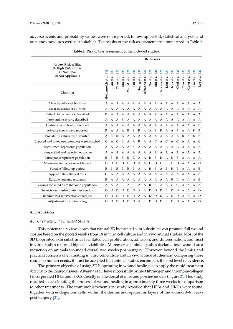
3.6. Quality Evaluation
The risk of bias of the included studies was conducted using a modified version of the OHAT.In general, the experimental conditions of all reported bioinks were duly mentioned, and almostall studies have low reporting and performance risk of bias. Five of the six in vivo studies have alow risk of bias due to reporting outcome details and fulfilling the selection criteria. Four of twelvein vitro studies showed a low risk of bias as well. In contrast, eight studies have a moderate risk ofbias due to the lack of skin cell representation and short follow-up periods, and only one study wasfound to have a high risk of bias due to high reporting and selection biases (i.e., finding was not clear,
Polymers 2020, 12, 1782 12 of 19
adverse events and probability values were not reported; follow-up period, statistical analysis, andoutcomes measures were not suitable). The results of the risk assessment are summarized in Table 4.
Table 4. Risk of bias assessment of the included studies.
References
A: Low Risk of BiasB: High Risk of Bias
C: Not ClearD: Not Applicable
Hei
denr
eich
etal
.[19
]
Xu
etal
.[23
]
Che
net
al.[
20]
Shie
tal.
[24]
Osi
dak
etal
.[25
]
Liu
etal
.[26
]
Hua
nget
al.[
33]
Alb
anna
etal
.[31
]
Xu
etal
.[27
]
Shie
tal.
[28]
Noc
era
etal
.[29
]
Kim
etal
.[34
]
Dat
taet
al.[
30]
Cho
ieta
l.[2
1]
Che
net
al.[
35]
Xio
nget
al.[
36]
Liu
etal
.[32
]
Lee
etal
.[22
]
Checklist
Clear hypothesis/objectives A A A A A A A A A A A A A A A A A A
Clear measures of outcome A A A A A A A A A A A A A A A A A A
Patient characteristics described B A A A A A A A A A A A A A A A A A
Interventions clearly described A A A B A A A A A A A A A A A A A A
Findings were clearly described A A A A A A A A A A A A A A A A A B
Adverse events were reported B A A A B B B A A B B A A B A A B B
Probability values were reported A B B A A A A A A A A A A A B B B B
Exposed and unexposed numbers were matched C A C B A A B A A C A C A C A A A C
Recruitment represents population A A A A A B A A A A A A A A A A A A
Pre-specified and reported outcomes A A A A A A A A A A A A A A A A A A
Participants represent population B B B B B C A A B B B A B B A A A A
Measuring outcomes were blinded D D D D D D A A D D D B D D A A A D
Suitable follow-up period B B B B B B A A B B A B B B A A A B
Appropriate statistical tests C A A A A A A A A A A A A A A A A B
Reliable outcome measures B A A A A A A A A A A A B A A A A B
Groups recruited from the same population A A A B A B A A B B A A C C A A A A
Subjects randomized into intervention D D D D D D A A D D D B D D A A A D
Randomized intervention concealed D D D D D D A A D D D A D D A A A D
Adjustment for confounding D D D D D D A B D D D B D D A A A D
4. Discussion
4.1. Overview of the Included Studies
This systematic review shows that natural 3D bioprinted skin substitutes can promote full woundclosure based on the pooled results from 18 in vitro cell culture and in vivo animal studies. Most of the3D bioprinted skin substitutes facilitated cell proliferation, adhesion, and differentiation, and mostin vitro studies reported high cell viabilities. Moreover, all animal studies declared total wound areareduction on animals wounded dorsal two weeks post-surgery. However, beyond the limits andpractical concerns of evaluating in vitro cell culture and in vivo animal studies and comparing theseresults to human needs, it must be accepted that animal studies encompass the first level of evidence.
The primary objective of using 3D bioprinting in wound healing is to apply the rapid treatmentdirectly to the injured tissues. Albanna et al. have successfully printed fibrinogen and thrombin/collagenI incorporated HFBs and HKCs directly on the dorsal of mice and porcine models (Figure 2). This studyresulted in accelerating the process of wound healing in approximately three weeks in comparisonto other treatments. The immunohistochemistry study revealed that HFBs and HKCs were found,together with endogenous cells, within the dermis and epidermis layers of the wound 3–6 weekspost-surgery [31].

Polymers 2020, 12, 1782 13 of 19
Polymers 2020, 12, x FOR PEER REVIEW 14 of 20
4.2.2. Gelatin
Gelatin is another commonly used bioink that presented high degradability, biocompatibility, and suitable rheological properties. Nevertheless, pure gelatin solutions have weak mechanical strength and low viscosity above 27±1°C, and that limits gelatin usage in 3D bioprinting. It is often mixed with other natural biomaterials, such as alginate [26,28,32] and silk-fibroin [33], to overcome the low formability. Moreover, gelatin methacrylate (GelMA) is also a potential wound healing bioink due to its high thermal sensitivity and photo-crosslinking ability. GelMA is also known to have good biocompatibility, and of promoting cell to cell interaction and cell migration. Furthermore, the advantageous mechanical stability of GelMA after UV crosslinking was used to induce a high shape fidelity of natural-based bioinks, such as cellulose nanofibrils [23] and silk sericin [20].
Figure 2. Example of in situ skin bioprinting process, where, (a) Markers are placed around the wound area as reference points; (b) Wound area scanned with a hand-held ZScanner™ (Z700 scanner); (c) Geometric information obtained via scanning is then inputted in the form of an STL file to orient the scanned images to the standard coordinate system; (d) The scanned data with its coordinate system is used to generate the fill volume, and the path points for nozzle head to travel to print the fill volume; (e, f). Output code is then provided to the custom bioprinter control interface for generation of nozzle path needed to print fill volume. Figure and caption reused from Albanna et al. [31]. Used under the Creative Commons License (http://creativecommons.org/licenses/by/4.0/).
4.2.3. Alginate
Alginate has been used in different 3D bioprinting applications because of its high shear-thinning and rapid gelation post-printing. However, alginate has many limitations as crosslinking delay may reduce the shape fidelity of the bioprinted constructs, low cell viability as rapid crosslinking limit cell-to-materials interaction. An attempt was conducted by Datta et al. to overcome those limitations by decreasing alginate viscosity using honey to increase cell viability without altering alginate printability. While alginate is qualified for most of the physicochemical properties needed for 3D bioprinting, it suffers poor cell adhesion properties, requiring efforts to enhance the cell adhesion without sacrificing the physicochemical properties [30]. Printing simple alginate solutions were found to have low shape fidelity, although researchers attempted to increase alginate viscosity or extrude it with chemical crosslinkers such as Ca+2 [26].
Figure 2. Example of in situ skin bioprinting process, where, (a) Markers are placed around thewound area as reference points; (b) Wound area scanned with a hand-held ZScanner™ (Z700 scanner);(c) Geometric information obtained via scanning is then inputted in the form of an STL file to orient thescanned images to the standard coordinate system; (d) The scanned data with its coordinate system isused to generate the fill volume, and the path points for nozzle head to travel to print the fill volume;(e,f). Output code is then provided to the custom bioprinter control interface for generation of nozzlepath needed to print fill volume. Figure and caption reused from Albanna et al. [31]. Used under theCreative Commons License (http://creativecommons.org/licenses/by/4.0/).
4.2. Bioinks Materials & Combinations
Many types of natural-based bioinks, composite or stand-alone materials, have been proposed torestore the skin integrity and accelerate the wound healing process due to their desirable properties,such as resembling skin ECM, high printability, and excellent biocompatibility as hydrogels are themost commonly used biomaterials [14].
4.2.1. Collagen
Collagen, as a hydrogel, exhibited desirable biodegradability, high shape consistency at37 ◦C, and excellent microstructure of micro-and macropores that promote cellular attachmentand proliferation [29]. However, collagen direct 3D bioprinting is still limited as collagen solutionshave poor printability, especially when incorporated with cells or tissue spheroids [25]. Notably,despite the limited collagen printability, no chemical crosslinking was applied over most of the studies.Instead, this property was overcome by either admixing with other materials such as fibrinogen andthrombin [31], chitosan [19], by using fibrillar collagen [29], by using low concentrations of collagen(2–4%) [25], or by controlling cell suspensions and densities [22]. In the same context, proteins gelationof matrices such as collagen is usually initiated by pH or temperature control or by both. Although thisapproach is valid for thin structures, it showed diffusion or thermal transference limitations in thickstructures (1 to 3 mm), which may lead to the appearance of gelled and non-gelled regions. High levelsof pH or temperature may also lead to severe harm to cells [22].

Polymers 2020, 12, 1782 14 of 19
4.2.2. Gelatin
Gelatin is another commonly used bioink that presented high degradability, biocompatibility, andsuitable rheological properties. Nevertheless, pure gelatin solutions have weak mechanical strengthand low viscosity above 27 ± 1 ◦C, and that limits gelatin usage in 3D bioprinting. It is often mixedwith other natural biomaterials, such as alginate [26,28,32] and silk-fibroin [33], to overcome thelow formability. Moreover, gelatin methacrylate (GelMA) is also a potential wound healing bioinkdue to its high thermal sensitivity and photo-crosslinking ability. GelMA is also known to havegood biocompatibility, and of promoting cell to cell interaction and cell migration. Furthermore,the advantageous mechanical stability of GelMA after UV crosslinking was used to induce a highshape fidelity of natural-based bioinks, such as cellulose nanofibrils [23] and silk sericin [20].
4.2.3. Alginate
Alginate has been used in different 3D bioprinting applications because of its high shear-thinningand rapid gelation post-printing. However, alginate has many limitations as crosslinking delay mayreduce the shape fidelity of the bioprinted constructs, low cell viability as rapid crosslinking limitcell-to-materials interaction. An attempt was conducted by Datta et al. to overcome those limitations bydecreasing alginate viscosity using honey to increase cell viability without altering alginate printability.While alginate is qualified for most of the physicochemical properties needed for 3D bioprinting,it suffers poor cell adhesion properties, requiring efforts to enhance the cell adhesion without sacrificingthe physicochemical properties [30]. Printing simple alginate solutions were found to have low shapefidelity, although researchers attempted to increase alginate viscosity or extrude it with chemicalcrosslinkers such as Ca+2 [26].
4.2.4. Skin-decellularized Extracellular Matrix (S-dECM)
Extracellular matrix (ECM) represents the non-cellular part of a tissue or an organ, and it mainlyassembles the microenvironment network for the cell to perform specific functions. Each tissuehas its well-constructed ECM, which consists of several components and proteins that maintain thenative structure and support cell migration. Interestingly, the ECM can be derived by using anappropriate protocol and reused as a scaffold for tissue regeneration [37]. Kim et al. successfullydecellularized porcine skin-tissue and formed a printable dECM bioink. They found that, in comparisonto collagen bioink, the 3D bioprinted skin equivalent using derived ECM bioink promoted dermalcompartment stabilization, enhanced epidermal organization, and provided more physiologicalrelevant skin functions in vitro. Moreover, dECM-based 3D skin encapsulated EPCs, and ASCspromoted neovascularization and re-epithelialization as well as wound closure in vivo [34].
4.3. Bioink Biocompatibility & Cellular Behavior
Bioinks biocompatibility was duly investigated, and some of the possible reasons that mayaffect cell viability, adhesion, proliferation, migration, and differentiation were reported. In general,cytotoxicity should be evaluated when proposing a potential material for medical use. Most of theincluded studies performed MTT assay to ensure no cytotoxicity or inflammation caused by thecell-to-materials chemical interaction. Notably, only silk sericin/GelMA bioink was found to causeacute inflammation on the 7th day, which disappeared at the end of the follow-up period [35].
Bioink pore size should also be considered when choosing a bioink as small pore sizes cause a lackof nutrition and oxygen supply, which led to low cell viability and slower cell migration. Choi et al.studied the effect of gelatin pore size on cell behavior and found that the proliferation rate of HDFsincreased by 14% in pore size of 580 µm compared to 435 µm after 14 days [21]. However, using naturalbioinks is favorable because of their suitable inter-molecular network. For example, fibrillar collagen iswell-known to have a suitable micro- and macropores structure, which was found to highly intervenein increasing cell viability and promoting high cell attachment and proliferation [29].

Polymers 2020, 12, 1782 15 of 19
In the same context, bioink concentration crucially affects cell viability as high concentrations leadto compacted cells. An evaluation of the impact of using different collagen concentration into viscollon cell viability, found that decreasing collagen concentration from 4% to 2% resulted in increasing thecell viability from 87.2% ± 2.1% to 97.2% ± 1.2% (p < 0.05) [25]. Nocera et al. studied the effect of usingsmaller collagen extract on NIH 3T3 cell viability and found that decreasing the concentration from100-extract to 25-extract promoted cell viability from 85.07 ± 6.73% to 111.31 ± 3.65% (p < 0.05) [29].Xu et al. also studied the effect of admixing small concentrations of GelMA with cellulose nanofibrils(CNFs) on cell proliferation. They found that three days after culture, there was twice the number ofcells on CNF/GelMA bioink compared with CNF bioink alone [23].
On the other hand, growth factors are essential morphogenetic proteins that influence cell activityand direct tissue repair and regeneration [38]. Xiong et al. studied the effect of using a fibroblastgrowth factor (FGF2) on cell proliferation. They found that adding 100 ng/mL of FGF2 growth factor tothe scaffold significantly enhanced the proliferation rate (~40% to ~75%), tissue morphology, and theassembly of the collagen fibril (Figure 3) [36].
Polymers 2020, 12, x FOR PEER REVIEW 16 of 20
4.4. Structural Design & Mechanical Properties
An effective bioink should possess excellent mechanical properties and should not breakdown post-printing. A bioink should also have high swelling ratios to maintain moisture wound area to exchange nutrients and facilitate cell proliferation. In the literature, human skin was found to have a young’s modulus average of 100 to 1100 kPa [34]. The swelling ratio has an inversible relationship with young’s modulus values, whereas increasing the dSIS filament distances from 500 to 700 μm increased the swelling ratio from 69% to 79% and decreased the Young’s modulus from 26.6 ± 3.8 to 9.7 ± 3.1 kPa (p < 0.05) [24]. The same results were reported with CNF crosslinked BDDE [27] and alginate/gelatin [28].
Bioinks should maintain their shape once they leave the tip of the printing nozzle. Overall, proper bioink viscosity ensures high shape fidelity and minimizes the possibility of structural collapse after printing [32]. Shear-thinning is another critical parameter as bioinks should have excellent shear-thinning properties to avoid clogging during the printing process and to regain immediate structural consistency post-printing to be ready to support the next layer [19,26,27]. For example, a period of 1 min was required to ensure the transformation of collagen to gel-state to preserve a solid base for the printing of the next layer [22]. Additionally, the rigidity of the printed scaffolds appeared to affect cell proliferation profoundly. As the rigidity of CNF increases within a tunable range of 3–8 kPa, cell proliferation was promoted [27].
Figure 3. Epidermis and blood vessel formation in skin defects. Immunohistochemical staining of wound sections to detect expression of cytokeratin, SMA and CD31, after implantation with 3DG-SF-SO3 and 3DG-SF- SO3-FGF scaffolds at day 28 post-surgery. Scale bars = 50 μm. Figure and caption reused from Xiong et al. [36]. Used under the Creative Commons License (http://creativecommons.org/licenses/by/4.0/).
4.5. Animal Models & Wound Healing
Early treatment of wounds is critical to avoid wound aggravation and tissue damage over time, due to the hypertrophic scarring. Patients undergoing late treatment often experience severe scarring.
Figure 3. Epidermis and blood vessel formation in skin defects. Immunohistochemical staining ofwound sections to detect expression of cytokeratin, SMA and CD31, after implantation with 3DG-SF-SO3and 3DG-SF- SO3-FGF scaffolds at day 28 post-surgery. Scale bars = 50 µm. Figure and caption reusedfrom Xiong et al. [36]. Used under the Creative Commons License (http://creativecommons.org/licenses/by/4.0/).
Cell suspension densities is another critical factor, as using high densities cause low cell viability.Lee et al. reported the use of an inkjet bioprinting system and studied the effect of using differentcell suspension densities and droplet size on cell viability. The study found that cell viabilityvaried proportionally with cell suspension density and inversely with the space between dropletsfor both keratinocytes (KCs) and fibroblasts (FBs) skin cells. At very low cell suspension density(0.5 million cells/mL) and large droplet spacing (400 mm), FBs cell viability was moderate (84%).Similarly, at a high cell suspension density (5 million cells/mL) and small droplet spacing (400 µm),KCs cell viability was lower (94%) [22]. Moreover, cell adhesion is profoundly affected by the matrix

Polymers 2020, 12, 1782 16 of 19
thickness, whereas a higher percentage of cell attachment was observed with 3 mm samples than with2 mm thick samples. A large thickness scaffold promoted cells to adhere [27].
4.4. Structural Design & Mechanical Properties
An effective bioink should possess excellent mechanical properties and should not breakdownpost-printing. A bioink should also have high swelling ratios to maintain moisture wound area toexchange nutrients and facilitate cell proliferation. In the literature, human skin was found to have ayoung’s modulus average of 100 to 1100 kPa [34]. The swelling ratio has an inversible relationshipwith young’s modulus values, whereas increasing the dSIS filament distances from 500 to 700 µmincreased the swelling ratio from 69% to 79% and decreased the Young’s modulus from 26.6 ± 3.8 to9.7 ± 3.1 kPa (p < 0.05) [24]. The same results were reported with CNF crosslinked BDDE [27] andalginate/gelatin [28].
Bioinks should maintain their shape once they leave the tip of the printing nozzle. Overall,proper bioink viscosity ensures high shape fidelity and minimizes the possibility of structural collapseafter printing [32]. Shear-thinning is another critical parameter as bioinks should have excellentshear-thinning properties to avoid clogging during the printing process and to regain immediatestructural consistency post-printing to be ready to support the next layer [19,26,27]. For example,a period of 1 min was required to ensure the transformation of collagen to gel-state to preserve asolid base for the printing of the next layer [22]. Additionally, the rigidity of the printed scaffoldsappeared to affect cell proliferation profoundly. As the rigidity of CNF increases within a tunablerange of 3–8 kPa, cell proliferation was promoted [27].
4.5. Animal Models & Wound Healing
Early treatment of wounds is critical to avoid wound aggravation and tissue damage over time,due to the hypertrophic scarring. Patients undergoing late treatment often experience severe scarring.Before proposing the capacity of using an effective wound treatment, it must demonstrate highbiocompatibility and non-cytotoxicity in vitro. It should also stimulate wound healing and tissuere-epithelization in vivo. Bioprinting human cells resulted in rapid epithelialization represented by4–5 weeks of acceleration time of wound re-epithelialization [31].
Layering skin constructs in regular pore size and structure significantly influenced nutritionsupply and cell ingrowth in the wound area [33]. To ensure scarless wounds, the treatment shouldbe placed evenly in an organized manner on the wound. Xiong et al. reported that the applicationof the gelatin–sulfonic acid–FGF scaffold on rats’ wounded dorsal helped to smoothen the woundpost-surgery, and the cross-sectional results showed complete wound closure in addition to theexistence of more blood vessels [36]. Furthermore, the cross-sectional area treated by SS/GelMAshowed the formation of new collagen with high fibroblast proliferation similar to the healthy tissueseven days post-surgery followed by complete wound closure on the 4th week, thus proving excellentwound healing properties [35].
For the survival or integration of the new tissue or organ into the surrounding tissue, suitablevascularization is required. Many attempts have been made to build vascularized skin scaffold byusing natural-based biopolymers [36] or by printing with interconnective pores sizes between 50 and500 µm and micropores with diameters lower than 10 µm [29], or by decellularizing skin ECM [34].
5. Limitations of the Present Review
This systematic review has several limitations. No specific risk of bias checklist was found toassess in vitro studies. Instead, the OHAT tool was adapted to evaluate both in vitro and in vivostudies. Furthermore, using 3D bioprinting for wound healing is still undergoing animal studies,and no human randomized clinical trials were identified. Another limitation is that the observationtime and measurements vary among studies, which causes high heterogeneity in the results. Hence,a meta-analysis was impossible to be performed.

Polymers 2020, 12, 1782 17 of 19
6. Conclusions and Future Perspectives
This systematic review identified the potential in vitro cell culture and in vivo animal studiesreporting 3D bioprinting skin substitutes. First of all, this review confirms the significant benefits ofusing 3D printed natural-based bioinks for skin repair and regeneration. Natural bioinks showedexcellent ability to mimic the three-dimensional microenvironment structure of native skin tissue andto promote cell adhesion, proliferation, migration, and mobility. Furthermore, in vivo visualizationshowed full wound closure four weeks post-surgery with well-organized dermal and epidermal layers.This review reported the importance of many bioink properties that should be found to accelerate thewound healing process for a better therapeutic approach. We recommend the use of natural bioinksfor wound healing and suggest performing more in vitro studies with the use of a variety of skin cellrepresentations other than dermal fibroblasts, which is known to survive the harsh environment.
Despite the limited number of conducted studies, in situ bioprinting is one of the most promisingadvances in skin tissue engineering, which can be used by surgeons to print complex organs efficientlyand rapidly. Yet, the main challenge is the ability to build tissue details more precisely which requiredthe integration of different fields, including engineering, biology, and medical science. In addition,some new cross-linking techniques, such as two-photon cross-linking and directed on tip UV light,might promote structural control over the existing bioinks. Self-healing hydrogels constitute anotherinteresting direction as they can be printed, retain their pre-vascularized microstructure, and can beused as self-healing scaffolds for wound healing.
Author Contributions: A.S. and A.N. conceptualized the study. A.S., A.N., and M.B.F. undertook studies selectionand risk of bias assessment. A.S., A.N., and N.M.H. performed data extraction. A.S., M.B.F., I.A.A., and K.-Y.C.performed the results and discussion. All authors contributed to data interpretation. A.S. drafted the manuscript,which was critically reviewed and approved by A.N., M.B.F, N.M.H., K.-Y.C., and I.A.A. All authors have readand agreed to the published version of the manuscript.
Funding: This study was funded through the grant provided by Universiti Kebangsaan Malaysia Medical Centre& 3D Gens Sdn. Bhd. under Matching Grant (Code No: FF-2019-449/1 and FF-2019-449) in the form of databasesubscription. The funder does not have any contribution and decision to publish or preparation of the manuscript.
Acknowledgments: All the authors would like to express immense gratitude to the Faculty of Medicine, UKM,for the guidance and resources to complete this review.
Conflicts of Interest: The authors declare that the research was conducted in the absence of any commercial orfinancial relationships that could be construed as a potential conflict of interest.
References
1. Sen, C.K. Human Wounds and Its Burden: An Updated Compendium of Estimates. Adv. Wound Care 2019,8, 39–48. [CrossRef] [PubMed]
2. Beldon, P. Basic science of wound healing. Surgery 2010, 28, 409–412. [CrossRef]3. Cooper, E.H.; Anderson, C.K.; Steele, L.; O’Boyle, P. Assessment of bladder cancer. Cancer 1973, 32, 1263–1266.
[CrossRef]4. Dhivya, S.; Padma, V.V.; Santhini, E. Wound dressings—A review. BioMedicine 2015, 5, 22. [CrossRef]5. Chouhan, D.; Dey, N.; Bhardwaj, N.; Mandal, B.B. Emerging and innovative approaches for wound healing
and skin regeneration: Current status and advances. Biomaterials 2019, 216, 119267. [CrossRef]6. Melchels, F.P.W.; Domingos, M.A.N.; Klein, T.J.; Malda, J.; Bartolo, P.J.; Hutmacher, D.W.
Additive manufacturing of tissues and organs. Prog. Polym. Sci. 2012, 37, 1079–1104. [CrossRef]7. He, P.; Zhao, J.; Zhang, J.; Li, B.; Gou, Z.; Gou, M.; Li, X. Bioprinting of skin constructs for wound healing.
Burn. Trauma 2018, 6, 1–10. [CrossRef]8. Groll, J.; Burdick, J.A.; Cho, D.W.; Derby, B.; Gelinsky, M.; Heilshorn, S.C.; Jüngst, T.; Malda, J.; Mironov, V.A.;
Nakayama, K.; et al. A definition of bioinks and their distinction from biomaterial inks. Biofabrication 2019,11, 013001. [CrossRef]
9. Gopinathan, J.; Noh, I. Recent trends in bioinks for 3D printing. Biomater. Res. 2018, 22, 1–15. [CrossRef]10. Panwar, A.; Tan, L.P. Current status of bioinks for micro-extrusion-based 3D bioprinting. Molecules 2016,
21, 685. [CrossRef]
Polymers 2020, 12, 1782 18 of 19
11. Xia, Z.; Jin, S.; Ye, K. Tissue and Organ 3D Bioprinting. SLAS Technol. 2018, 23, 301–314. [CrossRef]12. Ng, W.L.; Wang, S.; Yeong, W.Y.; Naing, M.W. Skin Bioprinting: Impending Reality or Fantasy?
Trends Biotechnol. 2016, 34, 689–699. [CrossRef] [PubMed]13. Kumar, A.; Starly, B. Large scale industrialized cell expansion: Producing the critical raw material for
biofabrication processes. Biofabrication 2015, 7, 44103. [CrossRef]14. Montero, F.E.; Rezende, R.A.; da Silva, J.V.L.; Sabino, M.A. Development of a Smart Bioink for Bioprinting
Applications. Front. Mech. Eng. 2019, 5, 1–12. [CrossRef]15. Valot, L.; Martinez, J.; Mehdi, A.; Subra, G. Chemical insights into bioinks for 3D printing. Chem. Soc. Rev.
2019, 48, 4049–4086. [CrossRef] [PubMed]16. Kim, J.E.; Kim, S.H.; Jung, Y. Current status of three-dimensional printing inks for soft tissue regeneration.
Tissue Eng. Regen. Med. 2016, 13, 636–646. [CrossRef] [PubMed]17. Liberati, A.; Altman, D.G.; Tetzlaff, J.; Mulrow, C.; Gøtzsche, P.C.; Ioannidis, J.P.A.; Clarke, M.; Devereaux, P.J.;
Kleijnen, J.; Moher, D. The PRISMA statement for reporting systematic reviews and meta-analyses of studiesthat evaluate health care interventions: Explanation and elaboration. J. Clin. Epidemiol. 2009, 62, e1–e34.[CrossRef]
18. National Toxicology Program(NTP). OHAT Risk of Bias Rating Tool for Human and Animal Studies. Availableonline: https://ntp.niehs.nih.gov/whatwestudy/assessments/noncancer/riskbias/index.html (accessed on10 January 2020).
19. Heidenreich, A.C.; Pérez-Recalde, M.; González Wusener, A.; Hermida, É.B. Collagen and chitosan blendsfor 3D bioprinting: A rheological and printability approach. Polym. Test. 2020, 82, 106297. [CrossRef]
20. Chen, X.; Yue, Z.; Winberg, P.C.; Dinoro, J.N.; Hayes, P.; Beirne, S.; Wallace, G.G. Development of rhamnose-richhydrogels based on sulfated xylorhamno-uronic acid toward wound healing applications. Biomater. Sci.2019, 7, 3497–3509. [CrossRef]
21. Choi, D.J.; Park, S.J.; Gu, B.K.; Kim, Y.J.; Chung, S.; Kim, C.H. Effect of the pore size in a 3D bioprinted gelatinscaffold on fibroblast proliferation. J. Ind. Eng. Chem. 2018, 67, 388–395. [CrossRef]
22. Lee, V.; Singh, G.; Trasatti, J.P.; Bjornsson, C.; Xu, X.; Tran, T.N.; Yoo, S.S.; Dai, G.; Karande, P. Design andfabrication of human skin by three-dimensional bioprinting. Tissue Eng. Part C Methods 2014, 20, 473–484.[CrossRef] [PubMed]
23. Xu, W.; Molino, B.Z.; Cheng, F.; Molino, P.J.; Yue, Z.; Su, D.; Wang, X.; Willför, S.; Xu, C.; Wallace, G.G. OnLow-Concentration Inks Formulated by Nanocellulose Assisted with Gelatin Methacrylate (GelMA) for 3DPrinting toward Wound Healing Application. ACS Appl. Mater. Interfaces 2019, 11, 8838–8848. [CrossRef][PubMed]
24. Shi, L.; Hu, Y.; Ullah, M.W.; Ullah, I.; Ou, H.; Zhang, W.; Xiong, L.; Zhang, X. Cryogenic free-form extrusionbioprinting of decellularized small intestinal submucosa for potential applications in skin tissue engineering.Biofabrication 2019, 11, 035023. [CrossRef] [PubMed]
25. Osidak, E.O.; Karalkin, P.A.; Osidak, M.S.; Parfenov, V.A.; Sivogrivov, D.E.; Pereira, F.D.A.S.;Gryadunova, A.A.; Koudan, E.V.; Khesuani, Y.D.; Kasyanov, V.A.; et al. Viscoll collagen solution asa novel bioink for direct 3D bioprinting. J. Mater. Sci. Mater. Med. 2019, 30, 31. Available online:https://pubmed.ncbi.nlm.nih.gov/30830351/ (accessed on 21 July 2020). [CrossRef]
26. Liu, P.; Shen, H.; Zhi, Y.; Si, J.; Shi, J.; Guo, L.; Shen, S.G. 3D bioprinting and in vitro study of bilayeredmembranous construct with human cells-laden alginate/gelatin composite hydrogels. Colloids SurfacesB Biointerfaces 2019, 181, 1026–1034. [CrossRef]
27. Xu, C.; Zhang Molino, B.; Wang, X.; Cheng, F.; Xu, W.; Molino, P.; Bacher, M.; Su, D.; Rosenau, T.; Willför, S.;et al. 3D printing of nanocellulose hydrogel scaffolds with tunable mechanical strength towards woundhealing application. J. Mater. Chem. B 2018, 6, 7066–7075. [CrossRef]
28. Shi, L.; Xiong, L.; Hu, Y.; Li, W.; Chen, Z.C.; Liu, K.; Zhang, X. Three-dimensional printing alginate/gelatinscaffolds as dermal substitutes for skin tissue engineering. Polym. Eng. Sci. 2018, 58, 1782–1790. [CrossRef]
29. Nocera, A.D.; Comín, R.; Salvatierra, N.A.; Cid, M.P. Development of 3D printed fibrillar collagen scaffoldfor tissue engineering. Biomed. Microdevices 2018, 20, 1–13. [CrossRef]
30. Datta, S.; Sarkar, R.; Vyas, V.; Bhutoria, S.; Barui, A.; Roy Chowdhury, A.; Datta, P. Alginate-honey bioinkswith improved cell responses for applications as bioprinted tissue engineered constructs. J. Mater. Res. 2018,33, 2029–2039. [CrossRef]

Polymers 2020, 12, 1782 19 of 19
31. Albanna, M.; Binder, K.W.; Murphy, S.V.; Kim, J.; Qasem, S.A.; Zhao, W.; Tan, J.; El-Amin, I.B.; Dice, D.D.;Marco, J.; et al. In Situ Bioprinting of Autologous Skin Cells Accelerates Wound Healing of ExtensiveExcisional Full-Thickness Wounds. Sci. Rep. 2019, 9, 1–15. [CrossRef]
32. Liu, J.; Chi, J.; Wang, K.; Liu, X.; Liu, J.; Gu, F. Full-thickness wound healing using 3D bioprintedgelatin-alginate scaffolds in mice: A histopathological study. Int. J. Clin. Exp. Pathol. 2016, 9, 11197–11205.
33. Huang, L.; Du, X.; Fan, S.; Yang, G.; Shao, H.; Li, D.; Cao, C.; Zhu, Y.; Zhu, M.; Zhang, Y. Bacterial cellulosenanofibers promote stress and fidelity of 3D-printed silk based hydrogel scaffold with hierarchical pores.Carbohydr. Polym. 2019, 221, 146–156. [CrossRef] [PubMed]
34. Kim, B.S.; Kwon, Y.W.; Kong, J.S.; Park, G.T.; Gao, G.; Han, W.; Kim, M.B.; Lee, H.; Kim, J.H.; Cho, D.W. 3Dcell printing of in vitro stabilized skin model and in vivo pre-vascularized skin patch using tissue-specificextracellular matrix bioink: A step towards advanced skin tissue engineering. Biomaterials 2018, 168, 38–53.[CrossRef] [PubMed]
35. Chen, C.S.; Zeng, F.; Xiao, X.; Wang, Z.; Li, X.L.; Tan, R.W.; Liu, W.Q.; Zhang, Y.S.; She, Z.D.; Li, S.J.Three-Dimensionally Printed Silk-Sericin-Based Hydrogel Scaffold: A Promising Visualized DressingMaterial for Real-Time Monitoring of Wounds. ACS Appl. Mater. Interfaces 2018, 10, 33879–33890. [CrossRef][PubMed]
36. Xiong, S.; Zhang, X.; Lu, P.; Wu, Y.; Wang, Q.; Sun, H.; Heng, B.C.; Bunpetch, V.; Zhang, S.; Ouyang, H. AGelatin-sulfonated Silk Composite Scaffold based on 3D Printing Technology Enhances Skin Regenerationby Stimulating Epidermal Growth and Dermal Neovascularization. Sci. Rep. 2017, 7, 1–12. [CrossRef][PubMed]
37. Dzobo, K.; Motaung, K.S.C.M.; Adesida, A. Recent trends in decellularized extracellular matrix bioinks for3D printing: An updated review. Int. J. Mol. Sci. 2019, 20, 4628. [CrossRef]
38. Mitchell, A.C.; Briquez, P.S.; Hubbell, J.A.; Cochran, J.R. Engineering growth factors for regenerative medicineapplications. Acta Biomater. 2016, 30, 1–12. [CrossRef]
© 2020 by the authors. Licensee MDPI, Basel, Switzerland. This article is an open accessarticle distributed under the terms and conditions of the Creative Commons Attribution(CC BY) license (http://creativecommons.org/licenses/by/4.0/).