Young vs. Old Dental Pulp Treatment: Repair vs. Regeneration
Upload
eustacia-austinCategory
view
229download
6
WOUND HEALINGWOUND HEALING
REPAIR + REGENERATIONREPAIR + REGENERATION
NEW EPITHELIUM GROWTHNEW EPITHELIUM GROWTH
TISSUE REPAIRTISSUE REPAIR
Replacement of injured or dead Replacement of injured or dead cells is critical to survival.cells is critical to survival.
Repair involves mainly two Repair involves mainly two processesprocesses
1.1. RegenerationRegeneration
2.2. Replacement by connective tissue Replacement by connective tissue or fibroplasia.or fibroplasia.
Wound healingWound healing is an intricate process is an intricate process where the skin (or another organ-tissue) where the skin (or another organ-tissue) repairs itself after injury. In normal skin, repairs itself after injury. In normal skin, the the epidermis and and dermis exist in a exist in a steady-state equilibrium, forming a steady-state equilibrium, forming a protective barrier against the external protective barrier against the external environment. Once the protective barrier environment. Once the protective barrier is broken, the normal (physiologic) is broken, the normal (physiologic) process of wound healing is immediately process of wound healing is immediately set in motion.set in motion.
Steps in wound healingSteps in wound healing
wound healing is divided into three or wound healing is divided into three or four sequential phases: (1) four sequential phases: (1) hemostasis (not considered a phase by some authors)(not considered a phase by some authors)
, (2) inflammation, (2) inflammation , (3) proliferation and, (3) proliferation and (4) remodeling.(4) remodeling. Upon injury to the skin, a set of complex Upon injury to the skin, a set of complex
biochemical events takes place to repair biochemical events takes place to repair the damage.the damage.
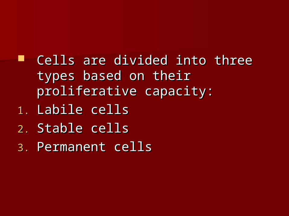
Cells are divided into three types Cells are divided into three types based on their proliferative based on their proliferative capacity:capacity:
1.1. Labile cellsLabile cells
2.2. Stable cellsStable cells
3.3. Permanent cellsPermanent cells

Labile cells: Labile cells: – This sub-population of cells is constantly turned over. The This sub-population of cells is constantly turned over. The
best examples are found in the epithelial cell population of best examples are found in the epithelial cell population of the the skinskin or or gutgut, and the hematopoetic cells in the , and the hematopoetic cells in the bone bone marrowmarrow. These cells have a short, finite life span, die via . These cells have a short, finite life span, die via apoptosis, and are rapidly replaced.apoptosis, and are rapidly replaced.
Permanent cells: Permanent cells: – Permanent cells are found in the CNS (Permanent cells are found in the CNS (neuronsneurons) and heart ) and heart
((Cardiac muscleCardiac muscle). Once they are destroyed, they cannot ). Once they are destroyed, they cannot regenerate. regenerate.
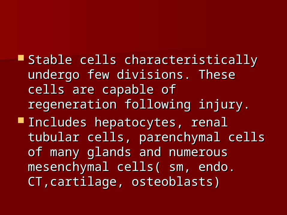
Stable cells characteristically Stable cells characteristically undergo few divisions. These cells undergo few divisions. These cells are capable of regeneration following are capable of regeneration following injury.injury.
Includes hepatocytes, renal tubular Includes hepatocytes, renal tubular cells, parenchymal cells of many cells, parenchymal cells of many glands and numerous mesenchymal glands and numerous mesenchymal cells( sm, endo. CT,cartilage, cells( sm, endo. CT,cartilage, osteoblasts)osteoblasts)

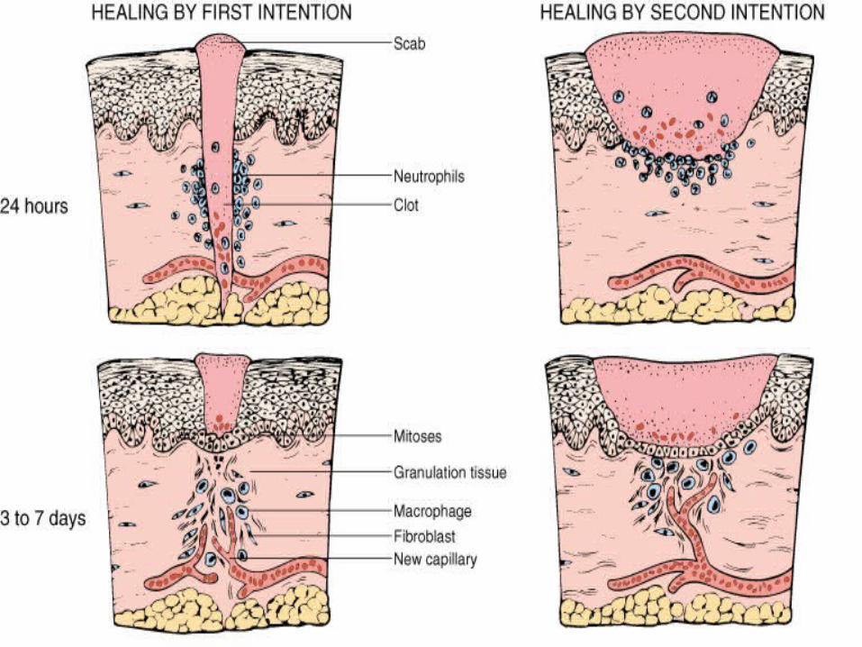
CLINICAL SURGICAL CORRELATES CLINICAL SURGICAL CORRELATES
OF WOUND HEALINGOF WOUND HEALING PRIMARY UNIONPRIMARY UNION HEALING BY “FIRST HEALING BY “FIRST
INTENTION”INTENTION” SUTURED SUTURED
SURGICAL SURGICAL INCISIONINCISION
SECONDARY UNIONSECONDARY UNION HEALING BY HEALING BY
“SECONDARY “SECONDARY INTENTION”INTENTION”
LARGE DEFECTS, LARGE DEFECTS, ULCERSULCERS
W.B. Saunders Company items and W.B. Saunders Company items and derived items Copyright (c) 1999 derived items Copyright (c) 1999
by W.B. Saunders Companyby W.B. Saunders CompanySlide 4.17

WOUND CONTRACTION IN A WOUND CONTRACTION IN A CENTRIPETALCENTRIPETAL FASHIONFASHION

WOUND CONTRACTION BY ACTION OF MYOFIBROBLASTS
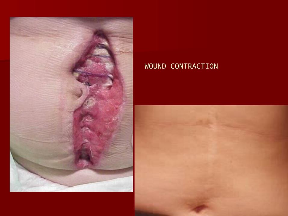
WOUND CONTRACTION
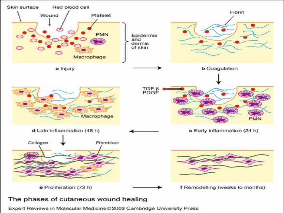

First 24 hrs; First 24 hrs; Neutrophils’ migration towards fibrin clot.Neutrophils’ migration towards fibrin clot. ACUTE INFLAMMATORY RESPONSEACUTE INFLAMMATORY RESPONSE ERYTHEMA, EDEMAERYTHEMA, EDEMA Basal cells exhibit mitotic activityBasal cells exhibit mitotic activity
24-48 hrs:24-48 hrs: Epithelial cell from edges migrate and Epithelial cell from edges migrate and
proliferate along dermis.proliferate along dermis. PLATELET/FIBRIN COVERINGPLATELET/FIBRIN COVERING MIGRATION TO MIDLINEMIGRATION TO MIDLINE
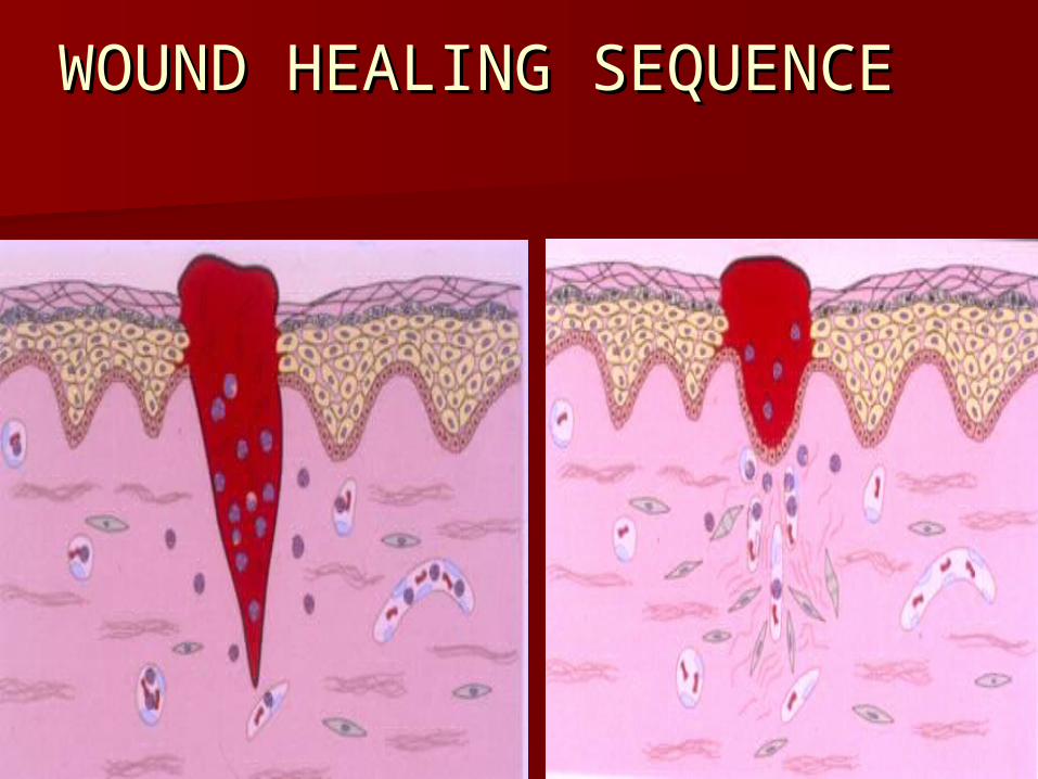
WOUND HEALING WOUND HEALING SEQUENCESEQUENCE
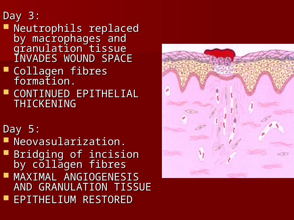
Day 3:Day 3: Neutrophils replaced by Neutrophils replaced by
macrophages and macrophages and granulation tissue granulation tissue INVADES WOUND SPACEINVADES WOUND SPACE
Collagen fibres formation.Collagen fibres formation. CONTINUED EPITHELIAL CONTINUED EPITHELIAL
THICKENINGTHICKENING
Day 5:Day 5: Neovasularization.Neovasularization. Bridging of incision by Bridging of incision by
collagen fibrescollagen fibres MAXIMAL ANGIOGENESIS MAXIMAL ANGIOGENESIS
AND GRANULATION AND GRANULATION TISSUETISSUE
EPITHELIUM RESTOREDEPITHELIUM RESTORED
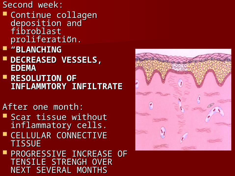
Second week:Second week: Continue collagen Continue collagen
deposition and fibroblast deposition and fibroblast proliferation.proliferation.
““BLANCHING”BLANCHING” DECREASED VESSELS, DECREASED VESSELS,
EDEMAEDEMA RESOLUTION OF RESOLUTION OF
INFLAMMTORY INFLAMMTORY INFILTRATEINFILTRATE
After one month:After one month: Scar tissue without Scar tissue without
inflammatory cells.inflammatory cells. CELLULAR CONNECTIVE CELLULAR CONNECTIVE
TISSUETISSUE PROGRESSIVE INCREASE OF PROGRESSIVE INCREASE OF
TENSILE STRENGH OVER TENSILE STRENGH OVER NEXT SEVERAL MONTHSNEXT SEVERAL MONTHS

Remodeling phaseRemodeling phase
3 week to 2 year3 week to 2 year New collagen forms which increase New collagen forms which increase
tensile strength to woundtensile strength to wound Balance of matrix degradation and Balance of matrix degradation and
collagen synthesiscollagen synthesis Scar tissue is only 80% as srtong as Scar tissue is only 80% as srtong as
original tissue.original tissue.

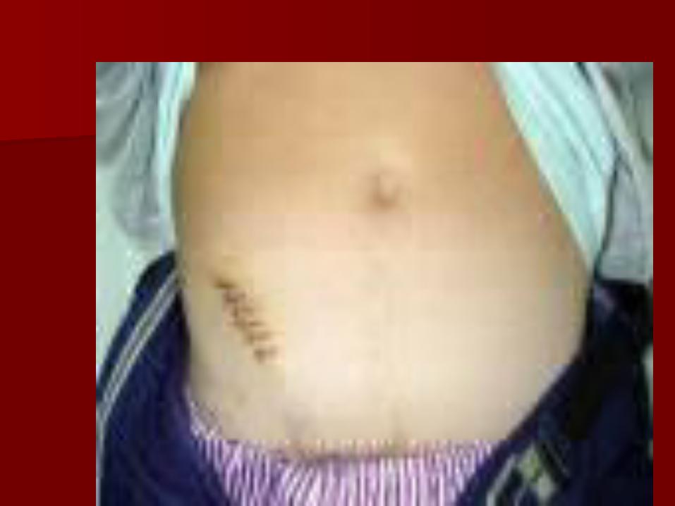
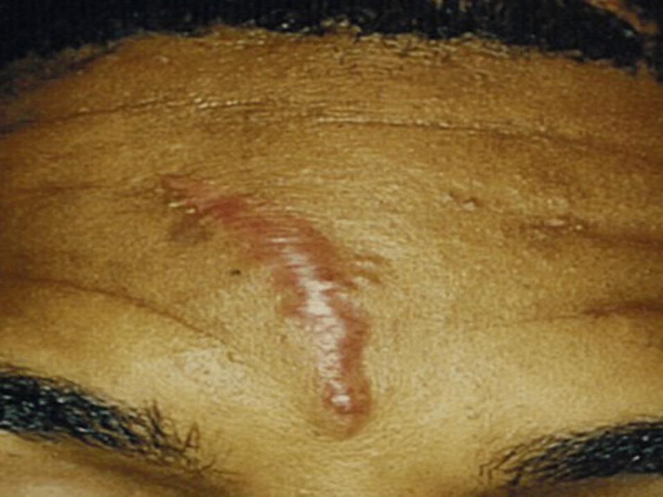
HYPERTROPHIC SCARHYPERTROPHIC SCAR
linear, red, RAISED,PRURITIC linear, red, RAISED,PRURITIC LESIONS,LESIONS,
confined to the original injury siteconfined to the original injury site
Common affect under constant Common affect under constant pressure and stretching area. pressure and stretching area.
Usually arise within one month of Usually arise within one month of injuryinjury
CAUSES OF HYPERTROPHIC CAUSES OF HYPERTROPHIC SCARSCAR
FOREIGN BODY IN THE WOUNDFOREIGN BODY IN THE WOUND SCRATCHINGSCRATCHING HEMATOMAHEMATOMA SECONDARY INTENTIONSECONDARY INTENTION EXCESSIVE TENSIONEXCESSIVE TENSION INADEQUATE WOUND CLOSUREINADEQUATE WOUND CLOSURE
KELOIDSKELOIDS
Form, irregularly shapeForm, irregularly shape Thin epi. Caused by surgical procedure, Thin epi. Caused by surgical procedure,
burn, trauma, inflamm.burn, trauma, inflamm. Spread beyond the limit of original Spread beyond the limit of original
injuryinjury Appear within week or yr. Appear within week or yr. LOCALLY INVASIVE BENIGN NEOPLASTIC LOCALLY INVASIVE BENIGN NEOPLASTIC
TISSUETISSUE Persist over time.RARELY REGRESSPersist over time.RARELY REGRESS
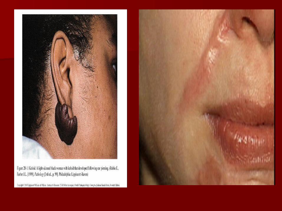
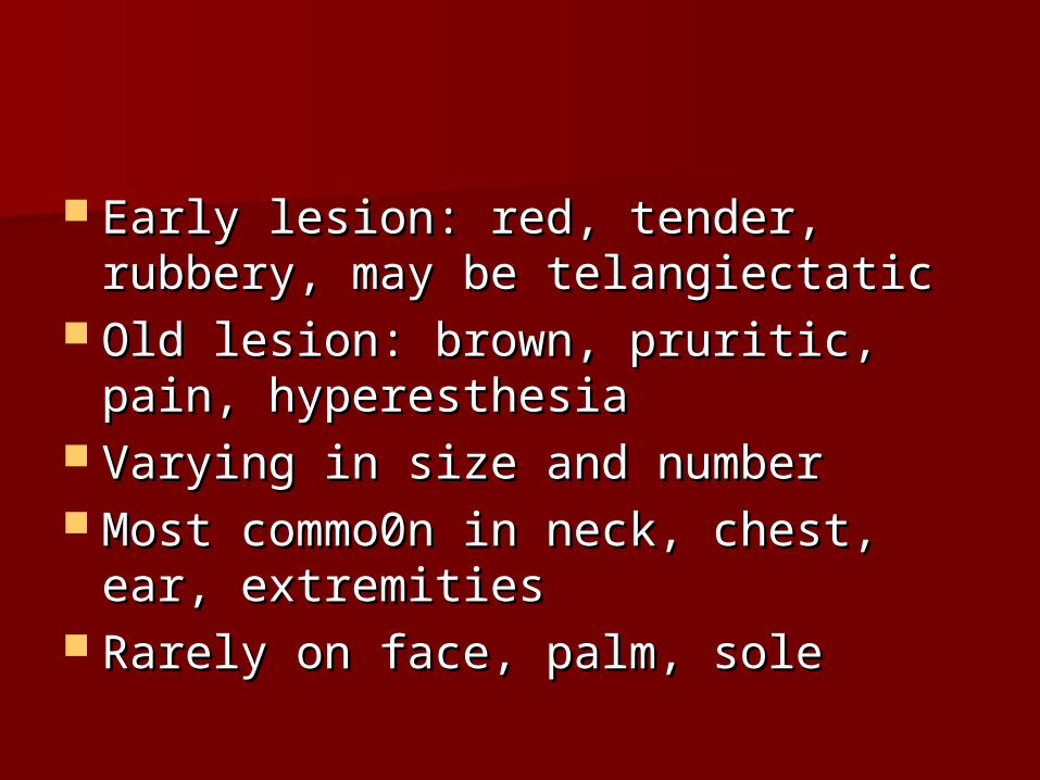
Early lesion: red, tender, rubbery, Early lesion: red, tender, rubbery, may be telangiectaticmay be telangiectatic
Old lesion: brown, pruritic, pain, Old lesion: brown, pruritic, pain, hyperesthesiahyperesthesia
Varying in size and numberVarying in size and number Most commo0n in neck, chest, ear, Most commo0n in neck, chest, ear,
extremitiesextremities Rarely on face, palm, soleRarely on face, palm, sole
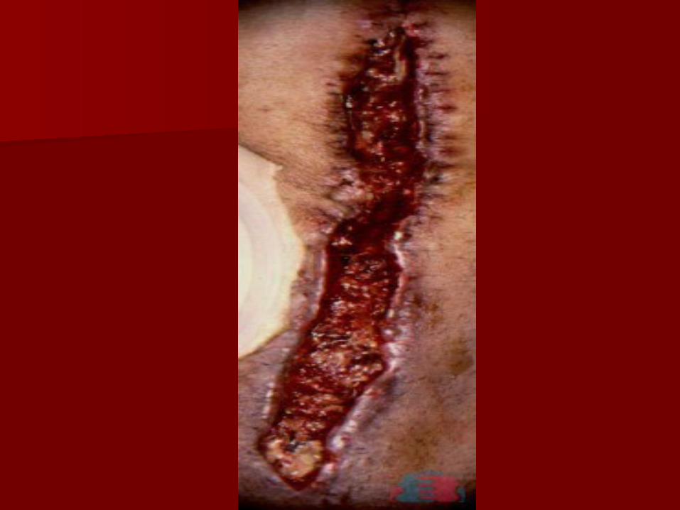
Complications of wound healingComplications of wound healing
- - Deficient scar formation-which can lead to Deficient scar formation-which can lead to rupture of the wound due to inadequate rupture of the wound due to inadequate formation –result in dehiscence and formation –result in dehiscence and ulcerationulceration
- Excessive scar formation of the repair Excessive scar formation of the repair components- components- KeloidKeloid & hypertrophic scar & hypertrophic scar

W.B. Saunders Company items and W.B. Saunders Company items and derived items Copyright (c) 1999 derived items Copyright (c) 1999
by W.B. Saunders Companyby W.B. Saunders Company
SUMMARY WOUND HEALING