BARD COSMAN, M.D. GEORGE F. CRIKELAIR, M. D. New York, New ...
8
Release of the Prolablum in the B||atera| Cleft Llp BARD COSMAN, M. D. GEORGE F. CRIKELAIR, M. D. New York, New York In certain bilateral cleft lip patients with an otherwise satisfactory initial repair, the prolabial segment of the upper lip remains adherent to the premaxilla, and the labial-alveolar suleus is absent. Lack of the sulcus poses a problem in orthodontic and prosthodontic treatment. The ad- herence and the suleus deficiency may contrlbute to immobility of the upper lip and a 'tied down' appearance. _- The paucity of local tissue, the presence of sear from prior lip repair, and the potential growth of the youthful patients involved are limiting factors to be considered in devising reparative methods. Experiences with several surgical techniques for the release of the prolabium and the reconstruction of the upper lip sulcus are presented here and evalu- ated on the basis of clinical observation. Material and Results Forty cases of bilateral cleft lip treated at Columbia-Presbyterian Medical Center from 1954 to 1964 were reviewed. In 12 patients from this group, 14 operations for the release of the prolabtum and the forma- tion of an upper labial-alveolar sulcus were performed. Details regard- ing procedures and results are present in Table 1. Partial ostectomy of the premaxilla with use of local mucus mem- brane flaps was successfully carried out in three cases. The degree of premaxillary protrusion involved in one of these patients was note- worthy (Figure 1). Split thickness skin graft to lip and premaxilla produced a good result in three of four cases in which this method was used (Figure 2). Signifi- cant hypertrophy of the donor site of one of these successful cases was noted, however. Failure occurred in a two-year-old in whom the initial lip release was not maintained in a three-year period of follow-up. The technique of full thickness free mucosal grafting of the labial surface, leaving the premaxillary surface bare, was carried out in six instances (Figure 3). Loss of the graft caused the single failure. The size of the mucosal graft required ranged from 2.5 by 1.0 em in a two and Dr. Cosman is Assistant Attending Surgeon and Instructor in Surgery, and Dr. Crikelair is Attending Surgeon and Professor of Clinical Surgery, the Division of Plastic Surgery, Department of Surgery, Presbyterian Hospital and College of Physi- cians and Surgeons, Columbia University, New York. 122
Transcript of BARD COSMAN, M.D. GEORGE F. CRIKELAIR, M. D. New York, New ...
Release of the Prolablum in the B||atera|Cleft Llp
BARD COSMAN, M. D.GEORGE F. CRIKELAIR, M. D.
New York, New York
In certain bilateral cleft lip patients with an otherwise satisfactoryinitial repair, the prolabial segment of the upper lip remains adherent tothe premaxilla, and the labial-alveolar suleus is absent. Lack of the sulcusposes a problem in orthodontic and prosthodontic treatment. The ad-herence and the suleus deficiency may contrlbute to immobility of the
upper lip and a 'tied down' appearance.
_- The paucity of local tissue, the presence of sear from prior lip repair,
and the potential growth of the youthful patients involved are limiting
factors to be considered in devising reparative methods. Experiences
with several surgical techniques for the release of the prolabium and
the reconstruction of the upper lip sulcus are presented here and evalu-
ated on the basis of clinical observation.
Material and Results
Forty cases of bilateral cleft lip treated at Columbia-Presbyterian
Medical Center from 1954 to 1964 were reviewed. In 12 patients from
this group, 14 operations for the release of the prolabtum and the forma-
tion of an upper labial-alveolar sulcus were performed. Details regard-
ing procedures and results are present in Table 1.
Partial ostectomy of the premaxilla with use of local mucus mem-
brane flaps was successfully carried out in three cases. The degree of
premaxillary protrusion involved in one of these patients was note-
worthy (Figure 1).
Split thickness skin graft to lip and premaxilla produced a good result
in three of four cases in which this method was used (Figure 2). Signifi-
cant hypertrophy of the donor site of one of these successful cases was
noted, however. Failure occurred in a two-year-old in whom the initial
lip release was not maintained in a three-year period of follow-up.
The technique of full thickness free mucosal grafting of the labial
surface, leaving the premaxillary surface bare, was carried out in six
instances (Figure 3). Loss of the graft caused the single failure. The size
of the mucosal graft required ranged from 2.5 by 1.0 em in a two and
Dr. Cosman is Assistant Attending Surgeon and Instructor in Surgery, and Dr.Crikelair is Attending Surgeon and Professor of Clinical Surgery, the Division ofPlastic Surgery, Department of Surgery, Presbyterian Hospital and College of Physi-cians and Surgeons, Columbia University, New York.
122
TABLE 1. Procedure and results for 12 individual patients.
RELEASE OF PROLABIUM 123
Age ai Follow-Case |operation Method up Result Comment
(years) (years)
A.W. 14> partial osteectomy of 1 good floating premaxilla
premaxilla; local mu- with irretrievablecosal flaps. dentition.
P.W. 14 partial osteectomy of 4 good as above.premaxilla; Z plasty
mucosal flaps; immedi-ate insertion of dentalprosthesis.
J .J. 5 partial osteectomy of 1 good massive premaxillarypremaxilla; local mu- protrusion. Figurecosal flaps. 1.
C.W. 6 split thickness skin graft 4 good lip appearance mark-to lip and premaxilla. edly improved.
Graft donor sitehypertrophic. Fig-ure 2.
M.J. 4 split thickness skin graft 4 good long term follow-upto lip and premaxilla.
E.M. 2 split thickness skin graft 3 failure release not main-' to lip and premaxilla. tained.
5 buccal mucosal transpo- 1 failure release not adequatesition flaps. lip bulky.
T.C. 12 mucosal graft to lip 1 good 3.5 by 2.5 cm lip de-alone. fect.
J.S. 12 mucosal graft to lip 2 good 4.0 by 2.5 em lip de-alone. fect. Figure 3.
R.B. 24 mucosal graft to lip 4 good 2.5 by 1.0 em lip de-alone. fect.
D .E. 8 mucosal graft to lip 4 good 3.0 by 1.0 em lip de-
alone. fect.M.G. 8 mucosal graft to lip 3 good
alone.
B.S. 414 mucosal graft to lip 1 failure loss of graft.alone.
6 split thickness skin graft 1 good
to lip and premaxilla.
one-half year old to 4.0 by 2.5 em in a 12-year-old. There was no diffi-
culty in primary closure of the buccal mucosa donor sites and no late
contractures were noted.
In one case, long laterally based buccal mucosal flaps were transposed
to cover both lip and premaxilla. The result was unsatisfactory in terms
of the release achieved and the bulkiness of the lip which was created.
Discussion
In a certain number of bilateral cleft lip patients, the initial condition
and/or its repair leads to adherence of the prolabial lip segment to the
=~, %
c JNC1S1ONS RESECTiON CLOSURE
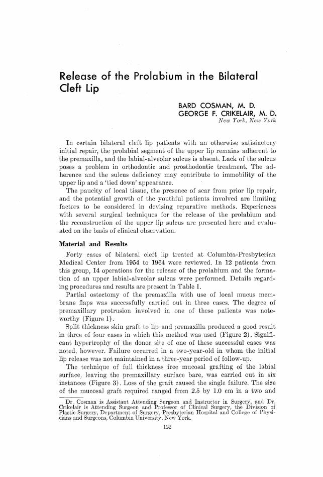
FIGURE 1. A. Five-year-old boy with massive protrusion of premasxilla despitesatisfactory lip repair in carly infancy. With teeth in occlusion, premaxilla overhangsthe lower lip. Upper lip adherent to premaxilla with anterior surface of premaxillacracked and crusted due to exposure.
B. Dental model for patient shown in Figure 1A showing alveolar arch in satis-factory alignment, with massive vomerine strut maintaining space where premaxillashould have been. Anterior surface of premaxilla 25 em anterior to anterior edgeof maxillary arch and premaxillary dentition on a level 1.5 em below the plane ofthe maxillary dentition.
C. For same patient, partial osteectomy avoiding trauma to premaxillary-yomerinesuture line and employing local mucosal flaps to reconstruct sulcus.
D. Intraoral view of same patient at end of operative procedurc.E. Appearance of same patient seven months after operation.
124
RELEASE OF PROLABIUM 125
premaxilla and to deficiency of the upper labial-alveolar sulcus. These
problems were significant enough to deserve operative revision in 12 of
the 40 bilateral cleft lip patients in our series.
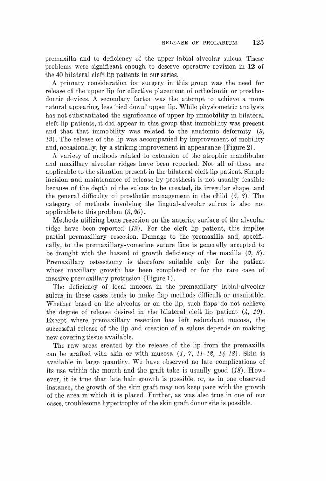
A primary consideration for surgery in this group was the need for
release of the upper lip for effective placement of orthodontic or prostho-
dontic devices. A secondary factor was the attempt to achieve a more
natural appearing, less 'tied down' upper lip. While physiometric analysis
has not substantiated the significance of upper lip immobility in bilateral
cleft lip patients, it did appear in this group that immobility was present
and that that immobility was related to the anatomic deformity (9,
13). The release of the lip was accompanied by improvement of mobility
and, occasionally, by a striking improvement in appearance (Figure 2).
A variety of methods related to extension of the atrophic mandibular
and maxillary alveolar ridges have been reported. Not all of these are
applicable to the situation present in the bilateral cleft lip patient. Simple
incision and maintenance of release by prosthesis is not usually feasible
because of the depth of the suleus to be created, its irregular shape, and
the general difficulty of prosthetic management in the child (5, 6). The
category of methods involving the lingual-alveolar suleus is also not
applicable to this problem (3, 20).
Methods utilizing bone resection on the anterior surface of the alveolar
ridge have been reported (12). For the cleft lip patient, this implies
partial premaxillary resection. Damage to the premaxilla and, specifi-
cally, to the premaxillary-vomerine suture line is generally accepted to
be fraught with the hazard of growth deficiency of the maxilla (2, 8).
Premaxillary osteectomy is therefore suitable only for the patient
whose maxillary growth has been completed or for the rare case of
massive premaxillary protrusion (Figure 1).
The deficiency of local mucosa in the premaxillary labial-alveolar
suleus in these cases tends to make flap methods difficult or unsuitable.
Whether based on the alveolus or on the lip, such flaps do not achieve
the degree of release desired in the bilateral cleft lip patient (4, 10).
Except where premaxillary resection has left redundant mucosa, the
successful release of the lip and creation of a suleus depends on making
new covering tissue available.
The raw areas created by the release of the lip from the premaxilla
can be grafted with skin or with mucosa (1, 7, 11-12, 14-18). Skin is
available in large quantity. We have observed no late complications of
its use within the mouth and the graft take is usually good (18). How-
ever, it is true that late hair growth is possible, or, as in one observed
instance, the growth of the skin graft may not keep pace with the growth
of the area in which it is placed. Further, as was also true in one of our
cases, troublesome hypertrophy of the skin graft donor site is possible.

126 Cosman, Crikelair
FIGURE 2. The 'tied down' appearance of upper lip is strikingly improved bysplit thickness skin graft to lip and premaxilla. Top, pre-operative full face andprofile; middle, post-operative full face and profile; bottom, skin graft suleus.

RELEASE OF PROLABIUM 127
s suicus bissEction RAW AREA
FIGURE 3. A. Pre-operative view demonstrating adherence of prolabial lip seg-ment to premaxilla.
B. Operative design involving dissection of lip from premaxilla and coverage oflabial surface alone by a full thickness mucosal graft held in place by a bolusdressing.
C. Operative view showing raw surfaces produced by dissection.D. Site of mucosal graft on lateral buccal surface.E. Mucosal graft in place with suture ready to be tied over bolus. Note remaining
exposed surface of premaxilla.
The use of a full thickness mucosal graft obviates most of these prob-
lems. The amount of mucosa available for donation is not unlimited
(15). By allowing the premaxillary surface to epithelize spontaneously,
the size of the defect requiring mucosal coverage has been routinely

128 Cosman, CUrikelarr
reduced to a size easily managed by a single, small mucosal graft. The
question of contracture consequent upon the secondary epithelization of
the premaxilla can be raised. Both experimental and clinical observations
suggest that some shrinkage of sulcus depth occurs under all conditions
(18, 19). However, lip release appears to be maintained by this method as
effectively as when both lip and premaxillary surfaces are covered by a
skin graft.
Conclusion
Fourteen surgical procedures for the late release of the prolabium and
creation of an upper labial-alveolar sulcus were carried out in 12 cases
in a series of 40 bilateral cleft lip patients. The techniques employed
were reviewed and their results compared. Dissection of the prolabium
from the premaxilla with full thickness free mucosal graft to the labial
surface leaving the premaxillary area bare was found to be an effective
method free of major objections.
180 Fort Washington Avenue
New York, New York 10082
References
1. AsHury, F. L., Senwartz, A. N., and M. F., A modified technique forcreating a lower lingual suleus. Plastic reconstr. Surg., 22, 204-213, 1958.
2. A. J., S., and Smon, B. E., Early and late management of theprotruding premaxilla. Plastic reconstr. Surg., 29, 58-70, 1962.
3. CampowrELL, J. B., Lingual ridge extension. J. oral Surg., 18, 287-292, 1955.4. Cmark, H. B., Jr., Deepening of labial sulcus by mucosal flap advancement:
report of case. J. oral Surg., 11, 165-168, 1953.5. ComnEtt, H. A., Immediate maxillary ridge extension. Dent. Digest, 60, 104-106,
1954.6. Coorry, D. O., A method for deepening the mandibular and maxillary sulci to
correct deficient denture ridges. J. oral Surg., 10, 279-289, 1952.. EssEr, J. F., Studies of plastic surgery of the face. Annals Surg., 65, 297-315, 1917.. Grover, D. M., and NEwcoms, M. R., Bilateral cleft lip repair and the floatingpremaxilla. Plastic reconstr. Surg., 28, 365-377, 1965.
9. Haring, F. N., and McCormack, R. M., A physiometric analysis of lip functionin cleft and noncleft subjects. Cleft Palate J., 1, 320-828, 1964.
10. KazanjIan, V. H., The surgical treatment of abnormalities of the edentulousalveolar processes and the palate. Amer. J. orthod. oral Surg., 26, 263-277, 1940.
11. KrucrEr, G. O., Ridge extension: review of indications and technics. J. oral Surg.,16, 191-201, 1958.
12. OsBwraersEr, H., Surgical preparation of the maxilla for prosthesis. J. oral Surg.,22, 127-134, 1964.
13. OrrErman, R. E., J. F., and SustEnv, J. D., Symmetry of lip activityin repaired unilateral clefts of the lip. Cleft Palate J., 1, 347-356, 1964.
14. PickrEriut, H. P., Intraoral skin grafting: the establishment of the buccal sulcus.Brit. J. dent. Serience, 62, 135-141, 1919. .
15. PropprEr, R. H., Simplified ridge extension using free mucosal grafts. J. oral Surg.,22, 469-474, 1964.
16. Prowurr, J. R., A skin-grafted ridge extension: new approach to atrophic ridgemanagement. J. oral Surg., 23, 123-129, 1965.
17. ReEmurmann, A., Creation of an alveolar ridge after bone transplantation to themandible. Plastic reconstr. Surg., 24, 188-189, 1959.
co[I

RELEASE OF PROLABIUM 129
18. StanEtTz, C. A., Jr., and Ranrow, R. M., The intra-oral use of split-thicknessskin grafts in head and neck surgery. Amer. J. Surg., 104, 721-726, 1962.
19. SprEnorurr, D. E., Haywarp, J. R., and ArBor, A., Study of suleus extension woundhealing in dogs. J. oral Surg., 22, 4183-421, 1964.
20. TrauNERr, R., Alveoplasty with ridge extension on the lingual side of the lowerjaw to solve the problem of a lower dental prosthesis. Oral Surg., oral Med., oralPath., 5, 340-346, 1952.