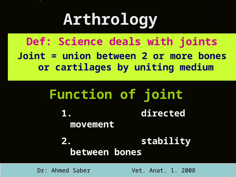
Arthrology Def: Science deals with joints Joint = union between 2 or more bones or cartilages by...
27
Arthrology Arthrology Def: Science deals with joints Joint = union between 2 or more bones or cartilages by uniting medium Function of joint 1. directed movement 2. stability between bones 3. bone growth Dr: Ahmed Saber Vet. Anat. 1. 2008
-
Upload
grace-dickens -
Category
Documents
-
view
213 -
download
0
Transcript of Arthrology Def: Science deals with joints Joint = union between 2 or more bones or cartilages by...
Arthrology Arthrology Def Science deals with joints
Joint = union between 2 or more bones or cartilages by uniting medium
Function of joint 1 directed
movement
2 stability between bones
3 bone growthDr Ahmed Saber Vet Anat 1 2008
Classification of jointsClassification of jointsAccording to types of uniting mediumAccording to types of uniting medium
Fibrous joints1 Uniting
medium is fibrous tissue
2 No joint cavity3 are temporary
(ossify with age)
4 No movement
Cartilagenous joints
1 Uniting medium is cartilage
2 are temporary3 No or very limited
movement
synovial joints1 uniting medium
is joint capsule2 Joint cavity
present3 Movable
Dr Ahmed Saber Vet Anat 1 2008
I- Fibrous joints (Synarthrosis)
1-Syndesmosis The uniting medium
is fibrous connective tissue (interosseous ligament)
ex Union of metacarpal bones radius and ulna and tibia and fibula
Types of fibrous joints
Large metacrpal
small metacrpal
syndes
mos
is
Dr Ahmed Saber Vet Anat 1 2008
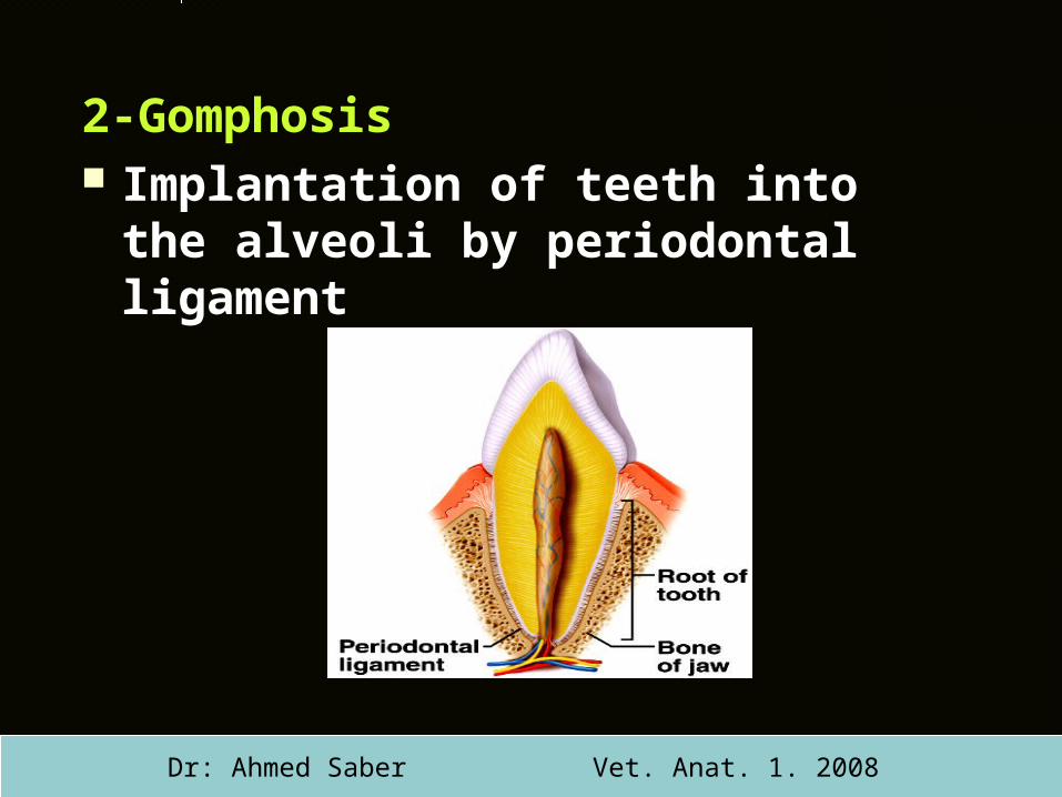
2-Gomphosis Implantation of teeth into the
alveoli by periodontal ligament
Dr Ahmed Saber Vet Anat 1 2008
3-Sutures
fibrous tissues bond the
bones of skull
Based on the edges of
the articulating bones
it may be
sutures
Dr Ahmed Saber Vet Anat 1 2008
A- Serrated sutures the interlocking borders are saw
like as interfrontal suture
B- Squamous sutures borders are flat and overlapped
as temporal and parietal suture
C- Plane suture the interlocking borders are
smooth as nasal suture
D- Foliate suture the edge of one bone fits into a
fissure in the adjacent bone as zygomatico-maxillary suture
A
B
C
D
Dr Ahmed Saber Vet Anat 1 2008
II- Cartilaginous joints
(amphiarthrosis)
1 Synchondrosis (hyaline cartilage joint)
the uniting medium is hyaline cartilage EX Costochondoral junction and epiphyseal and metaphyseal junction
Types of cartilaginous joints
Ribs
Costochondral junction
Dr Ahmed Saber Vet Anat 1 2008
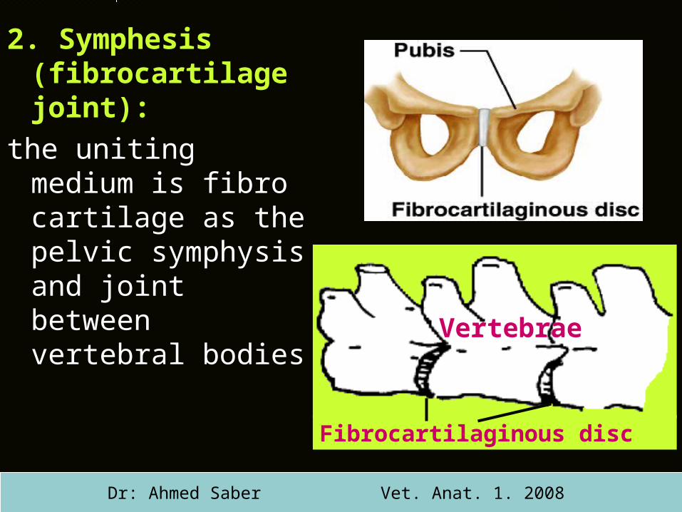
2 Symphesis (fibrocartilage joint)
the uniting medium is fibro cartilage as the pelvic symphysis and joint between vertebral bodies
Fibrocartilaginous disc
Vertebrae
Dr Ahmed Saber Vet Anat 1 2008
III Diarthroidal joint(Synovial movable or true
joint)
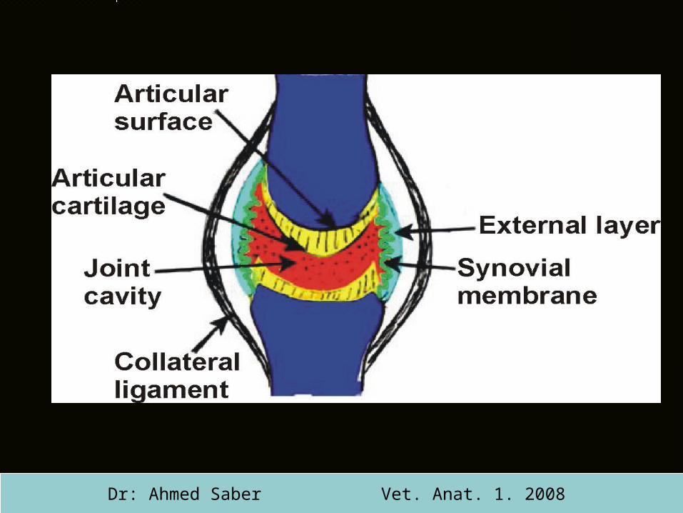
Structures of synovial joints
A-constant structures
Articular surface
Articular cartilage
Articular capsule
Joint cavity
B- inconstant (additional features)LigamentsArticular disc ampmenisciMarginal cartilage
Dr Ahmed Saber Vet Anat 1 2008
Dr Ahmed Saber Vet Anat 1 2008
A-constant structures ( essential)
I - Articular surfaces It is formed of compact bone It is smooth but sometime interrupted by
synovial fossa Covered by articular cartilage except
synovial fossa
Structures of synovial joints (cont)
Dr Ahmed Saber Vet Anat 1 2008
II - Articular cartilages It is smooth hyaline cartilage Cover the articular surfaces except
at the synovial fossa Almost one convex and the other is
concave The concave one is thickest at the
periphery and thin at the center and vise versa
Its function ndash increase the curvature of bonesndash facilitate movements and reduce
frictions
Dr Ahmed Saber Vet Anat 1 2008
III-Joint capsule It is fibroserous tube Originated from the periosteum of bones attached around the articular margins
and surrounds the joint cavity it is consisted of
1048678 Outer fibrous layer (external layer)ndash it may contain ligaments
1048678 Inner synovial layer (internal layer)ndash it is a thin single layer of epitheliumndash line the joint cavity except the articular
cartilagendash it secrete andor absorb synovia
Dr Ahmed Saber Vet Anat 1 2008
B- Additional features present in some joint
1- ligamentsStrong white fibrous band unite 2 or more
bonesA- internal (Intraarticular) lig placed within the fibrous layer covered with the synovial membrane Ex Cruciate ligamentB- Extrnal (periarticular) lig situated outside the joint capsuleEx Collateral ligaments
Structures of synovial joints (cont)
Dr Ahmed Saber Vet Anat 1 2008
2- Articular disc ampmeniscifibrocartilage plate present between the
articular cartilages divide the joint cavity partially (disc) completely
(menisci) into 2 partsEx Disc of tempro-mandibular joint menisci of
stifle jointfunction correct the incongruent surfaces anti concussion amp allow greater movement
Dr Ahmed Saber Vet Anat 1 2008
3- Marginal cartilage ring of fibrocartilage encircle the rim of
articular cartilageEx acetabulum of hip jointfunction enlarge the joint cavity protect the rim from fractures
Dr Ahmed Saber Vet Anat 1 2008
Synovia (synovial fluid)Synovia (synovial fluid) Secreted and absorbed by synovial
membrane It present within synovial joint around
some tendons and within bursae chemical composition (mucin salts
albumin fat droplets and cellular debris)
Function1 Lubricate the articular surfaces and tendons2 Nutrition of cartilage3 Remove waste metabolites4 Enable wandering leukocyte to circulate in the joint
cavity
Dr Ahmed Saber Vet Anat 1 2008
Blood and nerve Blood and nerve supply of synovial jointsupply of synovial joint
Synovial joint receives vessels from surrounding muscles
Sensory nerves terminates in the joint capsule and ligaments
Autonomic innervations terminate in the arteries
Dr Ahmed Saber Vet Anat 1 2008
Anatomical types of Anatomical types of synovial jointssynovial joints
I- According to the number of articulating bones
Simple joint between two bonesCompound joint between more than two
bonesII- according to fitting of articular surfacesCongruent joint adapted to each otherIn congruent joint are not adapted ex
stifle joint
Dr Ahmed Saber Vet Anat 1 2008
Types Shape Example 1- Ball and socket
SpheroidalEnartherosis
Ball and other isa cavityBall is the largestThe socket is the largest
Shoulderhip
2-Hinge (ginglymis)
Like door hinge one concave and other is convex
elbow
1
2
1
III-according to the shape of articular surfaces
Dr Ahmed Saber Vet Anat 1 2008
Types Types Shape Shape Example Example 3- Ellipsoid The convex andThe convex and
concave areconcave are
ellipsoidal ovalellipsoidal oval
AtlantooccipitalAtlantooccipital
radiocarpalradiocarpal
4- gliding (plane) Flat surfacesFlat surfaces
jointjointIntercarpalIntercarpal
skull
Atlas
3
Dr Ahmed Saber Vet Anat 1 2008
4
Types Shape Example 5- pivot One ring moved
around axis of otheratlanoaxial
6-saddle Concavoconvex as horse saddle
Caffin and pastern
5 Pastern joint
Coffinjoint
6
Dr Ahmed Saber Vet Anat 1 2008
Types Types Shape Shape Example Example 7- cochlear Cochlea (cavity)
receives ridge of other bone
Tibio-tarsal
8-Bi-condylar Both are convexcondyles
femorotibial
Tibia
Talus
Tars
al b
on
es
7
Tibia
Femur
8
Lateral condyle
of femur Lateral condyle of tibia
Dr Ahmed Saber Vet Anat 1 2008
Physiological types of synovial Physiological types of synovial jointsjointsI-According to the axis of movementuniaxial around one axis
Transverse axis as hing joints (elbow joint)Vertical axis as pivot joints (atlantoaxial)
biaxial arround two axisBiocodylar jointsEllipsoid jointsSaddle joints
multiaxial more than twoball and socket
Movements of synovial joints
A- According to causesActive movements due to muscle contractionPassive movements due to gravity or the
movement of other jointsB- According to the shape of articular
surface1-Gliding one surface glides over anotherEx Intercarpal femoropatellar and the joints
between the articular process of cervical vertebrae
Dr Ahmed Saber Vet Anat 1 2008
2- angular movementsA- flexion (decrease the joint angle)B- extension (increase the joint angle)C- adduction (towards the median plane)D-abduction (away from the median plane)E- circumduction (combination of ABC and
D)3- rotatory movementRotation of one segment around the
longitudinal axis of other segments atlantoaxial joint
Dr Ahmed Saber Vet Anat 1 2008
Factor affecting joint Factor affecting joint stabilitystability
11 Shape and size of articulating boneShape and size of articulating bone
22 Arrangements of articulating boneArrangements of articulating bone
33 Tone of surrounding musclesTone of surrounding muscles
44 Strength of the ligamentsStrength of the ligaments
To be continued in applied anatomy
Dr Ahmed Saber Vet Anat 1 2008
- Arthrology
- Classification of joints
- I- Fibrous joints (Synarthrosis)
- Slide 4
- Slide 5
- Slide 6
- II- Cartilaginous joints (amphiarthrosis)
- Slide 8
- III Diarthroidal joint (Synovial movable or true joint)
- Slide 10
- Slide 11
- Slide 12
- Slide 13
- Slide 14
- Slide 15
- Slide 16
- Synovia (synovial fluid)
- Blood and nerve supply of synovial joint
- Anatomical types of synovial joints
- Slide 20
- Slide 21
- Slide 22
- Slide 23
- Physiological types of synovial joints
- Movements of synovial joints
- Slide 26
- Factor affecting joint stability
-
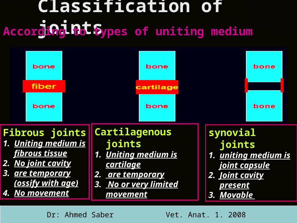
Classification of jointsClassification of jointsAccording to types of uniting mediumAccording to types of uniting medium
Fibrous joints1 Uniting
medium is fibrous tissue
2 No joint cavity3 are temporary
(ossify with age)
4 No movement
Cartilagenous joints
1 Uniting medium is cartilage
2 are temporary3 No or very limited
movement
synovial joints1 uniting medium
is joint capsule2 Joint cavity
present3 Movable
Dr Ahmed Saber Vet Anat 1 2008
I- Fibrous joints (Synarthrosis)
1-Syndesmosis The uniting medium
is fibrous connective tissue (interosseous ligament)
ex Union of metacarpal bones radius and ulna and tibia and fibula
Types of fibrous joints
Large metacrpal
small metacrpal
syndes
mos
is
Dr Ahmed Saber Vet Anat 1 2008
2-Gomphosis Implantation of teeth into the
alveoli by periodontal ligament
Dr Ahmed Saber Vet Anat 1 2008
3-Sutures
fibrous tissues bond the
bones of skull
Based on the edges of
the articulating bones
it may be
sutures
Dr Ahmed Saber Vet Anat 1 2008
A- Serrated sutures the interlocking borders are saw
like as interfrontal suture
B- Squamous sutures borders are flat and overlapped
as temporal and parietal suture
C- Plane suture the interlocking borders are
smooth as nasal suture
D- Foliate suture the edge of one bone fits into a
fissure in the adjacent bone as zygomatico-maxillary suture
A
B
C
D
Dr Ahmed Saber Vet Anat 1 2008
II- Cartilaginous joints
(amphiarthrosis)
1 Synchondrosis (hyaline cartilage joint)
the uniting medium is hyaline cartilage EX Costochondoral junction and epiphyseal and metaphyseal junction
Types of cartilaginous joints
Ribs
Costochondral junction
Dr Ahmed Saber Vet Anat 1 2008
2 Symphesis (fibrocartilage joint)
the uniting medium is fibro cartilage as the pelvic symphysis and joint between vertebral bodies
Fibrocartilaginous disc
Vertebrae
Dr Ahmed Saber Vet Anat 1 2008
III Diarthroidal joint(Synovial movable or true
joint)
Structures of synovial joints
A-constant structures
Articular surface
Articular cartilage
Articular capsule
Joint cavity
B- inconstant (additional features)LigamentsArticular disc ampmenisciMarginal cartilage
Dr Ahmed Saber Vet Anat 1 2008
Dr Ahmed Saber Vet Anat 1 2008
A-constant structures ( essential)
I - Articular surfaces It is formed of compact bone It is smooth but sometime interrupted by
synovial fossa Covered by articular cartilage except
synovial fossa
Structures of synovial joints (cont)
Dr Ahmed Saber Vet Anat 1 2008
II - Articular cartilages It is smooth hyaline cartilage Cover the articular surfaces except
at the synovial fossa Almost one convex and the other is
concave The concave one is thickest at the
periphery and thin at the center and vise versa
Its function ndash increase the curvature of bonesndash facilitate movements and reduce
frictions
Dr Ahmed Saber Vet Anat 1 2008
III-Joint capsule It is fibroserous tube Originated from the periosteum of bones attached around the articular margins
and surrounds the joint cavity it is consisted of
1048678 Outer fibrous layer (external layer)ndash it may contain ligaments
1048678 Inner synovial layer (internal layer)ndash it is a thin single layer of epitheliumndash line the joint cavity except the articular
cartilagendash it secrete andor absorb synovia
Dr Ahmed Saber Vet Anat 1 2008
B- Additional features present in some joint
1- ligamentsStrong white fibrous band unite 2 or more
bonesA- internal (Intraarticular) lig placed within the fibrous layer covered with the synovial membrane Ex Cruciate ligamentB- Extrnal (periarticular) lig situated outside the joint capsuleEx Collateral ligaments
Structures of synovial joints (cont)
Dr Ahmed Saber Vet Anat 1 2008
2- Articular disc ampmeniscifibrocartilage plate present between the
articular cartilages divide the joint cavity partially (disc) completely
(menisci) into 2 partsEx Disc of tempro-mandibular joint menisci of
stifle jointfunction correct the incongruent surfaces anti concussion amp allow greater movement
Dr Ahmed Saber Vet Anat 1 2008
3- Marginal cartilage ring of fibrocartilage encircle the rim of
articular cartilageEx acetabulum of hip jointfunction enlarge the joint cavity protect the rim from fractures
Dr Ahmed Saber Vet Anat 1 2008
Synovia (synovial fluid)Synovia (synovial fluid) Secreted and absorbed by synovial
membrane It present within synovial joint around
some tendons and within bursae chemical composition (mucin salts
albumin fat droplets and cellular debris)
Function1 Lubricate the articular surfaces and tendons2 Nutrition of cartilage3 Remove waste metabolites4 Enable wandering leukocyte to circulate in the joint
cavity
Dr Ahmed Saber Vet Anat 1 2008
Blood and nerve Blood and nerve supply of synovial jointsupply of synovial joint
Synovial joint receives vessels from surrounding muscles
Sensory nerves terminates in the joint capsule and ligaments
Autonomic innervations terminate in the arteries
Dr Ahmed Saber Vet Anat 1 2008
Anatomical types of Anatomical types of synovial jointssynovial joints
I- According to the number of articulating bones
Simple joint between two bonesCompound joint between more than two
bonesII- according to fitting of articular surfacesCongruent joint adapted to each otherIn congruent joint are not adapted ex
stifle joint
Dr Ahmed Saber Vet Anat 1 2008
Types Shape Example 1- Ball and socket
SpheroidalEnartherosis
Ball and other isa cavityBall is the largestThe socket is the largest
Shoulderhip
2-Hinge (ginglymis)
Like door hinge one concave and other is convex
elbow
1
2
1
III-according to the shape of articular surfaces
Dr Ahmed Saber Vet Anat 1 2008
Types Types Shape Shape Example Example 3- Ellipsoid The convex andThe convex and
concave areconcave are
ellipsoidal ovalellipsoidal oval
AtlantooccipitalAtlantooccipital
radiocarpalradiocarpal
4- gliding (plane) Flat surfacesFlat surfaces
jointjointIntercarpalIntercarpal
skull
Atlas
3
Dr Ahmed Saber Vet Anat 1 2008
4
Types Shape Example 5- pivot One ring moved
around axis of otheratlanoaxial
6-saddle Concavoconvex as horse saddle
Caffin and pastern
5 Pastern joint
Coffinjoint
6
Dr Ahmed Saber Vet Anat 1 2008
Types Types Shape Shape Example Example 7- cochlear Cochlea (cavity)
receives ridge of other bone
Tibio-tarsal
8-Bi-condylar Both are convexcondyles
femorotibial
Tibia
Talus
Tars
al b
on
es
7
Tibia
Femur
8
Lateral condyle
of femur Lateral condyle of tibia
Dr Ahmed Saber Vet Anat 1 2008
Physiological types of synovial Physiological types of synovial jointsjointsI-According to the axis of movementuniaxial around one axis
Transverse axis as hing joints (elbow joint)Vertical axis as pivot joints (atlantoaxial)
biaxial arround two axisBiocodylar jointsEllipsoid jointsSaddle joints
multiaxial more than twoball and socket
Movements of synovial joints
A- According to causesActive movements due to muscle contractionPassive movements due to gravity or the
movement of other jointsB- According to the shape of articular
surface1-Gliding one surface glides over anotherEx Intercarpal femoropatellar and the joints
between the articular process of cervical vertebrae
Dr Ahmed Saber Vet Anat 1 2008
2- angular movementsA- flexion (decrease the joint angle)B- extension (increase the joint angle)C- adduction (towards the median plane)D-abduction (away from the median plane)E- circumduction (combination of ABC and
D)3- rotatory movementRotation of one segment around the
longitudinal axis of other segments atlantoaxial joint
Dr Ahmed Saber Vet Anat 1 2008
Factor affecting joint Factor affecting joint stabilitystability
11 Shape and size of articulating boneShape and size of articulating bone
22 Arrangements of articulating boneArrangements of articulating bone
33 Tone of surrounding musclesTone of surrounding muscles
44 Strength of the ligamentsStrength of the ligaments
To be continued in applied anatomy
Dr Ahmed Saber Vet Anat 1 2008
- Arthrology
- Classification of joints
- I- Fibrous joints (Synarthrosis)
- Slide 4
- Slide 5
- Slide 6
- II- Cartilaginous joints (amphiarthrosis)
- Slide 8
- III Diarthroidal joint (Synovial movable or true joint)
- Slide 10
- Slide 11
- Slide 12
- Slide 13
- Slide 14
- Slide 15
- Slide 16
- Synovia (synovial fluid)
- Blood and nerve supply of synovial joint
- Anatomical types of synovial joints
- Slide 20
- Slide 21
- Slide 22
- Slide 23
- Physiological types of synovial joints
- Movements of synovial joints
- Slide 26
- Factor affecting joint stability
-
Dr Ahmed Saber Vet Anat 1 2008
A-constant structures ( essential)
I - Articular surfaces It is formed of compact bone It is smooth but sometime interrupted by
synovial fossa Covered by articular cartilage except
synovial fossa
Structures of synovial joints (cont)
Dr Ahmed Saber Vet Anat 1 2008
II - Articular cartilages It is smooth hyaline cartilage Cover the articular surfaces except
at the synovial fossa Almost one convex and the other is
concave The concave one is thickest at the
periphery and thin at the center and vise versa
Its function ndash increase the curvature of bonesndash facilitate movements and reduce
frictions
Dr Ahmed Saber Vet Anat 1 2008
III-Joint capsule It is fibroserous tube Originated from the periosteum of bones attached around the articular margins
and surrounds the joint cavity it is consisted of
1048678 Outer fibrous layer (external layer)ndash it may contain ligaments
1048678 Inner synovial layer (internal layer)ndash it is a thin single layer of epitheliumndash line the joint cavity except the articular
cartilagendash it secrete andor absorb synovia
Dr Ahmed Saber Vet Anat 1 2008
B- Additional features present in some joint
1- ligamentsStrong white fibrous band unite 2 or more
bonesA- internal (Intraarticular) lig placed within the fibrous layer covered with the synovial membrane Ex Cruciate ligamentB- Extrnal (periarticular) lig situated outside the joint capsuleEx Collateral ligaments
Structures of synovial joints (cont)
Dr Ahmed Saber Vet Anat 1 2008
2- Articular disc ampmeniscifibrocartilage plate present between the
articular cartilages divide the joint cavity partially (disc) completely
(menisci) into 2 partsEx Disc of tempro-mandibular joint menisci of
stifle jointfunction correct the incongruent surfaces anti concussion amp allow greater movement
Dr Ahmed Saber Vet Anat 1 2008
3- Marginal cartilage ring of fibrocartilage encircle the rim of
articular cartilageEx acetabulum of hip jointfunction enlarge the joint cavity protect the rim from fractures
Dr Ahmed Saber Vet Anat 1 2008
Synovia (synovial fluid)Synovia (synovial fluid) Secreted and absorbed by synovial
membrane It present within synovial joint around
some tendons and within bursae chemical composition (mucin salts
albumin fat droplets and cellular debris)
Function1 Lubricate the articular surfaces and tendons2 Nutrition of cartilage3 Remove waste metabolites4 Enable wandering leukocyte to circulate in the joint
cavity
Dr Ahmed Saber Vet Anat 1 2008
Blood and nerve Blood and nerve supply of synovial jointsupply of synovial joint
Synovial joint receives vessels from surrounding muscles
Sensory nerves terminates in the joint capsule and ligaments
Autonomic innervations terminate in the arteries
Dr Ahmed Saber Vet Anat 1 2008
Anatomical types of Anatomical types of synovial jointssynovial joints
I- According to the number of articulating bones
Simple joint between two bonesCompound joint between more than two
bonesII- according to fitting of articular surfacesCongruent joint adapted to each otherIn congruent joint are not adapted ex
stifle joint
Dr Ahmed Saber Vet Anat 1 2008
Types Shape Example 1- Ball and socket
SpheroidalEnartherosis
Ball and other isa cavityBall is the largestThe socket is the largest
Shoulderhip
2-Hinge (ginglymis)
Like door hinge one concave and other is convex
elbow
1
2
1
III-according to the shape of articular surfaces
Dr Ahmed Saber Vet Anat 1 2008
Types Types Shape Shape Example Example 3- Ellipsoid The convex andThe convex and
concave areconcave are
ellipsoidal ovalellipsoidal oval
AtlantooccipitalAtlantooccipital
radiocarpalradiocarpal
4- gliding (plane) Flat surfacesFlat surfaces
jointjointIntercarpalIntercarpal
skull
Atlas
3
Dr Ahmed Saber Vet Anat 1 2008
4
Types Shape Example 5- pivot One ring moved
around axis of otheratlanoaxial
6-saddle Concavoconvex as horse saddle
Caffin and pastern
5 Pastern joint
Coffinjoint
6
Dr Ahmed Saber Vet Anat 1 2008
Types Types Shape Shape Example Example 7- cochlear Cochlea (cavity)
receives ridge of other bone
Tibio-tarsal
8-Bi-condylar Both are convexcondyles
femorotibial
Tibia
Talus
Tars
al b
on
es
7
Tibia
Femur
8
Lateral condyle
of femur Lateral condyle of tibia
Dr Ahmed Saber Vet Anat 1 2008
Physiological types of synovial Physiological types of synovial jointsjointsI-According to the axis of movementuniaxial around one axis
Transverse axis as hing joints (elbow joint)Vertical axis as pivot joints (atlantoaxial)
biaxial arround two axisBiocodylar jointsEllipsoid jointsSaddle joints
multiaxial more than twoball and socket
Movements of synovial joints
A- According to causesActive movements due to muscle contractionPassive movements due to gravity or the
movement of other jointsB- According to the shape of articular
surface1-Gliding one surface glides over anotherEx Intercarpal femoropatellar and the joints
between the articular process of cervical vertebrae
Dr Ahmed Saber Vet Anat 1 2008
2- angular movementsA- flexion (decrease the joint angle)B- extension (increase the joint angle)C- adduction (towards the median plane)D-abduction (away from the median plane)E- circumduction (combination of ABC and
D)3- rotatory movementRotation of one segment around the
longitudinal axis of other segments atlantoaxial joint
Dr Ahmed Saber Vet Anat 1 2008
Factor affecting joint Factor affecting joint stabilitystability
11 Shape and size of articulating boneShape and size of articulating bone
22 Arrangements of articulating boneArrangements of articulating bone
33 Tone of surrounding musclesTone of surrounding muscles
44 Strength of the ligamentsStrength of the ligaments
To be continued in applied anatomy
Dr Ahmed Saber Vet Anat 1 2008
- Arthrology
- Classification of joints
- I- Fibrous joints (Synarthrosis)
- Slide 4
- Slide 5
- Slide 6
- II- Cartilaginous joints (amphiarthrosis)
- Slide 8
- III Diarthroidal joint (Synovial movable or true joint)
- Slide 10
- Slide 11
- Slide 12
- Slide 13
- Slide 14
- Slide 15
- Slide 16
- Synovia (synovial fluid)
- Blood and nerve supply of synovial joint
- Anatomical types of synovial joints
- Slide 20
- Slide 21
- Slide 22
- Slide 23
- Physiological types of synovial joints
- Movements of synovial joints
- Slide 26
- Factor affecting joint stability
-
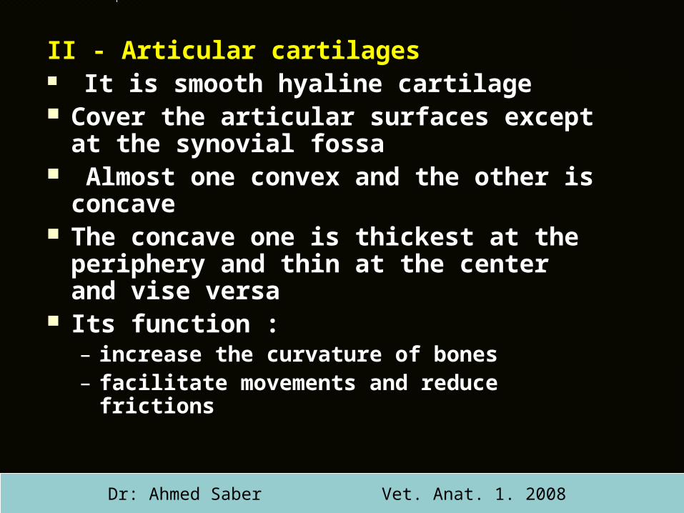
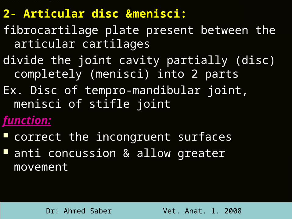
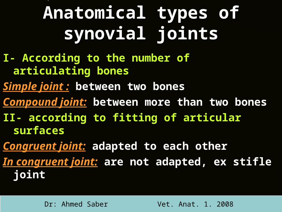
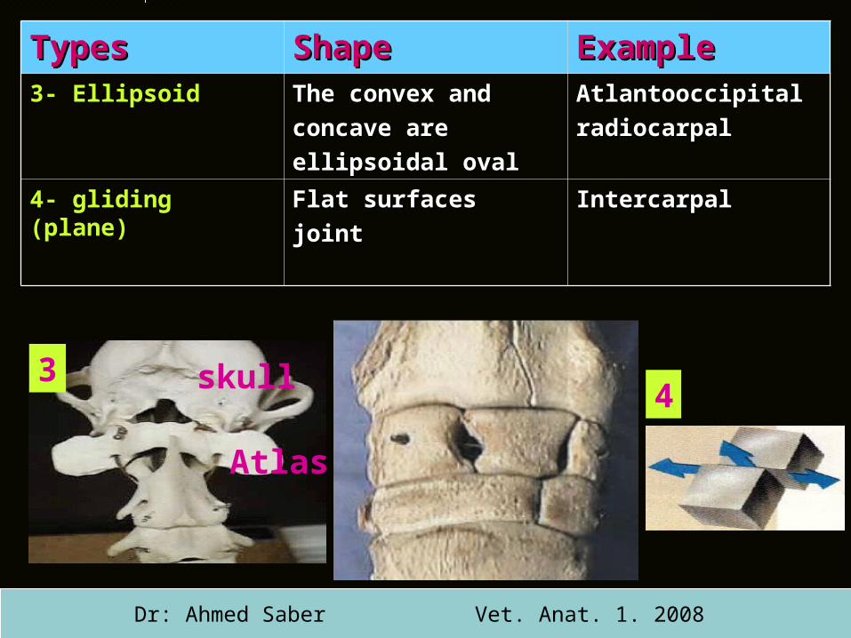
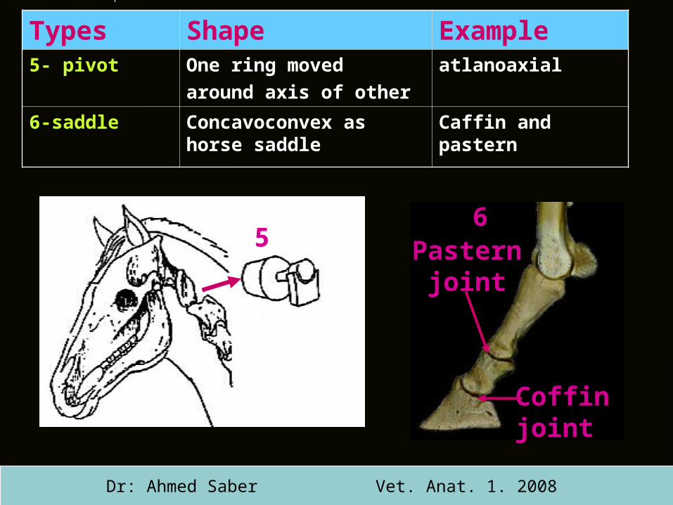
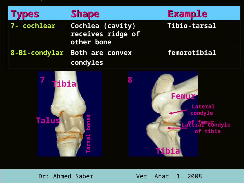
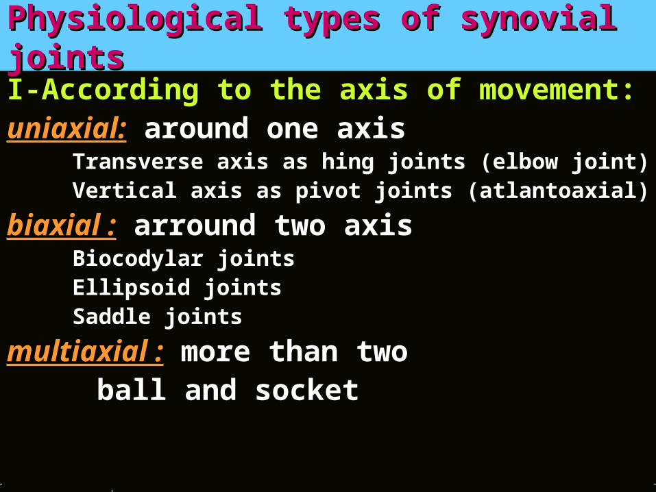
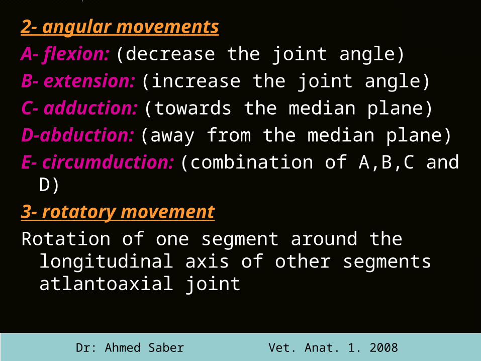