8-1 Articulations or Joints Articulation or Joint –Place where two bones (or bone and cartilage)...
16
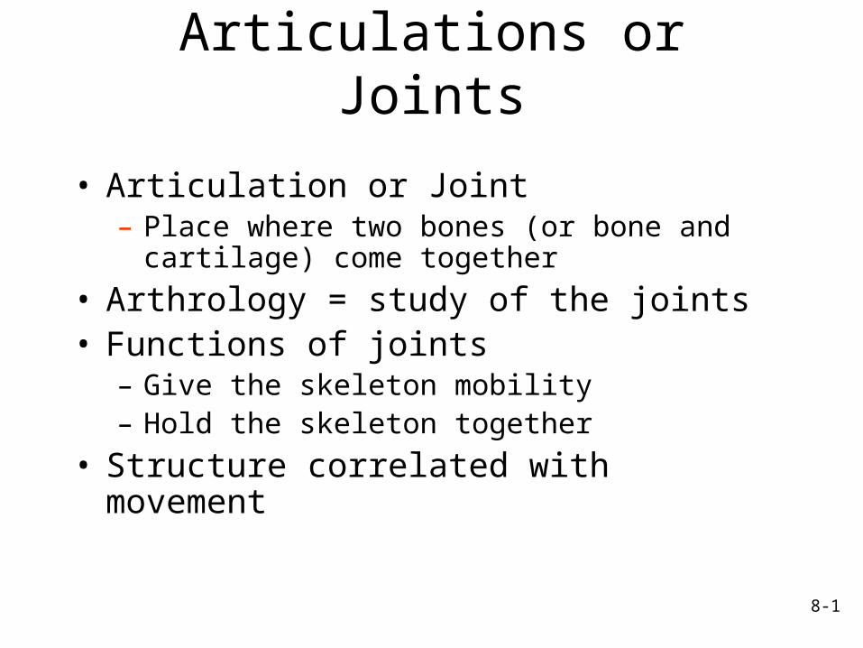
8-1 Articulations or Joints • Articulation or Joint – Place where two bones (or bone and cartilage) come together • Arthrology = study of the joints • Functions of joints – Give the skeleton mobility – Hold the skeleton together • Structure correlated with movement
-
Upload
ruby-osborne -
Category
Documents
-
view
222 -
download
1
Transcript of 8-1 Articulations or Joints Articulation or Joint –Place where two bones (or bone and cartilage)...
8-1
Articulations or Joints
• Articulation or Joint– Place where two bones (or bone and cartilage)
come together
• Arthrology = study of the joints• Functions of joints
– Give the skeleton mobility– Hold the skeleton together
• Structure correlated with movement
8-2
Classification of Joints• Structural classes: based on type
of connective tissue type that binds bones and whether or not a joint cavity is present– Fibrous– Cartilaginous– Synovial
• Functional classes: based on degree of motion – Synarthrosis: non-movable– Amphiarthrosis: slightly movable– Diarthrosis: freely movable
8-3
Fibrous Joints
• Characteristics– United by fibrous connective tissue– Have no joint cavity– Move little or none
• Types: – Sutures– Syndesmoses
8-4
Fibrous Joints: Syndesmoses
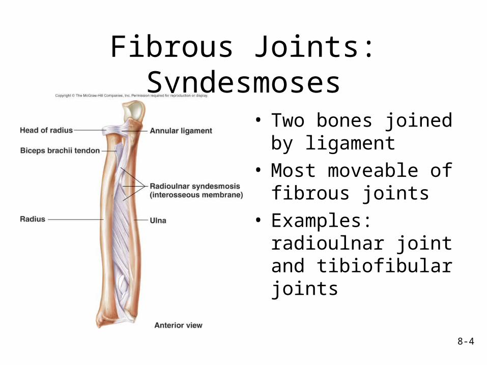
• Two bones joined by ligament
• Most moveable of fibrous joints
• Examples: radioulnar joint and tibiofibular joints
8-5
Cartilaginous Joints
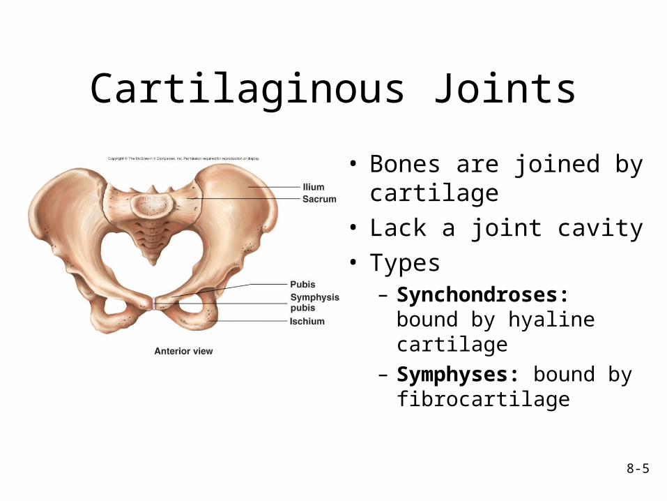
• Bones are joined by cartilage
• Lack a joint cavity
• Types– Synchondroses: bound by hyaline cartilage
– Symphyses: bound by fibrocartilage
8-6
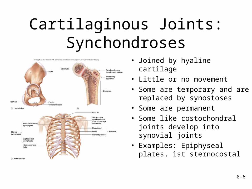
Cartilaginous Joints: Synchondroses
• Joined by hyaline cartilage
• Little or no movement• Some are temporary and are replaced by synostoses
• Some are permanent• Some like costochondral joints develop into synovial joints
• Examples: Epiphyseal plates, 1st sternocostal
8-7
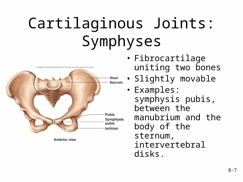
Cartilaginous Joints: Symphyses
• Fibrocartilage uniting two bones
• Slightly movable• Examples: symphysis pubis, between the manubrium and the body of the sternum, intervertebral disks.
8-8
Synovial Joints
• Contain synovial fluid in a joint cavity called the synovial cavity
• Allow considerable movement (diarthroses)
• Most joints that unite bones of appendicular skeleton reflecting greater mobility of appendicular skeleton compared to axial
8-9
Structure of
Synovial Joints
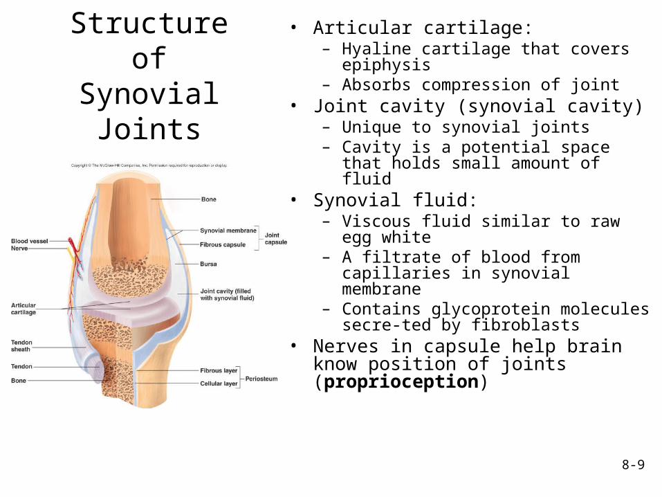
• Articular cartilage: – Hyaline cartilage that covers
epiphysis– Absorbs compression of joint
• Joint cavity (synovial cavity)– Unique to synovial joints– Cavity is a potential space
that holds small amount of fluid
• Synovial fluid: – Viscous fluid similar to raw
egg white– A filtrate of blood from
capillaries in synovial membrane
– Contains glycoprotein molecules secre-ted by fibroblasts
• Nerves in capsule help brain know position of joints (proprioception)
8-10
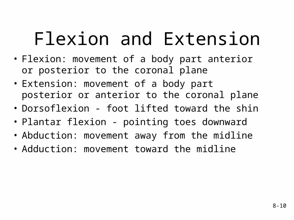
Flexion and Extension• Flexion: movement of a body part anterior or posterior to the coronal plane
• Extension: movement of a body part posterior or anterior to the coronal plane
• Dorsoflexion - foot lifted toward the shin• Plantar flexion - pointing toes downward• Abduction: movement away from the midline• Adduction: movement toward the midline

8-11
Types of Synovial Joints:Plane Joints• Plane or gliding
joints– Monaxial. One flat bone surface glides or slips over another similar surface
– Sometimes considered an amphiarthrosis
– Examples: intervertebral, intercarpal, intertarsal acromioclavicular, carpometacarpal, tarsometatarsal,
8-12
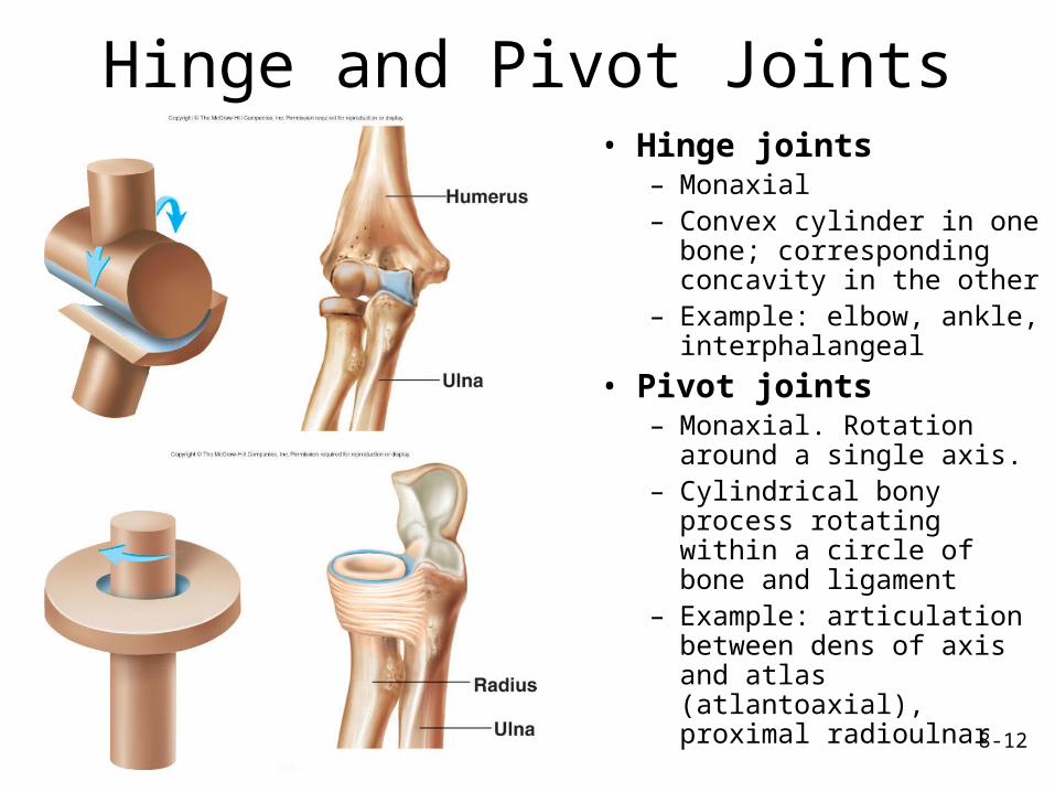
Hinge and Pivot Joints• Hinge joints
– Monaxial– Convex cylinder in one bone; corresponding concavity in the other
– Example: elbow, ankle, interphalangeal
• Pivot joints– Monaxial. Rotation around a single axis.
– Cylindrical bony process rotating within a circle of bone and ligament
– Example: articulation between dens of axis and atlas (atlantoaxial), proximal radioulnar
8-13
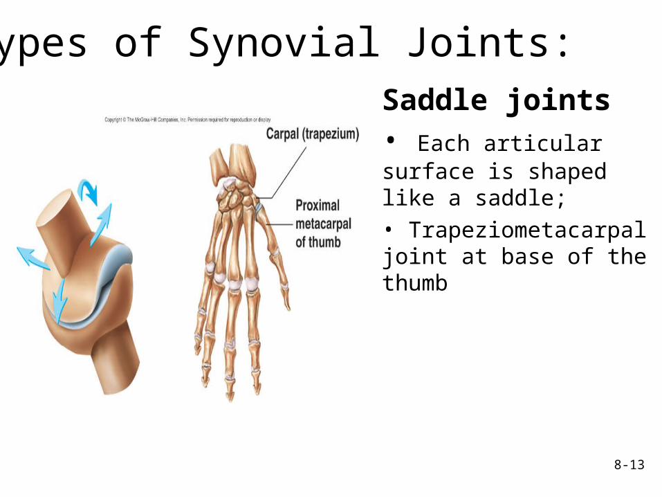
Saddle joints• Each articular surface is shaped like a saddle; • Trapeziometacarpal joint at base of the thumb
Types of Synovial Joints:
8-14
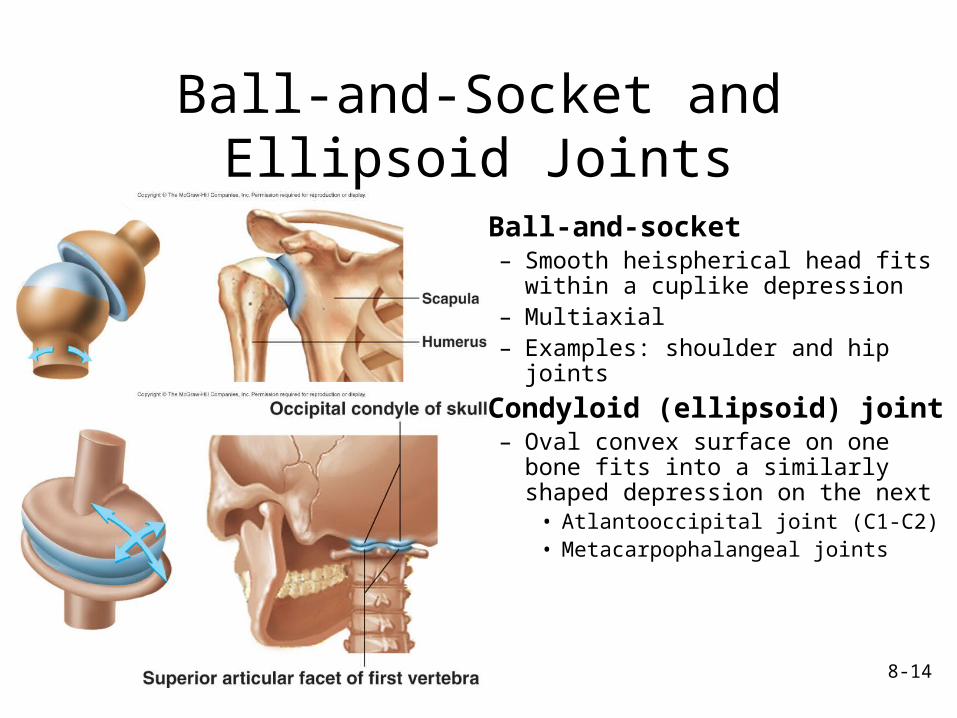
Ball-and-Socket and Ellipsoid Joints
• Ball-and-socket– Smooth heispherical head fits within a cuplike depression
– Multiaxial– Examples: shoulder and hip joints
• Condyloid (ellipsoid) joint– Oval convex surface on one bone fits into a similarly shaped depression on the next• Atlantooccipital joint (C1-C2)• Metacarpophalangeal joints
8-15
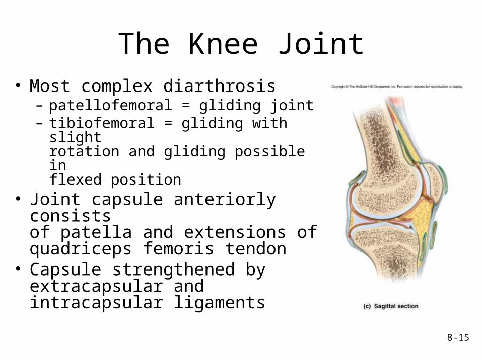
The Knee Joint• Most complex diarthrosis
– patellofemoral = gliding joint– tibiofemoral = gliding with slight rotation and gliding possible in flexed position
• Joint capsule anteriorly consists of patella and extensions of quadriceps femoris tendon
• Capsule strengthened by extracapsular and intracapsular ligaments
8-16
Effects of Aging on Joints
• Tissue repair slows; rate of new blood vessel development decreases
• Articular cartilages wear down and matrix becomes more rigid
• Production of synovial fluid declines• Ligaments and tendons become shorter and less flexible: decrease in range of motion (ROM)
• Muscles around joints weaken• A decrease in activity causes less flexibility and decreased ROM