Antagonist effect between violaxanthin and de-epoxidated pigments
11
Antagonist effect between violaxanthin and de-epoxidated pigments in nonphotochemical quenching induction in the qE deficient brown alga Macrocystis pyrifera Héctor Ocampo-Alvarez a , Ernesto García-Mendoza a, ⁎, Govindjee b, c, d a Departamento de Oceanografía Biológica / Centro de Investigación Científica y de Educación Superior de Ensenada (CICESE), Carretera Ensenada-Tijuana no. 3918, Ensenada, Baja California, México b Department of Plant Biology, University of Illinois at Urbana-Champaign, 265 Morrill Hall, 505 South Goodwin Avenue, Urbana, IL 61801-3707, USA c Department of Biochemistry, University of Illinois at Urbana-Champaign, 265 Morrill Hall, 505 South Goodwin Avenue, Urbana, IL 61801-3707, USA d Center of Biophysics & Computational Biology, University of Illinois at Urbana-Champaign, 265 Morrill Hall, 505 South Goodwin Avenue, Urbana, IL 61801-3707, USA abstract article info Article history: Received 30 May 2012 Received in revised form 13 December 2012 Accepted 14 December 2012 Available online 31 December 2012 Keywords: Xanthophyll cycle pool size Zeaxanthin-dependent quenching (qZ) De-epoxidation rate control Brown alga Macrocystis pyrifera Nonphotochemical quenching (NPQ) of Photosystem II fluorescence is one of the most important photoprotection responses of phototropic organisms. NPQ in Macrocystis pyrifera is unique since the fast induction of this response, the energy dependent quenching (qE), is not present in this alga. In contrast to higher plants, NPQ in this organism is much more strongly related to xanthophyll cycle (XC) pigment interconversion. Characterization of how NPQ is controlled when qE is not present is important as this might represent an ancient response to light stress. Here, we describe the influence of the XC pigment pool (ΣXC) size on NPQ induction in M. pyrifera. The sum of violaxanthin (Vx) plus antheraxanthin and zeaxanthin (Zx) represents the ΣXC. This pool was three-fold larger in blades collected at the surface of the water column (19 mol mol -1 Chl a ×100) than in blades collected at 6 m depth. Maximum NPQ was not different in samples with a ΣXC higher than 12 mol mol -1 Chl a × 100; however, NPQ induction was faster in blades with a large ΣXC. The increase in the NPQ induction rate was associated with a faster Vx to Zx conversion. Further, we found that NPQ depends on the de-epoxidation state of the ΣXC, not on the absolute concentration of Zx and antheraxanthin. Thus, there was an antagonist effect between Vx and de-epoxidated xanthophylls for NPQ. These results indicate that in the absence of qE, a large ΣXC is needed in M. pyrifera to respond faster to light stress conditions. © 2012 Elsevier B.V. All rights reserved. 1. Introduction The nonphotochemical quenching (NPQ) of Photosystem II (PSII) chlorophyll (Chl) a emission is a proxy to measure the thermal dissi- pation in the photosynthetic apparatus of plants and algae [1]. Dissi- pation of excess energy as heat is one of the most important photoprotection mechanisms of phototropic organisms. This process confers strong fitness to plants grown under field conditions [2] and provides resistance to environmental stress [3]. NPQ is a complex and finely regulated process that in higher plants consists of four different components: (1) qE, energy or ΔpH-dependent quenching; (2) qT, state transition quenching; (3) qI, photoinhibitory quenching and, (4) qZ, a zeaxanthin (Zx) dependent quenching [4]. The caroten- oid Zx plays an important role in the qE, qZ and qI components of NPQ, either as a direct quencher or as a modulator of these processes in the photosynthetic apparatus of higher plants [5,6]. Zx is formed under saturating light conditions. A pH lower than 6 in the thylakoid lumen activates the violaxanthin de-epoxidase enzyme (VDE) that converts violaxanthin (Vx) into antheraxanthin (Ax) and then into Zx. The back conversion reaction takes place in darkness and is medi- ated by the Zx epoxidase enzyme [7]. The Vx to Zx conversion in high light and the back reaction in darkness are known as the xanthophyll cycle (XC) [7]. The XC is present in higher plants, mosses, lichens, green algae and brown algae [6,8,9], while in diatoms, xanthophytes, haptophytes and dinoflagellates a homologous cycle exists in which diadinoxanthin is interconverted into diatoxanthin, known as diadinoxanthin cycle [6,10]. Of the different components involved in photoprotection, qE is the fastest component of NPQ since it is induced in high light and Biochimica et Biophysica Acta 1827 (2013) 427–437 Abbreviations: Ax, antheraxanthin; Chl, chlorophyll; Dd, diadinoxanthin; DPS, de-epoxidation state of the xanthophyll cycle pigment pool; Dt, diatoxanthin; ΔpH, proton gradient across the thylakoid membrane; FCP, fucoxanthin-chlorophyll protein; HL, high light; HPLC, high performance liquid chromatography; LHC, light harvesting complex; LL, low light; MGDG, monogalactosyldiacylglycerol; NPQ, nonphotochemical quenching of PSII florescence; PSII, photosystem II; qE, energy-dependent quenching; qI, photoinhibitory quenching; qZ, zeaxanthin-dependent quenching; qT, state transi- tion quenching; Vx, violaxanthin; VDE, violaxanthin de-epoxidase; XC, xanthophyll cycle; ΣXC, xanthophyll cycle pigment pool; Zx, zeaxanthin; ZE, zeaxanthin epoxidase ⁎ Corresponding author at: Biological Oceanography Department/CICESE, P.O Box 430222 San Diego, CA, 92143-0222, USA. Tel.: +52 646 1750500; fax: +52 646 175 0587. E-mail addresses: [email protected] (H. Ocampo-Alvarez), [email protected] (E. García-Mendoza), [email protected] (Govindjee). 0005-2728/$ – see front matter © 2012 Elsevier B.V. All rights reserved. http://dx.doi.org/10.1016/j.bbabio.2012.12.006 Contents lists available at SciVerse ScienceDirect Biochimica et Biophysica Acta journal homepage: www.elsevier.com/locate/bbabio
Transcript of Antagonist effect between violaxanthin and de-epoxidated pigments

Biochimica et Biophysica Acta 1827 (2013) 427–437
Contents lists available at SciVerse ScienceDirect
Biochimica et Biophysica Acta
j ourna l homepage: www.e lsev ie r .com/ locate /bbabio
Antagonist effect between violaxanthin and de-epoxidated pigments innonphotochemical quenching induction in the qE deficient brown algaMacrocystis pyrifera
Héctor Ocampo-Alvarez a, Ernesto García-Mendoza a,⁎, Govindjee b,c,d
a Departamento de Oceanografía Biológica / Centro de Investigación Científica y de Educación Superior de Ensenada (CICESE), Carretera Ensenada-Tijuana no. 3918, Ensenada,Baja California, Méxicob Department of Plant Biology, University of Illinois at Urbana-Champaign, 265 Morrill Hall, 505 South Goodwin Avenue, Urbana, IL 61801-3707, USAc Department of Biochemistry, University of Illinois at Urbana-Champaign, 265 Morrill Hall, 505 South Goodwin Avenue, Urbana, IL 61801-3707, USAd Center of Biophysics & Computational Biology, University of Illinois at Urbana-Champaign, 265 Morrill Hall, 505 South Goodwin Avenue, Urbana, IL 61801-3707, USA
Abbreviations: Ax, antheraxanthin; Chl, chlorophyde-epoxidation state of the xanthophyll cycle pigmentproton gradient across the thylakoid membrane; FCP, fuHL, high light; HPLC, high performance liquid chromatcomplex; LL, low light; MGDG, monogalactosyldiacylglyquenching of PSII florescence; PSII, photosystem II; qE,qI, photoinhibitory quenching; qZ, zeaxanthin-dependetion quenching; Vx, violaxanthin; VDE, violaxanthin dcycle; ΣXC, xanthophyll cycle pigment pool; Zx, zeaxan⁎ Corresponding author at: Biological Oceanography
430222 San Diego, CA, 92143-0222, USA. Tel.: +52 640587.
E-mail addresses: [email protected] (H. [email protected] (E. García-Mendoza), [email protected]
0005-2728/$ – see front matter © 2012 Elsevier B.V. Allhttp://dx.doi.org/10.1016/j.bbabio.2012.12.006
a b s t r a c t
a r t i c l e i n f oArticle history:Received 30 May 2012Received in revised form 13 December 2012Accepted 14 December 2012Available online 31 December 2012
Keywords:Xanthophyll cycle pool sizeZeaxanthin-dependent quenching (qZ)De-epoxidation rate controlBrown algaMacrocystis pyrifera
Nonphotochemical quenching (NPQ) of Photosystem II fluorescence is one of themost important photoprotectionresponses of phototropic organisms. NPQ inMacrocystis pyrifera is unique since the fast induction of this response,the energy dependent quenching (qE), is not present in this alga. In contrast to higher plants, NPQ in this organismismuchmore strongly related to xanthophyll cycle (XC) pigment interconversion. Characterization of howNPQ iscontrolledwhenqE is not present is important as thismight represent an ancient response to light stress. Here, wedescribe the influence of the XC pigment pool (ΣXC) size on NPQ induction inM. pyrifera. The sum of violaxanthin(Vx) plus antheraxanthin and zeaxanthin (Zx) represents the ΣXC. This pool was three-fold larger in bladescollected at the surface of the water column (19 mol mol−1 Chl a×100) than in blades collected at 6 m depth.Maximum NPQ was not different in samples with a ΣXC higher than 12 mol mol−1 Chl a×100; however, NPQinductionwas faster in bladeswith a largeΣXC. The increase in theNPQ induction ratewas associatedwith a fasterVx to Zx conversion. Further, we found that NPQ depends on the de-epoxidation state of the ΣXC, not on theabsolute concentration of Zx and antheraxanthin. Thus, there was an antagonist effect between Vx andde-epoxidated xanthophylls for NPQ. These results indicate that in the absence of qE, a large ΣXC is needed inM. pyrifera to respond faster to light stress conditions.
© 2012 Elsevier B.V. All rights reserved.
1. Introduction
The nonphotochemical quenching (NPQ) of Photosystem II (PSII)chlorophyll (Chl) a emission is a proxy to measure the thermal dissi-pation in the photosynthetic apparatus of plants and algae [1]. Dissi-pation of excess energy as heat is one of the most importantphotoprotection mechanisms of phototropic organisms. This processconfers strong fitness to plants grown under field conditions [2] and
ll; Dd, diadinoxanthin; DPS,pool; Dt, diatoxanthin; ΔpH,
coxanthin-chlorophyll protein;ography; LHC, light harvestingcerol; NPQ, nonphotochemicalenergy-dependent quenching;nt quenching; qT, state transi-e-epoxidase; XC, xanthophyllthin; ZE, zeaxanthin epoxidaseDepartment/CICESE, P.O Box
6 1750500; fax: +52 646 175
o-Alvarez),du (Govindjee).
rights reserved.
provides resistance to environmental stress [3]. NPQ is a complexand finely regulated process that in higher plants consists of fourdifferent components: (1) qE, energy or ΔpH-dependent quenching;(2) qT, state transition quenching; (3) qI, photoinhibitory quenchingand, (4) qZ, a zeaxanthin (Zx) dependent quenching [4]. The caroten-oid Zx plays an important role in the qE, qZ and qI components ofNPQ, either as a direct quencher or as a modulator of these processesin the photosynthetic apparatus of higher plants [5,6]. Zx is formedunder saturating light conditions. A pH lower than 6 in the thylakoidlumen activates the violaxanthin de-epoxidase enzyme (VDE) thatconverts violaxanthin (Vx) into antheraxanthin (Ax) and then intoZx. The back conversion reaction takes place in darkness and is medi-ated by the Zx epoxidase enzyme [7]. The Vx to Zx conversion in highlight and the back reaction in darkness are known as the xanthophyllcycle (XC) [7]. The XC is present in higher plants, mosses, lichens,green algae and brown algae [6,8,9], while in diatoms, xanthophytes,haptophytes and dinoflagellates a homologous cycle exists in whichdiadinoxanthin is interconverted into diatoxanthin, known asdiadinoxanthin cycle [6,10].
Of the different components involved in photoprotection, qE is thefastest component of NPQ since it is induced in high light and
428 H. Ocampo-Alvarez et al. / Biochimica et Biophysica Acta 1827 (2013) 427–437
disappears in darkness in seconds to minutes [1]. This component isactivated by a drop in the pH of the thylakoid lumen (formation of atransthylakoid proton gradient, ΔpH) that is sensed by the PsbSprotein in higher plants [11] or LHCSR proteins in green algae [12].Protonation of PsbS induces conformational changes of PSII antennacomplexes that promote thermal dissipation in this system [11]. Zxmodulates the PsbS-dependent quenching since the presence of thispigment enhances the formation of qE and retards its relaxation[13]. Also, lutein accelerates qE formation in a similar way as Zx[14] in some tropical plants that have a lutein cycle (formation oflutein from lutein epoxide) involved in photoprotection [15]. In con-trast to qE, qZ is a slowly developing (10 to 30 min after light expo-sure) and slowly relaxing (10 to 60 min in darkness) component ofNPQ that depends on the formation of Zx and is indirectly dependenton ΔpH since a low pH is necessary to activate the VDE [4,5]. qI is thelongest lasting component of all the NPQ components [16]; it hasbeen associated with PSII damage and its relaxation requires the re-pair of this system [17]. Zx also participates in qI since any sustaineddown regulation or inactivation of PSII is accompanied by the reten-tion of this pigment [3]. The relative contribution of each componentto NPQ development depends on the intensity and the duration of theexposure of the organism to light [5]. In higher plants, qE is the mostimportant response to short term (min) exposure to saturating lightsince it allows a flexible and rapid switching between a lightharvesting and energy dissipation function of the light harvesting an-tenna system [2].
The brown alga Macrocystis pyrifera has a slow induction of NPQupon exposure to high light, and the fast NPQ relaxation phase thattakes place in the first few minutes after light to dark transition isabsent in this alga [18]. In contrast to higher plants, preilluminationof this alga does not accelerate NPQ induction [18]. NPQ follows theaccumulation of Zx upon illumination in any pre-acclimation condi-tion, and the disruption of the ΔpH in light does not collapse NPQ inM. pyrifera [19]. Thus, qE is not present here and NPQ is strongly asso-ciated with XC activity [18,19]. In other Chl c containing organismssuch as diatoms, NPQ is also closely related to XC pigment intercon-version [6,20]. However, diatoms show a ΔpH related NPQ formedimmediately upon illumination [21]. The lack of qE has been reportedonly for M. pyrifera [18] but several other brown algae show similarcharacteristics of NPQ control [22,23].
There are important evolutionary implications of the differentialcontrol of NPQ between higher plants and brown algae [18]. Probablythe xanthophyll cycle appeared before the separation of the greenand red algae (brown algae) lineages and light harvesting evolution-ary events must have influenced the mechanisms of photoprotectionrelated to this cycle [18]. It is not known how NPQ is controlled whenit is independent of the ΔpH, but strongly related to the XC.M. pyrifera is a good model to investigate the response to light stressof an organism in which NPQ is independent of the ΔpH (absence ofqE that has a fast response). In this work, we characterize the effectof the xanthophyll cycle pigment pool (ΣXC) on the rate and the ex-tent of NPQ induction and its relaxation in M. pyrifera. The size ofthe ΣXC in this alga varies significantly in blades exposed to differentlight conditions [24]. An increase in the concentration of XC pigmentsis characteristic of sun-acclimated organisms [25–27]. In higherplants a high concentration of XC pigments slows the formation andrelaxation of NPQ [28]. A 40% decrease in the rate of NPQ inductionwas detected in Arabidopsis mutants that overexpress XC pigments[28]. Similar reduction in NPQ induction rate has been reported fororganisms grown in intermittent light that possess a large ΣXC [29].In an organism that lacks the fast ΔpH-dependent NPQ component,a large ΣXC might represent a physiological constraint if this poolnegatively affects the rate of NPQ induction. Here we have investigatedhow the size of the ΣXC affects the development of NPQ in M. pyrifera.We show that a large ΣXC is associated with an increase in the ratebut not in the amplitude of NPQ induction. We also found that this
process is related to the depoxidation state of the xanthophyll cycleand not to the absolute concentration of the photoprotective pigmentszeaxanthin and antheraxanthin.
2. Materials and methods
2.1. Plant material
Macrocystis pyrifera (L) C. Agardh samples were collected by scubadiving from Campo Kennedy kelp forest (31° 41.96 N; 116° 40.90 W),near Ensenada Baja California,México inNovember of 2009. Three bladeswere collected from the surface, and then at depths of 1, 2, 3 and 6 m inthe water column. Blades were collected from a single organism early inthe morning, tagged, kept in darkness and transported in coolers withseawater to the laboratory. Tissue discs (1.2 cm diameter) from eachblade were cut with a cork borer 10 cm above the pneumatocyst, alongthe central axis of each blade. The discs of the three blades from thesame depth were pooled and maintained in 250 mL Erlenmeyer flaskswith 200 mL of filtered seawater at 17±0.5 °C. The discs weremaintained under illumination (75 μmol photons m−2 s−1; cool-whitefluorescent bulbs) for 4 h, followed by at least 8 h of dark incubation.This light to dark treatment favored epoxidation of xanthophyll cyclepigments. After this treatment, the discs showed a maximum Photosys-tem II (PSII) quantum efficiency higher than 0.7, which is characteristicof non-stressed photosynthetic tissue of M. pyrifera [24]; this efficiencywas estimated from the Fv/Fm ratio, where Fv is the variable chlorophyll(Chl) fluorescence and Fm is the maximal Chl fluorescence, see e.g. [1].
At the sampling site, irradiance in the 400 to 700 nm range wasmeasured in the water columnwith a quantum scalar PAR (Photosyn-thetic Active Radiation) irradiance sensor QSPL-2101 (BiosphericalInstruments, San Diego CA, USA). Three profiles of the irradiancechanges with the depth were measured during the sampling period.Irradiance data were fitted using the Marquardt–Levenberg algorithmof Sigma Plot software (Jandel SSP Scientific SPSS, Chicago, IL, USA) tothe Lambert–Beer model for the reduction of irradiance in the watercolumn: Ez=E0 e−kd z, where Ez is the irradiance at depth z, E0 isthe irradiance at surface and the light attenuation coefficient is repre-sented by kd [30].
2.2. Experimental setup
To investigate the effect of the xanthophyll cycle pool size (ΣXC)and light intensity on the de-epoxidation and NPQ formation inM. pyrifera, the algal discs, collected from different depths, were ex-posed to five different light intensities. PSII florescence and pigmentconcentration were measured in samples exposed for 10 min to 45,90, 550, 950, and 1550 μmol photons m−2 s−1. Light was providedby 50-Watt halogen dichroic lamps (Techno Lite) and the intensitywas adjusted by using homemade neutral density filters (plasticsieves) placed between the lamp and the sample. During the light pe-riod, algal discs were placed in homemade acrylic chambers with con-tinuous flow of seawater maintained at 17±0.5 °C andsupplemented with 2 mM NaHCO3 to avoid any possible CO2 limita-tion. The light intensity was measured in situ with a cosine-typequantum sensor of a pulse amplitude modulated fluorometer(Diving-PAM; Heinz Walz, Effeltrich, Germany).
To evaluate NPQ relaxation and XC epoxidation, an independent ex-perimentwasperformed. Surface sampleswere collected in June of 2010from a different organism than the one in which induction experimentswere performed. The samples were acclimated as mentioned above(maintained at 75 μmol photons m−2 s−1 during 4 h, followed by 8 hof dark incubation) and then exposed to 1000 μmol photons m−2 s−1
for 60 min. Further samples were taken at 4, 6, 10, 20, 50 min of lightexposure and maintained in darkness for up to 60 min. NPQ and theconcentrations of the XC pigments concentration were monitoredduring light and dark treatments in each subsample. According to

Table 1Concentration of xanthophyll cycle pigments in dark-adapted blades of Macrocystispyrifera, collected from different depths in the water column in the ocean. The concentra-tions, as mol mol−1 Chl a×100, of violaxanthin (Vx), antheraxanthin (Ax), zeaxanthin(Zx), the size of the XC pigment pool (ΣXC) and the de-epoxidation state of this pool(DPS) are presented.
Depth Vx Ax Zx ΣXC⁎ Chl c:Chl a DPS
0 19.1±0.9 0.5±0.3 0.1±0.1 19.7±1 0.13±0.01 1.87±0.71 14.3±0.7 0.3±0.01 0.1±0.1 14.7±0.3 0.13±0.01 1.7±0.72 12.2±0.6 0.3±0.1 n.d 12.5±0.3 0.13±0.01 1.2±0.43 9.7±0.8 0.2±0.0 n.d 9.9±0.3 0.13±0.01 1.03±0.86 6.0±0.5 0.1±0.1 n.d 6.1±0.2 0.13±0.03 1.07±0.3
⁎ The ΣXC is the sum of [Vx]+[Ax]+[Zx]. The DPS is expressed as ([Zx]+0.5[Ax]/ΣXC)×100. Results represent the average of 5 independent measurements±SD. n.d.,not detected.
Fig. 1. Depth variation of the xanthophyll cycle pigment pool (ΣXC) in Macrocystispyrifera blades and irradiance (E) in the water column. Irradiance is normalized tothe intensity reaching the surface. Light data was fitted to the Lambert–Beer modelof reduction of irradiance in the water column with an extinction coefficient (kd) of0.4 m−1 (continuous blue line). Also, ΣXC variation was modeled with an exponentialmodel of reduction of this pool with depth (continuous red line).
429H. Ocampo-Alvarez et al. / Biochimica et Biophysica Acta 1827 (2013) 427–437
Nilkens et al. [4], NPQ and DPS relaxation data were evaluated using theequation y=A1exp (−t/τ)+y0, where, A represents the amplitude andτ the lifetimeof thefirst kinetic component, and y0 represents the ampli-tude of the second component.
2.3. Photosystem II chlorophyll a fluorescence measurements
Photosystem II chlorophyll a emission wasmeasured with a Diving-PAM (Walz, Germany); it wasmonitored by placing the PAM fiber opticat a 60° angle with respect to the light exposed side of the discs and thisoptical geometry was maintained during the duration of the experi-ments. NPQ was monitored in tissue discs illuminated for 10 min atfive different light intensities (see Section 2.2). To measure the maxi-mum florescence emission of PSII (Fm or Fm′; without or withnon-photochemical quenching, respectively, i.e., in dark-adapted, orlight-exposed samples) saturating flashes (halogen dichroic lamp ofthe Diving-PAM; 0.8 s; 3500 μmol photons m−2 s−1) were appliedevery 30 s. NPQ was calculated as (Fm−Fm′)/Fm′.
2.4. Pigment analysis
During the NPQ induction and relaxation experiments, samples forpigment analysis were taken at different time points during the illu-mination period and after the light was switched off. A tissue discwas collected from an individual chamber and immediately frozenand stored in liquid nitrogen (77 K). Pigments were extracted and an-alyzed as described in [24]. Pigments were extracted in 3 mL 100%cold acetone by grinding the discs that had been earlier frozen withliquid nitrogen. The extracts were maintained overnight at 4 °C andcentrifuged at 13,000×g (4 °C) for 5 min, and stored at −20 °C be-fore analysis by HPLC [24]. The HPLC was done using a ShimadzuAV-10 system (Kyoto, Japan), equipped with a Zorbax Eclipse XDBC8 (Agilent Technologies, Englewood, CO) reverse-phase column(4.6 mm×150 mm, 3.5 μm sizeparticles). The system was calibratedwith external standards (DHI Inc., Hoersholm, Denmark).
The de-epoxidation state (DPS) of the xanthophyll cycle pigmentpool was calculated as [Zx]+0.5[Ax]/ΣXC [31]; here, Ax and Zx standfor antheraxanthin and zeaxanthin, respectively, and ΣXC is thesum of Vx, Ax and Zx concentrations. The DPS represents thephotoprotective state of the XC, as the carotenoids Ax and Zx are in-volved in the dissipation of energy as heat that confers photoprotectionto the photosynthetic apparatus [32].
3. Results
3.1. Concentration of xanthophyll cycle pigments in blades fromdifferent depths
The concentration of the XC pigments in the blades ofM. pyriferadecreased with the depth of collection of the samples (Table 1). Thede-epoxidation state (DPS=[Zx]+0.5[Ax] /ΣXC) of the XC pig-ments was lower than 2% in all samples (Table 1). The DPS was asso-ciated mainly with the presence of Ax. The concentration of thispigment was five times higher in surface blades than in 6 m depthsamples (Table 1). Zx was detected only in samples collected atthe surface and 1 m depth (Table 1). Surface blades had a Vx con-centration of ~19 mol mol−1 Chl a×100; and a ΣXC concentrationof ~20 mol mol−1 Chl a×100 (Table 1). Vx decreased with depthand was three-fold lower in blades collected at 6 m. Further, therewas a 25% reduction of ΣXC in 1 m depth blades; its size was morethan three times lower in blades collected at 6 m depth than in sur-face blades (Table 1). Fig. 1 shows that there is an exponential re-duction of the ΣXC with depth. The change in the size of the XCpigment content follows the reduction of light intensity withdepth (Fig. 1). A 30% reduction of the intensity reaching the surfacewas detected at 1 m depth and at 6 m the reduction increased to
88% (Fig. 1). These results show that light plays an important rolein the regulation of the size of the ΣXC.
3.2. NPQ and XC pigment de-epoxidation in samples fromdifferent depths
Tissues collected from different depths were exposed to five differ-ent light intensities and NPQ induction was monitored for 10 min. Ashort illumination period of 10 min was chosen for the analysis of XCpigment de-epoxidation effect on NPQ induction in order to reducethe effect of PSII damage, especially in samples exposed to high light in-tensities. Two light intensities (45 and 90 μmol photons m−2 s−1)were below the light-saturation parameter (Ek) reported for bladesgrown above 9 m (102 μmol photons m−2 s−1; [24]) and three otherlight intensities (550, 950 and 1550 μmol photons m−2 s−1) werehigher than Ek. Samples collected from all depths showed an increasein NPQ upon exposure to light. Transient NPQ formation was observedin surface blades when exposed to 45 and 90 μmol photons m−2 s−1.NPQ in the samples increased in the first 4 min after light exposureand decreased after this illumination period (Fig. 2). This transientNPQ induction was also observed in 1 m and 2 m depth samples. Thelowest NPQ was measured in blades collected from 6 m depth (Fig. 2).NPQ at 10 minwas higher in samples from all depths exposed to higher

Fig. 2. Time course of nonphotochemical quenching (NPQ) in Macrocystis pyriferablades collected from different depths. Triplicate samples of each depth were exposedto five different irradiances. The data represent the average of the three replicates.
430 H. Ocampo-Alvarez et al. / Biochimica et Biophysica Acta 1827 (2013) 427–437
light intensities (Fig. 2). Maximum NPQ after 10 min of light exposurewas similar in samples collected from 3 m to the surface; however,the induction of this process was faster in surface than in deeper blades(Fig. 2). Half of the NPQ measured at 10 min (approximately 3 units ofNPQ) was detected at ~2, 2.5, 3.5 and 4.5 min in samples collected atsurface, 1, 2, and 3 m, respectively.
In M. pyrifera, NPQ is not dependent on the formation of thetransthylakoid proton gradient (ΔpH), but it is closely associated withXCactivity [18,19]. Therefore, the change in the concentration of XCpig-ments wasmeasured under all light treatments to relate NPQ inductionto XC de-epoxidation. The change in the concentration of XC pigmentsin samples collected at different depths and exposed to five irradiancesis shown in Fig. 3. Data of the change of XC pigments was normalizedto Chl a concentration. There were no significant changes in Chl or
carotenoid content on leaf area basis during the experimentation peri-od. In surface samples, Vx concentration decreased from 18.7 to12.5 mol mol−1 Chl a×100 in 10 min of exposure to1550 μmol photons m−2 s−1 (Fig. 3). Vx de-epoxidation decreasedwith the collection depth of the samples and with light intensity(Fig. 3). A transient decrease in Vx was observed in surface and 1 mdepth samples exposed to 45 and 90 μmol photons m−2 s−1 (Fig. 3).This transient change was also observed in Ax and Zx. The concentra-tion of these pigments increased in samples from surface and 1 mdepths in the first minutes after exposure to 45 and90 μmol photons m−2 s−1 and decreased towards the end of the expo-sure period. Ax showed a net accumulation upon exposure to light andreached a maximum concentration after 5 min in all samples exposedto 550 μmol photons m−2 s−1 and higher irradiance (Fig. 3). A fast ac-cumulation of Axwas observed under all the light conditions for surfaceblades, and there was a decrease in the rate of accumulation of this pig-ment in samples collected at depth (Fig 3). In contrast to Ax, the in-crease in Zx concentration was slower in the first minutes of lightexposure (Fig. 3). The time lag for Zx appearance increased with thedepth of collection of the samples. In 3 m blades Zx accumulation wasdetected after 2 min of light exposure. Therefore, there was a faster ac-cumulation of de-epoxidated pigment in samples acclimated to higherirradiances.
The change in the concentration of XC pigments was reflected inDPS variation during the light exposure period. DPS increased in sam-ples from all depths upon exposure to light due to the accumulationof Ax and Zx. Maximum DPS was detected after 10 min and washigher in samples from all depths exposed to irradiances above550 μmol photons m−2 s−1 (Fig. 3). DPS at 10 min of light exposurewas similar (~26) in samples collected from 3 m to the surface;however, similar to NPQ induction, the rise of this parameter intime was faster in surface than in deeper blades (Fig. 3).
3.3. NPQ and XC pigment de-epoxidation in relation to ΣXC size
The effect of the ΣXC size on NPQ induction and DPS was analyzed.Maximum NPQ formation was related to the ΣXC size of the blades(Fig. 4A). When exposed to 550, 950 and 1550 μmol photons m−2 s−1,the samples with a ΣXC of ~6 mol mol−1 Chl a×100 (6 m depth)showed significantly lower (Two-way ANOVA, pb0.05; Tukey aposteriori test) NPQ than samples with a larger xanthophyll cycle pig-ment pool (Fig. 3A). NPQ levels in blades collected from 0 m, 1 m, 2 mand 3 m depths (ΣXC larger than 6 mol mol−1 Chl a×100)were similarafter 10 min of exposure to different light treatments (Two-way ANOVA,pb0.05; Fig. 4A). The NPQwas ~6 in these samples after 10 min of expo-sure to 1550 μmol photons m−2 s−1 and a 20% and 30% reduction wasobserved in tissues exposed to 990 and 550 μmol photons m−2 s−1,respectively (Fig. 4A).
The de-epoxidation state of the XC pigments showed a similar re-lation with the size of the ΣXC as NPQ (Fig. 4B). The lowest DPS after10 min of light exposure was detected in samples with the smallestΣXC (6 m depth samples; Fig. 4B). DPS after 10 min of light exposurewas not different in samples with a ΣXC size higher than~12 mol mol−1 Chl a×100 (Fig. 4B). The NPQ formed within10 min of light exposure was similar in 3, 2, and 1 m, and surfacesamples. However, maximum DPS was different at each light intensi-ty. Maximum DPS after 10 min of light exposure was approximately27% in samples exposed to 1500 μmol photons m−2 s−1, and de-creased in samples exposed to 950 and 550 μmol photons m−2 s−1
(Fig. 4B).
3.4. Relation between NPQ induction and XC pigment de-epoxidation
The size of the ΣXC and the irradiance level influenced DPS andNPQ induction in M. pyrifera. The formation of de-epoxidated pig-ments and NPQ induction followed a similar trend between different

Fig. 3. Time course of the change of violaxanthin (Vx), antheraxanthin (Ax) and zeaxanthin (Zx) concentration in Macrocystis pyrifera blades collected from different depths. Thechange of the de-epoxidation state of the XC pigment pool (DPS) is also presented. Triplicate samples of each depth were exposed to five different irradiances. The data representthe average of the three replicates. Note the change in the scale of the Y axis in graphs of each collection depth.
431H. Ocampo-Alvarez et al. / Biochimica et Biophysica Acta 1827 (2013) 427–437
light treatments and in samples from different depths (cf. Figs. 2 and3, Fig. S1 in the supplementary material). The linear regression anal-ysis between NPQ and the amount of Zx formed in the differentsamples and light treatments yielded an r2 of 0.87 (Fig. 5A). Theunexplained variance of this linear relation could be associated with
Fig. 4. Relation of the xanthophyll cycle pool size (ΣXC) and the non-photochemical quenchthe XC pigment pool (DPS) is presented. The data represent the average of the three repMacrocystis pyrifera blades collected from different depths.
the lack of accumulation of Zx at the beginning of the light treatments(cf. Fig. 3). It is known that Ax also contributes to NPQ mechanism[31,33]. This pigment was rapidly formed upon light exposure(cf. Fig. 3). The relation of the concentration of Ax plus Zx and NPQis shown in Fig. 5B. An interesting pattern appeared from this result;
ing (NPQ; Panel A). In Panel B, the relation of the ΣXC with the de-epoxidation state oflicates±standard deviation of each variable measured at 10 min of light exposure in

Fig. 5. Relation of nonphotochemical quenching (NPQ)of Photosystem II fluorescence, andthe concentration of zeaxanthin (Panel A), antheraxanthin plus zeaxanthin (Panel B), andde-epoxidation state of the XC pool (Panel C) inMacrocystis pyrifera blades collected fromdifferent depths andexposed to different irradiances. The colors of the symbols and regres-sion lines represent data from samples collected at different depths.
Fig. 6. Time course of nonphotochemical quenching (NPQ) induction and relaxation inMacrocystis pyrifera blades collected from the surface. Samples were exposed toapproximately 60 min to 1000 μmol photon m−2 s−1 and samples (in triplicate)were collected at 4, 6, 10, 20 and 50 min. These subsamples were maintained in dark-ness to evaluate NPQ relaxation. The data represent the average of the three replicates.
432 H. Ocampo-Alvarez et al. / Biochimica et Biophysica Acta 1827 (2013) 427–437
NPQ was linearly related to Ax plus Zx concentration with an r2
higher than 0.96 when the relation is constructed with data obtainedfrom each individual collection depth (Fig. 5B). The steepest slope of
this relation was detected in the tissue collected at 6 m depth thatshowed the smallest ΣXC (Fig. 5B; cf. Fig. 5C). The slope of the relationdecreased from 6 m to surface samples (Fig. 5B). This indicates that ahigher concentration of Ax plus Zx is needed to induce the same NPQin samples with a larger ΣXC than in tissue with lower concentrationof XC pigments. However, when the relative amount ofde-epoxidated pigments is considered instead of the absolute concen-tration of these pigments, comparable NPQ appears to be induced insamples with a different ΣXC size exposed to the different light inten-sities. The linear relationship between NPQ and DPS has an r2 of 0.99considering all the data of the different light treatments, exposuretime and collection depths (Fig. 5C). Therefore, NPQ formation inM. pyrifera depends on the relative increase of de-epoxidated pig-ments and not on the absolute concentration of these pigments.This indicates that there is an antagonist effect for NPQ induction be-tween de-epoxidated pigments and Vx.
3.5. Relation between NPQ relaxation and epoxidation of XC pigments
The antagonist effect between de-epoxidated and epoxidated pig-mentswas also investigated in samplesmaintained in darkness after dif-ferent preilumination periods. NPQ relaxation in darkness is slow andfollows Zea epoxidation [19]. However, the reappearance of Vx in dark-ness and its effect on NPQ relaxation have not yet been investigated.
NPQ relaxation in samples collected at surface and exposed to differ-ent periods to high light (4, 6, 10, 20, and 50 min) and the change of XCpigment concentration are presented in Figs. 6 and 7, respectively. Theblades collected for this experiment showed a smaller ΣXC than surfaceblades used in the NPQ induction experiment. Vx concentration was~14 mol mol−1 Chl a×100 (Fig. 7). Also, maximum DPS was ~40%after the exposure of the sample to 1000 μmol photon m−2 s−1 for60 min (Fig. 7). NPQ relaxation (Fig. 6) and Zx epoxidation (Fig. 7)after high light exposure followed a monophasic exponential decay.Two NPQ relaxation phases were detected inM. pyrifera. Themaximumamplitude (A) of the main NPQ relaxation phase was fully developedafter 10 min of preillumination and represented more that 80% of theNPQ induced in light (Table 2). Lifetime (τ1) of this phase was between2 to 6 min (Table 2). The second component (y0 in Table 2; see also [4])was detected in all treatments and its contribution to NPQ increasedwith the length of the preillumination period (Fig. 6; Table 2). Relaxa-tion of NPQ was incomplete even in samples preilluminated for 4 min(Fig. 6). In samples preilluminated for 50 min, the slow relaxationphase represented more than 20% of the total NPQ; further, this phasedid not relax within the time range of the experiment (Fig. 6).
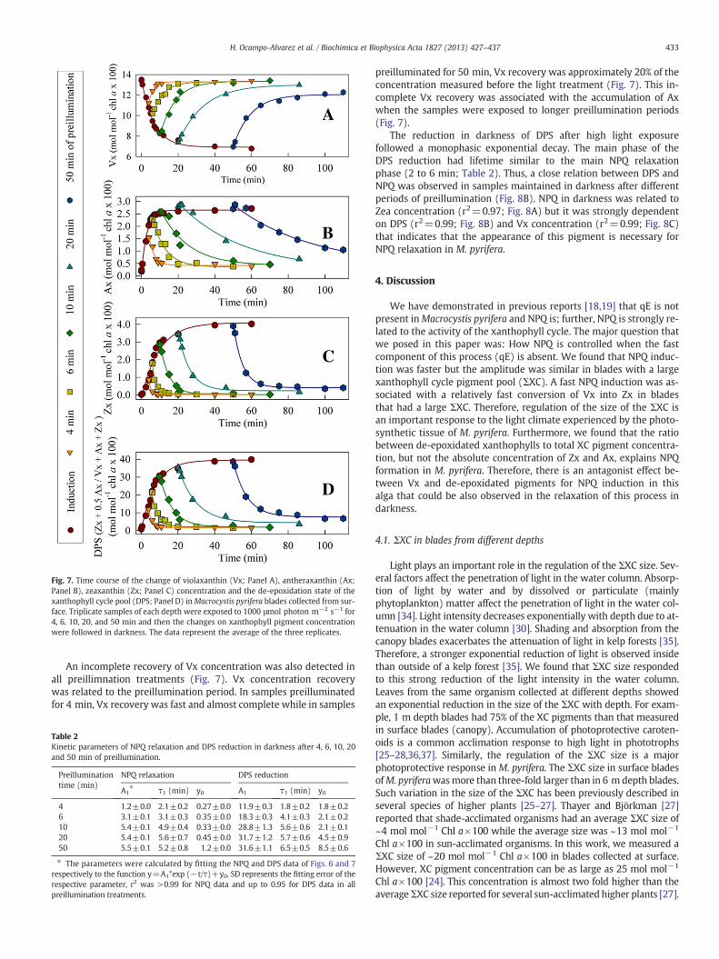
Fig. 7. Time course of the change of violaxanthin (Vx; Panel A), antheraxanthin (Ax;Panel B), zeaxanthin (Zx; Panel C) concentration and the de-epoxidation state of thexanthophyll cycle pool (DPS; Panel D) inMacrocystis pyrifera blades collected from sur-face. Triplicate samples of each depth were exposed to 1000 μmol photon m−2 s−1 for4, 6, 10, 20, and 50 min and then the changes on xanthophyll pigment concentrationwere followed in darkness. The data represent the average of the three replicates.
433H. Ocampo-Alvarez et al. / Biochimica et Biophysica Acta 1827 (2013) 427–437
An incomplete recovery of Vx concentration was also detected inall preillimnation treatments (Fig. 7). Vx concentration recoverywas related to the preillumination period. In samples preilluminatedfor 4 min, Vx recovery was fast and almost complete while in samples
Table 2Kinetic parameters of NPQ relaxation and DPS reduction in darkness after 4, 6, 10, 20and 50 min of preillumination.
Preilluminationtime (min)
NPQ relaxation DPS reduction
A1⁎ τ1 (min) y0 A1 τ1 (min) y0
4 1.2±0.0 2.1±0.2 0.27±0.0 11.9±0.3 1.8±0.2 1.8±0.26 3.1±0.1 3.1±0.3 0.35±0.0 18.3±0.3 4.1±0.3 2.1±0.210 5.4±0.1 4.9±0.4 0.33±0.0 28.8±1.3 5.6±0.6 2.1±0.120 5.4±0.1 5.6±0.7 0.45±0.0 31.7±1.2 5.7±0.6 4.5±0.950 5.5±0.1 5.2±0.8 1.2±0.0 31.6±1.1 6.5±0.5 8.5±0.6
⁎ The parameters were calculated by fitting the NPQ and DPS data of Figs. 6 and 7respectively to the function y=A1*exp (−t/τ)+y0. SD represents the fitting error of therespective parameter, r2 was >0.99 for NPQ data and up to 0.95 for DPS data in allpreillumination treatments.
preilluminated for 50 min, Vx recovery was approximately 20% of theconcentration measured before the light treatment (Fig. 7). This in-complete Vx recovery was associated with the accumulation of Axwhen the samples were exposed to longer preillumination periods(Fig. 7).
The reduction in darkness of DPS after high light exposurefollowed a monophasic exponential decay. The main phase of theDPS reduction had lifetime similar to the main NPQ relaxationphase (2 to 6 min; Table 2). Thus, a close relation between DPS andNPQ was observed in samples maintained in darkness after differentperiods of preillumination (Fig. 8B). NPQ in darkness was related toZea concentration (r2=0.97; Fig. 8A) but it was strongly dependenton DPS (r2=0.99; Fig. 8B) and Vx concentration (r2=0.99; Fig. 8C)that indicates that the appearance of this pigment is necessary forNPQ relaxation in M. pyrifera.
4. Discussion
We have demonstrated in previous reports [18,19] that qE is notpresent inMacrocystis pyrifera and NPQ is; further, NPQ is strongly re-lated to the activity of the xanthophyll cycle. The major question thatwe posed in this paper was: How NPQ is controlled when the fastcomponent of this process (qE) is absent. We found that NPQ induc-tion was faster but the amplitude was similar in blades with a largexanthophyll cycle pigment pool (ΣXC). A fast NPQ induction was as-sociated with a relatively fast conversion of Vx into Zx in bladesthat had a large ΣXC. Therefore, regulation of the size of the ΣXC isan important response to the light climate experienced by the photo-synthetic tissue of M. pyrifera. Furthermore, we found that the ratiobetween de-epoxidated xanthophylls to total XC pigment concentra-tion, but not the absolute concentration of Zx and Ax, explains NPQformation in M. pyrifera. Therefore, there is an antagonist effect be-tween Vx and de-epoxidated pigments for NPQ induction in thisalga that could be also observed in the relaxation of this process indarkness.
4.1. ΣXC in blades from different depths
Light plays an important role in the regulation of the ΣXC size. Sev-eral factors affect the penetration of light in the water column. Absorp-tion of light by water and by dissolved or particulate (mainlyphytoplankton) matter affect the penetration of light in the water col-umn [34]. Light intensity decreases exponentially with depth due to at-tenuation in the water column [30]. Shading and absorption from thecanopy blades exacerbates the attenuation of light in kelp forests [35].Therefore, a stronger exponential reduction of light is observed insidethan outside of a kelp forest [35]. We found that ΣXC size respondedto this strong reduction of the light intensity in the water column.Leaves from the same organism collected at different depths showedan exponential reduction in the size of the ΣXC with depth. For exam-ple, 1 m depth blades had 75% of the XC pigments than that measuredin surface blades (canopy). Accumulation of photoprotective caroten-oids is a common acclimation response to high light in phototrophs[25–28,36,37]. Similarly, the regulation of the ΣXC size is a majorphotoprotective response in M. pyrifera. The ΣXC size in surface bladesofM. pyriferawasmore than three-fold larger than in 6 m depth blades.Such variation in the size of the ΣXC has been previously described inseveral species of higher plants [25–27]. Thayer and Björkman [27]reported that shade-acclimated organisms had an average ΣXC size of~4 mol mol−1 Chl a×100 while the average size was ~13 mol mol−1
Chl a×100 in sun-acclimated organisms. In this work, we measured aΣXC size of ~20 mol mol−1 Chl a×100 in blades collected at surface.However, XC pigment concentration can be as large as 25 mol mol−1
Chl a×100 [24]. This concentration is almost two fold higher than theaverageΣXC size reported for several sun-acclimated higher plants [27].

Fig. 8. Relation of nonphotochemical quenching (NPQ) of Photosystem II fluorescence, and zeaxanthin concentration (Panel A) and the de-epoxidation state of the XC pigment pool(DPS; Panel B) and the violaxanthin (Vx) concentration (Panel C) in Macrocystis pyrifera blades collected from surface. Samples were exposed to 1000 μmol photon m−2 s−1 andthen maintained in darkness after different periods of preillumination (see legends of Figs. 6 and 7 for the description of the experiment).
434 H. Ocampo-Alvarez et al. / Biochimica et Biophysica Acta 1827 (2013) 427–437
4.2. ΣXC size effect on NPQ induction and de-epoxidation of XC pigments
The physiological implications of the increase of the ΣXC size inM. pyrifera were evident when the relationship between the size ofthis pool and NPQ induction was analyzed. In the absence of the fastqE component of NPQ, a large ΣXC represents a faster response tolight stress inM. pyrifera. Although NPQ at 10 minwas not significant-ly different between surface and 3 m blades, induction of this processwas two times faster in surface blades.ΣXC in surface bladeswasmorethan two times larger than in 3 m blades. The effect of a large ΣXC sizeon the rate of NPQ induction is not clear in higher plants since themajor proportion of this process is dominated by the ΔpH-PsbS drivenantenna rearrangement that can be activated in the absence of Zx [38].Therefore, NPQ does notmatch the kinetics of Zx formation during thefirst minutes of light exposure in higher plants. However, it has beendocumented that the induction of qE is slower in the sChyB Arabidopsisthaliana mutant, which shows an increase in the expression ofβ-carotene hydroxylase [28]. Slow qE induction was associated witha relatively slow change in the DPS since the ΣXC size is three-foldlarger in the sChyBmutant than in the wild-type genotype [28]. In di-atoms, the ΣXC size influences the NPQ extent [39]. However, atwo-fold increase in the size of the ΣXC did not accelerate NPQ forma-tion in Phaeodactylum tricornutum [40].
Similar to NPQ, the DPS was not significantly different betweenthe surface and 3 m blades. The increase of the ΣXC in high light ac-climated blades was not accompanied by an increase of the DPS. Incontrast to this observation in our system, DPS increases as the sizeof XC pool size becomes larger in higher plants [3,13,27]. This is asso-ciated with the reduction of peripheral antenna size in high light ac-climated plants and, as a consequence, an increase in the pool offree xanthophyll cycle pigments[41]. Probably in M. pyrifera, the in-crease of the ΣXC is co-regulated with xanthophyll binding proteins(most probably LHCSR proteins; see Section 4.3). This type of regula-tion has been observed in the green alga Chlamydomonas reinhartdiiwhere in high light acclimated cells, PSII antenna size does not de-crease but rather the concentration of LHCSR proteins increases [36].
In M. pyrifera similar maximum DPS can be established in tissueacclimated to different growth light conditions but the kinetics aredifferent, DPS increase was faster in samples with a large ΣXC size.An increase in the rate of the Vx to Ax conversion seems to be associ-ated with the faster accumulation of de-epoxidated xanthophylls.However, the accumulation of Zx during the first minutes of illumina-tion was the dominant factor for the rapid DPS increase in surface andclose to the surface samples. The more rapid conversion of Vx to Zx in
blades grown under higher light intensities is likely to be related to anincreased fraction of XC pigments that are (more easily) accessible tothe Vx de-epoxidase enzyme (VDE). In higher plants, different poolsof Vx can be identified according to the accessibility of this carotenoidto the VDE [41]. The de-epoxidation of Vx occurs in inverted hexago-nal phase domains formed by monogalactosyldiacylglycerol (MGDG)in the thylakoid membrane [41,42]. Therefore, Vx must detach fromthe antenna protein pigment complexes to be converted into Axand Zx [41]. Consequently, de-epoxidation of this pigment dependson the binding affinity to specific PSII antenna subunits [41]. In higherplants, Vx bound to some minor antenna complexes is not accessibleto the VDE enzyme while the pigment associated with the peripheralantenna of PSII is rapidly de-epoxidated [43]. Schaller et al. [42]reported that a pool of Vx is not bound to any protein complex, butis found dissolved in the MGDG lipid that surrounds the PSII antennaof higher plants. A free MGDG-related pool of Dx has been alsodetected in diatoms and its size has been shown to increase in highlight [44]. Therefore, a larger pool of easily accessible Vx can explainthe increase in the de-epoxidation rate of this pigment and as conse-quence, the faster NPQ induction in M. pyrifera blades acclimated tohigh light. This Vx could be loosely bound to a specific protein com-plex important for photoprotection in this alga (see section below)or free in the thylakoid membrane (Fig. 9).
4.3. The de-epoxidation state of the XC pigments and NPQ induction
Important observations on the regulation of NPQ induction in theabsence of a fast response are described in this paper. We found thatin M. pyrifera a higher concentration of Ax and Zx is needed to inducethe same NPQ in samples with a larger ΣXC size than in a tissue witha lower concentration of XC pigments. It seems paradoxical thatphotoprotective pigments are more efficient in inducing NPQ in lowlight-acclimated tissues than in tissues acclimated to high light condi-tions. However, this can be explained if we consider that low light accli-mated samples might have a lower number of quenching sites and thatthey saturate at low concentration of de-epoxidated pigments (Fig. 9).
We found that NPQ induction in different light intensities could beexplained by the de-epoxidation state of the XC pigment pool inblades with different acclimation characteristics (different samplingdepths). Also, transient NPQ induction observed at subsaturatinglight intensities in samples with a large ΣXC is related to the intercon-version of XC pigments.
Transient NPQ has been described in higher plants and its induc-tion has been related to the transient accumulation of protons in
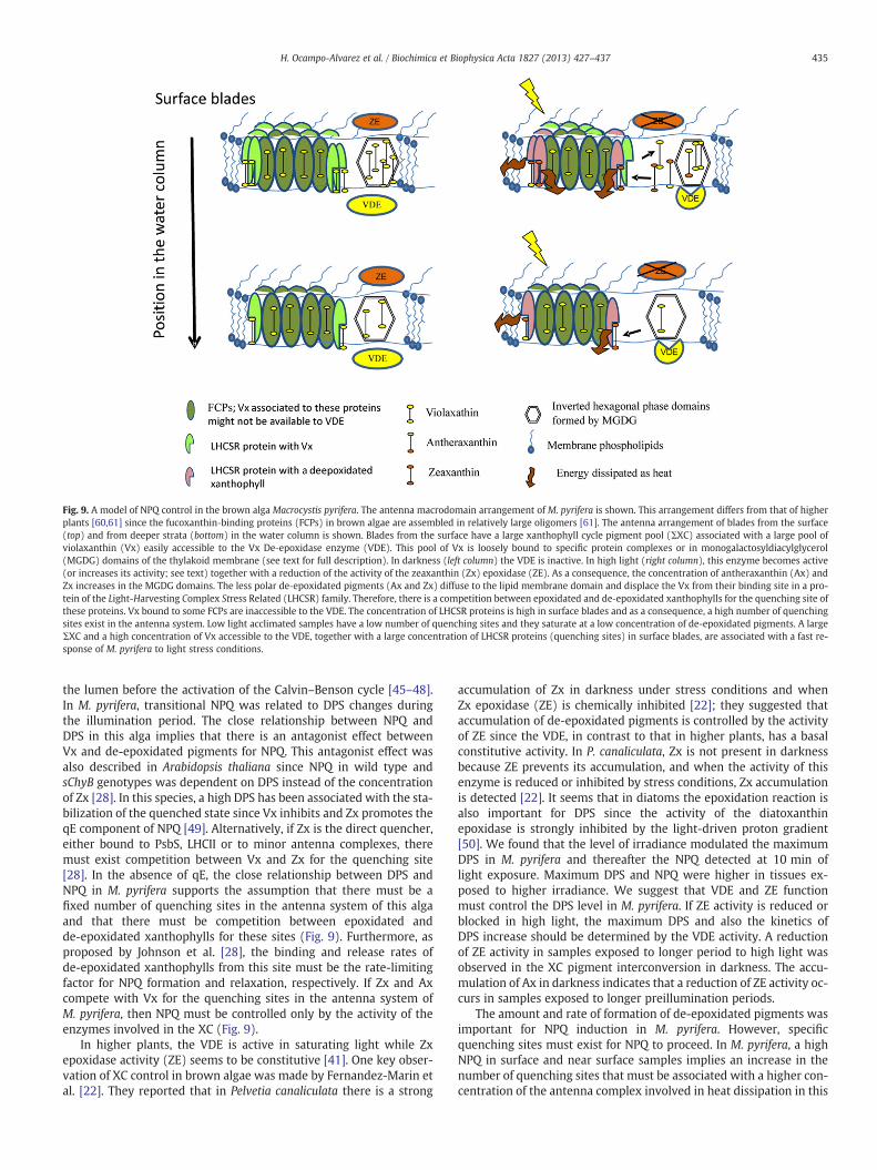
Fig. 9. A model of NPQ control in the brown alga Macrocystis pyrifera. The antenna macrodomain arrangement of M. pyrifera is shown. This arrangement differs from that of higherplants [60,61] since the fucoxanthin‐binding proteins (FCPs) in brown algae are assembled in relatively large oligomers [61]. The antenna arrangement of blades from the surface(top) and from deeper strata (bottom) in the water column is shown. Blades from the surface have a large xanthophyll cycle pigment pool (ΣXC) associated with a large pool ofviolaxanthin (Vx) easily accessible to the Vx De‐epoxidase enzyme (VDE). This pool of Vx is loosely bound to specific protein complexes or in monogalactosyldiacylglycerol(MGDG) domains of the thylakoid membrane (see text for full description). In darkness (left column) the VDE is inactive. In high light (right column), this enzyme becomes active(or increases its activity; see text) together with a reduction of the activity of the zeaxanthin (Zx) epoxidase (ZE). As a consequence, the concentration of antheraxanthin (Ax) andZx increases in the MGDG domains. The less polar de‐epoxidated pigments (Ax and Zx) diffuse to the lipid membrane domain and displace the Vx from their binding site in a pro-tein of the Light-Harvesting Complex Stress Related (LHCSR) family. Therefore, there is a competition between epoxidated and de‐epoxidated xanthophylls for the quenching site ofthese proteins. Vx bound to some FCPs are inaccessible to the VDE. The concentration of LHCSR proteins is high in surface blades and as a consequence, a high number of quenchingsites exist in the antenna system. Low light acclimated samples have a low number of quenching sites and they saturate at a low concentration of de-epoxidated pigments. A largeΣXC and a high concentration of Vx accessible to the VDE, together with a large concentration of LHCSR proteins (quenching sites) in surface blades, are associated with a fast re-sponse of M. pyrifera to light stress conditions.
435H. Ocampo-Alvarez et al. / Biochimica et Biophysica Acta 1827 (2013) 427–437
the lumen before the activation of the Calvin–Benson cycle [45–48].In M. pyrifera, transitional NPQ was related to DPS changes duringthe illumination period. The close relationship between NPQ andDPS in this alga implies that there is an antagonist effect betweenVx and de-epoxidated pigments for NPQ. This antagonist effect wasalso described in Arabidopsis thaliana since NPQ in wild type andsChyB genotypes was dependent on DPS instead of the concentrationof Zx [28]. In this species, a high DPS has been associated with the sta-bilization of the quenched state since Vx inhibits and Zx promotes theqE component of NPQ [49]. Alternatively, if Zx is the direct quencher,either bound to PsbS, LHCII or to minor antenna complexes, theremust exist competition between Vx and Zx for the quenching site[28]. In the absence of qE, the close relationship between DPS andNPQ in M. pyrifera supports the assumption that there must be afixed number of quenching sites in the antenna system of this algaand that there must be competition between epoxidated andde-epoxidated xanthophylls for these sites (Fig. 9). Furthermore, asproposed by Johnson et al. [28], the binding and release rates ofde-epoxidated xanthophylls from this site must be the rate-limitingfactor for NPQ formation and relaxation, respectively. If Zx and Axcompete with Vx for the quenching sites in the antenna system ofM. pyrifera, then NPQ must be controlled only by the activity of theenzymes involved in the XC (Fig. 9).
In higher plants, the VDE is active in saturating light while Zxepoxidase activity (ZE) seems to be constitutive [41]. One key obser-vation of XC control in brown algae was made by Fernandez-Marin etal. [22]. They reported that in Pelvetia canaliculata there is a strong
accumulation of Zx in darkness under stress conditions and whenZx epoxidase (ZE) is chemically inhibited [22]; they suggested thataccumulation of de-epoxidated pigments is controlled by the activityof ZE since the VDE, in contrast to that in higher plants, has a basalconstitutive activity. In P. canaliculata, Zx is not present in darknessbecause ZE prevents its accumulation, and when the activity of thisenzyme is reduced or inhibited by stress conditions, Zx accumulationis detected [22]. It seems that in diatoms the epoxidation reaction isalso important for DPS since the activity of the diatoxanthinepoxidase is strongly inhibited by the light-driven proton gradient[50]. We found that the level of irradiance modulated the maximumDPS in M. pyrifera and thereafter the NPQ detected at 10 min oflight exposure. Maximum DPS and NPQ were higher in tissues ex-posed to higher irradiance. We suggest that VDE and ZE functionmust control the DPS level in M. pyrifera. If ZE activity is reduced orblocked in high light, the maximum DPS and also the kinetics ofDPS increase should be determined by the VDE activity. A reductionof ZE activity in samples exposed to longer period to high light wasobserved in the XC pigment interconversion in darkness. The accu-mulation of Ax in darkness indicates that a reduction of ZE activity oc-curs in samples exposed to longer preillumination periods.
The amount and rate of formation of de-epoxidated pigments wasimportant for NPQ induction in M. pyrifera. However, specificquenching sites must exist for NPQ to proceed. In M. pyrifera, a highNPQ in surface and near surface samples implies an increase in thenumber of quenching sites that must be associated with a higher con-centration of the antenna complex involved in heat dissipation in this

436 H. Ocampo-Alvarez et al. / Biochimica et Biophysica Acta 1827 (2013) 427–437
alga (Fig. 9). In contrast, there must be fewer quenching sites in lowlight acclimated samples, resulting in their saturation at low concen-trations of de-epoxidated pigments (Fig. 9). It is not yet known as towhich proteins or complexes are involved in heat dissipation inbrown algae. This group of organisms does not have homologouschlorophyll a/b binding proteins (CABs) [51,52]. Given that CABs arenot present, photoprotective pigments must be associated with pro-teins different from those which play a role in qE control in higherplants. The candidates are proteins that belong to the light harvestingstress response protein family (LHCSR) [52]. It was shown recently[53] that of all the light harvesting complex genes, those that belongto the LHCSR clade showed the highest expression at surface com-pared to depth photosynthetic tissue of M. pyrifera.
LHCSR proteins are essential in the modulation of NPQ in otherheterokonts as diatoms [54,55]. Specifically, two proteins of this fam-ily, the LHCX1 and LHCX6 seem to play an important role in the con-trol of NPQ in diatoms. LHCX6 is strongly and rapidly induced inThalassiosira pseudonana by high light; further, the steady state levelof this protein parallels the increase in the qI component of NPQ[55]. In contrast to LHCX6, LHCX1 was not directly involved in NPQdevelopment [55]; rather it has a structural function within thePSII-FCP supercomplex. However, LHCX1 is essential for NPQ devel-opment in Phaeodactylum tricornutum [54]. LHCX1-less mutants ofthis species showed a significantly reduced NPQ capacity and a re-duced fitness [54]. Furthermore, the capacity of NPQ developmentwas directly related to the expression of LHCX1 in different ecotypesof P. tricornutum [54]. How these proteins act in the NPQ process ofdiatoms is not clear. The evidence that two LHCSR proteins are impor-tant for NPQ indicates that diatoms have overlapping means to dissi-pate excess energy as heat [56]. Biochemical and structuralinformation on LHCX proteins and their arrangement between thepigment–protein complexes is needed to know how the NPQ is con-trolled in diatoms [57,58]. Similar to those in diatoms, LHCSR proteinsmight be important for NPQ control in M. pyrifera (Fig. 9) and theymust be identified and characterized biochemically and structurally.We propose that these proteins are the sites of quenching and theslow NPQ development might be controlled by the binding ofde-epoxidated pigments to these proteins in M. pyrifera (Fig. 9).
The analysis of NPQ relaxation kinetics is a useful tool to charac-terize the components involved in this process. A two exponentialdecay model is necessary to explain NPQ relaxation in Arabidopsissince at least three different components are involved in NPQ devel-opment in higher plants: qE, qZ and qI [4]. The rapidly relaxingphase that is the dominating component in higher plants is absentin M. pyrifera and as a consequence, NPQ relaxation can be describedby a single exponential decay model. In samples exposed to longerillumination periods a long-lasting component was observed that issimilar to the qI component described in higher plants [5]. This lastcomponent was related to a reduction of the conversion of Ax intoVx after long preillumination periods. This component represents aminor proportion of the relaxation kinetics of NPQ. The major propor-tion of NPQ formed in light disappears in minutes and is related to there-appearance of Vx in darkness. The similar lifetime of the main NPQrelaxation phase and DPS reduction in darkness confirms the antago-nist effect between violaxanthin and de-epoxidated pigments for NPQdevelopment. Relaxation kinetics of this NPQ phase resembles theones reported for the qZ component described in higher plants[4,5]. Probably a qZ type NPQ (or the one observed inM. pyrifera) rep-resents an ancient response to high light. It has been speculated thatLHCSR probably evolved a function different from that of lightharvesting and they may have appeared in ancestral chlorophyll cfucoxanthin-containing organisms and were possibly acquired bygreen algae later [52]. The ancestral LHCSR mechanism found ingreen algae and mosses was probably functionally replaced by PsbSduring the evolution of land plants [59]. However, a most ancientmechanism of NPQ control related to these proteins might be present
in brown alga that is not related to allosteric changes in the PSII an-tenna and is controlled mainly by the presence of Zx and Ax (Fig. 9).
5. Conclusions
From the data presented in this paper, it is obvious that ΣXC sizemodulates the NPQ kinetics ofM. pyrifera. In the absence of a rapid in-ducible NPQ component controlled by the ΔpH-PsbS driven antennarearrangements as in higher plants, M. pyrifera relies on the increaseof the ΣXC size to accelerate the formation of the photoprotectivepigments zeaxanthin and antheraxanthin. The relationship betweenepoxidated and de-epoxidated xanthophylls, but not to the absoluteconcentration of zeaxanthin and antheraxanthin, explains the NPQformation inM. pyrifera. Therefore, there must be an antagonist effectbetween violaxanthin and de-epoxidated pigments for NPQ. NPQinduction was faster in blades with a large ΣXC and the size of thispool follows closely the light conditions experienced by the photo-synthetic tissue.
Supplementary data to this article can be found online at http://dx.doi.org/10.1016/j.bbabio.2012.12.006.
Acknowledgements
This work was supported by the SEP-CONACYT project CB-2005-01-49615. O-A received a scholarship from CONACyT, and projectSEP-CONACYT CB-2005-01-49615 for his Ph.D. studies. Also, thesupport of CICESE postgraduate department is acknowledged. Wethank Ricardo Cruz López, Felipe Gómez Valdivia, Carolina CastañedaVega and Amayaly Becerril Espinosa for help with the sampling andimmersions to collect the samples. O-A thanks Andrea Liévana forher help in the English revision of an early version of the manuscript.We thank two anonymous reviewers for their suggestions that haveimproved our paper. Govindjee thanks the Fulbright SpecialistAward to him during October–November, 2012, to India, when thismanuscript was finalized.
References
[1] G.H. Krause, P. Jahns, Non-photochemical energy dissipation determined by chloro-phyll fluorescence quenching: characterization and function, in: G.C. Papageorgiou,Govindjee (Eds.), Chlorophyll a Fluorescence: a Signature of Photosynthesis, Springer,Dordrecht, The Netherlands, 2004, pp. 464–495.
[2] C. Kulheim, J. Agren, S. Jansson, Rapid regulation of light harvesting and plantfitness in the field, Science 297 (2002) 91–93.
[3] B. Demmig-Adams, W.W. Adams, Photoprotection in an ecological context: theremarkable complexity of thermal energy dissipation, New Phytol. 172 (2006)11–21.
[4] M. Nilkens, E. Kress, P. Lambrev, Y. Miloslavina, M. Muller, A.R. Holzwarth, P. Jahns,Identification of a slowly inducible zeaxanthin-dependent component of non-photochemical quenching of chlorophyll fluorescence generated under steady-state conditions in Arabidopsis, Biochim. Biophys. Acta 1797 (2010) 466–475.
[5] P. Jahns, A.R. Holzwarth, The role of the xanthophyll cycle and of lutein inphotoprotection of photosystem II, Biochim. Biophys. Acta 1817 (2012) 182–193.
[6] R. Goss, T. Jakob, Regulation and function of xanthophyll cycle-dependentphotoprotection in algae, Photosynth. Res. 106 (2010) 103–122.
[7] H.Y. Yamamoto, T.O.M. Nakayama, C.O. Chichester, Studies on the light and darkinterconversions of leaf xanthophylls, Arch. Biochem. Biophys. 97 (1962)168–173.
[8] U. Heber, Conservation and dissipation of light energy in desiccation-tolerantphotoautotrophs, two sides of the same coin, Photosynth. Res. 113 (2012) 5–13,http://dx.doi.org/10.1007/s11120-11012-19738-11125.
[9] U. Heber, O.L. Lange, V.A. Shuvalov, Conservation and dissipation of light energy ascomplementary processes: homoiohydric and poikilohydric autotrophs, J. Exp.Bot. 57 (2006) 1211–1223.
[10] H. Stransky, A. Hager, The carotenoid pattern and the occurrence of thelight-induced xanthophyll cycle in various classes of algae. VI. Chemosystematicstudy, Arch. Microbiol. 73 (1970) 315–323.
[11] X.P. Li, A.M. Gilmore, S. Caffarri, R. Bassi, T. Golan, D. Kramer, K.K. Niyogi, Regula-tion of photosynthetic light harvesting involves intrathylakoid lumen pH sensingby the PsbS protein, J. Biol. Chem. 279 (2004) 22866–22874.
[12] G. Peers, T.B. Truong, E. Ostendorf, A. Busch, D. Elrad, A.R. Grossman, M. Hippler,K.K. Niyogi, An ancient light-harvesting protein is critical for the regulation ofalgal photosynthesis, Nature 462 (2009) 518–521.
437H. Ocampo-Alvarez et al. / Biochimica et Biophysica Acta 1827 (2013) 427–437
[13] A.V. Ruban, P. Horton, The xanthophyll cycle modulates the kinetics ofnonphotochemical energy dissipation in isolated light-harvesting complexes,intact chloroplasts, and leaves of spinach, Plant Physiol. 119 (1999) 531–542.
[14] S. Matsubara, Y.-C. Chen, R. Caliandro, Govindjee, R.M. Clegg, Photosystem II fluo-rescence lifetime imaging in avocado leaves: contributions of the lutein-epoxideand violaxanthin cycles to fluorescence quenching, J. Photochem. Photobiol. B104 (2011) 271–284.
[15] J.I. Garcia-Plazaola, S. Matsubara, B. Osmond, The lutein epoxide cycle in higherplants: its relationships to other xanthophyll cycles and possible functions,Funct. Plant Biol. 34 (2007) 759–773.
[16] G.H. Krause, Photoinhibition of photosynthesis. An evaluation of damaging andprotective mechanisms, Physiol. Plant. 74 (1988) 566–574.
[17] N. Murata, S. Takahashi, Y. Nishiyama, S.I. Allakhverdiev, Photoinhibition of pho-tosystem II under environmental stress, Biochim. Biophys. Acta 1767 (2007)414–421.
[18] E. Garcia-Mendoza, H. Ocampo-Alvarez, Govindjee, Photoprotection in the brownalga Macrocystis pyrifera: Evolutionary implications, J. Photochem. Photobiol. B104 (2011) 377–385.
[19] E. Garcia-Mendoza, M.F. Colombo-Pallotta, The giant kelp Macrocystis pyriferapresents a different nonphotochemical quenching control than higher plants,New Phytol. 173 (2007) 526–536.
[20] A.V. Ruban, J. Lavaud, B. Rousseau, G. Guglielmi, P. Horton, A.L. Etienne, Thesuper-excess energy dissipation in diatom algae: comparative analysis withhigher plants, Photosynth. Res. 82 (2004) 165–175.
[21] I. Grouneva, T. Jakob, C.Wilhelm, R. Goss, A newmulticomponent NPQmechanismin the diatom Cyclotella meneghiniana, Plant Cell Physiol. 49 (2008) 1217–1225.
[22] B. Fernandez-Marin, F. Miguez, J. Becerril, J. Garcia-Plazaola, Activation ofviolaxanthin cycle in darkness is a common response to different abiotic stresses:a case study in Pelvetia canaliculata, BMC Plant Biol. 11 (2011) 181–192.
[23] M.A. Rodrigues, C.P. Dos Santos, A.J. Young, D. Strbac, D.O. Hall, A smaller andimpaired xanthophyll cycle makes the deep sea macroalgae Laminaria Abyssalis(Phaeophyceae) highly sensitive to daylight when compared with shallowwater Laminaria Digitata, J. Phycol. 38 (2002) 939–947.
[24] M.F. Colombo-Pallotta, E. Garcia-Mendoza, L.B. Ladah, Photosynthetic performance,light absorption, and pigment composition of Macrocystis pyrifera (Laminariales,Phaeophyceae) blades from different depths, J. Phycol. 42 (2006) 1225–1234.
[25] G.N. Johnson, J.D. Scholes, P. Horton, A.J. Young, Relationships between carotenoidcomposition and growth habit in British plant species, Plant Cell Environ. 16(1993) 681–686.
[26] S. Matsubara, G.H. Krause, J. Aranda, A. Virgo, K.G. Beisel, P. Jahns, K. Winter, Sun-shade patterns of leaf carotenoid composition in 86 species of neotropical forestplants, Funct. Plant Biol. 36 (2009) 20–36.
[27] S.S. Thayer, O. Björkman, Leaf xanthophyll content and composition in sun andshade determined by HPLC, Photosynth. Res. 23 (1990) 331–343.
[28] M.P. Johnson, P.A. Davison, A.V. Ruban, P. Horton, The xanthophyll cycle pool sizecontrols the kinetics of non-photochemical quenching in Arabidopsis thaliana,FEBS Lett. 582 (2008) 262–266.
[29] P. Jahns, G.H. Krause, Xanthophyll cycle and energy-dependent fluorescencequenching in leaves from pea plants grown under intermittent light, Planta 192(1994) 176–182.
[30] J.T.O. Kirk, Light and Photosynthesis in Aquatic Ecosystems, Third editionCambridge University Press, 2010.
[31] A. Gilmore, H. Yamamoto, Linear models relating xanthophylls and lumen acidityto non-photochemical fluorescence quenching. Evidence that antheraxanthinexplains zeaxanthin-independent quenching, Photosynth. Res. 35 (1993) 67–78.
[32] E. Pfündel, W. Bilger, Regulation and possible function of the violaxanthin cycle,Photosynth. Res. 42 (1994) 89–109.
[33] A.M. Gilmore, T.L. Hazlett, Govindjee, Xanthophyll cycle-dependent quenching ofphotosystem II chlorophyll a fluorescence: formation of a quenching complexwith a short fluorescence lifetime, Proc. Natl. Acad. Sci. U. S. A. 92 (1995)2273–2277.
[34] C.J. Lorenzen, Extinction of light in the ocean by phytoplankton, ICES J. Mar. Sci.34 (1972) 262–267.
[35] V.A. Gerard, The light environment in a giant kelp forest: influence of Macrocystispyrifera on spatial and temporal variability, Mar. Biol. 84 (1984) 189–195.
[36] G. Bonente, S. Pippa, S. Castellano, R. Bassi, M. Ballottari, Acclimation ofChlamydomonas reinhardtii to different growth irradiances, J. Biol. Chem. 287(2012) 5833–5847.
[37] B. Demmig, K. Winter, A. Krüger, F.-C. Czygan, Photoinhibition and zeaxanthinformation in intact leaves : a possible role of the xanthophyll cycle in the dissipa-tion of excess light energy, Plant Physiol. 84 (1987) 218–224.
[38] M.G. Müller, P. Lambrev, M. Reus, E. Wientjes, R. Croce, A.R. Holzwarth, Singletenergy dissipation in the photosystem II light-harvesting complex does notinvolve energy transfer to carotenoids, ChemPhysChem 11 (2010) 1289–1296.
[39] J. Lavaud, B. Rousseau, H.J. van Gorkom, A.L. Etienne, Influence of thediadinoxanthin pool size on photoprotection in the marine planktonic diatomPhaeodactylum tricornutum, Plant Physiol. 129 (2002) 1398–1406.
[40] J. Lavaud, B. Rousseau, A.L. Etienne, In diatoms, a transthylakoid proton gradientalone is not sufficient to induce a non-photochemical fluorescence quenching,FEBS Lett. 523 (2002) 163–166.
[41] P. Jahns, D. Latowski, K. Strzalka, Mechanism and regulation of the violaxanthincycle: the role of antenna proteins and membrane lipids, Biochim. Biophys. Acta1787 (2009) 3–14.
[42] S. Schaller, D. Latowski, M. Jemiola-Rzeminska, C. Wilhelm, K. Strzalka, R. Goss,The main thylakoid membrane lipid monogalactosyldiacylglycerol (MGDG) pro-motes the de-epoxidation of violaxanthin associated with the light-harvestingcomplex of photosystem II (LHCII), Biochim. Biophys. Acta 1797 (2010) 414–424.
[43] P. Jahns, A. Wehner, H. Paulsen, S. Hobe, De-epoxidation of violaxanthin after re-constitution into different carotenoid binding sites of light-harvesting complex II,J. Biol. Chem. 276 (2001) 22154–22159.
[44] B. Lepetit, D. Volke, M. Gilbert, C.Wilhelm, R. Goss, Evidence for the existence of oneantenna-associated, lipid-dissolved and two protein-bound pools of diadinoxanthincycle pigments in diatoms, Plant Physiol. 154 (2010) 1905–1920.
[45] G. Finazzi, G.N. Johnson, L. Dallosto, P. Joliot, F.-A. Wollman, R. Bassi, A zeaxanthin-independent nonphotochemical quenching mechanism localized in the photosys-tem II core complex, Proc. Natl. Acad. Sci. U. S. A. 101 (2004) 12375–12380.
[46] L. Kalituho, K.C. Beran, P. Jahns, The transiently generated nonphotochemicalquenching of excitation energy in Arabidopsis leaves is modulated by zeaxanthin,Plant Physiol. 143 (2007) 1861–1870.
[47] P. Cardol, R. De Paepe, F. Franck, G. Forti, G. Finazzi, The onset of NPQ and ΔμH+
upon illumination of tobacco plants studied through the influence of mitochon-drial electron transport, Biochim. Biophys. Acta 1797 (2010) 177–188.
[48] P.A. Joliot, G. Finazzi, Proton equilibration in the chloroplast modulatesmultiphasic kinetics of nonphotochemical quenching of fluorescence in plants,Proc. Natl. Acad. Sci. 107 (2010) 12728–12733.
[49] P. Horton, M. Wentworth, A. Ruban, Control of the light harvesting function ofchloroplast membranes: the LHCII-aggregation model for non-photochemicalquenching, FEBS Lett. 579 (2005) 4201–4206.
[50] R. Goss, E. Ann Pinto, C. Wilhelm, M. Richter, The importance of a highly active andΔpH-regulated diatoxanthin epoxidase for the regulation of the PS II antenna func-tion in diadinoxanthin cycle containing algae, J. Plant Physiol. 163 (2006) 1008–1021.
[51] J.M. Cock, L. Sterck, P. Rouze, D. Scornet, A.E. Allen, G. Amoutzias, V. Anthouard, F.Artiguenave, J.-M. Aury, J.H. Badger, B. Beszteri, K. Billiau, E. Bonnet, J.H. Bothwell,C. Bowler, C. Boyen, C. Brownlee, C.J. Carrano, B. Charrier, G.Y. Cho, S.M. Coelho, J.Collen, E. Corre, C. Da Silva, L. Delage, N. Delaroque, S.M. Dittami, S. Doulbeau, M.Elias, G. Farnham, C.M.M. Gachon, B. Gschloessl, S. Heesch, K. Jabbari, C. Jubin, H.Kawai, K. Kimura, B. Kloareg, F.C. Kupper, D. Lang, A. Le Bail, C. Leblanc, P. Lerouge,M. Lohr, P.J. Lopez, C. Martens, F. Maumus, G. Michel, D. Miranda-Saavedra, J.Morales, H. Moreau, T. Motomura, C. Nagasato, C.A. Napoli, D.R. Nelson, P.Nyvall-Collen, A.F. Peters, C. Pommier, P. Potin, J. Poulain, H. Quesneville, B.Read, S.A. Rensing, A. Ritter, S. Rousvoal, M. Samanta, G. Samson, D.C.Schroeder, B. Segurens, M. Strittmatter, T. Tonon, J.W. Tregear, K. Valentin, P.von Dassow, T. Yamagishi, Y. Van de Peer, P. Wincker, The Ectocarpus genomeand the independent evolution of multicellularity in brown algae, Nature 465(2010) 617–621.
[52] S. Dittami, G. Michel, J. Collen, C. Boyen, T. Tonon, Chlorophyll-binding proteinsrevisited — a multigenic family of light-harvesting and stress proteins from abrown algal perspective, BMC Evol. Biol. 10 (2010) 365–379.
[53] T.H. Konotchick, Transcriptomic profiling of the giant kelp, Macrocystis pyrifera,across environmental gradients, PhDThesis, University of California, San Diego, 2012.
[54] B. Bailleul, A. Rogato, A. de Martino, S. Coesel, P. Cardol, C. Bowler, A. Falciatore, G.Finazzi, An atypical member of the light-harvesting complex stress-related proteinfamily modulates diatom responses to light, Proc. Natl. Acad. Sci. 107 (2010)18214–18219.
[55] S.-H. Zhu, B.R. Green, Photoprotection in the diatom Thalassiosira pseudonana:role of LI818-like proteins in response to high light stress, Biochim. Biophys.Acta 1797 (2010) 1449–1457.
[56] H. Wu, S. Roy, M. Alami, B.R. Green, D. Campbell, Photosystem II photoinactivation,repair, and protection inmarine centric diatoms, Plant Physiol. 160 (2012) 464–476.
[57] F. Depaw, A. Rogato, M. Ribera d'Alcala, A. Falciatore, Exploring the molecularbasis of responses to light in marine diatoms, J. Exp. Bot. 63 (2012) 1575–1591.
[58] I. Grouneva, A. Rokka, E.-M. Aro, The thylakoid membrane proteome of twomarine diatoms outlines both diatom-specific and species-specific features ofthe photosynthetic machinery, J. Proteome Res. 10 (2011) 5338–5353.
[59] A. Alboresi, C. Gerotto, G.M. Giacometti, R. Bassi, T. Morosinotto, Physcomitrellapatensmutants affected on heat dissipation clarify the evolution of photoprotectionmechanisms upon land colonization, Proc. Natl. Acad. Sci. U. S. A. 107 (2010)11128–11133.
[60] T. Katoh, T. Ehara, Supramolecular assembly of fucoxanthin-chlorophyll-proteincomplexes isolated from a brown alga, Petalonia fascia. Electron microscopicstudies, Plant Cell Physiol. 31 (1990) 439–447.
[61] D. Douady, B. Rousseau, L. Caron, Fucoxanthin-chlorophyll a/c light-harvestingcomplexes of Laminaria saccharina: partial amino acid sequences and arrange-ment in thylakoid membranes, Biochemistry 33 (1994) 3165–3170.