AMP-activated protein kinase activation and NADPH oxidase ... · nitrite can be ascribed to nitrit...
10
AMP-activated protein kinase activation and NADPH oxidase inhibition by inorganic nitrate and nitrite prevent liver steatosis Isabel Cordero-Herrera a , Mikael Kozyra a , Zhengbing Zhuge a , Sarah McCann Haworth a , Chiara Moretti a , Maria Peleli a , Mayara Caldeira-Dias a , Arghavan Jahandideh a , Han Huirong a , Josiane de Campos Cruz a , Andrei L. Kleschyov a,b , Marcelo F. Montenegro a , Magnus Ingelman-Sundberg a , Eddie Weitzberg a,c , Jon O. Lundberg a,1,2 , and Mattias Carlstrom a,1,2 a Department of Physiology and Pharmacology, Karolinska Institutet, 17177 Stockholm, Sweden; b Laboratory of Biophysics, Freiberg Instruments Ltd., 09599 Freiberg, Germany; and c Department of Perioperative Medicine and Intensive Care, Karolinska University Hospital, 17176 Stockholm, Sweden Edited by Louis J. Ignarro, University of California, Los Angeles School of Medicine, Beverly Hills, CA, and approved November 16, 2018 (received for review June 4, 2018) Advanced age and unhealthy dietary habits contribute to the in- creasing incidence of obesity and type 2 diabetes. These metabolic disorders, which are often accompanied by oxidative stress and compromised nitric oxide (NO) signaling, increase the risk of ad- verse cardiovascular complications and development of fatty liver disease. Here, we investigated the therapeutic effects of dietary nitrate, which is found in high levels in green leafy vegetables, on liver steatosis associated with metabolic syndrome. Dietary nitrate fuels a nitrate–nitrite–NO signaling pathway, which prevented many features of metabolic syndrome and liver steatosis that de- veloped in mice fed a high-fat diet, with or without combination with an inhibitor of NOS (L-NAME). These favorable effects of ni- trate were absent in germ-free mice, demonstrating the central importance of host microbiota in bioactivation of nitrate. In a hu- man liver cell line (HepG2) and in a validated hepatic 3D model with primary human hepatocyte spheroids, nitrite treatment reduced the degree of metabolically induced steatosis (i.e., high glucose, insulin, and free fatty acids), as well as drug-induced steatosis (i.e., amiodar- one). Mechanistically, the salutary metabolic effects of nitrate and nitrite can be ascribed to nitrite-derived formation of NO species and activation of soluble guanylyl cyclase, where xanthine oxidore- ductase is proposed to mediate the reduction of nitrite. Boosting this nitrate–nitrite–NO pathway results in attenuation of NADPH oxidase- derived oxidative stress and stimulation of AMP-activated protein kinase and downstream signaling pathways regulating lipogenesis, fatty acid oxidation, and glucose homeostasis. These findings may have implications for novel nutrition-based preventive and therapeutic strat- egies against liver steatosis associated with metabolic dysfunction. nitrate | nitrite | nitric oxide | steatosis | microbiota O ccurrence of metabolic syndrome has increased dramati- cally worldwide, and, consequently, the prevalence of type 2 diabetes (T2D) is soon reaching 10%. Reports from the World Health Organization (WHO) project the number of adults living with T2D in 2040 to be >650 million globally, with more than 20% of the US population affected. Increased prevalence of T2D is largely reflected by being overweight or obesity in the society, and associated morbidities and mortalities are immense. Nonalcoholic fatty liver disease (NAFLD) is the most common liver disease in the world and is strongly associated with being overweight, obesity, and the metabolic syndrome (1). In the United States, 25% of the population is affected, and almost half of these have received a diagnosis of T2D. NAFLD can be re- versed by weight loss and exercise but can also progress to nonalcoholic steatohepatitis and further to liver fibrosis and cirrhosis. Attempts have been made to identify a common un- derlying molecular mechanism that can explain the various fea- tures of the metabolic syndrome and associated complications (2, 3). One such candidate mechanism, linking cardiometabolic disease in humans, is impaired nitric oxide (NO) bioavailability and signaling (4, 5). NO synthase (NOS) is generally thought to be the source of endogenously produced NO, a pleiotropic signaling molecule involved in cardiovascular and metabolic regulation. Endothelial NOS (eNOS) gene polymorphisms have been associated with metabolic syndrome and T2D in hu- mans (6–8), and eNOS-deficient mice display hypertension, dyslipidemia, insulin resistance, and accumulation of visceral adipose tissue, which are the key features of metabolic syndrome (9–11). In addition to the classical L-arginine–dependent NOS system, an alternative route for NO generation has been identified, wherein the inorganic anions nitrate and nitrite are serially re- duced to NO and other bioactive nitrogen oxide intermediates (12). Bioactivation of nitrate involves its selective accumulation in the salivary glands and active secretion into the oral cavity, followed by reduction to the more reactive nitrite anion. This nitrite formation is believed to involve oral commensal nitrate-reducing Significance Liver steatosis, or fatty liver, is the most common liver disease in the world, affecting up to 25% of all Americans. There is currently no approved drug available for this condition, which may progress to serious disease, including steatohepatitis, fi- brosis, and cirrhosis. Here, we show in rodent and human models of metabolic syndrome that steatosis can be prevented by a simple dietary approach. Inorganic nitrate, present in green leafy vegetables, is converted in vivo to nitric oxide (NO) in a process involving symbiotic host bacteria. NO then induces key metabolic regulatory pathways to ultimately reduce oxi- dative stress and improve cardiometabolic functions. Clinical trials would be helpful to tell if dietary nitrate is useful in treatment and prevention of fatty liver disease. Author contributions: A.L.K., M.F.M., M.I.-S., E.W., J.O.L., and M.C. designed research; I.C.-H., M.K., Z.Z., S.M.H., C.M., M.P., M.C.-D., A.J., H.H., J.C.C., A.L.K., M.F.M., and M.C. performed research; I.C.-H., M.K., C.M., A.L.K., M.F.M., M.I.-S., and M.C. contributed new reagents/analytic tools; I.C.-H., M.K., Z.Z., S.M.H., C.M., M.P., M.C.-D., A.J., H.H., J.C.C., A.L.K., M.F.M., M.I.-S., E.W., J.O.L., and M.C. analyzed data; and E.W., J.O.L., and M.C. wrote the paper. Conflict of interest statement: J.O.L. and E.W. are coinventors on patent applications related to the therapeutic use of inorganic nitrate. M.I.-S. is cofounder of HepaPredict AB. This article is a PNAS Direct Submission. This open access article is distributed under Creative Commons Attribution-NonCommercial- NoDerivatives License 4.0 (CC BY-NC-ND). 1 J.O.L. and M.C. contributed equally to this work. 2 To whom correspondence may be addressed. Email: [email protected] or mattias. [email protected]. This article contains supporting information online at www.pnas.org/lookup/suppl/doi:10. 1073/pnas.1809406115/-/DCSupplemental. Published online December 17, 2018. www.pnas.org/cgi/doi/10.1073/pnas.1809406115 PNAS | January 2, 2019 | vol. 116 | no. 1 | 217–226 MEDICAL SCIENCES
Transcript of AMP-activated protein kinase activation and NADPH oxidase ... · nitrite can be ascribed to nitrit...
AMP-activated protein kinase activation and NADPHoxidase inhibition by inorganic nitrate and nitriteprevent liver steatosisIsabel Cordero-Herreraa, Mikael Kozyraa, Zhengbing Zhugea, Sarah McCann Hawortha, Chiara Morettia, Maria Pelelia,Mayara Caldeira-Diasa, Arghavan Jahandideha, Han Huironga, Josiane de Campos Cruza, Andrei L. Kleschyova,b,Marcelo F. Montenegroa, Magnus Ingelman-Sundberga, Eddie Weitzberga,c, Jon O. Lundberga,1,2,and Mattias Carlstroma,1,2
aDepartment of Physiology and Pharmacology, Karolinska Institutet, 17177 Stockholm, Sweden; bLaboratory of Biophysics, Freiberg Instruments Ltd.,09599 Freiberg, Germany; and cDepartment of Perioperative Medicine and Intensive Care, Karolinska University Hospital, 17176 Stockholm, Sweden
Edited by Louis J. Ignarro, University of California, Los Angeles School of Medicine, Beverly Hills, CA, and approved November 16, 2018 (received for reviewJune 4, 2018)
Advanced age and unhealthy dietary habits contribute to the in-creasing incidence of obesity and type 2 diabetes. These metabolicdisorders, which are often accompanied by oxidative stress andcompromised nitric oxide (NO) signaling, increase the risk of ad-verse cardiovascular complications and development of fatty liverdisease. Here, we investigated the therapeutic effects of dietarynitrate, which is found in high levels in green leafy vegetables, onliver steatosis associated with metabolic syndrome. Dietary nitratefuels a nitrate–nitrite–NO signaling pathway, which preventedmany features of metabolic syndrome and liver steatosis that de-veloped in mice fed a high-fat diet, with or without combinationwith an inhibitor of NOS (L-NAME). These favorable effects of ni-trate were absent in germ-free mice, demonstrating the centralimportance of host microbiota in bioactivation of nitrate. In a hu-man liver cell line (HepG2) and in a validated hepatic 3D model withprimary human hepatocyte spheroids, nitrite treatment reduced thedegree of metabolically induced steatosis (i.e., high glucose, insulin,and free fatty acids), as well as drug-induced steatosis (i.e., amiodar-one). Mechanistically, the salutary metabolic effects of nitrate andnitrite can be ascribed to nitrite-derived formation of NO speciesand activation of soluble guanylyl cyclase, where xanthine oxidore-ductase is proposed to mediate the reduction of nitrite. Boosting thisnitrate–nitrite–NO pathway results in attenuation of NADPH oxidase-derived oxidative stress and stimulation of AMP-activated proteinkinase and downstream signaling pathways regulating lipogenesis,fatty acid oxidation, and glucose homeostasis. These findingsmay haveimplications for novel nutrition-based preventive and therapeutic strat-egies against liver steatosis associated with metabolic dysfunction.
nitrate | nitrite | nitric oxide | steatosis | microbiota
Occurrence of metabolic syndrome has increased dramati-cally worldwide, and, consequently, the prevalence of type
2 diabetes (T2D) is soon reaching 10%. Reports from the WorldHealth Organization (WHO) project the number of adults livingwith T2D in 2040 to be >650 million globally, with more than20% of the US population affected. Increased prevalence ofT2D is largely reflected by being overweight or obesity in thesociety, and associated morbidities and mortalities are immense.Nonalcoholic fatty liver disease (NAFLD) is the most common
liver disease in the world and is strongly associated with beingoverweight, obesity, and the metabolic syndrome (1). In theUnited States, 25% of the population is affected, and almost halfof these have received a diagnosis of T2D. NAFLD can be re-versed by weight loss and exercise but can also progress tononalcoholic steatohepatitis and further to liver fibrosis andcirrhosis. Attempts have been made to identify a common un-derlying molecular mechanism that can explain the various fea-tures of the metabolic syndrome and associated complications (2,3). One such candidate mechanism, linking cardiometabolic
disease in humans, is impaired nitric oxide (NO) bioavailabilityand signaling (4, 5). NO synthase (NOS) is generally thought tobe the source of endogenously produced NO, a pleiotropicsignaling molecule involved in cardiovascular and metabolicregulation. Endothelial NOS (eNOS) gene polymorphismshave been associated with metabolic syndrome and T2D in hu-mans (6–8), and eNOS-deficient mice display hypertension,dyslipidemia, insulin resistance, and accumulation of visceraladipose tissue, which are the key features of metabolic syndrome(9–11).In addition to the classical L-arginine–dependent NOS system,
an alternative route for NO generation has been identified,wherein the inorganic anions nitrate and nitrite are serially re-duced to NO and other bioactive nitrogen oxide intermediates(12). Bioactivation of nitrate involves its selective accumulationin the salivary glands and active secretion into the oral cavity,followed by reduction to the more reactive nitrite anion. This nitriteformation is believed to involve oral commensal nitrate-reducing
Significance
Liver steatosis, or fatty liver, is the most common liver diseasein the world, affecting up to 25% of all Americans. There iscurrently no approved drug available for this condition, whichmay progress to serious disease, including steatohepatitis, fi-brosis, and cirrhosis. Here, we show in rodent and humanmodels of metabolic syndrome that steatosis can be preventedby a simple dietary approach. Inorganic nitrate, present ingreen leafy vegetables, is converted in vivo to nitric oxide (NO)in a process involving symbiotic host bacteria. NO then induceskey metabolic regulatory pathways to ultimately reduce oxi-dative stress and improve cardiometabolic functions. Clinicaltrials would be helpful to tell if dietary nitrate is useful intreatment and prevention of fatty liver disease.
Author contributions: A.L.K., M.F.M., M.I.-S., E.W., J.O.L., and M.C. designed research;I.C.-H., M.K., Z.Z., S.M.H., C.M., M.P., M.C.-D., A.J., H.H., J.C.C., A.L.K., M.F.M., and M.C.performed research; I.C.-H., M.K., C.M., A.L.K., M.F.M., M.I.-S., and M.C. contributed newreagents/analytic tools; I.C.-H., M.K., Z.Z., S.M.H., C.M., M.P., M.C.-D., A.J., H.H., J.C.C.,A.L.K., M.F.M., M.I.-S., E.W., J.O.L., and M.C. analyzed data; and E.W., J.O.L., and M.C.wrote the paper.
Conflict of interest statement: J.O.L. and E.W. are coinventors on patent applicationsrelated to the therapeutic use of inorganic nitrate. M.I.-S. is cofounder of HepaPredict AB.
This article is a PNAS Direct Submission.
This open access article is distributed under Creative Commons Attribution-NonCommercial-NoDerivatives License 4.0 (CC BY-NC-ND).1J.O.L. and M.C. contributed equally to this work.2To whom correspondence may be addressed. Email: [email protected] or [email protected].
This article contains supporting information online at www.pnas.org/lookup/suppl/doi:10.1073/pnas.1809406115/-/DCSupplemental.
Published online December 17, 2018.
www.pnas.org/cgi/doi/10.1073/pnas.1809406115 PNAS | January 2, 2019 | vol. 116 | no. 1 | 217–226
MED
ICALSC
IENCE
S

bacteria (13). Further metabolism of nitrite to form NO and otherbioactive nitrogen oxides occurs systemically in blood and tissuesand is catalyzed by a variety of proteins and enzymes, includingdeoxygenated hemoglobin (14) and xanthine oxidoreductase(XOR) (15). Accumulating evidence clearly shows that the nitrate–nitrite–NO pathway is involved in regulation of cardiometabolicfunctions in both healthy and disease states (12, 16, 17). Dietarysupplementation with inorganic nitrate, to boost this alternativepathway of NO formation, has therapeutic effects in experimentalmodels of metabolic syndrome and T2D (18–20), which, at least inpart, may be linked to modulation of mitochondrial function (21),reduction of NADPH oxidase (NOX)-derived oxidative stress (18,22, 23), and immune cell regulation (24, 25). Moreover, epidemi-ological dietary studies have shown that increased intake of vege-tables which are rich in nitrate conveys antiobesity and antidiabeticeffects and protects against development of cardiovascular diseaseand T2D (26–29).Considering the important role of NO in regulation of metabolic
functions, the aim of the present study was to investigate potentialsalutary effects of nitrate administration on the development orprogression of liver steatosis associated with obesity and metabolicsyndrome and to explore possible underlying mechanisms. Ourresults from the in vivo disease model in mice and in vitro studiesusing HepG2 cells and primary human hepatocyte spheroids in-dicate that stimulation of the nitrate–nitrite–NO pathway may at-tenuate the development of liver steatosis via mechanisms thatinvolve activation of AMP-activated protein kinase (AMPK) sig-naling and reduction of NOX-derived oxidative stress.
ResultsAn overview of the experimental protocol is shown in SI Ap-pendix, Fig. S1.
In Vivo Mouse Model (Part I).Dietary nitrate alleviates features of metabolic syndrome. To see if theanimals developed metabolic syndrome following chronic treatmentwith a high-fat diet (HFD) and a NOS inhibitor [Nω-Nitro-L-arginine methyl ester hydrochloride (L-NAME)], body weight andcomposition, glucose clearance, insulin sensitivity, blood pressure,and vascular reactivity were measured. Mice fed with an HFDsignificantly increased in body weight, fat mass, and percentage offat, without changes in lean mass (Table 1). Similar abnormalitieswere observed in mice receiving nitrate supplementation (Table 1).HFD-treated mice also had increased fasting blood glucose levels,and these were significantly lower in the nitrate group (Table 1). Inaddition, an impaired glucose clearance was seen in mice fed anHFD, and this was prevented by nitrate (Fig. 1 A and B). HFD-treated mice developed insulin resistance, which was prevented bynitrate treatment (Fig. 1 C and D). HFD treatment was also linkedwith increased blood pressure (Fig. 1E) and impaired endothelium-dependent vasorelaxation to acetylcholine (Fig. 1F). The higherblood pressure was most likely mainly driven by the L-NAMEtreatment added to the HFD. Nitrate completely prevented the risein blood pressure and significantly preserved endothelial function.
In a separate series, the effects of dietary nitrate in animals fed anHFD alone (without L-NAME) were tested. The cardiometabolicabnormalities that developed with the HFD were largely preventedalso in this model (SI Appendix, Figs. S2 and S3).Dietary nitrate restores nitrate, nitrite, and iron-nitrosylated hemoglobinlevels in blood and dinitrosyl–iron complexes in liver. The model ofmetabolic syndrome used here utilizes an HFD in combination withan NOS inhibitor. Therefore, one would expect to see a decrease inplasma and tissue levels of nitrate and nitrite. Indeed, nitrate levelsin plasma and liver from HFDmice were reduced, and, as expected,these were significantly increased by dietary nitrate (Fig. 2 A–C).Plasma nitrite was also reduced by the HFD and restored to controllevels in the nitrate group. There were no differences in liver nitritelevels among the groups (Fig. 2D). EPR analysis of blood and liversamples directly demonstrated the restoration of compromised tis-sue NO levels by dietary nitrate. Thus, the treatment with HFD+L-NAME markedly decreased the blood iron-nitrosylated hemoglobin(Hb-NO) and liver dinitrosyl–iron complex (DNIC) levels; thesewere largely prevented by dietary nitrate (Fig. 2 E and F).Nitrate prevents liver steatosis and attenuates NOX activity. Liversections stained with Oil Red O demonstrated that HFD in the
Table 1. Metabolic characteristics of mice treated with an HFD
Parameter Control HFD HFD + nitrate
N 9 9 10BW start, g 22.4 ± 0.4 21.8 ± 0.2 21.9 ± 0.5BW end, g 25.0 ± 0.5* 26.7 ± 0.5*,# 27.6 ± 0.9*,#
Total lean mass, g 21.2 ± 0.6 20.1 ± 0.4 20.5 ± 0.4Total fat, g 3.8 ± 0.1 6.5 ± 0.5# 6.6 ± 0.6#
Total fat/lean mass, % 18.1 ± 0.6 32.4 ± 2.9# 31.8 ± 2.8#
Abdominal fat/BW, % 14.8 ± 0.5 24.5 ± 2.2# 23.7 ± 1.8#
Fasting blood glucose, mM 7.2 ± 0.2 10.1 ± 0.4# 8.6 ± 0.1
BW, body weigh; n, number of mice per group.*P < 0.05 vs. start (within group).#P < 0.05 vs. control group.
0 15 30 45 60 75 90 105 1200
100
150
200
250
300
350
Time (min)
Blo
od G
luco
se -
IPG
TT(%
Cha
nges
vs
Base
line)
Control
HFD HFD + Nitrate
aa
a
b
c c
Control HFD HFD+ Nitrate0
15000
17500
20000
22500
25000
27500
IPG
TT(A
UC
, 0 -1
20 m
in)
**** ***
0 15 30 45 60 75 90 105 1200
25
50
75
100
125
150
Time (mins)
Blo
od G
luco
se -
IPIT
T(%
Cha
nge
vs B
asel
ine)
Control
HFDHFD+Nitrate
a
bc c
a a
c
Control HFD HFD+Nitrate
-5000
-4000
-3000
-2000
-1000
0
IPIT
T(A
UC
, 0-1
20 m
in)
*** ***
Control HFD HFD + Nitrate0
90
100
110
120
130
140
Mea
n A
rteria
l Pre
ssur
e (m
mH
g)
** *
-9 -8 -7 -6 -5 -40
20
40
60
80
100
Acetylcholine (log mol/L)
Vaso
rela
xatio
n(%
of P
heny
leph
rine)
Control
HFDHFD+Nitrate
*
***
A B
C D
E F
Fig. 1. Cardiovascular and metabolic phenotype. (A) After 5 wk of dietarytreatment with an HFD in combination with the NOS inhibitor L-NAME, micewere subjected to an i.p. glucose tolerance test (IPGTT) (2 g/kg body weight),and time-course changes in glucose levels were measured. (B) Area underthe curve (AUC) calculated from IPGTT data. (C) Intraperitoneal insulin tol-erance tests (IPITTs) were performed after 6 wk of treatment (0.75 IU/kgbody weight), and glucose levels were followed for 2 h. (D) Inverse AUCcalculated from IPITT data. (E) Mean arterial blood pressure was recorded bythe noninvasive tail monitoring system at week 6 of treatment. (F) At killing(i.e., 7 wk of treatment), fresh mesenteric arteries from the mice were iso-lated, and reactivity to increasing concentrations of acetylcholine wasmeasured. Data are presented as the mean ± SEM. n = 6 to 10 mice pergroup. *, **, ***, **** denote P < 0.05, P < 0.01, P < 0.001, and P < 0.0001,respectively, between indicated groups. An “a” denotes P < 0.05 betweenControl vs. HFD, “b” denotes P < 0.05 between Control vs. HFD+Nitrate, and“c” denotes P < 0.05 between HFD vs. HFD+Nitrate. Tests were performed bytwo-way repeated measures (RM) ANOVA (A, C, and F) or one-way ANOVAfollowed by Holm–Sidak test (B, E, and D). IPGTT, i.p. glucose tolerance test;IPITT, i.p. insulin tolerance test; HFD, high-fat diet+L-NAME.
218 | www.pnas.org/cgi/doi/10.1073/pnas.1809406115 Cordero-Herrera et al.
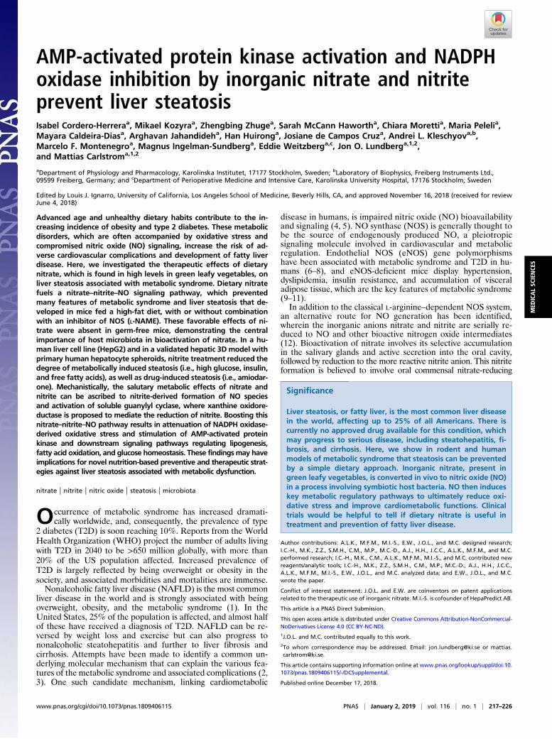
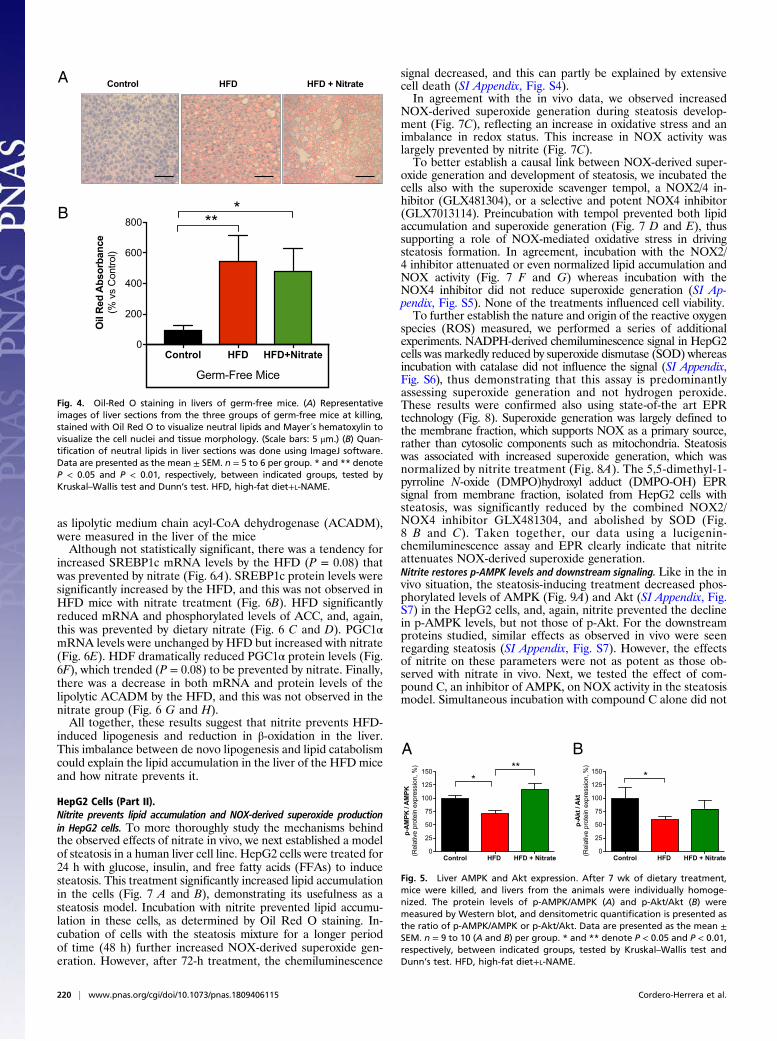
presence of L-NAME led to increased lipid accumulation comparedwith the control group, and this was fully prevented by nitrate (Fig. 3A and B). HFD also increased NOX-derived superoxide productionin the liver (Fig. 3C), and this was prevented by nitrate. Moreover,there was a positive correlation between lipid accumulation andNOX activity (Fig. 3F). To examine how nitrate could modulateNOX activity, the levels of the NOX subunit p67phox and NOX2(gp91phox) were measured by Western blot. HFD significantly in-creased p67phox levels (twofold increase). In line with the resultsabove, nitrate prevented p67phox up-regulation (Fig. 3D). Nochanges in the expression of NOX2 were seen (Fig. 3E).Host microbiota are necessary for dietary nitrate to prevent steatosis.Oral bacteria are believed to be central in bioactivation of di-etary nitrate as they catalyze its reduction to the more reactivenitrite anion, a reaction that cannot be as effectively performedby mammalian enzymes (30). When germ-free mice were fed anHFD and L-NAME for the same duration as in conventionalmice, they also developed liver steatosis. However, while nitrateprevented steatosis in conventional mice (Fig. 3 A and B), it waswithout effect in germ-free mice (Fig. 4), indicating the centralinvolvement of commensal bacteria in bioactivation of nitrate toelicit protective nitric oxide signaling. Similarly, abnormal glu-cose tolerance, increased fat content, and cardiac hypertrophy ingerm-free mice were also not improved by the nitrate treatment(SI Appendix, Table S1).Nitrate prevents the AMPK inhibition induced by an HFD. AMPK isconsidered a central regulator of glucose and lipid metabolism inthe liver and plays a key role in preventing lipid accumulation inhepatocytes. Akt is one of the downstream proteins of the insulin-signaling pathway, which is often disrupted in the metabolic syn-
drome and type 2 diabetes. Decreased phosphorylated levels ofAMPK and Akt were observed in the livers of HFD-fed mice (Fig. 5).Livers from nitrate-supplemented mice displayed phospho-AMPK(p-AMPK)/AMPK ratios similar to control levels while thephospho-Akt (p-Akt)/Akt ratios were not significantly af-fected. Thus, the preventive effect of nitrate on steatosis likelyinvolves preserved AMPK signaling.Nitrate modulates genes and proteins involved in lipid metabolism. Withindications of the AMPK signaling pathway being involved in thesalutary effects of nitrate, we next analyzed some of its keydownstream target proteins involved in cholesterol and fatty acidsynthesis, fatty acid oxidation, and mitochondrial biogenesis.Messenger RNA expression and protein levels of the lipogenictranscription factor sterol regulatory element-binding protein 1(SREBP1c), acetyl-CoA carboxylase (ACC), and peroxisomeproliferator-activated receptor γ coactivator 1 (PGC1α), as well
Control HFD HFD + Nitrate 0
25
50
75
100
125
150
175
Plas
ma
Nitr
ate
(M
)
****
*
Control HFD HFD + Nitrate 0
20
40
60
80
100
120
Live
r Nitr
ate
(M
)
****
Control HFD HFD + Nitrate0
10
20
30
40
50
Nitr
osyl
- Hem
oglo
bin
(Arb
itrar
y U
nits
)
*
Control HFD HFD + Nitrate 0.0
0.1
0.2
0.3
0.4
0.5
0.6
Plas
ma
Nitr
ite(
M)
*** *
Control HFD HFD + Nitrate 0.0
1.0
2.0
3.0
4.0
5.0Li
ver N
itrite
(M
)
Control HFD HFD + Nitrate0
20
40
60
80
Din
itros
yl Ir
on C
ompl
exes
(DN
IC)
(Arb
itrat
y U
nits
)
**
A B
C D
E F
Fig. 2. Nitrate, nitrite, and iron-nitrosylated species levels in blood and liver.In tissues obtained after 7 wk of dietary treatment with an HFD in combi-nation with the NOS inhibitor L-NAME, (A) plasma nitrate, (B) plasma nitrite,(C) liver nitrate, (D) liver nitrite, (E) blood iron-nitrosylated-hemoglobin, and(F) liver dinitrosyl iron complexes (DNICs) were measured for the three animalgroups. Data are presented as the mean ± SEM. n = 6 to 9 per group. *, **,and *** denote P < 0.05, P < 0.01, and P < 0.001, respectively, between in-dicated groups tested by Kruskal–Wallis test and Dunn’s test (A–E). HFD, high-fat diet+L-NAME.
Control HFD HFD + NitrateA
B C
D EControl HFD HFD + Nitrate
0
100
200
300
Oil
Red
Abs
orba
nce
(% v
s C
ontro
l)
** *
Control HFD HFD + Nitrate
90
100
110
120
130
RLU
/ m
in(%
vs
Con
trol)
** *
2000 2500 3000 3500 40000
1 108
2 108
3 108
4 108
Oil
Red
Abs
orba
nce
( A.U
.)
RLU / min (A. U.)
r = 0.69p = 0.0005
Control HFD HFD + Nitrate0
25
50
75
100
125
150N
OX2
(gp9
1pho
x)(R
elat
ive
prot
ein
expr
essi
on, %
)
Control HFD HFD + Nitrate0
50
100
150
200
250
p67p
hox
(Rel
ativ
e pr
otei
n ex
pres
sion
, %) * *
F
Fig. 3. Oil-Red O staining and NADPH oxidase activity in liver. (A) Represen-tative images of liver sections from the three mice groups at the moment ofkilling stained withOil RedO to visualize neutral lipids andMayer´s hematoxylin tovisualize the cell nuclei and tissue morphology. (Scale bars: 1 μm.) Insets show fourtimes magnified details of the images to highlight the lipid-staining morphology.(B) Quantification of neutral lipids in liver sections using ImageJ software. (C)NADPH oxidase-derived superoxide and hydrogen peroxide production was mea-sured by Amplex red and expressed as relative light units (RLUs), in the liver of thethree groups of mice at killing. (D and E) p67phox and NOX2 (gp91phox) proteinlevels in the liver were determined byWestern blot. Densitometric quantification ispresented as p67phox/vinculin and NOX2/vinculin. (F) Correlation between Oil RedO staining and NADPH oxidase activity in the liver. Data are presented as themean ± SEM. n = 5 to 9 per group. * and ** denote P < 0.05 and P < 0.01, re-spectively, between indicated groups, tested by Kruskal–Wallis test and Dunn’s test(B, C, and E) or Pearson correlation test (D). HFD, high-fat diet+L-NAME.
Cordero-Herrera et al. PNAS | January 2, 2019 | vol. 116 | no. 1 | 219
MED
ICALSC
IENCE
S
as lipolytic medium chain acyl-CoA dehydrogenase (ACADM),were measured in the liver of the miceAlthough not statistically significant, there was a tendency for
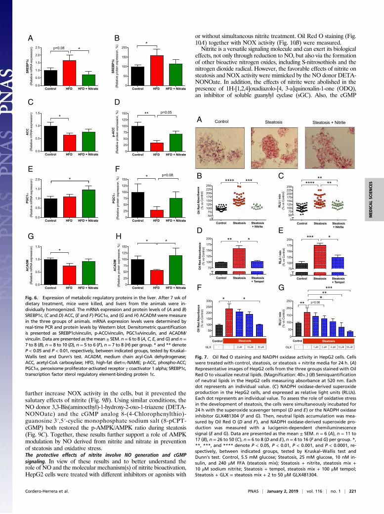
increased SREBP1c mRNA levels by the HFD (P = 0.08) thatwas prevented by nitrate (Fig. 6A). SREBP1c protein levels weresignificantly increased by the HFD, and this was not observed inHFD mice with nitrate treatment (Fig. 6B). HFD significantlyreduced mRNA and phosphorylated levels of ACC, and, again,this was prevented by dietary nitrate (Fig. 6 C and D). PGC1αmRNA levels were unchanged by HFD but increased with nitrate(Fig. 6E). HDF dramatically reduced PGC1α protein levels (Fig.6F), which trended (P = 0.08) to be prevented by nitrate. Finally,there was a decrease in both mRNA and protein levels of thelipolytic ACADM by the HFD, and this was not observed in thenitrate group (Fig. 6 G and H).All together, these results suggest that nitrite prevents HFD-
induced lipogenesis and reduction in β-oxidation in the liver.This imbalance between de novo lipogenesis and lipid catabolismcould explain the lipid accumulation in the liver of the HFD miceand how nitrate prevents it.
HepG2 Cells (Part II).Nitrite prevents lipid accumulation and NOX-derived superoxide productionin HepG2 cells. To more thoroughly study the mechanisms behindthe observed effects of nitrate in vivo, we next established a modelof steatosis in a human liver cell line. HepG2 cells were treated for24 h with glucose, insulin, and free fatty acids (FFAs) to inducesteatosis. This treatment significantly increased lipid accumulationin the cells (Fig. 7 A and B), demonstrating its usefulness as asteatosis model. Incubation with nitrite prevented lipid accumu-lation in these cells, as determined by Oil Red O staining. In-cubation of cells with the steatosis mixture for a longer periodof time (48 h) further increased NOX-derived superoxide gen-eration. However, after 72-h treatment, the chemiluminescence
signal decreased, and this can partly be explained by extensivecell death (SI Appendix, Fig. S4).In agreement with the in vivo data, we observed increased
NOX-derived superoxide generation during steatosis develop-ment (Fig. 7C), reflecting an increase in oxidative stress and animbalance in redox status. This increase in NOX activity waslargely prevented by nitrite (Fig. 7C).To better establish a causal link between NOX-derived super-
oxide generation and development of steatosis, we incubated thecells also with the superoxide scavenger tempol, a NOX2/4 in-hibitor (GLX481304), or a selective and potent NOX4 inhibitor(GLX7013114). Preincubation with tempol prevented both lipidaccumulation and superoxide generation (Fig. 7 D and E), thussupporting a role of NOX-mediated oxidative stress in drivingsteatosis formation. In agreement, incubation with the NOX2/4 inhibitor attenuated or even normalized lipid accumulation andNOX activity (Fig. 7 F and G) whereas incubation with theNOX4 inhibitor did not reduce superoxide generation (SI Ap-pendix, Fig. S5). None of the treatments influenced cell viability.To further establish the nature and origin of the reactive oxygen
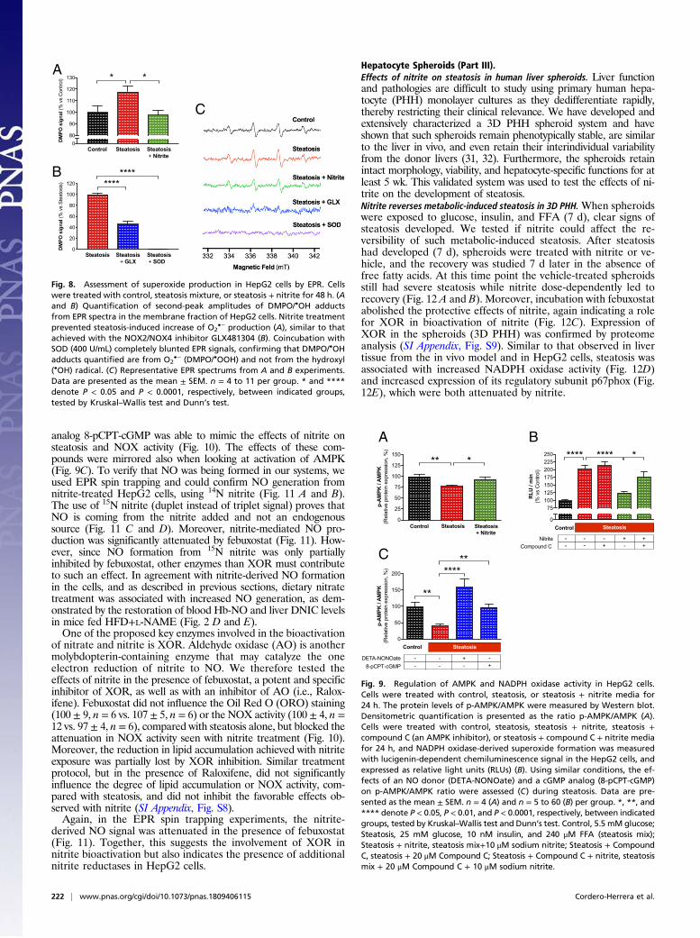
species (ROS) measured, we performed a series of additionalexperiments. NADPH-derived chemiluminescence signal in HepG2cells was markedly reduced by superoxide dismutase (SOD) whereasincubation with catalase did not influence the signal (SI Appendix,Fig. S6), thus demonstrating that this assay is predominantlyassessing superoxide generation and not hydrogen peroxide.These results were confirmed also using state-of-the art EPRtechnology (Fig. 8). Superoxide generation was largely defined tothe membrane fraction, which supports NOX as a primary source,rather than cytosolic components such as mitochondria. Steatosiswas associated with increased superoxide generation, which wasnormalized by nitrite treatment (Fig. 8A). The 5,5-dimethyl-1-pyrroline N-oxide (DMPO)hydroxyl adduct (DMPO-OH) EPRsignal from membrane fraction, isolated from HepG2 cells withsteatosis, was significantly reduced by the combined NOX2/NOX4 inhibitor GLX481304, and abolished by SOD (Fig.8 B and C). Taken together, our data using a lucigenin-chemiluminescence assay and EPR clearly indicate that nitriteattenuates NOX-derived superoxide generation.Nitrite restores p-AMPK levels and downstream signaling. Like in the invivo situation, the steatosis-inducing treatment decreased phos-phorylated levels of AMPK (Fig. 9A) and Akt (SI Appendix, Fig.S7) in the HepG2 cells, and, again, nitrite prevented the declinein p-AMPK levels, but not those of p-Akt. For the downstreamproteins studied, similar effects as observed in vivo were seenregarding steatosis (SI Appendix, Fig. S7). However, the effectsof nitrite on these parameters were not as potent as those ob-served with nitrate in vivo. Next, we tested the effect of com-pound C, an inhibitor of AMPK, on NOX activity in the steatosismodel. Simultaneous incubation with compound C alone did not
Control HFD HFD+Nitrate0
200
400
600
800
Oil
Red
Abs
orba
nce
(% v
s C
ontro
l)
***
Control HFD HFD + NitrateA
B
Germ-Free Mice
Fig. 4. Oil-Red O staining in livers of germ-free mice. (A) Representativeimages of liver sections from the three groups of germ-free mice at killing,stained with Oil Red O to visualize neutral lipids and Mayer´s hematoxylin tovisualize the cell nuclei and tissue morphology. (Scale bars: 5 μm.) (B) Quan-tification of neutral lipids in liver sections was done using ImageJ software.Data are presented as the mean ± SEM. n = 5 to 6 per group. * and ** denoteP < 0.05 and P < 0.01, respectively, between indicated groups, tested byKruskal–Wallis test and Dunn’s test. HFD, high-fat diet+L-NAME.
Control HFD HFD + Nitrate0
25
50
75
100
125
150
p-A
kt /
Akt
(Rel
ativ
e pr
otei
n ex
pres
sion
, %)
*
Control HFD HFD + Nitrate0
25
50
75
100
125
150
p-A
MPK
/ A
MPK
(Rel
ativ
e pr
otei
n ex
pres
sion
, %) **
*
A B
Fig. 5. Liver AMPK and Akt expression. After 7 wk of dietary treatment,mice were killed, and livers from the animals were individually homoge-nized. The protein levels of p-AMPK/AMPK (A) and p-Akt/Akt (B) weremeasured by Western blot, and densitometric quantification is presented asthe ratio of p-AMPK/AMPK or p-Akt/Akt. Data are presented as the mean ±SEM. n = 9 to 10 (A and B) per group. * and ** denote P < 0.05 and P < 0.01,respectively, between indicated groups, tested by Kruskal–Wallis test andDunn’s test. HFD, high-fat diet+L-NAME.
220 | www.pnas.org/cgi/doi/10.1073/pnas.1809406115 Cordero-Herrera et al.
further increase NOX activity in the cells, but it prevented thesalutary effects of nitrite (Fig. 9B). Using similar conditions, theNO donor 3,3-Bis(aminoethyl)-1-hydroxy-2-oxo-1-triazene (DETA-NONOate) and the cGMP analog 8-(4-Chlorophenylthio)-guanosine 3′,5′-cyclic monophosphate sodium salt (8-pCPT-cGMP) both restored the p-AMPK/AMPK ratio during steatosis(Fig. 9C). Together, these results further support a role of AMPKmodulation by NO derived from nitrite and nitrate in preventionof steatosis and oxidative stress.The protective effects of nitrite involve NO generation and cGMPsignaling. In view of these results and to better understand therole of NO and the molecular mechanism(s) of nitrite bioactivation,HepG2 cells were treated with different inhibitors or agonists with
or without simultaneous nitrite treatment. Oil Red O staining (Fig.10A) together with NOX activity (Fig. 10B) were measured.Nitrite is a versatile signaling molecule and can exert its biological
effects, not only through reduction to NO, but also via the formationof other bioactive nitrogen oxides, including S-nitrosothiols and thenitrogen dioxide radical. However, the favorable effects of nitrite onsteatosis and NOX activity were mimicked by the NO donor DETA-NONOate. In addition, the effects of nitrite were abolished in thepresence of 1H-[1,2,4]oxadiazolo-[4, 3-a]quinoxalin-1-one (ODQ),an inhibitor of soluble guanylyl cyclase (sGC). Also, the cGMP
Control HFD HFD + Nitrate 0.0
0.5
1.0
1.5
2.0
2.5
SREB
P1c
(Rel
ativ
e m
RN
A ex
pres
sion
) *p=0.08
Control HFD HFD + Nitrate 0.0
0.5
1.0
1.5
AC
AD
M(R
elat
ive
mR
NA
expr
essi
on)
*
Control HFD HFD + Nitrate0
50
100
150
200
SREB
P1c
(Rel
ativ
e pr
otei
n ex
pres
sion
, %) *
Control HFD HFD + Nitrate0
25
50
75
100
125
150
AC
AD
M(R
elat
ive
prot
ein
expr
essi
on, %
) * *
A B
C D
E F
G H
Control HFD HFD + Nitrate 0.0
0.5
1.0
1.5
AC
C(R
elat
ive
mR
NA
expr
essi
on)
*
Control HFD HFD + Nitrate0
25
50
75
100
125
150p-
AC
C(R
elat
ive
prot
ein
expr
essi
on, %
)** p=0.05
Control HFD HFD + Nitrate 0.0
0.5
1.0
1.5
2.0
PGC
1(R
elat
ive
mR
NA
expr
essi
on) *
Control HFD HFD + Nitrate0
25
50
75
100
125
150
PGC
1(R
elat
ive
prot
ein
expr
essi
on, %
) * p=0.08
Fig. 6. Expression of metabolic regulatory proteins in the liver. After 7 wk ofdietary treatment, mice were killed, and livers from the animals were in-dividually homogenized. The mRNA expression and protein levels of (A and B)SREBP1c, (C and D) ACC, (E and F) PGC1α, and (G and H) ACADMwere measurein the three groups of animals. mRNA expression levels were determined byreal-time PCR and protein levels by Western blot. Densitometric quantificationis presented as SREBP1c/vinculin, p-ACC/vinculin, PGC1α/vinculin, and ACADM/vinculin. Data are presented as themean ± SEM. n= 6 to 8 (A, C, E, andG) and n=7 to 8 (B), n = 8 to 10 (D), n = 5 to 6 (F), n = 7 to 8 (H) per group. * and ** denoteP < 0.05 and P < 0.01, respectively, between indicated groups, tested by Kruskal–Wallis test and Dunn’s test. ACADM, medium chain acyl-CoA dehydrogenase;ACC, acetyl-CoA carboxylase; HFD, high-fat diet+L-NAME; p-ACC, phospho-ACC;PGC1α, peroxisome proliferator-activated receptor γ coactivator 1 alpha; SREBP1c,transcription factor sterol regulatory element-binding protein 1c.
05075
100125150175200225250
****
Oil
Red
Abs
orba
nce
(% v
s C
ontro
l)
Control Steatosis Steatosis + Nitrite
***
05075
100125150175200225250
RLU
/ m
in(%
vs
Con
trol)
Control Steatosis Steatosis + Nitrite
**** ****
Control Steatosis Steatosis + NitriteA
B C
0
75
100
125
150
175
200
Oil
Red
Abs
orba
nce
(% v
s C
ontro
l)
Control Steatosis Steatosis + Tempol
** *
0
75
100
125
150
175
200
RLU
/ m
in(%
vs
Con
trol)
Control Steatosis Steatosis + Tempol
*** *D E
0
50
100
150
200
250
300
Oil
Red
Abs
orba
nce
(% v
s C
ontro
l)
Control Steatosis
GLX 2 M 10 M 50 M--
* *
0
50
100
150
200
250
RLU
/ m
in(%
vs
Con
trol)
Control Steatosis
GLX 2 M 10 M 50 M--
***
***
p=0.06
4 M1 M
**F G
Fig. 7. Oil Red O staining and NADPH oxidase activity in HepG2 cells. Cellswere treated with control, steatosis, or steatosis + nitrite media for 24 h. (A)Representative images of HepG2 cells from the three groups stained with OilRed O to visualize neutral lipids. (Magnification: 40×.) (B) Semiquantificationof neutral lipids in the HepG2 cells measuring absorbance at 520 nm. Eachdot represents an individual value. (C) NADPH oxidase-derived superoxideproduction in the HepG2 cells, and expressed as relative light units (RLUs).Each dot represents an individual value. To assess the role of oxidative stressin the development of steatosis, the cells were simultaneously incubated for24 h with the superoxide scavenger tempol (D and E) or the NADPH oxidaseinhibitor GLX481304 (F and G). Then, neutral lipids accumulation was mea-sured by Oil Red O (D and F), and NADPH oxidase-derived superoxide pro-duction was measured with a lucigenin-dependent chemiluminescencesignal (E and G). Data are presented as the mean ± SEM. n = 6 (A), n = 11 to17 (B), n = 26 to 50 (C), n = 6 to 8 (D and E), n = 4 to 16 (F and G) per group. *,**, ***, and **** denote P < 0.05, P < 0.01, P < 0.001, and P < 0.0001, re-spectively, between indicated groups, tested by Kruskal–Wallis test andDunn’s test. Control, 5.5 mM glucose; Steatosis, 25 mM glucose, 10 nM in-sulin, and 240 μM FFA (steatosis mix); Steatosis + nitrite, steatosis mix +10 μM sodium nitrite; Steatosis + tempol, steatosis mix + 100 μM tempol;Steatosis + GLX = steatosis mix + 2 to 50 μM GLX481304.
Cordero-Herrera et al. PNAS | January 2, 2019 | vol. 116 | no. 1 | 221
MED
ICALSC
IENCE
S
analog 8-pCPT-cGMP was able to mimic the effects of nitrite onsteatosis and NOX activity (Fig. 10). The effects of these com-pounds were mirrored also when looking at activation of AMPK(Fig. 9C). To verify that NO was being formed in our systems, weused EPR spin trapping and could confirm NO generation fromnitrite-treated HepG2 cells, using 14N nitrite (Fig. 11 A and B).The use of 15N nitrite (duplet instead of triplet signal) proves thatNO is coming from the nitrite added and not an endogenoussource (Fig. 11 C and D). Moreover, nitrite-mediated NO pro-duction was significantly attenuated by febuxostat (Fig. 11). How-ever, since NO formation from 15N nitrite was only partiallyinhibited by febuxostat, other enzymes than XOR must contributeto such an effect. In agreement with nitrite-derived NO formationin the cells, and as described in previous sections, dietary nitratetreatment was associated with increased NO generation, as dem-onstrated by the restoration of blood Hb-NO and liver DNIC levelsin mice fed HFD+L-NAME (Fig. 2 D and E).One of the proposed key enzymes involved in the bioactivation
of nitrate and nitrite is XOR. Aldehyde oxidase (AO) is anothermolybdopterin-containing enzyme that may catalyze the oneelectron reduction of nitrite to NO. We therefore tested theeffects of nitrite in the presence of febuxostat, a potent and specificinhibitor of XOR, as well as with an inhibitor of AO (i.e., Ralox-ifene). Febuxostat did not influence the Oil Red O (ORO) staining(100 ± 9, n = 6 vs. 107 ± 5, n = 6) or the NOX activity (100 ± 4, n =12 vs. 97 ± 4, n = 6), compared with steatosis alone, but blocked theattenuation in NOX activity seen with nitrite treatment (Fig. 10).Moreover, the reduction in lipid accumulation achieved with nitriteexposure was partially lost by XOR inhibition. Similar treatmentprotocol, but in the presence of Raloxifene, did not significantlyinfluence the degree of lipid accumulation or NOX activity, com-pared with steatosis, and did not inhibit the favorable effects ob-served with nitrite (SI Appendix, Fig. S8).Again, in the EPR spin trapping experiments, the nitrite-
derived NO signal was attenuated in the presence of febuxostat(Fig. 11). Together, this suggests the involvement of XOR innitrite bioactivation but also indicates the presence of additionalnitrite reductases in HepG2 cells.
Hepatocyte Spheroids (Part III).Effects of nitrite on steatosis in human liver spheroids. Liver functionand pathologies are difficult to study using primary human hepa-tocyte (PHH) monolayer cultures as they dedifferentiate rapidly,thereby restricting their clinical relevance. We have developed andextensively characterized a 3D PHH spheroid system and haveshown that such spheroids remain phenotypically stable, are similarto the liver in vivo, and even retain their interindividual variabilityfrom the donor livers (31, 32). Furthermore, the spheroids retainintact morphology, viability, and hepatocyte-specific functions for atleast 5 wk. This validated system was used to test the effects of ni-trite on the development of steatosis.Nitrite reverses metabolic-induced steatosis in 3D PHH.When spheroidswere exposed to glucose, insulin, and FFA (7 d), clear signs ofsteatosis developed. We tested if nitrite could affect the re-versibility of such metabolic-induced steatosis. After steatosishad developed (7 d), spheroids were treated with nitrite or ve-hicle, and the recovery was studied 7 d later in the absence offree fatty acids. At this time point the vehicle-treated spheroidsstill had severe steatosis while nitrite dose-dependently led torecovery (Fig. 12 A and B). Moreover, incubation with febuxostatabolished the protective effects of nitrite, again indicating a rolefor XOR in bioactivation of nitrite (Fig. 12C). Expression ofXOR in the spheroids (3D PHH) was confirmed by proteomeanalysis (SI Appendix, Fig. S9). Similar to that observed in livertissue from the in vivo model and in HepG2 cells, steatosis wasassociated with increased NADPH oxidase activity (Fig. 12D)and increased expression of its regulatory subunit p67phox (Fig.12E), which were both attenuated by nitrite.
080
90
100
110
120
130
DM
PO s
igna
l (%
vs
Con
trol) *
Control Steatosis Steatosis + Nitrite
*
0
20
40
60
80
100
120
DM
PO s
igna
l (%
vs
Stea
tosi
s)
Steatosis Steatosis+ GLX
Steatosis+ SOD
********
A
B
C
Fig. 8. Assessment of superoxide production in HepG2 cells by EPR. Cellswere treated with control, steatosis mixture, or steatosis + nitrite for 48 h. (Aand B) Quantification of second-peak amplitudes of DMPO/•OH adductsfrom EPR spectra in the membrane fraction of HepG2 cells. Nitrite treatmentprevented steatosis-induced increase of O2
•− production (A), similar to thatachieved with the NOX2/NOX4 inhibitor GLX481304 (B). Coincubation withSOD (400 U/mL) completely blunted EPR signals, confirming that DMPO/•OHadducts quantified are from O2
•− (DMPO/•OOH) and not from the hydroxyl(•OH) radical. (C) Representative EPR spectrums from A and B experiments.Data are presented as the mean ± SEM. n = 4 to 11 per group. * and ****denote P < 0.05 and P < 0.0001, respectively, between indicated groups,tested by Kruskal–Wallis test and Dunn’s test.
A B
0
25
50
75
100
125
150
p-A
MPK
/ A
MPK
(Rel
ativ
e pr
otei
n ex
pres
sion
, %)
** *
Control Steatosis Steatosis + Nitrite
0
75100125150175200225250
RLU
/ m
in(%
vs
Con
trol)
****
Control Steatosis
NitriteCompound C
****
- + +--- - +- +
*
0
50
100
150
200
p-A
MPK
/ A
MPK
(Rel
ativ
e pr
otei
n ex
pres
sion
, %)
Control Steatosis
DETA-NONOate8-pCPT-cGMP
+-
--
--
-+
**
******C
Fig. 9. Regulation of AMPK and NADPH oxidase activity in HepG2 cells.Cells were treated with control, steatosis, or steatosis + nitrite media for24 h. The protein levels of p-AMPK/AMPK were measured by Western blot.Densitometric quantification is presented as the ratio p-AMPK/AMPK (A).Cells were treated with control, steatosis, steatosis + nitrite, steatosis +compound C (an AMPK inhibitor), or steatosis + compound C + nitrite mediafor 24 h, and NADPH oxidase-derived superoxide formation was measuredwith lucigenin-dependent chemiluminescence signal in the HepG2 cells, andexpressed as relative light units (RLUs) (B). Using similar conditions, the ef-fects of an NO donor (DETA-NONOate) and a cGMP analog (8-pCPT-cGMP)on p-AMPK/AMPK ratio were assessed (C) during steatosis. Data are pre-sented as the mean ± SEM. n = 4 (A) and n = 5 to 60 (B) per group. *, **, and**** denote P < 0.05, P < 0.01, and P < 0.0001, respectively, between indicatedgroups, tested by Kruskal–Wallis test and Dunn’s test. Control, 5.5 mM glucose;Steatosis, 25 mM glucose, 10 nM insulin, and 240 μM FFA (steatosis mix);Steatosis + nitrite, steatosis mix+10 μM sodium nitrite; Steatosis + CompoundC, steatosis + 20 μM Compound C; Steatosis + Compound C + nitrite, steatosismix + 20 μM Compound C + 10 μM sodium nitrite.
222 | www.pnas.org/cgi/doi/10.1073/pnas.1809406115 Cordero-Herrera et al.

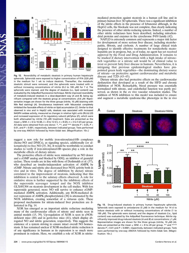
Nitrite attenuates drug-induced steatosis in 3D PHH. Next, we tested ifsimultaneous incubation with nitrite could attenuate drug-induced steatosis. When spheroids were exposed to the hepato-toxic drug amiodarone (14 d), clear signs of steatosis had de-veloped (Fig. 13). Coincubation with nitrite clearly preventedlipid accumulation. Remarkably, these favorable effects of nitriteon metabolic- and drug-induced steatosis were significant al-ready at 1 μM, which is similar to the levels of endogenous nitritefound in healthy tissues under basal conditions.
DiscussionHere, we show that dietary inorganic nitrate preserves metabolicand cardiovascular function and markedly attenuates liver stea-tosis in mice with diet-induced metabolic syndrome. The favorableeffects of nitrate on development of steatosis occurred despitesimilar accumulation of fat mass. These salutary effects werereflected in experiments using HepG2 cells and in 3D spheroidsfrom primary human liver cells. Dietary nitrate in vivo and nitritein vitro activated AMPK and reduced NOX activity, leading toenhanced NO bioavailability and cardiometabolic improvement.Dietary nitrate is metabolized in vivo to first form the more
reactive nitrite anion (12). It has been suggested that oral bac-teria have a key role in nitrate bioactivation, but mammaliansystems have also been demonstrated (33). Our data from nor-mal and germ-free mice indicate a central role of bacteria innitrate bioactivation. Thus, the protective effects of nitrate ondevelopment of liver steatosis were absent in germ-free animals.Because the nitrate ion in itself is more or less biologically inert,we later used nitrite in all subsequent in vitro experiments. Whenhuman liver spheroids were exposed to nitrite, the degree ofsteatosis induced by either nutritional overload or amiodaronewas significantly decreased. Remarkably, the effects of nitrite invitro were seen already at submicromolar levels: i.e., close towhat is seen in tissues under basal conditions and definitely whatis achieved after ingestion of nitrate-rich vegetables (12, 17).Mechanistically, the effects of nitrite seemed to be mediated by
its reduction to NO and a further cGMP-mediated activation ofAMPK, a master regulator of cellular metabolism and energyhomeostasis. Formation of NO from nitrate in vivo was verifiedwith EPR methodology detecting two specific NO-derived speciesin blood and liver tissue and from nitrite in cells using a spin trap.The reduction of nitrate to NO-species in vivo was confirmed by anEPRmethod detecting Hb-NO in whole blood and DNICs in liver.Additionally, the NO (15N) formation from nitrite (15N) in cul-tured cells was demonstrated by EPR spin trapping using Fe(II)-diethyldithiocarbamate colloid (Fe-DETC) (34). Recent studies
075
100125150175200225
Oil
Red
Abs
orba
nce
(% v
s C
ontro
l)
Control Steatosis
Nitrite
ODQFebuxostat
DETA-NONOate- + + +--
---
8-pCPT-cGMP
-
++-
--
--
- --
-
---++
-----
----
**** *****
*
***
*
A
B
075
100125150175200225
RLU
/ m
in(%
vs
Con
trol)
Control Steatosis
Nitrite
ODQFebuxostat
DETA-NONOate- + + +--
---
8-pCPT-cGMP
-
++-
--
--
- --
-
---++
-----
----
**** **
*****
****
Fig. 10. Mechanisms contributing to nitrite-mediated protection against stea-tosis in HepG2 cells. Cells were treated with control, steatosis, steatosis + nitrite,steatosis + DETA-NONOate (a slow releasing NO-donor), steatosis + cGMP ana-log (8-pCPT-cGMP), steatosis + Febuxostat (a specific inhibitor of XOR), or stea-tosis + ODQ (an inhibitor of soluble guanylyl cyclase) media for 24 h. (A)Accumulation of neutral lipids was measured by Oil Red O, and (B) NADPHoxidase-derived superoxide formation was measured with lucigenin-dependent chemiluminescence signal, and expressed as relative lightunits (RLUs). Data are presented as the mean ± SEM. n = 5 to 32 (A) and n =5 to 60 (B) per group. *, **, ***, and **** denote P < 0.05, P < 0.01, P <0.001, and P < 0.0001, respectively, between indicated groups, tested byKruskal–Wallis test and Dunn’s test. Control, 5.5 mM glucose; Steatosis,25 mM glucose, 10 nM insulin, and 240 μM FFA (steatosis mix); Steatosis +nitrite, steatosis mix + 10 μM sodium nitrite; Steatosis + DETA-NONOate,steatosis mix+ 5 μM DETA-NONOate; Steatosis + cGMP analog, steatosis mix +10 μM cGMP analog; Steatosis + Febuxostat, steatosis mix + 50 nM Febuxostat;Steatosis + ODQ, steatosis mix + 10 μM ODQ.
0
20
40
60
80
Fe-D
ETC
sig
nal (
Arbi
trary
Uni
ts)
Vehicle Nitrite 14N Nitrite 14N+ Febuxostat
******
Magnetic Field (mT)
A B
0
10
20
30
Fe-D
ETC
sig
nal (
Arbi
trary
Uni
ts)
Vehicle Nitrite 15N+ Febuxostat
Nitrite 15N
******C D
Magnetic Field (mT)
Nitrite 14N+ Febuxostat
Nitrite 14N
Vehicle
Nitrite 15N+ Febuxostat
Nitrite 15N
Vehicle
Fig. 11. Affirmation of nitrite-mediated NO production in HepG2 cells byEPR. Changes in first-peak amplitudes from EPR spectrums, together withrepresentative spectrums, for NO production using Fe(DETC)2 as spin trap.HepG2 cells were incubated with vehicle or Febuxostat (50 nM) for 20 minand then treated with nitrite 14N (500 μM) or nitrite 15N (500 μM) for 1 h inthe presence of 0.5 mM colloid Fe(DETC)2 for NO detection. Shown arequantified EPR data for nitrite 14N (A and B) and nitrite 15N (C and D) to-gether with representative spectrums for the different experiments. Dataare presented as the mean ± SEM. n = 3 to 4 per group. ** and **** denoteP < 0.01 and P < 0.0001, respectively, between indicated groups, tested byKruskal–Wallis test and Dunn’s test.
Cordero-Herrera et al. PNAS | January 2, 2019 | vol. 116 | no. 1 | 223
MED
ICALSC
IENCE
S
suggest a new role for mobile iron-nitrosylated-Hb complexes(heme-NO and DNICs), as signaling species, additionally (or al-ternatively) to free NO (35, 36). It would be worthwhile to conductstudies to verify if iron-nitrosylated-Hb species play a role in themetabolic effects of dietary nitrate.The protective effects of nitrite were mimicked by an NO donor
and a cGMP analog and blocked by ODQ, an inhibitor of guanylylcyclase. These results are in line with those of Deshmukh et al. (37),who described an insulin-independent activation of AMPK bycGMP. Nitrate and nitrite also decreased liver NOX activity both invivo and in vitro. The degree of inhibition by dietary nitratecorrelated to the improvement of steatosis, indicating that thisinhibition is central to the salutary effects observed. A role foroxidative stress is further supported by the inhibitory effects ofthe superoxide scavenger tempol and the NOX inhibitorGLX481304 on steatosis development in the cell studies. With lesssuperoxide generated, more NO will survive to enhance cGMP-mediated AMPK activation. Additionally, any ROS-mediated di-rect repression of AMPK phosphorylation (38) is attenuated by theNOX inhibition, creating somewhat of a virtuous cycle. Theseproposed mechanisms for nitrate-induced liver protection are il-lustrated in Fig. 14.XOR has emerged as an important nitrite reductase mediating
many of the cardiovascular effects of nitrate and nitrite seen inanimal models (15, 39). Up-regulation of XOR is seen in eNOS-deficient mice (40) and in germ-free mice (41), which display ab-rogated NO and nitrite generation, respectively. This suggests theexistence of a system striving to ensure intact overall NO homeo-stasis. It has remained unclear if XOR-mediated nitrite reduction isof any significance in humans as its expression is so much moreprominent in rodents. Here, we establish a role of XOR in nitrite-
mediated protection against steatosis in a human cell line and inprimary human liver 3D spheroids. There was a significant inhibitionof the nitrite effects in the presence of febuxostat although, in theHepG2 cells, the blockade was not complete, thus clearly indicatingthe presence of other nitrite-reducing systems. Indeed, a number ofother nitrite reductases have been described, including mitochon-drial proteins and enzymes in the cytochrome P450 family (42).NAFLD is extremely common and represents a major risk factor
for development of more serious liver disease, including steatohe-patitis, fibrosis, and cirrhosis. A number of large clinical trialsdesigned to identify effective treatments for nonalcoholic steato-hepatitis are in progress, but, as of today, no agent has yet receivedapproval by the Food and Drug Administration. It remains tobe studied if dietary intervention with a high intake of nitrate-rich vegetables or a nitrate salt would be of clinical value totreat or prevent fatty liver disease in humans. Nevertheless, it isintriguing that previous epidemiological studies have pin-pointed green leafy vegetables—the dominating dietary sourceof nitrate—as protective against cardiovascular and metabolicdisease and T2D (43–45).Dietary nitrate also had protective effects on the cardiovascular
dysfunction that developed as a result of the HFD and chronicinhibition of NOS. Remarkably, blood pressure was essentiallynormalized with nitrate, and endothelial function was partly pre-served, as shown in the ex vivo vascular relaxation studies. Theaddition of NOS inhibition to this model was included to ensureand augment a metabolic syndrome-like phenotype in the in vivo
0.0
0.5
1.0
1.5
2.0
2.5
Lipi
ds(R
elat
ive
Con
trol)
Control Steatosis
Nitrite (µM)
10 40 100 10.2 0.6
* ***
0
50
100
150
200
250
RLU
/ m
in(%
vs
Con
trol)
*** *
Control Steatosis Steatosis + Nitrite
*
0.0
0.5
1.0
1.5
2.0
p67p
hox
(Rel
ativ
e m
RN
A ex
pres
sion
)
* **
Control Steatosis Steatosis + Nitrite
Control
Steatosis
Steatosis + Nitrite
A BC
D
0.0
0.5
1.0
1.5
Lipi
ds(R
elat
ive
Stea
tosi
s)
** *
Steatosis
FebuxostatNitrite
++
--
-+
E
Fig. 12. Reversibility of metabolic steatosis in primary human hepatocytespheroids. Spheroids were exposed to higher concentration of FFA (320 μM)in the medium for 1 wk to induce steatosis. Thereafter, the metabolicsteatotic stimuli were removed, and the spheroids were treated with orwithout increasing concentrations of nitrite (0.2 to 100 μM) for 7 d. Thespheroids were stained, and the degree of steatosis (i.e., lipid content) wasevaluated by the AdipoRed fluorescence technique. Nitrite improved reversibilityof metabolic-induced steatosis in a dose-dependent way (A and B), being sig-nificant compared with the steatosis group at concentrations ≥0.6 μM. Repre-sentative images are shown for the three groups (nitrite, 10 μM) (staining withNile Red staining) (A). Simultaneous treatment with febuxostat completelyinhibited the favorable effects of nitrite on lipid accumulation (C). Similar to thatobserved in vivo and in HepG2 cells, steatosis was associated with increasedNADPH oxidase activity, measured as changes in relative light units (RLUs) (D)and increased expression of its regulatory subunit p67phox (E), which wereboth attenuated by nitrite (10 μM) treatment. Data are presented as themean ± SEM. n = 6 to 16 (B), n = 8 to 12 (C), n = 8 (D), n = 3 to 5 (E) per group.All data were obtained from one donor. *, **, and *** denote P < 0.05, P <0.01, and P < 0.001, respectively, between indicated groups. Tests performedby one-way ANOVA followed by Holm–Sidak test. (Magnification: 10×.)
0.0
0.5
1.0
1.5
2.0
2.5
Lipi
ds(R
elat
ive
Con
trol)
Control Steatosis
Nitrite (µM)10 40 100 1
** ******
**
Control Steatosis Steatosis+NitriteA
B
Fig. 13. Drug-induced steatosis in primary human hepatocyte spheroids.Spheroids were exposed to amiodarone (5 μM) in the medium for 14 d toinduce steatosis with or without increasing concentrations of nitrite (1 to100 μM). The spheroids were stained, and the degree of steatosis (i.e., lipidcontent) was evaluated by the AdipoRed fluorescence technique. Nitrite sig-nificantly improved drug-induced steatosis (A and B) at concentrations ≥1 μM.Representative images are shown for the three groups (nitrite, 10 μM) (A).Data are presented as the mean ± SEM. n = 8 to 20 per group. ** and ****denote P < 0.01 and P < 0.0001, respectively, between indicated groups. Testswere performed by one-way ANOVA followed by Holm–Sidak test. (Magni-fication: 10×.)
224 | www.pnas.org/cgi/doi/10.1073/pnas.1809406115 Cordero-Herrera et al.
studies (Fig. 1). With this combination, one cannot exactly judge therelative contribution of the HFD vs. the L-NAME treatment to theoverall phenotype, and it also makes conclusions about the exactmechanisms more complicated. However, one should note that asimilar cardiometabolic protection was seen also in additional ex-periments without systemic NOS inhibition and that L-NAME wasnot a part of any of the subsequent cell studies. So, clearly, allpathways affected by nitrate/nitrite were so also in the presence of afunctional NOS. Moreover, the favorable effects of nitrate on liversteatosis appear to be independent of changes in body weight oraccumulation of visceral adipose tissue as protection was observedin both mice fed with HFD+L-NAME (i.e., no effect of nitrate onbody weight and fat content) and HFD alone (i.e., body weight andfat content were reduced by nitrate).
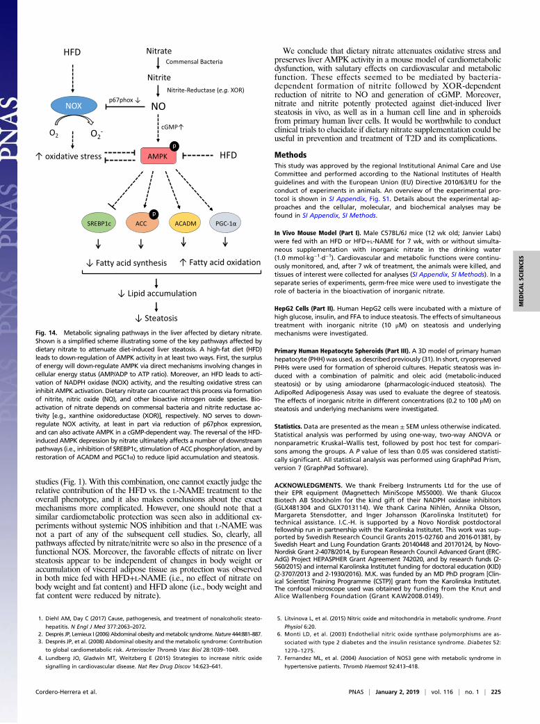
We conclude that dietary nitrate attenuates oxidative stress andpreserves liver AMPK activity in a mouse model of cardiometabolicdysfunction, with salutary effects on cardiovascular and metabolicfunction. These effects seemed to be mediated by bacteria-dependent formation of nitrite followed by XOR-dependentreduction of nitrite to NO and generation of cGMP. Moreover,nitrate and nitrite potently protected against diet-induced liversteatosis in vivo, as well as in a human cell line and in spheroidsfrom primary human liver cells. It would be worthwhile to conductclinical trials to elucidate if dietary nitrate supplementation could beuseful in prevention and treatment of T2D and its complications.
MethodsThis study was approved by the regional Institutional Animal Care and UseCommittee and performed according to the National Institutes of Healthguidelines and with the European Union (EU) Directive 2010/63/EU for theconduct of experiments in animals. An overview of the experimental pro-tocol is shown in SI Appendix, Fig. S1. Details about the experimental ap-proaches and the cellular, molecular, and biochemical analyses may befound in SI Appendix, SI Methods.
In Vivo Mouse Model (Part I). Male C57BL/6J mice (12 wk old; Janvier Labs)were fed with an HFD or HFD+L-NAME for 7 wk, with or without simulta-neous supplementation with inorganic nitrate in the drinking water(1.0 mmol·kg−1·d−1). Cardiovascular and metabolic functions were continu-ously monitored, and, after 7 wk of treatment, the animals were killed, andtissues of interest were collected for analyses (SI Appendix, SI Methods). In aseparate series of experiments, germ-free mice were used to investigate therole of bacteria in the bioactivation of inorganic nitrate.
HepG2 Cells (Part II). Human HepG2 cells were incubated with a mixture ofhigh glucose, insulin, and FFA to induce steatosis. The effects of simultaneoustreatment with inorganic nitrite (10 μM) on steatosis and underlyingmechanisms were investigated.
Primary Human Hepatocyte Spheroids (Part III). A 3D model of primary humanhepatocyte (PHH)was used, as described previously (31). In short, cryopreservedPHHs were used for formation of spheroid cultures. Hepatic steatosis was in-duced with a combination of palmitic and oleic acid (metabolic-inducedsteatosis) or by using amiodarone (pharmacologic-induced steatosis). TheAdipoRed Adipogenesis Assay was used to evaluate the degree of steatosis.The effects of inorganic nitrite in different concentrations (0.2 to 100 μM) onsteatosis and underlying mechanisms were investigated.
Statistics. Data are presented as the mean ± SEM unless otherwise indicated.Statistical analysis was performed by using one-way, two-way ANOVA ornonparametric Kruskal–Wallis test, followed by post hoc test for compari-sons among the groups. A P value of less than 0.05 was considered statisti-cally significant. All statistical analysis was performed using GraphPad Prism,version 7 (GraphPad Software).
ACKNOWLEDGMENTS. We thank Freiberg Instruments Ltd for the use oftheir EPR equipment (Magnettech MiniScope MS5000). We thank GlucoxBiotech AB Stockholm for the kind gift of their NADPH oxidase inhibitors(GLX481304 and GLX7013114). We thank Carina Nihlén, Annika Olsson,Margareta Stensdotter, and Inger Johansson (Karolinska Institutet) fortechnical assistance. I.C.-H. is supported by a Novo Nordisk postdoctoralfellowship run in partnership with the Karolinska Institutet. This work was sup-ported by Swedish Research Council Grants 2015-02760 and 2016-01381, bySwedish Heart and Lung Foundation Grants 20140448 and 20170124, by Novo-Nordisk Grant 2-4078/2014, by European Research Council Advanced Grant (ERC-AdG) Project HEPASPHER Grant Agreement 742020, and by research funds (2-560/2015) and internal Karolinska Institutet funding for doctoral education (KID)(2-3707/2013 and 2-1930/2016). M.K. was funded by an MD PhD program [Clin-ical Scientist Training Programme (CSTP)] grant from the Karolinska Institutet.The confocal microscope used was obtained by funding from the Knut andAlice Wallenberg Foundation (Grant KAW2008.0149).
1. Diehl AM, Day C (2017) Cause, pathogenesis, and treatment of nonalcoholic steato-
hepatitis. N Engl J Med 377:2063–2072.2. Després JP, Lemieux I (2006) Abdominal obesity andmetabolic syndrome.Nature 444:881–887.3. Després JP, et al. (2008) Abdominal obesity and the metabolic syndrome: Contribution
to global cardiometabolic risk. Arterioscler Thromb Vasc Biol 28:1039–1049.
4. Lundberg JO, Gladwin MT, Weitzberg E (2015) Strategies to increase nitric oxide
signalling in cardiovascular disease. Nat Rev Drug Discov 14:623–641.
5. Litvinova L, et al. (2015) Nitric oxide and mitochondria in metabolic syndrome. Front
Physiol 6:20.6. Monti LD, et al. (2003) Endothelial nitric oxide synthase polymorphisms are as-
sociated with type 2 diabetes and the insulin resistance syndrome. Diabetes 52:
1270–1275.7. Fernandez ML, et al. (2004) Association of NOS3 gene with metabolic syndrome in
hypertensive patients. Thromb Haemost 92:413–418.
Fig. 14. Metabolic signaling pathways in the liver affected by dietary nitrate.Shown is a simplified scheme illustrating some of the key pathways affected bydietary nitrate to attenuate diet-induced liver steatosis. A high-fat diet (HFD)leads to down-regulation of AMPK activity in at least two ways. First, the surplusof energy will down-regulate AMPK via direct mechanisms involving changes incellular energy status (AMP/ADP to ATP ratio). Moreover, an HFD leads to acti-vation of NADPH oxidase (NOX) activity, and the resulting oxidative stress caninhibit AMPK activation. Dietary nitrate can counteract this process via formationof nitrite, nitric oxide (NO), and other bioactive nitrogen oxide species. Bio-activation of nitrate depends on commensal bacteria and nitrite reductase ac-tivity [e.g., xanthine oxidoreductase (XOR)], respectively. NO serves to down-regulate NOX activity, at least in part via reduction of p67phox expression,and can also activate AMPK in a cGMP-dependent way. The reversal of the HFD-induced AMPK depression by nitrate ultimately affects a number of downstreampathways (i.e., inhibition of SREBP1c, stimulation of ACC phosphorylation, and byrestoration of ACADM and PGC1α) to reduce lipid accumulation and steatosis.
Cordero-Herrera et al. PNAS | January 2, 2019 | vol. 116 | no. 1 | 225
MED
ICALSC
IENCE
S
8. Bressler J, Pankow JS, Coresh J, Boerwinkle E (2013) Interaction between the NOS3 geneand obesity as a determinant of risk of type 2 diabetes: The atherosclerosis risk in com-munities study. PLoS One 8:e79466.
9. Cook S, et al. (2003) Clustering of cardiovascular risk factors mimicking the humanmetabolic syndrome X in eNOS null mice. Swiss Med Wkly 133:360–363.
10. Huang PL (2009) eNOS, metabolic syndrome and cardiovascular disease. TrendsEndocrinol Metab 20:295–302.
11. Carlström M, et al. (2010) Dietary inorganic nitrate reverses features of metabolicsyndrome in endothelial nitric oxide synthase-deficient mice. Proc Natl Acad Sci USA107:17716–17720.
12. Lundberg JO, Weitzberg E, Gladwin MT (2008) The nitrate-nitrite-nitric oxide path-way in physiology and therapeutics. Nat Rev Drug Discov 7:156–167.
13. Lundberg JO, Govoni M (2004) Inorganic nitrate is a possible source for systemicgeneration of nitric oxide. Free Radic Biol Med 37:395–400.
14. Helms CC, Liu X, Kim-Shapiro DB (December 21, 2016) Recent insights into nitritesignaling processes in blood. Biol Chem, 10.1515/hsz-2016-0263.
15. Cantu-Medellin N, Kelley EE (2013) Xanthine oxidoreductase-catalyzed reduction ofnitrite to nitric oxide: Insights regarding where, when and how. Nitric Oxide 34:19–26.
16. Lundberg JO, et al. (2009) Nitrate and nitrite in biology, nutrition and therapeutics.Nat Chem Biol 5:865–869.
17. Lundberg JO, Carlström M, Larsen FJ, Weitzberg E (2011) Roles of dietary inorganicnitrate in cardiovascular health and disease. Cardiovasc Res 89:525–532.
18. Hezel M, et al. (2016) Dietary nitrate improves age-related hypertension and meta-bolic abnormalities in rats via modulation of angiotensin II receptor signaling andinhibition of superoxide generation. Free Radic Biol Med 99:87–98.
19. Peleli M, et al. (2015) In adenosine A2B knockouts acute treatment with inorganicnitrate improves glucose disposal, oxidative stress, and AMPK signaling in the liver.Front Physiol 6:222.
20. Nyström T, et al. (2012) Inorganic nitrite stimulates pancreatic islet blood flow andinsulin secretion. Free Radic Biol Med 53:1017–1023.
21. Larsen FJ, et al. (2011) Dietary inorganic nitrate improves mitochondrial efficiency inhumans. Cell Metab 13:149–159.
22. Gao X, et al. (2015) NADPH oxidase in the renal microvasculature is a primary targetfor blood pressure-lowering effects by inorganic nitrate and nitrite. Hypertension 65:161–170.
23. Montenegro MF, et al. (2011) Sodium nitrite downregulates vascular NADPH oxidaseand exerts antihypertensive effects in hypertension. Free Radic Biol Med 51:144–152.
24. Yang T, et al. (2017) Dietary nitrate attenuates renal ischemia-reperfusion injuries bymodulation of immune responses and reduction of oxidative stress. Redox Biol 13:320–330.
25. Yang T, et al. (2015) Inorganic nitrite attenuates NADPH oxidase-derived superoxidegeneration in activated macrophages via a nitric oxide-dependent mechanism. FreeRadic Biol Med 83:159–166.
26. Appel LJ, et al.; DASH Collaborative Research Group (1997) A clinical trial of the ef-fects of dietary patterns on blood pressure. N Engl J Med 336:1117–1124.
27. Liese AD, Nichols M, Sun X, D’Agostino RB, Jr, Haffner SM (2009) Adherence to theDASH diet is inversely associated with incidence of type 2 diabetes: The insulin re-sistance atherosclerosis study. Diabetes Care 32:1434–1436.
28. Ahluwalia A, et al. (2016) Dietary nitrate and the epidemiology of cardiovasculardisease: Report from a National Heart, Lung, and Blood Institute Workshop. J AmHeart Assoc 5:e003402.
29. Ghasemi A, Jeddi S (2017) Anti-obesity and anti-diabetic effects of nitrate and nitrite.Nitric Oxide 70:9–24.
30. Winerdal M, et al. (2017) Single dose caffeine protects the neonatal mouse brainagainst hypoxia ischemia. PLoS One 12:e0170545.
31. Bell CC, et al. (2016) Characterization of primary human hepatocyte spheroids as amodel system for drug-induced liver injury, liver function and disease. Sci Rep 6:25187.
32. Kozyra M, et al. (2018) Human hepatic 3D spheroids as a model for steatosis andinsulin resistance. Sci Rep 8:14297.
33. Jansson EA, et al. (2008) A mammalian functional nitrate reductase that regulatesnitrite and nitric oxide homeostasis. Nat Chem Biol 4:411–417.
34. Kleschyov AL, et al. (2000) Spin trapping of vascular nitric oxide using colloid Fe(II)-diethyldithiocarbamate. Biochem Biophys Res Commun 275:672–677.
35. Vanin AF (2016) Dinitrosyl iron complexes with thiol-containing ligands as a “workingform” of endogenous nitric oxide. Nitric Oxide 54:15–29.
36. Kleschyov AL (2017) The NO-heme signaling hypothesis. Free Radic Biol Med 112:544–552.
37. Deshmukh AS, et al. (2010) Nitric oxide increases cyclic GMP levels, AMP-activatedprotein kinase (AMPK)alpha1-specific activity and glucose transport in human skeletalmuscle. Diabetologia 53:1142–1150.
38. Ruderman NB, Carling D, Prentki M, Cacicedo JM (2013) AMPK, insulin resistance, andthe metabolic syndrome. J Clin Invest 123:2764–2772.
39. Khambata RS, Ghosh SM, Ahluwalia A (2015) “Repurposing” of xanthine oxidore-ductase as a nitrite reductase: A new paradigm for therapeutic targeting in hyper-tension. Antioxid Redox Signal 23:340–353.
40. Peleli M, et al. (2016) Enhanced XOR activity in eNOS-deficient mice: Effects on thenitrate-nitrite-NO pathway and ROS homeostasis. Free Radic Biol Med 99:472–484.
41. Huang L, Borniquel S, Lundberg JO (2010) Enhanced xanthine oxidoreductase ex-pression and tissue nitrate reduction in germ free mice. Nitric Oxide 22:191–195.
42. van Faassen EE, et al. (2009) Nitrite as regulator of hypoxic signaling in mammalianphysiology. Med Res Rev 29:683–741.
43. Bazzano LA, Li TY, Joshipura KJ, Hu FB (2008) Intake of fruit, vegetables, and fruitjuices and risk of diabetes in women. Diabetes Care 31:1311–1317.
44. Carter P, Gray LJ, Troughton J, Khunti K, Davies MJ (2010) Fruit and vegetable intakeand incidence of type 2 diabetes mellitus: Systematic review and meta-analysis. BMJ341:c4229.
45. Bondonno CP, et al. (2017) Association of vegetable nitrate intake with carotidatherosclerosis and ischemic cerebrovascular disease in older women. Stroke 48:1724–1729.
226 | www.pnas.org/cgi/doi/10.1073/pnas.1809406115 Cordero-Herrera et al.


![Recent developments in the C NMR spectroscopic analysis of ......oxygenase) [6, 7], and regulatory functions based on nitric oxide (guanylyl cyclase, nitrophorins) [8, 9], to name](https://static.fdocuments.net/doc/165x107/5e2f94c6c3c9e74bdb12d59c/recent-developments-in-the-c-nmr-spectroscopic-analysis-of-oxygenase-6.jpg)


![Pharmacokinetics and Catabolism of [3H]TAK-164, a Guanylyl … · 2020. 8. 25. · 1 Pharmacokinetics and Catabolism of [3H]TAK-164, a Guanylyl Cyclase C Targeted Antibody-Drug Conjugate](https://static.fdocuments.net/doc/165x107/608e421808011232b77aaca5/pharmacokinetics-and-catabolism-of-3htak-164-a-guanylyl-2020-8-25-1-pharmacokinetics.jpg)