
AJMB_2013012311303349
7
American Journal of Molecular Biology, 2013, 3, 10-16 AJMB doi:10.4236/ajmb.2013.31002 Published Online January 2013 ( http://www.sc irp.org/journ al/ajmb/ ) Bax promoter G(−248)A polymorphism in a Turkish clinical breast cancer patients: A case-control study Yemliha Yildiz 1 , IlhanYaylim 1* , Nazli Ezgi Ozkan 1 , Soykan Arikan 2 , Saime Turan 1 , Seden Kucucuk 3 , Ender Coskunpinar 1 , Turgay Isbir 4 1 Institute of Experimental Medicine, Department of Molecular Medicine, Istanbul University, Istanbul, Turkey 2 Istanbul Research and Education Hospital, Surgery Clinic, Istanbul, Turkey 3 Institute of Oncology, Istanbul University, Istanbul, Turkey 4 Institute of Health Sciences, Multidiciplinary Molecular Medicine Department, Yeditepe University, Istanbul, Turkey Email: [email protected] , * [email protected] , [email protected] , [email protected] , [email protected] , [email protected] , [email protected] , [email protected] Received 8 November 2012; revised 8 December 2102; accepted 15 December 2012 ABSTRACT Bax is an important protein involved in apoptotic process. Mutations of the Bax gene are known to af- fect protein expression and function. A promotor polymorphism G(−248)A in the 5’ UTR of Bax gene may alter regulation of apoptosis in carcinogenesis. Our study was performed to test the association be- tween G(−248)A polymorphisms in the Bax gene and breast cancer risk and progression. G(−248)A poly- morphism was genotyped in a Turkish breast cancer, case-control population including 53 female cancer patients (mean age ± SD: 60.9 ± 11.9 years) and 82 controls (mean age ± SD: 57.2 ± 17.5 years) using PCR-RFLP analysis. Genotype and allele frequencies were assessed with the chi-square test. There was no difference in the distribution of Bax genotypes, and allele frequencies were G (87.7% versus 88.4%) and A (12.3% versus 11.6%) in the cancer patients and controls, respectively. Within the cancer group, the presence of a polymorphic Bax G( −248)A allele was not associated with clinico-pathological parameters such as advanced tumor stage, lymph node or distant metastasis. We present the first to report on Bax G(−248)A polymorphisms in breast cancer. Our re- sults suggest that Bax G(−248)A polymorphism do not modify individual susceptibility to invasive breast cancer in Turkish women. Keywords: Bcl-2 Associated-X-Protein; SNP; Apoptosis; PCR; Susceptibility; Carcinogenesis 1. INTRODUCTION Breast cancer is the most frequent and lethal cancer among the women all around the world. According to statistics 226,870 women will be diagnosed as breast cancer and 39,510 deaths will be reported relevant to the disease in the US by 2012 [1]. A variety of factors play role in disease progression. Although heredity plays an important role in carcinogenesis, environmental factors, nutrition habits and somatic mutations also increase the possibility of the d isease. Early diagnosis, which is spe- cifically important for prolonged survival of the patients, has a great importance also in breast cancer as well as all other cancer types. Gene polymorphisms are known to be cancer risk detection parameters and a variety of genes which are related to increased cell proliferation and lost of apoptosis are being investigated worldwide. As all other cancers, altered apoptosis regulation is one of the major problems in breast cancer. Apoptosis in breast cancer has two different pathways. The first pathway is known as extrinsic pathway, and it is initiated by the death receptors which are the sub group of TNF receptor family [2]. In our earlier studies, we demonstrated the connection between breast cancer and polymorphisms of TNF related apoptosis inducing ligand (TRAIL) [3]. The second pathway is mitochondria centered and known as the intrinsic pathway. Regulation of this pathway is car- ried out by Bcl-2 family proteins that control the mito- chondrial release of death factors such as cytochrome c, AIF, Smac/DIABLO and endonuclease C [4-7]. BAX (bcl-2 associated × protein) protein is one of the proapo- ptotic members of bcl-2 family and in vivo studies in animal models and cancer cell lines demonstrate that it induces the apoptosis as a tumor suppressor [8]. Bax gene is the first defined apoptotic gene that directly acti- vated by p53 [9]. This direct regulation puts a spotlight on Bax gene especially for understanding the mechanism underlying the carcinogenesis process and possible treat- ment approaches. * Corresponding author. Published Online January 2013 in SciRes. http://www.scirp.org/journal/ajmb
description
....
Transcript of AJMB_2013012311303349

7/17/2019 AJMB_2013012311303349
http://slidepdf.com/reader/full/ajmb2013012311303349 1/7
American Journal of Molecular Biology, 2013, 3, 10-16 AJMB
doi:10.4236/ajmb.2013.31002 Published Online January 2013 (http://www.scirp.org/journal/ajmb/)
Bax promoter G(−248)A polymorphism in a Turkish
clinical breast cancer patients: A case-control study
Yemliha Yildiz1, IlhanYaylim
1*, Nazli Ezgi Ozkan
1, Soykan Arikan
2, Saime Turan
1, Seden Kucucuk
3,
Ender Coskunpinar1, Turgay Isbir
4
1Institute of Experimental Medicine, Department of Molecular Medicine, Istanbul University, Istanbul, Turkey2Istanbul Research and Education Hospital, Surgery Clinic, Istanbul, Turkey3Institute of Oncology, Istanbul University, Istanbul, Turkey
4Institute of Health Sciences, Multidiciplinary Molecular Medicine Department, Yeditepe University, Istanbul, Turkey
Email: [email protected], *[email protected], [email protected], [email protected],[email protected], [email protected] , [email protected] , [email protected]
Received 8 November 2012; revised 8 December 2102; accepted 15 December 2012
ABSTRACT
Bax is an important protein involved in apoptotic
process. Mutations of the Bax gene are known to af-
fect protein expression and function. A promotor
polymorphism G(−248)A in the 5’ UTR of Bax gene
may alter regulation of apoptosis in carcinogenesis.
Our study was performed to test the association be-
tween G(−248)A polymorphisms in the Bax gene and
breast cancer risk and progression. G(−248)A poly-
morphism was genotyped in a Turkish breast cancer,
case-control population including 53 female cancer
patients (mean age ± SD: 60.9 ± 11.9 years) and 82controls (mean age ± SD: 57.2 ± 17.5 years) using
PCR-RFLP analysis. Genotype and allele frequencies
were assessed with the chi-square test. There was no
difference in the distribution of Bax genotypes, and
allele frequencies were G (87.7% versus 88.4%) and
A (12.3% versus 11.6%) in the cancer patients and
controls, respectively. Within the cancer group, the
presence of a polymorphic Bax G(−248)A allele was
not associated with clinico-pathological parameters
such as advanced tumor stage, lymph node or distant
metastasis. We present the first to report on Bax
G(−248)A polymorphisms in breast cancer. Our re-
sults suggest that Bax G(−248)A polymorphism do
not modify individual susceptibility to invasive breast
cancer in Turkish women.
Keywords: Bcl-2 Associated-X-Protein; SNP; Apoptosis;
PCR; Susceptibility; Carcinogenesis
1. INTRODUCTION
Breast cancer is the most frequent and lethal cancer
among the women all around the world. According to
statistics 226,870 women will be diagnosed as breast
cancer and 39,510 deaths will be reported relevant to the
disease in the US by 2012 [1]. A variety of factors play
role in disease progression. Although heredity plays an
important role in carcinogenesis, environmental factors,
nutrition habits and somatic mutations also increase the
possibility of the disease. Early diagnosis, which is spe-
cifically important for prolonged survival of the patients,
has a great importance also in breast cancer as well as all
other cancer types. Gene polymorphisms are known to be
cancer risk detection parameters and a variety of genes
which are related to increased cell proliferation and lost
of apoptosis are being investigated worldwide. As all
other cancers, altered apoptosis regulation is one of the
major problems in breast cancer. Apoptosis in breast
cancer has two different pathways. The first pathway is
known as extrinsic pathway, and it is initiated by the
death receptors which are the sub group of TNF receptor
family [2]. In our earlier studies, we demonstrated the
connection between breast cancer and polymorphisms of
TNF related apoptosis inducing ligand (TRAIL) [3]. The
second pathway is mitochondria centered and known as
the intrinsic pathway. Regulation of this pathway is car-
ried out by Bcl-2 family proteins that control the mito-chondrial release of death factors such as cytochrome c,
AIF, Smac/DIABLO and endonuclease C [4-7]. BAX
(bcl-2 associated × protein) protein is one of the proapo-
ptotic members of bcl-2 family and in vivo studies in
animal models and cancer cell lines demonstrate that it
induces the apoptosis as a tumor suppressor [8]. Bax
gene is the first defined apoptotic gene that directly acti-
vated by p53 [9]. This direct regulation puts a spotlight
on Bax gene especially for understanding the mechanism
underlying the carcinogenesis process and possible treat-
ment approaches.*Corresponding author.
Published Online January 2013 in SciRes. http://www.scirp.org/journal/ajmb

7/17/2019 AJMB_2013012311303349
http://slidepdf.com/reader/full/ajmb2013012311303349 2/7
Y. Yildiz et al . / American Journal of Molecular Biology 3 (2013) 10-16 11
Bax gene is located in chromosome 19q13.3 and mu-
tations in the promoter and coding region of it are known
to alter the protein expression and function thus, affect-
ing the ability of apoptosis [10]. 50% of human colon
adenocarcinomas with the microsatellite mutator phenol-
type and 20% of human hematopoetic malignancies werefound to harbor Bax mutations [11,12].
Bax protein has Bcl-2 homologous domains (BH1,
BH2, BH3 and pseudo BH4) as other Bcl-2 family mem-
bers. Among these conserved domains, BH3 is the most
important one in the protein functions since it is known
to be essential in both hetero- and homo dimerization
proses and pro apoptotic ability [13-15].
Alterations in BH1, BH3 domains and upstream of
BH3 domains were found in human malignant hemato-
poetic cell lines. Colorectal and gastric carcinoma with
replication error positive phenotype are found to have
50% and 69% G [8] alteration in BH3 upstream regioncausing the synthesis of shorten Bax protein. However,
Peng et al . reported that tumor-associated mutations of
bax gene are very rare events in common types of nodal
and extranodal B-cell lymphomas [11,12,16-19].
In a similar aspect Bax mutations are seem to be in-
frequent in AIDS-NHL (Non-Hodgkin’s Lymphomas)
and deregulated apoptosis is not caused by Bax muta-
tions [20]. In addition studies indicating the role of Bax
mutations in cancer with microsatellite instability in-
cluding colorectal cancers [21,22] and gastric carcinomas
[23,24]. Aframeshift mutation of the Bax gene in basal
cell carcinoma was reported in Korean patients [25]. Be-side to structural, thus functional, effects; alterations in
Bax protein expression are other consequence of the mu-
tations in Bax gene. Altered Bax protein expression has
been observed in a variety of cancer [26].
Poor clinical outcome in breast carcinoma [27], non-
Hodgkin lymphoma [28], ovarian [29] and colon cancer
[11] is shown to be related with reduced Bax expression.
Thus, mutations in the promotor region of the Bax
gene are specifically taking attention. A guanine adenine
substitution in 248 nucleotides upstream from the trans-
lation start point of the Bax is reported to be relevant
with different conditions in different cancer types. Rela-
tion of this SNP with treatment resistance and shorter
survival in chronic lymphocytic leukemia was reported
[30]. In another study, G(−248)A change was shown to
reduce the gene expression [31] Influence of G(−248)A
SNP in chronic lymhocytic leukemia Binet stage was
also identified [32]. In a more recent research, the effect
of a single nucleotide polymorphism in the bax gene pro-
moter in transcription and cell death in retinal ganglion
cells was shown in knockout mice [33]. Influence of Bax
in the therapy response is also investigated in the expres-
sion levels. It was indicated that bax has the second larg-
est influence on intrinsic cisplatin sensivity after bcl-2
[34].
This promotor SNP has another possible importance in
means of defining risk factors in cancer prediction. An
elevated risk of squamous cell carcinoma of head and
neck in patients having AA genotype of G(−248)A SNP
was reported [10].Taken together, these data suggest a key role of Bax in
carcinogenesis, direct effect of the G(−248)A promoter
polymorphism is still controversial. Regarding the lack
of a study in this matter, we aimed to investigate
G(−248)A polymorphism in breast cancer.
2. MATERIALS AND METHODS
2.1. Study Subjects
Fifty-three breast cancer patients (mean age 60.9 ± 11.9
years) and 82 healthy females (mean age 57.2 ± 17.5
years) were collected between 2007 and 2011 for thestudy. All participants provided written informed con-
stant prior to study. Subjects were provided from Istanbul
Research and Education Hospital, Surgery Clinic and
Istanbul Oncology Institute, Radiation Oncology De-
partment. A standardized questionnaire, pathological re-
cords and medical records were received to confirm the
diagnosis and cancer status. The control subjects, which
were not taking any regular medication by that time,
were randomly selected among volunteers. The blood
samples were collected after pathological diagnosis and
prior to any chemotherapeutic or radiation therapy from
those patients who had not undergone blood transfusion.
The study was approved by the Medical Ethics Commit-
tee of Istanbul Medical Faculty.
2.2. Methods
Blood sampled were collected into EDTA containing
tubes. DNA was isolated from White blood cells by salt-
ing out procedure; ammonium acetate extraction and
ethanol precipitation fallowing sodium dodecyl sul-
phatelysis and proteinase K digestion [35].
Promotor region of the Bax gene was amplified by
using 10 pmol forward (5’-CGG GGT TAT CTC TTG
GGC–3’) and reverse (5’-GTG AGA GCC CCG CTG
AAC-3’) primers. The PCR reaction mixture contained
150 ng DNA template, 15 mM MgCl2, 50 mM KCl, 10
mM Tris-HCl (pH 8.4), 200 µM each od deoxynucleotide
triphosphate (MBI Fermentase, Vilnius, Lithuania) and 2
units of Taq DNA polymerase (MBI Fermentase, Vilnius,
Lithuania). The DNA was denaturated at 95˚C for 5 min-
utes and temperature cycling was set at 94˚C for 30 sec-
onds, 56˚C for 30 seconds, and 72˚C for 45 seconds for
40 cycles, followed by a final extension at 72˚C for 5
minutes.
438 bp PCR products were digested by AciI (MBI
Fermentase, Vilnius, Lithuania) enzyme as manufacturer
Copyright © 2013 SciRes. AJMB
7/17/2019 AJMB_2013012311303349
http://slidepdf.com/reader/full/ajmb2013012311303349 3/7
Y. Yildiz et al . / American Journal of Molecular Biology 3 (2013) 10-1612
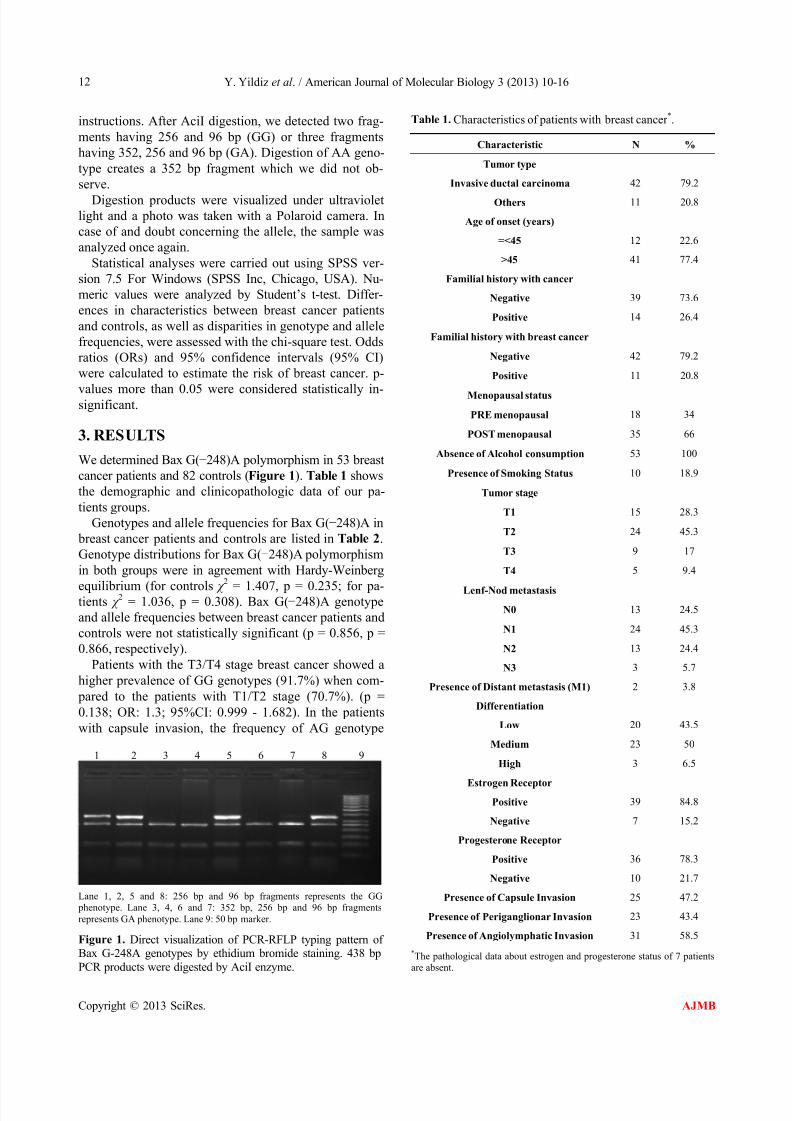
instructions. After AciI digestion, we detected two frag-
ments having 256 and 96 bp (GG) or three fragments
having 352, 256 and 96 bp (GA). Digestion of AA geno-
type creates a 352 bp fragment which we did not ob-
serve.
Digestion products were visualized under ultravioletlight and a photo was taken with a Polaroid camera. In
case of and doubt concerning the allele, the sample was
analyzed once again.
Statistical analyses were carried out using SPSS ver-
sion 7.5 For Windows (SPSS Inc, Chicago, USA). Nu-
meric values were analyzed by Student’s t-test. Differ-
ences in characteristics between breast cancer patients
and controls, as well as disparities in genotype and allele
frequencies, were assessed with the chi-square test. Odds
ratios (ORs) and 95% confidence intervals (95% CI)
were calculated to estimate the risk of breast cancer. p-
values more than 0.05 were considered statistically in-significant.
3. RESULTS
We determined Bax G(−248)A polymorphism in 53 breast
cancer patients and 82 controls (Figure 1). Table 1 shows
the demographic and clinicopathologic data of our pa-
tients groups.
Genotypes and allele frequencies for Bax G(−248)A in
breast cancer patients and controls are listed in Table 2.
Genotype distributions for Bax G(−248)A polymorphism
in both groups were in agreement with Hardy-Weinberg
equilibrium (for controls χ
2
= 1.407, p = 0.235; for pa-tients χ
2 = 1.036, p = 0.308). Bax G(−248)A genotype
and allele frequencies between breast cancer patients and
controls were not statistically significant (p = 0.856, p =
0.866, respectively).
Patients with the T3/T4 stage breast cancer showed a
higher prevalence of GG genotypes (91.7%) when com-
pared to the patients with T1/T2 stage (70.7%). (p =
0.138; OR: 1.3; 95%CI: 0.999 - 1.682). In the patients
with capsule invasion, the frequency of AG genotype
1 2 3 4 5 6 7 8 9
Lane 1, 2, 5 and 8: 256 bp and 96 bp fragments represents the GG
phenotype. Lane 3, 4, 6 and 7: 352 bp, 256 bp and 96 bp fragments
represents GA phenotype. Lane 9: 50 bp marker.
Figure 1. Direct visualization of PCR-RFLP typing pattern ofBax G-248A genotypes by ethidium bromide staining. 438 bp
PCR products were digested by AciI enzyme.
Table 1. Characteristics of patients with breast cancer *.
Characteristic N %
Tumor type
Invasive ductal carcinoma 42 79.2
Others 11 20.8
Age of onset (years)
=<45 12 22.6
>45 41 77.4
Familial history with cancer
Negative 39 73.6
Positive 14 26.4
Familial history with breast cancer
Negative 42 79.2
Positive 11 20.8
Menopausal status
PRE menopausal 18 34
POST menopausal 35 66
Absence of Alcohol consumption 53 100
Presence of Smoking Status 10 18.9
Tumor stage
T1 15 28.3
T2 24 45.3
T3 9 17
T4 5 9.4
Lenf-Nod metastasis
N0 13 24.5
N1 24 45.3
N2 13 24.4
N3 3 5.7
Presence of Distant metastasis (M1) 2 3.8
Differentiation
Low 20 43.5
Medium 23 50
High 3 6.5
Estrogen Receptor
Positive 39 84.8
Negative 7 15.2
Progesterone Receptor
Positive 36 78.3
Negative 10 21.7
Presence of Capsule Invasion 25 47.2
Presence of Periganglionar Invasion 23 43.4
Presence of Angiolymphatic Invasion 31 58.5
*The pathological data about estrogen and progesterone status of 7 patients
are absent.
Copyright © 2013 SciRes. AJMB
7/17/2019 AJMB_2013012311303349
http://slidepdf.com/reader/full/ajmb2013012311303349 4/7
Y. Yildiz et al . / American Journal of Molecular Biology 3 (2013) 10-16
Copyright © 2013 SciRes.
13
AJMB
(32%) was higher than those without capsule invasion
(17.9%), however the difference was not statistically
significant (p = 0.232; OR: 1.792; 95% CI: 0.674 -
4.768). We observed that two of the fifty-three patient,
who has distant metastasis, both have the GG genotypes.
In patients with angiolymphatic invasion, GG genotype
frequency (83.9%) was higher than in patients without
vascular invasion (63.6) but this is not statistically sig-
nificant (p = 0.092). Distributions of Bax G(−248)A
genotypes according to clinical parameters and tumor
characteristics of breast cancer patients are summarized
in Table 3. Among breast cancer patients, there were no
Table 2. Genotypes and allele frequencies for Bax G(−248A) in breast patients and controls.
Genotype Patients n = 53 % Controls n = 82 % #OR (95 % Cl) * p value
GG 40 75.5 63 76.8 0.982 (0.809 - 1.193) 0.856
GA 13 24.5 19 23.2 1.059 (0.572 - 1.958) 0.856
AA 0 0 0 0 - -
Alleles
G 93 87.7 145 88.4 0.866
A 13 12.3 19 11.6
*
p-value was obtained by chi-square test.
#
Odds ratio for genotype was calculated as selected genotype vs. Other genotypes. OR indicates crude odds ratio.
Table 3. Distribution of Bax (G−248A) genotypes with clinicopathological features in breast cancer patients*.
Histopathological parameters N % GG (%) GA (%)
Tumor Stage
T1, T2 41 77.3 29 (70.7) 12 (29.3)
T3, T4 12 22.7 11 (91.7) 1 (8.3)
Lyph node status
N(+) 37 69.8 27 (73) 10 (27)
N(−) 16 30.2 13 (81.3) 3 (18.8)
Distant metastasis
M(+) 2 3.8 2 (%100) 0
M(−) 51 96.2 38 (74.5) 13 (25.5)
Oestrogen receptor
Negative 7 15.2 5 (71.4) 2 (28.6)
Positive 39 84.8 28 (71.8) 11 (28.2)
Progesterone receptor
Negative 10 21.7 6 (60) 4 (40)
Positive 36 78.3 27 (75) 9 (25)
Capsule Invasion
Negative 28 52.8 23 (82.1) 5 (17.9)
Positive 25 47.2 17 (68) 8 (32)
Periganglionar Invasion
Negative 30 56.6 22 (73.3) 8 (26.7)
Positive 23 43.4 18 (78.3) 5 (21.7)
Angiolymphatic Invasion
Negative 22 41.5 14 (63.6) 8 (36.4)
Positive 31 58.5 26 (83.9) 5 (16.1)
Differentiation
Low 20 43.5 15 (75) 5 (25)
High 26 56.5 19 (73.1) 7 (26.9)
*AA genotype was not detected in study groups.
7/17/2019 AJMB_2013012311303349
http://slidepdf.com/reader/full/ajmb2013012311303349 5/7
Y. Yildiz et al . / American Journal of Molecular Biology 3 (2013) 10-1614
significant association between the Bax G(−248)A geno-
types and some clinical parameters including age at di-
agnosis, family history, menopausal status, smoking,
using alcohol and some pathological parameters such as
tumor stage, lymph node metastasis, distant metastasis ,
periganglionar invasion, estrogen and progesterone re-
ceptor status and also tumor differentiation (data are not
shown).
4. DISCUSSION
Bax gene which is a member of Bcl-2 family is an im-
portant regulator of apoptosis. Although altered expres-
sion of Bax protein seems to be more relevant with car-
cinogenesis process, mutations leading this disregulation
and correlations of these mutations with different cancers
are hotspots among the researchers. Promotor region of
the Bax gene contains p53 response elements thus affects
gene expression [31].
Different mutations of Bax gene have been examined
in different studies so far. Saxana et al . defined the
G(−248)A SNP in chronic lymphocytic leukemia (CLL)
in 2002 and suggested a relation between disease pro-
gression and treatment resistance with 34 patients [30].
In control group, genotypic frequencies were 94.3% and
5.7% for GG and GA genotypes; and allelic frequencies
were 0.97 and 0.03 for G and A alleles respectively. Al-
though the differences in distribution of alleles; absence
of AA genotype neither in control nor in patient group,
supports our findings.
In a more recent study, Skogsberg et al . used a biggerstudy group (463 cases and 207 controls) and found no
significant differences between Bax G(−248)A polymer-
phism and CLL progression or treatment resistant [36].
Genotyping frequencies in control group were 79%, 19%
and 2% for GG, GA and AA genotypes respectively,
again indicating a rare presence of A allele. These results
are highly relevant to our findings (76.8% GG, 23.2%
GA, 0%AA; n = 82) if we consider the number of sub-
jects that has been examined. It is possible to get more
similar results with a higher number of subject.
In this study, we did not record a statistically signifi-
cant difference in Bax G(−248)A genotype and allele
frequencies between breast cancer patients and control.
However we observed that some of the clinic-patho-
logical data may be related with specific genotypes.
GG genotype frequency (83.9%) was higher in pa-
tients with angiolymphatic invasion when compared to
those without (63.6%). These results suggest that GG
genotype in the BAX promoter region may increase an-
giolymphatic invasion in breast cancer and consequently
increase metastatic spread and be an adverse prognostic
factor.
A similar relationship was also observed in patients
carrying the Bax AG genotype, with 1.79 fold increased
risk for capsule invasion. Capsular invasion at metastatic
nodes in breast cancer was shown to be strongly associ-
ated with further regional nodes metastasis [37,38]. Thus,
such a possible increased risk may be related to breast
cancer progression, specifically in later stages of the dis-
ease.
This paper represents the first study on Bax G(−245)A
polymorphism in breast cancer and gives an opinion
about the possible effects of this polymorphism in breast
cancer progression. However further validations in larger
studies are needed to clarify the role of G(−248)A poly-
morphism in breast cancer.
5. ACKNOWLEDGEMENTS
This study was supported by a grant from Istanbul University, Research
Foundation (project number: 1715), Turkey.
REFERENCES
[1] Siegel, R., Naishadham, D. and Jemal, A. (2012) Cancer
statistics. CA: A Cancer Journal for Clinicians, 62, 10-29.
doi:10.3322/caac.20138
[2] Ashkenazi, A. and Dixit, V.M. (1998) Death receptors:
Signaling and modulation. Science, 281, 1305-1308.
doi:10.1126/science.281.5381.1305
[3] Yildiz, Y., Yaylim-Eraltan, I., Arikan, S., Ergen, H.A.,
Küçücük, S. and Isbir, T. (2010) Is there any correlation
between TNF-related apoptosis-inducing ligand (TRAIL)
genetic variants and breast cancer? Archives of Medical
Science, 6, 932-936. doi:10.5114/aoms.2010.19304 [4]
Susin, S.A., Lorenzo, H.K., Zamzami, N., Marzo, I., Snow,
B.E., Brothers, G.M., Mangion, J., Jacotot, E., Costantini,
P., Loeffler, M., Larochette, N., Goodlett, D.R., Aeber-
sold, R., Siderovski, D.P., Penninger, J.M. and Kroemer,
G. (1999) Molecular characterization of mitochondrial
apoptosis-inducing factor. Nature, 397, 441-446.
doi:10.1038/17135
[5]
Du, C., Fang, M., Li, Y., Li, L. and Wang, X. (2000)
Smac, a mitochondrial protein that promotes cytochrome
c-dependent caspase activation by eliminating IAP inhi-
bition. Cell , 102, 33-42.
doi:10.1016/S0092-8674(00)00008-8
[6] Verhagen, A.M., Ekert, P.G., Pakusch, M., Silke, J., Con-
nolly, L.M., Reid, G.E., Moritz, R.L., Simpson, R.J. and
Vaux, D.L. (2000) Identification of DIABLO, a mamma-
lian protein that promotes apoptosis by binding to and
antagonizing IAP proteins. Cell , 102, 43-53.
doi:10.1016/S0092-8674(00)00009-X
[7] Li, K., Li, Y., Shelton, J.M., Richardson, J.A., Spencer,
E., Chen, Z.J., Wang, X. and Williams, R.S. (2000) Cyto-
chrome c deficiency causes embryonic lethality and at-
tenuates stress-induced apoptosis. Cell ,101, 389-399.
doi:10.1016/S0092-8674(00)80849-1
[8] Zhang, L., Yu, J., Park, B.H., Kinzler, K.W. and Vogel-
stein, B. (2000) Role of BAX in the apoptotic response to
anticancer agents. Science, 290, 989-992.
Copyright © 2013 SciRes. AJMB

7/17/2019 AJMB_2013012311303349
http://slidepdf.com/reader/full/ajmb2013012311303349 6/7
Y. Yildiz et al . / American Journal of Molecular Biology 3 (2013) 10-16 15
doi:10.1126/science.290.5493.989
[9] Miyashita, T. and Reed, J.C. (1995) Tumor suppressor p53 is a direct transcriptional activator of the human bax
gene. Cell , 80, 293-299.doi:10.1016/0092-8674(95)90412-3
[10]
Chen, K., Hu, Z., Wang, L.E., Sturgis, E.M., El-Naggar,A.K., Zhang, W. and Wei, Q. (2007) Single-nucleotide polymorphisms at the TP53-binding or responsive pro-moter regions of BAX and BCL2 genes and risk of squa-
mous cell carcinoma of the head and neck. Carcino- genesis, 28, 2008-2012.
doi:10.1093/carcin/bgm172
[11] Rampino, N., Yamamoto, H., Ionov, Y., Li, Y., Sawai, H.,Reed, J.C. and Perucho, M. (1997) Somatic frameshiftmutations in the BAX gene in colon cancers of the micro-satellite mutator phenotype. Science, 275, 967-969.doi:10.1126/science.275.5302.967
[12] Meijerink, J.P., Mensink, E.J., Wang, K., Sedlak, T.W.,Slöetjes, A.W., de Witte, T., Waksman, G. and Korsme-yer, S.J. (1998) Hematopoietic malignancies demonstrateloss-of-function mutations of BAX. Blood , 91, 2991-2997.
[13] Adams, J.M. and Cory, S. (1998) The Bcl-2 protein family:Arbiters of cell survival. Science, 281, 1322-1326.doi:10.1126/science.281.5381.1322
[14] Yin, X.M., Oltvai, Z.N. and Korsmeyer, S.J. (1994) BH1and BH2 domains of Bcl-2 are required for inhibition ofapoptosis and heterodimerization with Bax. Nature, 369,321-323. doi:10.1038/369321a0
[15] Zha, H., Aimé-Sempé, C., Sato, T. and Reed, J.C. (1996)Proapoptotic protein Bax heterodimerizes with Bcl-2 andhomodimerizes with Bax via a novel domain (BH3) dis-tinct from BH1 and BH2. The Journal of Biological Che-
mistry, 271, 7440-7444. doi:10.1074/jbc.271.13.7440 [16] Peng, H., Aiello, A., Packham, G., Isaacson, P.G. and Pan,
L. (1998) Infrequent bax gene mutations in B-cell lym- phomas. The Journal of Pathology, 186, 378-382.
doi:10.1002/(SICI)1096-9896(199812)186:4<378::AID-PATH203>3.0.CO;2-5
[17] Meijerink, J.P., Smetsers, T.F., Slöetjes, A.W., Linders,
E.H. and Mensink, E.J. (1995) Bax mutations in cell linesderived from hematological malignancies. Leukemia, 9,1828-1832.
[18] Yamamoto, H., Sawai, H. and Perucho, M. (1997) Frame-
shift somatic mutations in gastrointestinal cancer of themicrosatellite mutator phenotype. Cancer Reserch, 57,
4420-4426.[19] Brimmell, M., Mendiola, R., Mangion, J. and Packham,
G. (1998) BAX frameshift mutations in cell lines derivedfrom human haemopoietic malignancies are associatedwith resistance to apoptosis and microsatellite instability.Oncogene, 16, 1803-1812.doi:10.1038/sj.onc.1201704
[20] Gaidano, G., Vivenza, D., Forconi, F., Capello, D., Glo-ghini, A., Bhatia, K., Gutierrez, M., Gallicchio, M., Avan-
zi, G.C., Fassone, L., Ariatti, C., Buonaiuto, D., Cingo-lani, A., Saglio, G., Tirelli, U., Larocca, L.M., Dalla-Favera, R. and Carbone, A. (2000) Mutation of BAXoccurs infrequently in acquired immunodeficiency syn-drome-related non-Hodgkin’s lymphomas. Genes Chro-
mosomes Cancer , 27, 177-182.doi:10.1002/(SICI)1098-2264(200002)27:2<177::AID-GCC9>3.0.CO;2-O
[21] Yashiro, M., Hirakawa, K. and Boland, C.R. (2010) Mu-tations in TGFbeta-RII and BAX mediate tumor progres-
sion in the later stages of colorectal cancer with micro-satellite instability. BMC Cancer , 10, 303.doi:10.1186/1471-2407-10-303
[22] Shima, K., Morikawa, T., Yamauchi, M., Kuchiba, A.,Imamura, Y., Liao, X., Meyerhardt, J.A., Fuchs, C.S. andOgino, S. (2011) TGFBR2 and BAX mononucleotidetract mutations, microsatellite instability, and prognosisin 1072 colorectal cancers. PLoS One, 6, e25062.doi:10.1371/journal.pone.0025062
[23] Oliveira, C., Seruca, R., Seixas, M. and Sobrinho-Simões,M. (1998) The clinicopathological features of gastric car-cinomas with microsatellite instability may be mediated by mutations of different “target genes”: A study of theTGFbeta RII, IGFII R, and BAX genes. The American
Journal of Pathology, 153, 1211-1219.doi:10.1016/S0002-9440(10)65665-9
[24] Iacopetta, B.J., Soong, R., House, A.K. and Hamelin, R.(1999) Gastric carcinomas with microsatellite instability:Clinical features and mutations to the TGF-beta type IIreceptor, IGFII receptor, and BAX genes. The Journal of Pathology, 187, 428-432.doi:10.1002/(SICI)1096-9896(199903)187:4<428::AID-PATH264>3.0.CO;2-A
[25] Cho, S., Hahm, J.H. and Hong, Y.S. (2001) Analysis of p53 and BAX mutations, loss of heterozygosity, p53 andBCL2 expression and apoptosis in basal cell carcinoma inKorean patients. British Journal of Dermatology, 144,841-848. doi:10.1046/j.1365-2133.2001.04142.x
[26]
Addeo, R., Crisci, S., D’Angelo, V., Vincenzi, B., Casale,F., Pettinato, G., Donofrio, V., Boldrini, R., Alaggio, R.,Collini, P., Bertorelle, R., Di Tullio, M.T., Caraglia, M.,Terenziani, M., Lo Curto, M. and Indolfi, P. (2007) Baxmutation and overexpression inversely correlate with im-mature phenotype and prognosis of childhood germ celltumors. Oncology Reports, 17, 1155-1161.
[27] Krajewski, S., Blomqvist, C., Franssila, K., Krajewska,M., Wasenius, V.M., Niskanen, E., Nordling, S. and Reed,J.C. (1995) Reduced expression of proapoptotic geneBAX is associated with poor response rates to combi-nation chemotherapy and shorter survival in women withmetastatic breast adenocarcinoma. Cancer Reserch, 55,4471-4478.
[28] Gascoyne, R.D., Krajewska, M., Krajewski, S., Connors,J.M. and Reed, J.C. (1997) Prognostic significance ofBax protein expression in diffuse aggressive non-Hodg-kin’s lymphoma. Blood , 90, 3173-3178.
[29] Tai, Y.T., Lee, S., Niloff, E., Weisman, C., Strobel, T.and Cannistra, S.A. (1998) BAX protein expression andclinical outcome in epithelial ovarian cancer. Journal ofClinical Oncology, 16, 2583-2590
[30]
Saxena, A., Moshynska, O., Sankaran, K., Viswanathan,S. and Sheridan, D.P. (2002) Association of a novelsingle nucleotide polymorphism, G(-248)A, in the 5’-UTR of BAX gene in chronic lymphocytic leukemia withdisease progression and treatment resistance. Cancer
Copyright © 2013 SciRes. AJMB
7/17/2019 AJMB_2013012311303349
http://slidepdf.com/reader/full/ajmb2013012311303349 7/7
Y. Yildiz et al . / American Journal of Molecular Biology 3 (2013) 10-16
Copyright © 2013 SciRes.
16
AJMB
Letters, 10, 187, 199-205.doi:10.1016/S0304-3835(02)00378-6
[31] Moshynska, O., Moshynskyy, I., Misra, V. and Saxena, A.(2005) G125A single nucleotide polymorphism in the hu-
man BAX promoter affects gene expression. Oncogene,
24, 2042-2049. doi:10.1038/sj.onc.1208377 [32] Lahiri, O., Harris, S., Packham, G. and Howell, M. (2007)
p53 pathway gene single nucleotide polymorphisms and
chronic lymphocytic leukemia. Cancer Genetics and Cyto- genetics, 179, 36-44.doi:10.1016/j.cancergencyto.2007.07.013
[33] Semaan, S.J., Li, Y. and Nickells, R.W. (2010) A singlenucleotide polymorphism in the Bax gene promoter af-
fects transcription and influences retinal ganglion celldeath. ASN Neuro, 2, e00032. doi:10.1042/AN20100003
[34] Farnebo, L., Jedlinski, A., Ansell, A., Vainikka, L., Thu-nell, L.K., Grénman, R., Johansson, A.C. and Roberg, K.
(2009) Proteins and single nucleotide polymorphisms in-volved in apoptosis,growth control, and DNA repair pre-
dict cisplatin sensitivity in head and neck cancer cell lines. International Journal of Molecular Medicine, 24, 549-
556.
[35]
Miller, S.A., Dykes, D.D. and Polesky, H.F. (1988) A
simple salting out procedure for extracting DNA fromhuman nucleated cells. Nucleic Acids Research, 16, 1215.doi:10.1093/nar/16.3.1215
[36] Skogsberg, S., Tobin, G., Kröber, A., Kienle, D., Thun- berg, U., Aleskog, A., Karlsson, K., Laurell, A., Merup,
M., Vilpo, J., Sundström, C., Roos, G., Jernberg-Wiklund,H., Döhner, H., Nilsson, K., Stilgenbauer, S. and Rosen-
quist, R. (2006) The G(-248)A polymorphism in the pro-moter region of the Bax gene does not correlate with
prognostic markers or overall survival in chronic lym- phocytic leukemia. Leukemia, 20, 77-81.
doi:10.1038/sj.leu.2404030
[37]
Fujii, T., Tabe, Y., Yajima, R., Yamaguchi, S., Tsutsumi,
S., Asao, T. and Kuwano, H. (2011) Extracapsular inva-sion as a risk factor for disease recurrence in colorectal
cancer. World Journal of Gastroenterology, 17, 2003-2006. doi:10.3748/wjg.v17.i15.2003
[38] Fujii, T., Yanagita, Y., Fujisawa, T., Hirakata, T., Iijima,M. and Kuwano, H. (2010) Implication of extracapsular
invasion of sentinel lymph nodes in breast cancer: Pre-diction of nonsentinel lymph node metastasis. World
Journal of Surgery, 34, 544-548.doi:10.1007/s00268-009-0389-4


