Acute Limb Ischemia - prd-medweb … · • Up to 15% of cases of acute limb ischemia occur in...
58
Acute Limb Ischemia R. J. Guzman M. J. Osgood 10/7/11
-
Upload
truongtuong -
Category
Documents
-
view
218 -
download
1
Transcript of Acute Limb Ischemia - prd-medweb … · • Up to 15% of cases of acute limb ischemia occur in...
Acute Limb Ischemia
R. J. Guzman M. J. Osgood
10/7/11
Outline
• Etiologies • Classification • Presentation • Workup/diagnosis • Management • Outcomes
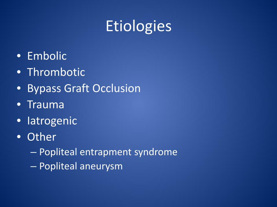
Etiologies
• Embolic • Thrombotic • Bypass Graft Occlusion • Trauma • Iatrogenic • Other
– Popliteal entrapment syndrome – Popliteal aneurysm
Embolic Sources
• Cardiac – Atrial – Ventricular - mural thrombus – Paradoxical – Endocarditis – Cardiac tumor (atrial myxoma)
• Noncardiac – Atheroembolism – Aortic mural thrombus
Embolism
• Emboli usually lodge at arterial bifurcations • Most common locations:
– Common femoral artery – Popliteal artery
• Embolic ischemia is usually poorly tolerated because it often occurs in non-diseased peripheral arteries without established collaterals
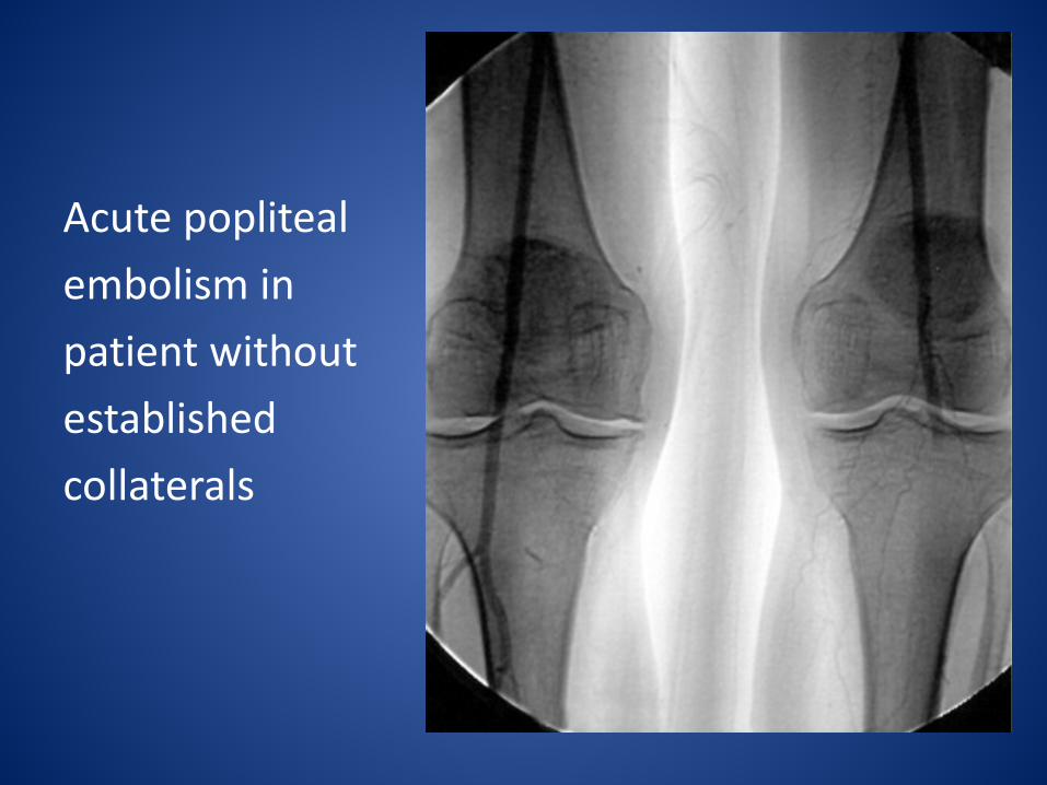
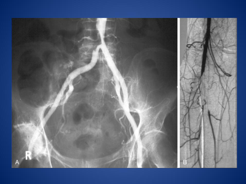
Acute popliteal embolism in patient without established collaterals
Embolism
• Embolic ischemia is progressive because thrombus propagates proximal and distal to the embolus

Embolism • Embolic ischemia is progressive because
thrombus propagates proximal and distal to the embolus
Thrombotic Sources
• Atherosclerotic obstruction • Hypercoagulable states • Aortic or arterial dissection
Atherosclerotic obstruction
• Acute thrombosis of a narrowed, atherosclerotic peripheral artery
• Usually better tolerated because involved vessel has well-developed collaterals
• May be asymptomatic or present as acute claudication or rest pain
• May result from plaque disruption or from global reduction in cardiac output in patients with atherosclerotic burden
Hypercoagulability
• Inherited and acquired thrombophilias • Low arterial flow (relative stasis) • Hyperviscosity • Malignancy • Thrombocytosis / thrombocythemia /
myeloproliferative disorders • HIT
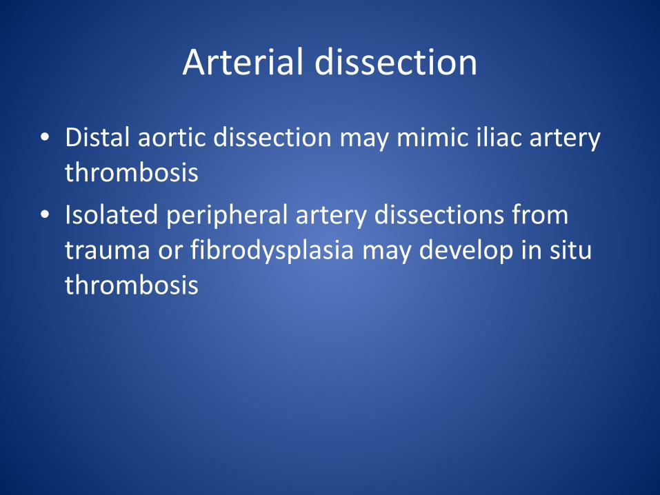
Arterial dissection
• Distal aortic dissection may mimic iliac artery thrombosis
• Isolated peripheral artery dissections from trauma or fibrodysplasia may develop in situ thrombosis
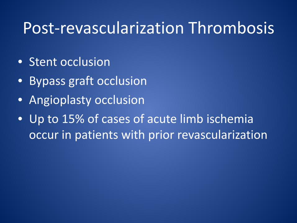
Post-revascularization Thrombosis
• Stent occlusion • Bypass graft occlusion • Angioplasty occlusion • Up to 15% of cases of acute limb ischemia
occur in patients with prior revascularization
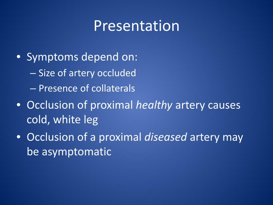
Presentation
• Symptoms depend on: – Size of artery occluded – Presence of collaterals
• Occlusion of proximal healthy artery causes cold, white leg
• Occlusion of a proximal diseased artery may be asymptomatic
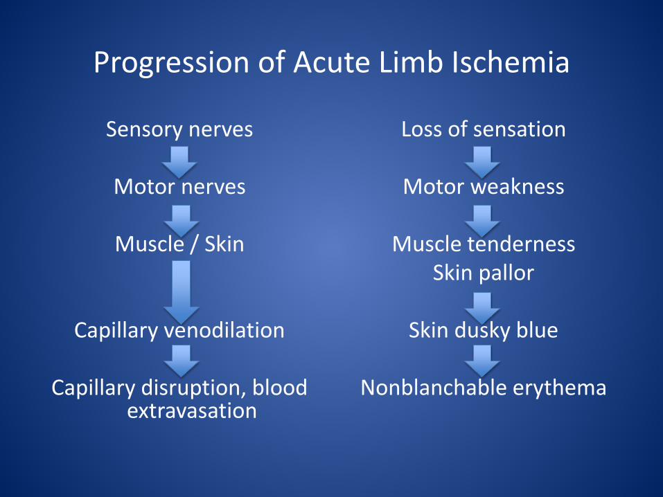
Progression of Acute Limb Ischemia
Sensory nerves
Motor nerves
Muscle / Skin
Capillary venodilation
Capillary disruption, blood extravasation
Loss of sensation
Motor weakness
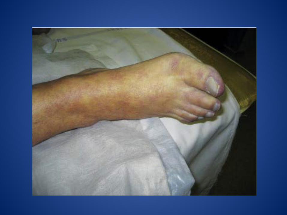
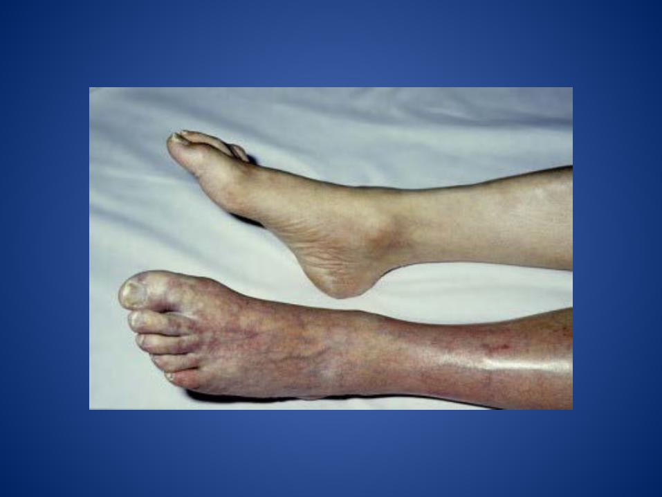
Muscle tenderness Skin pallor
Skin dusky blue
Nonblanchable erythema
Clinical Assessment
• **** HPI **** • Past medical history • Family history • Physical exam • Hand-held doppler signals
HPI
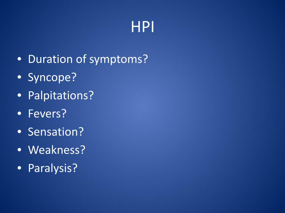
• Duration of symptoms? • Syncope? • Palpitations? • Fevers? • Sensation? • Weakness? • Paralysis?
PMHx / Social Hx / Family Hx
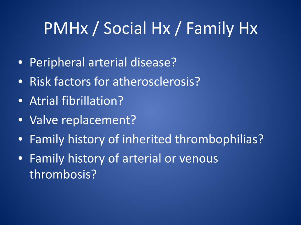
• Peripheral arterial disease? • Risk factors for atherosclerosis? • Atrial fibrillation? • Valve replacement? • Family history of inherited thrombophilias? • Family history of arterial or venous
thrombosis?
Physical Exam
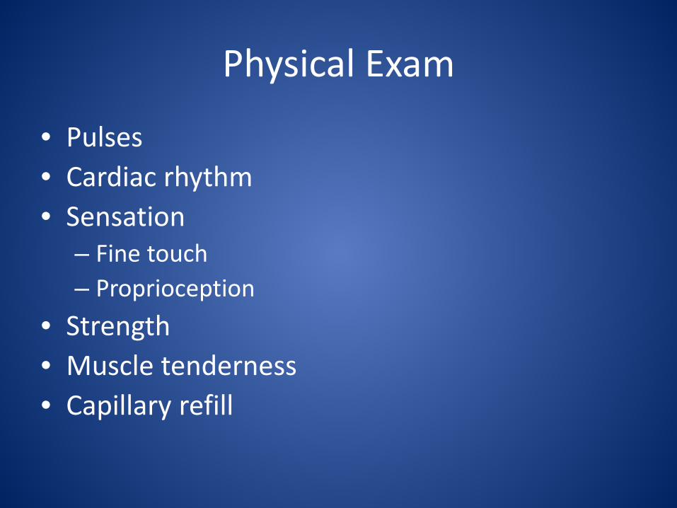
• Pulses • Cardiac rhythm • Sensation
– Fine touch – Proprioception
• Strength • Muscle tenderness • Capillary refill
Hand-held dopplers
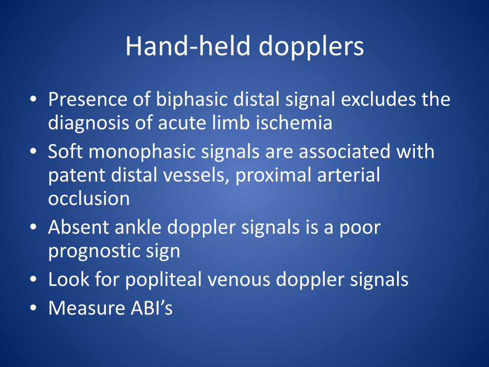
• Presence of biphasic distal signal excludes the diagnosis of acute limb ischemia
• Soft monophasic signals are associated with patent distal vessels, proximal arterial occlusion
• Absent ankle doppler signals is a poor prognostic sign
• Look for popliteal venous doppler signals • Measure ABI’s
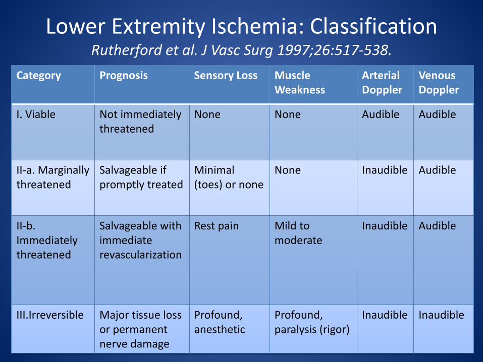
Lower Extremity Ischemia: Classification Rutherford et al. J Vasc Surg 1997;26:517-538.
Category Prognosis Sensory Loss Muscle Weakness
Arterial Doppler
Venous Doppler
I. Viable Not immediately threatened
None None Audible Audible
II-a. Marginally threatened
Salvageable if promptly treated
Minimal (toes) or none
None Inaudible Audible
II-b. Immediately threatened
Salvageable with immediate revascularization
Rest pain Mild to moderate
Inaudible
Audible
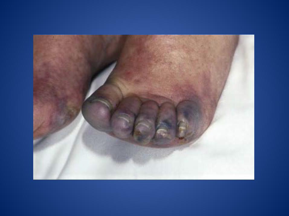
III.Irreversible Major tissue loss or permanent nerve damage
Profound, anesthetic
Profound, paralysis (rigor)
Inaudible
Inaudible

Diagnosis: Level of Occlusion
• Aortic occlusion presents with mottled bilateral lower extremities, bilateral leg paralysis, loss of pulses in BLE
• Iliac occlusion resembles aortic occlusion, but is unilateral
• Femoropopliteal occlusion is most common; degree of symptoms depends on involvement of profunda
• Popliteal or infrapopliteal occlusion presents with ischemic calf muscles and intact femoral pulse (i.e., popliteal entrapment syndrome, popliteal aneurysm)
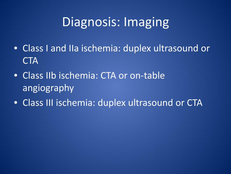
Diagnosis: Imaging
• Class I and IIa ischemia: duplex ultrasound or CTA
• Class IIb ischemia: CTA or on-table angiography
• Class III ischemia: duplex ultrasound or CTA
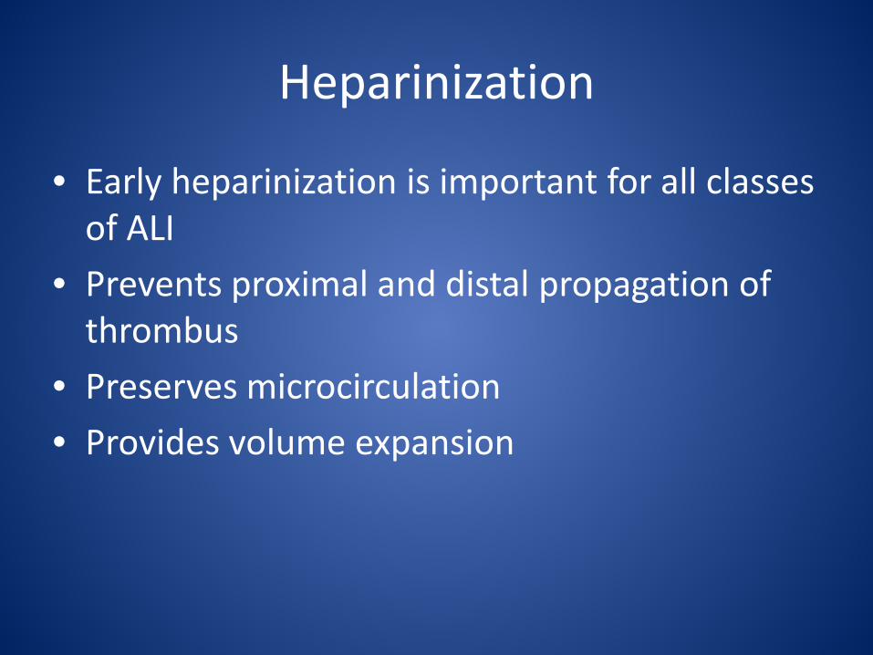
Heparinization
• Early heparinization is important for all classes of ALI
• Prevents proximal and distal propagation of thrombus
• Preserves microcirculation • Provides volume expansion
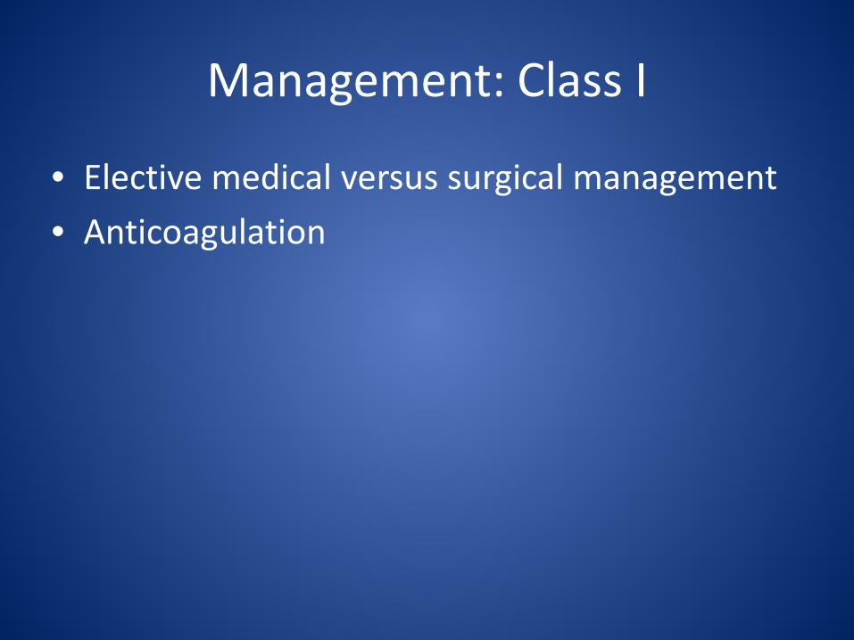
Management: Class I
• Elective medical versus surgical management • Anticoagulation
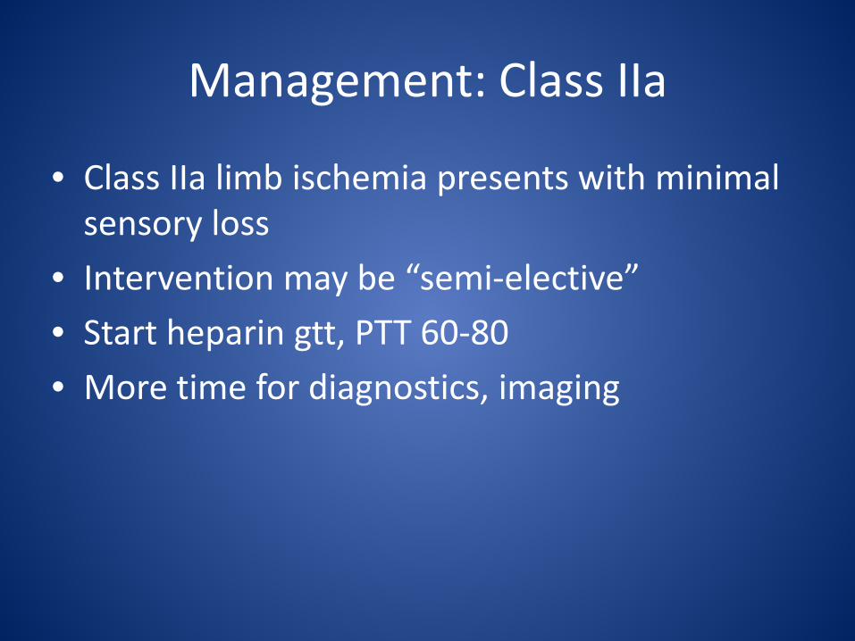
Management: Class IIa
• Class IIa limb ischemia presents with minimal sensory loss
• Intervention may be “semi-elective” • Start heparin gtt, PTT 60-80 • More time for diagnostics, imaging
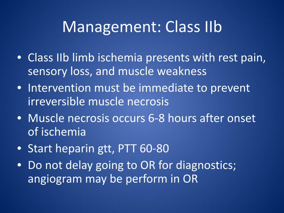
Management: Class IIb
• Class IIb limb ischemia presents with rest pain, sensory loss, and muscle weakness
• Intervention must be immediate to prevent irreversible muscle necrosis
• Muscle necrosis occurs 6-8 hours after onset of ischemia
• Start heparin gtt, PTT 60-80 • Do not delay going to OR for diagnostics;
angiogram may be perform in OR
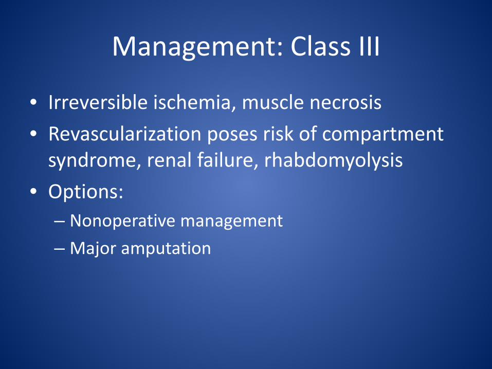
Management: Class III
• Irreversible ischemia, muscle necrosis • Revascularization poses risk of compartment
syndrome, renal failure, rhabdomyolysis • Options:
– Nonoperative management – Major amputation
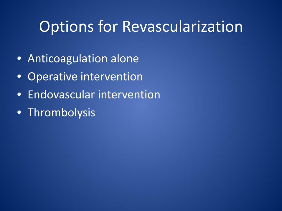
Options for Revascularization
• Anticoagulation alone • Operative intervention • Endovascular intervention • Thrombolysis
Anticoagulation
• Safe and effective for Class I ischemia and may be definitive therapy in this group
• Improves results after embolectomy (i.e., Class II ischemia)
• Allows stabilization of patient in Class III ischemia while awaiting definitive management (i.e., major amputation)
Operative Intervention
• Embolectomy or thrombectomy with balloon embolectomy catheters
• Endarterectomy • Vascular reconstruction using bypass graft in
patients with coexisting PAD

Fasciotomy
• Any patient with early motor changes (Rutherford class IIb or III) should have fasciotomy after open surgical revascularization or successful PMT
• If no fasciotomy performed, patient must be closely monitored for compartment syndrome
Endovascular: Percutaneous Mechanical Thrombectomy
• Hydrodynamic: removes thrombus from the peripheral arteries using a stream of fluid and hydrodynamic forces to extract the thrombotic material from the lumen (AngioJet, Hydrolyser, Oasis catheters)
• Rotational: fragment thrombus without actually aspirating the fragments have been designed to establish arterial recanalization
Catheter-Directed Thrombolysis
• Compared with surgical thrombectomy, thrombolytics have the advantage of lysing clots in large arteries, distal smaller arteries, arterioles, and capillaries
• Good option for Class I and IIa ALI • Good option when runoff vessels appear
occluded with thrombus • All available thrombolytic agents are plasminogen
activators • Achieves regional thrombus dissolution with
minimal systemic fibrinolysis
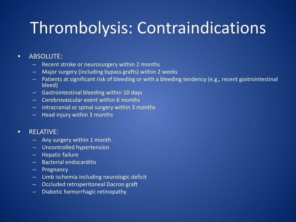
Thrombolysis: Contraindications • ABSOLUTE:
– Recent stroke or neurosurgery within 2 months – Major surgery (including bypass grafts) within 2 weeks – Patients at significant risk of bleeding or with a bleeding tendency (e.g., recent gastrointestinal
bleed) – Gastrointestinal bleeding within 10 days – Cerebrovascular event within 6 months – Intracranial or spinal surgery within 3 months – Head injury within 3 months
• RELATIVE:
– Any surgery within 1 month – Uncontrolled hypertension – Hepatic failure – Bacterial endocarditis – Pregnancy – Limb ischemia including neurologic deficit – Occluded retroperitoneal Dacron graft – Diabetic hemorrhagic retinopathy

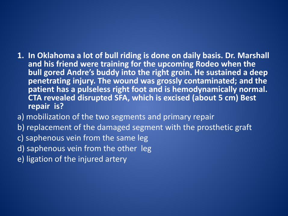
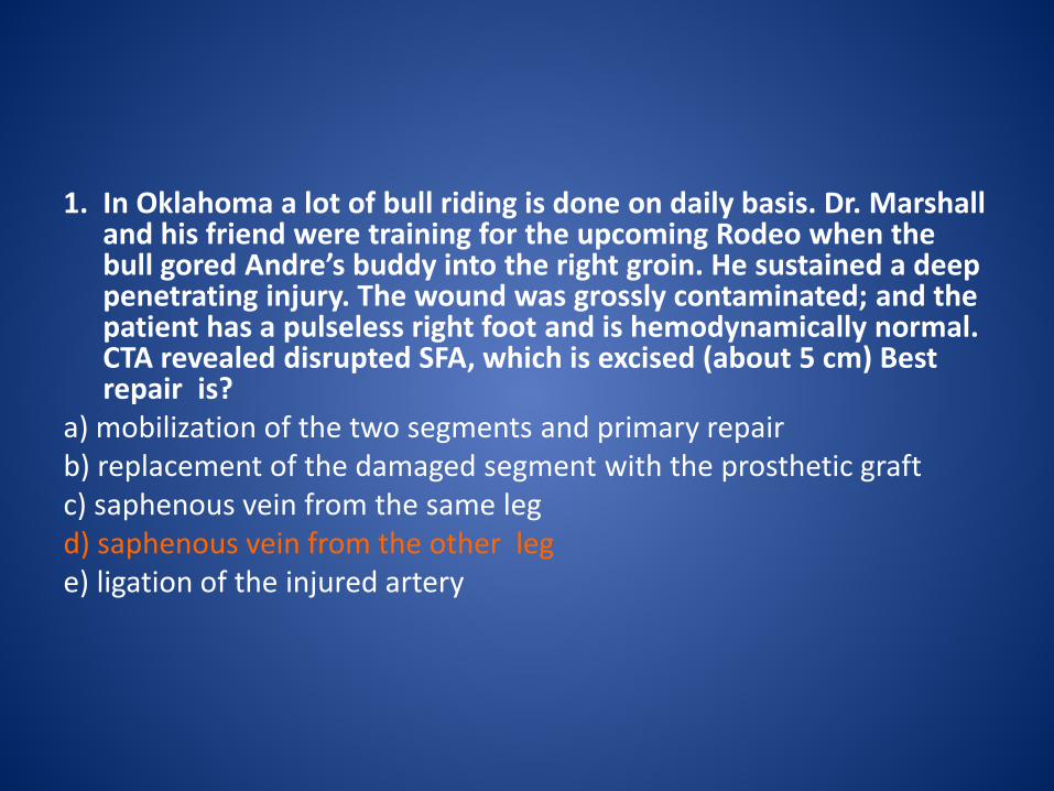
1. In Oklahoma a lot of bull riding is done on daily basis. Dr. Marshall and his friend were training for the upcoming Rodeo when the bull gored Andre’s buddy into the right groin. He sustained a deep penetrating injury. The wound was grossly contaminated; and the patient has a pulseless right foot and is hemodynamically normal. CTA revealed disrupted SFA, which is excised (about 5 cm) Best repair is?
a) mobilization of the two segments and primary repair b) replacement of the damaged segment with the prosthetic graft c) saphenous vein from the same leg d) saphenous vein from the other leg e) ligation of the injured artery
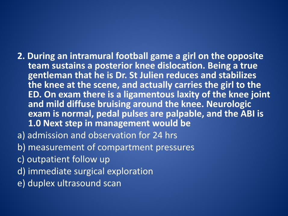
2. During an intramural football game a girl on the opposite team sustains a posterior knee dislocation. Being a true gentleman that he is Dr. St Julien reduces and stabilizes the knee at the scene, and actually carries the girl to the ED. On exam there is a ligamentous laxity of the knee joint and mild diffuse bruising around the knee. Neurologic exam is normal, pedal pulses are palpable, and the ABI is 1.0 Next step in management would be
a) admission and observation for 24 hrs b) measurement of compartment pressures c) outpatient follow up d) immediate surgical exploration e) duplex ultrasound scan
3. A patient with a history of atrial fibrillation with no history of PVD develops a cold painful right leg after forgetting to take his Coumadin. Pulses on the left side are normal. The most appropriate next step in management is
a) thrombolytics b) embolectomy c) arterial bypass d) do nothing

3. A patient with a history of atrial fibrillation with no history of PVD develops a cold painful right leg after forgetting to take his Coumadin. Pulses on the left side are normal. The most appropriate next step in management is
a) thrombolytics b) embolectomy c) arterial bypass d) do nothing
4. After successful treatment of the patient in question#3 his leg becomes swollen and he complains of pain on passive motion of the foot. The most appropriate next step is
a) thrombolytics b) emergent re-exploration with thrombectomy c) fasciotomy d) aggressive diuresis
4. After successful treatment of the patient in question#3 his leg becomes swollen and he complains of pain on passive motion of the foot. The most appropriate next step is
a) thrombolytics b) emergent re-exploration with thrombectomy c) fasciotomy d) aggressive diuresis
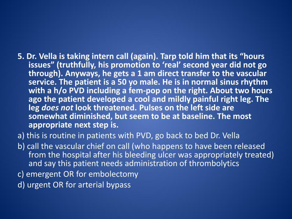
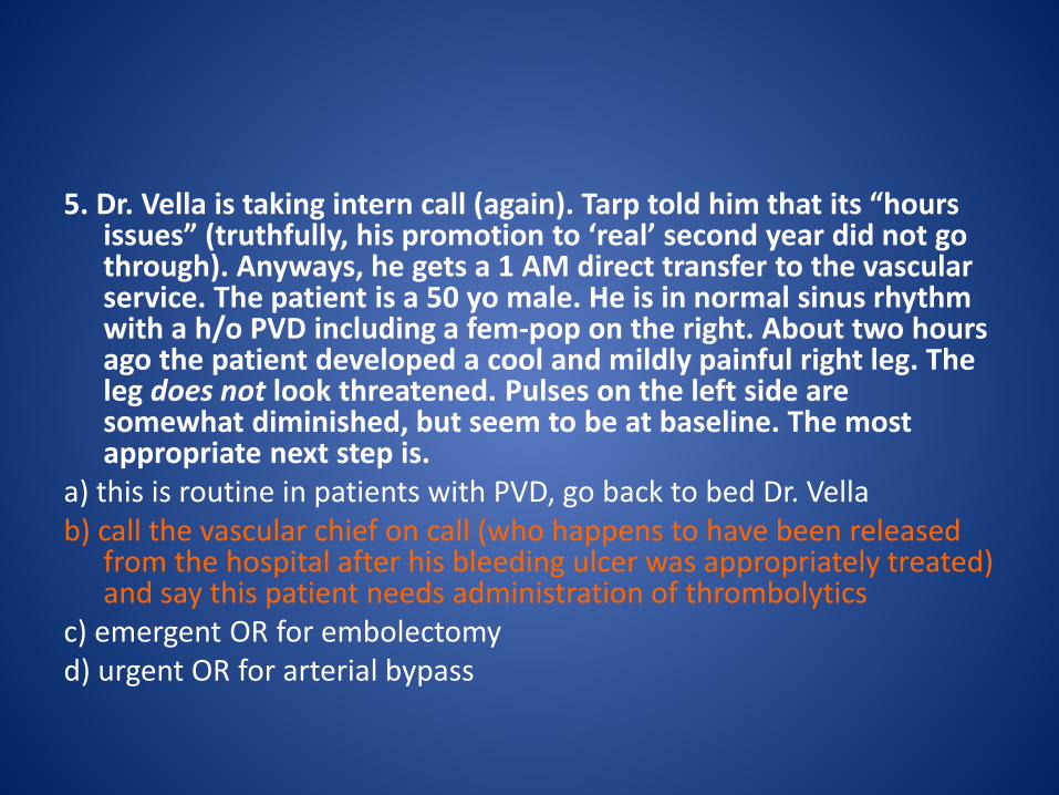
5. Dr. Vella is taking intern call (again). Tarp told him that its “hours issues” (truthfully, his promotion to ‘real’ second year did not go through). Anyways, he gets a 1 am direct transfer to the vascular service. The patient is a 50 yo male. He is in normal sinus rhythm with a h/o PVD including a fem-pop on the right. About two hours ago the patient developed a cool and mildly painful right leg. The leg does not look threatened. Pulses on the left side are somewhat diminished, but seem to be at baseline. The most appropriate next step is.
a) this is routine in patients with PVD, go back to bed Dr. Vella b) call the vascular chief on call (who happens to have been released
from the hospital after his bleeding ulcer was appropriately treated) and say this patient needs administration of thrombolytics
c) emergent OR for embolectomy d) urgent OR for arterial bypass
5. Dr. Vella is taking intern call (again). Tarp told him that its “hours issues” (truthfully, his promotion to ‘real’ second year did not go through). Anyways, he gets a 1 AM direct transfer to the vascular service. The patient is a 50 yo male. He is in normal sinus rhythm with a h/o PVD including a fem-pop on the right. About two hours ago the patient developed a cool and mildly painful right leg. The leg does not look threatened. Pulses on the left side are somewhat diminished, but seem to be at baseline. The most appropriate next step is.
a) this is routine in patients with PVD, go back to bed Dr. Vella b) call the vascular chief on call (who happens to have been released
from the hospital after his bleeding ulcer was appropriately treated) and say this patient needs administration of thrombolytics
c) emergent OR for embolectomy d) urgent OR for arterial bypass
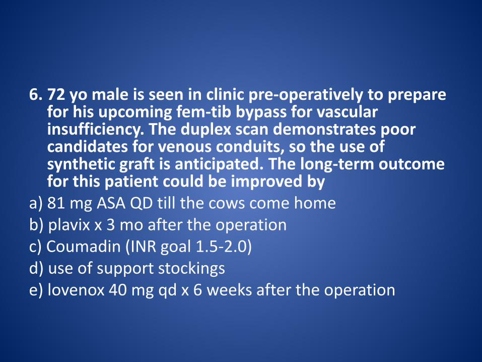
6. 72 yo male is seen in clinic pre-operatively to prepare for his upcoming fem-tib bypass for vascular insufficiency. The duplex scan demonstrates poor candidates for venous conduits, so the use of synthetic graft is anticipated. The long-term outcome for this patient could be improved by
a) 81 mg ASA QD till the cows come home b) plavix x 3 mo after the operation c) Coumadin (INR goal 1.5-2.0) d) use of support stockings e) lovenox 40 mg qd x 6 weeks after the operation
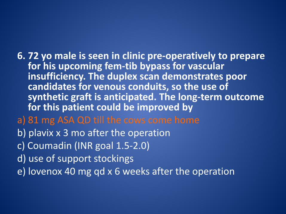
7. Its 2 am at the VA. The ER calls (freeeeaking out, as usual) about a 70 yo vet with an infected left second toe. He has been treated with antibiotics for 6 weeks, with no benefit. On exam he has a nonhealing infected ulcer on the volar aspect of his left second distal phalanx. The ulcer is purulent at the base. There is no ascending cellulitis. Patient is not toxic. Best management course is
a) iv abx b) single toe amputation c) single toe amputation and iv abx d) transmetatarsal amputation of the foot and iv abx e) guillotine BKA with delayed formalization and iv abx
8) Which of the following is not an immediate indication for an operative intervention?
a) pulsatile bleeding b) expanding hematoma c) arterial thrill by manual palpation over the
injured area d) loss of distal pulse e) auscultated bruit over the injured area f) diminished pulse compared to the contralateral
extremity
8) Which of the following is NOT an immediate indication for an operative intervention?
a) pulsatile bleeding b) expanding hematoma c) arterial thrill by manual palpation over the
injured area d) loss of distal pulse e) auscultated bruit over the injured area f) diminished pulse compared to the contralateral
extremity
9) Dr. Iranmanesh is a new trauma chief (M&M for the next 6 weeks is going to be fun as Dr. Sharp will rip him a new one a time or two). It’s Friday night and Nashville is bbbbumping. A 21 yo male comes in with a GSW to the thigh. He has one of the hard signs of a vascular injury with signs of distal ischemia, and Sina rolls to the OR. He sweats his way into proximal and distal control of the SFA injury, which is similar to that experienced when Andre’s buddy met an angry bull. However, Sina also notes a femoral vein injury. Patient is not in extremis, anesthesia is on top of their biz. Sina has adequate time to do this repair well, his approach will be
a) repair of the artery, followed by ligation of the vein b) ligation of both (just what Dr. Sharp would like to hear) c) repair the vein, followed by repair of the artery d) shunt the artery, ligate the vein, return later for definitive repair