Acute GI Bleeding - Amazon Web Services€¦ · Acute GI Bleeding: A ... • Rectal exam: is there...
130
Acute GI Bleeding: A few things that we “know” A few things that we need to “know” and Guidance on using Guidelines Surgery Resident Teaching Conference February 25 th 2011 Walter Smalley MD MPH
Transcript of Acute GI Bleeding - Amazon Web Services€¦ · Acute GI Bleeding: A ... • Rectal exam: is there...

Acute GI Bleeding:
A few things that we “know” A few things that we need to “know”
and Guidance on using Guidelines
Surgery Resident Teaching ConferenceFebruary 25th 2011
Walter Smalley MD MPH
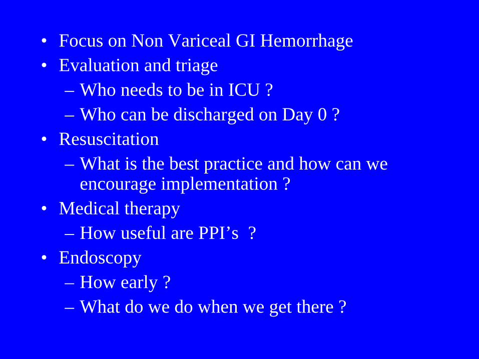
• Focus on Non Variceal GI Hemorrhage• Evaluation and triage
– Who needs to be in ICU ?– Who can be discharged on Day 0 ?
• Resuscitation– What is the best practice and how can we
encourage implementation ?• Medical therapy
– How useful are PPI’s ?• Endoscopy
– How early ? – What do we do when we get there ?

• Diagnose Upper GI bleeding• Triage according to risk• Stabilize patient : Start Resuscitation• Call GI and Surgery early• Initiate empiric therapy
• Decide about timing of endoscopy• Make diagnosis• Treat underlying condition
Virtually immediately (minutes)the job of
internist/ hospitalist/ surgeon
Hours:the job of GI and surgery

• If GI bleeding is acute – call the GI fellow on call ASAP – do not wait until the morning.
• Optimizes patient care• Optimizes utilization of resources for the patient
and the rest of the hospital

Is this an Upper GI bleed ?
• Diagnostic value
Red blood from mouth or rectum > History > exam >> labs, xrays

Evaluation: Clinical Bleeding History• Hematemesis
– Vomiting (“Not spitting”, “coughing”)– “Coffee grounds” ?
• likely not important in absence of other findings• Should we quit teaching medical professionals about
“coffee grounds” ?– Guaiac of emesis or NG aspirate?
• Will offer a cash reward for anecdotal evidence of benefit
• Melena– Black (NOT “dark”), tarry, “metallic” odor– Needs to be in the gut 4-6 hours– Hematochezia and shock may mean UGI bleed

Lessons from the literature – part 1

** “normals” were persons willing to drink blood and examine feces for medical science
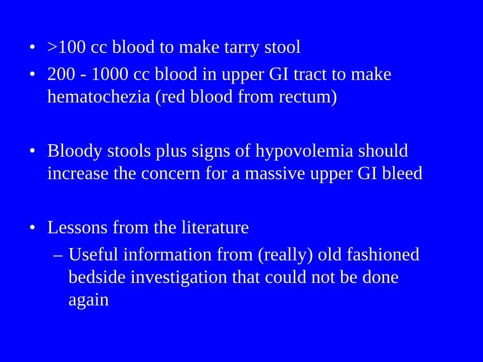
• >100 cc blood to make tarry stool• 200 - 1000 cc blood in upper GI tract to make
hematochezia (red blood from rectum)
• Bloody stools plus signs of hypovolemia should increase the concern for a massive upper GI bleed
• Lessons from the literature– Useful information from (really) old fashioned
bedside investigation that could not be done again
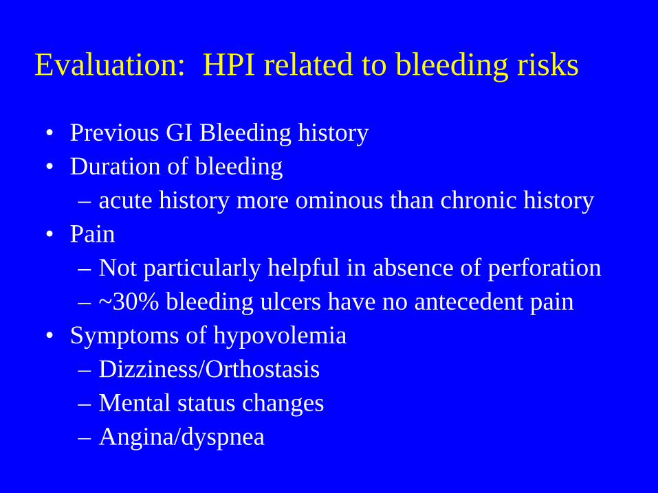
Evaluation: HPI related to bleeding risks
• Previous GI Bleeding history• Duration of bleeding
– acute history more ominous than chronic history• Pain
– Not particularly helpful in absence of perforation– ~30% bleeding ulcers have no antecedent pain
• Symptoms of hypovolemia– Dizziness/Orthostasis– Mental status changes– Angina/dyspnea
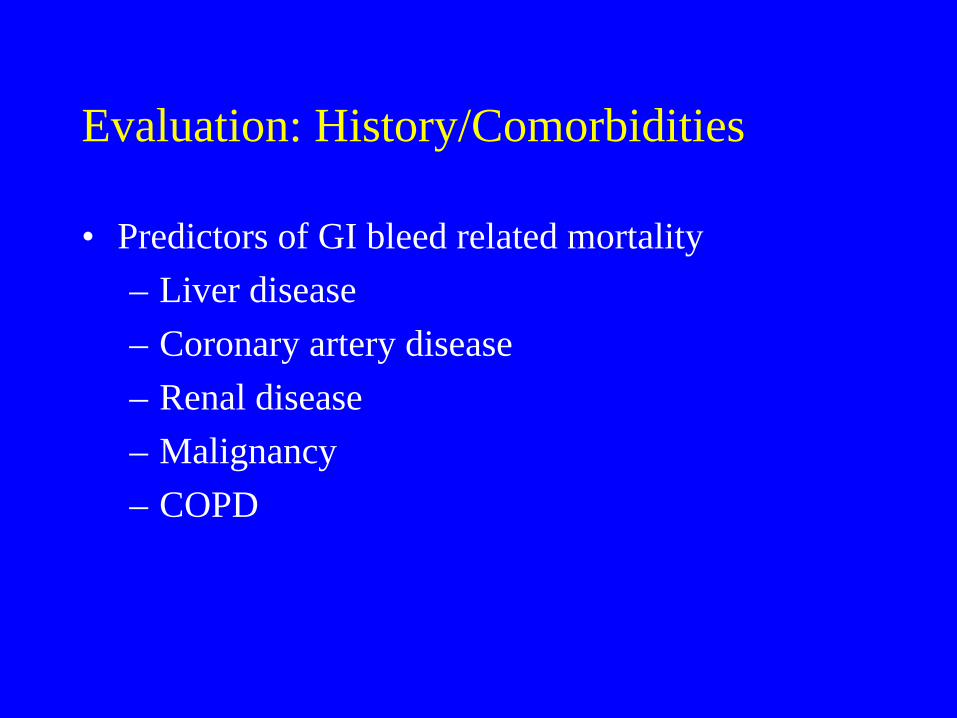
Evaluation: History/Comorbidities
• Predictors of GI bleed related mortality– Liver disease – Coronary artery disease– Renal disease – Malignancy – COPD
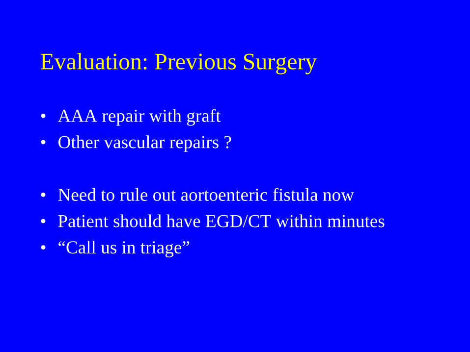
Evaluation: Previous Surgery
• AAA repair with graft• Other vascular repairs ?
• Need to rule out aortoenteric fistula now• Patient should have EGD/CT within minutes • “Call us in triage”
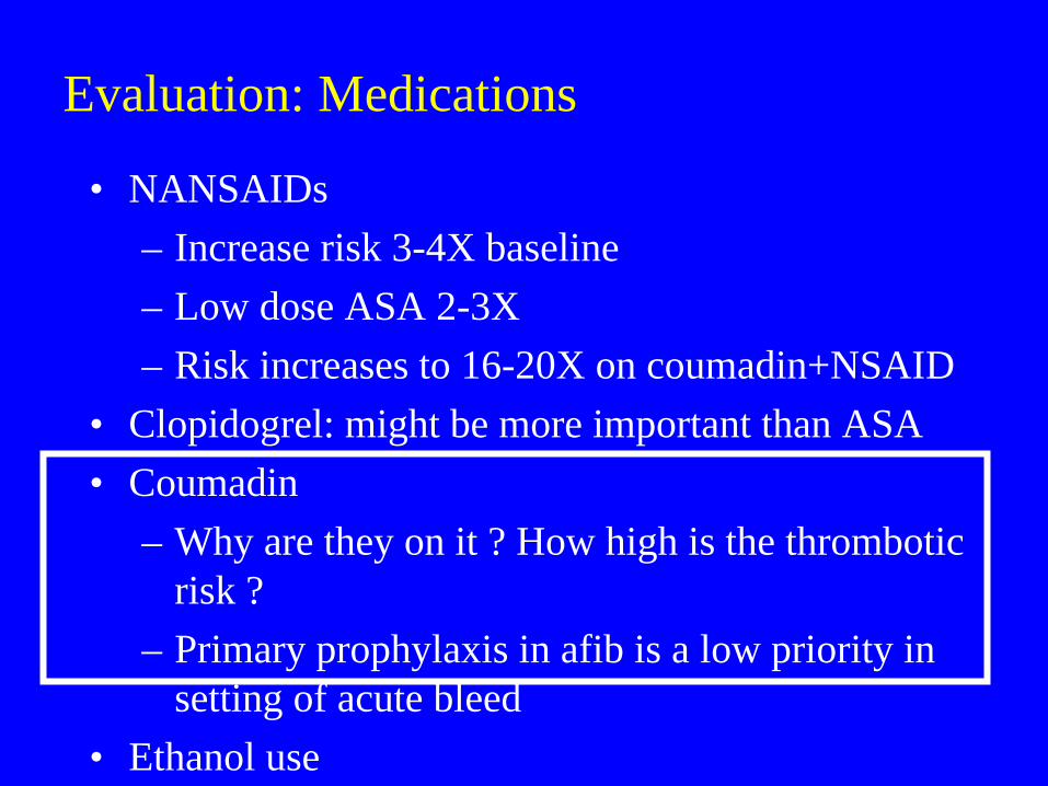
Evaluation: Medications
• NANSAIDs– Increase risk 3-4X baseline– Low dose ASA 2-3X– Risk increases to 16-20X on coumadin+NSAID
• Clopidogrel: might be more important than ASA• Coumadin
– Why are they on it ? How high is the thrombotic risk ?
– Primary prophylaxis in afib is a low priority in setting of acute bleed
• Ethanol use

Evaluation: Physical exam - I
• Vital signs– Resting HR– Orthostatics: Hypovolemia
• Orthostasis (drop 20 mm Hg, Increase in HR ~20)• Requires a ~20%volume loss• Capillary refill time
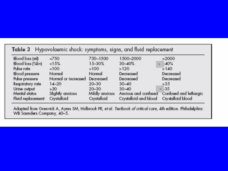
<
<

HCT may not drop acutely

Evaluation: Physical exam - II
• Oxygenation: can this person tolerate conscious sedation without intubation ?
• Mentation : – Can this person consent to invasive procedures ?
• Abdominal exam:– Are there signs of peritonitis ?
• Rectal exam: is there red blood ?• Peripheral signs of “cold” shock• Urine output

Evaluation: admission labs• PCV• Platelets• PT/INR• BUN/Cr.• Metabolic profile : is acidosis present ?• Type and Screen
– Blood type and screen for major Ab– Requires ~30 minute more work once decision to
crossmatch for transfusion is made• Type and crossmatch : reserves a unit of blood for a
patient


The Quality Based Pyramid of Information
http://library.downstate.edu/EBM2/2100.htm
Validity increases
Work increases
Relevance may increase or decrease
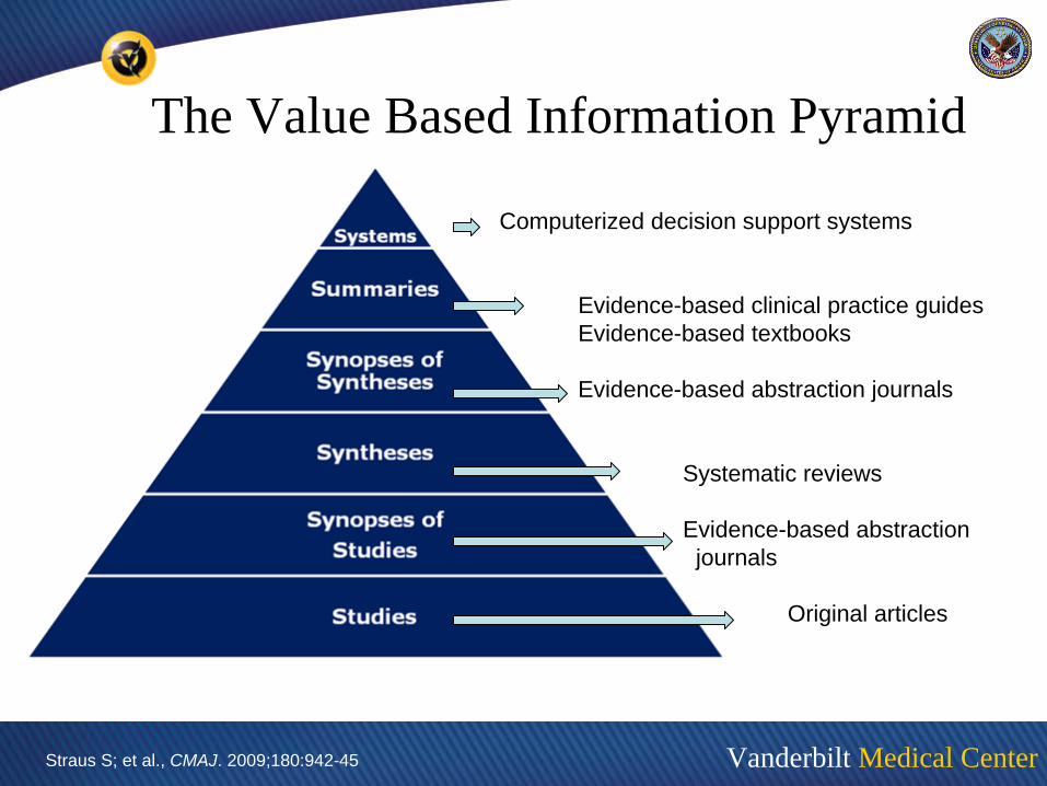
Vanderbilt Medical Center
The Value Based Information Pyramid
Computerized decision support systems
Evidence-based clinical practice guidesEvidence-based textbooks
Evidence-based abstraction journals
Systematic reviews
Evidence-based abstractionjournals
Original articles
Straus S; et al., CMAJ. 2009;180:942-45

Use of Clinical Practice Guidelines
• High level practitioners should be able to evaluate CPGs – Validity– Applicability to specific care environments– Applicability to specific patients incorporating
characteristics and preferences
• This will be a skill set that will differentiate medical doctors from other practitioners

“ Performing a coronary bypass isn’t difficult…..knowing when to do it is what is difficult…..”

Guideline development
• Multidisciplinary panel• Gather and synthesize evidence• Grade evidence• Develop specific guidelines• Distribute and implement guidelines• Measure effect of guidelines
• GRADE Guidelines– “guidelines on guideline development”– http://www.gradeworkinggroup.org

Methodology of Guideline development
• Expert panels– Vote on items or arrive at consensus by formal or
informal methods – Prone to undue influence by strong personalities– BOGSAR technique
• “Bunch of guys sitting around a room”
• Delphi Panels– Specific questions reviewed anonymously by members– Answers combined anonymously by leaders– Questions and answers resubmitted – Process repeated– Designed to eliminate domination by strong panel
members

Evaluation of a Clinical Practice Guideline• Were all important options and outcomes identified?
– What is the outcome of interest ?• Explicitly stated: Mortality ? Quality of life ? Costs ?
• Was an explicit and sensible process used to indentify, select and combine the evidence ?– Description of techniques used to gather best evidence
Adapted from: JAMA 274(7). 570-574
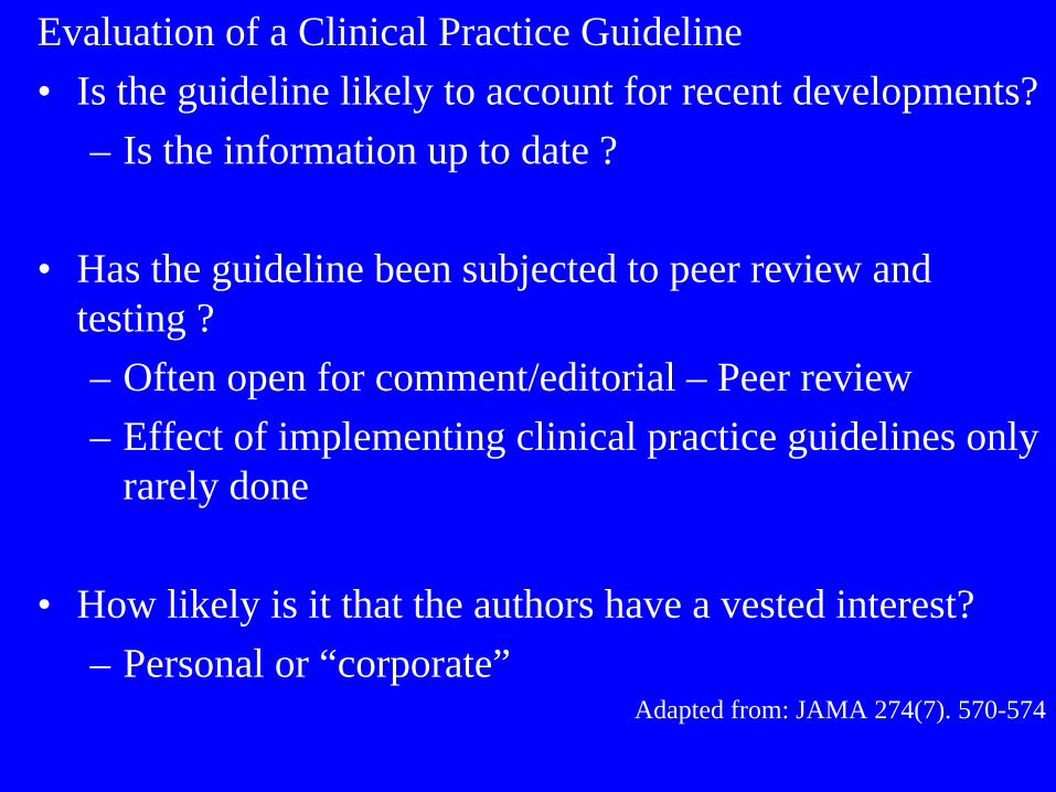
Evaluation of a Clinical Practice Guideline• Is the guideline likely to account for recent developments?
– Is the information up to date ?
• Has the guideline been subjected to peer review and testing ?– Often open for comment/editorial – Peer review– Effect of implementing clinical practice guidelines only
rarely done
• How likely is it that the authors have a vested interest?– Personal or “corporate”
Adapted from: JAMA 274(7). 570-574

Evaluation of Clinical Practice Guidelines
• Is the primary objective of the guideline consistent with the objectives you have for your patients
• Are the primary recommendations applicable to your patients?– Does patients have access to recommended
options ?Adapted from: JAMA 274(7). 570-574.

Evaluation of Clinical Practice Guidelines• Are practical clinically important
recommendations made?
• How strong are the recommendations?– Grading schemes need to be explicit – Variety of schemes
• What is the impact of uncertainty associated with the evidence and values used in the guidelines?– Would new evidence from an RCT likely
change the recommendation ?
Adapted from: JAMA 274(7). 570-574.

Explicit discussion on the search strategy used to find best evidence
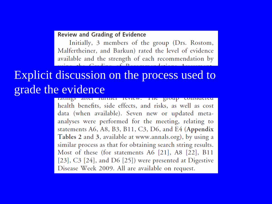
Explicit discussion on the process used to grade the evidence
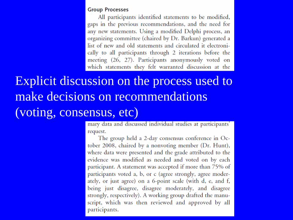
Explicit discussion on the process used to make decisions on recommendations (voting, consensus, etc)

Explicit discussion potential conflict of interests, including a member by member accounting and also an explanation of who is funding the process
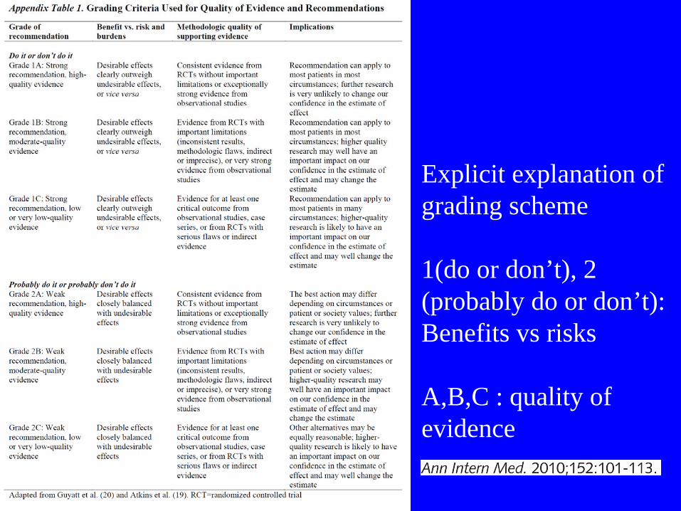
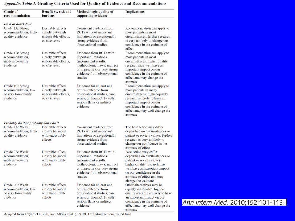
Explicit explanation of grading scheme
1(do or don’t), 2 (probably do or don’t):Benefits vs risks
A,B,C : quality of evidence
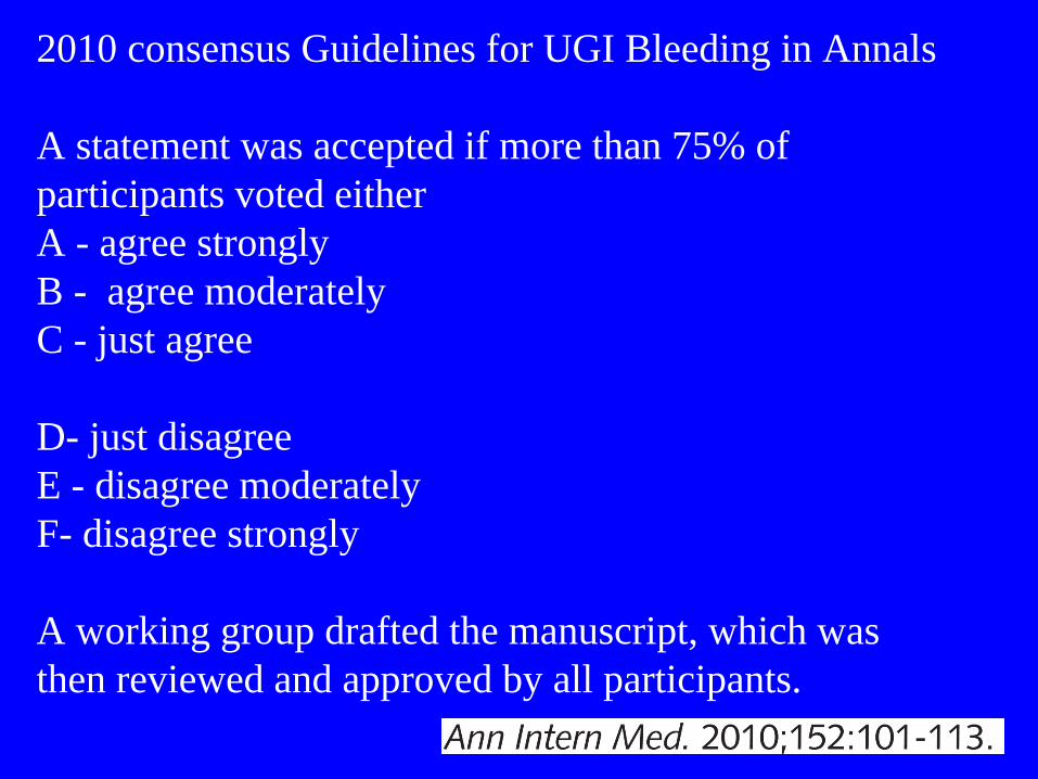
2010 consensus Guidelines for UGI Bleeding in Annals
A statement was accepted if more than 75% ofparticipants voted either A - agree stronglyB - agree moderatelyC - just agree
D- just disagreeE - disagree moderatelyF- disagree strongly
A working group drafted the manuscript, which was then reviewed and approved by all participants.
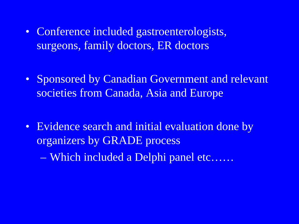
• Conference included gastroenterologists, surgeons, family doctors, ER doctors
• Sponsored by Canadian Government and relevant societies from Canada, Asia and Europe
• Evidence search and initial evaluation done by organizers by GRADE process– Which included a Delphi panel etc……

2010 consensus Guidelines for UGI Bleeding in Annals
A statement was accepted if more than 75% ofparticipants voted either A - agree stronglyB - agree moderatelyC - just agree
D- just disagreeE - disagree moderatelyF- disagree strongly
A working group drafted the manuscript, which was then reviewed and approved by all participants.

Who needs to be in a monitored bed or in the ICU ?Statement A2Prognostic scales are recommended for early stratification of patients into low- and high-risk categories for rebleeding and mortality. (Agree, 97% [Vote: a, 56%; b, 35%; c, 6%; d, 3%].Grade: Low, 1c, “do it”)
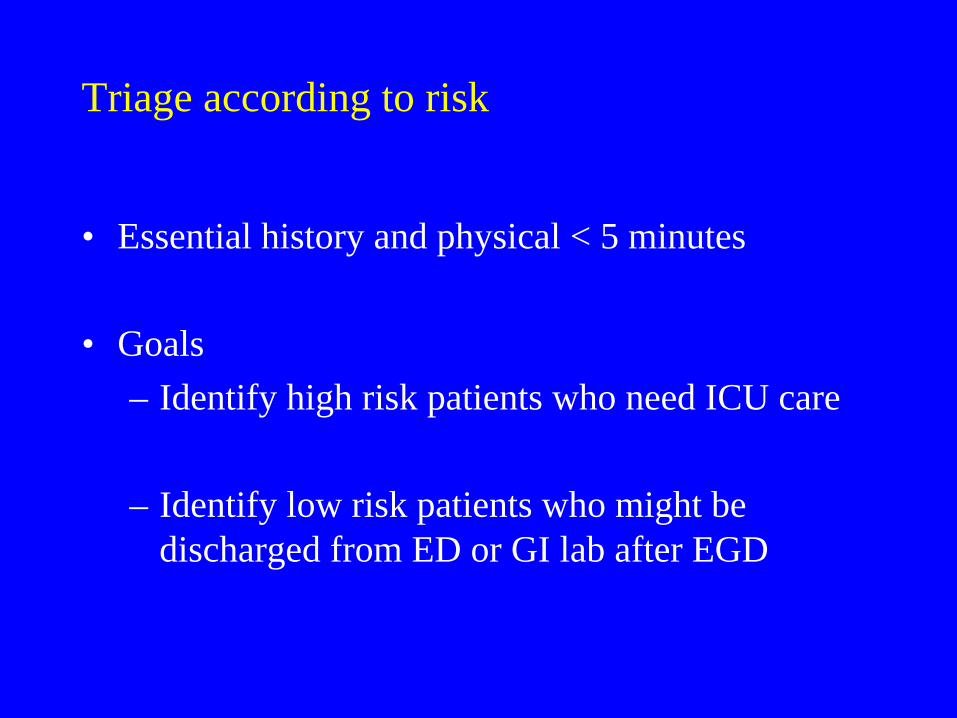
Triage according to risk
• Essential history and physical < 5 minutes
• Goals– Identify high risk patients who need ICU care
– Identify low risk patients who might be discharged from ED or GI lab after EGD

Risk stratification/ Triage
Clinical features
• History• Exam• Labs
Endoscopic features
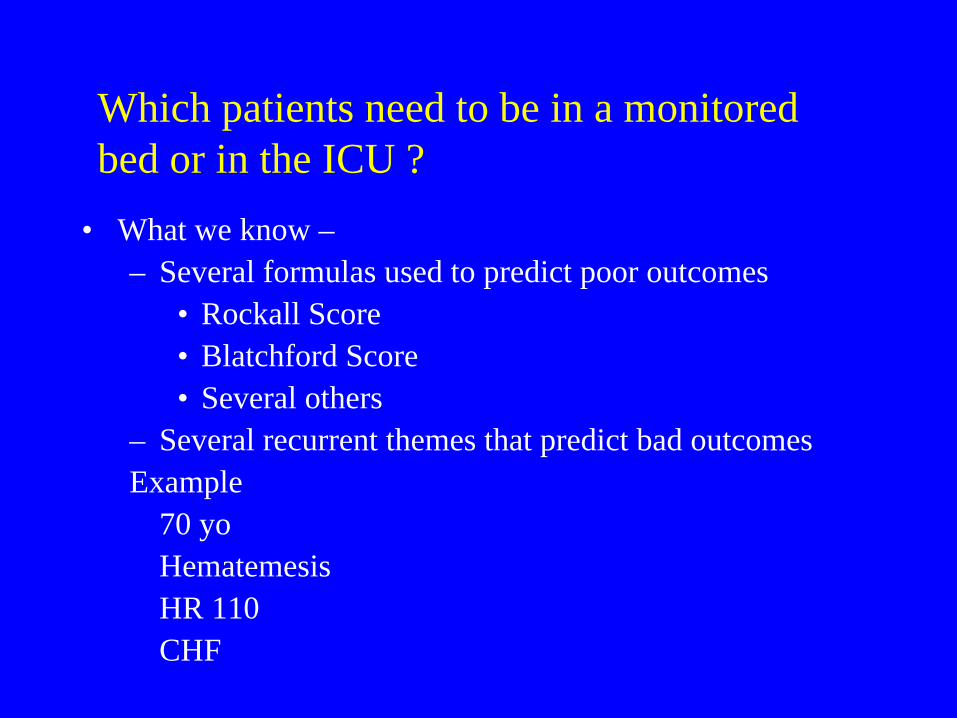
Which patients need to be in a monitored bed or in the ICU ?
• What we know –– Several formulas used to predict poor outcomes
• Rockall Score• Blatchford Score• Several others
– Several recurrent themes that predict bad outcomes Example
70 yoHematemesisHR 110CHF

<
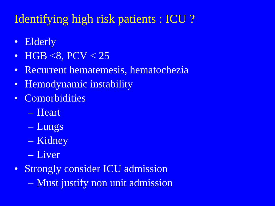
Identifying high risk patients : ICU ?
• Elderly• HGB <8, PCV < 25• Recurrent hematemesis, hematochezia• Hemodynamic instability• Comorbidities
– Heart – Lungs– Kidney– Liver
• Strongly consider ICU admission– Must justify non unit admission

Identifying low risk patients:
Who can be sent home from triage or discharged from ICU or floor ?
•Patient characteristics
•Non of the pre endoscopy factors that require ICU or monitored bed
•EGD findings •low risk findings: MW tear, esophagitis, ulcer with clean base

In low risk patients….
low risk EGD findings..
Might mean early discharge or no admission
•3 RCTs•Consistent results from all studies
Arch Intern Med. 2001;161:1393-1404
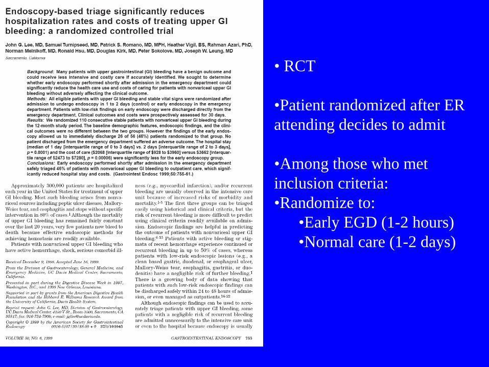
• RCT
•Patient randomized after ER attending decides to admit
•Among those who met inclusion criteria:•Randomize to:
•Early EGD (1-2 hours)•Normal care (1-2 days)
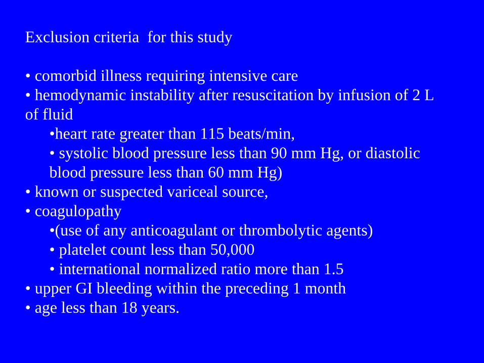
Exclusion criteria for this study
• comorbid illness requiring intensive care • hemodynamic instability after resuscitation by infusion of 2 L of fluid
•heart rate greater than 115 beats/min, • systolic blood pressure less than 90 mm Hg, or diastolic blood pressure less than 60 mm Hg)
• known or suspected variceal source, • coagulopathy
•(use of any anticoagulant or thrombolytic agents)• platelet count less than 50,000• international normalized ratio more than 1.5
• upper GI bleeding within the preceding 1 month• age less than 18 years.
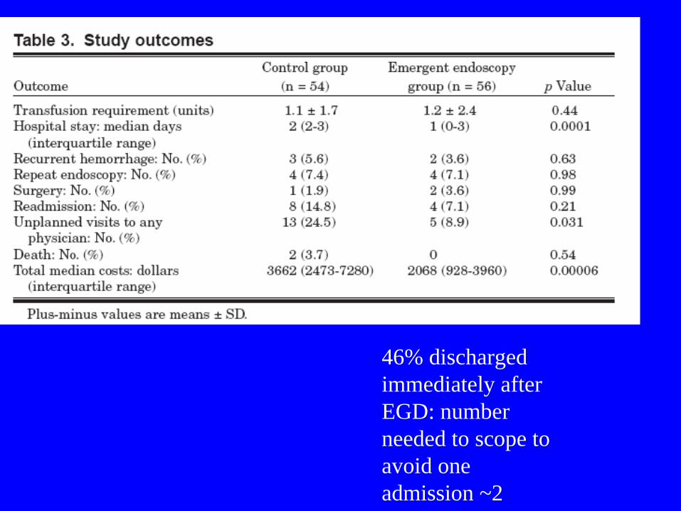
46% discharged immediately after EGD: number needed to scope to avoid one admission ~2

Identifying low risk patients: Who can be sent home from triage ?
• No hemodynamic instability• Limited hematemesis• Few/No comorbid conditions• Good support system
• Consider “Triage” endoscopy – EGD with MW tear or ulcer with clean base
• Consider outpatient management• RCT evidence to suggest this is safe

Who needs to be in a monitored bed or in the ICU ?Statement A2Prognostic scales are recommended for early stratification of patients into low- and high-risk categories for rebleeding and mortality. (Agree, 97% [Vote: a, 56%; b, 35%; c, 6%; d, 3%].Grade: Low, 1c, “do it”)
My personal assessment of guideline:The data supports using a systematic approach for triage and possible discharge
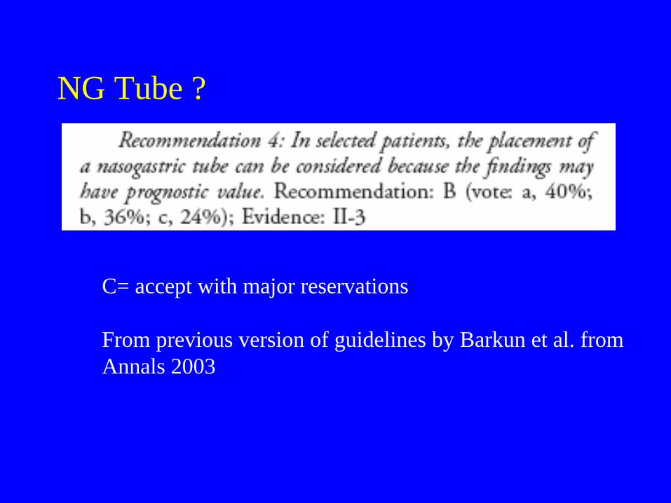
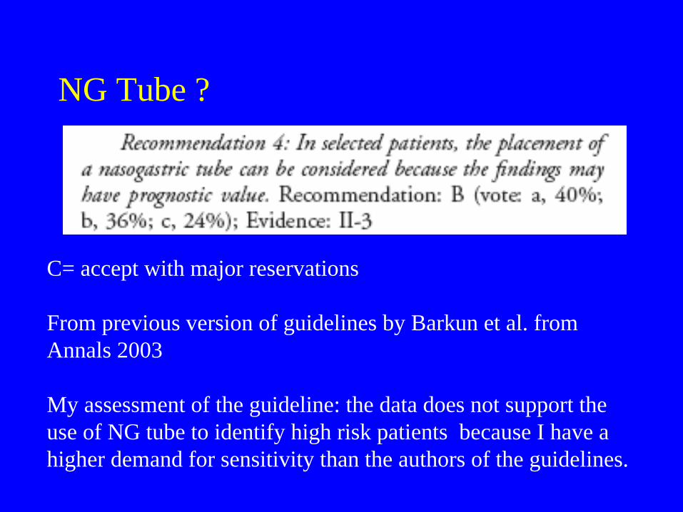
NG Tube ?
C= accept with major reservations
From previous version of guidelines by Barkun et al. from Annals 2003
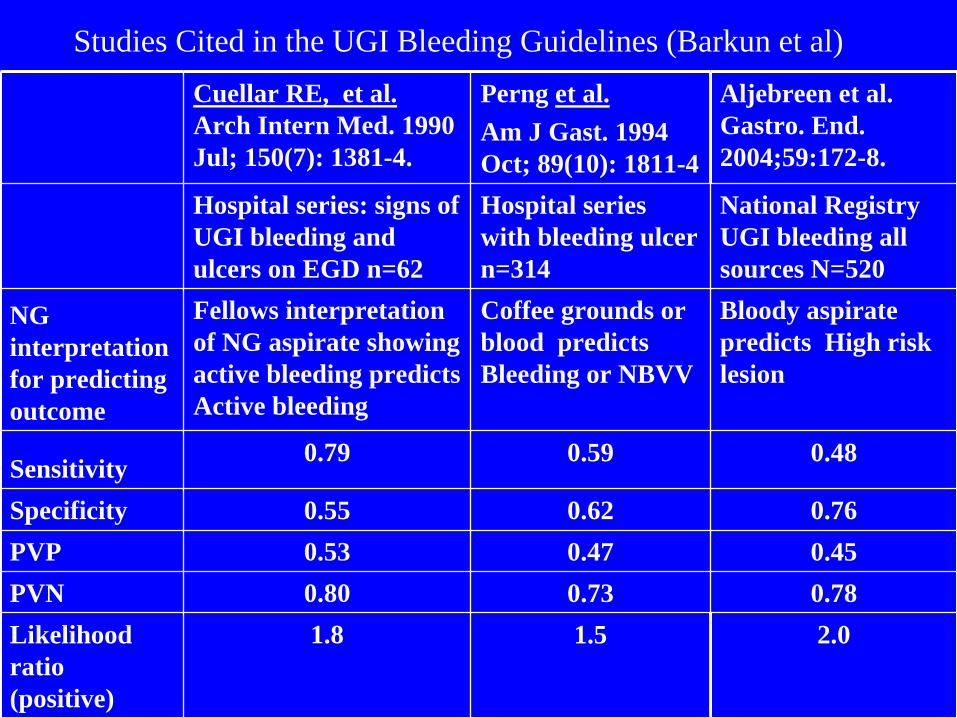
Cuellar RE, et al. Arch Intern Med. 1990 Jul; 150(7): 1381-4.
Perng et al.Am J Gast. 1994 Oct; 89(10): 1811-4
Aljebreen et al. Gastro. End. 2004;59:172-8.
Hospital series: signs of UGI bleeding and ulcers on EGD n=62
Hospital series with bleeding ulcer n=314
National Registry UGI bleeding all sources N=520
NG interpretation for predicting outcome
Fellows interpretation of NG aspirate showing active bleeding predicts Active bleeding
Coffee grounds or blood predicts Bleeding or NBVV
Bloody aspirate predicts High risk lesion
Sensitivity 0.79 0.59 0.48
Specificity 0.55 0.62 0.76PVP 0.53 0.47 0.45PVN 0.80 0.73 0.78Likelihood ratio (positive)
1.8 1.5 2.0
Studies Cited in the UGI Bleeding Guidelines (Barkun et al)

Studies in recent metanalysis of NG tube aspirate in ER patients withmelena or hematochezia without hematemesis
ACADEMIC EMERGENCY MEDICINE 2010; 17:126–132
Gold standard was EGD finding of in all studies
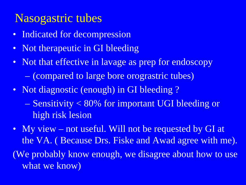
Nasogastric tubes• Indicated for decompression• Not therapeutic in GI bleeding• Not that effective in lavage as prep for endoscopy
– (compared to large bore orograstric tubes)• Not diagnostic (enough) in GI bleeding ?
– Sensitivity < 80% for important UGI bleeding or high risk lesion
• My view – not useful. Will not be requested by GI at the VA. ( Because Drs. Fiske and Awad agree with me).
(We probably know enough, we disagree about how to use what we know)
NG Tube ?
C= accept with major reservations
From previous version of guidelines by Barkun et al. from Annals 2003
My assessment of the guideline: the data does not support the use of NG tube to identify high risk patients because I have a higher demand for sensitivity than the authors of the guidelines.

• Diagnose Upper GI bleeding• Triage according to risk• Stabilize patient : Start Resuscitation• Call GI and Surgery early• Initiate empiric therapy
• Decide about timing of endoscopy• Make diagnosis• Treat underlying condition
Virtually immediately (minutes)the job of
internist/ hospitalist
This is what I emphasize to the Surgery housestaff and GI Fellows
Hours:the job of GI and surgery

Initial Treatment• Large bore IV access, multiple sites
– Don’t let central access delay other interventions• Volume replacement• Consider Pressors• Start Oxygen ( especially if conscious sedation is
anticipated)• Transfusion goals
– PCV > 25% and stable, >30% if Hx. CAD– INR < 1.5– Platelets >50K


Statement A4Blood transfusions should be administered to a patient with a hemoglobin level of 70 g/L or less.(Agree, 100% [Vote: a, 59%; b, 35%; c, 6%]. Grade: Low, 1c, “do it”)

Treatment of hemorrhagic shock: Liberal or Conservative Pressor therapy ? Liberal or Conservative volume resuscitation ?
• Goal directed therapy (MAP, etc) is an attractive concept but has not been proven outside setting of septic shock
• Pressors may have some role – there is very little comparative, human data in the setting of treating NVUGIH
• In general start with aggressive volume resuscitation– Crystalloid (20 ml per kg)– Blood when needed, ready
• We need to know more about optimal resuscitation for GI hemorrhage (in humans)

Statement A4Blood transfusions should be administered to a patient with a hemoglobin level of 70 g/L or less.(Agree, 100% [Vote: a, 59%; b, 35%; c, 6%]. Grade: Low, 1c, “do it”)
My assessment: Yes, despite the lack of RCT evidence I agree it is a good thing to treat shock……
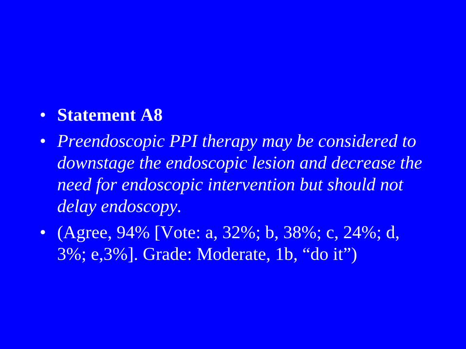
• Statement A8• Preendoscopic PPI therapy may be considered to
downstage the endoscopic lesion and decrease the need for endoscopic intervention but should not delay endoscopy.
• (Agree, 94% [Vote: a, 32%; b, 38%; c, 24%; d, 3%; e,3%]. Grade: Moderate, 1b, “do it”)
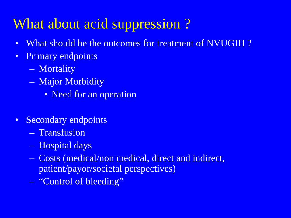
What about acid suppression ?• What should be the outcomes for treatment of NVUGIH ?• Primary endpoints
– Mortality– Major Morbidity
• Need for an operation
• Secondary endpoints– Transfusion– Hospital days– Costs (medical/non medical, direct and indirect,
patient/payor/societal perspectives)– “Control of bleeding”

Acid Suppression (Smalley : GI conference circa July 2008)• There is good evidence that acid suppression may
decrease– rebleeding rates – surgical rates
• There is very little evidence that it saves lives
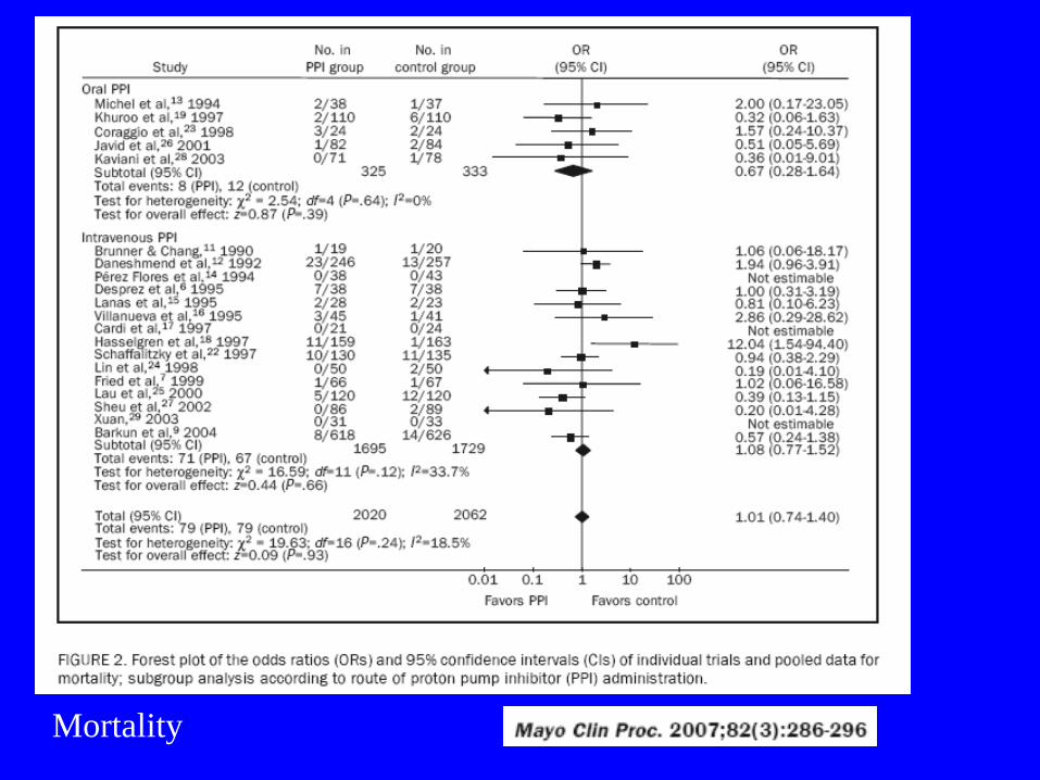
• A mortality benefit would be a difficult to meet standard to meet


Authors’ conclusionsPPI treatment in PU bleeding reduces rebleeding and surgery compared with placebo or H2RA, but there is no evidence of an overall effect on all-cause mortality.
Proton pump inhibitor treatment for acute peptic ulcer bleeding (Review)Copyright © 2008 The Cochrane Collaboration. Published by JohnWiley & Sons, Ltd
Lesson from the literature:Sometimes things getquoted in strange ways
Mortality

Multiple subgroup analysis were done………………


Metanalysis done for consensus guidelines preparation
31 RCTs (24) Total 5792
PPI treatment with or without endoscopic therapy compared with placebo or H2RA
1)Main outcome: Rebleeding: OR 0.45 (95%CI 0.36, 0.57)
2) Secondary outcome: Surgery: OR 0.56 (95%CI 0.45, 0.70)Mortality: OR 0.90 (95%CI 0.67, 1.19)
Online appendix table 2

PPI’s Summary
• There is no definitive improvement in mortality overall– Probably a benefit in those with high risk lesions– Demonstrating mortality benefit would be a very high standard
• PPI s seem to consistently decrease need for transfusion and need for operation
• The effects of oral PPI versus IV PPIs have not been directly compared (enough) – similar outcomes appear to be expected given the current data
• PPI’s seem to work better with a bolus followed by continuous infusion
• Timing of EGD should not be influenced by administration of PPI• PPIs are not likely to be harmful (in the short run)

• Statement A8• Preendoscopic PPI therapy may be considered to
downstage the endoscopic lesion and decrease the need for endoscopic intervention but should not delay endoscopy.
• (Agree, 94% [Vote: a, 32%; b, 38%; c, 24%; d, 3%; e,3%]. Grade: Moderate, 1b, “do it”)
• My assessment: PPIs are helpful, probably not harmful
• What to do about aspirin in patients with UGI bleeding ?
• Gastroenterologist/lawyer from the podium– “I can stop most GI bleeds.”– “I can’t stop most MIs”
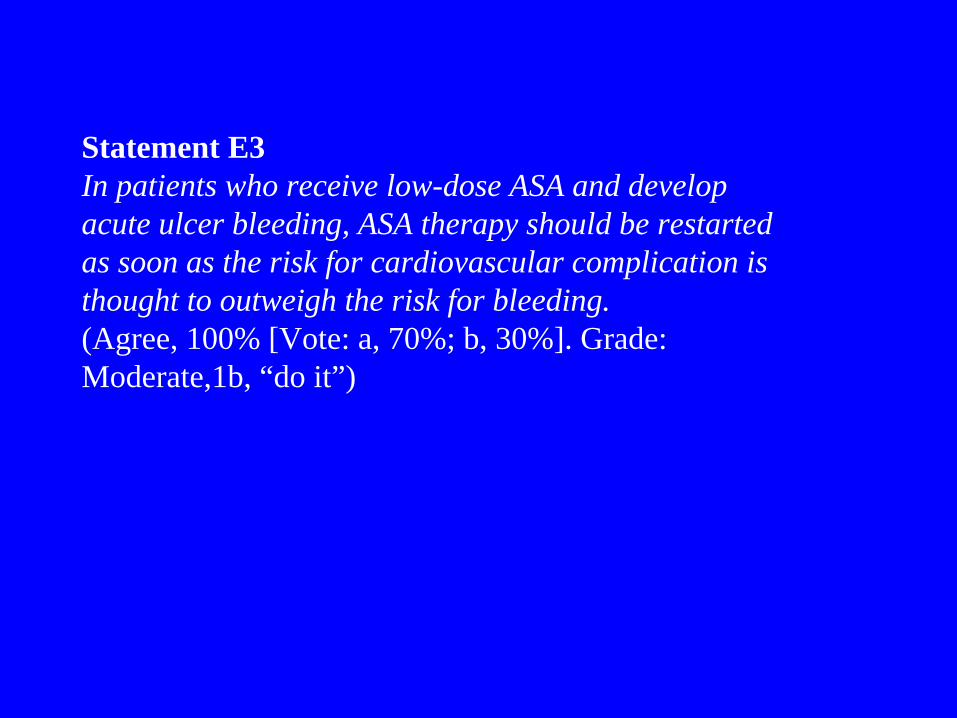
Statement E3In patients who receive low-dose ASA and develop acute ulcer bleeding, ASA therapy should be restarted as soon as the risk for cardiovascular complication is thought to outweigh the risk for bleeding.(Agree, 100% [Vote: a, 70%; b, 30%]. Grade: Moderate,1b, “do it”)


• RCT among persons with ulcer related bleeding requiring endoscopic treatment
• All had been on ASA prophylaxis for documented CVD or cerebrovascular disease
• All got PPI and HP testing and treatment
• ASA 85 mg vs placebo

ASA groupMore bleeding 10% vs 5% at 2 monthsLess dying 1% vs 12% at 2 months
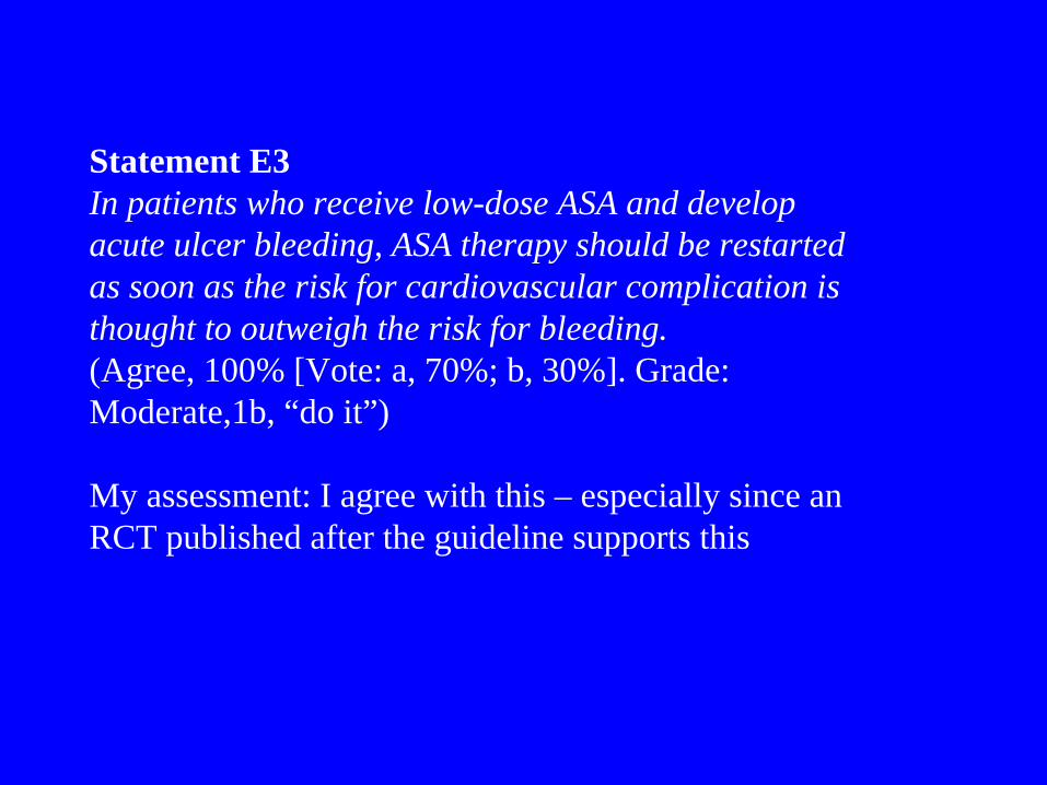
Statement E3In patients who receive low-dose ASA and develop acute ulcer bleeding, ASA therapy should be restarted as soon as the risk for cardiovascular complication is thought to outweigh the risk for bleeding.(Agree, 100% [Vote: a, 70%; b, 30%]. Grade: Moderate,1b, “do it”)
My assessment: I agree with this – especially since an RCT published after the guideline supports this

• Statement A5• In patients receiving anticoagulants, correction of
coagulopathy is recommended but should not delay endoscopy.
• (Agree, 97% [Vote: a, 38%; b, 44%; c, 15%; d, 3%]. Grade: Low, 2c, “probably do it”)

Antithrombotics in bleeding• Weak indication (Primary prophylaxis of afib etc.) : hold
or reverse anticoagulation• Among those with a strong indication for anticoagulation
• bare metal stents in first several weeks or drug eluting stents for 12 months
• Mitral valve, PE, Acute coronary syndrome etc.– Restart aspirin once bleeding is controlled– Hold the antithrombotics and non aspirin antiplatelet
agents until bleeding is controlled (usually < 24 hours)– Reverse the antithrombotics if bleeding is not
controlled and patient is in shock/may go to OR

• Diagnose Upper GI bleeding• Triage according to risk• Stabilize patient : Start Resuscitation• Call GI and Surgery early• Initiate empiric therapy
• Decide about timing of endoscopy• Make diagnosis• Treat underlying condition
Virtually immediately (minutes)the job of
internist/ hospitalist with input from GI
This is what I emphasize to the Surgery housestaff and GI Fellows
Hours:the job of GI and surgery


• Endoscopy in acute upper GI bleeding
• Timing• Preparation• Interventions

Statement B3• Early endoscopy (within 24 hours of presentation)
is recommended for most patients with acute upper gastrointestinal bleeding.
• (Agree, 100% [Vote: a, 85%; b, 12%; c, 3%]. Grade: Moderate, 1b, “do it”)
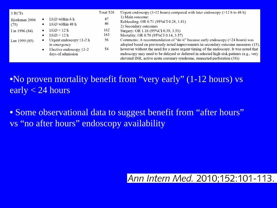
•No proven mortality benefit from “very early” (1-12 hours) vs early < 24 hours
• Some observational data to suggest benefit from “after hours” vs “no after hours” endoscopy availability
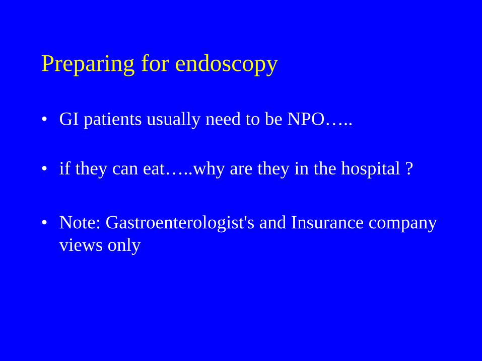
Preparing for endoscopy
• GI patients usually need to be NPO…..
• if they can eat…..why are they in the hospital ?
• Note: Gastroenterologist's and Insurance company views only

• Promotility agents should not be used routinely before endoscopy to increase the diagnostic yield.
• (Agree, 82% [Vote: a, 35%; b, 35%; c, 12%; d, 6%;e, 3%; f, 9%]. Grade: Moderate, 2b, “probably don’t do it”)

• “A meta-analysis (21) of 3 trials that evaluated erythromycin (60–62), comprising 316 patients, and 2 abstracts that evaluated metoclopramide (63, 64) found that use of a prokinetic agent significantly reduced the need for repeated endoscopy (odds ratio [OR], 0.51 [95% CI, 0.30 to 0.88]) in patients suspected of having blood in their stomach, compared with placebo or no treatment (Appendix Table2).”

• Promotility agents should not be used routinely before endoscopy to increase the diagnostic yield. (Agree, 82% [Vote: a, 35%; b, 35%; c, 12%; d, 6%;e, 3%; f, 9%]. Grade: Moderate, 2b, “probably don’t do it”)
• The data cited by the guideline document is consistently positive – 3/3 RCTs, double blinded etc– The guideline writers fail to make a case why not to
use promotility agents routinely– There possible risks to IV erythromycin but these
were unapparent in the trials
• Sometimes guideline voting does not follow the data presented
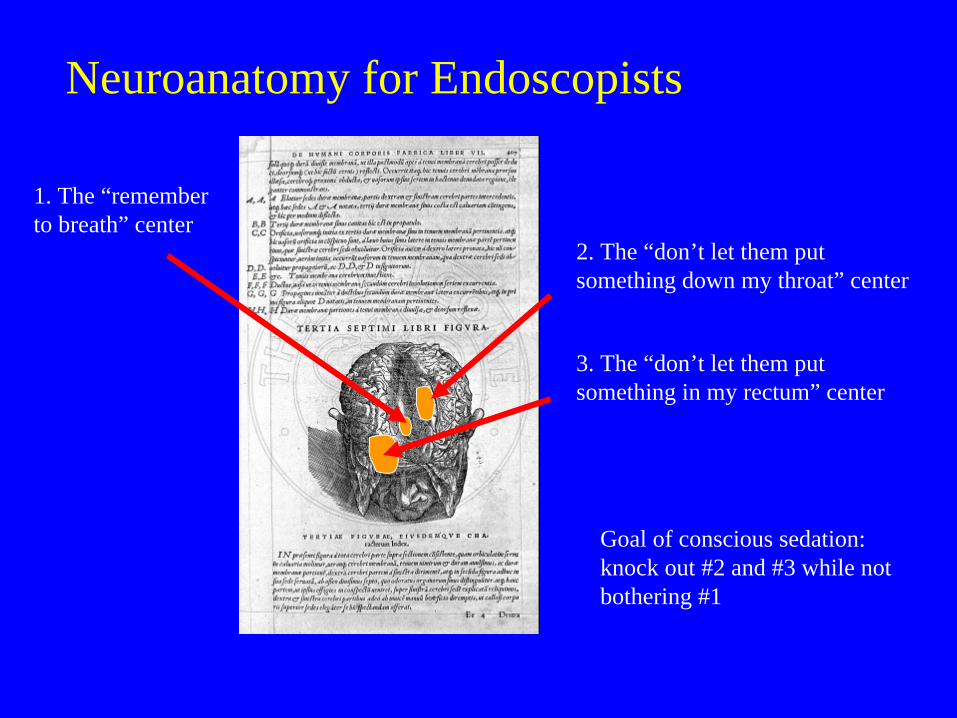
Neuroanatomy for Endoscopists
1. The “remember to breath” center
2. The “don’t let them put something down my throat” center
3. The “don’t let them put something in my rectum” center
Goal of conscious sedation: knock out #2 and #3 while not bothering #1

Endotracheal tubes• Protect airway
– “Elective intubation is better than emergent intubation”– Setting
• Massive bleeding (hematemesis) • Decreased mental status
– Allows for more aggressive conscious/deep sedation– Allows for more definitive endoscopic therapy in patients
with massive bleeding (“gourmet endoscopy”)
• ASGE Guidelines “Patients with ongoing, significant hematemesis or those who may not be able to protect their airway for any reason and are at risk for aspiration should be considered for endotracheal intubation before undergoing endoscopy.”

• Now that we are doing endoscopy what do we do when we get there ?

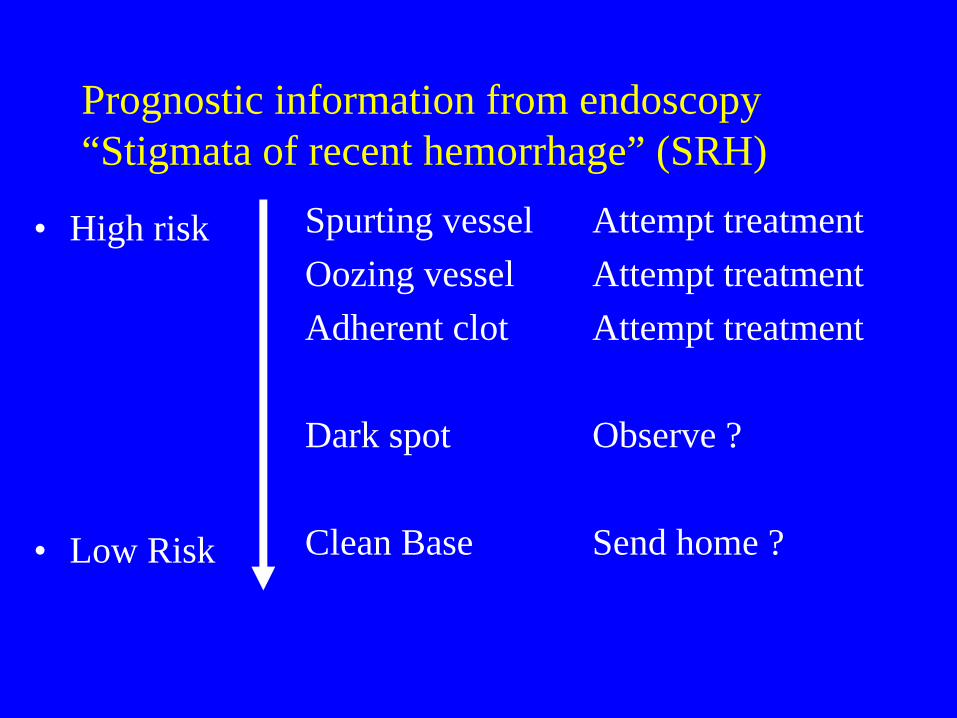
Prognostic information from endoscopy “Stigmata of recent hemorrhage” (SRH)
• High risk
• Low Risk
Spurting vesselOozing vesselAdherent clot
Dark spot
Clean Base
Attempt treatmentAttempt treatmentAttempt treatment
Observe ?
Send home ?

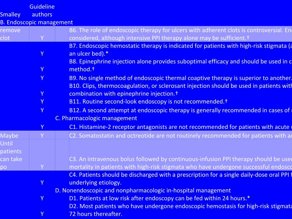
• B5. A finding of a clot in an ulcer bed warrants targeted irrigation in an attempt at dislodgement, with appropriate treatment of the underlying lesion.†
• B7. Endoscopic hemostatic therapy is indicated for patients with high-risk stigmata (active bleeding or a visible vessel in an ulcer bed).*
• B8. Epinephrine injection alone provides suboptimal efficacy and should be used in combination with another method.†

• B9. No single method of endoscopic thermal coaptive therapy is superior to another.*
• B10. Clips, thermocoagulation, or sclerosant injection should be used in patients with high-risk lesions, alone or in combination with epinephrine injection.†
• B11. Routine second-look endoscopy is not recommended.†
• B12. A second attempt at endoscopic therapy is generally recommended in cases of rebleeding.*

“Endoscopic therapy for patients with UGIB caused by PUD has been studied in randomized, controlled trials.
Laser therapy; monopolar electrocautery ; bipolar electrocautery; heat probe; epinephrine injection; and epinephrine injection with additives, such as the sclerosants ethanolamine and polidocanol,
are all effective when compared with no therapy or sham therapy.”
ASGE guideline: the role of endoscopy in acute non-variceal upper-GI hemorrhage 2004

“Numerous prospective randomized studies of endoscopic treatment methods have been performed.
No single modality has been shown to be superior for treating UGIB caused by PUD.
For epinephrine injection, the addition of a second modality (combination therapy) reduces further bleeding, the need for surgery, and mortality.
Operator experience plays a significant role in modality choice and in achieving hemostasis.”
Pick one or two modalities – use them a lot.
ASGE guideline: the role of endoscopy in acute non-variceal upper-GI hemorrhage 2004

In 2008 most would agree that endoscopically placed clips also should be included in the list of modalities effective in treating ulcer bleeding

Adherent clots: amorphous red clots attached to Adherent clots: amorphous red clots attached to an ulcer base which did not wash away with an ulcer base which did not wash away with vigorous irrigationvigorous irrigation

What should we do when we see an adherent clot ?
• John Tarpley– “Don’t poke a skunk”
• Dennis Jensen (UCLA)– Shave off the clot from (currently) non bleeding
lesions and treat

This RCT influences us to be more aggressive in Nonbleeding lesions withstigmata of recenthemorrhage
Gastro:Jensen:2002

Rebleeding Rates in RCT’s of Treatment of Adherent Clots
35.0%34.3%
0.0%4.8%0%
10%20%30%40%50%
Mayo ClinicMulticenter Trial
UCLA CUREMulticenter Trial
Medical Therapy Endotherapy
P < 0.05
N = 56 N = 32

• Limitation of procedural RCTs
• Are our endoscopies (endoscopists) like endoscopies (endoscopists) from UCLA or Mayo?

Routine Repeat Endoscopy?
• Review of 6 randomized trials– No reduction in risk of rebleeding– Increased number of procedures– Possibly increase risk from unnecessary
retreatment
Romagnuolo J. Can J Gastroenterol 18(6): 401 2004

Lau N Engl J Med 1999;340:751
Endoscopy vs. Surgery for Recurrent Bleeding• 100 patients with rebleeding after endoscopic control randomized
to repeat endoscopy (n=48) or direct surgery (n=44)
• 13 (23%) patients in endoscopy group had salvage surgery compared to 100% in the surgery arm– NNT (repeat scope) to prevent one operation < 2
• Overall similar outcomes (mortality, length of hospital stay, number of blood transfusions)
• Complications higher with direct surgery (16 vs. 7, p=0.03)
• Analysis of endoscopy failures: ulcers> 2cm, hypotension at randomization
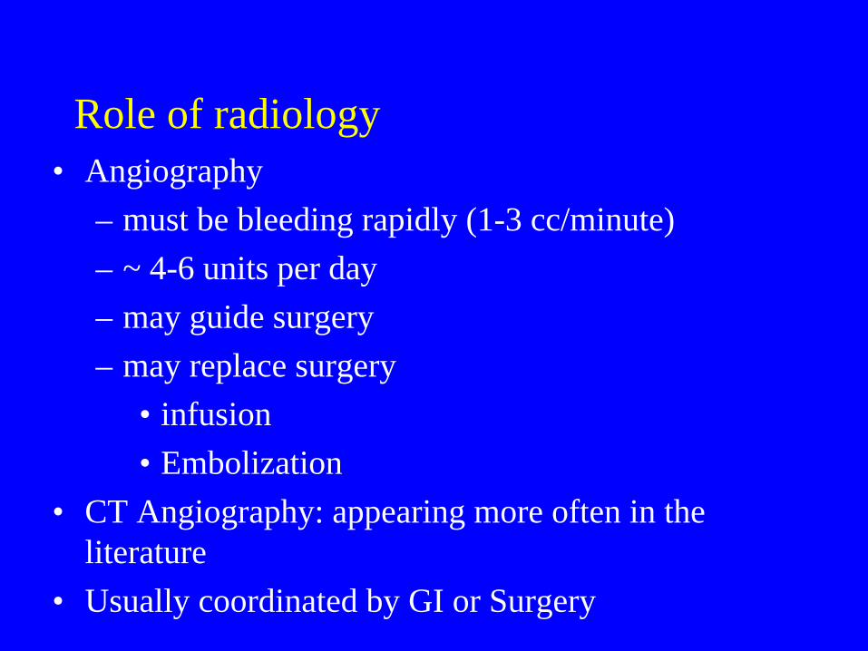
Role of radiology• Angiography
– must be bleeding rapidly (1-3 cc/minute)– ~ 4-6 units per day– may guide surgery– may replace surgery
• infusion• Embolization
• CT Angiography: appearing more often in the literature
• Usually coordinated by GI or Surgery

Role of nuclear medicine: Tagged cell scan
• Hypothetically < 1 unit per day• Early (15 minute) scan is most useful• Early scans done at Vanderbilt• Not usually done at VA• Not utilized by VA GI service very often• REQUIRED by VUMC angiographers prior to
angio attempt : “facilitates selective angiography”

Lower GI bleeding• Self limited in > 80% cases• Most common
– Diverticular disease– AVMs
• Less common– Colitis (Inflammatory or infectious)– Tumors– Hemorhoids– Miscellaneous ulcers– Ischemia
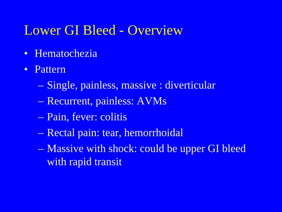
Lower GI Bleed - Overview• Hematochezia• Pattern
– Single, painless, massive : diverticular– Recurrent, painless: AVMs– Pain, fever: colitis– Rectal pain: tear, hemorrhoidal– Massive with shock: could be upper GI bleed
with rapid transit

Lower GI bleed : Overview• Evaluate and triage according to risk
– (Similar to UGI Bleed)– Age, comorbid conditions, hemodynamics
• Stabilize : replace volume• Call GI and Surgery Consultants early
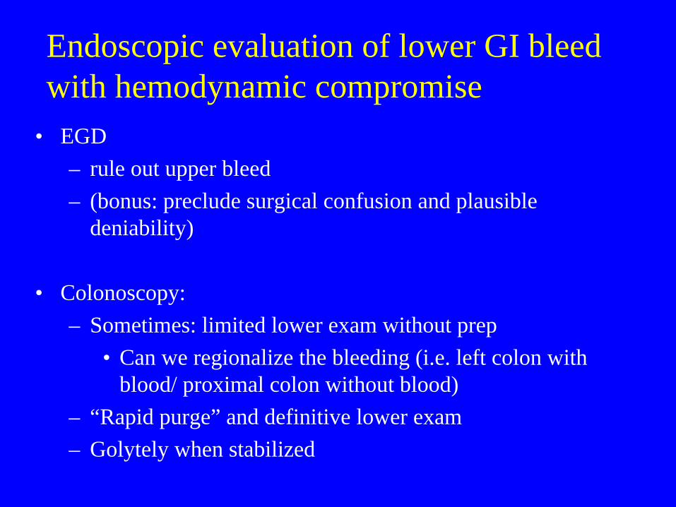
Endoscopic evaluation of lower GI bleed with hemodynamic compromise
• EGD– rule out upper bleed – (bonus: preclude surgical confusion and plausible
deniability)
• Colonoscopy:– Sometimes: limited lower exam without prep
• Can we regionalize the bleeding (i.e. left colon with blood/ proximal colon without blood)
– “Rapid purge” and definitive lower exam– Golytely when stabilized

Not an RCT“We treated it when we saw it”

PEG solution was either orally (in the case of 67 percent of patients) or by nasogastric tube (in the case of 33 percent) to rid the colon of clots, stool, and blood.
The procedure usually required 5 to 6 liters of purge and three to four hours before the colon was clean.
Urgent colonoscopy was defined as colonoscopy performedat the bedside 6 to 12 hours after hospitalization or thediagnosis of hematochezia and within 1 hour after clearance ofstool, blood, and clots, as documented by a physician.
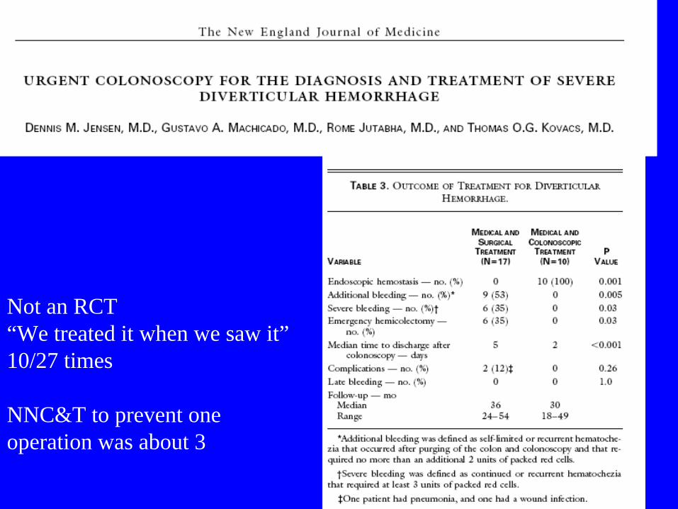
Not an RCT“We treated it when we saw it”10/27 times
NNC&T to prevent one operation was about 3

Randomized Trial of Urgent vs. Elective Colonoscopy in Patients Hospitalized With Lower GI Bleeding
Laine et al. Am J Gastroenterol 2010; 105:2636–2641;
85 patients with shock and BRBPREGD then randomization to urgent (< 12 H ) or routine colonoscopy
15% had UGI source
Among those with lower GI source no difference in outcomes
Need for operationHospital days, volume of transfusion, costs

Endoscopy in acute lower GI bleeding
• More purely diagnostic that in UGI bleed• Identify level of bleeding
– examination of ileum if possible• Therapeutic:
– AVM cautery/injection– Diverticular bleed (Only at UCLA ?)
• Will typically need rapid colon prep after EGD


• A major role of the physician will be to parse Clinical Practice Guidelines to most effectively use the available resources to a patient with individual risk factors and preferences.

SmalleyGuideline
authors
A. Resuscitation, risk assessment, and preendoscopy management
YA1. Immediately evaluate and initiate appropriate resuscitation.*A2. Prognostic scales are recommended for early stratification of patients into low‐
and
categories for rebleeding and mortality.†
no YA3. Consider placement of a nasogastric tube in selected patients because the findings
prognostic value.*
YA4. Blood transfusions should be administered to a patient with a hemoglobin level 70 A5. In patients receiving anticoagulants, correction of coagulopathy is recommended b
not delay endoscopy.
maybe YA6. Promotility agents should not be used routinely before endoscopy to increase the d
yield.
Y
A7. Selected patients with acute ulcer bleeding who are at low risk for rebleeding on th
clinical and endoscopic criteria may be discharged promptly after endoscopy.†A8. Preendoscopic
PPI therapy may be considered to downstage
the endoscopic lesion
decrease the need for endoscopic intervention but should not delay endoscopy.†
B. Endoscopic management
YB1. Develop institution‐specific protocols for multidisciplinary management.* Include a
an endoscopist trained in endoscopic hemostasis.*B2. Have available on an urgent basis support staff trained to assist in endoscopy.*
< 6 hours YB3. Early endoscopy (within 24 hours of presentation) is recommended for most patien
acute upper gastrointestinal bleeding.†
Y
B4. Endoscopic hemostatic therapy is not indicated for patients with low‐risk stigmata (
based ulcer or a nonprotuberant
pigmented dot in an ulcer bed).*B5. A finding of a clot in an ulcer bed warrants targeted irrigation in an attempt at dislo
with appropriate treatment of the underlying lesion.†
SmalleyGuideline
authors
B. Endoscopic managementremove
clot Y
B6. The role of endoscopic therapy for ulcers with adherent clots is controversial. End
considered, although intensive PPI therapy alone may be sufficient.†
YB7. Endoscopic hemostatic therapy is indicated for patients with
high‐risk stigmata (a
an ulcer bed).*
YB8. Epinephrine injection alone provides suboptimal efficacy and
should be used in co
method.†
Y B9. No single method of endoscopic thermal coaptive
therapy is superior to another.
YB10. Clips, thermocoagulation, or sclerosant injection should be
used in patients with
combination with epinephrine injection.†
Y B11. Routine second‐look endoscopy is not recommended.†Y B12. A second attempt at endoscopic therapy is generally recommended in cases of r
C. Pharmacologic managementY C1. Histamine‐2 receptor antagonists are not recommended for patients with acute u
Maybe Y C2. Somatostatin and octreotide are not routinely recommended for patients with acUntil
patients
can take
po Y
C3. An intravenous bolus followed by continuous‐infusion PPI therapy should be used
mortality in patients with high‐risk stigmata who have undergone successful endosco
YC4. Patients should be discharged with a prescription for a single daily‐dose oral PPI f
underlying etiology.
D. Nonendoscopic and nonpharmacologic in‐hospital managementY D1. Patients at low risk after endoscopy can be fed within 24 hours.*
YD2. Most patients who have undergone endoscopic hemostasis for high‐risk stigmata
72 hours thereafter.

SmalleyGuideline
authors
B. Endoscopic managementD. Nonendoscopic and nonpharmacologic in‐hospital management
Y D1. Patients at low risk after endoscopy can be fed within 24 hours.*
YD2. Most patients who have undergone endoscopic hemostasis for high‐risk stigmata
72 hours thereafter.
Y D3. Seek surgical consultation for patients for whom endoscopic therapy has failed.*
YD4. Where available, percutaneous embolization can be considered
as an alternative
endoscopic therapy has failed.
YD5. Patients with bleeding peptic ulcers should be tested for H.
pylori and receive era
with confirmation of eradication.†
Maybe Y D6. Negative H. pylori diagnostic tests obtained in the acute setting should be repeatE. Postdischarge, ASA, and NSAIDs
would not
use COX‐II Y
E1. In patients with previous ulcer bleeding who require an NSAID, it should be recogn
traditional NSAID plus PPI or a COX‐2 inhibitor alone is still associated with a clinically
bleeding.
would not
use COX‐II Y
E2. In patients with previous ulcer bleeding who require an NSAID, the combination of
recommended to reduce the risk for recurrent bleeding from that of COX‐2 inhibitors
YE3. In patients who receive low‐dose ASA and develop acute ulcer bleeding, ASA thera
the risk for cardiovascular complication is thought to outweigh the risk for bleeding.
YE4. In patients with previous ulcer bleeding who require cardiovascular prophylaxis, it
clopidogrel alone has a higher risk for rebleeding than ASA combined with a PPI.