ABDOMINAL OESOPHAGUS, STOMACH - دانشکده پزشکی...
40
ABDOMINAL OESOPHAGUS, STOMACH Dr. Zahiri
Transcript of ABDOMINAL OESOPHAGUS, STOMACH - دانشکده پزشکی...

ABDOMINAL OESOPHAGUS, STOMACH
Dr. Zahiri
Transpyloric plane An upper transverse line
=Addison's Plane located halfway between the jugular notch and the
upper border of the pubic symphysis It is also said to lie roughly a hand's breadth beneath
the xiphoid process of the human sternum. (9th costal cartilages and the lower border of the L1)
Dr. Maria Zahiri
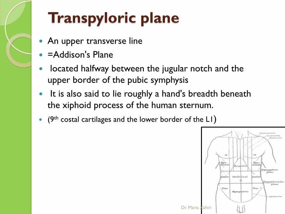
Structures crossed
the fundus of the gallbladder the origin of the superior
mesenteric artery (termination of the superior mesenteric
vein)
hilum of the kidney the root of the transverse
mesocolon duodenojejunal flexure
Dr. Maria Zahiri
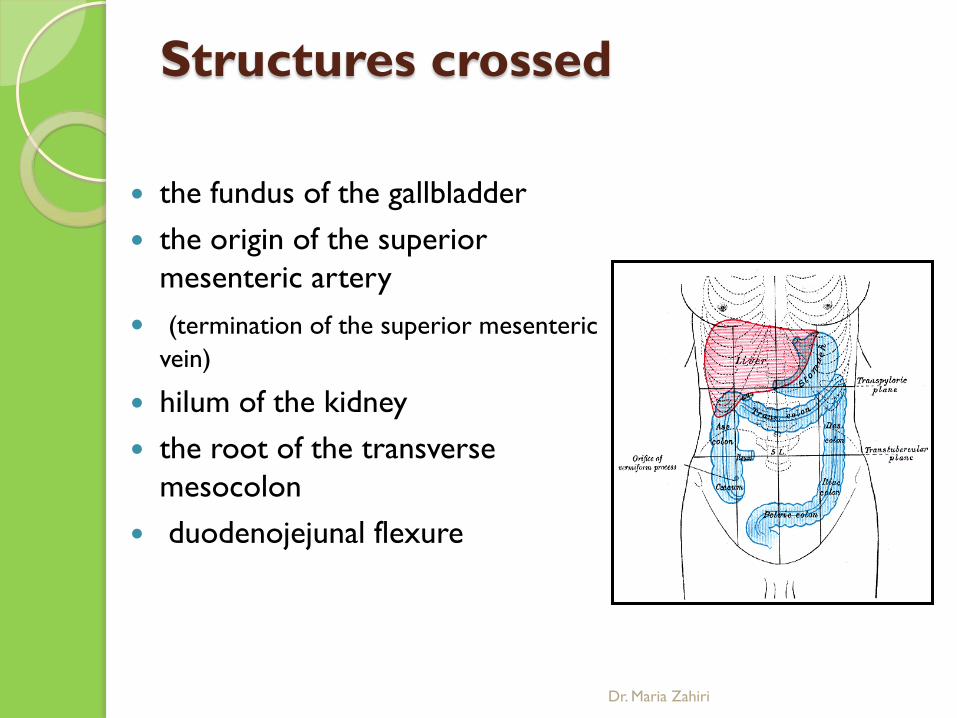
intertubercular plane (or transtubercular lower transverse line midway between the upper transverse and the upper
border of the pubic symphysis passing through the iliac tubercles L5
Dr. Maria Zahiri
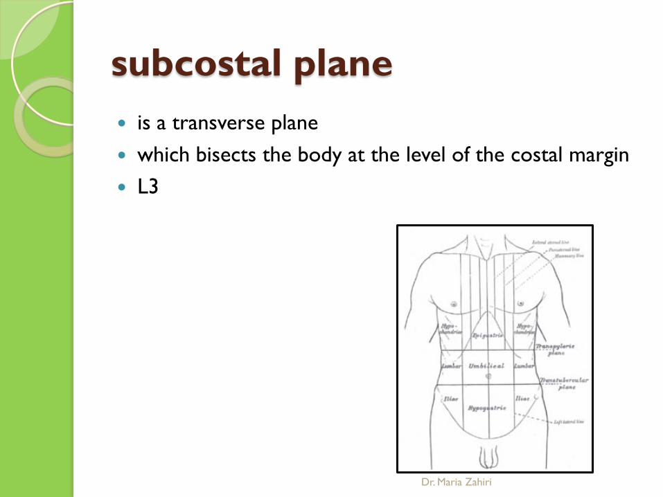
subcostal plane is a transverse plane which bisects the body at the level of the costal margin L3
Dr. Maria Zahiri
interspinous line Transumbilical line (L3- L4)
Dr. Maria Zahiri

Deep surface of the anterior abdominal wall
3 ligament & peritoneal fold 3 peritoneal fossae
Dr. Maria Zahiri

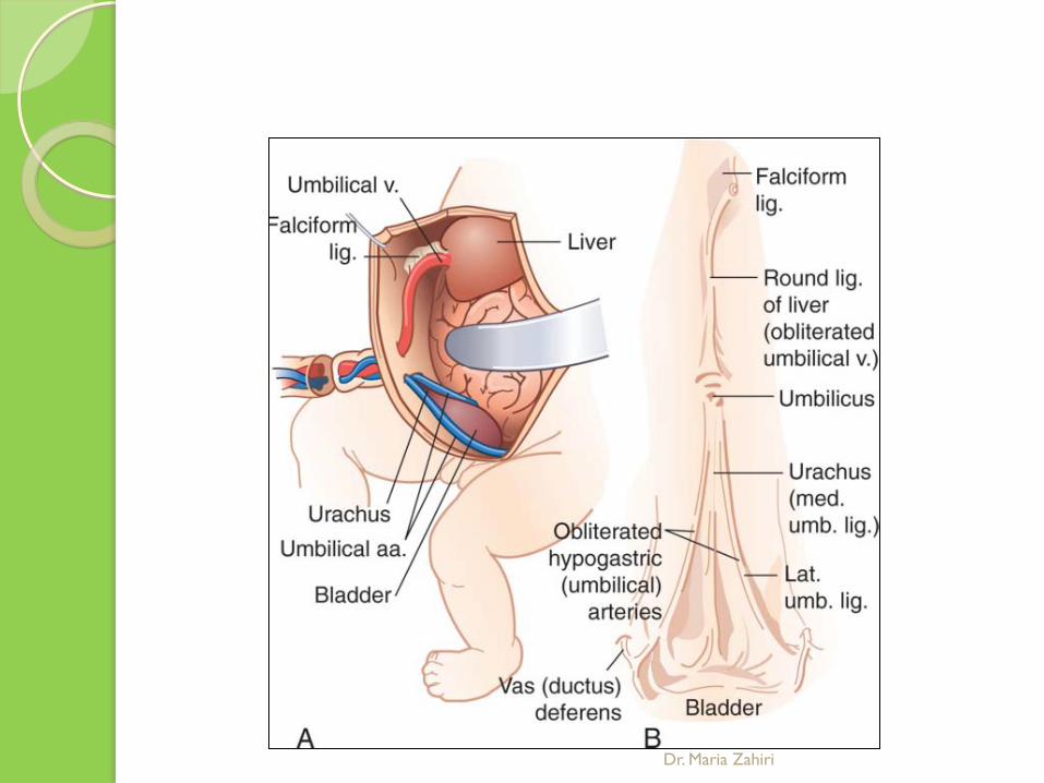
median umbilical ligament Remnant of the embryonic urachus. It extends from the apex of the bladder to the umbilicus It is covered by the median umbilical fold
Dr. Maria Zahiri

medial umbilical ligament = cord of umbilical artery is a paired structure is covered by the medial umbilical folds It represents the remnant of the fetal umbilical arteries used as a landmark for surgeons exploring the medial
inguinal fossa during laparoscopic inguinal hernia repair.
Dr. Maria Zahiri

lateral umbilical fold overlies the inferior epigastric artery (a branch of the
external iliac artery remain functional after birth It originates just medial to the deep inguinal ring
Dr. Maria Zahiri
Dr. Maria Zahiri

The lateral umbilical fold is an important reference site with regards to hernia classification.
A direct hernia occurs medial to the lateral umbilical
fold, whereas an indirect hernia originates lateral to the fold.
Dr. Maria Zahiri
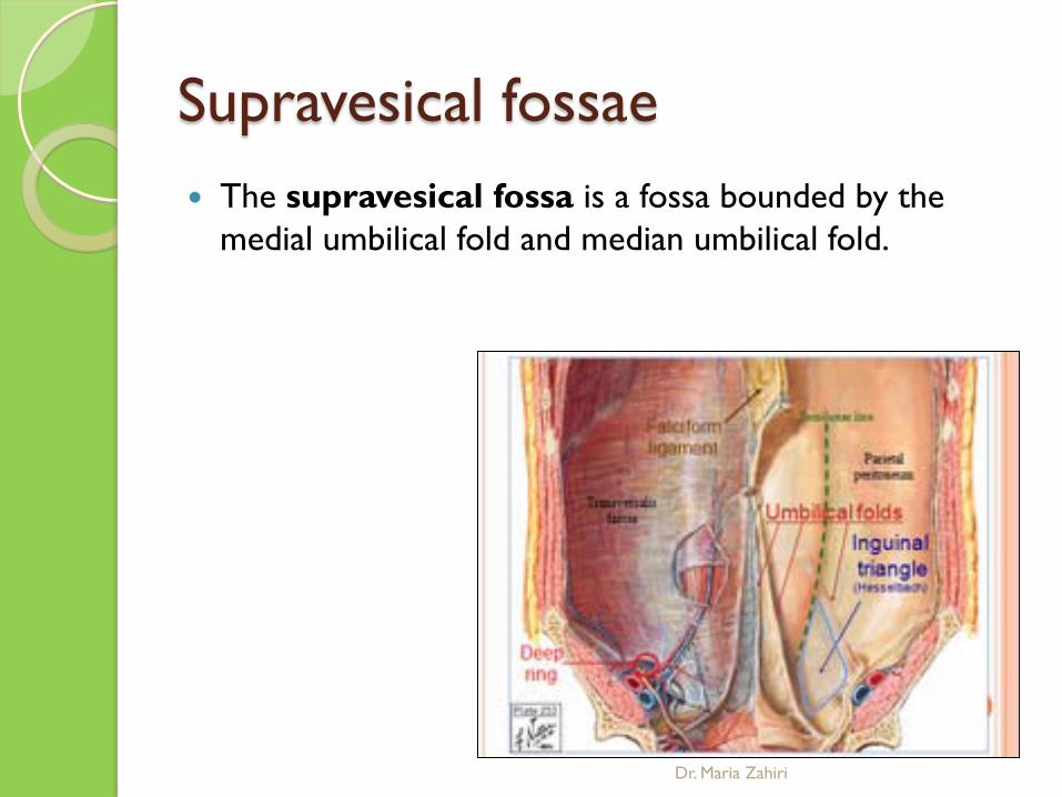
Supravesical fossae The supravesical fossa is a fossa bounded by the
medial umbilical fold and median umbilical fold.
Dr. Maria Zahiri

Medial inguial fossae a fossa bounded by the median umbilical fold and lateral
umbilical fold .
Dr. Maria Zahiri
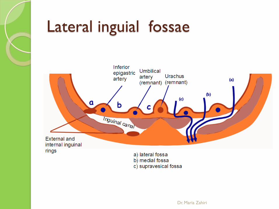
Lateral inguial fossae
Dr. Maria Zahiri

Abdominal esophagus
Dr. Maria Zahiri

ANATOMY OF OESOPHAGUS
Muscular tube of 25cm length Connects the pharynx to the stomach Flattened anterioposterioly Begins in the neck at lower border of cricoid
cartilage (C6 vertebra) Pierces the diaphragm at T10 Opens into the stomach at T11
Dr. Maria Zahiri

CONSTRICTIONS OF THE OESOPHAGUS
• There are 4 constrictions 1) * At the beginning – pharyngeo oesophageal
junction 2) Where it is crossed by the aortic arch 3) Where it is crossed by the left bronchus 4) Where it pierces the diaphragm
Dr. Maria Zahiri

BLOOD SUPPLY Arterial Supply - Inferior thyroid artery - Oesophageal branches of aorta - Oesophageal branches of the left gastric artery Venous drainage - Upper part drains into brachiocephalic vein - Middle part drains into azygous vein - Lower part drains into the left gastric vein
Dr. Maria Zahiri
LYMPHATIC DRAINAGE
Cervical part drains into the deep cervical lymph nodes Posterior part into the posterior mediastinal lymph
nodes Abdominal part into the left gastric nodes
Dr. Maria Zahiri
NERVE SUPPLY
Upper half is supplied by the recurrent laryngeal nerve
Lower part by the oesophageal plexus but mainly
by the vagus nerve
Dr. Maria Zahiri

OESOPHAGUS - DIVISIONS
1. Cervical part – ends at the lower border of T1
2. Thoracic part – ends at T10 (where is pierces the diaphragm along with vagus nerve and oesophageal branches of the left gastric artery)
3. Abdominal part – ends at the cardiac end of the stomach
Dr. Maria Zahiri
PHYSIOLOGY
Upper oesophageal sphincter - (UOS)
lower oesophageal sphincter - physiological (LOS) LOS prevents gastric reflux
Dr. Maria Zahiri
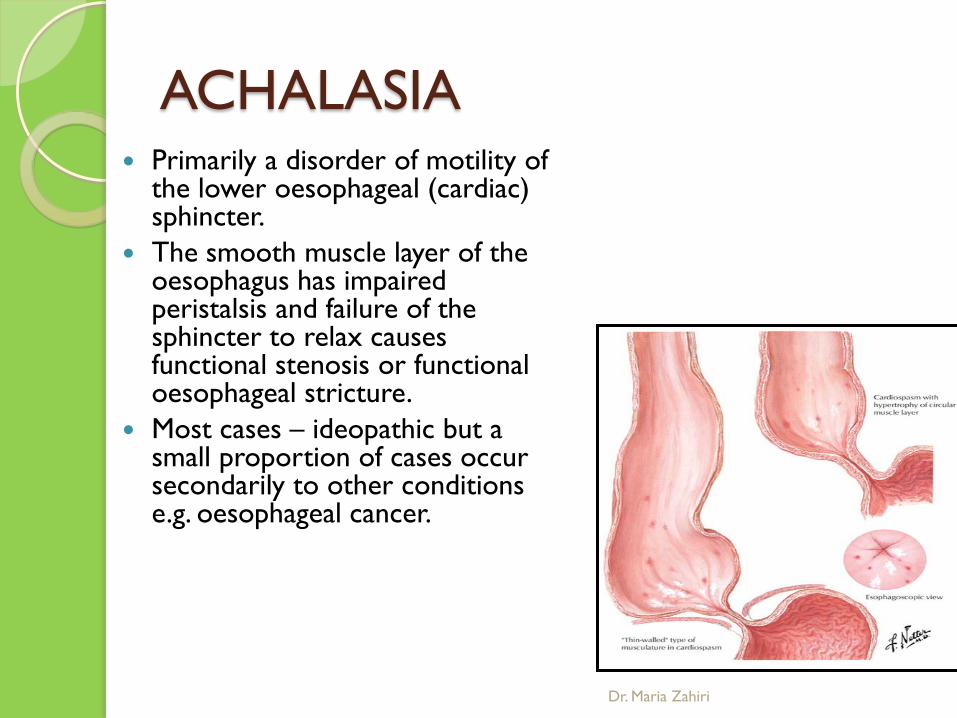
ACHALASIA Primarily a disorder of motility of
the lower oesophageal (cardiac) sphincter.
The smooth muscle layer of the oesophagus has impaired peristalsis and failure of the sphincter to relax causes functional stenosis or functional oesophageal stricture.
Most cases – ideopathic but a small proportion of cases occur secondarily to other conditions e.g. oesophageal cancer.
Dr. Maria Zahiri
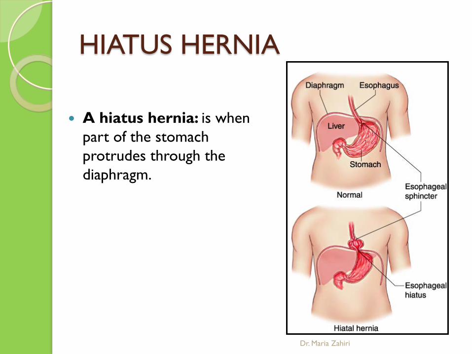
HIATUS HERNIA
A hiatus hernia: is when
part of the stomach protrudes through the diaphragm.
Dr. Maria Zahiri

STOMACH
Dr. Maria Zahiri

Dr. Maria Zahiri
The stomach is a “J” shaped hollow, muscular organ suspended under the diaphragm.
The upper larger portion of the stomach or Fundus is
situated in the upper left quadrant of the abdomen and entrance to the stomach is gained through the esophagus through the Gastroesophageal Juncture (GE juncture or sometimes called the cardiac sphincter).
Dr. Maria Zahiri
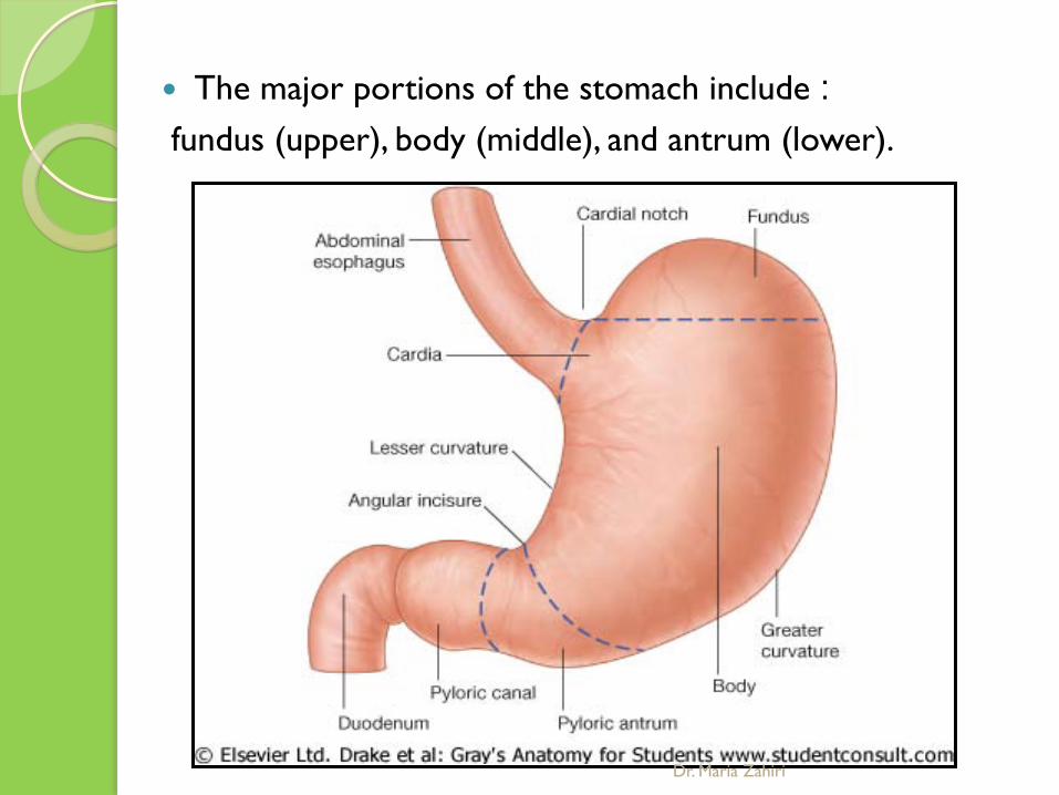
The major portions of the stomach include : fundus (upper), body (middle), and antrum (lower).
Dr. Maria Zahiri

Dr. Maria Zahiri
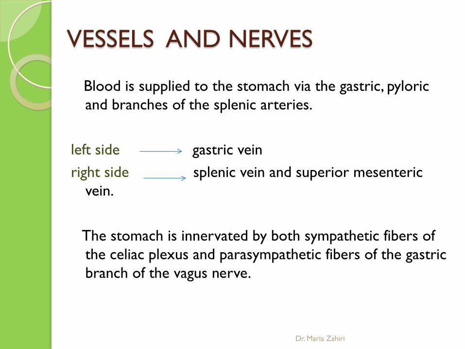
VESSELS AND NERVES
Blood is supplied to the stomach via the gastric, pyloric and branches of the splenic arteries.
left side gastric vein right side splenic vein and superior mesenteric
vein. The stomach is innervated by both sympathetic fibers of
the celiac plexus and parasympathetic fibers of the gastric branch of the vagus nerve.
Dr. Maria Zahiri

Blood supply The celiac artery, also known as the celiac trunk, is
the first major branch of the abdominal aorta. It is 1.25 cm in length Branching from the aorta anterior to the upper border
of L1 vertebra it is one of three anterior/ midline branches of the
abdominal aorta (the others are the superior and inferior mesenteric arteries).
Dr. Maria Zahiri

Region supplied The celiac artery supplies oxygenated blood to the liver,
stomach, abdominal esophagus, spleen and the superior half of both the duodenum and the pancreas.
These structures correspond to the embryonic foregut. (Similarly, the superior mesenteric artery and inferior mesenteric artery feed structures arising from the embryonic midgut and hindgut respectively.
obstruction of the celiac artery will lead to necrosis of the structures it supplies.
Dr. Maria Zahiri
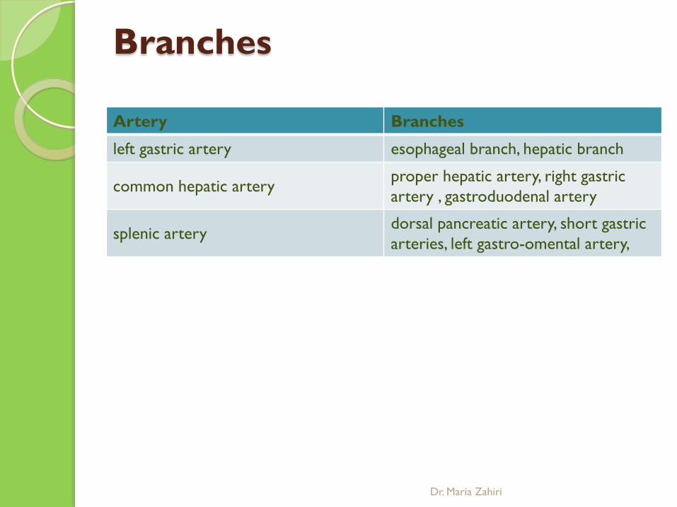
Branches Artery Branches
left gastric artery esophageal branch, hepatic branch
common hepatic artery proper hepatic artery, right gastric artery , gastroduodenal artery
splenic artery dorsal pancreatic artery, short gastric arteries, left gastro-omental artery,
Dr. Maria Zahiri

Supradeodenal artery
= gastroepiploic
Dr. Maria Zahiri

vein drainage of stomach
Dr. Maria Zahiri

lymphatic drainage of stomach
Dr. Maria Zahiri

GASTRIC INNERVATION The gastric sympathetic innervation is derived from
preganglionic fibers arising predominantly from T6 to T8 spinal nerves, which synapse within the bilateral celiac ganglia to neurons whose postganglionic fibers course through the celiac plexus along the vascular supply of the stomach. Accompanying these sympathetic nerves are afferent pain-transmitting fibers from the stomach and motor fibers to the pyloric sphincter.
The parasympathetic innervation is via the right and left vagus nerves, which form the distal esophageal plexus, and gives rise to the posterior and anterior vagal trunks near the gastric cardia.
The trunks contain preganglionic parasympathetic fibers, as well as afferent fibers from the viscera. Both trunks give rise to celiac and hepatic branches before continuing on …
Dr. Maria Zahiri

GASTRIC INNERVATION
Dr. Maria Zahiri

روزگار خوشDr. Maria Zahiri


![In silico experiment with an antigen-toll-like receptor-5 ...med.bpums.ac.ir/UploadedFiles/CourseFiles/2016_4_30/9e98584ce9__db682a... · duodenal ulcer to gastric cancer. [2,3] This](https://static.fdocuments.net/doc/165x107/5e1b8534c24775600c471325/in-silico-experiment-with-an-antigen-toll-like-receptor-5-medbpumsaciruploadedfilescoursefiles20164309e98584ce9db682a.jpg)