Merida virus, a putative novel rhabdovirus discovered in Culex and ...

Vol. 1: 204-217. 1986 DISEASES OF AQUATIC ORGANISMS Dis. aquat. Org.
Published October 15
A new rhabdovirus isolated in Japan from cultured hirame (Japanese flounder) Paralichthys olivaceus
and ayu Plecoglossus altivelis
' Laboratory of Microbiology, Faculty of Fisheries, Hokkaido University, Minato, Hakodate 041. Japan Hyogo Prefectural Fisheries Experimental Station, Akashi, Hyogo 673. Japan
ABSTRACT: In March 1984, a new rhabdovlrus was isolated from moribund cultured hlrame (Japanese flounder) Parahchthys ohvaceus and from ayu Plecoglossus alhvelis fry In Hyogo Prefecture, Japan. From February through May 1985, the virus was again isolated from hirame in seawater tanks in Hyogo and Kagawa Prefecture, and in Hokkaldo, Japan. At temperatures between 5 and 20 "C the vlrus replicated and induced cytopathic effects (CPE), which progressed to eventual cytolysis, in susceptible cell lines, ~ncludmg FHM, EPC, BF-2, RTG-2, STE-137, HF-l , BB, YNK, CCO, and EK-l. The CHSE- 214, KO-6 and CHH-l cell lines were refractory. Maximum virus titers of about log to 10g8 TCID,, rill-l were obtained In FHM or EPC cell hnes, and virus replicated optimally at 15 to 20 "C. Virus part~cles were bullet-shaped and measured 80 nm X 180 to 200 nm. The isolate was sensitive to pH 3, to dlethyl ether, and to heat (50 "C, 2 mm). Virus did not hemagglutinate human 0 type erythrocytes. Viral rephcabon was not inhib~ted by 104 M 5-~ododeoxyundlne. Infect~vity was not reduced by antisera aginst infectious hematopoletic necrosis v ~ r u s (IHNV), v~ra l hemorrhagic septicemia virus (VHSV), spring viremia of carp virus (SVCV), pike fry rhabdovirus (PFRV), eel vlrus-America (EVA), and eel wus-European (EVX). The viral isolate was pathogen~c for rainbow trout Salmo gairdnen by injection but not for chum salmon Oncorhynchus keta, coho salmon 0 . krsutch, masu salmon 0. masou, or ayu fry by water-borne exposure. The new virus has been named Rhabdovirus olivaceus (hirame rhabdovirus, HRV).
INTRODUCTION
An unknown disease occurred dunng March 1984 in cultured hirame (Japanese flounder) Paralichthys olivaceus held in pens in Fukura Bay, and in cultured ayu Plecoglossus altivelis fry held in seawater tanks at the Hyogo Prefectural Fisheries Experimental Station, Hyogo Prefecture, Japan (Gorie et al. 1985a, b). Com- mon gross signs of the dsease in hlrame were conges- tion of the gonads, focal hemorrhage of the skeletal muscle and fins and accumulation of ascitic fluid; characteristic microscopic signs were pyknosis and necrosis of the hematopoietic tissue (Kmura et al. unpubl. data). Infected ayu fry exhibited exophthalmia and pethechiae on the opercula. The following year, in February to May 1985, the disease was again observed among the hirame cultured in seawater tanks at the Hyogo Prefectural Fisheries Experimental Station,
O Inter-Research/Pnnted In F. R. Germany
Negi Island, Kagawa Prefecture, and at Yagshin Island, Hokkaido. Examination of diseased fish failed to show any bacterial, fungal or parasitic agents known to infect these hosts.
During the course of viral examination, infectious hematopoietic necrosis virus (1HNV)-Like cytopathic effects (CPE) were observed in the RTG-2 cell cultures. The responsible agent was not neutrchzed with anti- sera prepared against known fish rhabdoviruses: IHNV, vlral hemorrhagic septicemia virus (VHSV), spring viremia of carp virus (SVCV), pike fry rhab- dovirus (PFRV), eel virus-America (EVA) and eel virus- Europe (EVX). Pathogenicity and virulence of the iso- late to the hirame was demonstrated by intraperitoneal injection (Gorie et al. 1985a).
In this report we describe the characteristics and pathogenicity of this new virus, named Rhabdovirus olivaceus or hirame rhabdovirus (HRV).

210 Dis. aquat. Org. 1: 209-217, 1986
MATERIALS AND METHODS
Viruses. Five isolates of HRV were used for virologi- cal and serological studies. The HRV-8401H isolate originated from diseased hirame and the HRV-A 8401H isolate was cultured from moribund ayu fry at Hyogo Prefecture in March 1984 (Gone et al. 1985a, b). The HRV-8501H, 8501K and 8501Y isolates were obtained from diseased hirame in Hyogo Prefecture in February, Kagawa Prefecture in March, and Hokkaido in April 1985, respectively. The fish rhabdoviruses, SVCV, PFRV, EVA and EVX, and IHNV (strains IHNV- H, ChAb, HV-l) were used as reference viruses. Two of the IHNV strains (ChAb and HV-l) were stock viruses in our laboratory. The IHNV-H, SVCV, EVA and EVX were provided by Di-. B. J. Hill, Fish Disedses Ldburd- tory, Ministry of Agriculture, Fisheries and Food, U. K., and PFRV was provided by Dr. P. de Kinkelin, Institut National de la Research Agronomique, France. Stock strains of infectious pancreatic necrosis virus (IPNV, serotype VR-299, Ab, and Sp) and Oncorhynchus masou virus (OMV, 00-7812) were also used as refer- ence viruses.
Cell Lines. The rainbow trout gonad cell line (RTG-2: Wolf & Quimby 1962) was used for primary isolation and for routine propagation of the virus. The cell lines of fathead minnow (FHM: Gravell & Malsburger 1965), epithelioma papilosum cyprini (EPC: Tomasec & Fijan 1971). blue gill fry (BF-2: Wolf & Quimby 1966), steel- head trout embryo (STE-137: Lannan et al. 1984), yamabe kidney (YNK: Watanabe et al. 1978), brown bullhead (BB: Wolf & Quimby 19661, channel catfish ovary (CCO: Bowser & Plumb 1980), eel kidney (EK-1: Chen et al. 1982), chinook salmon embryo (CHSE-214: Lannan et al. 1984), chum salmon heart (CHH-1: Lan- nan et al. 1984), kokanee salmon ovary (KO-6: Lannan et al. 1984) and hirame fin (HF-1: Yoshimizu & Kimura unpubl.) were used for virus susceptibility tests. All cells were grown in Eagle's minimum essential medium prepared with Earle's salts (MEM; Gibco, Grand Island, New York, USA) and supplemented with 10 O/O fetal bovine serum (FBS; Flow Laboratories, McLean, Virginia, USA) and antibiotics (100 I. U. penicdhn ml-'; 100 pg streptomycin ml-l). Except where noted, cells and virus were incubated at 15 "C.
Antisera. Antiserum aginst IHNV (HV-1) was pro- vided by the Ministry of Agriculture and Fisheries, Japan, and antisera against IHNV-H, SVCV, EVA and EVX were provided by Dr. B. J. Hdl (50 O/O plaque neutralization titers of these sera were 13 000, 30 000, 600 000 and 680 000, respectively). Anti-PFRV and polyvalent anti-VHSV sera were provided by Dr. P. de Kinkelin (50 % plaque neutralization titers were 4000 and greater than 6400, respectively). Anti-VHSV serum was also provided by Dr. W. Ahne, Institut fiir
Zoologie und Hydrobiologie, Universitat Miinchen, F. R. Germany (log 10 NI = 3.0 at 1:lOO). Anti-IPNV (VR 299, Ab, and Sp) and anti-OMV sera were prepared in this laboratory (50 % neutrahzation titers were 42 000, 3500, 14 000, and 80 respectively).
Virus assay. The concentrations of infectious virus in characterization and neutralization studies were mea- sured by TCIDSo assay using RTG-2 cells incubated at 15 "C for 7 d. The titer was determined using 96-well microplates (Flow Laboratories) and the endpoints were calculated by the method of Reed & Muench (1938).
Ether sensitivity. One m1 of diethyl ether was added to 4 m1 of clarified supernatant from an infected cell culture. This mixture and a control tube containing Hd~lks' bdd11ced sd t s01utiu11 ('BBS) i r ~ pldce v1 the ether were allowed to stand at 4 "C for 18 h. The ether was then removed using nitrogen gas and the virus in the aqueous phase and the BBS contro! was titrated.
Resistance to 5-iododeoxyundine (IUdR). Monolayer cultures of RTG-2 cells were exposed to 10-4 M IUdR (test cultures) or to BSS (control cultures). The test and control cultures were infected with virus, incubated at 15 "C for 10 d, and the virus was then titrated.
StabAty to pH 3. Stability of HRV to pH 3 was tested by incubating a suspension of virus in MEM adjusted to this pH. Controls consisted of the virus suspended in MEM at pH 7.2. After 3 h incubation at 15 ' C , the virus was titrated.
Hemagglutination with human Type 0 erythrocytes. The potential of HRV to hemagglutinate human Type 0 erythrocytes (0.5 O/O suspension) at pH 7.2 and 20 "C was assessed using a standard microassay procedure commonly employed in mammalian virology (Hsiung 1982).
Heat inactivation. The virus suspension was distri- buted in 0.5 m1 aliquots into Kahn tubes which were placed in a 50 or 60 "C water bath. At specified times thereafter samples were removed, rapidly cooled, and the remaining infectious virus was titrated.
Serological characterization tests. Neutralization of viral infectivity by antisera against HRV, IHNV, IPNV, VHSV, SVCV, EVA, EVX, PFRV, and OMV was tested by malung serial 10-fold dilutions in BSS of the super- natant from HRV-infected or reference virus-infected cultures and reacting 0.2 m1 of each hlution with 0.2 m1 of normal rabbit serum (1:100 in BSS) or rabbit antisera (HRV; IHNV; IPNV [VR-299, Ab, Sp]; SVCV; EVA and EVX, 1:100; IHNV [HV-l], 1:90; PFRV, 1:80; and OMV, 1:20). After incubation for 60 min at 15 "C, replicate pomons (0.1 ml) of each m~vture were removed and assayed in FHM cells for detectable CPE.
The serological relations between HRV and the reference rhabdoviruses were determined by cross- neutralization tests using rabbit antisera. Antisera

lQmura et al.. New rhabdov~rus from cultured hlrame and ayu 211
were diluted with BSS at pH 7.2 in either 2 or 3-fold steps. Each virus was harvested and diluted with BSS to give approximately 100 TCID5n 0.05 ml-l. Aliquots (0.05 ml) of the virus suspension were reacted with 0.05 in1 of each antlserum dilution at 15 'C for 1 h following w h ~ c h a 0.1 m1 volume of FHM cells (2.0 X 10⁴ cells 0.1 ml-l) was added to each reaction mixture. The result- ing cultures were incubated at 15 "C for 10 d and then observed for CPE. Neutralizing antibody titers were expressed as the reciprocal of the highest dilution of antiserum protecting 50 O/O of the Inoculated cell cul- tures.
Cell line susceptibility and virus replication. Mono- layer cultures of FHM, EPC, BF-2, STE-137, RTG-2, HF-1, CCO, YNK, EK-1, CHSE-214, CHH-1, and KO-6 cells were prepared in 96-well plates. Stock cultures of HRV and IHNV (ChAb) were diluted 10-' to lO- ' in BSS and 0.05 m1 of each dilution was inoculated into 4 wells of each cell line and the virus titers determined. The cell lines mentioned were also tested for their a b h t y to support HRV growth. Cells were seeded in 25 cm2 flasks (Falcon) at a concentration of 1.0 X 10⁶ flask-' in MEM with 10 O/O FBS (MEM-10) and incu- bated for 3 d. The cultures were then inoculated w ~ t h HRV at a multiplicity of infection (MOI) of 0.1, and 0.1 m1 of culture medium was removed from each flask at Day 1, 3, 7, and 10, and the virus titer determined.
The optimum temperature for virus replication was determined using RTG-2 and EPC cells. Monolayer cultures of these cell llnes were prepared in 25 cm2 flasks and infected with virus (HRV, IHNV, IPNV) at MO1 = 1. After 2 h adsorption, cells were washed with BSS and then MEM-10 was added. The cultures were incubated at 5, 10, 15, 20, and 25 "C. At 1, 4, 8, and 11 d post-infection, the tlter in each flask was determined.
Stability o f HRVin seawater. The stabhty of HRV in seawater was compared with its s tabhty in cell culture medium. One m1 of harvested virus was added to 9 volumes of autoclaved seawater or to 9 m1 of MEM-10. Both mixtures were held at 15 "C and the virus titers
were determined after 2, 5 , 7, 10, 20, 30, 60, 96 and 152 d.
Electron microscopy. Ultrathin sections of RTG-2 cells infected wlth HRV were prepared for examina- tion by electron microscopy. Monolayer cell cultures were lnfected with HRV (MO1 = 0.1). After 24 h incu- bation at 15 "C, cells were scraped from the flasks and fixed with 3 O/O glutaraldehyde in 0.1 M sodium cacodylate buffer, pH 7.4 for 1 h. Cells were washed with 0.1 M sodium cacodylate buffer by centrifugation (167 x g for 10 min) and then post-fixed with 1 'Yo O s 0 4 in 0.2 M sodium cacodylate buffer for 30 min. Pelleted cells were embedded in epoxy resin, sectioned, and stained with 2 % uranyl acetate. The size of the virus particles was measured by comparison with polysty- rene latex beads measuring 0.23 pm in diameter (Oken Shoji Co., Tokyo).
Virus purification and antiserum preparation. Con- centratlon of HRV-8401H was achieved by polyethy- lene glycol precipitation followed by centrifugation at 100 000 x g for 1 h through 0.25 '10 sucrose percol (Pharmacia Fine Chenlicals). The virus band was removed and washed by centrifugation (100 000 x g for 1 h) In phosphate buffered saline (PBS; pH 7.2). The pelleted virus was then resuspended in TNE buffer (0.2 M tris, 0.15 M NaC1, 0.001 M Na2EDTA, pH 7.2) and dialyzed overnight against TNE buffer.
A New Zealand white rabbit was injected intraven- ously with purified virus both intramuscularly and subcutaneously with an emulsion of purified virus and Freund's incomplete adjuvant (1: 1). The intramuscular and subcutaneous injections were carried out on Day 30, 50 and 60 after primary inoculahon. The rabbit was bled 10 d after the last injection. Serum was collected, sterilized by membrane filtration, inactivated at 56 "C for 30 min and stored at -80 "C until used.
Pathogenicity o f HRV. The pathogenicity of the virus was tested in specific-pathogen-free chum salmon, masu salmon, coho salmon, rainbow trout and ayu. Ralnbow trout (8 g) , and masu salmon (6 g) were
Table 1. Biochemical and biophys~cal characterization of HRV
Characteristics Log TCID,, ml-l HRV 8401H HRV A-8401H
Control 6.05 6.05 Ether treatment, 5 "C, 18 h < 1.80 < 1.80 pH 3.0, 3 h 4.05 4.30 50 "C, 2 min <0.80 <0.80 60 "C, 1 min <0.80 <0.80 IUdR, 50 pg ml-" 6.05 6.30 Hemagglutinatlon of human 0 type erythrocytes' Negative Negabve
See text

212 Dis. aquat. Org. 1 209-217, 1986
injected intramuscularly with 0.1 m1 of MEM contain- ing 1040 TCIDSO ml-' of virus. Groups of 10 fish each were injected with virus. Two groups received only MEM. All were held in 20 l aquaria at 12 to 13 "C for 21 d and observed daily for mortality. Fifty chum and coho salmon, 30 masu salmon and 15 ayu, weighing approx- imately 0.2, 0.5, 0.2 and 0.5 g , respectively, were infected by the immersion method using 103.0 TCIDSo ml-'. Fish were exposed to the virus at 10 OC for 60 min and then held for 21 d in 3 1 aquaria at 12 to 14 "C (or at 16 to 17 'C in the case of the ayu). After 21 d of observation, samples of 5 fish from each challenged group were assayed for virus. Samples consisted of fish that had died during the observation period and fish that had survived. The fish were homogenized in BSS (1:!0 w/v) using a stomacher. The homogenates were filtered through a 0.45 pm pore diameter filter and the virus titers determined.
RESULTS
Biochemical and biophysical characterization
Biochemical and biophysical characteristics of the HRV-8401H and HRV-A-8401H isolates are sum- marized in Table 1. Ether treatment markedly reduced infectivity of the viruses. The BSS-treated control had a titer of 106.05 TCIDSo ml-l while the ether-treated sus- pension gave a titer less than lol.* TCIDSO ml-'. This sensitivity to ether is evidence that the virus has an essential lipid-containing envelope.
The deoxyuridine analogue IUdR was not effective in blocking viral replication. Control cultures and IUdR treated cultures had similar titers ( lo6.05 TCIDSO
Table 2. Cross-neutralization tests with HRV,
ml-l). The lack of inhibition by halogenated pyrimi- dines is evidence that the virus possesses an RNA genome.
Both isolates were inactivated by exposure to pH 3.0. No hemagglutination by HRV was observed with human 0 type erythrocytes.
Electron microscopy
Virus particles in HRV-infected RTG-2 cells were bullet-shaped (Fig. 1). Measurements on 30 particles revealed them to be between 180 and 200 nm long and 80 nm in diameter. These morphological and biophysi- cal features are characteristic of viruses of the Rhab- doviridae.
Serological characterization
The 5 isolates of HRV used were obtained from 2 host species at 3 different locations over a 2 yr period. All were neutralized with anti-HRV 8401H rabbit serum. No neutralization of HRV infectivity by antisera against IHNV, IPNV, OMV, VHSV, SVCV, PFRV, EVA or EVX was observed. The infectivity of each control virus used was reduced over 100-fold by the homolo- gous antiserum.
Cross-neutralization tests indicated that HRV was clearly distinguishable from the 5 reference rhabdo- viruses (Table 2). The HRV was neutralized with homologous antiserum at a titer of 160 but was not neutralized with the heterologous antisera tested. Anti-IHNV, SVCV, EVA and EVX rabbit sera were also highly specific. Cross reactions were not observed except between EVA and EVX.
IHNV, VHSV, SVCV, EVA, EVX and PFRV
Virus Virus titer Anti-sera (NDS0)'
Log HRV IHNV VHSV SVCV PFRV EVA EVX TCID5* 8401H HV-1' H3 K' A~ H K K H H well-'
HRV 8401H 1.75 160 < I f t 4 2 < 4 2 c 4 2 < 4 2 c 4 2 1 1 7 1 4 2 <42 IHNV HV-1 2.00 < 17 90 ND ND ND ND ND ND ND ND IHNV H 1.75 < 17 ND 141 < 4 2 < 4 2 < 4 2 < 4 2 < l 7 < 4 2 <42 SVCV H 2.00 t 17 ND .=42 < 4 2 < 4 2 141 ND < l 7 1 4 2 <42 EVA H 1.55 1 1 7 ND c 4 2 <42 ND c 4 2 < 4 2 ND 2 2 8 1 2 2 8 1 PFRV K 2.25 < 17 ND < 4 2 ND ND ND ND 2 1 1 2 ND ND
' ND,,, = 50 '10 neutralization titers Provided by Ministry of Agriculture and Fisheries ' Provided by Dr. B J. Hill ' Provided by Dr P. de Kinkehn; antiserum against VHSV was polyvalent
Provided by Dr. W. Ahne ND: not determined

Kirnura et al.: New rhabdovirus from cultured hirame and ayu 213
Fig. 1. Electron micrograph of an ultrathin section showing large numbers of bullet-shaped vlrus particles in rainbow trout gonad (RTG-2) cells infected with HRV. Stained with 2 % uranyl acetate (Bar = 200 nm)
Cell line susceptibility and virus replication
The HRV was originally isolated using RTG-2 cells. Of the 12 other fish cell lines also evaluated for HRV susceptibility, the EPC, FHM, BF-2, YNK, STE-137, BB, EK-l, CCO, and HF-1 lines were susceptible to HRV infection. The EPC and FHM cell lines showed the greatest sensitivity, while no CPE developed in CHH- 1, CHSE-214 and KO-6 cells (Table 3). The last 3 cell lines were susceptible to IHNV, but the HF-l, BB, EK-1 and CC0 cells were not.
As judged by virus yield (Fig. 2), the FHM and RTG- 2 cells were the most efficient at replicating HRV. An incubation time of 3 to 5 d was required to observe CPE in EPC, HF-1, STE-137, and BF-2 cells; and the virus yields were 10*.*~ TCIDS0 ml-' or greater. No CPE developed in CHSE-214, CHH-l, or KO-6 cells even after 10 d incubation at 15 "C.
The temperature range for viral replication was determined to be 5 to 20 'C; no replication occurred at
Table 3. Comparison of cell h e susceptibility to HRV and IHNV infection
Cell lines HRV-8401H IHNV ChAb CPE' Titer2 CPE Titer
RTG-2 + 6.30 + 4.30 EPC + 7.30 + 6.30 FHM + 6.80 + 5.80 BF-2 + 5.55 + 4.55 YNK + 6.30 + 4.55 STE-137 + 5.05 + 4.80
CHSE-214 - t2.80 + 5.55 CHH-1 + 53.05 + 6.30 KO-6 - t2.80 + 4.80
HF- l + 5.80 - <2.80 BB + 5.30 <2.80 EK- 1 + 3.80 < 2.80 C C 0 + 4.55 < 2.80
Cytopathic effects Log TCIDso ml-'

214 Dis. aquat. Org. 1: 209-217, 1986
Fig. 2. Replication of HRV in selected fish cell lines incubated at 15 "C. MO1 = 0.1
, , 2 1 ) .
I 4 8 I I t:ne .n duvs
Fig. 3. Effect of temperature on replication of HRV in RTG-2 cells. ( A ) 5 "C; (0) 10 "C; (0) 15 "C; (a) 20 "C
p m V
I 4 8 I I t i r e In dcys
Fig. 4. Effect of temperature on replication of HRV In EPC cells. (0) 15 'C; (e) 20 "C; (0) 25 "C
25 "C (Fig. 3 & 4 ) . The RTG-2 cell line was used for those experiments performed at 5 to 20 "C and the EPC cells, which tolerate higher temperatures, were used for tests at 15 to 25 "C. The extent of CPE correlated with the virus titer produced over the range of 5 to 20 "C. At 10 "C, viral replication was slower and CPE less extensive than at 15 or 20 'C.
Stability of HRV in seawater
Virus was suspended in seawater or MEM at an initial concentration of 106.'0 TCIDso ml-l. The loss of
infectivity of HRV in seawater appeared to follow first- order kinehcs for the first 5 d but at least 104'0 TCIDs0 ml-' remained after 96 d. Stability was slightly less in the seawater than in MEM.
Pathogenicity
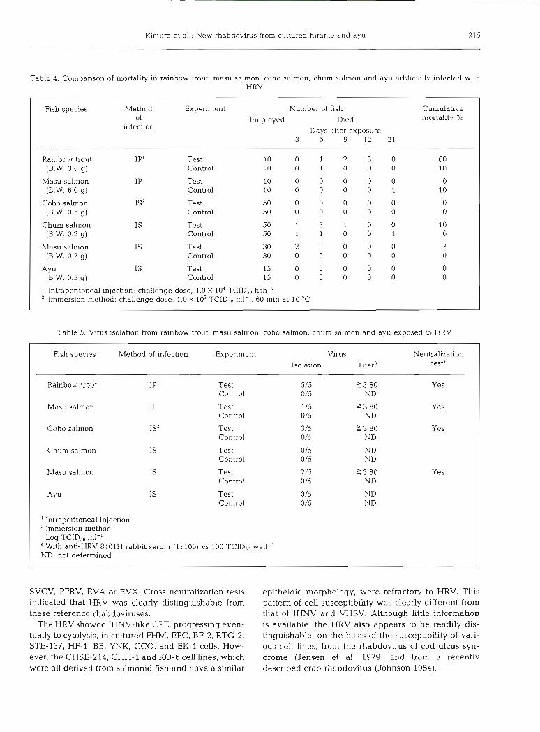
Table 4 gives the results of the pathogenicity tests. Of the 2 species of salmonids challenged with HRV by injection, only 1 (rainbow trout) proved susceptible. In addition, none of the 3 species of salmonids and ayu challenged with HRV by the immersion method developed signs of disease even though 1 of them (the ayu) was known to be suspectible to the virus under hatche~y conditions.
Virus isolations were carried out on 5 dead or surviv- ing fish from each virus-challenged and control group. Virus was recovered from 5 individual rainbow trout (all mortalities); from 1 of 2 dead masu salmon fry and from 1 of 3 surviving masu salmon fry; and from 3 of 5 surviving coho salmon fry (Table 5). Signs of disease were only observed in rainbow trout. All the isolates were neutralized by anti-HRV serum.
Virus titers in tissues of infected rainbow trout were determined for kidney and spleen pools, liver and pancreas pools, heart, intestine, swimbladder and muscle. The virus was recovered from all tissues examined. &dney and spleen pools had the highest titers (i.e. l o 6 0 5 to 106.90 TCIDso ml-l, Table 6).
DISCUSSION
The virus described here, provisionally termed hirame rhabdovirus (HRV) was isolated from cultured hirame and seawater-cultured ayu fry. The virus appears to be a new pathogen of marine fish. No evidence of a similar agent is noted among recent listings of fish viruses by Pllcher & Fryer (1980), Wolf & Mann (1980), Agius (1982), Johnson (1984) or Wolf (1984).
The HRV replicated in several fish cell lines at 5 to 20 "C. It was sensitive to pH 3, diethyl ether, and heat (50 "C for 2 min), but replication was not inhibited by IUdR. The virion was bullet-shaped and measured 80 nm X 180 to 200 nm. These morphological and bio- physical features are characteristic of viruses belong- ing to the famdy Rhabdoviridae. Several other rhabdo- viruses, including IHNV, VHSV, SVCV, PFRV, EVA and EVX are well-known fish pathogens. Another virus, swimbladder inflammation (SBI) virus, proved to be morphologically and serologically identical to SVCV (Bachmann & Ahne 1973, 1974). The HRV was not neutralized with the antisera against IHNV, VHSV,
h m u r a et al.: New rhabdovirus from cultured hirame and ayu
Table 4. Comparison of mortality in rainbow trout, masu salmon, coho salmon, chum salmon and ayu artificially infected with HRV
Fish species Method Experiment Number of fish Cumulative of Died mortal~ty '10
infection Days after exposure 3 6 9 12 21
Rainbow trout IP1 Test 10 0 1 2 3 0 60 (B.W. 3.0 g) Control 10 0 1 0 0 0 10
Employed
Masu salmon (B.W. 6.0 g)
Coho salmon (B.W. 0.5 g)
Chum salmon (B.W. 0.2 g)
Masu salmon (B.W. 0.2 g)
(B.W. 0.5 g) I
IS'
IS
Test Control
Test Control
Test Control
Test Control
Test Control
' Intraperitoneal injechon: challenge dose, 1.0 X 10⁴ TCIDS0 fish-' Immersion method: challenge dose, 1.0 X 10³ TCID,, ml-', 60 min at 10 "C
Table 5. Virus isolation from rainbow trout, masu salmon, coho salmon, chum salmon and ayu exposed to HRV
Fish species Method of infection Experiment Virus Neutralization Isolation Titer3 test4
Rainbow trout IP1 Test 5/5 23.80 Yes Control 0/5 ND
Masu salmon IP Test 1 /5 2 3.80 Yes Control 0/5 ND
Coho salmon IS Test 3/5 23 .80 Yes Control 0/5 ND
Chum salmon IS Test 0/5 ND Control 0/5 ND
Masu salmon IS Test 2/5 2 3.80 Control 0/5 ND
AY u IS Test 0/5 ND Control 0/5 ND
l Intraperitoneal injection ' Immersion method "og TCID,, ml-'
With anti-HRV 8401H rabbit serum (1 : 100) vs 100 TCID,, well-' ND: not determined
Yes
SVCV, PFRV, EVA or EVX. Cross-neutralization tests indicated that HRV was clearly distinguishable from these reference rhabdoviruses.
The HRV showed IHNV-like CPE, progressing even- tually to cytolysis, in cultured FHM, EPC, BF-2, RTG-2, STE-137, HF-1, BB, YNK, CCO, and EK-1 cells. How- ever, the CHSE-214, CHH-l and KO-6 cell lines, which were all derived from salmonid fish and have a similar
epitheloid morphology, were refractory to HRV. This pattern of cell susceptibihty was clearly different from that of IHNV and VHSV. Although little information is available, the HRV also appears to be readily dis- tinguishable, on the basis of the susceptibility of vari- ous cell lines, from the rhabdovirus of cod ulcus syn- drome (Jensen et al. 1979) and from a recently described crab rhabdovirus (Johnson 1984).

Dis. aquat. Org. 1: 20S217, 1986
Table 6. Virus titers in ludney, spleen, liver, pancreas and swim bladder of HRV-infected rainbow trout
Specimen Organ Titer' Log TCID,, g- '
I Kidney + spleen 6.05 Liver + pancreas 4.30 Heart 3.55 Intestine 5.80
k d n e y + spleen 6.80 hver + pancreas 4.05 Swimbladder 5.80 Muscle 4.80
RTG-2 cells used for assay
Although most rhabdoviruses isolated from non-sal- monid fishes (for example SVCV, PFRV, EVA and EVX, the vims isolated from grass carp iAnnt: 13751 and the virus from a North American cichlid [Mals- berger & Lautenslager 19801) replicate at 25 "C or above, HRV did not replicate at 25 "C. A comparison between HRV and Rhabdovirus salmonis (Osadchaya & Nakonechnaya 1981) could not be made because of the limited information available.
The pathogenicity of HRV for hirame was observed following intraperitoneal injection into 100 to 250 g fish (Gorie et al. 1986). The cumulative mortahty was 20 %, but mortality was not observed at 16 "C or above. The mortalities of hirame in natural outbreaks in Hyogo were 25 % (1984) and 7.2 % (1985). For Kagawa Prefecture the mortality was 3.3 % but for Hokkaido it was greater than 90 %. The size of fish affected ranged from 100 to 700 g. The high mortality rate among naturally infected hirame observed in Hokkaido may have been due to the low rearing temperatures in the winter (2 to 5 "C). We are now investigating the ultra- structure of tissues from naturally HRV-infected hirame and will publish the results in the near future.
Experimentally, HRV was lethal for rainbow trout, the cumulative mortality being 60 % . The signs of HRV infection in rainbow trout were similar to those caused by the salmonid virus, VHSV, because hemorrhaging in muscles was commonly seen. Interestingly, Castric & de Kinkelin (1984) have reported that VHSV is also pathogenic for sea ba.ss and turbot. The gross signs and histopathological data reported for these 2 marine species were similar to those observed in HRV- infected hirame.
Chum, coho and masu salmon fry and ayu fry experi- enced low or no mortalities when exposed to HRV by an immersion method. It may therefore be that these species are resistant to the vlrus, or that, in the case of the ayu, the experimental temperatures were too high to favor the virus. However, HRV was isolated from
some of the dead and surviving fish exposed to virus. If the experiment had been extended, higher mortalities might therefore have been observed.
From the evidence obtained thus far, HRV is a new pathogenic virus of fish. This virus is now considered to be an important pathogen of cultured hirame and salmonids in Japan. We propose the name Rhab- dovirus olivaceus n.sp., for this new rhabdovirus because it was first isolated from diseased hirame (Japanese flounder) Paralichthys olivaceus.
Acknowledgements. The authors express their sincere grabtude to Dr. B. J. Hill, Fish Disease Laboratory, Mmistry of Agriculture, Fisheries and Food, U. K.; Dr. P. de Kmkelin. Institute National de la Research Agronornique, France; Dr W. Ahne, lnstitut fiir Zoologie und Hydrobiologie, Universitat ivlunchen, F. R. Germany; and the staff of Hyogo, Kagawa and Okayama Prefectural Fisheries Experimental Stations for their assistance. We thank Drs. J . L. Fryer, J . Rohovec and J . R . Winton, Oregon State University, USA, for their critics! reviews of the paper and their valuable suggestions.
LITERATURE CITED
Aglus, C. (1982). Virus diseases of warm water fish. In: Roberts, R. T. (ed.) Microbial diseases of fish. Academic Press, New York
Ahne, W. (1975). A rhabdovirus isolated from grass carp (Ctenopharyngodon idella var.). Arch. Virol. 48: 181-185
Bachmann, P. A., Ahne, W. (1973). Isolation and charactenza- tion of agent causing swimbladder inflammation in carp. Nature, Lond. 244: 235237
Bachmann, P. A., Ahne, W. (1974). Biological properties and identification of the agent causing swimbladder inflam- mation in carp. Arch. ges. Virusforschung 44: 261-269
Bowser, P. R., Plumb, J. A. (1980). Fish cell lines: establish- ment of a h e from ovaries of channel catfish. In Vitro 16: 365-368
Castric, J. , de Kmkelin, P. (1984). Experimental study of the susceptibility of two marine fish species, sea bass (Picen- trarchus labrax) and turbot (Scophthalmus maximus), to viral haemorrhagic septicaemia. Aquaculture 46: 203-212
Chen, S. W., Ueno, Y., Kou, G. H. (1982). A cell line derived from Japanese eel ( A n g d a japonica) kidney. Proc. Natl. Sci. Coun., ROC 6: 93-100
Gorie, S., Nakamoto, K., Katashima, K (1985a). Diseases of cultured hirame (Japanese flounder, Paralichthys olivaceus) - I. Prehminary report on a disease of marine pen cultured hirame may be caused by viral infection. Bull. Hyogo Pref. Fish. Exp. Stn 23: 6 6 6 8
Gorie, S., Katashima, K. , Tahata, K. (1985b). Disease of cul- tured ayu (Plecoglossus altivelis). Ann. Rep. Hyogo Pref. Fish. Exp. Stn, 297-300
Gone, S., Katashlma, K., Tahata, K . (1986). Disease of cul- tured hrarne (Japanese flounder, Parahchthys olivaceus) - 11. Pathogenicity of a virus isolated from cultured hirame. Fish Pathol. 21: in press
Gravel], M., Malsburger, R. G. (1965). A permanent cell line from the fathead rmnnow (hmephalespromelas). Ann. N. Y Acad. Sci. 126: 555-565
Hs~ung, G . D. (1982). Hemagglutination and the hernaggluti- nation-mhibition test. In: Diagnostic virology. Yale Unl- versity Press, New Haven and London. p. 276
Kimura et al.: New rhabdovirus from cultured hirame and ayu
Jensen, N. I., Bloch, B., Larsen, J . L. (1979). The ulcus-syn- drorne in cod (Gadus morhual-111. A preliminary virolog- cal report. Nordisk Veterinaernledicln 31: 436-442
Johnson, P. T. (1984). Viral hseases of marine invertebrates. Helgolander Meeresunters. 37: 65-98
Lannan, C. N., Winton, J . R., Fryer, J. L. (1984). Fish cell lines: Establishment and characterization of nine cell lines from salrnonids. In Vitro 20: 671-676
Malsberger, R. G., Lautenslager, G. (1980). Fish viruses: rhab- dovirus isolated from a species of the family Cichlidae. Fish Health News 9: i-ii
Osadchaya, Ye. F., Nakonechnaya, M. G. (1981). Rhabdovirus salmonis, the cause of a new disease In rainbow trout, Salmo gairdnen. J . Ichthyol. 21: 113-121
Pilcher, K. S., Fryer. J. L. (1980). The viral diseases of fish: a review through 1978. In: Isenberg, H. D. (ed.) CRC critical reviews in microbiology, Vol 7, p. 287-363; Vol 8, p. 1-25.
Reed, L. J . , Muench, H. (1938). A simple method of estimating fifty percent end points. Am. J . Hygiene 27: 493-497
Tomasec, J., Fijan, N. (1971). Virusne bolestiriba (viral dis- eases of fish). Final report on research under a part of project 6 nl1966, Zagreb
Watanabe, T., Kobayash~, N , Sato, Y., Ishizah, Y. (1978). Continuous cell line derived from ladney of Yamame. Oncorhynchus masou. Bull. Jap. Soc. scient Fish. 44: 415-418
Wolf, K. (1984). Diseases of pisces; Diseases caused by mic- roorganisms; Agents. Virales. In- k n n e , 0. (ed.) Diseases of marine animals. Vol. IV, Part 1. Biologische Anstalt Helgoland, Hamburg, p. 17-47
Wolf, K., Mann, J . A. (1980). Poilulotherm vertebrate cell lines and viruses: A current listing for fishes. In Vitro 16: 168-179
Wolf, K., Quimby, M. C. (1962). Established eurythermic line of fish cells in vitro. Science 135: 10651066
Wolf, K., Quimby, M. C. (1966). Lymphocystis virus: Isolation and propagahon in centrarchid fish cell h e s . Science 151: 1004-1005
Responsible Subject Editor: Dr. T. Evelyn; accepted for printing on July 4. 1986