A Case of Clinical Transformation between C3 ... · SPEP/UPEP, ANA, and ANCA titers were negative,...
3
Remedy Publications LLC. Journal of Clinical Nephrology & Kidney Diseases 2017 | Volume 2 | Issue 1 | Article 1010 1 Case Presentation A 49-year-old African American male, with a self-reported history of hypertension and colitis, who presented to the hospital with complaints of abdominal pain, dyspnea on exertion, and new- onset lower extremity edema. e patient’s home medications included lisinopril, amlodipine, hydrocaholorothiazide, and acetaminophen. Physical exam was significant for +2 pedal edema. His hemoglobin and hematocrit were low at 6.2 gm/dl and 20%, respectively, and his platelet count was 285,000 thou/cmm. His BUN and creatinine at that time were 29 mg/dl and 2.2 mg/dl, respectively, with a serum albumin of 1.9 gm/dL and 2+ proteinuria on his dipstick and no microscopic findings on UA. Nephrology consultation was not requested on this admission. He underwent colonoscopy which showed severe ulcerative colitis spanning the length of his colon. He was treated with mesalamine, given two units of packed red blood cells, and was eventually discharged home aſter symptomatic relief. Approximately one month later, the patient was admitted again to the hospital with complaints of recurrent pedal edema and poorly controlled hypertension (BP: 160/90 mmHg). At that time, his hemoglobin and hematocrit were 8.2 g/dl and 24%, respectively and platelet count of 270 thou/cmm. His creatinine increased to 2.96 mg/dl, his UA showed pH of 7.0, specific gravity of 1.009, >100 RBC per HPF, 14 WBC and showed 2+ proteinuria, with a random urine protein/creatinine ratio of 8 g/g. A serologic work-up was positive only for a low C3 of 53. C4 was within normal limits at 35.5; SPEP/UPEP, ANA, and ANCA titers were negative, as were hepatitis and HIV serologies. Renal ultrasound demonstrated normal sized kidneys with no evidence of hydronephrosis or obstruction. He underwent a renal biopsy. Results showed proliferative and crescentic glomerulonephritis staining for C3 only, (Figure 1), including some glomeruli with collapsing features, the presence of sub epithelial humps on EM, and a moderate degree of tubular atrophy and interstitial fibrosis (Figure 2 and 3), and no evidence of thrombotic microangiopathy. As the biopsy diagnosis was consistent with C3 Glomerulonephritis (C3GN), a disease for which there is no well-defined treatment, he continued to be treated conservatively with renin-angiotensin blockade and was referred into a clinical trial for immune therapy. Less than 2 weeks aſter the biopsy results returned, the patient presented again to the hospital with complaints of shortness of breath. Physical examination was significant for worsening pedal edema; neither skin rash nor petechiae were seen. At the time of admission, he was profoundly anemic, with a HCT of 10.9%, and thrombocytopenia with a platelet count of 53,000 thou/cmm. Haptoglobin was undetectable and LDH was 2,317 U/L. He was found to have a creatinine of 15 A Case of Clinical Transformation between C3 Glomerulonephritis and Atypical HUS OPEN ACCESS *Correspondence: Faizan Babar, Department of Nephrology, George Washington University, Washington DC 20052, USA, E-mail: [email protected] Received Date: 02 Jun 2017 Accepted Date: 24 Jul 2017 Published Date: 02 Aug 2017 Citation: Babar F, Cohen S, Brown E. A Case of Clinical Transformation between C3 Glomerulonephritis and Atypical HUS. J Clin Nephrol Kidney Dis. 2017; 2(1): 1010. Copyright © 2017 Faizan Babar. This is an open access article distributed under the Creative Commons Attribution License, which permits unrestricted use, distribution, and reproduction in any medium, provided the original work is properly cited. Case Report Published: 02 Aug, 2017 Faizan Babar 1 *, Scott Cohen 1 and Eric Brown 2 1 Department of Nephrology, George Washington University, Washington DC, USA 2 Mid-Atlantic Nephrology Associates, Glen Burnie, Maryland, USA Abstract e clinical and pathological approach to (MPGN) has evolved in recent years. Traditionally classified into types I, II, and III MPGN based on electron microscopic findings, MPGN is now being classified by immunofluorescence microscopy. is shiſt in classification aims to define MPGN by the underlying pathogenic process as opposed to the histologic appearance alone, which was flawed due to significant overlap among the different types [1]. Currently, the bulk of MPGN is designated either as an immune-complex-mediated lesion or a lesion due to persistent activation of the alternative complement pathway, though MPGN caused by direct endothelial injury has also been described [2]. In the following case, we present a patient with complement-mediated MPGN, defined ultra-structurally as C3GN, who went on to develop aHUS. Keywords: C3 glomerulonephritis; Atypical HUS
Transcript of A Case of Clinical Transformation between C3 ... · SPEP/UPEP, ANA, and ANCA titers were negative,...
Remedy Publications LLC.
Journal of Clinical Nephrology & Kidney Diseases
2017 | Volume 2 | Issue 1 | Article 10101
Case PresentationA 49-year-old African American male, with a self-reported history of hypertension and colitis,
who presented to the hospital with complaints of abdominal pain, dyspnea on exertion, and new-onset lower extremity edema. The patient’s home medications included lisinopril, amlodipine, hydrocaholorothiazide, and acetaminophen. Physical exam was significant for +2 pedal edema. His hemoglobin and hematocrit were low at 6.2 gm/dl and 20%, respectively, and his platelet count was 285,000 thou/cmm. His BUN and creatinine at that time were 29 mg/dl and 2.2 mg/dl, respectively, with a serum albumin of 1.9 gm/dL and 2+ proteinuria on his dipstick and no microscopic findings on UA. Nephrology consultation was not requested on this admission. He underwent colonoscopy which showed severe ulcerative colitis spanning the length of his colon. He was treated with mesalamine, given two units of packed red blood cells, and was eventually discharged home after symptomatic relief.
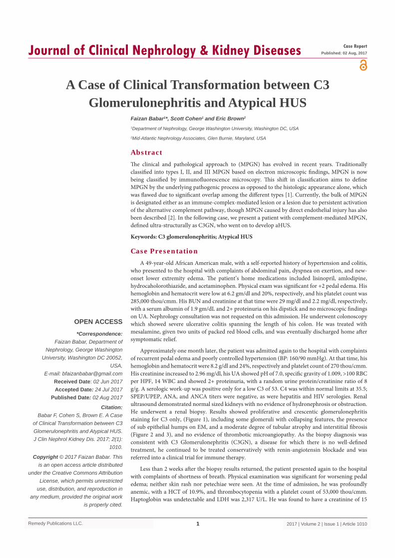
Approximately one month later, the patient was admitted again to the hospital with complaints of recurrent pedal edema and poorly controlled hypertension (BP: 160/90 mmHg). At that time, his hemoglobin and hematocrit were 8.2 g/dl and 24%, respectively and platelet count of 270 thou/cmm. His creatinine increased to 2.96 mg/dl, his UA showed pH of 7.0, specific gravity of 1.009, >100 RBC per HPF, 14 WBC and showed 2+ proteinuria, with a random urine protein/creatinine ratio of 8 g/g. A serologic work-up was positive only for a low C3 of 53. C4 was within normal limits at 35.5; SPEP/UPEP, ANA, and ANCA titers were negative, as were hepatitis and HIV serologies. Renal ultrasound demonstrated normal sized kidneys with no evidence of hydronephrosis or obstruction. He underwent a renal biopsy. Results showed proliferative and crescentic glomerulonephritis staining for C3 only, (Figure 1), including some glomeruli with collapsing features, the presence of sub epithelial humps on EM, and a moderate degree of tubular atrophy and interstitial fibrosis (Figure 2 and 3), and no evidence of thrombotic microangiopathy. As the biopsy diagnosis was consistent with C3 Glomerulonephritis (C3GN), a disease for which there is no well-defined treatment, he continued to be treated conservatively with renin-angiotensin blockade and was referred into a clinical trial for immune therapy.
Less than 2 weeks after the biopsy results returned, the patient presented again to the hospital with complaints of shortness of breath. Physical examination was significant for worsening pedal edema; neither skin rash nor petechiae were seen. At the time of admission, he was profoundly anemic, with a HCT of 10.9%, and thrombocytopenia with a platelet count of 53,000 thou/cmm. Haptoglobin was undetectable and LDH was 2,317 U/L. He was found to have a creatinine of 15
A Case of Clinical Transformation between C3 Glomerulonephritis and Atypical HUS
OPEN ACCESS
*Correspondence:Faizan Babar, Department of
Nephrology, George Washington University, Washington DC 20052,
USA,E-mail: [email protected]
Received Date: 02 Jun 2017Accepted Date: 24 Jul 2017
Published Date: 02 Aug 2017
Citation: Babar F, Cohen S, Brown E. A Case
of Clinical Transformation between C3 Glomerulonephritis and Atypical HUS. J Clin Nephrol Kidney Dis. 2017; 2(1):
1010.
Copyright © 2017 Faizan Babar. This is an open access article distributed
under the Creative Commons Attribution License, which permits unrestricted
use, distribution, and reproduction in any medium, provided the original work
is properly cited.
Case ReportPublished: 02 Aug, 2017
Faizan Babar1*, Scott Cohen1 and Eric Brown2
1Department of Nephrology, George Washington University, Washington DC, USA
2Mid-Atlantic Nephrology Associates, Glen Burnie, Maryland, USA
AbstractThe clinical and pathological approach to (MPGN) has evolved in recent years. Traditionally classified into types I, II, and III MPGN based on electron microscopic findings, MPGN is now being classified by immunofluorescence microscopy. This shift in classification aims to define MPGN by the underlying pathogenic process as opposed to the histologic appearance alone, which was flawed due to significant overlap among the different types [1]. Currently, the bulk of MPGN is designated either as an immune-complex-mediated lesion or a lesion due to persistent activation of the alternative complement pathway, though MPGN caused by direct endothelial injury has also been described [2]. In the following case, we present a patient with complement-mediated MPGN, defined ultra-structurally as C3GN, who went on to develop aHUS.
Keywords: C3 glomerulonephritis; Atypical HUS

Faizan Babar, et al., Journal of Clinical Nephrology & Kidney Diseases
Remedy Publications LLC. 2017 | Volume 2 | Issue 1 | Article 10102
mg/dl, significantly elevated from his previous of ~2.9 mg/dl. A hematopathologist personally reviewed the smear and confirmed suspected hemolysis with a large schistocytes burden. He was started on dialysis and plasmapheresis pending the results of an ADAMTS13 level. Once the ADAMTS13 returned in the normal range (activity 43%, antibody 11 u/ml), plasmapheresis was stopped and he was started on eculizumab. The patient received ciprofloxacin and meningioccocal vaccination prior to initiating eculizumab. At the end of the patient’s hospital stay of ten days, all hematologic parameters (HCT, platelets, LDH, haptoglobin) had normalized but he remained dialysis dependent. Genotyping, performed to screen for mutations in genes known to code for complement regulatory factors, returned negative.
DiscussionC3 Glomerulopathy is a term that encompasses dense deposit
disease (DDD) and C3 glomerulonephritis. It is characterized by a glomerular lesion with predominant C3 complement deposition (typically in the absence of C1q, C4, IgG and IgA) [3-5]. On newer IF classification criteria, C3 Glomerulopathy is described as a staining pattern that is C3 dominant and at least two orders of magnitude more intense than any combination of IgG, IgM, IgA, and C1q [6]. On electron microscopy, C3 GN is characterized by sub epithelial or mesangium electron-dense deposits compared to predominant intra membranous deposits seen in DDD [3-5]. Membranoproliferative glomerulonephritis (MPGN) is the most common light microscopic finding in C3 glomerulopathy [7].
The complement system is an integral part of innate immunity, divided into three different pathways, the lectin, classical, and alternate pathways (AP). Studies have shown the role of AP dysregulation in pathogenesis of C3GN and aHUS. In contrast to the specific protein:protein or protein:carbohydrate interactions that characterize classical and lectin pathway activation, the unique feature of AP is the
auto activation by a process termed as physiologic tick over of C3 [8]. The conformationally altered C3 known as C3(H2O) is capable of binding and activating other factors leading to AP activation. Alternate complement pathway also serves as amplification loop for fixed C3b generated by lectin and classical pathway. C3b then binds the factor b resulting in confirmation change in factor B that allows factor D to cleave it similarly to tick over process [8].
The activity of alternate complement pathway is regulated by various complement factors (complement factor H, complement factor I and membrane complex protein) found in serum and on cell surface. Inherited and acquired abnormalities in complement regulatory proteins lead to AP dysregulation. One study found similar genetic mutations in C3GN and aHUS [9]. This suggests that genetically predisposed individuals might be at risk for having aHUS on a background of C3GN. More than 100 mutations have been identified in genes coding for complement factor H, MCP, and factor I. Most of the mutation confers a distinct phenotype and the extension of disease spectrum in humans is very rare. Mutation in CFH gene is associated with both C3GN and aHUS [9]. CFH is single-polypeptide chain glycoprotein and consists of 20 short consensus repeats (SCRs)
Age Complement levels CFH, MCP, CFI mutations
C3Nef, Anit-CFH antibody
Time interval to the onset of aHUS
CFH haplotype/ SNP in CHF,CFI, MCP
4 months Normal C3,MCP,CFHR1/R3 AntiCFH+ 6 years N/A
13 years Low C3 CFH p.I216T;SCR4 Not detected 3 years H1/H3CFH
54 years Low C3 No mutations Not detected 4 years H1/H3CFH
22 years Normal N/A Not detected 2.5 years H1/H3CFH
36 years Low C3 No mutations AntiCFH + 3 years N/A
1.5 years Low C3 CFH mutation Not detected 2 years SNP in MCP
New born Low C3 CFH mutation N/A 3 years N/A
Table 1: Reported cases of atypical HUS and C3 GN manifesting together.
Figure 1: Immunoflourescence studies glomeruli stain for C3+, IgM,IgG C1q- Figure 2: H&E stain glomeruli showing MPGN changes.
Figure 3: EM glomeruli shows sub epithelial immune deposits, mild tubular atrophy.

Faizan Babar, et al., Journal of Clinical Nephrology & Kidney Diseases
Remedy Publications LLC. 2017 | Volume 2 | Issue 1 | Article 10103
with two main functional domains positioned at the opposite ends of the protein. The N terminus (SCRs 1-4) is responsible for the fluid-phase complement regulatory functions and C-terminus (SCRs1 9-20) regulates cellular phase functions. Genetic mutations associated with C3GN are concentrated at N terminus, whereas genetic mutations in aHUS are clustered at C-terminus (SCR19-20) [10]. Similarly, different autoantibodies against CFH cause distinct functional abnormalities. C3 nephritic factor autoantibody sometimes seen in C3GN binds CFH at SCR 1-4 N-terminus whereas anti-CFH antibody seen aHUS binds CFH at the C-terminus SCR19-20 [10]. These findings suggest specific abnormalities in the CFH protein lead to distinct phenotypes. Single nucleotide polymorphism (SNP) in CFH gene is reported with different phenotypes [10]. There have been seven cases reported in the literature with the diagnoses of C3GN and aHUS in the same individual [7,11,12]. We reviewed these cases to determine the disease onset, presence of a genetic mutations or antibodies against complement proteins, and the time interval between C3GN and aHUS (Table 1). Three out of seven patients carried mutation in CFH gene; one patient had mutation in C3, MCP, CFI genes. Three patients had CFH1/H3 haploinsufficiency and one patient with SNP in MCP gene. The phenotypic variation between C3GN and aHUS was prevalent in transplant kidneys. Time interval between C3GN and aHUS is 6m to 6y. In 4 of 7 reported cases including our case, there was no identifiable cause. The intriguing finding we learned in our case was the acute clinical transformation from C3GN to aHUS and the presence of additional immune mediated disease, not seen in other cases.
The exact mechanism of acute transformation between C3GN and aHUS in the same individual is not well understood. We hypothesize that ulcerative colitis may contribute to hyper-activation of the AP of complement leading to acute transformation between the relatively clinically indolent C3GN and systemic TMA secondary to aHUS. We think AP pathway also enhances the role of the classical and lectin pathways in ulcerative colitis by an amplification loop, thereby increasing the generation of surface C3b which ultimately leads to greater production of C5a and MAC (membrane attack complex). The pro-inflammatory activity of C5a might play a crucial role in acute phenotypic variation between C3GN and aHUS as suggested in animal models [13]. Pickering et al. [5] demonstrated that mice with partial hepatocyte-CFH deficiency developed aHUS in the background of C3GN by adding a second insult in the form of a nephrotoxic agent. The experiment suggested the significance of C5a in the phenotypic variation between C3GN and systemic TMA. This may indicate the role of therapies targeting terminal complement blockade in preventing the phenotypic extension of diseases caused by uncontrolled AP activity.
ConclusionThe clinical transformation of C3GN and aHUS is very rare and
poorly understood. There is no genetic mutation reported in some
of the cases of C3GN and aHUS, which suggests some unidentified mutations causing similar phenotypes. Dysregulation of AP activity is common to both diseases, and the presence of additional immunologic disease may potentiate the activity of AP pathway leading to clinical transformation between C3GN and aHUS.
References1. Bomback AS, Appel GB. Pathogenesis of the C3 glomerulopathies and
reclassification of MPGN. Nat Rev Nephrol. 2012;8(11):634-42.
2. Sethi S, Nester CM, Smith RJ. Membranoproliferative glomerulonephritis and C3 glomerulopathy: resolving the confusion. Kidney Int. 201;81(5):434-41.
3. Medjeral-Thomas NR, O'Shaughnessy MM, O'Regan JA, Traynor C, Flanagan M, Wong L, et al. C3 glomerulopathy: clinicopathologic features and predictors of outcome. Clin J Am Soc Nephrol. 2014;9(1):46-53.
4. Walker PD, Ferrario F, Joh K, Bonsib SM. Dense deposit disease is not a membranoproliferative glomerulonephritis. Mod Pathol. 2007;20(6):605-16.
5. Pickering M, D'Agati VD, Nester CM, Smith RJ, Haas M, Appel GB, et al. C3 glomerulopathy: consensus report. Kidney Int. 2013;84(6):1079-89.
6. Hou J, Markowitz GS, Bomback AS, Appel GB, Herlitz LC, Barry SM, et al. Towards a working definition of C3 glomerulopathy by immunofluorescence. Kidney int. 2014;85(2):450-6.
7. Manenti L, Gnappi E, Vaglio A, Allegri L, Noris M, Bresin E, et al. Atypical haemolytic uraemic syndrome with underlying glomerulopathies. A case series and a review of the literature. Nephrol Dial Transplant. 2013;28(9):2246-59.
8. Muller-Eberhard HJ. Molecular organization and function of the complement system. Annu Rev Biochem. 1988;57:321-47.
9. Servais S, Frémeaux‐Bacchi V, Lequintrec M, Salomon R, Blouin J, Knebelmann B, et al. Primary glomerulonephritis with isolated C3 deposits: a new entity which shares common genetic risk factors with HUS. J Med Genet. 2007;44(3):193-9.
10. de Córdoba SR, de Jorge EG. Translational mini-review series on complement factor H: genetics and disease associations of human complement factor H. Clin Exp Immunol. 2008;151(1):1-13.
11. Lorcy N, Rioux-Leclercq N, Lombard ML, Le Poqamp P, Viqneau C. Three kidneys, two diseases, one antibody? Nephrol Dial Transplant. 2011;26(11):3811-3.
12. Boyer O, Noël LH, Balzamo E, Guest G, Biebuyck N, Charbit M, et al. Complement Factor H deficinecy and posttranslation glomerulonephritis with isolated C3GN deposits. Am J Kidney Dis. 2008;51(4):671-7.
13. Vernon KA, Ruseva MM, Cook HT, Botto M, Malik TH, Pickering MC. Partial Complement Factor H Deficiency Associates with C3 Glomerulopathy and Thrombotic Microangiopathy. J Am Soc Nephrol. 2016;27(5):1334-42.

![osteoporosispatients2015 [Read-Only] · • SPEP/UPEP • TTGIGA (celiac) • Phosphorus • Magnesium • PTH. 6/11/2015 7 Who do we treat? 6/11/2015 8. 6/11/2015 9 FRAX. 6/11/2015](https://static.fdocuments.net/doc/165x107/5aec58b47f8b9a90318e2a89/osteoporosispatients2015-read-only-spepupep-ttgiga-celiac-phosphorus.jpg)