Paraproteinaemias. Multiple myeloma. Amyloidosis. Part 1...
54
Paraproteinaemias. Multiple myeloma. Amyloidosis. Part 1 Dr. Gábor Mikala
Transcript of Paraproteinaemias. Multiple myeloma. Amyloidosis. Part 1...

Paraproteinaemias. Multiplemyeloma. Amyloidosis.
Part 1
Dr. Gábor Mikala

Subtypes of Plasma Cell Disorders
• Increased Plasma Cells and Paraproteins
– Monoclonal Gammopathy (of unknown significance, MGUS)
– Multiple Myeloma (MM)
– Waldenström’s Macroglobulinemia (WM, IgM)
• Increased / Altered Products of Plasma Cells (that may be few)
– Light Chain Amyloidosis
– Light Chain Deposition Disease
Lymphoblast
Lymphoplasmacyte
Germinal Center
SOMATIC
IgM
Lymph node
B cell Development As a Framework for Malignancies
BoneMarrow
G,A,E Plasma cell
Virgin B cell
Lymphoblast
Plasmablast
Pre-B cell
HYPERMUTATION
V(D)J RECOMBINATION
SWITCHRECOMBINATION

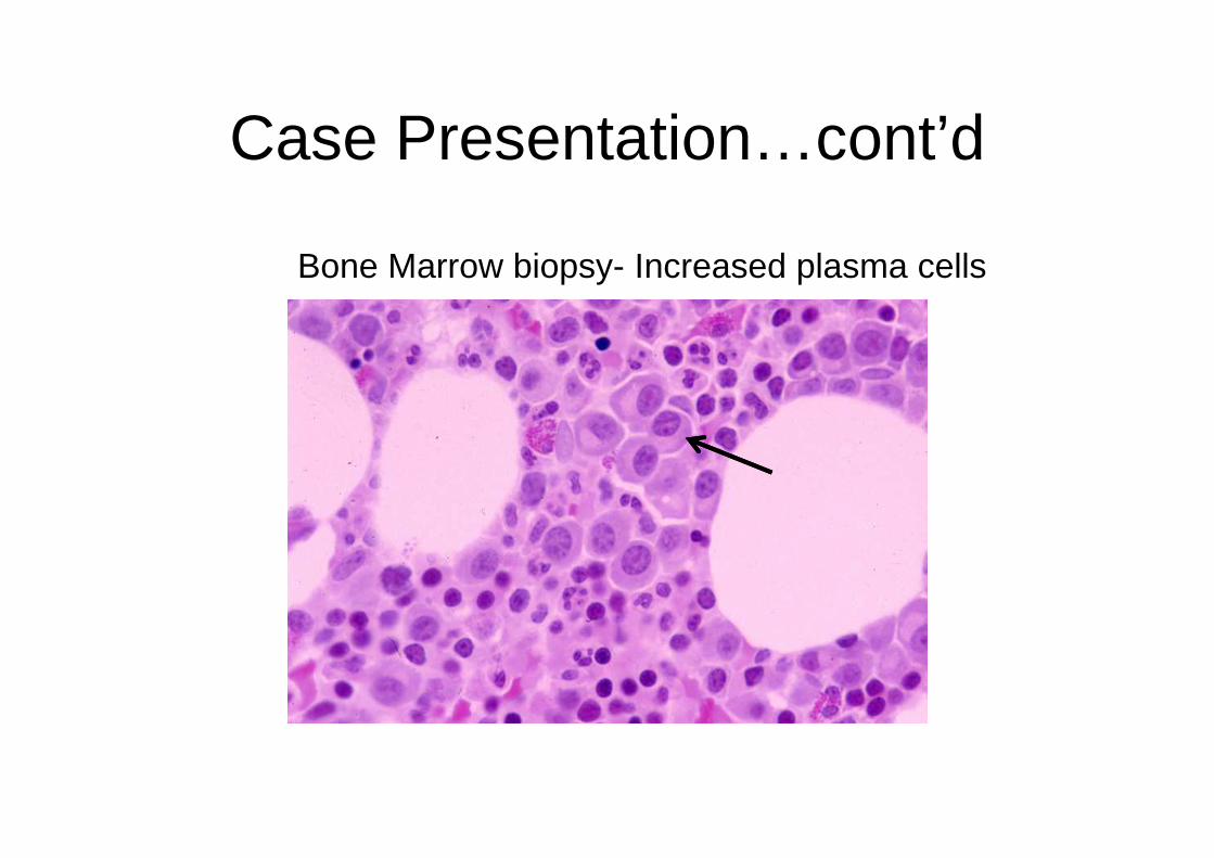
B cell Development As a Framework for Malignancies
Lymphoblast
Lymphoplasmacyte
Germinal Center
SOMATIC
IgM WALDENSTROM’S
Lymph node
BoneMarrow
MULTIPLEMYELOMA
G,A,E Plasma cell
Virgin B cell
Lymphoblast
Plasmablast
Pre-B cell
HYPERMUTATION
CLL
BURKITT’SLYMPHOMAFOLLICULAR
LYMPHOMA
ALL

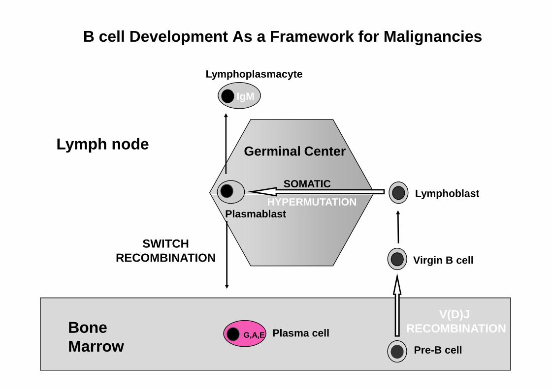
Case Presentation
• 48 yo male• Admitted with 2 wk history of fatigue and diffuse
bone pain• Evaluation: • Evaluation:
– Increased serum creatinine 500 umol/l – Increased serum calcium 2.9 mmol/l– Increased serum globulin 50 g/l (normal < 3 g/dl)

Evaluation of Abnormal Serum Globulins
Serum
Serum/Urine Protein Electrophoresis (S/UPEP)
ImmunofixationElectrophoresis (IFE)
Urine
Serum M spike 40 g/l ; typed as IgG kappa monoclonal Ig
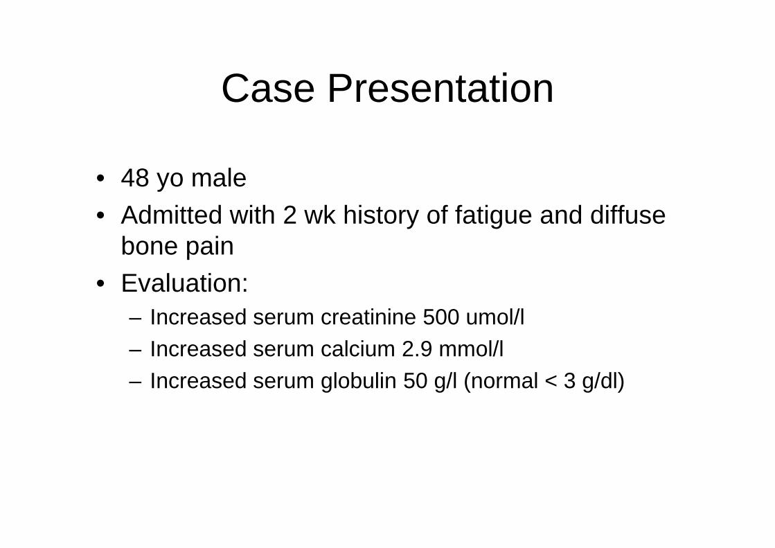
Case Presentation…cont’d
Bone Marrow biopsy- Increased plasma cells
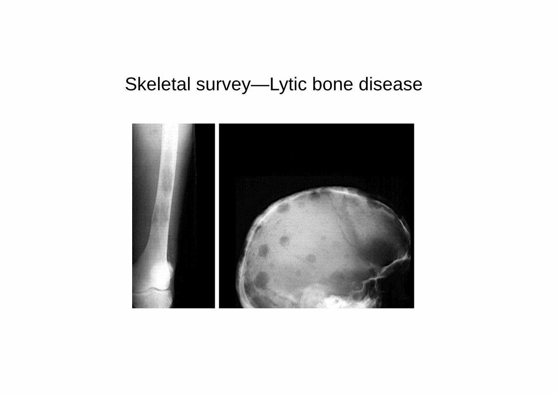
Skeletal survey—Lytic bone disease
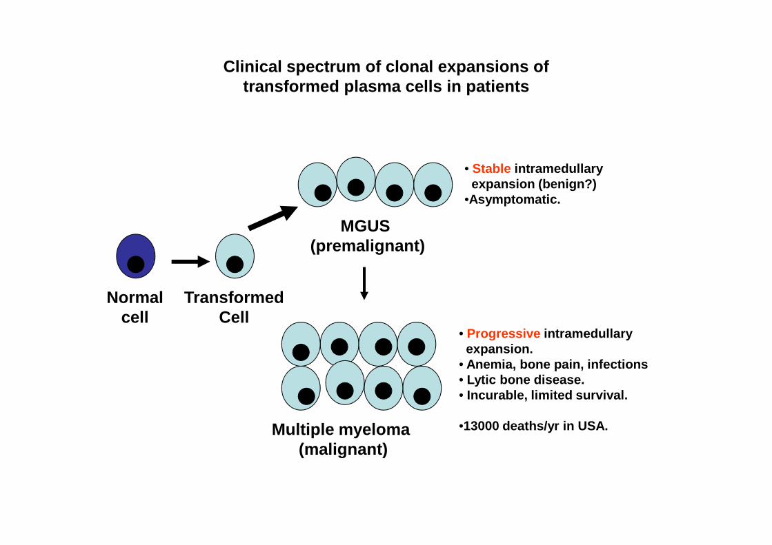
MGUS (premalignant)
Clinical spectrum of clonal expansions of transformed plasma cells in patients
• Stable intramedullaryexpansion (benign?)
•Asymptomatic.
Normalcell
TransformedCell
Multiple myeloma (malignant)
• Progressive intramedullaryexpansion.
• Anemia, bone pain, infections• Lytic bone disease.• Incurable, limited survival.
•13000 deaths/yr in USA.


Diagnostic Criteria in Monoclonal Gammopathies
• MGUS– < 10% bone marrow plasma cells and M spike < 3 g/dl– Monoclonal protein / clonal plasma cell population– No End organ damage
• Myeloma• Myeloma– > 10% clonal bone marrow plasma cells– End Organ Damage or very high risk thereof
• Indolent / Smoldering Myeloma– > 10% marrow plasma cells or M spike > 3 g/dl– No End organ damage and no high risk of that

Criteria for End-Organ Damage
in Monoclonal Gammopathies
• CRAB– Calcium > 0.3 mmol/l above ULN– Renal Insufficiency (> 180 umol/l)– Anemia (< 100 g/l)– Anemia (< 100 g/l)– Bone Lesions (lytic lesions or osteopenia)
• High risk of progression to symptomatic disease– >60% marrow plasma cells– FLC ratio >100– >1 focal lesion on MRI

A Model for Pathogenesis of Myeloma


Monoclonal Gammopathy of Undetermined
Significance (MGUS)
• Common, age-related• Prevalence: 3.2% in persons over 50 yrs old (Minnesota)
– ~5% in age >70
• Higher prevalence in African populations.• ? Association with inflammatory states: obesity, Gaucher’s disease
• Increased risk for thrombosis, neuropathy and fractures
• Risk of progression in entire population: 1% /yr
• Risk factors for progression: %PC, level M spike, Free light chain, IgA protein, ?Decline in uninvolved Ig’s

Smoldering Myeloma (SMM)
• Patients with PC > 10% or M spike > 3 g/dl, but lacking CRAB
symptoms.
• 10% per yr progression to overt MM
• Most eventually require therapy.
• Current recommendation is observation until progressive disease
– Except in patient with extremely high risk of progression (defining events).

Disease Progression in MGUS and SMM
Kyle et al. NEJM 356: 2582, 2007

Risk Groups in Asymptomatic Myeloma
G1: BMPC >10% M > 3 g/dl;
G2: BMPC >10% M < 3 g/dl
G3: BMPC <10% M > 3 g/dl
Kyle et al. NEJM 356: 2582, 2007

Multiple myeloma
• Uncontrolled proliferation of Ig secreting plasma cells
– most commonly IgG (57%), IgA (21%) or light chain only (18%)
• Twice as frequent in men as • Twice as frequent in men as women, and in blacks as whites
• 1% of all cancers
– 2% in African Americans
• Incurable
• Median survival 4-6 years

5 year survival (SEER)

Work-up in suspected myeloma
• Assessment of serum/urine protein– SPEP/IF, 24 hr urine for UPEP/IF – Free light chains (kappa, lambda)
• CBC, sCr, Calcium, Albumin, LDH, • Serum beta 2 microglobulin (B2M)• Skeletal survey or low dose total body CT• Skeletal survey or low dose total body CT• Bone marrow aspirate and/or biopsy
– Cytogenetics (including FISH)• Under investigation:
– MRI spine– PET scans– Bone densitometry, Urine n-telopeptide

* Kappa and Lambda FLC’s are both increased in renal failure/CKD

Key clinical aspects of myeloma
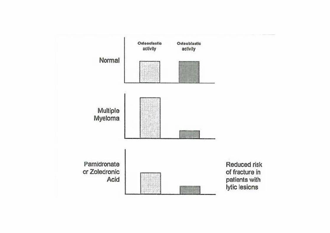
• Predominantly intra-medullary growth.• Absence of clinical LN or spleen involvement.• Low proliferative fraction.• Long periods of stability in MGUS.• Osteoclast activation, osteoblast inhibition, and bone loss.• Multi-focal growth of tumor cells.

Clinical presentation

OsteoclastOsteoblast
LYTIC BONE DZHYPERCALCEMIA
Ig depositionCast nephropathyRENAL FAILURE
Manifestations of Clonal Plasma Cell Proliferation
ErythropoiesisANEMIA
Immune-paresisHypogammINFECTION

Historical aside…
At age 39, developed fatigue and bone pain from several fractures. She died 4 years later; autopsy showed that her marrow was replaced by a red, gelatinous substance

Multiple Myeloma: Skeletal Complications


Renal Pathology in MMLight Chain Deposition Disease
Light Chain Cast Nephropathy AL Amyloid


International Staging System
• Stage I (B2M <3.5 mg/l; Albumin >35 g/l)
– Median OS 62 months
• Stage III (B2M >5.5 mg/l)
– Median OS 29 months– Median OS 29 months
• Stage II (Neither Stage I or III)
– Median OS 44 months

Principles Of Treatment
• No evidence that early treatment prolongs survival – with the
exception of extremely high risk of progression
• Wait for symptoms, or evidence of disease progression, to start
treatment
• Supportive measures are critically important• Supportive measures are critically important
– drink plenty of fluids daily
– treat infections promptly
– prophylactic bisphosphonates reduce skeletal complications in
patients with osteopenia and lytic bone disease
– anemia often responds to erythropoietin.

“Myeloma treatment is a marathon, not a sprint”


JCO, 30(4), Feb 2012


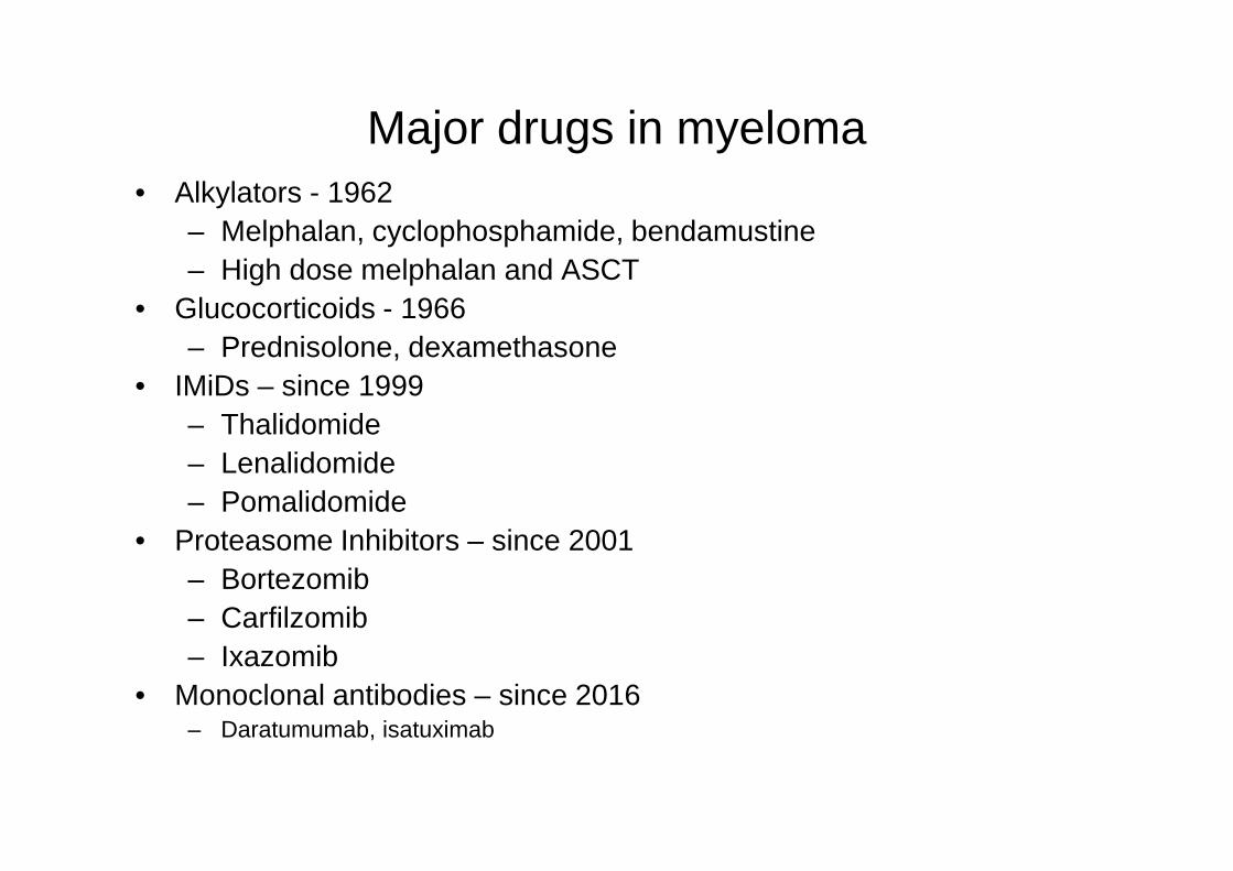
Major drugs in myeloma• Alkylators - 1962
– Melphalan, cyclophosphamide, bendamustine– High dose melphalan and ASCT
• Glucocorticoids - 1966– Prednisolone, dexamethasone
• IMiDs – since 1999– Thalidomide – Thalidomide – Lenalidomide– Pomalidomide
• Proteasome Inhibitors – since 2001– Bortezomib– Carfilzomib– Ixazomib
• Monoclonal antibodies – since 2016– Daratumumab, isatuximab

Treatment course
Asymptomatic
MGUSStable MM
Symptomatic Acute
PancytopeniaPlasma cell leukemiaStable MM Plasma cell leukemia
Years Months Days
Treatments
M protein

Cytogenetic Profiles of Myeloma
Carrasco et al Cancer Cell, Sawyer et al CGG, 2011

Percentage
of patients
Clinical and laboratory features
Hyperdiploid 45 More favorable, IgG-κ, older patients.
Non-hyperdiploid 40 Aggressive, IgA-λ, younger individuals
Cyclin D
translocation
18
t(11;14)(q13;q32) 16 Upregulation of CCND1; favorable prognosis; bone lesions. Two subtypes
by GEP; CD20+ in one subset
t(6;14q)(p21;32) 2 Probably same as CCND1
IMWG Molecular Classification of Myeloma
t(6;14q)(p21;32) 2 Probably same as CCND1
t(12;14)(p13;q32) <1 Rare
MMSET
translocation
15
t(4;14)(p16;q32) 15 Upregulation of MMSET; upregulation of FGFR3 in 75%
unfavorable prognosis with conventional therapy; bone lesions less
frequent, responds well to bortezomib
MAF translocation 8 Aggressive
t(14;16)(q32;q23) 5 Confirmed as aggressive by at least two series
t(14;20)(q32;q11) 2 One series shows more aggressive disease.
t(8;14)(q24;q32) 1 Unknown effect on outcome but presumed aggressive.
Unclassified (other) 15 Various subtypes and some with overlap
IMWG. Leukemia 2010

Factors Associated with Increased Disease Risk in MM
• Gene expression profile (GEP) 70 (or GEP15) high risk signature
• FISH:
– t(4:14); t(14:16)
– Del 17p
– 1q amp; hypodiploidy– 1q amp; hypodiploidy
• Any abnormal cytogenetics by metaphase, including del chr 13
• ISS Stage 3 (increased beta 2 microglobuline)
• High LDH
• > 10 focal lesions on MR

Initial therapy in myeloma
• Current approach based on risk status and potential candidacy for
stem cell transplantation.
• Combination therapy superior to single agents: standard is triple
combination (steroid + 2 active drugs)combination (steroid + 2 active drugs)

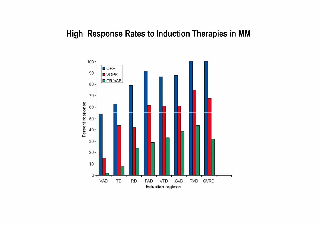
High Response Rates to Induction Therapies in MM


High dose therapy in myeloma
• Small randomized trials showing superiority to conventional therapy without ASCT (two with mature data).
• Early ASCT not superior to late ASCT.• No conditioning regimen superior to melphalan alone.• No conditioning regimen superior to melphalan alone.• No benefit from CD34+ selected grafts.
• Superiority of SCT is being tested in the setting of new drugs – benefit probably stays

Initial Therapy: Transplant Ineligible
• Melphalan + Prednisone was once the standard approach.
• RCTs show superiority of addition of thalidomide (MPT) or
bortezomib (MPV) to MP.bortezomib (MPV) to MP.
• Lenalidomide and dexamethasone is also active (probably superior
to MPT, if maintained)

The future…
JCO, 30(4), Feb 2012