4.1 The individual cell - link.springer.com · Shown here are different cell types, cell walls,...
18
11 4.3 cells of Solanum tuberosum, polar- ized light. 4.4 Chloroplasts in peripheral cells of a needle of Pinus nigra. 4.5 Tannins in heartwood cells of the dwarf shrub Globularia cordi- folia. 4.6 Calcium oxalate crystals in enlarged parenchyma cells of the herb Gypsophila repens, polarized light. 4. Cellular composition of the plant body 4.1 The individual cell Single cells can only be observed through microscopes. The cell is anatomically and physiologically complex. It principally consists of protoplasts, which contain various organelles, the vacuole and the cell wall. The following section introduces ele- ments that are visible under normal and polarized light without special microscopic equipment. Shown here are different cell types, cell walls, nuclei and plas- tids and ergastic substances (non-protoplasm material such as crystals, resins, tannins etc.). Nuclei, plastids, vacuoles and cell walls are recognizable by light microscopy. Nuclei Plastids 4.1 Viscum album. 4.2 Picea abies. Ergastic substances 50 μm 25 μm 25 μm 25 μm 100 μm 100 μm F. H. Schweingruber, A. Börner, The Plant Stem, https://doi.org/10.1007/978-3-319-73524-5_4 © The Author(s) 2018
Transcript of 4.1 The individual cell - link.springer.com · Shown here are different cell types, cell walls,...

11
4.3 cells of Solanum tuberosum, polar-ized light.
4.4 Chloroplasts in peripheral cells of a needle of Pinus nigra.
4.5 Tannins in heartwood cells of the dwarf shrub Globularia cordi-folia.
4.6 Calcium oxalate crystals in enlarged parenchyma cells of the herb Gypsophila repens, polarized light.
4. Cellular composition of the plant body
4.1 The individual cell
Single cells can only be observed through microscopes. The cell is anatomically and physiologically complex. It principally consists of protoplasts, which contain various organelles, the vacuole and the cell wall. The following section introduces ele-ments that are visible under normal and polarized light without special microscopic equipment.
Shown here are different cell types, cell walls, nuclei and plas-tids and ergastic substances (non-protoplasm material such as crystals, resins, tannins etc.).
Nuclei, plastids, vacuoles and cell walls are recognizable by light microscopy.
Nuclei
Plastids
4.1 Viscum album. 4.2 Picea abies.
Ergastic substances
50 μm 25 μm
25 μm 25 μm
100 μm100 μm
F. H. Schweingruber, A. Börner, The Plant Stem, https://doi.org/10.1007/978-3-319-73524-5_4 © The Author(s) 2018

12 Ch 4. Cellular composition of the plant body
Primary meristems in shoots & roots Embryonic cells in shoot tips, primary meristem
4.2 Meristematic initials – The source of new cells
Meristematic cells are anatomically undifferentiated and capa-ble of dividing. Meristematic initials are living cells and have
Primary meriste-matic initials are arranged in the tip of longitudinal shoots and roots (vegetation point). Secondary meristematic initials are arranged around the shoot and are the initial part of the cam-bium. Tertiary meristematic initials are a product of parenchyma cells in the bark. They represent the cork cambium (phellogen).
Primary meristems produce the cortex and the pith in stems. Secondary meristems (cambium) are a product of primary meri-stems and occur around stems where they produce the xylem and phloem. Tertiary meristems are a product of living paren-chyma cells in the cortex, the phloem, and rarely in the xylem, where they produce mainly cork cells. Meristematic cells have no pits.
4.7 Primary meristems in a sapling of Carpinus betulus.
4.8 Location of a primary meri-stem in a shoot tip Acer pseu-doplatanus.
4.9 Microscopic aspect of embryonic cells in the primary meristem in a shoot of Larix decidua.
4.12 Cells with nuclei in the tertiary meristem (phellogen) of Paeonia suffruticosa.
4.13 Location of the secondary and tertiary meristems in Pinus mugo.
Juvenile cells in secondary and tertiary meristems
4.10 Macrosopic aspect of the location of a primary meristem in a root tip of Allium ursinum.
4.11 Embryonic cells in the primary meristem in a root of Allium ursinum.
Embryonic cells in root tips, primary meristem
4.14 Juvenile cells with nuclei in secondary meristem and adjacent xylem/phloem in Viscum album.
embr
yoni
c ce
llem
bryo
nic
cell
embr
yoni
c ce
ll
livin
gde
ad
phloem xylem
secondarymeristem
tertiarymeristem
lateral bud
tips of secondary roots
terminal bud
50 μm 25 μm
25 μm
25 μm
1 mm
100 μm

13
Macroscopic aspect Thin and thick cuticles
4.15 Leaves with a glossy surface
leaves of .
4.17 Stem of Ipomaea tricolor on a shadowy site, with a thin cuticula.
4.16 Stem of a water plant (Potamo-geton coloratus) without cuticula.
4.18 Leaf with a thick, unstructured cuticula of Zamia sp. on a very dry site.
Rippled cuticles Structured cuticulae
4.19 Rippled cuticula of an annual twig of Ephedra viridis.
4.21 Layered cuticula on a young stem of the mistletoe Viscum album.
4.20 Thick, rippled cuticula on the underside of a leaf of Buxus sem-pervirens on a dry site.
4.22 Granulated cuticula on a leaf of Welwitschia mirabilis.
4.3 The cuticula – Protection against dehydration
Cells exposed to the air, mostly epidermis cells, protect internal plant cells from dehydration. The cuticle at the external surface of the epidermis is an effective transpiration protection layer.
those with rippled cuticles are matte.
Cuticles mainly consist of pectin and cutin. The soluble extract polymerizes after the full development of the organs. Cuticles occur on leaves and young stems without periderm in all taxo-nomic units of vascular plants. They are absent in roots.
Cuticles are generally absent in water plants, they are thin in plants growing in shadowy conditions, and thick in plants at dry sites. The structure of the surfaces is homogeneous, layered or even granular. Chemically related to cutin are waxes, suberin and sporopollenin.
50 μm
25 μm
25 μm 25 μm 25 μm
25 μm 25 μm

14 Ch 4. Cellular composition of the plant body
Macroscopic aspect of terrestrial plants Epidermis cells in a red leaf
4.4 Epidermis – The skin of plants
The epidermis covers the products of primary meristems of most plants. Examples of stems and leaves from ferns, coni-fers, monocotyledonous and dicotyledonous plants are shown. Epidermis cells form a uniseriate layer of generally isodiamet-ric cells at the periphery of primary plant bodies. Epidermis cells protect internal tissues from dehydration. Local cell wall expansion and cell division form bulliform cells or a variety of trichoms and hairs with special functions.
The epidermis of green plants is punctuated with stomata, allowing gas exchange between the atmosphere and the plant tissue. Since epidermis cell walls are transparent, photosynthe-sis is possible in all cells below it even when the vacuoles are
anthocyanins (pigments).
4.25 in the epidermis of a fruit of Euonymus europaeus.
4.23 All leaves and young twigs of peren-nial plants are covered with an epidermis, like here in
4.24 Red leaves appear red because the -
ment anthocyanin, like here in Berberis sp.
Anatomy of a thin-walled epidermisWater plants Terrestrial plants
4.26 Very thin-walled epi-dermis in the water plant Elodea canadensis.
4.29 Externally thick-walled,
shoot of Asparagus sp. on a dry site.
4.28 -dermis of the herb Adoxa moschatellina on a wet site.
4.27 -dermis in the swamp plant Scheuchzeria palustris.
4.30 Very thin-walled epi-dermis in a Sphagnum sp.
250 μm50 μm 25 μm 25 μm 100 μm
100 μm

15
4.39 Unicellular hairs in Lilium mar-tagon.
4.41 Multicellular hairs in Ptiloste-mon chamaepeuce.
4.40 Bicellular hairs in Scrophularia peregrina.
4.42 Stem of the moss Thuidium tamariscinum.
4.37 Excretion in Lysimachia vul-garis.
4.35 Locally enlarged epidermis cells in Asparagus scoparius.
Anatomy of elongated epidermis cells Glandulous excretion elements
4.38 Excretion in Salvia pratensis.4.36 on a shoot of the monocotyledon-ous Carex glareosa.
Anatomy of a thick-walled epidermis
Trichoms
Stomata
Rhizoids
4.31 Externally thick-walled and Psi-
lotum nudum on a wet site.
4.33 -mis on a needle of the conifer Pinus nigra.
4.32 Very thick-walled epidermis in Zamia sp.
4.34 Sunken stoma in the epidermis of Elymus farctus.
stoma
stoma
stomata
50 μm 50 μm
50 μm
25 μm 25 μm
25 μm 25 μm
25 μm 25 μm
100 μm
100 μm
100 μm

16 Ch 4. Cellular composition of the plant body
4.5 Collenchyma – Local peripheral stability
Collenchyma functions as a stabilizing element in edges and ridges of herbaceous plant stems of various genera in dicotyle-donous and monocotyledonous plants. They are a product of pri-mary meristems and occur in the cortex of stems. Collenchyma cells are similar to parenchyma cells but are normally longer and have pointed axial ends. Characteristic are the partially
thickened primary cell walls. The walls contain cellulose, and a large amount of pectin, which is indicated by blue to purple staining with Astrablue/Safranin. The appearance largely var-ies: some cells are only slightly and some intensively thickened (lamellar collenchyma) and some only in the angles (angular col-lenchyma). At least some cells contain protoplasts and nuclei.
4.43 Stachys sylvatica, a Lamiaceae with quadrangular stems.
4.46 Rumex alpinus, a Polygona-ceae with longitudinally ribbed annual shoots.
4.49 Tamus communis, a monocoty-ledonous plant, belonging to the Dioscoreaceae, with lightly-ribbed annual liana-like shoots.
4.44 The quadrangular stem of Stachys sylvatica is sta-bilized by collenchyma in the cortex.
4.47 The ridges in the stems of Rumex alpinus are stabi-lized by collenchyma in the cortex.
4.50 Morphologically hardly differentiated collenchyma in Tamus communis. Only the different reaction to the Astrablue/Safranin staining (purple coloring) indicates the collenchyma.
4.45 Cortex with collenchyma, bordered towards the outside by an epidermis and towards the inside by paren-chyma cells. Thick edges characterize the collenchyma.
4.48 Living collenchyma cells with intensively thick-ened angles (angular collenchyma). Most cells contain protoplasts. In a few of them, nuclei are visible.
Collenchyma occurs mainly in the cortex of dicotyledonous plants
Collenchyma is rare in monocotyledonous plants
500 μm
500 μm
50 μm
50 μm
25 μm

17
4.51 Potatoes (Solanum tuberosum) consist mainly of parenchyma cells. They are able to produce new shoots.
4.52 chyma cells. Parenchyma cells in the phloem can change their mode of behavior and form new shoots.
4.53 Scar on the stem of Acer pseudoplatanus. Living parenchyma cells repair wounds and pro-tect the stem against destructive organisms.
4.6 Parenchyma cells – Storage and repair
Humans would not be able to exist without plant parenchyma cells because their cell contents, especially carbohydrates, are essential components of our diet. Parenchyma cells are pres-ent in all plants. They are mostly small isodiametric or slightly elongated cells without pointed ends. Cell walls of parenchyma
perforated with simple pits. Parenchyma cells primarily func-tion as storage cells. Living parenchyma cells are totipotent. They have the potential to regenerate new cell types or entire plants under suitable environmental conditions. They play an
essential role in regeneration processes after injury (see Chapter 10).
and seeds. Abundant amounts of parenchyma cells occur in thickened belowground organs. The life span of parenchyma cells of perennial plants is normally very long. Ray-parenchyma cells can live for more than 100 years in the sapwood of coni-fers or more than 200 years in dwarf shrubs of the arctic.
Parenchyma cells occur in all terrestrial plants
4.59 Water-storing paren-chyma cells in the succu-lent Sedum acre.
4.62 Parenchyma cells alternate with sieve cells in the bark of conifers such as Abies alba.
4.61 Parenchyma cells sur-round air canals in the cortex of the swamp plant Orontium aquaticum.
4.60 Very small parenchyma cells between air-conducting spaces in the pith of swamp plant Scirpus radicans.
4.63 Long-living parenchy-matic ray cells with nuclei (blue) in Picea abies.
Shape of parenchyma cells
4.54 Parenchyma cells are
grains in the rhizome of Anemone nemorosa, polar-ized light.
4.57 Simple pits are char-acteristic for parenchyma cells. Ray cells in the bark of Paeonia suffruticosa.
4.56 Parenchyma cells in the xylem of Sonchus lep-tophyllus. Axially sectioned cells are round, radially sec-tioned cells are elongated.
4.55 Isodimetric paren-chyma cells in the stem of the moss Polytrichum com-mune.
4.58 Irregularly formed paren chyma cells, the callus cells, appear after wounding in Picea abies.
nucleus
50 μm50 μm
50 μm
25 μm25 μm
25 μm25 μm25 μm
100 μm
100 μm

18 Ch 4. Cellular composition of the plant body
4.68 Fibers in stems of dicotyledonous dwarf shrubs like Sibbaldia procumbens.
4.69 Fibers in culms and leaves of monocotyledonous herbs like Festuca alpina (top) or Festuca erecta.
4.64 Fibers are omnipresent in human life in the form of wood products.
4.65 200 μm) in shrubs, such as Buxus sempervirens.
4.66 >1000 μm) in trees, such as Fagus sylvatica.
4.67 Long (1,000–3,000 μm),
for pulp) in Pinus sylvestris.
Use of
Fibers occur in all vascular plants
4.7 Fibers and tracheids – Stabilisation and water conduction
paper, are substantial basics of our daily life. The main func-tion of stabilization, while tracheids are stabilizing and conduct water.
Fibers and tracheids are long cells with elongated, pointed tips. Tips are a result of post-cambial axial elongation. Fibers or tra-cheids occur in all growth forms and on all sites in spore plants
(ferns, horsetails), in conifers, in monocotyledonous and dicot-yledonous plants. They are part of annual and perennial stems
walls. Characteristic is the presence of secondary walls. Pits in simple or slightly bordered. Pits in tracheids
are bordered (see Chapter 5.2, Fig. 5.19). Fibers occur in the xylem, phloem and cortex, tracheids only in the xylem.
250 μm
250 μm 250 μm 250 μm
100 μm

19
4.77 Thick-walled tracheids in Larix decidua. 4.78 Eryobotrya japonica.
4.75 Fibers in the bark of Chrysothamnus parryi.
4.79 Salix foetida.
4.76 Fibers in a culm of Sesleria coerulea.4.74 Fibers in a shoot (cortex) of Cucumis sativus.
Fibers occur in all parts of stems
Fibers occur in all parts of plants
4.70 Fibers occur in stems, roots and leaves. Cas-tanea sativa.
4.72 Fibers in a root of Ledum palustre. 4.73 Fibers in a leaf of Carex sempervirens.
4.71 Fibers in a shoot of Ranunculus lanuginosus.
leaf
shoot
root
250 μm250 μm
25 μm
1 mm
100 μm 100 μm
100 μm
100 μm100 μm

20 Ch 4. Cellular composition of the plant body
4.8 Sclereids in the bark – Extraordinary cell-wall thickening
We notice sclereids when eating pears: The granules remain-ing between your teeth are sclereids. Sclereids are absent in the xylem but frequent in the phloem, cortex and pith of trees and herbs. They also occur in fruits and nut shells, and rarely in leaves. Characteristic for sclereids are the irregularly formed cells with thick, secondary walls, distinct simple pits and often
distinct growth layers. A special case are star-like sclereids in the aerenchyma of the water lily (Nuphar sp). Sclereids in the bark normally occur in the non-conducting (adult) phloem and in rays. Sclereids originate from parenchyma cells with a short-term accelerated growth of secondary walls.
Sclereids in stems
4.80 Hard bark of Fagus sylvatica. 4.82 Flexible and soft culms and a petiole of the water lily Nuphar lutea.
4.81 Many sclereids in a ray and the cortex in Fagus sylvatica, polarized light.
4.83 Star-like sclereids in air-con-ducting channels (aerenchyma) in Nuphar lutea, polarized light.
4.88 Group of sclereids in the bark of Picea abies. 4.89 Isolated sclereids in the fruit of Mespilus germanica.
4.90 Isolated sclereids with distinct layers in a leaf of Welwitschia mirabilis.
Single and groups of sclereids layers
Sclereids in fruits
4.84 Fruit of Mespilus germanica. 4.86 Fruits of the hazelnut (Corylus avellana).
4.85 Sclereids in the peripheral part of the fruit of Mespilus germanica, polarized light).
4.87 Sclereids in the shell of a hazel-nut.
250 μm
25 μm 25 μm 25 μm
100 μm
250 μm 50 μm

21
4.9 Vessels – Water conduction
Plant life on terrestrial sites would not exist without the water conducting -sels with bare eyes, because their diameter is usually below the resolution of human eyes. Vessels consist of vessel elements. Fully developed vessel elements are dead and more or less
perforation plates at their distal ends and pits at the longitudinal walls. Lig-
Vessels occur in the xylem of most spore plants (ferns, horsetails, lycopods) and most monocotyledons and dicotyledons, except in conifers and few others where they are replaced by tracheids. Vessels occur in all growth forms. Diameters are usually large
in lianas (>200–500 μm), smaller in trees (50–200 μm) and small in herbs (20–50 μm). The length of vessel elements var-ies from <100 μm in small herbs to >1,500 μm in trees. Vessel elements are normally short in ring-porous and long in diffuse-porous tree species.
Vessels conduct water mainly in axial direction from the root to the leaves. Lateral perforations (bordered pits) also indicate a lateral water transport. Vessels exist in the xylem of all plant organs in various dimensions. Vessels are normally larger in roots than in stems, and even smaller in leaves.
For a discussion of cell-wall structures see Chapter 5.1.
4.91 Vessels in the earlywood of ring-porous spe-cies (e.g. Quercus sp.) are visible to the bare eye.
4.94 Lycopods: clubmoss Lycopodium clavatum.
4.92 Long vessel element (500 μm) with helical thickenings in Tilia sp.
4.95 Monocotyledons: stem of palm Phoenix dactylifera.
4.93 Short and wide vessel in the earlywood of Fraxinus excelsior. Reprinted from Greguss 1945.
4.96 Dicotyledons: stem of apple tree Malus domestica.
Macroscopic aspect Anatomy of vessel elements
Vessels occur in all vascular plants
perforation plateperforation platebordered pits
250 μm250 μm 500 μm

22 Ch 4. Cellular composition of the plant body
4.101 Small vessel diameters (20 μm) in a vascu-lar bundle of a thick leaf of Clusea rosea.
4.100 Diameter of earlywood vessels in the stem (top) is 70 μm, in the root (bottom) 250 μm in Prunus amygdalus.
4.97 Very large vessel diameters (550 μm) in lianas Celastrus sp. (top) and Thunbergia sp. (bottom).
4.98 Large vessel diameter (80 μm) in the dicoty-ledonous tree Fagus sylvatica.
4.99 Very small vessel diameters (20 μm) in the 5 cm-tall annual herb Erophila verna.
Vessels occur in all growth forms
Vessels occur in most parts of plants
stem
root
100 μm
100 μm 100 μm
100 μm100 μm100 μm

23
4.10 Cork cells – Defense against organisms, heat and cold
Cork is a perfect insulation material. We use it to seal wine -
ally used cork is a product of cork oak (Quercus suber). Plants insulate their stems against extreme environmental conditions
cortex of most taxa and growth forms in conifers and dicoty-ledons. They are rare in monocotyledonous plants, and occur
there only in trees. Cork cell walls consist of suberin, which is decompose for many fungi. Cork cells of any form
can be thin- or thick-walled.
Cork formation occurs usually on the outside of stems. How-ever, a few species produce cork rhythmically within the xylem. Small, long-lived herbs compartmentalize their center by form-ing cork internally within the stem.
Cork cells in stems of seed plants of all growth forms
4.102 Conifers: Layered bark of Pinus sylvestris.
4.104 Dicotyledonous plants: Lay-ered bark of a small cushion of the alpine Saxifraga oppositifolia.
4.103 Dicotyledonous plants: Lay-ered bark of the tree Quercus suber.
4.105 Monocotyledonous plants: Cork cells in the tree Dracaena ser-rulata.
Thin- and thick-walled cork cells Cork cells within the xylem
4.106 A small mantle of thin-walled cork cells surrounds the water-stor-ing cortex of the succulent Sedum acre.
4.108 Tangential cork layers between compartments of xylem in Artemisia tridentata.
4.107 A large zone of thick-walled, rectangular cork cells surrounds the
cushion plant Saxifraga caesia.
4.109 Cork layers in a stem separate the living from the dead xylem in the center of the stem of Scorzonera virgata.
cork
cel
ls
cork
cel
ls cork
cel
ls
250 μm 500 μm
500 μm
1 mm
1 mm1 mm
100 μm
500 μm

24 Ch 4. Cellular composition of the plant body
Anatomy of sieve elements and companion cells
Sieve elements in ferns, conifers, dicotyledonous and monocotyledonous plants
4.117 Sieve cells in the phloem of the fern Cryptogramma crispa.
4.113 Phloem of the tree Adansonia digitata, consisting of sieve tubes, companion cells and parenchyma cells.
4.119 Sieve tube in the phloem of the dicotyledonous Quercus ilex.
4.115 Sieve plates of sieve tubes in Adansonia digitata.
4.118 Sieve cells in the phloem of the conifer Picea abies.
4.114 Phloem of the shrub Lonicera alpigena, consisting of sieve tubes, companion cells and parenchyma cells.
4.120 Sieve tubes in a vascular bundle of the monocotyledonous Arundo donax.
4.116 Sieve elements of the dicoty-ledonous herb Bryonia dioeca (left) with sieve plates at distal ends, and of the conifer Larix decidua with lat-eral sieve plates (right).
4.11 Sieve cells, sieve tubes and companion cells – Conduction of assimilates
Sieve elements are part of the bast. Neolithic settlers knew sieve cells and bast to braid baskets and tissues. Today, papier mâché (“chewed paper”) is probably the only remaining useful product.
Sieve cells and sieve tubes are a major part of the phloem, which mainly conducts assimilates from leaves to parenchyma cells. Sieve elements are accompanied by parenchyma cells and companion cells. Sieve cells have -eral sides, while sieve tubes have plates at their distal ends and
on their lateral walls. However, the anatomical differentiation
do not contain nuclei. The metabolism of the sieve elements is maintained by the nuclei in the companion cells. Companion cells are smaller and always adjacent to sieve tubes.
Sieve cells occur in all vascular plants from ferns to dicotyle-dons. Sieve cells and sieve tubes often collapse following their death. More information is given in Chapter 6.2.8.
4.110 Neolithic tissues made of Tilia bast. Photo: Amt für Städtebau Unterwasserarchäologie, Univ. Zürich.
4.111 Stripped bark of Populusdismantled from the xylem in the cambial zone.
4.112 Large layered phloem between the xylem and cork in Juglans regia.
Macroscopic aspect of the phloem
sitube
sitube
comp com
p
pa
pa
papa
pa
si si
si
si
pa
pa
pa
comp
dead bark
phloem
xylem
500 μm50 μm 50 μm
50 μm50 μm
25 μm
1 mm 1 mm1 mm
25
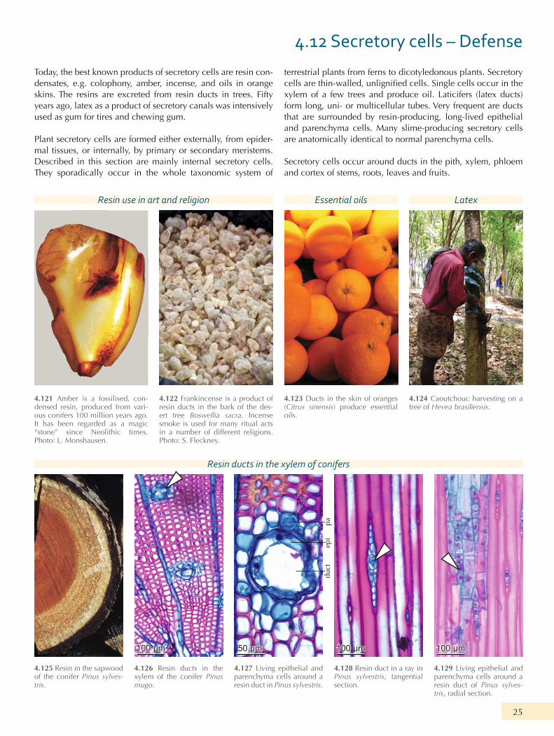
4.12 Secretory cells – Defense
Today, the best known products of secretory cells are resin con-densates, e.g. colophony, amber, incense, and oils in orange skins. The resins are excreted from resin ducts in trees. Fifty years ago, latex as a product of secretory canals was intensively used as gum for tires and chewing gum.
Plant secretory cells are formed either externally, from epider-mal tissues, or internally, by primary or secondary meristems. Described in this section are mainly internal secretory cells. They sporadically occur in the whole taxonomic system of
terrestrial plants from ferns to dicotyledonous plants. Secretory
xylem of a few trees and produce oil. Laticifers (latex ducts) form long, uni- or multicellular tubes. Very frequent are ducts that are surrounded by resin-producing, long-lived epithelial and parenchyma cells. Many slime-producing secretory cells are anatomically identical to normal parenchyma cells.
Secretory cells occur around ducts in the pith, xylem, phloem and cortex of stems, roots, leaves and fruits.
Resin use in art and religion
Resin ducts in the xylem of conifers
Essential oils Latex
4.125 Resin in the sapwood of the conifer Pinus sylves-tris.
4.128 Resin duct in a ray in Pinus sylvestris, tangential section.
4.127 Living epithelial and parenchyma cells around a resin duct in Pinus sylvestris.
4.126 Resin ducts in the xylem of the conifer Pinus mugo.
4.129 Living epithelial and parenchyma cells around a resin duct of Pinus sylves-tris, radial section.
4.121 Amber is a fossilised, con-densed resin, produced from vari-ous conifers 100 million years ago. It has been regarded as a magic “stone” since Neolithic times. Photo: L. Mons hausen.
4.123 Ducts in the skin of oranges (Citrus sinensis) produce essential oils.
4.122 Frankincense is a product of resin ducts in the bark of the des-ert tree Boswellia sacra. Incense smoke is used for many ritual acts in a number of different religions. Photo: S. Fleckney.
4.124 Caoutchouc harvesting on a tree of Hevea brasiliensis.
paep
idu
ct
50 μm 100 μm 100 μm100 μm

26 Ch 4. Cellular composition of the plant body
Anatomy of ducts in stems of dicotyledonous plants
Anatomy of oil cells Anatomy of laticifers
4.130 Resin ducts in the phloem of the incense tree Boswellia sacra.
4.138 Enlarged oil cells in the xylem of the tree Phoebe nanmu, tangential section. It was the pre-ferred wood for the construction of the Forbidden City in Beijing.
4.132 Macroscopic aspect of trau-matic “resin” ducts (kino veins) in the xylem of Eucalyptus obliqua. Photo: P. Majewski.
4.140 Latex-producing laticifers in the phloem and the cortex of the shrub Euphorbia armena.
4.131 Traumatic “resin” ducts in the xylem of an almond tree (Prunus amygdalus).
4.139 Enlarged oil cells in the xylem of Phoebe nanmu, radial section.
4.133 Microscopic aspect of trau-matic “resin” ducts (kino vein) in the xylem of a Eucalyptus sp.
Anatomy of ducts in various parts of plants
4.134 Resin duct in a needle of the conifer Picea abies.
4.136 Ducts in the pith of a twig of Grewia villosa.
4.135 Duct in the cortex of the bark of the herb Petasites paradoxus.
4.137 Ducts in the shell of a hazel-nut (Corylus avellana).
4.141 A single laticifer, consisting of several secretory elements in Scrophularia dentata.
250 μm
250 μm 250 μm 250 μm 250 μm
250 μm
50 μm1 mm 1 mm
500 μm100 μm

27
4.13 Intercellulars and aerenchyma – Air circulation within the plant
The very light-weighted shoots of reed (Phragmites communis) and many other grass-like shore plants have been used for the
Hay from wet meadows in the European Alps is used as bed-ding in cattle stables. These practical uses are based on the hollow stems, and the presence of aerenchyma in the shoots. Intercellulars of any form occur mainly in wetland plants. They guarantee the gas exchange from the stomata in the leaves to all
-chyma. Hollow shoots and aerenchyma in water plants allow
and cortex of plants in all taxonomic units of vascular plants, and aerenchyma with particular structures occur in plants of wet environments, e.g. in swamps and lakes (helophytes and hydrophytes).
Intercellulars are small spaces between round cells. Aeren-chyma consists of parenchyma cells surrounding the intercellu-lars. They form nets, sponge-like tissues in thick stems, star-like groups, channels, irregular and radial spaces and lacunas.
Use of wetland plants
Aerenchyma
Anatomy of “air bags” Aerenchyma
4.142 Phrag-mites communis).
4.146 cortex of a rhizome of Juncus arcti-cus.
4.144 Hollow pith as the result of extreme stem expansion in Polygo-num amphibium.
4.148 Small air tubes conduct air from the leaf to the root in Potamo-geton natans.
4.143 Stacks of dry, stiff, nutrient-poor grasses and sedges in a wet montane meadow. The material is used instead of cereal straw for bedding in cattle stables. Photo: M. Küchler.
4.147 Large air tubes conduct air from the leaf to the root in Nym-phaea alba.
4.145 Stellate combined cells form Juncus
conglomeratus.
4.149 Small lacunas within vascular bundles conduct air from the leaf to the roots in Dacytlis glomerata.
500 μm
500 μm
500 μm500 μm 500 μm
100 μm

28 Ch 4. Cellular composition of the plant body
Open Access This chapter is licensed under the terms of the Creative Commons Attribution 4.0 International License (http://creativecommons.org/licenses/by/4.0/), which permits use, sharing, adaptation, distribution and reproduction in any medium or format, as long as you give appropriate credit to the original author(s) and the source, provide a link to the Creative Commons license and indicate if changes were made.
The images or other third party material in this chapter are included in the chapter's Creative Commons license, unless indicated otherwise in a credit line to the material. If material is not included in the chapter's Creative Commons license and your intended use is not permitted by statutory regulation or exceeds the permitted use, you will need to obtain permission directly from the copyright holder.