2012.8.14 1 Department of Biomedical Engineering and Environmental Sciences, National Tsing Hua...
22
2012.8.14 1 DNA-Conjugated Gold Nanoparticles as Ultrasound- Responsive Drug Delivery Systems Department of Biomedical Engineering and Environmental Sciences, National Tsing Hua University Hsinchu, Taiwan. Shih-Tsung Kang, Yun-Ling Luo, Yu-Fen Huang, Chih-Kuang Yeh
-
Upload
shon-miller -
Category
Documents
-
view
220 -
download
0
Transcript of 2012.8.14 1 Department of Biomedical Engineering and Environmental Sciences, National Tsing Hua...
1
2012.8.14
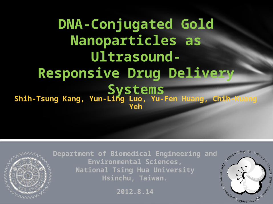
DNA-Conjugated Gold Nanoparticles as
Ultrasound-Responsive Drug Delivery
Systems
Department of Biomedical Engineering and Environmental Sciences,
National Tsing Hua UniversityHsinchu, Taiwan.
Shih-Tsung Kang, Yun-Ling Luo, Yu-Fen Huang, Chih-Kuang Yeh
2
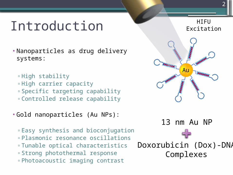
Introduction
• Nanoparticles as drug delivery systems:
▫ High stability▫ High carrier capacity▫ Specific targeting capability▫ Controlled release capability
• Gold nanoparticles (Au NPs):
▫ Easy synthesis and bioconjugation▫ Plasmonic resonance oscillations▫ Tunable optical characteristics▫ Strong photothermal response▫ Photoacoustic imaging contrast
Au
13 nm Au NP
Doxorubicin (Dox)-DNAComplexes
HIFUExcitation
3
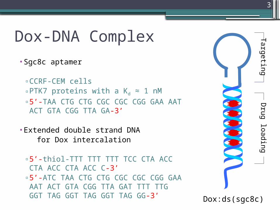
Dox-DNA Complex• Sgc8c aptamer
▫ CCRF-CEM cells ▫ PTK7 proteins with a Kd ≈ 1 nM▫ 5’-TAA CTG CTG CGC CGC CGG GAA
AAT ACT GTA CGG TTA GA-3’
• Extended double strand DNA for Dox intercalation
▫ 5’-thiol-TTT TTT TTT TCC CTA ACC CTA ACC CTA ACC C-3’
▫ 5’-ATC TAA CTG CTG CGC CGC CGG GAA AAT ACT GTA CGG TTA GAT TTT TTG GGT TAG GGT TAG GGT TAG GG-3’
Targ
etin
gD
rug
load
ing
Dox:ds(sgc8c)

4
Dox:ds(sgc8c)-Au NPs
• Incubation of ds(sgc8c)-Au NPs (9.7 nM) with Dox (4 μM) for 2 h yields the nanoconjugates loaded with 2.7 μM Dox
68% loading efficiency
• For a single 13-nm Au NP▫ 61 ± 10 Dox-DNA complexes ▫ 280 ± 23 Dox molecules▫ 4-5 Dox molecules per DNA
• Hydrodynamic diameter change: ▫ 19.9 ±0.6 nm of citrate-stabilized Au NPs▫ 42.1 ±4.0 nm of ds(sgc8c)-Au NPs Au
Gold-Thiol Bonding

5
http://oncozine.ning.com/profiles/blogs/new-drug-delivery-system-gold
PT
K7 CEM Cell
Nucleus
HIFUTreatment
Au
Sgc8c aptamer
Dox-DNA Complex
Au
Cells (2×10⁵ cells/well) were incubated with Dox-loaded nanoconjugates (4.9 nM) loaded with 1.35-μM Dox in RPMI-1640 medium for 2 h.
Parameters:• Frequency: 10 MHz• Acoustic pressure: 4 MPa, • Duty cycle: 10%• Treatment time: 10 min

10-MHz HIFU Transducer
AgarPhantom
Waveform Generator 2
Power Amplifier
Waveform Generator 1
Trigger
Experimental Setup
• Flow cytometry: Mean Fluorescence Intensity (MFI)
• MTT assay: Cell Viability
Immediately
24 h later
7
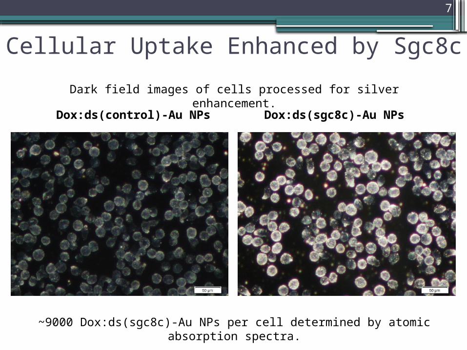
Cellular Uptake Enhanced by Sgc8c
Dark field images of cells processed for silver enhancement.
Dox:ds(control)-Au NPs Dox:ds(sgc8c)-Au NPs
~9000 Dox:ds(sgc8c)-Au NPs per cell determined by atomic absorption spectra.
8
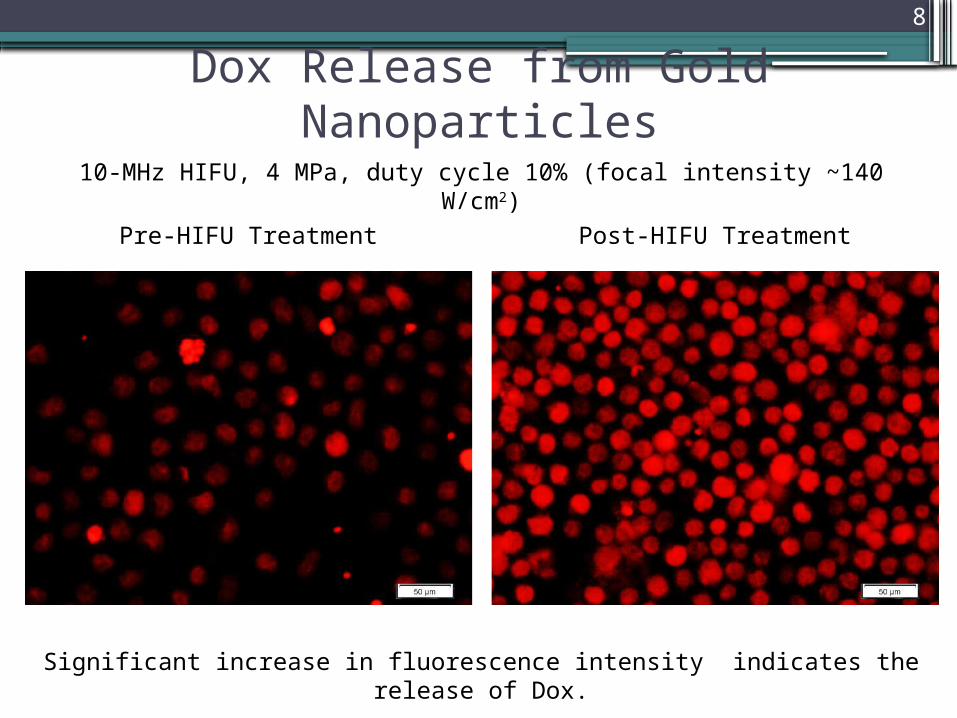
Dox Release from Gold Nanoparticles10-MHz HIFU, 4 MPa, duty cycle 10% (focal intensity ~140 W/cm2)
Significant increase in fluorescence intensity indicates the release of Dox.
Pre-HIFU Treatment Post-HIFU Treatment
9
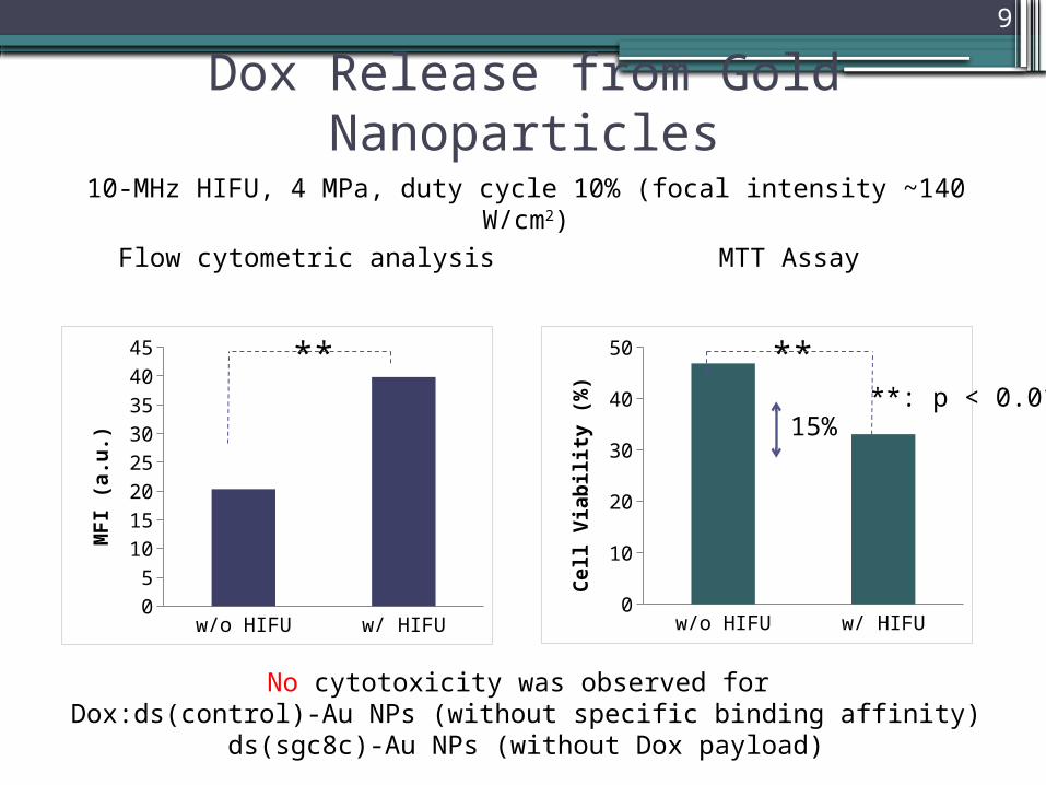
Dox Release from Gold Nanoparticles
Flow cytometric analysis MTT Assay
w/o HIFU w/ HIFU05
1015202530354045
MF
I (a
.u.)
**
w/o HIFU w/ HIFU05
101520253035404550
Cell
Via
bil
ity (
%)
**
10-MHz HIFU, 4 MPa, duty cycle 10% (focal intensity ~140 W/cm2)
No cytotoxicity was observed for Dox:ds(control)-Au NPs (without specific binding affinity)
ds(sgc8c)-Au NPs (without Dox payload)
**: p < 0.0115%

10
Pure
Cells
Dox:d
s(sg
c8c)
PDPH-D
ox-sgc
8c-A
u N
Ps
Dox:d
s(sg
c8c)
-Au
NPs
05
1015202530354045
Pre-HIFUPost-HIFU
MF
I (a
.u.)
Comparisons of Different Cases
Dox release was only achieved when both Dox-DNA complexes and Au NPs were present.
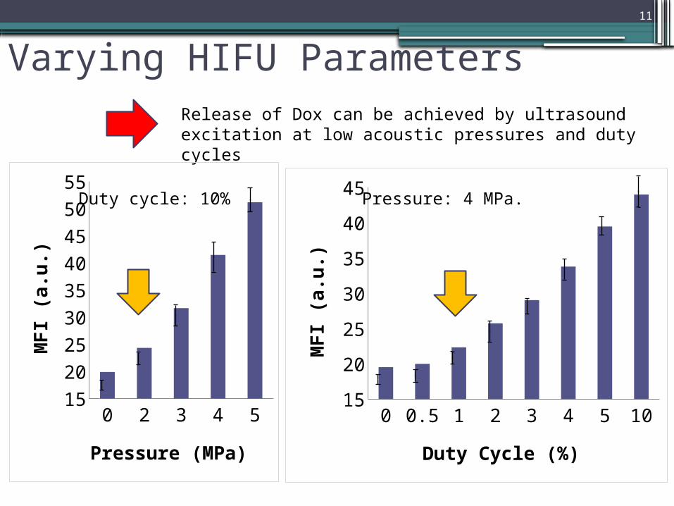
Varying HIFU Parameters11
Release of Dox can be achieved by ultrasound excitation at low acoustic pressures and duty cycles
0 2 3 4 515
20
25
30
35
40
45
50
55
Pressure (MPa)
MF
I (a
.u.)
0 0.5 1 2 3 4 5 1015
20
25
30
35
40
45
Duty Cycle (%)
MF
I (a
.u.)
Pressure: 4 MPa.Duty cycle: 10%
Sonication Time (s)
Tem
pera
ture
(˚
C)
ΔM
FI
(a.u
.)Δ
MFI
(a.u
.)
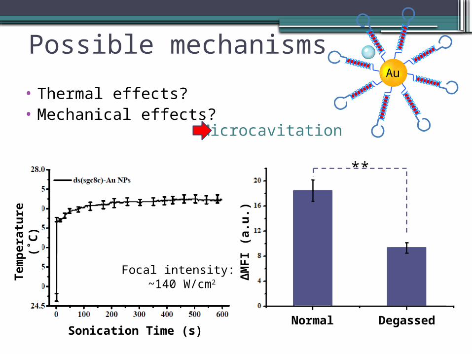
Normal Degassed
**
Au
Possible mechanisms
• Thermal effects?• Mechanical effects?
Microcavitation
Focal intensity: ~140 W/cm2
13
Pure Cells ds(sgc8c)-Au NP Cell with H2O20
5
10
15
20
25
30
Pre-HIFUPost-HIFUM
FI
(a. u
.)
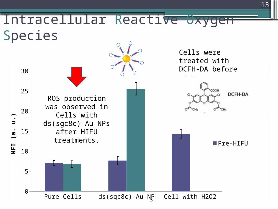
Intracellular Reactive Oxygen Species
Cells were treated with DCFH-DA before HIFU treatments
ROS production was observed in Cells with ds(sgc8c)-Au NPs after HIFU
treatments.
s
14
Summary• Au NPs conjugated with Dox-DNA complexes have shown to
be ultrasound-responsive drug delivery systems. The drug release mechanism was found to relate to microcavitation.
• The presence of Au NPs might provide nucleation sites for facilitating gas nuclei on the surface of Au NPs to grow into cavitation bubbles under HIFU excitation.
• Further investigation is required to clarify which phenomena during microcavitation responsible for the Dox release.
• ROS scavenger treatment.• DNA integrity evaluation.• Different HIFU parameters.• Different types of nanoparticles.
Thanks for your attention.
15
16
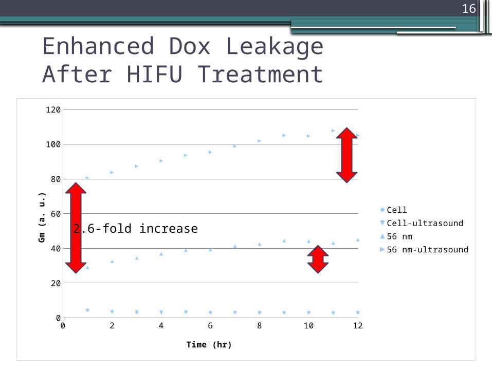
Enhanced Dox LeakageAfter HIFU Treatment
0 2 4 6 8 10 120
20
40
60
80
100
120
CellCell-ultrasound56 nm56 nm-ultrasound
Time (hr)
Gm
(a.
u.)
2.6-fold increase
17
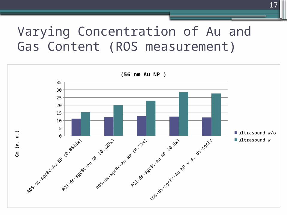
Varying Concentration of Au and Gas Content (ROS measurement)
ROS-ds-sg
c8c-Au N
P (0.0
625x)
ROS-ds-sg
c8c-Au N
P (0.1
25x)
ROS-ds-sg
c8c-Au N
P (0.2
5x)
ROS-ds-sg
c8c-Au N
P (0.5
x)
ROS-ds-sg
c8c-Au N
P v.s. d
s-sg
c8c
0
5
10
15
20
25
30
35
(56 nm Au NP )
ultrasound w/o
ultrasound w
Gm
(a. u
.)
18
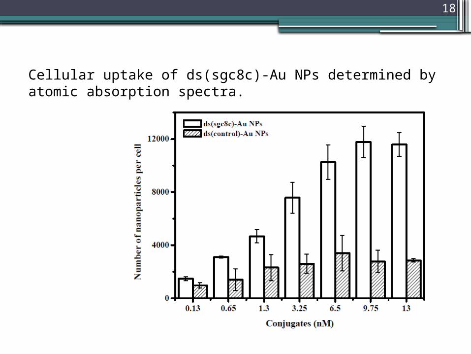
Cellular uptake of ds(sgc8c)-Au NPs determined by atomic absorption spectra.
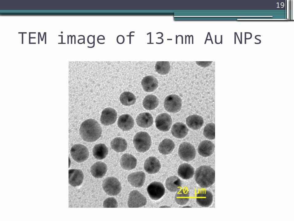
19
TEM image of 13-nm Au NPs
20 μm
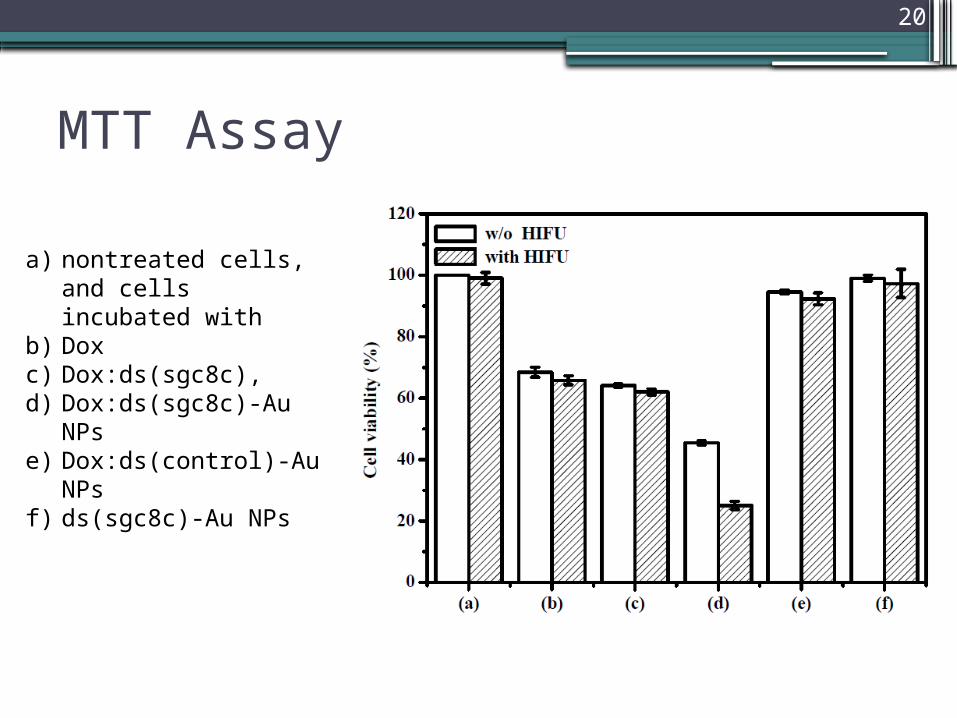
20
MTT Assay
a) nontreated cells, and cells incubated with
b) Doxc) Dox:ds(sgc8c), d) Dox:ds(sgc8c)-Au NPse) Dox:ds(control)-Au
NPsf) ds(sgc8c)-Au NPs
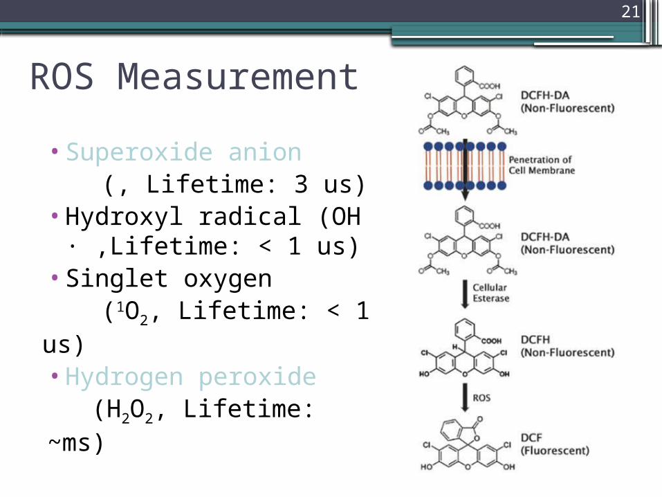
21
ROS Measurement
•Superoxide anion (, Lifetime: 3 us)•Hydroxyl radical (OH
· ,Lifetime: < 1 us) •Singlet oxygen (1O2, Lifetime: < 1 us)•Hydrogen peroxide (H2O2, Lifetime: ~ms)

22
Au
Possible Mechanisms
•Pyrolysis in bubble interior.▫Dissociation due to high temperature.
•Hydroxylation at bubble interface.▫Reaction with Reactive Oxygen Species.
•Super-critical water at bubble interface.
I. Hua, R. H. Hoechemer, and M. R. Hoffmann, “Sonolytic Hydrolysis of p-Nitrophenyl Acetate: The Role of Supercritical Water,” The Journal of Physical Chemistry, vol. 99, no. 8, pp. 2335-2342, Feb. 1995.