19-1 Anatomy and Physiology, Sixth Edition Rod R. Seeley Idaho State University Trent D. Stephens...
32
19-1 Anatomy and Physiology, Sixth Edition Rod R. Seeley Idaho State University Trent D. Stephens Idaho State University Philip Tate Phoenix College Copyright © The McGraw-Hill Companies, Inc. Permission required for reproduction or display. *See PowerPoint Image Slides for all figures and tables pre-inserted into PowerPoint without notes. Chapter 19 Chapter 19 Lecture Lecture Outline Outline *
-
Upload
nickolas-conley -
Category
Documents
-
view
229 -
download
1
Transcript of 19-1 Anatomy and Physiology, Sixth Edition Rod R. Seeley Idaho State University Trent D. Stephens...

19-1
Anatomy and Physiology, Sixth Edition
Rod R. SeeleyIdaho State UniversityTrent D. StephensIdaho State UniversityPhilip TatePhoenix College
Copyright © The McGraw-Hill Companies, Inc. Permission required for reproduction or display.
*See PowerPoint Image Slides for all figures and tables pre-inserted into PowerPoint without notes.
Chapter 19Chapter 19
Lecture OutlineLecture Outline**

19-2
Chapter 19
Cardiovascular SystemBloodBlood

19-3
Functions of Blood
• Transport of: – Gases, nutrients, waste products– Processed molecules– Regulatory molecules
• Regulation of pH and osmosis
• Maintenance of body temperature
• Protection against foreign substances
• Clot formation
19-4
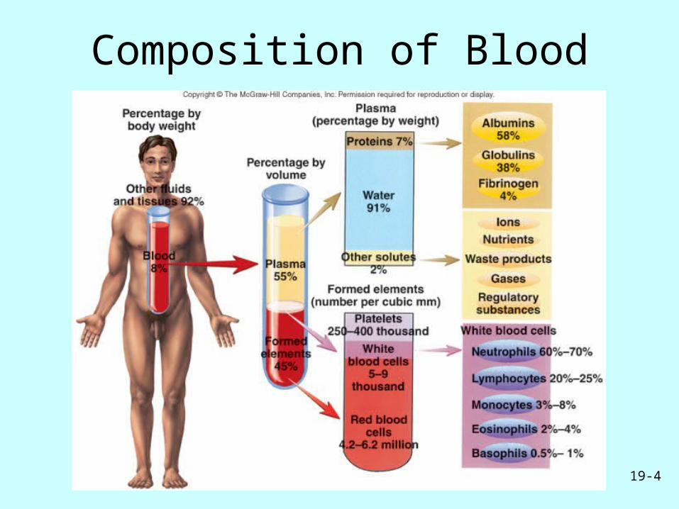
Composition of Blood

19-5
Plasma• Liquid part of blood
– Pale yellow made up of 91% water, 9% other
• Colloid: Liquid containing suspended substances that don’t settle out– Albumin: Important in regulation of water
movement between tissues and blood– Globulins: Immune system or transport
molecules– Fibrinogen: Responsible for formation of blood
clots

19-6
Formed Elements• Red blood cells (erythrocytes)• White blood cells (leukocytes)
– Granulocytes• Neutrophils• Eosinophils• Basophils
– Agranulocytes • Lymphocytes• Monocytes
• Platelets (thrombocytes)

19-7
Production of Formed Elements
• Hematopoiesis or hemopoiesis: Process of blood cell production
• Stem cells: All formed elements derived from single population– Proerythroblasts: Develop into red blood cells– Myeloblasts: Develop into basophils,
neutrophils, eosinophils– Lymphoblasts: Develop into lymphocytes– Monoblasts: Develop into monocytes– Megakaryoblasts: Develop into platelets

19-8
Hematopoiesis

19-9
Erythrocytes• Structure
– Biconcave, anucleate
• Components– Hemoglobin
– Lipids, ATP, carbonic anhydrase
• Function– Transport oxygen from
lungs to tissues and carbon dioxide from tissues to lungs

19-10
Hemoglobin
• Consists of: – 4 globin molecules: Transport carbon dioxide (carbonic
anhydrase involved), nitric oxide
– 4 heme molecules: Transport oxygen• Iron is required for oxygen transport

19-11
Erythropoiesis
• Production of red blood cells– Stem cells proerythroblasts early erythroblasts
intermediate late reticulocytes
• Erythropoietin: Hormone to stimulate RBC production

19-12
Hemoglobin Breakdown

19-13
Leukocytes
• Protect body against microorganisms and remove dead cells and debris
• Movements– Ameboid
– Diapedesis
– Chemotaxis
• Types– Neutrophils: Small
phagocytic cells
– Eosinophils: Reduce inflammation
– Basophils: Release histamine and increase inflammatory response
– Lymphocytes: Immunity
– Monocytes: Become macrophages

Leukocytes


• Macrophages:
- are the main phagocytes of the body.
• Neutrophils:
- are the first responders and become phagocytic when they encounter infectious material.

• Eosinophils:
- are weakly phagocytic but are important in defending the body against parasitic worms.
• Mast cells:
- have the ability to bind with, ingest, and kill a wide range of bacteria.

Natural killer cells
• They are able to lyse and kill :
- cancer cells
- virally infected cells
before the adaptive immune system has been activated
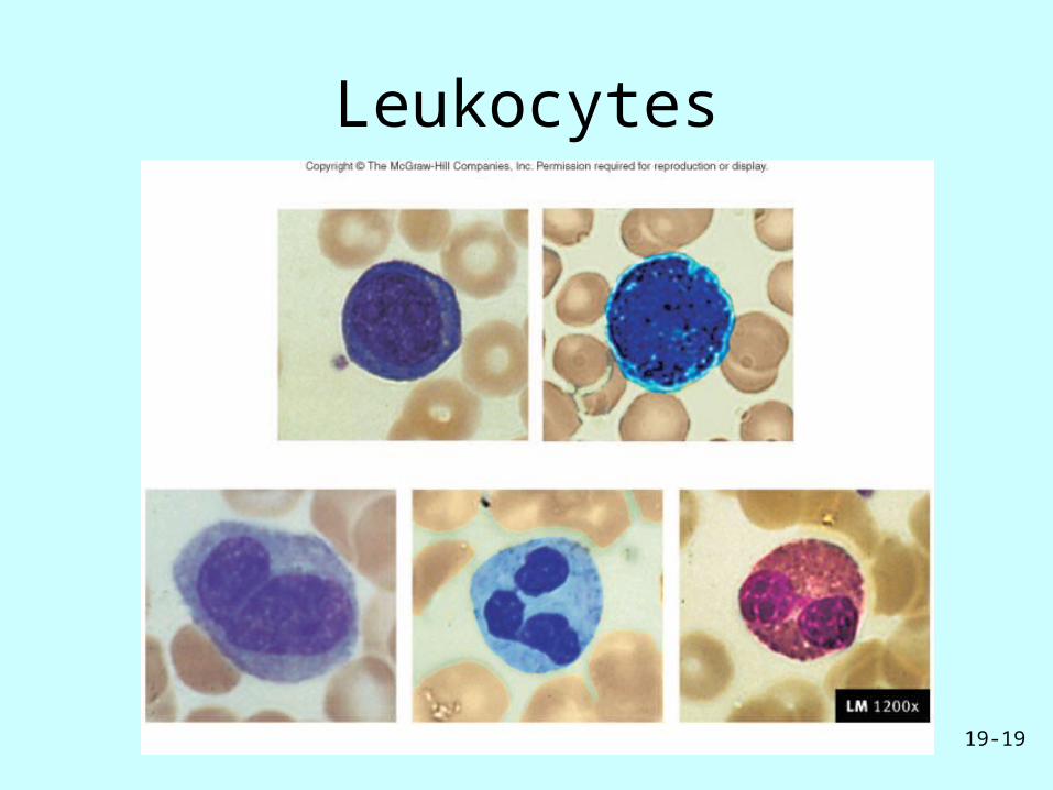
19-19
Leukocytes

19-20
Thrombocytes
• Cell fragments pinched off from megakaryocytes in red bone marrow
• Important in preventing blood loss– Platelet plugs
– Promoting formation and contraction of clots

19-21
Hemostasis
• Arrest of bleeding
• Events preventing excessive blood loss– Vascular spasm: Vasoconstriction of damaged
blood vessels– Platelet plug formation – Coagulation or blood clotting

19-22
Platelet Plug Formation

19-23
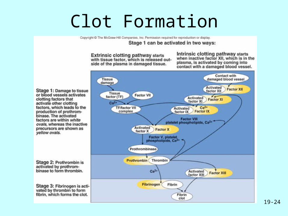
Coagulation
• Stages– Activation of
prothrombinase
– Conversion of prothrombin to thrombin
– Conversion of fibrinogen to fibrin
• Pathways– Extrinsic
– Intrinsic
19-24
Clot Formation

19-25
Fibrinolysis
• Clot dissolved by activity of plasmin, an enzyme which hydrolyzes fibrin

19-26
Blood Grouping
• Determined by antigens (agglutinogens) on surface of RBCs
• Antibodies (agglutinins) can bind to RBC antigens, resulting in agglutination (clumping) or hemolysis (rupture) of RBCs
• Groups– ABO and Rh

19-27
ABO Blood Groups

19-28
Agglutination Reaction

19-29
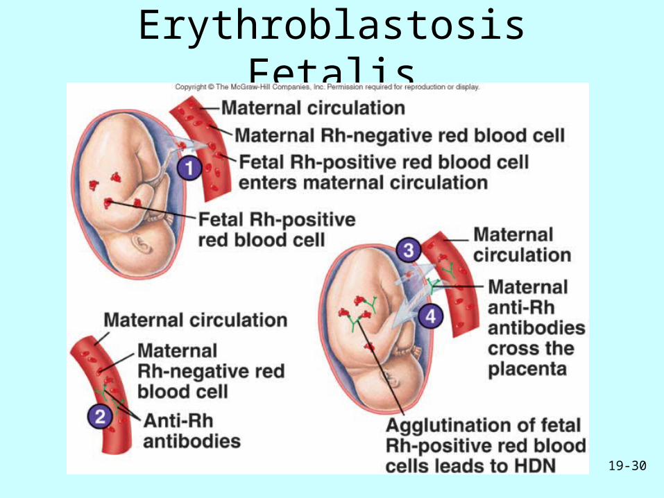
Rh Blood Group
• First studied in rhesus monkeys
• Types– Rh positive: Have these antigens present on
surface of RBCs– Rh negative: Do not have these antigens present
• Hemolytic disease of the newborn (HDN)– Mother produces anti-Rh antibodies that cross
placenta and cause agglutination and hemolysis of fetal RBCs
19-30
Erythroblastosis Fetalis

19-31
Diagnostic Blood Tests
• Type and crossmatch• Complete blood count
– Red blood count
– Hemoglobin measurement
– Hematocrit measurement
• White blood count• Differential white blood
count• Clotting

19-32
Blood Disorders
• Erythrocytosis: RBC overabundance
• Anemia: Deficiency of hemoglobin– Iron-deficiency
– Pernicious
– Hemorrhagic
– Hemolytic
– Sickle-cell
• Hemophilia• Thrombocytopenia• Leukemia• Septicemia• Malaria• Infectious
mononucleosis• Hepatitis