10-1 Anatomy and Physiology, Seventh Edition Rod R. Seeley Idaho State University Trent D. Stephens...
39
10-1 Anatomy and Physiology, Seventh Edition Rod R. Seeley Idaho State University Trent D. Stephens Idaho State University Philip Tate Phoenix College Copyright © The McGraw-Hill Companies, Inc. Permission required for reproduction or display. *See PowerPoint Image Slides for all figures and tables pre-inserted into PowerPoint without notes. Chapter 10 Chapter 10 Lecture Lecture Outline Outline *
-
Upload
homer-george -
Category
Documents
-
view
218 -
download
2
Transcript of 10-1 Anatomy and Physiology, Seventh Edition Rod R. Seeley Idaho State University Trent D. Stephens...

10-1
Anatomy and Physiology, Seventh Edition
Rod R. SeeleyIdaho State UniversityTrent D. StephensIdaho State UniversityPhilip TatePhoenix College
Copyright © The McGraw-Hill Companies, Inc. Permission required for reproduction or display.
*See PowerPoint Image Slides for all figures and tables pre-inserted into PowerPoint without notes.
Chapter 10Chapter 10
Lecture OutlineLecture Outline**

10-2
Muscular SystemGross Anatomy
Chapter 10

10-3
General Principles• Tendons: attach muscles to bones
– Aponeurosis: a very broad tendon• Muscle terminology
– Origin or head: muscle end attached to more stationary of two bones– Insertion: muscle end attached to bone with greatest movement– Belly: largest portion of the muscle between origin and insertion– Agonist: muscle that, when it contracts, causes an action– Antagonist: a muscle working in opposition to agonist
• Example: the biceps brachii can be used to lift weights and is the agonist, but when you move a bowling ball back to prepare to bowl, the biceps is the antagonist
– Synergists: muscles that work together to cause a movement• Prime mover: plays major role in accomplishing movement• Fixators: stabilize joint/s crossed by the prime mover; prevent movement of
the origin of the prime mover.

10-4
Muscle attachment

10-5
Muscle Types

10-6
Examples of Muscle Shapes

10-7
Nomenclature
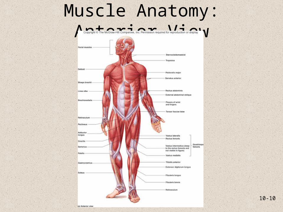
• Muscles are named according to:– Location: pectoralis gluteus, brachial– Size: maximus, minimus, longus, brevis– Shape: deltoid, quadratus, teres– Orientation: rectus– Origin and insertion: sternocleidomastoid,
brachioradialis– Number of heads: biceps, triceps– Function: abductor, adductor, masseter

10-8
Muscle Movements
• Muscles and their tendons and bones act together as lever systems to move either parts of the body or the whole body. Muscle contractions are a pull or force by relative positions of– Lever: rigid shaft or bone– Fulcrum: pivot point or joint– Weight or resistance (force of gravity either in the
form of the weight of the body parts or the weight of an object being lifted, pulled, or pushed)

10-9
Classes of Levers
• Class I– Fulcrum between force and
weight– Seesaw– Head movement at the
atlantooccipital joint
• Class II– Weight is between fulcrum
and pull– Wheelbarrow – Standing on toes;
metatarsophalangeal joint
• Class III– Pull located between fulcrum
and weight– Person using a shovel– Most common: biceps brachii
with elbow as fulcrum
10-10
Muscle Anatomy: Anterior View

10-11
Muscle Anatomy: Posterior View

10-12
Head and Neck Muscles• Flexion: muscles deep within
the neck along the anterior margins of the vertebral bodies
• Extension: posterior neck muscles attached to occipital bone
• Rotation and abduction: lateral and posterior groups
• Examples: sternocleidomastoid, trapezius, splenius muscles

10-13
Posterior Deep Neck Muscles

10-14
Muscles of Facial Expression• Cutaneous; origin and
insertion in the superficial fascia.
• Confined primarily to head and neck.
• Move the skin; some act as sphincters.
• Examples: orbicularis oris, orbicularis oculi, platysma.

10-15
Muscles of Mastication and Hyoid
• Mastication: chewing. Involves elevation/depression of the mandible and excursion to grind the teeth together
• Act with the muscles of hyoid in movement of the mandible
• Muscles of the cheek and tongue aid mastication by pushing the food under the teeth
• Examples: masseter, temporalis, pterygoids, digastrics

10-16
Muscles of the Hyoid

10-17
Tongue Movements• Important in speech:
changes shape• Swallowing
– Moves food around in mouth– Holds food in place during
grinding– Pushes food up to palate and
back toward pharynx
• Intrinsic: entirely within the tongue and allow change in shape
• Extrinsic: insert in tongue and allow change in shape and movement

10-18
Swallowing and the Larynx• Hyoid muscles: infra- and
suprahyoid groups– Suprahyoid muscles fix the
hyoid, then thyrohyoid can elevate larynx
– When infrahyoid group fixes hyoid, suprahyoid muscles can help depress the mandible
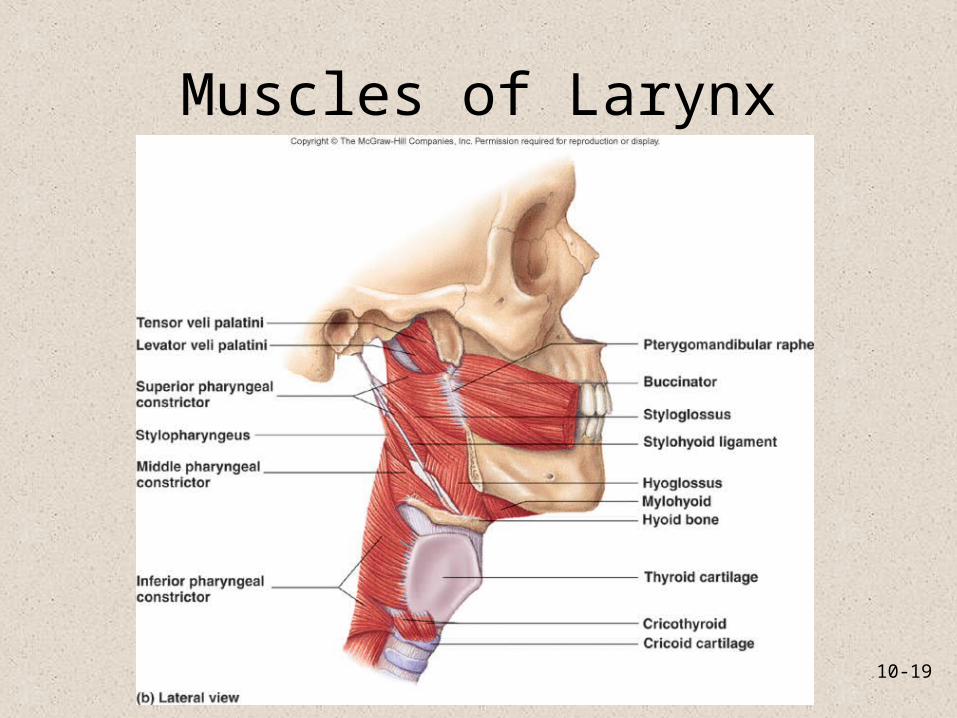
• Swallowing: Elevation of pharynx and larynx
• Constriction of the pharynx from superior to inferior
• Salpingopharyngeus opens auditory tubes to equalize pressure between middle ear and atmosphere
10-19
Muscles of Larynx

10-20
Extrinsic Muscles of the Eye• Rectus muscles: insert on
sclera anterior to center of sphere. Move eyeball and thus pupil laterally, superiorly, inferiorly, and medially
• Oblique muscles: insert onto the posterolateral margin of the eyeball and both laterally deviate the eyeball. The superior oblique passes through a pulley-like trochlea

10-21
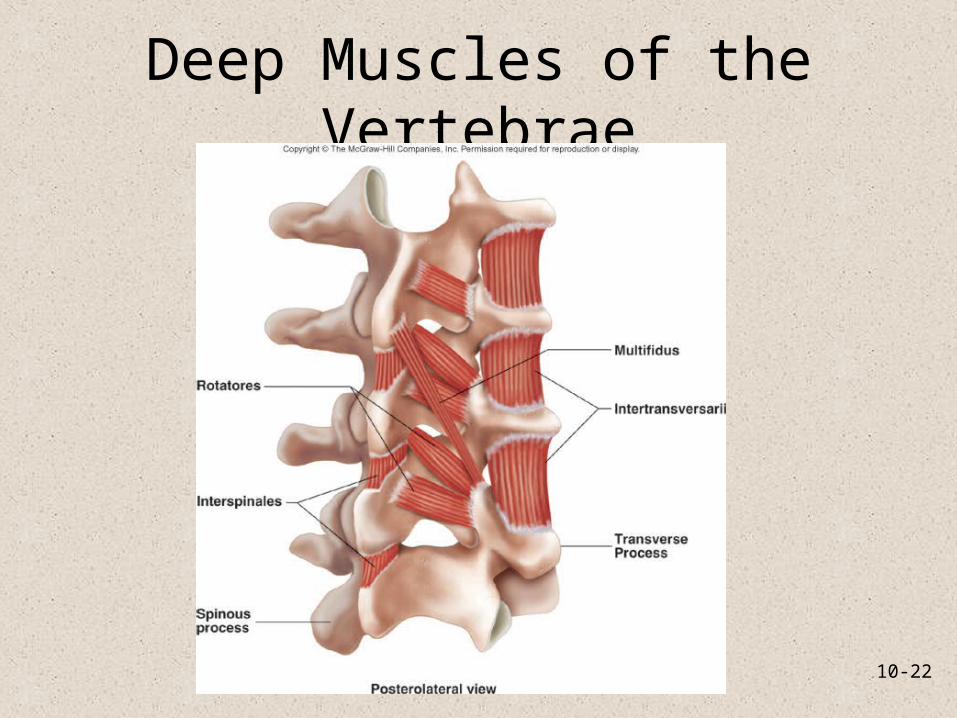
Muscles that Move the Vertebral Column
• Muscles that extend, laterally flex, and rotate the vertebral column. Used to produce erect posture
• Divided into deep and superficial groups– Deep group: from vertebra to
vertebra
– Superficial group extend from vertebrae to ribs
10-22
Deep Muscles of the Vertebrae

10-23
Thoracic Muscles• Involved in breathing• Four groups associated with
rib cage– Scalenes: elevate first two ribs
during inspiration– External intercostals: elevate
the ribs– Internal intercostals: depress
ribs during expiration– Transversus thoracis: depresses
ribs during expiration– Diaphragm: major movement
of inspiration. Flattens during contraction and increases the volume of the thoracic cavity

10-24
Abdominal Wall
• Flex and rotate vertebral column, decrease volume of abdominal and thoracic cavities
• Aid in forced expiration, vomiting, defecation, urination, childbirth
• Crossing pattern of muscles adds strength to abdominal wall to support organs

10-25
Abdominal Wall
• Rectus Abdominis– Linea alba in center– Covered by rectus sheath– Tendinous intersections divided muscle into sections– Flexes vertebral column
• External abdominal oblique: flexes and rotates abdomen• Internal abdominal oblique: flexes and rotates abdomen• Transversus abdominis: compresses abdominal wall

10-26
Muscles of Pelvic Floor and Perineum• Pelvic diaphragm:
Funnel-shaped, supports the pelvic viscera. Pierced by anal canal, urethra and (vagina).
• Perineum: diamond-shaped area inferior to pelvic diaphragm. Anterior half of diamond is urogenital triangle; posterior half is anal triangle

10-27
Scapular Movements
• Muscles that attach the upper limb to the body and move or stabilize the scapula and clavicle.
• Originate on the axial skeleton.
• Trapezius, levator scapulae, rhomboideus, serratus anterior, pectoralis minor

10-28
Arm Movements• Muscles that attach arm
to thorax: pectoralis major, latissimus dorsi
• Deltoid and pectoralis major both act as flexors and extensors of the shoulder
• Deltoid abducts and medially and laterally rotates arm

10-29
Rotator Cuff• Primary muscles holding
humerus in the glenoid cavity
• Form a cuff or cap over the proximal humerus
• Involved in flexion, extension, abduction, adduction, rotation and circumduction
• Infraspinatus, subscapularis, supraspinatus, teres minor

10-30
Forearm Movement
• Movements at the elbow
• Extension: triceps brachii and anconeus
• Flexion: biceps brachii, brachioradialis, and brachialis
• Supination and pronation: – Supination: supinator and
biceps brachii
– Pronation: pronator quadratus and pronator teres
10-31
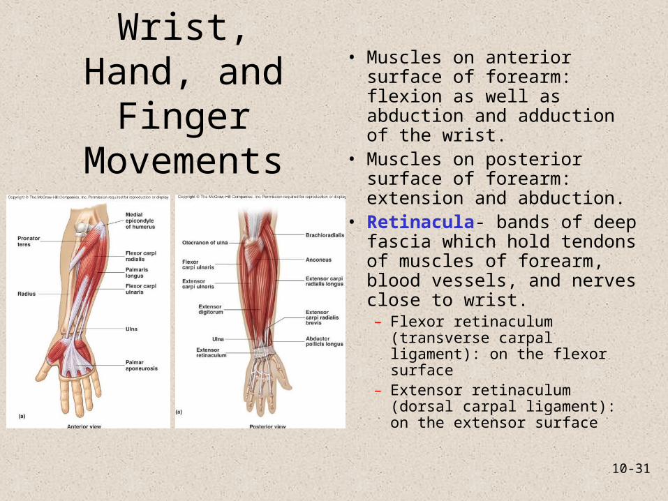
Wrist, Hand, and Finger
Movements
• Muscles on anterior surface of forearm: flexion as well as abduction and adduction of the wrist.
• Muscles on posterior surface of forearm: extension and abduction.
• Retinacula- bands of deep fascia which hold tendons of muscles of forearm, blood vessels, and nerves close to wrist. – Flexor retinaculum (transverse
carpal ligament): on the flexor surface
– Extensor retinaculum (dorsal carpal ligament): on the extensor surface

10-32
Intrinsic Muscles of the Hand
• Originate and insert in hand.

10-33
Thigh Movement• Originate on coxa; insert
onto femur• Anterior, posterolateral,
deep– Anterior: flex hip. Iliacus
and psoas major often referred to as iliopsoas since they share a tendon of insertion
– Posterolateral: gluteals and tensor fasciae latae Extension of thigh
– Deep: thigh rotators

10-34
Leg Movements• Quadriceps femoris: anterior surface of thigh
– Extension of the leg at the knee. – Rectus femoris also flexes the hip.– Insert by common tendon (patellar tendon) on and
around the patella– Patellar tendon extends from patella to tibial tuberosity
• Sartorius: flexes hip and knee, laterally rotates thigh
• Medial thigh muscles: adduction• Posterior thigh muscles: hamstrings. Flexion and
rotation of the knee

10-35
Muscles that Move the Leg

10-36
Ankle, Foot, and Toe
Movements
• Extrinsic foot muscles• Three leg compartments
– Anterior compartment: extensors involved in dorsiflexion and eversion/inversion of foot; extension of toes. Dorsiflex foot, extend toes.
– Lateral compartment: plantar flexion and eversion.
– Posterior compartment-• Superficial muscles
(plantar flexion) have common tendon of insertion called the calcaneal (Achilles) tendon. Gastrocnemius, plantaris
• Deep muscles: plantar flex and invert foot

10-37
Extrinsic Muscles of the Foot

10-38
Extrinsic Muscles of the Foot

10-39
Intrinsic Muscles of the Foot• Analogous to intrinsics
of hand, but serve in support and locomotion
• Flexion, extension, abduction and adduction of toes
• Deep fascia forms plantar aponeurosis.