Notes: Muscle Types & Function. (1) Types of Muscle Skeletal Cardiac Smooth.
Upload
blaise-gainesCategory
view
217download
3
1
2
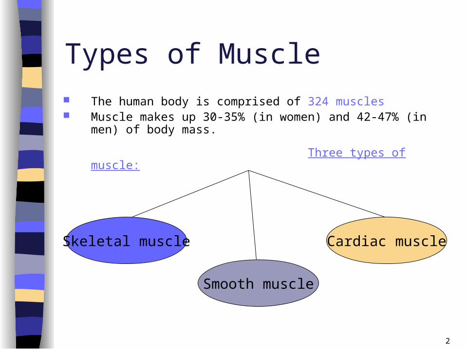
Types of Muscle The human body is comprised of 324 muscles Muscle makes up 30-35% (in women) and 42-47% (in men) of
body mass.
Three types of muscle:
Skeletal muscle
Smooth muscle
Cardiac muscle

3
A. Skeletal (Striated) Muscle Connects the various parts of the skeleton through one or more
connective tissue tendons During muscle contraction, skeletal muscle shortens and moves
various parts of the skeleton Activated through signals carried to the muscles via nerves (voluntary
control) Repeated activation of a skeletal muscle can lead to fatigue

4
B. Smooth Muscle
Located in the blood vessels, the
respiratory tract, the iris of the eye,
the gastro-intestinal tract
The contractions are slow and
uniform
Activation is involuntary

5
C. Cardiac Muscle
Has characteristics of both
skeletal and smooth muscle
Functions to provide the
contractile activity of the heart
Is very fatigue resistant
Activation of cardiac muscle is
involuntary (like smooth muscle)
6
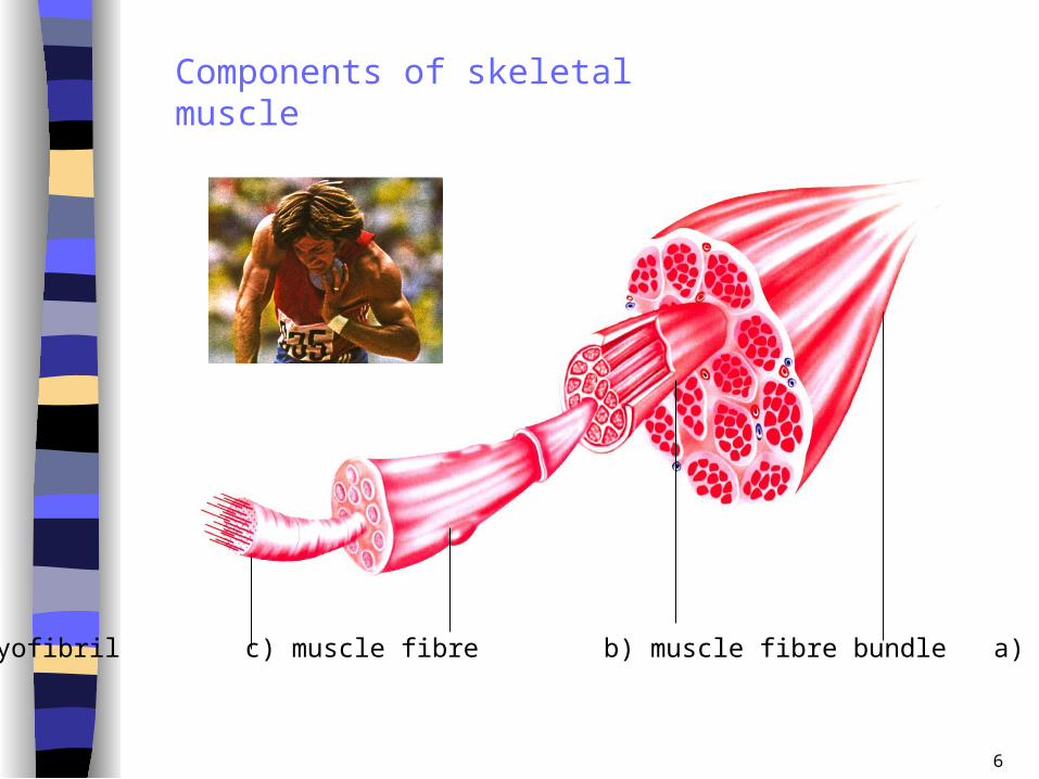
d) myofibril c) muscle fibre b) muscle fibre bundle a) Muscle belly
Components of skeletal muscle
7
Muscle Fibres Each fibre is made up of a number of myofilaments
Surrounded by a connective tissue sheath called
Sarcolemma
Many fibres are enclosed by connective tissue sheath
Perimycium to form bundle of fibres
Group of fibres activated via same nerve: motor unit
Each fibre has capillaries that supply nutrients and
eliminate waste
8
Muscle Teamwork Agonist (prime mover):
- the muscle or group of muscles producing a desired effect
Antagonist:
- the muscle or group of muscles opposing the action
Synergist: - the muscles surrounding the joint being moved
Fixators:
- the muscle or group of muscles that steady joints closer to the body axis so that the desired action can occur

9
Bending or straightening of elbow requires the coordinated interplay of the biceps and triceps muscles
10
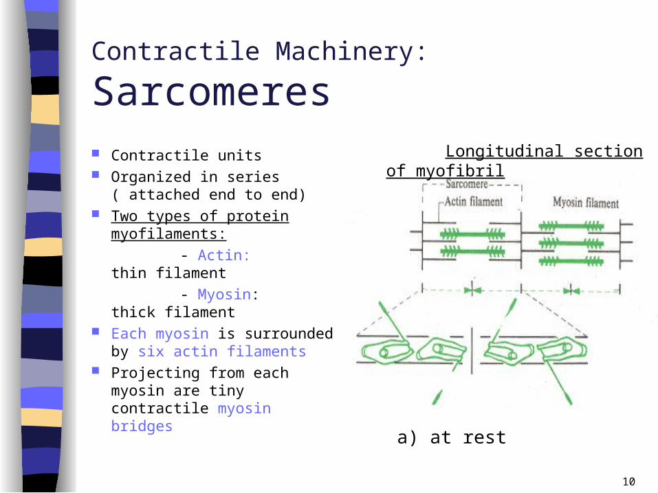
Contractile Machinery:
Sarcomeres Contractile units Organized in series ( attached
end to end) Two types of protein
myofilaments:
- Actin: thin filament
- Myosin: thick filament Each myosin is surrounded by
six actin filaments Projecting from each myosin
are tiny contractile myosin bridges
Longitudinal section of myofibril
a) at rest

11
Contractile Machinery:Crossbridge formation and movement
Cross bridge formation: - a signal comes from the motor nerve activating the fibre - the heads of the myosin filaments temporarily attach themselves to the actin filaments
Cross bridge movement: - similar to the stroking of the oars and movement of rowing shell- movement of myosin filaments in relation to actin filaments- shortening of the sarcomere- shortening of each sarcomere is additive
b) contraction
Longitudinal section of myofibril

12
Contractile Machinery:Optimal Crossbridge formation
Sarcomeres should be optimal distance apart
If the sarcomeres are stretched farther apart than optimal distance:
- fewer cross bridges can form less force produced
If the sarcomeres are too close together:
- cross bridges interfere with one another as they form less force produced
Longitudinal section of myofibril
c) Powerful stretching
d) Powerful contraction

13
Contractile Machinery:
Optimal muscle length and optimal joint
angle
The distance between sarcomeres is dependent on the stretch of
the muscle and the position of the joint
Maximal muscle force occurs at optimal muscle length (lo)
Maximal muscle force occurs at optimal joint angle
Optimal joint angle occurs at optimal muscle length

14
Muscle tension during elbow flexion at constant speed
15
Contractile Machinery:
Tendons, origin, insertion
In order for muscles to contract, they must be attached to the bones to create movement
Tendons: strong fibrous tissues at the ends of each muscle that attach muscle to bone
Origin: the end of the muscle attached to the bone that does not move
Insertion: the point of attachment of the muscle on the bone that moves

16
Muscle Fibre Types
Slow twitch fibres:
Slow Oxidative (Type I)
Fast twitch fibres: Fast Glycolytic (Type IIb) Fast Oxidative Glyc. (Type IIb)
17
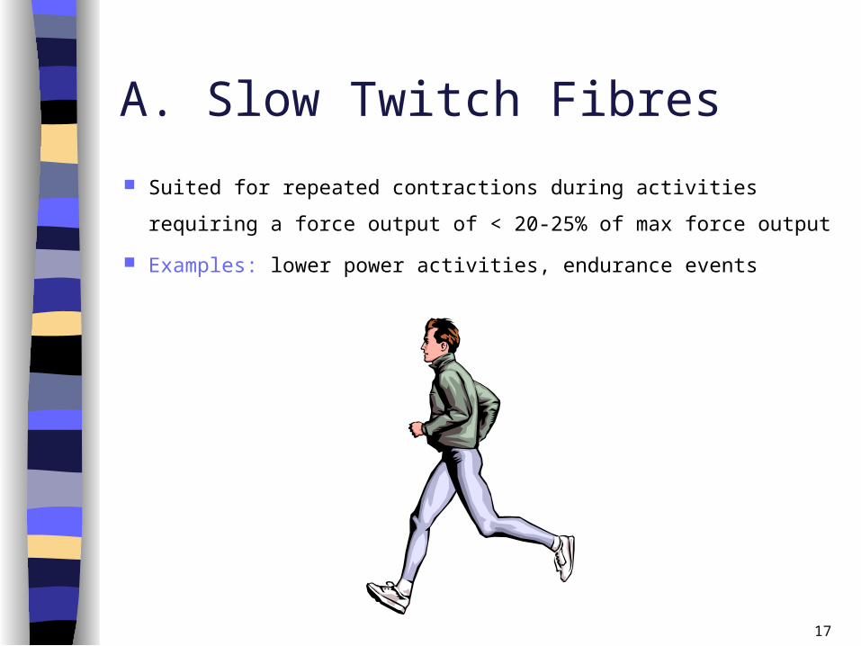
A. Slow Twitch Fibres
Suited for repeated contractions during activities requiring a
force output of < 20-25% of max force output
Examples: lower power activities, endurance events

18
B) Fast Twitch Fibres Significantly greater force and speed generating capability than
slow twitch fibres
Well suited for activities involving high power
Examples: sprinting, jumping, throwing

19
The Muscle Biopsy
Used to determine muscle fibre type
1. Injection of local anesthetic into the muscle being sampled
2. Incision of approximately 5-7mm is made in the skin and fascia
of the muscle
3. The piece of tissue (250-300mg) removed via the biopsy needle
is imbedded in OCT compound
4. The sample is frozen in isopentane cooled to –180C

20
Nerve-Muscle Interaction
Skeletal muscle activation is initiated through neural activation
NS can be divided into central (CNS) and peripheral (PNS)
The NS can be divided in terms of function: motor and sensory
activity
Sensory: collects info from the various sensors located
throughout the body and transmits the info to the brain
Motor: conducts signals to activate muscle contraction
21
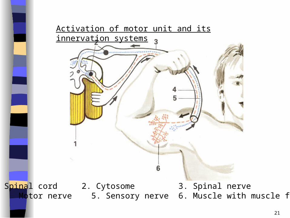
Activation of motor unit and its innervation systems
1. Spinal cord 2. Cytosome 3. Spinal nerve 4. Motor nerve 5. Sensory nerve 6. Muscle with muscle fibres
22
Motor Unit
Motor nerves extend from the spinal cord to the muscle fibres
Each fibre is activated through impulses delivered via motor end plate
Motor unit: a group of fibres activated via the same nerve Muscles needed to perform precise movements generally
consist of a large number of motor units and few muscle fibres
Less precise movements are carried out by muscles composed of fewer motor units with many fibres per unit

23
Muscle’s Adaptation to Strength Training
Individual’s performance improvements occur through a process of biological adaptation, which is reflected in the body’s increased strength
Adaptation process proceeds at different time rates for different functional systems and physiological processes
Adaptation depends on intensity levels used in training and on athlete’s unique biological make-up
Enzymes adapt within hours, cardiovascular adaptation within 10 to 14 days