Introduction Hand anatomy Ligament and dislocation injuries Fractures in hand Compartment...
87
INJURIES OF THE HAND
-
Upload
ralf-sparks -
Category
Documents
-
view
224 -
download
0
Transcript of Introduction Hand anatomy Ligament and dislocation injuries Fractures in hand Compartment...

INJURIES OF THE HAND

Contents
Introduction Hand anatomy Ligament and dislocation injuries Fractures in hand Compartment syndrome Nerve injuries Arterial injuries Zones of the hand and their injuries POP slab application
INTRODUCTION
The hand is a very complex organ with multiple joints, different types of ligament, tendons and nerves. With constant use, it is no wonder that hand disease injuries are common in society.
Hand injuries can result from excessive use, degenerative disorders or trauma.
Trauma to the finger or the hand is quite common in society.
In some particular cases, the entire finger may be subject to amputation.
The majority of traumatic injuries are work-related. Today, skilled hand surgeons can sometimes reattach the finger or thumb using microsurgery.
Hand Disease Retrieved on 2010-01-20
Hand Anatomy
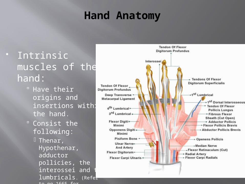
Intrinsic muscles of the hand:
Have their origins and insertions within the hand.
Consist the following: Thenar, Hypothenar,
adductor pollicies, the interossei and the lumbricals. (Refer to pg 1665 for anatomical description)
Hand Anatomy
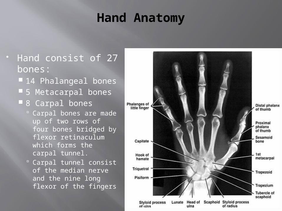
Hand consist of 27 bones: 14 Phalangeal bones 5 Metacarpal bones 8 Carpal bones
Carpal bones are made up of two rows of four bones bridged by flexor retinaculum which forms the carpal tunnel.
Carpal tunnel consist of the median nerve and the nine long flexor of the fingers

Hand Anatomy
Extensor Tendons: Courses over the dorsal side of the forearm,
wrist and hand. 9 extensor tendons pass under the extensor
retinaculum and separate into 6 compartments
Schmidt, Hans-Martin; Lanz, Ulrich (2003). Surgical Anatomy of the Hand. Thieme. ISBN 1-58890-007X.
Surface anatomy of the hand.
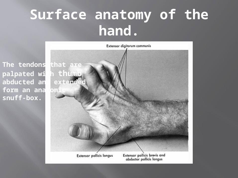
The tendons that are palpated with thumb abducted and extended form an anatomic snuff-box.

Hand Anatomy
Flexor Tendons: Courses over the volar side of the forearm,
wrist, and hand.
Unlike the extensor tendons, the flexor tendons are enclosed in synovial sheaths making them prone to deep space infections.
Schmidt, Hans-Martin; Lanz, Ulrich (2003). Surgical Anatomy of the Hand. Thieme.
ISBN 1-58890-007-X.
Hand Anatomy
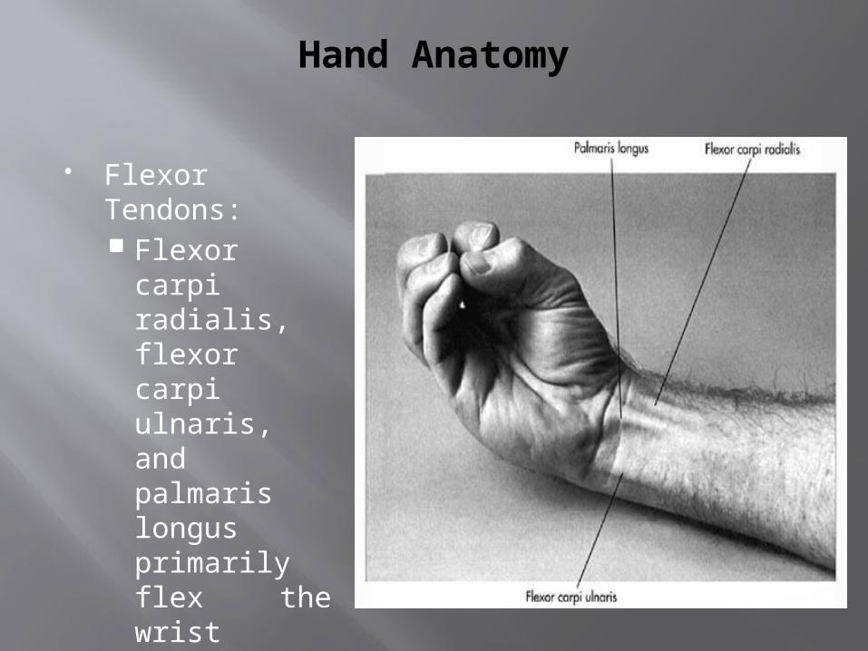
Flexor Tendons: Flexor carpi
radialis, flexor carpi ulnaris, and palmaris longus primarily flex the wrist
Hand Anatomy
Hand Anatomy
Blood supply (BS):
Proximal portions of the hand (BS) come from the deep and superficial arches on the palmar and dorsal side.
BS of the fingers is distributed by the digital arteries that arises from the superficial palmer arch.
Schmidt, Hans-Martin; Lanz, Ulrich (2003). Surgical Anatomy of the Hand Thieme. ISBN 1-58890-007-X..
Hand Anatomy
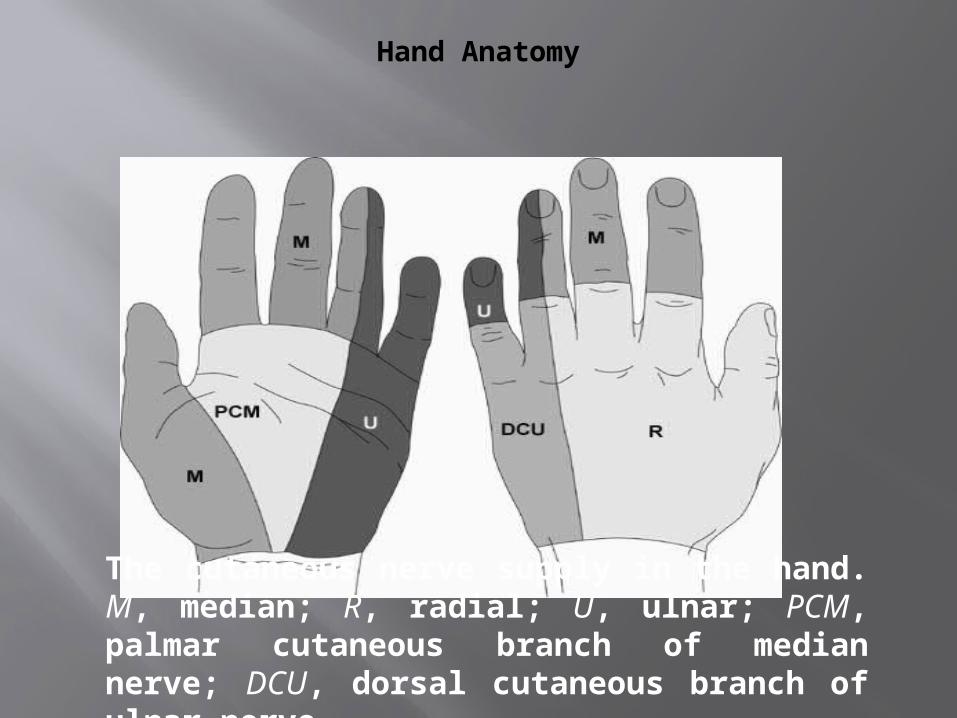
The cutaneous nerve supply in the hand. M, median; R, radial; U, ulnar; PCM, palmar cutaneous branch of median nerve; DCU, dorsal cutaneous branch of ulnar nerve
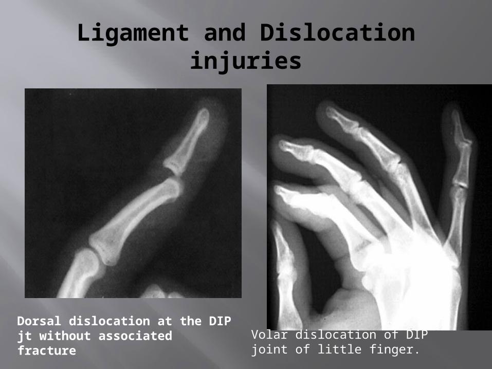
Ligament and Dislocation injuries
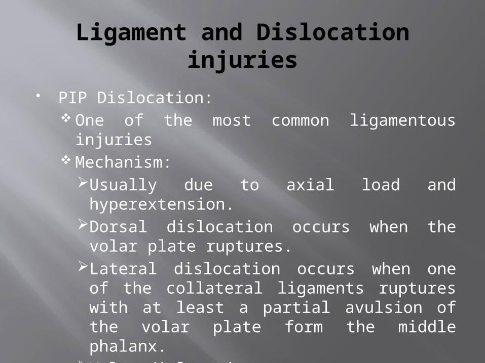
PIP Dislocation: One of the most common ligamentous injuries Mechanism:
Usually due to axial load and hyperextension.
Dorsal dislocation occurs when the volar plate ruptures.
Lateral dislocation occurs when one of the collateral ligaments ruptures with at least a partial avulsion of the volar plate form the middle phalanx.
Volar dislocations are rare.
Ligament and Dislocation injuries
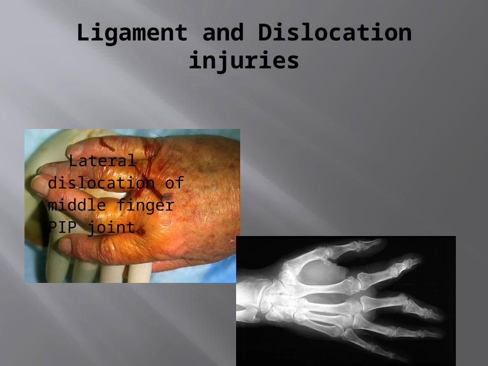
Lateral dislocation of middle finger PIP joint.
Ligament and Dislocation injuries
PIP Dislocation: Reduction
Perform similarly to DIP dorsal dislocationsActive ROM and strength should be tested
after reduction.If testing is normal, then splint in 30-degree
flexion for 3 wks.If the joint is irreducible or there is evidence
of complete ligamentous disruption, operative repair is required.
Ligament and Dislocation injuries
MP dislocation: Less common than at the PIP. Mechanism:
Usually due to hyperextension forces that rupture the volar plate causing dorsal dislocation.
In subluxation (simple dislocation) – the jt appears to be hyperextended 60 – 90 degrees and the articular surfaces are still in contact.
Volar dislocation are rare and usually require operative reduction.
Ligament and Dislocation injuries
MP dislocation: Reduction:
Does not involve hyperextension (this might convert it from a simple to a complex dislocation)
Performed with the wrist flexed to relaxed the flexor tendon and applying pressure over the dorsum of the proximal phalanx in a distal and volar direction.
Splint the MP jt in flexion after reduction.
Ligament and Dislocation injuries
CMC jt dislocation:
Are uncommon because the jt is supported by strong dorsal, volar, and interosseous ligaments and reinforced by the broad insertions of the wrist flexors and extensors.

Ligament and Dislocation injuries
CMC jt dislocation: Mechanism:
Usually due to high-speed mechanisms (MVC, falls, crushes, or clenched fist trauma).
Usually occurs via dorsal and with associated fractures.
Ligament and Dislocation injuries
CMC jt dislocation: Reduction:
Attempt after regional anesthesia with traction and flexion with simultaneous longitudinal pressure on the metacarpal base.
Pt need early referral after reduction to determine if further fixation is needed.
Ligament and Dislocation injuries
DIP Dislocation at DIP are uncommon because of
the firm attachments of the skin and subcutaneous tissue to the underlying bone.
Dislocations at the DIP are usually dorsal. Reduction can be done by longitudinal traction
and hyperextension, followed by direct dorsal pressure to the base of the distal phalanx after a digital block.
Ligament and Dislocation injuries
Volar dislocation of DIP joint of little finger.
Dorsal dislocation at the DIP jt without associated fracture
Fractures
Fractures
Metacarpal (II to V) Fractures 2nd and 3rd metacarpals are relatively immobile
and fractures require anatomic reduction. 4th and 5th MC have 15 to 20-degree AP
motion, which allows for some compensation. MC fractures are categorized as head, neck,
shaft, or base fractures.
Fractures
Metacarpal (II to V) Fractures Head:
Usually caused by a direct blow, crush or missile.
Fractures are distal to the insertion of the collateral ligaments and are often comminuted.
If a laceration is present a human bite must be considered.
Treatment: Ice, elevation, immobilization, and referral to a hand surgeon.

Fractures
Metacarpal (II to V) Fractures Neck:
Usually caused by a directed impaction force.
Fracture of the fifth MC neck is often referred to as a boxer’s fracture
Fracture are usually unstable with volar angulation.
Angulation of < 20 degrees in the 4th and 40 degrees in the 5th MC will not result in functional impairment
Fractures
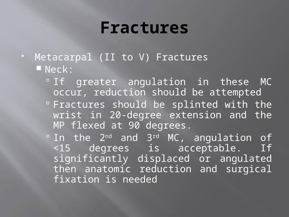
Metacarpal (II to V) Fractures Neck:
If greater angulation in these MC occur, reduction should be attempted
Fractures should be splinted with the wrist in 20-degree extension and the MP flexed at 90 degrees.
In the 2nd and 3rd MC, angulation of <15 degrees is acceptable. If significantly displaced or angulated then anatomic reduction and surgical fixation is needed

Fractures
Metacarpal (II to V) Fractures Shaft:
Usually occur via a direct blow Rotational deformity and shortening are
more often in shaft fractures than in neck fractures.
If reduction is needed, than operative fixation is usually indicated.
Fractures
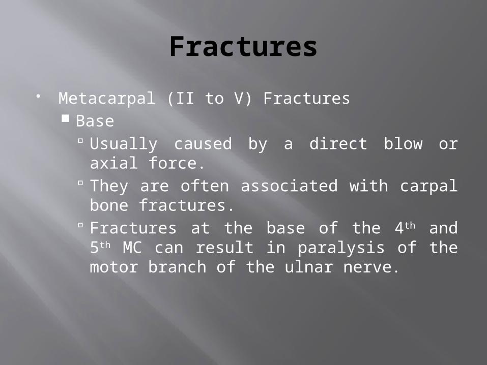
Metacarpal (II to V) Fractures Base
Usually caused by a direct blow or axial force.
They are often associated with carpal bone fractures.
Fractures at the base of the 4th and 5th MC can result in paralysis of the motor branch of the ulnar nerve.

Fractures
Thumb MC Because of the mobility of the thumb MC,
shaft fractures are uncommon Fractures usually involve the base. Two type:
Extraarticular Intraarticular
Fractures
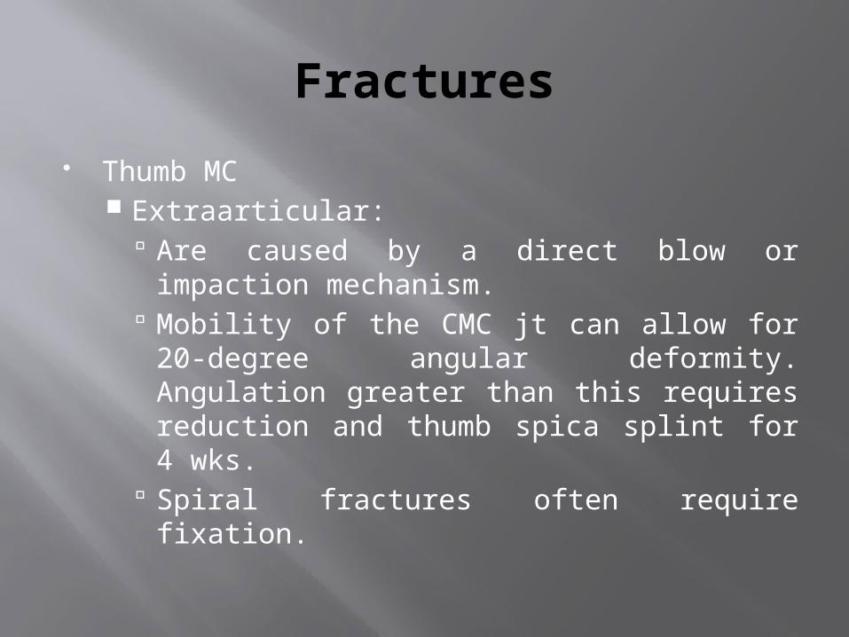
Thumb MC Extraarticular:
Are caused by a direct blow or impaction mechanism.
Mobility of the CMC jt can allow for 20-degree angular deformity. Angulation greater than this requires reduction and thumb spica splint for 4 wks.
Spiral fractures often require fixation.

Fractures
Thumb MC Intraarticular
Caused by impaction from striking a fixed object (two type)
Bennett fx Is an intraarticular fx with associated subluxation or dislocation at the CMC jt.
The ulnar portion of the MC usually remains in place.
The distal portion usually subluxes radially and dorsally from the pull of abduction pollicis longus and the adductor pollicis
Treatment – thumb spica and referralSoyer AD. Fractures of the base of the first metacarpal: current treatment options. J Am Acad Orthop Surg. Nov-Dec 1999;7(6):403-12.
Fractures
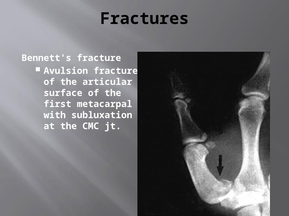
Bennett's fracture Avulsion fracture
of the articular surface of the first metacarpal with subluxation at the CMC jt.
Fractures
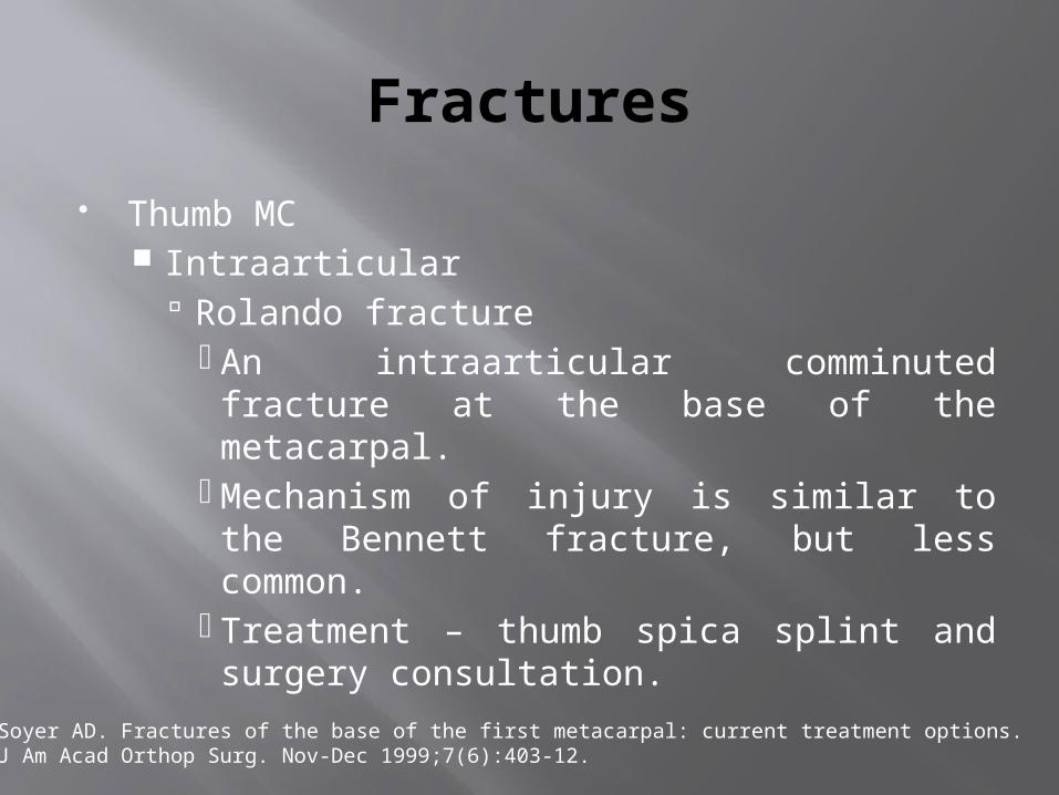
Thumb MC Intraarticular
Rolando fracture An intraarticular comminuted fracture at the base of the metacarpal.
Mechanism of injury is similar to the Bennett fracture, but less common.
Treatment – thumb spica splint and surgery consultation.
Soyer AD. Fractures of the base of the first metacarpal: current treatment options. J Am Acad Orthop Surg. Nov-Dec 1999;7(6):403-12.

Fractures
Thumb CMC: Isolated dislocation is rare compared to the
more common Bennett fracture dislocation. Easy to reduce but unstable after reduction. Apply thumb spica splint after reduction. Need surgical referral.
COMPARTMENT SYNDROME
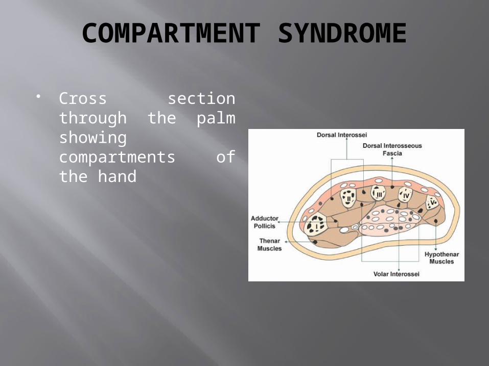
Cross section through the palm showing compartments of the hand
COMPARTMENT SYNDROME
Edema of tissues or hemorrhage within any of these compartments may lead to elevated pressures that result in tissue necrosis and subsequent loss of hand function due to contracture.
Sign and symptoms: Pain and paresthesias occur early
Paralysis and pulselessness occurring later
Konstantakos EK, Dalstrom DJ, Nelles ME, Laughlin RT, Prayson MJ (December 2007). "Diagnosis and management of extremity compartment syndromes: an orthopaedic perspective". Am Surg 73 (12): 1199–209. PMID 1818637.
COMPARTMENT SYNDROME
Diagnosis Confirmed by compartment pressure
measurement – high rate of false readings.
Treatment In the setting of severe crush injury with signs
and symptoms suggestive of compartment syndrome, emergent hand surgeon consultation for fasciotomy is mandatory.
Konstantakos EK, Dalstrom DJ, Nelles ME, Laughlin RT, Prayson MJ (December 2007). "Diagnosis and management of extremity compartment syndromes: an orthopaedic perspective". Am Surg 73 (12): 1199–209. PMID 18186372.
Nerve Injuries
Median and Ulnar- refer for immediate or delayed repair (10days).
Radial nerve repairs may delayed up to 3 months
Arterial Injuries
Radial/Ulnar artery injuries need referral
Digital arterial injuries: assess clinically- if no ischemia, does not need repair (collateral circulation)
Assess for associated nerve injury
POP SLABS FOR HAND INJURIES
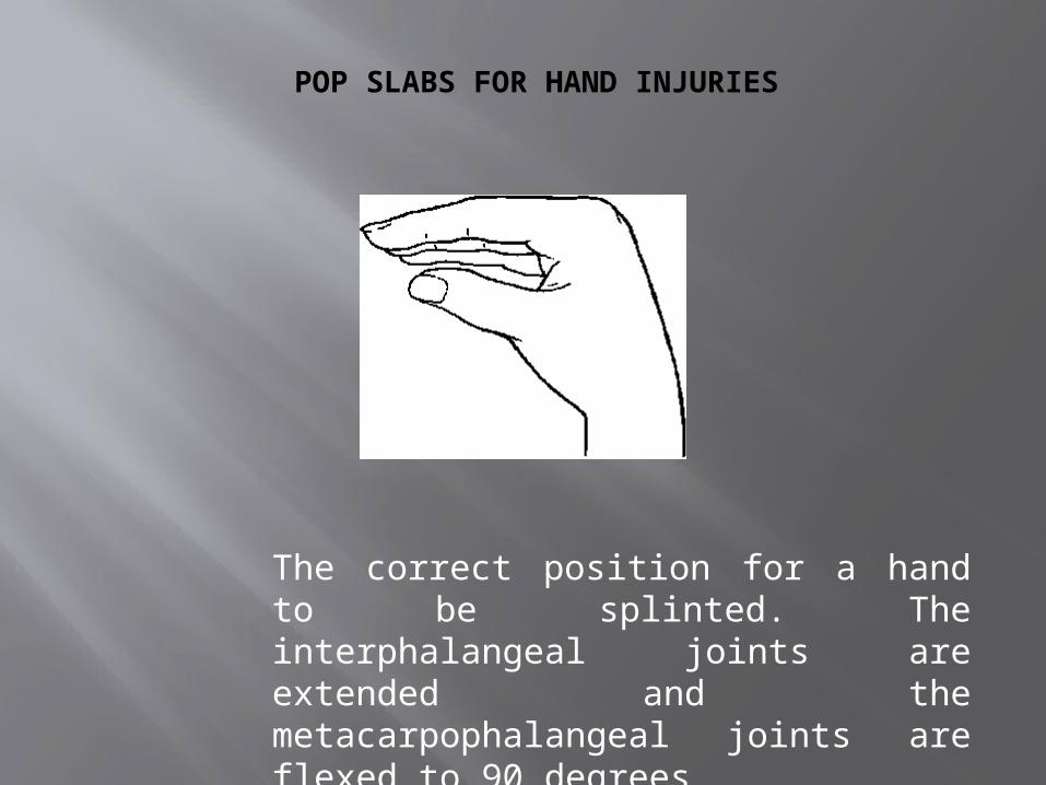
The correct position for a hand to be splinted. The interphalangeal joints are extended and the metacarpophalangeal joints are flexed to 90 degrees
POP SLABS FOR HAND INJURIES
An injured hand should be elevated and immobilized with the metacarpophalangeal joints in 90 degrees of flexion and the interphalangeal joints in full extension.
In this position, the capsule and collateral ligaments of these joints are maximally stretched and cannot contract.
This facilitates subsequent restoration of function. The presence of open wounds of the hand does
not prevent the use of this ideal position. It can be achieved by fully extending the wrist and applying a slab.
UNDERSTANDING WRIST
LINK BETWEEN FOREARM & HAND
JOINTS: DRUJ, RADIOCARPAL, MID-CARPAL
15 BONES: DISTAL RADIUS & ULNA
TWO ROWS OF CARPUS: Some Lovers Try Positions
That They Can't Handle.
BASE OF FIVE METACARPALS
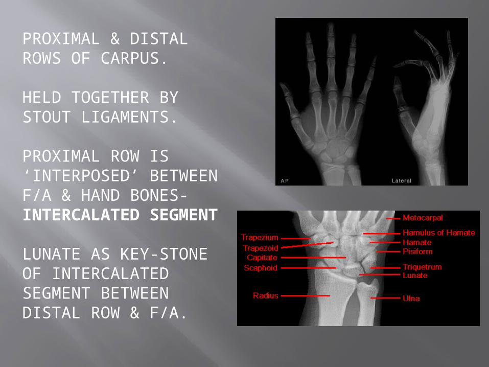
PROXIMAL & DISTAL ROWS OF CARPUS.
HELD TOGETHER BY STOUT LIGAMENTS.
PROXIMAL ROW IS ‘INTERPOSED’ BETWEENF/A & HAND BONES- INTERCALATED SEGMENT
LUNATE AS KEY-STONE OF INTERCALATED SEGMENT BETWEEN DISTAL ROW & F/A.
NO MUSCLE ATTACHMENT, EXCEPT PISIFORM~ SISAMOID.
POSITION OF FUNCTION / MAXIMUM STABILITY- WITH THE TIGHTENING OF LONG MUSCLES WITH WRIST IN 30 deg. ADDUCTION, CARPUS ARE DRAGED TO THE RADIAL SOCKET SECURELY- POSITION DURING POWER GRIP.
SCAPHOID- POTENTIALLY MOST UNSTABLE.
AS THE WRIST MOVES SO DOES SCAPHOID- LUNATE & TRIQUETRUM FOLLOWS-GIUDED BY INTEROSSEOUSLIG.
LIGAMENTS
EXTRINSIC: (CONNECTS FOREARM WITH CARPUS)
-RADIOCARPAL -CARPOMETACARPAL INTRINSIC: INTERCARPAL
ALL ARE INTRACAPSULAR EXCEPT-FLEXORRETINACULM -PISOHAMATE -PISOMETACARPAL
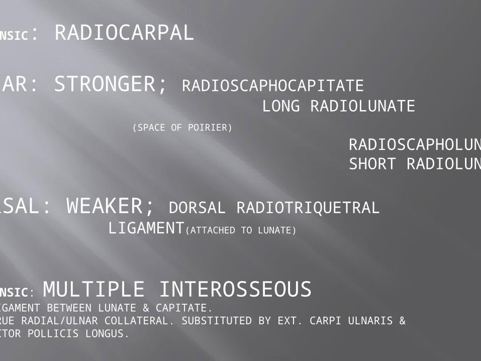
EXTRINSIC: RADIOCARPAL
VOLAR: STRONGER; RADIOSCAPHOCAPITATE LONG RADIOLUNATE
(SPACE OF POIRIER)
RADIOSCAPHOLUNATE SHORT RADIOLUNATE
DORSAL: WEAKER; DORSAL RADIOTRIQUETRAL LIGAMENT(ATTACHED TO LUNATE)
INTRINSIC: MULTIPLE INTEROSSEOUS-NO LIGAMENT BETWEEN LUNATE & CAPITATE.-NO TRUE RADIAL/ULNAR COLLATERAL. SUBSTITUTED BY EXT. CARPI ULNARIS & ABDUCTOR POLLICIS LONGUS.
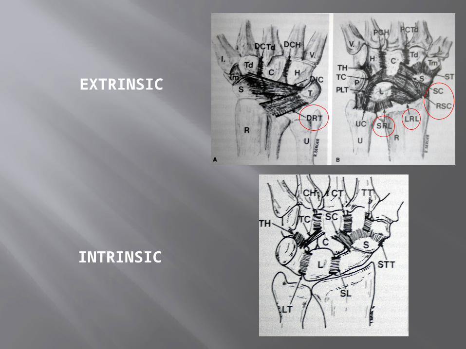
EXTRINSIC
INTRINSIC
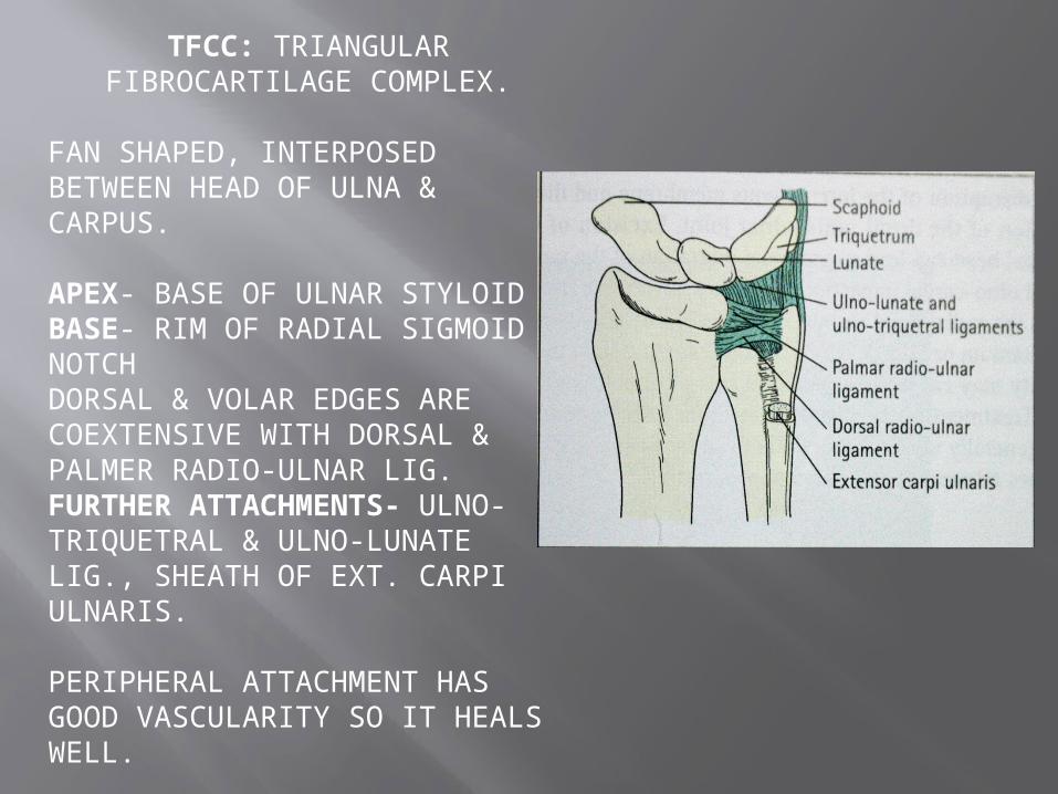
TFCC: TRIANGULAR FIBROCARTILAGE COMPLEX.
FAN SHAPED, INTERPOSED BETWEEN HEAD OF ULNA & CARPUS.
APEX- BASE OF ULNAR STYLOIDBASE- RIM OF RADIAL SIGMOID NOTCHDORSAL & VOLAR EDGES ARE COEXTENSIVE WITH DORSAL & PALMER RADIO-ULNAR LIG.FURTHER ATTACHMENTS- ULNO-TRIQUETRAL & ULNO-LUNATE LIG., SHEATH OF EXT. CARPI ULNARIS.
PERIPHERAL ATTACHMENT HAS GOOD VASCULARITY SO IT HEALS WELL.
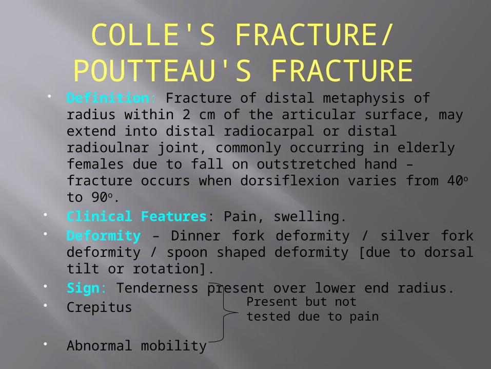
Definition: Fracture of distal metaphysis of radius within 2 cm of the articular surface, may extend into distal radiocarpal or distal radioulnar joint, commonly occurring in elderly females due to fall on outstretched hand – fracture occurs when dorsiflexion varies from 40o to 90o.
Clinical Features: Pain, swelling. Deformity – Dinner fork deformity / silver fork deformity /
spoon shaped deformity [due to dorsal tilt or rotation]. Sign: Tenderness present over lower end radius. Crepitus
Abnormal mobility
COLLE'S FRACTURE/ POUTTEAU'S FRACTURE
Present but not tested due to pain
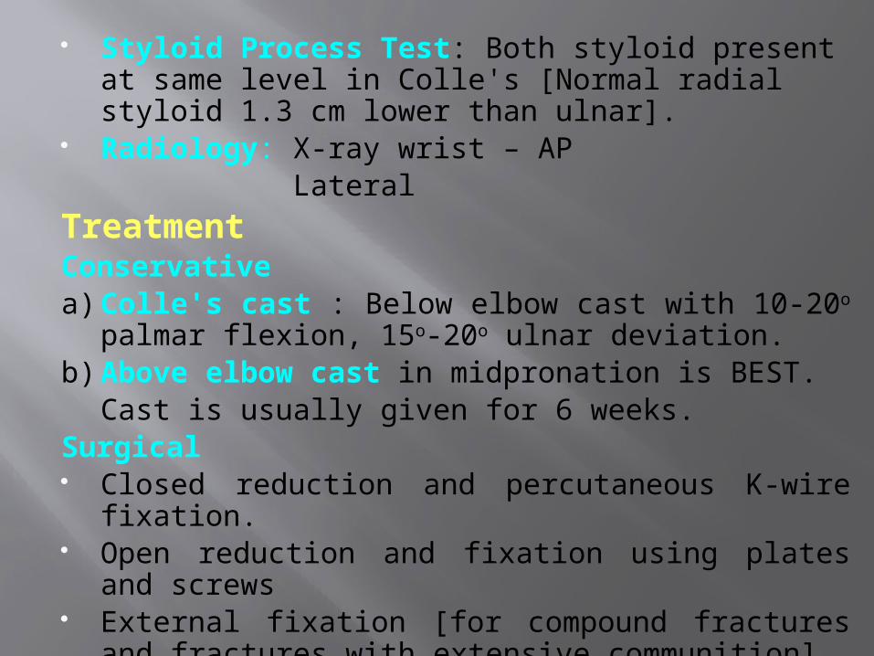
Styloid Process Test: Both styloid present at same level in Colle's [Normal radial styloid 1.3 cm lower than ulnar].
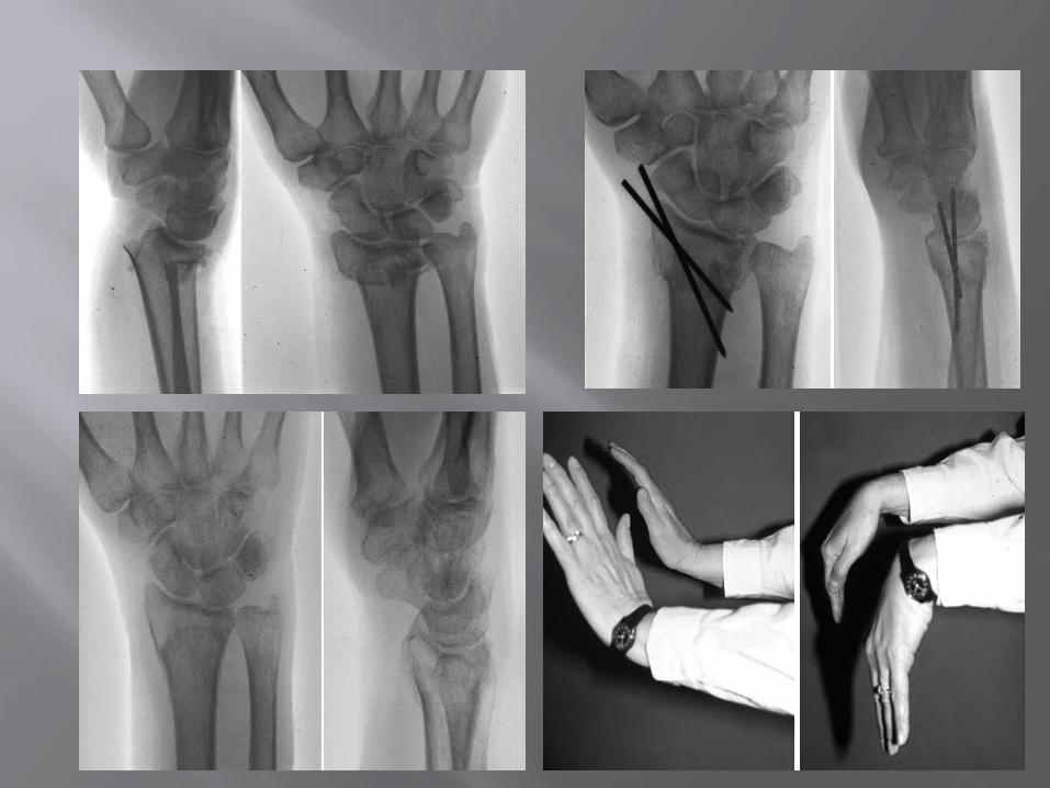
Radiology: X-ray wrist – AP Lateral
TreatmentConservativea) Colle's cast : Below elbow cast with 10-20o palmar
flexion, 15o-20o ulnar deviation.b) Above elbow cast in midpronation is BEST.
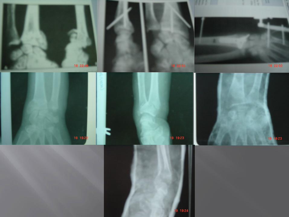
Cast is usually given for 6 weeks.Surgical Closed reduction and percutaneous K-wire fixation. Open reduction and fixation using plates and screws External fixation [for compound fractures and fractures
with extensive communition].
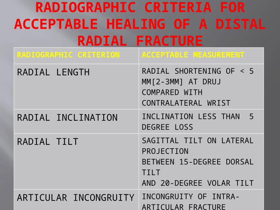
RADIOGRAPHIC CRITERIA FOR ACCEPTABLE HEALING OF A DISTAL RADIAL FRACTURE
RADIOGRAPHIC CRITERION ACCEPTABLE MEASUREMENT
RADIAL LENGTH RADIAL SHORTENING OF < 5 MM[2-3MM] AT DRUJ COMPARED WITH CONTRALATERAL WRIST
RADIAL INCLINATION INCLINATION LESS THAN 5 DEGREE LOSS
RADIAL TILT SAGITTAL TILT ON LATERAL PROJECTIONBETWEEN 15-DEGREE DORSAL TILTAND 20-DEGREE VOLAR TILT
ARTICULAR INCONGRUITY
INCONGRUITY OF INTRA-ARTICULAR FRACTUREIS ≤ 2 MM AT RADIOCARPAL JOINT
Complications:Early: (a) Unstable reduction
(b) Median or ulnar nerve stretch.
(c) Post reduction swelling.
(d) Compartment syndrome
(e) Distal radioulnar subluxation/dislocation.
Late: (a) Stiffness of finger and wrist joint.
(b) Malnunion.
(c) Rupture of extensor policis tendon.
(d) Sudeck's osteodystrophy.
(e) Frozen shoulder / shoulder hand syndrome / mental amputation.
(f) Carpal tunnel syndrome.
Definition: Fracture of distal one third of radius with palmar or volar displacement.
Mechanism of injury:a) Fall on back of dorsum of handb) Fall on forearm in supinationc) Direct blow on flexed hand.
Clinical Features: Pain, swellingDeformity Garden Spade deformity.Loss of wrist functions.
Radiology: X-ray wrist – AP view– Lateral view
– Carpus displaced proximally– Fracture fragment displaced anteriorly with palmar angulation.
SMITH FRACTURE / REVERSE COLLE'S
FRACTURE
Conservative Treatment: Reduction setting, above elbow POP casts [forearm in supination, wrist in extension].
Surgical: For unstable fractures – open/close reduction and fixation by K-wire, plate or screws.
• Complication: Usually arises due to misdiagnosis as Colle's other complications are similar to Colle's.

Definition: It is an intrarticular fracture dislocation or subluxation in which rim of distal radius is displaced either dorsally or volarly along with carpal bones.
Dislocation is most clinically and radiographically obvious abnormality.
BARTON FRACTURE
Dorsal Barton Volar Barton
Type Posterior marginal type Anterior marginal type
Variant Variant of Colle's Variant of Smith
Mechanism Fall with dorsiflexion & pronation of distal forearm on a flexed wrist.
Due to palmar tensile stress or dorsal shear stress.
Treatment Below elbow POP with wrist in neutral position.
Above elbow POP after reduction.
Operative Percutaneous K-wire fixation. External fixation.
Incidence : 60% of all carpal bone fracture. It articulates with 5 bones (radius, lunate, triquetral,
trapezium, capitulum) and lies at 45o to longitudinal axis of Zrows.
Central indentation known as waist.
Blood Supply:
67% of scaphoid have arterial foramina throughout its length.
13% - predominant in distal 1/3rd.
20% - waist. Proximal 1/3rd without adequate blood supply – Prone to
AVN.
Age Group: Young adults.
Mode of Injury: Fall on outstretched hand with hyper-extension and slight radial deviation at wrist.
SCAPHOID FRACTURE

Anatomical classification:
Waist fracture - m.c. 70%
Proximal pole fracture - 20%
Distal body fracture -
Tuberosity fracture -
Clinical Feature: Pain, swelling over wrist, inability/difficulty to use wrist.
Tenderness present in Anatomical snuff box.
Radiologically: AP
Lat
Oblique view Radiographs should be repeated at 10-14 days (local
decalcification after such an interval may reveal previously hidden fractures).
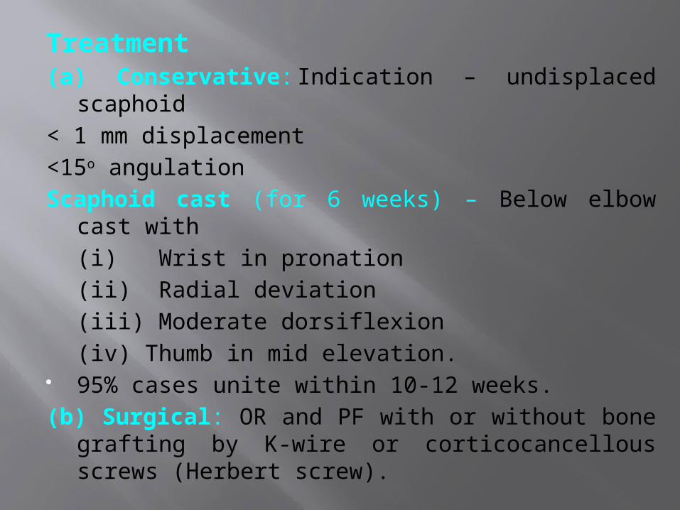
Treatment(a) Conservative: Indication – undisplaced scaphoid
< 1 mm displacement
<15o angulation
Scaphoid cast (for 6 weeks) – Below elbow cast with
(i) Wrist in pronation
(ii) Radial deviation
(iii) Moderate dorsiflexion
(iv) Thumb in mid elevation. 95% cases unite within 10-12 weeks.
(b) Surgical: OR and PF with or without bone grafting by K-wire or corticocancellous screws (Herbert screw).

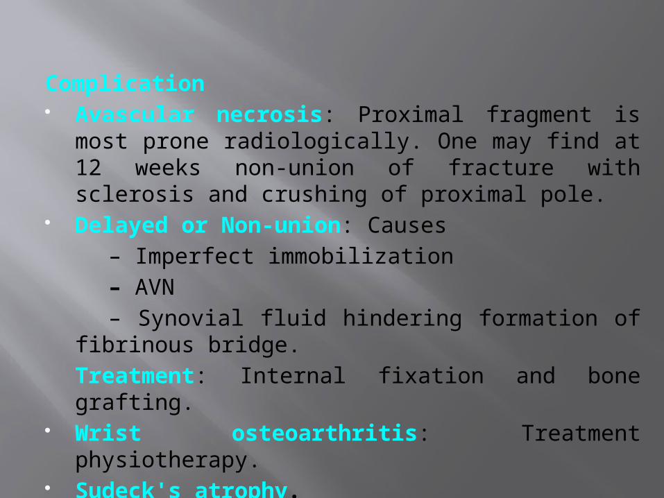
Complication Avascular necrosis: Proximal fragment is most prone
radiologically. One may find at 12 weeks non-union of fracture with sclerosis and crushing of proximal pole.
Delayed or Non-union: Causes
– Imperfect immobilization
– AVN
– Synovial fluid hindering formation of fibrinous bridge.
Treatment: Internal fixation and bone grafting. Wrist osteoarthritis: Treatment physiotherapy. Sudeck's atrophy.

Thank You
1. When performing a replant of an amputated finger, which of the following is the correct order of surgery?
a. Bone, Artery, Extensor, Flexor, Nerve, Vein.
b. Artery, Bone, Vein, Extensor, Flexor, Nerve.
c. Artery, Bone, Extensor, Flexor, Vein, Nerve.
d. Bone, Extensor, Flexor, Artery, Nerve, Vein.
e. Bone, Extensor, Flexor, Artery, Vein, Nerve.

a. Bone, Artery, Extensor, Flexor, Nerve, Vein. b. Artery, Bone, Vein, Extensor, Flexor, Nerve. c. Artery, Bone, Extensor, Flexor, Vein, Nerve. d. Bone, Extensor, Flexor, Artery, Nerve, Vein. e. Bone, Extensor, Flexor, Artery, Vein, Nerve. 2. d. Bone, Extensor, Flexor, Artery, Nerve,
Vein. This is a well-known order. A useful way of remembering it is BE a FAN of V. A stable platform is needed for reconstruction. Then the deep structures must be repaired before the delicate arterial and nerve repairs.
2. When reducing a Smith’s or volar Barton’s fracture, the reduction manoeuvre should include?
a. Supination only. b. Extension only. c. Extension and supination. d. Extension and pronation. e. Flexion and supination.
2. When reducing a Smith’s or volar Barton’s fracture, the reduction manoeuvre should include?
a. Supination only. b. Extension only. c. Extension and supination. d. Extension and pronation. e. Flexion and supination. 6. c. Extension and supination. This question
tests the understanding of the deforming forces of a fracture. Extension and supination are necessary to overcome the pronation rotatory deformity that the volar displaced fragment undergoes.
3. A 22-year-old medical student was slightly intoxicated and fell onto his extended wrist while his forearm was pronated. He has pain and a clicking sensation on the ulnar side of his wrist. X-rays and nerve conduction studies are normal. The most likely diagnosis is?
a. Scapholunate dissociation. b. Hook of hamate fracture. c. Triangular fibrocartilage complex (TFCC)
tear. d. Piso-triquetral subluxation. e. Extensor carpi ulnaris (ECU) subluxation.
3. A 22-year-old medical student was slightly intoxicated and fell onto his extended wrist while his forearm was pronated. He has pain and a clicking sensation on the ulnar side of his wrist. X-rays and nerve conduction studies are normal. The most likely diagnosis is?
a. Scapholunate dissociation. b. Hook of hamate fracture. c. Triangular fibrocartilage complex (TFCC) tear. d. Piso-triquetral subluxation. e. Extensor carpi ulnaris (ECU) subluxation. 7. c. Triangular fibrocartilage complex (TFCC) tear. Once again
mechanism of injury and mechanics are key to understanding the injury. Wrist pain must always be divided into radial, dorsal and ulna. Then according to the anatomy of the region, specific signs and limited special investigations a diagnosis can be made. TFCC tears are either acute or chronic and have been classified by Palmer: Class 1 – Traumatic A – central perforation or tear B – ulnar avulsion with or without ulnar styloid fracture C – distal avulsion D – radial avulsion with or without sigmoid notch fracture Class 2 – Degenerative stage A – TFCC wear B – TFCC wear with lunate and/or ulnar chondromalacia C – TFCC perforation with lunate and/or ulnar chondromalacia D – TFCC perforation with lunate and/or ulnar chondromalacia and lunotriquetral (LT) ligament perforation E – TFCC perforation with lunate and/or ulnar chondromalacia, LT ligament perforation, and ulnocarpal arthritis

4.The following are all good prognosis after nerve injury except?
a. Young age. b. Low velocity injury. c. Sharp (knife) injury. d. Proximal injury. e. Early exploration.
4.The following are all good prognosis after nerve injury except?
a. Young age. b. Low velocity injury. c. Sharp (knife) injury. d. Proximal injury. e. Early exploration.
12.d. Proximal injury. A more distal low velocity injury with a sharp object will have a better potential for healing. The long distance to the motor endplate from a proximal injury may preclude recovery. Younger patients have far higher potential for full recovery than adults.

5.Which of the following is a rule of tendon transfer?
a. The donor muscle must be at least MRC grade 3. b. Joints can have 50% maximum contracture.
c. Tendon pull must be synergistic. d. Line of pull should be orthogonal. e. Tendon excursions of the finger
extensors is longer than the flexors.

5.Which of the following is a rule of tendon transfer?
a. The donor muscle must be at least MRC grade 3. b. Joints can have 50% maximum contracture. c. Tendon pull must be synergistic. d. Line of pull should be orthogonal. e. Tendon excursions of the finger extensors is longer than the flexors.
15.c. Tendon pull must be synergistic. These rules must be appreciated and short cuts will only lead to disaster. Donor muscles must be expendable and have adequate power, ideally MRC grade 5. Joints must be mobile with no contracture.

6.A 23-year-old was intoxicated at a wedding and fell through a glass window. He presents to the emergency department with a radial wrist laceration with arterial bleeding. With regards to the timing of surgery the major blood supply to the hand is provided by which of the following?
a. Deep branch of the radial artery. b. Radial artery. c. Deep palmar arch. d. Superficial palmar arch. e. Interosseous artery.
answer
d. Superficial palmar arch. The superficial palmar arch is a continuation of the ulna artery. In the majority of patients (78%) this arch iscompletedbybranches from thedeep palmar,radialormedianarteries. This explains why even with significant lacerations to the ulna artery a hand can be well perfused.
Ques – bennets fracture is a fracture dislocation of base of ---- metacarpal?
A-1 B-2 C-3 D-4
Ques – bennets fracture is a fracture dislocation of base of ---- metacarpal?
A-1 B-2 C-3 D-4 Answer –1st
Which carpal bone fracture cause median nerve involvement
A-scaphoid B-lunate C-triquetral D-hamate
Which carpal bone fracture cause median nerve involvement
A-scaphoid B-lunate C-triquetral D-hamate Answer—b
Most common site of scaphoid fracture A-neck B-waist C-proximal fragent D-distal fragment
Most common site of scaphoid fracture A-neck B-waist C-proximal fragent D-distal fragment Answer-b

Most common complication of colles fracture
A-malunion B-avn C-stiffness of finger D-rupture of epl tendon
Most common complication of colles fracture
A-malunion B-avn C-stiffness of finger D-rupture of epl tendon
Answer-c
All are injuries of lower end radius except A-smith B-colles C-night stick D-barton
All are injuries of lower end radius except A-smith B-colles C-night stick D-barton
Answer-c