


Languages
Pages
Legal


UTERUS BODY PATHOLOGY
DR. MONA RASHED

BODY OF UTERUS
• The uterine corpus is composed of endometrial mucosa and the underlying smooth muscle myometrium
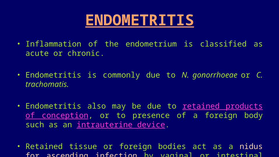
ENDOMETRITIS• Inflammation of the endometrium is classified as acute or chronic.
• Endometritis is commonly due to N. gonorrhoeae or C. trachomatis.
• Endometritis also may be due to retained products of conception, or to presence of a foreign body such as an intrauterine device.
• Retained tissue or foreign bodies act as a nidus for ascending infection by vaginal or intestinal tract flora.

ENDOMETRITIS; cont.• Histologic examination reveals a neutrophilic infiltrate
coexisting with a stromal lymphoplasmacytic infiltrate in the superficial endometrium and glands
• Clinically, endometritis manifest with fever, abdominal pain, and menstrual abnormalities.
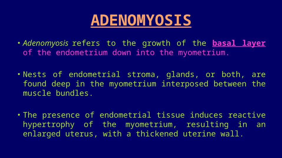
ADENOMYOSIS• Adenomyosis refers to the growth of the basal layer of the
endometrium down into the myometrium.
• Nests of endometrial stroma, glands, or both, are found deep in the myometrium interposed between the muscle bundles.
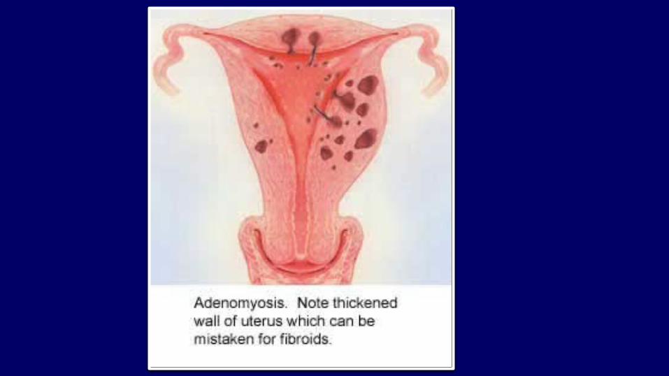
• The presence of endometrial tissue induces reactive hypertrophy of the myometrium, resulting in an enlarged uterus, with a thickened uterine wall.
ADENOMYOSIS; cont.• Because the glands in adenomyosis derive from the stratum
basalis of the endometrium, they do not undergo cyclic bleeding.
• Clinically; adenomyosis may produce menorrhagia, dysmenorrhea, and pelvic pain before the onset of menstruation.
ENDOMETRIOSIS
• Definition: Endometrial glands or stroma outside the uterus • Common sites of endometriosis: Ovaries, uterine ligaments,
recto-vaginal septum and pelvic peritoneum, scars, umbilicus, vagina, vulva and appendix
ENDOMETRIOSIS; cont.• Three hypotheses explain the endometriosis:
1. The regurgitation theory, menstrual backflow through the fallopian tubes leads to implantation.
2. The metaplastic theory, endometrial differentiation of coelomic epithelium (from which endometrium originates) as the source.
3. Vascular or lymphatic dissemination theory.

ENDOMETRIOSIS; MORPHOLOGY• Endometriosis almost always contains functioning endometrium,
which undergoes cyclic bleeding with menstruation.
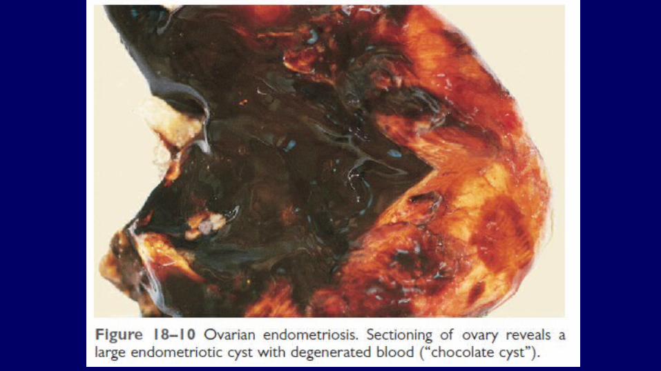
• Because blood collects in these foci, they usually appear grossly as red-brown nodules.
• When the ovaries are involved, the lesions may form large, blood-filled cysts that turn brown (chocolate cysts) as the blood ages.
ENDOMETRIOSIS; MORPHOLOGY; cont.• Organization of the blood causes widespread fibrosis, leading to
adhesions among pelvic structures, sealing of the tubal fimbriated ends, and distortion of the oviducts and ovaries.
• Clinical Effects of endometriosis: Infertility, dysmenorrhea, pelvic pain
• The histologic diagnosis at all sites is the presence of: endometrial glands, endometrial stroma, and hemosiderin pigment (old blood).


PROLIFERATIVE LESIONS OF THE ENDOMETRIUM AND MYOMETRIUM
PROLIFERATIVE LESIONS OF THE ENDOMETRIUM AND MYOMETRIUM
• The most common proliferative lesions of the uterine corpus are:
1. Endometrial hyperplasia2. Endometrial carcinomas3. Endometrial polyps4. Smooth muscle tumors: leiomyoma & leiomyosarcoma
• All tend to produce abnormal uterine bleeding as their earliest manifestation.

Endometrial Hyperplasia• Excess of estrogen, can induce exaggerated endometrial
proliferation (hyperplasia), which is an important precursor of endometrial carcinoma.
• Causes of estrogen excess include
1. Anovulation 2. Polycystic ovarian disease 3. Functioning granulosal cell tumors of ovary 4. Estrogen replacement therapy 5. Obesity, as adipose tissue converts steroid precursors into estrogens.
Classification of endometrial hyperplasia
1. Hyperplasia Without Atypia (low grade) – Simple hyperplasia:. Rarely progress to carcinoma – Complex hyperplasia: Glandular crowding and branching with a complex growth
pattern. Less than 5% progress to carcinoma
2. Atypical Hyperplasia (high grade)
– Histopathology: there are atypical cellular and nuclear features. Approximately 25% progress to carcinoma
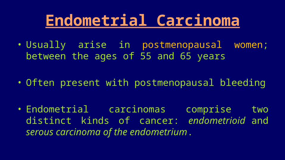
Endometrial Carcinoma• Usually arise in postmenopausal women; between the ages of
55 and 65 years
• Often present with postmenopausal bleeding
• Endometrial carcinomas comprise two distinct kinds of cancer: endometrioid and serous carcinoma of the endometrium.
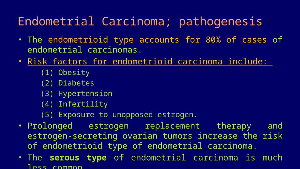
Endometrial Carcinoma; pathogenesis• The endometrioid type accounts for 80% of cases of endometrial
carcinomas. • Risk factors for endometrioid carcinoma include:
(1) Obesity(2) Diabetes(3) Hypertension(4) Infertility(5) Exposure to unopposed estrogen.
• Prolonged estrogen replacement therapy and estrogen-secreting ovarian tumors increase the risk of endometrioid type of endometrial carcinoma.
• The serous type of endometrial carcinoma is much less common.

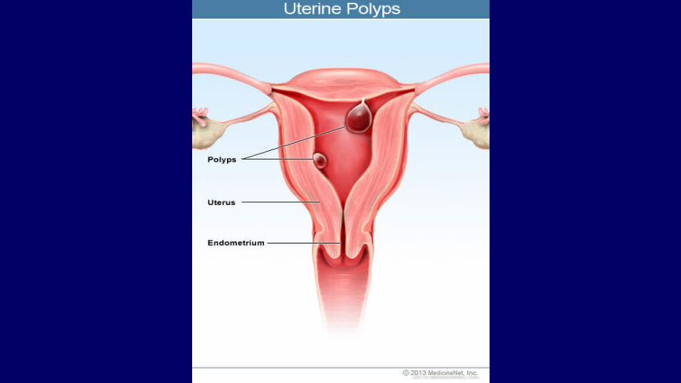
Endometrial Polyps• Sessile lesions range from 0.5 to 3 cm in diameter.
• Larger polyps may project from the endometrial mucosa into the uterine cavity.
• On histologic examination, they are composed of endometrium resembling the basalis, frequently with small muscular arteries.
• Clinically; endometrial polyps cause abnormal uterine bleeding.
• Commonly are detected around the time of menopause.

Leiomyoma• Benign tumors that arise from the smooth muscle cells in the
myometrium; termed leiomyomas.
• Leiomyomas are the most common benign tumor in females, affecting 30% to 50% of women of reproductive age.

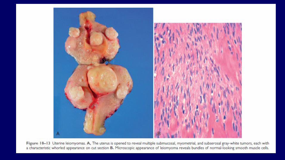
Leiomyoma; morphology• Leiomyomas are typically well circumscribed, firm gray-white masses with a
characteristic whorled cut surface.
• They may occur singly, or multiple within the uterus myometrium, ranging from small nodules to large tumors.
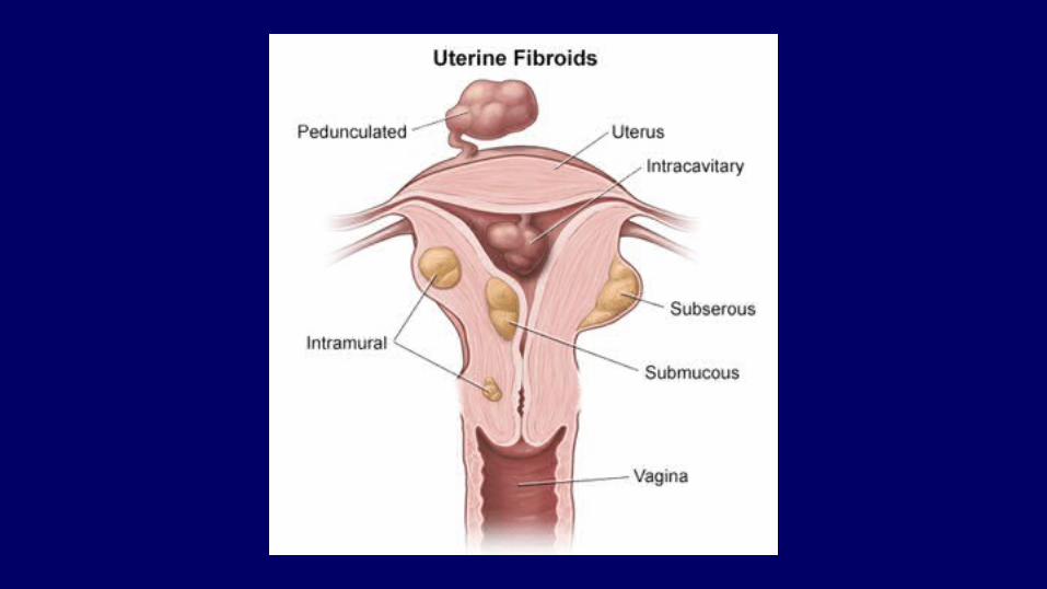
• Leiomyoma may be embedded within the myometrium (intramural), whereas others may lie directly beneath the endometrium (sub-mucosal) or directly beneath the serosa (sub-serosal).
• On histologic examination, the tumors are characterized by bundles of smooth muscle cells mimicking the appearance of normal myometrium.

Leiomyoma; Clinically • The most frequent presenting sign is menorrhagia, with or
without metrorrhagia.
• Large leiomyomas may be palpated by the affected woman or may produce a dragging sensation.
• Leiomyomas almost never transform into sarcomas, and the presence of multiple lesions does not increase the risk of malignancy.

Leiomyosarcoma• Leiomyosarcomas do not arise from a preexisting leiomyomas.
• They are almost always solitary and most often occur in postmenopausal women, in contradistinction to leiomyomas, which frequently are multiple and usually arise premenopausally.
• Leiomyosarcomas typically take the form of soft, hemorrhagic, necrotic masses.
• Recurrence after removal is common, and they metastasize, typically to the lungs.

References: ROBBINS Basic Pathology; 9th edition. Chapter 19: Female genital system and breast; Body of uterus, pages: 689-694
Top Related