


Languages
Pages
Legal


lable at ScienceDirect
Acta Materialia 109 (2016) 300e313
Contents lists avai
Acta Materialia
journal homepage: www.elsevier .com/locate/actamat
Full length article
Microstructural evolution and phase transformation in twinning-induced plasticity steel induced by high-pressure torsion
X.H. An a, *, Q.Y. Lin a, G. Sha b, M.X. Huang c, S.P. Ringer a, d, Y.T. Zhu b, e, X.Z. Liao a, **
a School of Aerospace, Mechanical and Mechatronic Engineering, The University of Sydney, Sydney, NSW 2006, Australiab School of Materials Science and Engineering, Nanjing University of Science and Technology, Jiangsu 210094, Chinac Department of Mechanical Engineering, The University of Hong Kong, Hong Kong, Chinad Australian Centre for Microscopy & Microanalysis, The University of Sydney, Sydney, NSW 2006, Australiae Department of Materials Science and Engineering, North Carolina State University, Raleigh, NC 27695, USA
a r t i c l e i n f o
Article history:Received 9 December 2015Received in revised form5 February 2016Accepted 19 February 2016Available online xxx
Keywords:Twinning-induced plasticity steelNanostructuresPhase transformationElement diffusion and segregationHigh-pressure torsion
* Corresponding author.** Corresponding author.
E-mail addresses: [email protected] (X.H. Anau (X.Z. Liao).
http://dx.doi.org/10.1016/j.actamat.2016.02.0451359-6454/© 2016 Acta Materialia Inc. Published by
a b s t r a c t
The microstructural evolution of twinning-induced plasticity steel during high-pressure torsion (HPT)processing at 573 K was systematically evaluated. Due to the high processing temperature, the formationof a homogeneous nanostructure was primarily dominated by complicated dislocation and grainboundary activities in lieu of deformation twinning. Apart from the grain refinement process, phasetransformation took place at late stages of deformation, resulting in the microstructural fingerprint ofequaxied nanograins with multiple phases in the steel. On account of remarkable elemental redistri-bution, the diffusion-controlled nature of the transformation was convincingly identified. During thetransformation, although the cementite also initially formed, austenite eventually decomposed intoferrite and Mn-riched M23C6 carbide, implying that Mn is the pivotal alloying element for the trans-formation kinetics. Owing to the sluggish bulk diffusivity of Mn, it is proposed that a high density ofdefects, nanostructures and the HPT processing play a crucial role in promoting the elemental diffusionand segregation and in stimulating the phase transformation.
© 2016 Acta Materialia Inc. Published by Elsevier Ltd. All rights reserved.
1. Introduction
Bulk ultrafine-grained (UFG, < 1000 nm) and nanocrystalline(NC, < 100 nm) materials have been extensively explored over thelast two decades due to their superior mechanical properties,including exceptionally high strength and hardness, excellent fa-tigue resistance and low-temperature superplasticity [1e5]. Severeplastic deformation (SPD) is among themost effective methods thatcan produce fully dense and contamination-free bulk UFG and NCmaterials [3e5]. By recourse to the SPD techniques, originallycoarse-grained structures can be substantially refined to UFG or NCstructures through a series of sophisticated grain refinement pro-cesses [3e5]. Comprehensive understanding of the fundamentalmechanisms of SPD-inducedmicrostructural evolution is crucial forachieving optimal mechanical properties of these materials viafine-tuning their microstructures.
It is well documented that dislocation activities and
Elsevier Ltd. All rights reserved.
deformation twinning are the two primary SPD-induced grainrefinement mechanisms [3]. For materials in which dislocation slipis the dominant deformation mechanism, the SPD-induced micro-structural evolution is mainly controlled by complicated dislocationactivities that subdivide a large grain into many smaller volumeelements through the formation of dislocation cells. Withincreasing plastic strain, the cell boundaries gradually evolve intolow-angle and then high-angle grain boundaries (LAGBs andHAGBs, whose misorientation angles are below and above 15�,respectively) [6,7]. In contrast, refinement via deformation twin-ning involves the formation of a high-density of nanoscale twins incoarse grains and the subsequent transformation of two-dimensional twin lamellae structures into three-dimensionalnanograins through extensive interactions between twin bound-aries (TBs) and other defects (mainly dislocations) [8]. The finalgrain sizes after the refinement by the two mechanisms aregenerally in the range of submicrometer and nanometer, respec-tively [8,9]. In these bulk UFG and NC materials, there are aconsiderable fraction of LAGBs and a high density of dislocationsresided in or near non-equilibrium HAGBs, which remarkably in-fluence the thermal stability and mechanical properties of thematerials [10e12].

X.H. An et al. / Acta Materialia 109 (2016) 300e313 301
In addition to grain refinement, SPD processing can also lead tothe formation of non-equilibrium solid solutions, disordering,amorphization, and phase transformations under high stress levelsin various alloys [13e16]. For example, SPD can dissolve cementitein pearlitic steels through extensive C-dislocation interactions [13],promoting a reverse phase transformation from ferrite to austenite[14]. Although the reverted austenitic phase is unstable at roomtemperature, the long-distance diffusion of C atoms, stemmed fromthe decomposition of cementite, stabilizes to some extent theaustenitic phase [14]. Therefore, alloying elements dissolved in thematrix phase play a significant role in SPD-induced microstructuralevolution of engineering materials.
As a promising candidate for numerous industrial applications,twinning-induced plasticity (TWIP) steels with the austenitic phasehave been extensively investigated since they possess excellentcombination of high tensile strength and good ductility, whichoriginates from their exceptionally large work-hardening rate[17e20]. It is generally recognized that the high work-hardeningrate is closely related to the formation of deformation twins[19,20]. TBs act as effective barriers for dislocation motion tostrengthen the materials and the increasing density of TBs withfurther straining results in a “dynamic Hall-Petch effect”. Addi-tionally, the extensive interaction between the interstitial C atomsof CeMn dipoles and dislocations produces a dynamic strain agingeffects, also leading to high work-hardening rate [21]. Based on thetraditional crystal plasticity theory, the activation of predominantdeformation mechanism is mainly controlled by the stacking faultenergy (SFE) [22], which is highly dependent on the chemicalcomposition of TWIP steels. Until now, most researches concerningTWIP steels focused on the dependence of deformation mecha-nisms on their alloying elements and SFE [23e26], and the prop-erties associated with microstructures [27e30]. Compared to otheradvanced steels [13,31e35], investigation on the microstructuralresponse of TWIP steels to SPD processing is still limited [37,38].However, fundamental exploration of the SPD-induced micro-structural evolution of TWIP steels is important both scientifically,for the comprehensive understanding of the grain refinementprocesses in the engineering metallic materials, and technologi-cally with respect to their energy absorption applications wherehigh strain will be imposed on the steels.
In present work, high-pressure torsion (HPT), which is one ofthe most popular SPD techniques [39], was applied to process aTWIP steel at 573 K. The HPT-induced microstructural evolutionwas systematically studied in virtue of various advanced charac-terization methods, including X-ray diffraction (XRD), conventionalelectron backscatter diffraction (EBSD), scanning electronmicroscope-transmission Kikuchi diffraction (SEM-TKD) andtransmission electron microscopy (TEM). During HPT processing,diffusional phase transformation surprisingly occurred via thedecomposition of austenite into ferrite and carbides. To link thephase transformation and elemental diffusion, atom probe to-mography (APT) was employed to analyze the elemental distribu-tions before and after HPT processing.
2. Experimental procedure
2.1. Materials and processing
The original TWIP steel used in this investigation is a commer-cial product produced by Ansteel Co. Ltd. in China. The nominalchemical composition of the TWIP steel is Fe-17.37Mn-3.31C-3.34Al-0.94Si in at.% or Fee18Mn-0.75C-1.7Al-0.5Si in wt.%.Detailed information about the material is available in Refs. [40,41].For HPT processing, the fully recrystallized steel was cut into discsamples with a diameter of 20 mm and a thickness of 1.5 mm. The
disks were HPT processed through 1, 5, 10, and 12 revolutions(thereafter referred to as 1R, 5R, 10R and 12R samples, respectively)under a quasi-constrained condition [39] with an imposed pressureof 4.0 GPa at 573 K. Before HPT processing, the anvils were pre-heated to 573 K and then held for 5 min to warm the samples.The temperature was kept at 573 ± 10 K during the process. Thesamples were quenched into water immediately after HPT pro-cessing. One or two revolutions of HPT were carried out for onetime and abovementioned procedure was repeated to reach thedesigned revolution numbers.
2.2. Materials characterization techniques
After HPT, disks with a diameter of 3 mm for microstructuralcharacterization were taken near the edge of the HPT samples andwere mechanically polished using 1200e4000 grit SiC paperssequentially to acquire a flat and smooth surface. All structuralcharacterization experiments were undertaken at positions of~1e2 mm away from the edge of disks and all micrographs weretaken from plan-view. For XRD and conventional EBSD experi-ments, electro-polishing was conducted to remove the strainedlayer caused by mechanical polishing using a Struers LectroPol-5unit and an electrolyte of 23% perchloric acid and 77% acetic acidunder the voltage of 20 V at the ambient temperature for 50 s. XRDmeasurements of all specimens were performed using a SiemensD5000 diffractometer to identify the phase information of the steelbefore and after HPT. EBSD analysis of the original, 1R and 5Rsamples were carried out using a Carl Zeiss Ultra Plus field emissiongun SEM with the operating voltage of 12 kV and the step sizes of200 nm, 100 nm and 60 nm, respectively.
For the 10R and 12R samples with much finer microstructures,SEM-TKD and TEM were applied to characterize the microstruc-tures and phase transformation. The samples were electropolishedusing a Struers TenuPol-5 jet electropolishing unit and a solution of23% perchloric acid and 77% acetic acid under an operating voltageof 20 V and at a temperature of�20 �C. SEM-TKD experiments wereperformed in the Zeiss Ultra SEM with operating voltage of 20 kVand the step size of 10 nm. The detailed experimental set-up ofSEM-TKD follows that presented in Ref. 42. The chemistry infor-mation of the samples was simultaneously collected using an Ox-ford Instruments AZtec EDS system with an X-Max 20 mm2 silicondrift detector during the SEM-TKD mapping. TEM characterizationwas performed using a JEOL 2200F operating at 200 kV and a JEOL3000F at 300 kV.
A local electrode atom probe was employed to acquire high-resolution compositional data of the coarse-grained sample and a12R sample. Tip samples for APT analysis were prepared using atwo-step electropolishing procedure with thin bars having cross-sections of 0.5 � 0.5 mm2. The first step used an electrolyte of25% perchloric acid in acetic acid at 15 V and the second step usedan electrolyte of 4% perchloric acid in 2-butoxethynal at 20 V. APTcharacterization was performed in a atom probe, CAMECA LEAP4000X Si at a specimen temperature of 20 K under a laser pulse atan energy of 40 pJ and with a target evaporation rate of 1%.Reconstruction and quantitative analysis of atom probe data wereperformed by using IVAS 3.6.6 software.
3. Experimental results
3.1. XRD results before and after HPT
XRD patterns of the original steel and HPT samples are pre-sented in Fig. 1. Within the XRD detection limit, only peaks from theaustenitic phase were identified in the initial TWIP steel and the 1Rand 5R samples. Although significant HPT-induced martensitic

X.H. An et al. / Acta Materialia 109 (2016) 300e313302
transformation at room temperature has been reported in a TWIPsteel [37], nomartensite formed in the present study, whichmay beattributed to the high processing temperature suppressing themartensitic transformation. With further deformation, the (110)peak of ferrite (thereafter referred to as (110)a) is clearly detected inthe 10R sample, signaling the phase transformation from austeniteto ferrite. The (110)a peak is more remarkable and the (211)a peak isalso notably identified in the 12R sample. Based on the XRD results,the volume fraction of ferrite is ~5.6% and ~20% in the 10R and 12Rsamples, respectively. It is well known that stress-inducedmartensitic transformation can be activated by mechanicalloading, since austenite in the TWIP steel is metastable [25,28,37].However, there was no report concerning deformation-inducedphase transformation from austenite to ferrite. To comprehen-sively understand the HPT-induced microstructural evolution andphase transformation of the TWIP steel, in-depth microstructuralinvestigations were performed by recourse of extensive EBSD andTKD characterization.
3.2. EBSD and TKD investigations of HPT-induced microstructuralevolution
The original TWIP steel is of a recrystallized microstructure withan average grain size of ~4 mm and only a few LAGBs, as shown inFig. 2 (a). In line with the XRD result, the phase map in Fig. 2(b)further confirms that the original TWIP steel contains onlyaustenite. After HPT for 1 revolution, the original grain featuresincluding their shape and GBs were still traceable although there isgradual variation in the crystallographic orientations within indi-vidual grains, while a high density of LAGB with a fraction of ~68%formed within the grains, as exhibited in Fig. 3 (aec). Largerchanges in orientation are adjacent to GBs and near triple junctions,which stemmed from abundant dislocation activities, leading to theformation of LAGBs [7,43]. Fig. 3 (a) also reveals a few deformationtwins, with TBs presented with red lines, in several grains whoseorientations may favor deformation twinning [44,45]. No phasetransformation was observed and only austenite existed in thesample (Fig. 3(b)). Usually, profuse twinning activities play a crucialrole in accommodating the plastic strain imposed on the TWIP[23e30]. In the present investigation, dislocation slip rather thantwinning was the dominant mechanism for the large shear defor-mation because of the high processing temperature, which will be
Fig. 1. XRD patterns of the original TWIP steel and those after different numbers ofrevolutions of HPT processing.
discussed in detail later.HPT processing for 5 revolutions led to significant change in the
microstructure. As exhibited in the orientation map in Fig. 3 (d),grains were strongly elongated with roughly parallel lamellarboundaries, interweaved with some nearly equiaxed ultrafinegrains. The average width of the lamellar structure is ~1 mm. Thephase map of Fig. 3 (e) shows only the austenitic phase in thesample. Compared to the 1R sample shown in Fig. 3 (c), the fractionof LAGB in the 5R sample was slightly reduced but was still as highas ~60%, while the density of deformation twins decreased as wellbased on the volume fraction of TBs, as illustrated in Fig. 3 (f). Thiscould be ascribed to the detwinning behavior due to the extensiveinteraction between dislocations [35] and TBs and the twinningactivation becomes increasingly difficult with the grain refinementin the micrometer range [44].
Owing to the limited spatial resolution of the conventional EBSDmethod, the microstructure characterization of the 10R and 12Rsamples was carried out by recourse of the SEM-TKD technique[42]. After HPT for 10 revolutions, although many grains are stillelongated, the aspect ratios of grain length to width were promi-nently reduced, while the number of equiaxed ultrafine andnanoscale grains increased remarkably, as exhibited in Fig. 4 (aec).In parallel with the XRD results, new phases with small grain sizesare detected in some “band-like” narrow local regions, as revealedin the phase map in Fig. 4 (c). Based on the Kikuchi patterns, thenew phases are determined as ferrite and two types of carbides ecementite and M23C6 e with volume fractions of ~5.2%, 1.2% and1.5%, respectively. Because of their small volume fractions, carbideswere not identified by XRD. Although the experimental set-up ofTKD and the nature of thin foil specimens make it difficult to collectcompositional information with high accuracy [42], the inhomo-geneous elemental distributions of Fe and Mn at places where thenew phases formed were still identified, as shown in Fig. 4 (d) and(e), indicating clearly the diffusional nature of the phase trans-formation. Thus, elemental diffusion and redistribution play acrucial role in the phase transformation, which is significantlydifferent from the martensitic transformation in TWIP steels[25,28,37].
The grain size distributions of austenite and ferrite in the 10Rsample are shown in Fig. 5 (a) and (b), respectively. The averagegrains size of austenite and ferrite was ~130 nm and ~65 nm,respectively, and the latter had a narrower grain size distributionrange. The grain sizes of two carbides were ~50 nm Fig. 5 (c) and (d)present the misorientation angle distribution of austenite andferrite, respectively. Compared to 1R and 5R samples, the volumefraction of LAGBs in austenitic grains is remarkably decreased to~26%, while there are only a few LAGBs with a fraction of ~10% inferrite phase. In fact, although plenty of ultrafine and nanoscalegrains were formed in many regions, combined with the significantreduction of LAGBs and the occurrence of the phase transformation,themicrostructure of the steel was quite inhomogeneous even afterHPT for 10 revolutions.
After HPT for 12 revolutions, the representative microstruc-tural trait of the steel was uniformly distributed equiaxed nano-grains, as detected in Fig. 6 (aec). Meanwhile, the phasetransformation proceeded, resulting in the formation of moreferrite and M23C6 carbide. However, the cementite phase wasbarely noticeable as shown in Fig. 6 (c). Based on the TKD result,the volume fractions of ferrite and M23C6 increased remarkably to23% and 3.2%, respectively. Accompanied with the occurrence ofextensive phase transformation, the distributions of Fe and Mnbecame increasingly inhomogeneous owing to the significantelemental redistribution, as exhibited in Fig. 6 (d) and (e). Quali-tatively, the Mn-enriched and -depleted regions roughly corre-spond to the M23C6 carbide and ferrite, respectively, in which Fe is
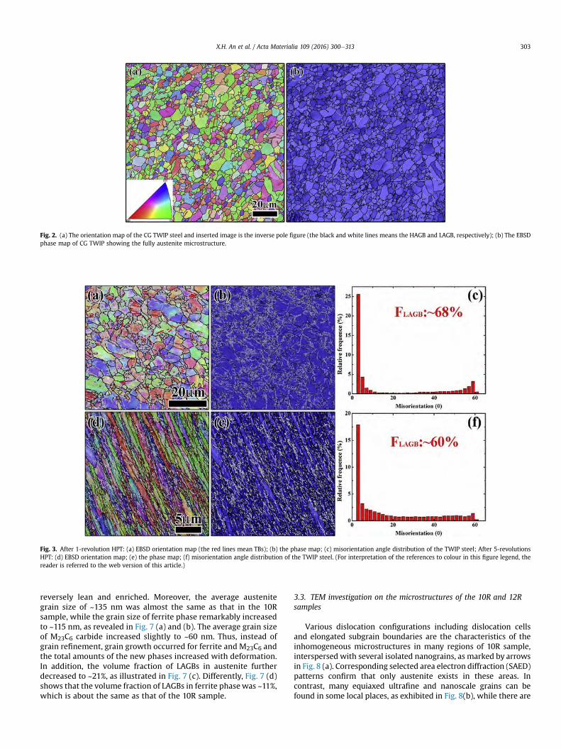
Fig. 2. (a) The orientation map of the CG TWIP steel and inserted image is the inverse pole figure (the black and white lines means the HAGB and LAGB, respectively); (b) The EBSDphase map of CG TWIP showing the fully austenite microstructure.
Fig. 3. After 1-revolution HPT: (a) EBSD orientation map (the red lines mean TBs); (b) the phase map; (c) misorientation angle distribution of the TWIP steel; After 5-revolutionsHPT: (d) EBSD orientation map; (e) the phase map; (f) misorientation angle distribution of the TWIP steel. (For interpretation of the references to colour in this figure legend, thereader is referred to the web version of this article.)
X.H. An et al. / Acta Materialia 109 (2016) 300e313 303
reversely lean and enriched. Moreover, the average austenitegrain size of ~135 nm was almost the same as that in the 10Rsample, while the grain size of ferrite phase remarkably increasedto ~115 nm, as revealed in Fig. 7 (a) and (b). The average grain sizeof M23C6 carbide increased slightly to ~60 nm. Thus, instead ofgrain refinement, grain growth occurred for ferrite and M23C6 andthe total amounts of the new phases increased with deformation.In addition, the volume fraction of LAGBs in austenite furtherdecreased to ~21%, as illustrated in Fig. 7 (c). Differently, Fig. 7 (d)shows that the volume fraction of LAGBs in ferrite phase was ~11%,which is about the same as that of the 10R sample.
3.3. TEM investigation on the microstructures of the 10R and 12Rsamples
Various dislocation configurations including dislocation cellsand elongated subgrain boundaries are the characteristics of theinhomogeneous microstructures in many regions of 10R sample,interspersed with several isolated nanograins, as marked by arrowsin Fig. 8 (a). Corresponding selected area electron diffraction (SAED)patterns confirm that only austenite exists in these areas. Incontrast, many equiaxed ultrafine and nanoscale grains can befound in some local places, as exhibited in Fig. 8(b), while there are
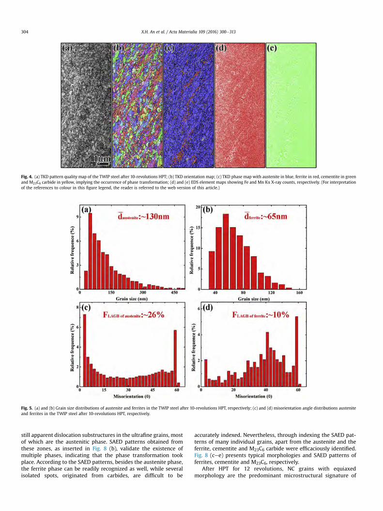
Fig. 4. (a) TKD pattern quality map of the TWIP steel after 10-revolutions HPT; (b) TKD orientation map; (c) TKD phase map with austenite in blue, ferrite in red, cementite in greenand M23C6 carbide in yellow, implying the occurrence of phase transformation; (d) and (e) EDS element maps showing Fe and Mn Ka X-ray counts, respectively. (For interpretationof the references to colour in this figure legend, the reader is referred to the web version of this article.)
Fig. 5. (a) and (b) Grain size distributions of austenite and ferrites in the TWIP steel after 10-revolutions HPT, respectively; (c) and (d) misorientation angle distributions austeniteand ferrites in the TWIP steel after 10-revolutions HPT, respectively.
X.H. An et al. / Acta Materialia 109 (2016) 300e313304
still apparent dislocation substructures in the ultrafine grains, mostof which are the austenitic phase. SAED patterns obtained fromthese zones, as inserted in Fig. 8 (b), validate the existence ofmultiple phases, indicating that the phase transformation tookplace. According to the SAED patterns, besides the austenite phase,the ferrite phase can be readily recognized as well, while severalisolated spots, originated from carbides, are difficult to be
accurately indexed. Nevertheless, through indexing the SAED pat-terns of many individual grains, apart from the austenite and theferrite, cementite and M23C6 carbide were efficaciously identified.Fig. 8 (cee) presents typical morphologies and SAED patterns offerrites, cementite and M23C6, respectively.
After HPT for 12 revolutions, NC grains with equiaxedmorphology are the predominant microstructural signature of

Fig. 6. (a) TKD pattern quality map of the TWIP steel after 12-revolutions HPT; (b) TKD orientation map; (c) TKD phase map with austenite in blue, ferrite in red and M23C6 carbidein yellow; (d) and (e) EDS element maps showing Fe and Mn Ka X-ray counts, respectively. (For interpretation of the references to colour in this figure legend, the reader is referredto the web version of this article.)
Fig. 7. (a) and (b) Grain size distributions of austenite and ferrites in the TWIP steel after 12-revolutions HPT, respectively; (c) and (d) misorientation angle distributions austeniteand ferrites in the TWIP steel after 12-revolutions HPT, respectively.
X.H. An et al. / Acta Materialia 109 (2016) 300e313 305
the steel in most regions, as shown in Fig. 9 (a) and (b). Thecorresponding SAED pattern inserted in Fig. 9 (a) identifies threephases, i.e., austenite, ferrite and M23C6 carbide. No cementitewas found. Typical atomic-scale TEM images and their corre-sponding fast Fourier transformation (FFT) patterns of austenite,ferrite and M23C6 carbides are shown in Fig. 9 (c)e(e). While theGBs in SPD materials processed at the ambient temperature are
usually blurred due to the significant internal stress and limiteddynamic recovery [8,35,46], most GBs in present study are quiteclear, as exhibited in Fig. 9 (a) and (b), implying that dynamicrecovery or recrystallization caused by the high processingtemperature, play an essential role in shaping the morphology ofthese fine grains. A few deformation twins were occasionallyfound in austenitic nanograins and M23C6 carbides. Some

Fig. 8. (a) and (b) Typical TEM images of the TWIP steel after 10-revolutions HPT, showing the only austenite and multiple phase microstructures, respectively and the insertedimages are corresponding SEAD patterns; (cee) typical morphology of ferrite, cementite and M23C6 carbide with their SEAD patterns, respectively.
Fig. 9. (a) and (b) Characteristic TEM images and the SEAD patterns of the TWIP steel microstructures after 12-revolutions HPT and the deformation twins are marked by the arrowsin (b); (cee) HRTEM images and corresponding FFT of austenite, ferrite and M23C6 carbide, respectively.
X.H. An et al. / Acta Materialia 109 (2016) 300e313306
twinned grains are marked by arrows in Fig. 9 (b). These twinsare expected to form via partial dislocation emissions from GBs[47,48]. In a whole, the microstructure in the 12R sample is quitehomogeneous in comparison to that in the 10R sample.
3.4. APT investigations of elemental distribution in the original steeland the 12R sample
Based on the SEM-TKD results, the phase transformation in the

X.H. An et al. / Acta Materialia 109 (2016) 300e313 307
present investigation is diffusion controlled. Therefore, informationon the elemental distributions before and after transformation iscrucial for comprehensive understanding of the phase trans-formation. Fig. 10 exhibits the typical APT results of the originalTWIP steel. Detailed elemental concentrations are summarized inTable 1. The concentrations of Mn and Si are slightly higher, whilethe contents of C and Al are lower than the nominal compositions.All elements are uniformly distributed without apparent segrega-tion, validating a single-phase with randomly distributed solutes inthe original steel. In contrast, as revealed in the APT maps in Fig. 11,inhomogeneous elemental distribution existed after HPT for 12revolutions. The segregation of the Mn and C atoms is more severethan that of Si and Al. This further substantiates the diffusionalnature of the phase transformation.
Referring to the EDS results, the C and Mn-enriched regionsidentified by iso-concentration surfaces is M23C6 carbide, while theMn-depleted zone is recognized as ferrite, as shown in Fig. 12 (a)and (b). One-dimensional compositional profiles across ferrite,carbide and austenite, as illustrated in Fig. 12 (cee), are used toquantify the elemental distribution in these phases. The apparentcompositional variation in ferrite, carbide and austenite is clearlydiscernable. The compositions of the three phases are listed inTable 1. As revealed in Fig. 12 (d) and (e), Mn partitioned morestrongly into the austenitic phase, but Fe partitioned more stronglyinto the ferrite while Si and Al showed no clear partitioning be-tween the two phases. The C concentration in ferrite is ~0.56 at.%,which is higher than that in conventional a-Fe (<0.1 at.%) [14]. Infact, the C content in austenite is also decreased to ~1.0 at.%, lowerthan that in the original TWIP steel, while Fe and Mn contents inaustenite slightly increased and decreased, respectively, after HPTprocessing. In the M23C6 carbide, the enrichment of Mn and C andthe leanness of Fe and Si are remarkable. The minimum of Si at thecenter of the M23C6 carbide is attributed to the diminishing equi-librium solubility of Si in carbides [49]. However, the C content of~11.5 at.% is lower than the typical stoichiometric value of 20.6 at.%in M23C6 carbide. Both Mn and C are well-known austenite stabi-lizers and Mn is also a carbide-forming element [23,49]. In asso-ciation with the prominent segregation of Mn and C atoms, thephase transformation should be intimately correlated to the diffu-sion of these two elements, resulting in the decomposition ofaustenite to ferrites and carbides.
4. Discussion
After HPT processing, the original coarse grains of the TWIP
Fig. 10. 3D atom map of alloying elements of the original TWIP steel: (a) Si, (b) Mn, (c) C58 nm � 58 nm � 110 nm).
steel were substantially refined, leading to the formation of ho-mogeneously distributed equaxied nanograins, while the phasetransformation at later stages of deformation endues the nano-structure with multiple phases in lieu of the initially singleaustenitic phase. Different from early studies about thedeformation-induced martensitic transformation in TWIP steels[25,28,37], herein, phase transformation involves significantelemental redistribution via diffusion, which was promoted by thenanostructures and HPT processing. In this section, the micro-structural evolution and phase transformation of the TWIP steelwill be discussed in detail.
4.1. HPT-induced microstructural evolution
As mentioned above, SPD-induced microstructural evolution ishighly dependent on the dominant deformationmechanism, whichis controlled by the SFE [3,45]. Deformation twinning is the favor-able deformation mechanism for TWIP steels with SFE in the rangeof 18e45 mJ m�2 [23]. The SFE of steels can be finely manipulatedthrough appropriate adjustment of the types and concentrations ofalloying elements [18,24e26]. However, processing temperature isanother factor that affects significantly the deformation behavior ofmaterials [23,30]. While low temperature favors deformationtwinning, high temperature promotes dislocation slip [23,30]. It hasbeen shown that the plastic deformation was mainly accommo-dated by the dislocations rather than twinning during warm rollingat 573 K in the same TWIP [41]. Therefore, different from the earlystudies on the twinning structure evolution of TWIP steels duringSPD [35e37], dislocation activities dominate the formation of HPT-induced nanostructures in the present study due to the high pro-cessing temperature [23,30].
Themicrostructure evolution during HPTcan be roughly dividedinto four steps, which are outlined schematically in Fig. 13. Duringearly stages of deformation (step 1), the original grain morphologyis still detectable and grain interior accommodates the major shearstrain since the GB regions are relatively “strong”. Profuse LAGBswith high-density dislocations accumulate drastically within thegrains and near the GBs, causing gradual variation of crystalorientation within the original individual grains, as detected inFig. 3(a). It has to be mentioned that the characterized regions inFig. 3 (a) were subjected to the von Mises strain ε of about 40(ε ¼ 2pNr=
ffiffiffiffiffiffi
3hp
, where r and h are the radius and thickness of thedisk, respectively, and N is the number of revolutions), while areaswith strain smaller than 40 present not much difference inmorphology but do show lower dislocation densities and lower
, (d) Al, exhibiting the homogeneous distribution of these elements (Box dimension:

Table 1APT results of the chemical compositions of the original TWIP steel and the austenite, ferrite and M23C6 carbide obtained after 12-revolutions HPT.
Materials Phases Fe (at%) Mn (at%) C (at%) Si (at%) Al (at%)
Original TWIP steel Austenite 77.65 18.26 2.95 0.98 0.16TWIP steel after 12-revolutions HPT Austenite 80.51 17.53 1.03 0.85 0.08
Ferrite 95.21 3.18 0.56 0.95 0.10M23C6 Carbide 48.56 39.66 11.53 0.18 0.07
Fig. 11. 3D atom map of alloying elements of the TWIP steel after 12-revolutions HPT: (a) Si, (b) Mn, (c) C, (d) Al, revealing the segregations of these elements (Box dimension:105 nm � 105 nm � 100 nm).
Fig. 12. (a) and (b) 3D maps of C and Mn with isosurfaces of a C-rich region and Mn-lean region, defined at 8 at% C and 10 at% Mn, respectively for the TWIP steel after 12-revolutions HPT; (c) 3D maps of C and Mn with isosurfaces defined at 8 at% C and 10 at% Mn and 1D concentration files are drawn along the bar; (d) the corresponding 1Dconcentration profiles of Fe, Mn and C, which helps distinguish the austenite, ferrite and M23C6 carbide; (e) the corresponding 1D concentration profiles of Al an Si.
X.H. An et al. / Acta Materialia 109 (2016) 300e313308
volume fractions of LAGBs. Generally, such high strain readily in-duces the severe deformation of the GBs [7,43,46]. In the presentstudy, large amounts of alloying elements in the TWIP steel,especially the CeMn dipoles, could decrease GB energy andmovingvelocity, and drag or suppress the GB motion [22,50]. With furtherdeformation, various defects are increasingly stored within grains,making the grain interior and GB comparably “strong”. Accordingly,GBs begin to play crucial roles in carrying the plastic strain duringstep 2 of the deformation, while the grains are prominently elon-gated and the grain thickness is apparently reduced. However, thethickness of elongated grains cannot be continuously decreased [7].
During the deformation in step 3, some microscale shear bandsformed in several regions due to the large strain or stress locali-zation. These bands penetrate GBs and cut the elongated grains,leading to formation of roughly equaxied nanograins within thebands. Besides, LAGBs evolve gradually into HAGBs via generation,deposition and recombination of more dislocations [6], whichcauses the notable reduction of grain length and the remarkabledecrease in the volume fraction of LAGBs. During step 4, theseprocesses are progressively proceeded, while HAGBs are signifi-cantly sharpened so that they are clearly recognizable throughboundary migration and dynamic recovery favored by the high

Fig. 13. A schematic diagram of the HPT-induced microstructure evolution of the TWIP steel.
X.H. An et al. / Acta Materialia 109 (2016) 300e313 309
processing temperature. Finally, the equiaxed nanoscale grainswith uniform distribution become the representative trait of mi-crostructures in TWIP steel.
It is referred above that alloying elements redistributed exten-sively during HPT that led to diffusional phase transformation,which also affected remarkably the nanostructural evolution.Compared to those imposed on various materials to achieve uni-formly distributed nanograins [7,42,45], ultra-high vonMises strainof ~500 is herein required. The dramatic segregation of elements inor near dislocations and GBs essentially slows down the dislocationrecovery and subgrain boundary migration, which are crucial forthe formation of nanograins [7,46,51]. In contrast, the occurrence ofthe phase transformation actually benefits the grain refinement.Firstly, the nanoscale ferritic grains and carbides directly formed viathe decomposition of the austenite phase. Secondly, owing to thevariations of the microstructures and properties among thesephases, large strain or stress incompatibility exists near their inter-phase boundaries, leading to additional accumulation of disloca-tions to accelerate the formation of nanostructures [3e5,35]. Asshown in Fig. 4 (b) and (c), equiaxed austenitic nanograins arereadily detected in the regions near ferrite and carbides, whileaustenitic grains elsewhere were still elongated. Therefore, themutual interaction of the grain refinement and phase trans-formation during SPD promotes a homogeneous nanostructurewith multiple phases.
4.2. HPT-induced phase transformation
Although the metastable austenite in the TWIP steel cantransform into martensite through a stress-induced transformationprocess [17,18,25,28,37], diffusional phase transformation is hereinpreferred, on account of the significant diffusion of alloying ele-ments as shown in Figs. 11 and 12. In principle, the diffusionaltransformation is primarily activated by the thermally activatedatomic diffusion at sufficiently high temperatures [52]. A recentinvestigation uncovered that isothermal aging at 773e873 K fortens of hours led to a partial phase transformation from austenite topearlite in a TWIP steel with 14% Mn content and that Mn parti-tioning is the key for the transformation [53]. Our HPT processingtemperature was only 573 K and the duration of HPT samplesstaying at the temperature was less than an hour, which is notenough to stimulate the diffusional phase transformation without
deformation due to the sluggish bulk diffusivity of Mn atoms [52].Therefore, it is expected that a high density of defects, includingvacancies, dislocations and GBs introduced by HPT processing,played an essential role in promoting the elemental diffusion andtriggering the decomposition of austenite into ferrite and carbides.
Figs. 11 and 12 indicate the inhomogeneous distribution of Mnand C atoms after HPT, substantiating the diffusional trans-formation nature. As an interstitial solute in iron lattice, C can easilydiffuse for a long distance even at room temperature [14,50]. Incontrast, Mn is a substitutional solutewith very low bulk diffusivity.For example, the diffusion distance of Mn is only ~0.5 nm in ferriteat 673 K for 30 min [50] and ~5 nm in austenite at 823 K �C for 10 h[53]. Accordingly, the diffusion of Mn at 573 K for ~35e42 minshould usually be negligible. However, the introduction of a highdensity of defects, including vacancies, dislocations and LAGBs, byHPT provides rapid diffusion channels for solute atoms, which in-creases their diffusion coefficients [14,49,50,52]. Besides, disloca-tions and GBs provide preferred nucleation sites for the formationof new phases, leading to their apparent segregation [54,55]. Sig-nificant segregation at defects changes the local chemical equilib-rium and then the difference in the Gibbs free energies of parentand new phases, offering the chemical driving force for the phasetransformation [52]. Meanwhile, the large external stress and strainlocalization facilitate the nucleation of ferrites and carbides [14,55].As shown in Fig.14 (a), the decomposition of austenite occurred in ahighly localized shear band, evidenced by the formation of theM23C6 carbide in Fig. 14 (b) and (c). This explains why most of thenewly formed ferrite and carbides present the “band-like” distri-bution at the early stages of phase transformation, as revealed inFig. 4 (c). It is well known that Mn is a carbide-forming element andits solubility in carbides is much higher than that in ferrite [50,54].After the nucleation of new phases, the significantly different sol-ubility yields a thermodynamic driving force that promotes diffu-sion of Mn along the subgrain boundaries or triple junction intocarbide nuclei [50]. With further deformation, the segregation andpartitioning of Mn and C atoms are more remarkable, while theformation of nanograins also benefits the diffusion process due tothe reduced length scale, both of which increase the kinetics of thetransformation and the growth of ferrite and carbides. Therefore,the grain sizes of ferrites and carbides increased during HPT in lieuof the grain refinement.
In the recent investigation, no new phases formed during warm

Fig. 14. (a) The phase transformation occurred with a shear band when the TWIP steel is processed by 10-revolutions HPT, evidenced by the formation of M23C6 carbide marked in asquare. The edge of the shear band, where GBs aligned, is indicated by arrows; (b) enlarged TEM images of the square in (a); (c) HRTEM image and FFT of the M23C6 carbide, showingthe formation of twins.
X.H. An et al. / Acta Materialia 109 (2016) 300e313310
rolling at 573 K in the same TWIP steel although high density ofdislocation was introduced [41]. Hence, the hydrostatic pressure,large shear stress and the formation of nanostructures provided byHPT crucially promote the phase transformation as well. It has beenproposed that the application of high external pressure couldremarkably influence the phase equilibrium by reducing the gap ofthe Gibbs free energies between parent and new phases, whileHPT-induced nanostructures significantly increase the overall freeenergy of the matrix [14,37,51], both of which may make the phasetransformation thermodynamically easier. Moreover, the highshear stress is generally prone to stimulate the transformation byproviding additional energy for the nucleation and propagation ofnew phases [14,37]. These factors may not only prefer the emer-gence of the phase transformation, but also affect the trans-formation processes.
It was revealed that the metastable austenite of a TWIP steelcould be partially transformed into the typical pearlite during thelong-term isothermal aging [53]. However, the austenite finally
decomposed into ferrite and M23C6 carbide rather than cementitein present study, although there was some cementite at early stagesof the phase transformation. To further identify the nature of thecarbide, its atomic structure was analyzed. Fig. 15 (a) shows atypical <110> high-resolution TEM image of the carbide, and theinset low magnification image exhibits the morphology of thewhole carbide particle. Through the FFT [Fig. 15 (b)] and Inverse FFT(IFFT) [Fig. 15 (c)], the atomic scale structure of the carbide outlinedin Fig. 15 (d) is consistent with the theoretically calculated latticestructure of Mn23C6 carbide [56], which substantially validates theidentification of the carbide. Due to the similar sizes of Fe and Mnatoms, it is difficult to distinguish them in the M23C6 carbide. TheAPT study revealed that the C content of the carbide is lower thanthe stoichiometric value of the M23C6. This may be attributed to thepartitioning of C from carbide into ferrites during deformation [54],while the C atoms could be readily attracted by the vacancies anddislocations in ferrite resulting in the higher C concentration inseverely deformed ferrite than in conventional ferrite [14,54].

Fig. 15. (a) The low magnification and HRTEM images of the M23C6 carbide; (b) the FFT of (a); (c) the enlarged IFFT image of the rectangle area in (a); (d) the enlarged image therectangle area in (c) and the theoretical atomic structure of Mn23C6 with Mn atoms in cyan and C atoms in red, which further confirm the identification of M23C6 carbide. (Forinterpretation of the references to colour in this figure legend, the reader is referred to the web version of this article.)
X.H. An et al. / Acta Materialia 109 (2016) 300e313 311
Concerning that the detailed transformation processes were rathercomplicated, the reason of low C content in the carbide needfurther exploration in future.
M23C6 carbides are common inMo andW-containing steels, lowalloy steels, and stainless steels [57e59], while Mn-enriched M23C6carbides are also detected in high-Mn steels, which involves theextensive diffusion of Mn atoms [60,61]. At the early stages of thephase transformation in present study, since C diffuses much fasterthan Mn, it is expected that cementite formed in regions where Mnsegregation is relatively inconspicuous and M23C6 carbides alsonucleated in areas where Mn atoms are substantially segregated.However, cementite disappeared with further deformation andtransformation, which may be attributed to the transformation ofcementite to M23C6 carbide [57,62]. Both experimental and theo-retical studies revealed that the cementite could be evolved intoM23C6 through the reaction of M3C þ g ¼ M23C6 þ a [56,62].Cementite could also be dissolved through Cedislocation in-teractions during HPT since cementite is unstable when subjectedto severe shear deformation [13,14,54]. Accordingly, more M23C6carbides precipitated owing to the increasingly remarkable segre-gation of Mn atoms, also accelerated by the plastic deformation[58]. The formation of M23C6 significantly denudes the Mn in thematrix and destabilizes the austenite, leading to simultaneoustransformation of austenite to ferrite. Therefore, the diffusion and
segregation of Mn atoms dominantly affect the phase trans-formation of the TWIP steel during HPT. Similar to the effect of SPDon the precipitation process in Al alloys [63], HPT processing notonly stimulates the diffusional phase transformation of the TWIPsteel, but also influences the transformation kinetics. In addition,such diffusional phase transformation may be employed to hier-archically manipulate the microstructures through adjusting theprocessing conditions to design advanced steels with superiormechanical properties.
5. Conclusions
The microstructural evolution and phase transformation of aTWIP steel processed by HPT at 573 K have been systematicallystudied by recourse to various advanced characterization tech-niques including XRD, EBSD, TKD, TEM and APT. The results aresummarized as follows.
(1) Because the high processing temperature of 573 K does notfavor deformation twinning, the grain refinement process ofthe TWIP steel was dominated by complex dislocation andGB activities.
(2) The HPT-induced microstructural evolution and grainrefinement of the TWIP steel during HPT can be divided into

X.H. An et al. / Acta Materialia 109 (2016) 300e313312
four stages: the accumulation of high-density defects withinthe grains, shear deformation of the GBs, fragmentation ofelongated grains, and eventual formation of uniformequaxied nanograins.
(3) Diffusional phase transformation, which leads to inhomo-geneous compositional distribution, took place at the laterstages of deformation. A high density of defects, nano-structures and the HPT processing play crucial roles in pro-moting the elemental diffusion and transformation.
(4) During the phase transformation, austenite decomposedfinally into ferrite and Mn-riched M23C6 carbides, althoughcementite formed at the early stages of the transformation.Mn was the dominant element for the transformationkinetics.
Acknowledgments
The authors acknowledge the scientific and technical input andsupport from the Australian Microscopy & Microanalysis ResearchFacility node at the University of Sydney. This project was sup-ported by University of Sydney Postdoctoral Research Fellowship,the Australian Research Council (Grant No. DP150101121). Dr. R.D.Liu and X. Wang of Ansteel are acknowledged for providing theTWIP steel samples.
References
[1] K.S. Kumar, H. Van Swygenhoven, S. Suresh, Mechanical behavior of nano-crystalline metals and alloys, Acta Mater. 51 (2003) 5743e5774.
[2] M.A. Meyers, A. Mishra, D.J. Benson, Mechanical properties of nanocrystallinematerials, Prog. Mater. Sci. 51 (2006), 427e556.
[3] R.Z. Valiev, R.K. Islamgaliev, I.V. Alexandrov, Bulk nanostructured materialsfrom severe plastic deformation, Prog. Mater. Sci. 45 (2000) 103e189.
[4] Y. Estrin, A. Vinogradov, Extreme grain refinement by severe plastic defor-mation: a wealth of challenging science, Acta Mater. 61 (2013) 782e817.
[5] R.Z. Valiev, Y. Estrin, Z. Horita, T.G. Langdon, M.J. Zehetbauer, Y.T. Zhu, Fun-damentals of superior properties in bulk nanospd materials, Mater. Res. Lett.(2015), http://dx.doi.org/10.1080/21663831.2015.1060543.
[6] Y. Iwahashi, Z. Horita, M. Nemoto, T.G. Langdon, An investigation of micro-structural evolution during equal-channel angular pressing, Acta Mater. 45(1997) 4733e4741.
[7] R. Pippan, S. Scheriau, A. Taylor, M. Hafok, A. Hohenwarter, A. Bachmaier,Saturation of fragmentation during severe plastic deformation, Annu. Rev.Mater. Res. 40 (2010) 319e343.
[8] S. Qu, X.H. An, H.J. Yang, C.X. Huang, G. Yang, Q.S. Zang, Z.G. Wang, S.D. Wu,Z.F. Zhang, Microstructural evolution and mechanical properties of CueAlalloys subjected to equal channel angular pressing, Acta Mater. 57 (2009)1586e1601.
[9] N.R. Tao, K. Lu, Nanoscale structural refinement via deformation twinning inface-centered cubic metals, Scr. Mater. 60 (2009) 1039e1043.
[10] J.Y. Huang, Y.T. Zhu, H. Jiang, T.C. Lowe, Microstructures and dislocationconfigurations in nanostructured Cu processed by repetitive corrugation andstraightening, Acta Mater. 49 (2001) 1497e1505.
[11] X.H. An, Q.Y. Lin, S.D. Wu, Z.F. Zhang, R.B. Figueiredo, N. Gao, T.G. Langdon, Theinfluence of stacking fault energy on the mechanical properties of nano-structured Cu and CueAl alloys processed by high-pressure torsion, Scr.Mater. 64 (2011) 954e957.
[12] X.H. An, Q.Y. Lin, S.D. Wu, Z.F. Zhang, Improved fatigue strengths of nano-crystalline Cu and CueAl Alloys, Mater. Res. Lett. 3 (2015) 135e141.
[13] Y. Ivanisenko, W. Lojkowski, R.Z. Valiev, H.J. Fecht, The mechanism of for-mation of nanostructure and dissolution of cementite in a pearlitic steelduring high pressure torsion, Acta Mater. 51 (2003) 5555e5570.
[14] Y. Ivanisenko, I. MacLaren, X. Sauvage, R.Z. Valiev, H.J. Fecht, Shear-induceda �g transformation in nanoscale FeeC composite, Acta Mater. 54 (2006)1659e1669.
[15] N. Li, Y.D. Wang, R. LinPeng, X. Sun, P.K. Liaw, G.L. Wu, L. Wang, H.N. Cai,Localized amorphism after high-strain-rate deformation in TWIP steel, ActaMater. 59 (2011) 6369e6377.
[16] C. Borchers, C. Garve, M. Tiegel, M. Deutges, A. Herz, K. Edalati, R. Pippan,Z. Horita, R. Kirchheim, Nanocrystalline steel obtained by mechanical alloyingof iron and graphite subsequently compacted by high-pressure torsion, ActaMater. 97 (2015) 207e215.
[17] O. Gr€assel, L. Krüger, G. Frommeyer, L.W. Meyer, High strength FeeMne(Al,Si) TRIP/TWIP steels development e properties e application, Int. J. Plast. 16(2000) 1391e1409.
[18] O. Bouaziz, S. Allain, C.P. Scott, P. Cugy, D. Barbier, High manganese austenitic
twinning induced plasticity steels: a review of the microstructure propertiesrelationships, Curr. Opin. Solid State Mater. Sci. 15 (2011) 141e168.
[19] I. Gutie
rrez-Urrutia, D. Raabe, Dislocation and twin substructure evolutionduring strain hardening of an Fee22 wt.% Mne0.6 wt.% C TWIP steel observedby electron channeling contrast imaging, Acta Mater. 59 (2011) 6449e6462.
[20] D.R. Steinmetz, T. J€apel, B. Wietbrock, P. Eisenlohr, I. Gutie
rrez-Urrutia,A. Saeed-Akbari, T. Hickel, F. Roters, D. Raabe, Revealing the strain-hardeningbehavior of twinning-induced plasticity steels: theory, simulations, experi-ments, Acta Mater. 61 (2013) 494e510.
[21] S.J. Lee, J. Kim, S.N. Kane, B.C. De Cooman, On the origin of dynamic strainaging in twinning-induced plasticity steels, Acta Mater. 59 (2011) 6809e6819.
[22] M.A. Meyers, K.K. Chawala, Mechanical Behavior of Materials, Prentice-HallInc, New Jersey, 1999.
[23] S. Curtze, V.T. Kuokkala, Dependence of tensile deformation behavior of TWIPsteels on stacking fault energy, temperature and strain rate, Acta Mater. 58(2010) 5129e5141.
[24] K. Jeong, J.E. Jin, Y.S. Jung, S. Kang, Y.K. Lee, The effects of Si on the mechanicaltwinning and strain hardening of Fee18Mne0.6C twinning e induced plas-ticity steel, Acta Mater. 61 (2013) 3399e3410.
[25] D.T. Pierce, J.A. Jime
nez, J. Bentley, D. Raabe, C. Oskay, J.E. Wittig, The influenceof manganese content on the stacking-fault and austenite/ε-martensiteinterfacial energies in FeeMne(AleSi) steels investigated by experiment andtheory, Acta Mater. 68 (2014) 238e253.
[26] D.T. Pierce, J.A. Jimenez, J. Bentley, D. Raabe, J.E. Wittig, The influence ofstacking fault energy on the microstructural and strain e hardening evolutionof FeeMneAleSi steels during tensile deformation, Acta Mater. 100 (2015)178e190.
[27] L. Bracke, K. Verbeken, L. Kestens, J. Penning, Microstructure and textureevolution during cold rolling and annealing of a high Mn TWIP steel, ActaMater. 57 (2009) 1512e1524.
[28] Y. Lu, B. Hutchinson, D.A. Molodov, G. Gottstein, Effect of deformation andannealing on the formation and reversion of ε-martensite in an FeeMneCalloy, Acta Mater. 57 (2009) 3079e3090.
[29] K. Jeong, J.-E. Jin, Y.-S. Jung, S. Kang, Y.-K. Lee, The effects of Si on the me-chanical twinning and strain hardening of Fee18Mne0.6C twinning-inducedplasticity steel, Acta Mater. 61 (2013) 3399e3410.
[30] I.-C. Jung, B.C. De Cooman, Temperature dependence of the flow stress of Fee18Mne0.6CexAl twinning-induced plasticity steel, Acta Mater. 61 (2013)6724e6735.
[31] A. Vorhauer, R. Pippan, On the homogeneity of deformation by high pressuretorsion, Scr. Mater. 51 (2004) 921e925.
[32] Y. Son, Y.K. Lee, K.T. Park, C.S. Lee, D.H. Shin, Ultrafine grained ferritemartensite dual phase steels fabricated via equal channel angular pressing:microstructure and tensile properties, Acta Mater. 53 (2005) 3125e3134.
[33] J. Zrnik, R. Pippan, S. Scheriau, L. Kraus, M. Fujda, Microstructure and me-chanical properties of UFG medium carbon steel processed by HPT atincreased temperature, J. Mater. Sci. 45 (2010) 4822e4826.
[34] F.K. Yan, G.Z. Liu, N.R. Tao, K. Lu, Strength and ductility of 316L austeniticstainless steel strengthened by nano-scale twin bundles, Acta Mater. 60(2012) 1059e1071.
[35] Y. Cao, Y.B. Wang, X.H. An, X.Z. Liao, M. Kawasaki, S.P. Ringer, T.G. Langdon,Y.T. Zhu, Concurrent microstructural evolution of ferrite and austenite in aduplex stainless steel processed by high-pressure torsion, Acta Mater. 63(2014) 16e29.
[36] Z. Yanushkevich, A. Belyakov, R. Kaibyshev, Microstructural evolution of a304-type austenitic stainless steel during rolling at temperatures of 773e1273K, Acta Mater. 82 (2015) 244e254.
[37] M.S. Matoso, R.B. Figueiredo, M. Kawasaki, D.B. Santos, T.G. Langdon, Pro-cessing a twinning-induced plasticity steel by high-pressure torsion, Scr.Mater. 67 (2012) 649e652.
[38] I.B. Timokhina, A. Medvedev, R. Lapovok, Severe plastic deformation of a TWIPsteel, Mater. Sci. Eng. A 593 (2014) 163e169.
[39] A.P. Zhilyaev, T.G. Langdon, Using high-pressure torsion for metal processing:fundamentals and applications, Prog. Mater. Sci. 53 (2008) 893e979.
[40] Z.Y. Liang, X. Wang, W. Huang, M.X. Huang, Strain rate sensitivity and evo-lution of dislocations and twins in a twinning-induced plasticity steel, ActaMater. 88 (2015) 170e179.
[41] Z.Y. Liang, Y.Z. Li, M.X. Huang, The respective hardening contributions ofdislocations and twins to the flow stress of a twinning-induced plasticitysteel, Scr. Mater. 112 (2016) 28e31.
[42] P.W. Trimby, Y. Cao, Z.B. Chen, S. Han, K.J. Hemker, J.S. Lian, X.Z. Liao,P. Rottmann, S. Samudrala, J.L. Sun, J.T. Wang, J. Wheeler, J.M. Cairney, Char-acterizing deformed ultrafine-grained and nanocrystalline materials usingtransmission Kikuchi diffraction in a scanning electron microscope, ActaMater. 62 (2014) 69e80.
[43] X.H. An, S.D. Wu, Z.F. Zhang, R.B. Figueiredo, N. Gao, T.G. Langdon, Evolution ofmicrostructural homogeneity in copper processed by high-pressure torsion,Scr. Mater. 63 (2010) 560e563.
[44] I. Gutierrez-Urrutia, S. Zaefferer, D. Raabe, The effect of grain size and grainorientation on deformation twinning in a Fee22 wt.%Mne0.6wt.%C TWIPsteel, Mater. Sci. Eng. A 527 (2010) 3552e3560.
[45] H. Beladi, I.B. Timokhina, Y. Estrin, J. Kim, B.C. De Cooman, S.K. Kim, Orien-tation dependence of twinning and strain hardening behaviour of a highmanganese twinning induced plasticity steel with polycrystalline structure,Acta Mater. 59 (2011) 7787e7799.

X.H. An et al. / Acta Materialia 109 (2016) 300e313 313
[46] X.H. An, Q.Y. Lin, S.D. Wu, Z.F. Zhang, R.B. Figueiredo, N. Gao, T.G. Langdon,Significance of stacking fault energy on microstructural evolution in Cu andCueAl alloys processed by high-pressure torsion, Philos. Mag. 91 (2011)3307e3326.
[47] Y.T. Zhu, X.Z. Liao, X.L. Wu, Deformation twinning in nanocrystalline mate-rials, Prog. Mater. Sci. 57 (2012) 1e62.
[48] X.H. An, S.M. Zhu, Y. Cao, M. Kawasaki, X.Z. Liao, S.R. Ringer, J.F. Nie,T.G. Langdon, Y.T. Zhu, Atomic-scale investigation of interface-facilitateddeformation twinning in severely deformed Cu-Ag nanolamellar compos-ites, App. Phys. Lett. 107 (2015) 011901.
[49] Y.J. Li, P. Choi, S. Goto, C. Borchers, D. Raabe, R. Kirchheim, Evolution ofstrength and microstructure during annealing of heavily cold-drawn 6.3 GPahypereutectoid pearlitic steel wire, Acta Mater. 60 (2012) 4005e4016.
[50] Y.J. Li, A. Kostka, P. Choi, S. Goto, D. Ponge, R. Kirchheim, D. Raabe, Mecha-nisms of subgrain coarsening and its effect on the mechanical properties ofcarbon-supersaturated nanocrystalline hypereutectoid steel, Acta Mater. 84(2015) 110e123.
[51] X.H. An, S.D. Wu, Z.G. Wang, Z.F. Zhang, Enhanced cyclic deformation re-sponses of ultrafine-grained Cu and nanocrystalline CueAl alloys, Acta Mater.74 (2014) 200e214.
[52] D.A. Porter, K.E. Easterling, Phase Transformations in Metals and Alloys, CRCPress, New York, 2004.
[53] R.T. van Tol, L. Zhao, J. Sietsma, Kinetics of austenite decomposition inmanganese-based steel, Acta Mater. 64 (2014) 33e40.
[54] Y.J. Li, P. Choi, C. Borchers, S. Westerkamp, S. Goto, D. Raabe, R. Kirchheim,Atomic-scale mechanisms of deformation-induced cementite decompositionin pearlite, Acta Mater. 59 (2011) 3965e3977.
[55] M. Kuzmina, M. Herbig, D. Ponge, S. Sandl€obes, D. Raabe, Linear complexions:confined chemical and structural states at dislocations, Science 349 (2015)1080e1083.
[56] J.Y. Xie, L.D. Teng, N.X. Chen, S. Seetharaman, Atomistic simulation on thestructural properties and phase stability for Cr23C6 and Mn23C6, J. AlloysCompd. 420 (2006) 269e272.
[57] A. Inoue, T. Masumoto, Carbide reactions (M3C->M7C3->M23C6->M6C) duringtempering of rapidly solidified high carbon Cr-W and Cr-Mo steels, Metall.Trans. A 11 (1980) 739e747.
[58] S.M. Hong, M.Y. Kim, D.J. Min, K.H. Lee, J.H. Shim, D.I. Kim, J.Y. Suh, W.S. Jung,I.S. Choi, Unraveling the origin of strain-induced precipitation of M23C6 in theplastically deformed 347 Austenite stainless steel, Mater. Char. 94 (2014)7e13.
[59] M.I. Isik, A. Kostka, V.A. Yardley, K.G. Pradeep, M.J. Duarte, P.P. Choi, D. Raabe,G. Eggeler, The nucleation of Mo-rich Laves phase particles adjacent to M23C6micrograin boundary carbides in 12% Cr tempered martensite ferritic steels,Acta Mater. 90 (2015) 94e104.
[60] Y.L. Lin, C.P. Chou, M23C6 Carbide in an Fe-26.6Mne8.8AI-0.61C alloy, Scr.Metall. Mater. 27 (1992) 67e70.
[61] L.M. Roncery, S. Weber, W. Theisen, Nucleation and precipitation kinetics ofM23C6 and M2N in an FeeMneCreCeN austenitic matrix and their relation-ship with the sensitization phenomenon, Acta Mater. 59 (2011) 6275e6286.
[62] D. Djurovic, B. Hallstedt, J.V. Appen, R. Dronskowski, Thermodynamicassessment of the FeeMneC system, Calphad 35 (2011) 479e491.
[63] G. Sha, Y.B. Wang, X.Z. Liao, Z.C. Duan, S.P. Ringer, T.G. Langdon, Influence ofequal-channel angular pressing on precipitation in an AleZneMgeCu alloy,Acta Mater. 57 (2009) 3123e3132.
Top Related