


Languages
Pages
Legal


What is funduscopy?And…
Why is it important to you?

Web sites of interest:
Welch Alleyn www.panoptic.welchallyn.com http://www.welchallyn.com/medical/ go to
“optometry student” menu drop down
Red Atlashttp://www.redatlas.com

Review of ocular anatomy

Retinal Layers

Optic Nerve Anatomy

Choroidal Vessels

Funduscopy Techniques/instruments
Direct Ophthalmoscopy
Indirect Ophthalmoscopy
Fundus Biomicroscopy
Fundus Contact Lens

Why do we dilate pupils?

Direct OphthalmoscopyAdvantages Portable Easy to use Upright image Magnification 15x Can use w/o dilation
Disadvantages Small field of view Lack of stereopsis Media opacities can degrade
image
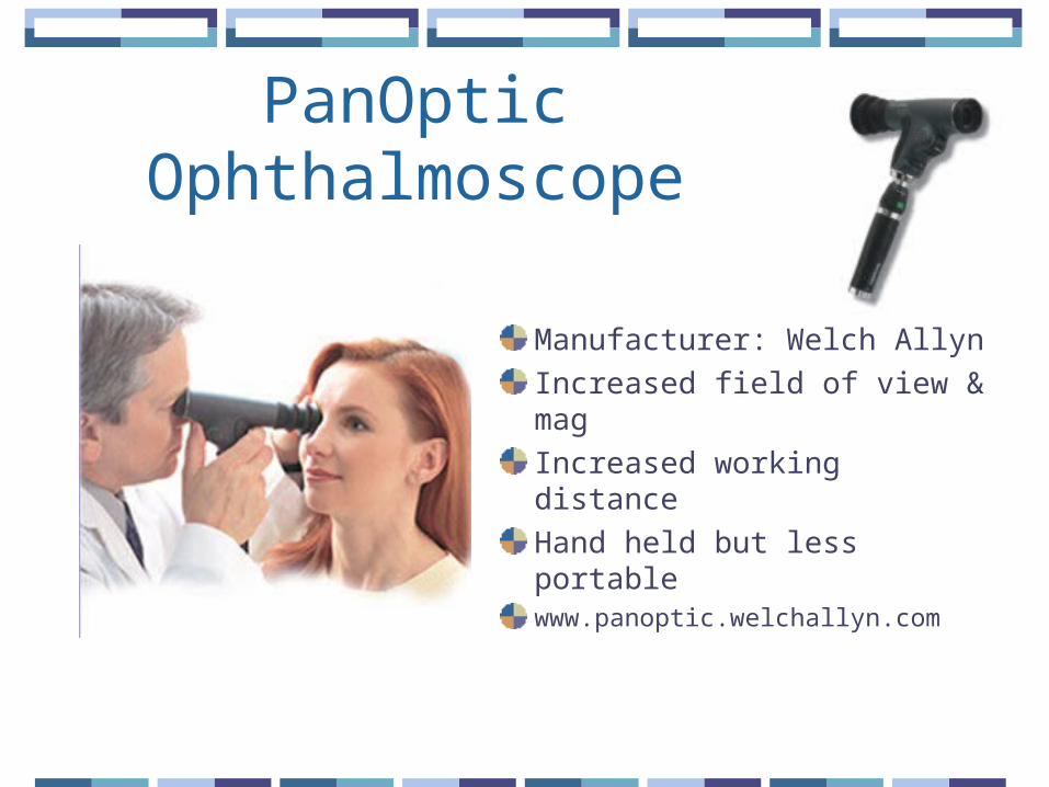
PanOptic Ophthalmoscope
Manufacturer: Welch Allyn
Increased field of view & mag
Increased working distance
Hand held but less portablewww.panoptic.welchallyn.com

Indirect Ophthalmoscopy
Monocular or binocularAdvantages: Wide field of view Binocular instruments
provide stereopsis
Disadvantages: Requires more skill Decreased magnification
(3x) Requires dilation Inverted image

Indirect Ophthalmoscopy

Fundus Biomicroscopy
Field of View & Mag: FOV <indirect but
>direct varies w/lens & slit
lamp mag
Inverted imageStereopsisDilated pupilRequires skill

Fundus Biomicroscopy

Fundus Contact LensRequires physical contact w/eye
Viewed w/Biomicroscope
Advanced dx & surgery
Field of view & Mag vary w/lens design

Direct Ophthalmoscopy: Basic skills
Optics: Illumination system Magnifier
Hyperopes myopes
Observation system Lens wheel Apertures


Direct Ophthalmoscopy: Basic skills
Viewing ocular media Observe red reflex Look for media
opacities Cataracts Corneal scars Large floaters

Direct Ophthalmoscopy: Basic skills
Proper position for central fundus viewing
Right eye to right eye
Left eye to left eye
Don’t rub noses…

Direct Ophthalmoscopy: Basic skills
Proper position for peripheral fundus viewing

Direct Ophthalmoscopy: Exam technique
Be systematicStart at optic disc & work radiallyObserve:Optic disc: C/D ratioVessels: course & caliber, AV ratio, light
reflex, crossings/bankingMaculaPeripheral fundus
Direct Ophthalmoscopy: Basic skills
Clinical pearlsFOV incr. when closer to Pt. Larger pupil increases FOV Contact lensesCheck lens wheel– watch accommodation

Normal Fundus

Viewing the Optic Nerve Head
Observe:SizeShapeColorMarginsCup to disc ratio (C/D) horiz & Vert
Blood Vessel Evaluation
Observe:Vessel diameterShape/tortuosityColorCrossingsLight reflexArtery/Vein (A/V) ratio: after 2nd bifurcation

Hypertensive Retinopathy
Scheie classification:I: Thinning of retinal arterioles relative
to veinsII: Obvious arteriolar narrowing w/focal areas
of attenuationIII: Stage II + cotton wool spots, exudates &
hemesIV: Stage III + swollen optic disk (similar to
papilledema)
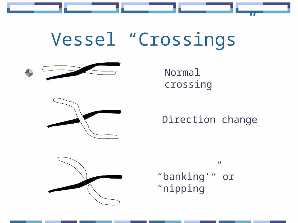
Vessel “Crossings”
Normal crossing
Direction change
“banking’” or “nipping”
Arteriolosclerosis
Increased light reflex (1/2)
“Copper wire” arterioles
“Silver wiring” arterioles whitish appearance w/continuing sclerosis
Increased A/V crossings

Macula
Lies about 2DD (disc diameters) temporal to the optic disc
Should be avascular
May appear darker red than surrounding retina
Should see bright foveal reflex on younger pts
Top Related