


Languages
Pages
Legal

Bioactive Molecules (GC-MS) of Endophytic Fungi,Xylaria from Nyctanthes arbor-trists (Linn)
Mamta Gokhale 1*, Monika Verma 1, Rumana Faraz 2 and Diwyansh Raj 1
1 Department of Botany & Microbiology, St. Aloysius College (Autonomous),Jabalpur-482001, Madhya Pradesh, India
2 Department of Biotechnology, Barkatullah University, Bhopal, Madhya Pradesh, India
Abstract: This study deals with isolation and identification (GC-MS) of bioactive molecules from anendophytic fungus isolated from a traditional medicinal shrub Nyctanthes arbor-trists (Linn). Total 11 compoundswere identified from the isolated fungal extract. On the basis of study of macroscopic and microscopic charactersand ITS sequencing, isolated endophytic fungus was identified as Xylaria. Secondary metabolites isolatedfrom Xylaria were screened for antimicrobial assay against three pathogenic bacteria. Crude extract of Xylariawas demonstrated strong inhibitor of three pathogenic bacteria Micrococcus luteus (clear zone 68.2 mm),Citrobacter freundii (clear zone of 59.16 mm) and Chromobacterium violaceum (clear zone 39.96 mm). Productionof hydrolyzing enzymes like amylase and lipase from Xylaria was also detected.
Key words: Endophytic fungi, Nyctanthes arbor-trists, Secondary metabolites, Xylaria.
IntroductionEndophytes are considered as non-aggressive,
often even postulating a mutualistic role within theirhost. Different groups of organisms such as fungi,bacteria, actinomycetes and mycoplasma are re-ported as endophytes of plants 1. Term “endo-phytes” refers to the organisms which through-out or part of their life cycle invade the tissue ofliving plant and cause asymptomatic infections.Most endophytes isolated to date have been asco-mycetes and their amorphs, however, Rungjin-damai et al.2 reported several endophytes mayalso be basidiomycetes. However, the coloniza-tion rate and isolation rate of endophytic fungi fromplants varied greatly. Some medicinal plants har-bored more endophytic fungi than others 3. Someof the common endophytes not only existed in mostplant hosts but also have higher relative frequen-cies within each of the hosts. In contrast, some
other endophytic fungi were detected in only planthost 3. Endophytic fungi are of biotechnologicalinterest due to their potential use as genetic vec-tors, metabolites and biological control agents.
Endophytes are ubiquitous and have been foundin all the species of plants. Many economicallyimportant grasses carry fungal endophytes someof which may enhance host growth, may improvethe plants ability to tolerate abiotic stress such asdrought, as well as improve their resistance to in-sect and mammalian herbivores 4. Some endo-phytes protect their host from insect by produc-ing bioactive metabolites 5. Recent studies sug-gest that endophytic fungi are not host specific 6.
Nyctanthes arbor-tristis is one of the mostuseful traditional medicinal plants, it is native toIndia. It is a night flowering sad tree of familyOleaceae is well known in India. It is a terrestrialwoody perennial having life span of 5-20 yrs. It is
ISSN Online: 2349-7785
*Corresponding author (Mamta Gokhale)E-mail: < [email protected] > © 2016, Har Krishan Bhalla & Sons
Received 17 January 2017; accepted in revised form 115 February 2017
JAM 3(1) 2017 pp 43 - 53 43
usually a shrub or a small tree having brilliant,highly fragrant flower, which bloom at night andfall off before sunrise, giving the ground under-neath a pleasing blend of white and red.
Nyctanthes arbor-tristis is a mythological plantand possesses high medicinal value in Ayurveda.The popular medicinal use of N. arbor-tristis in-clude anthelminthic, antipyretic, skin ailments andas a sedative. Each part of the plant has somemedicinal value and is thus commercially exploit-able. It is now considered as a valuable source ofseveral unique products for the medicines againstvarious diseases and also for the development ofsome industrial products.
ObjectiveObjectives of the present study were: 1. Isola-
tion of endophytic fungi, 2. Characterization andidentification of isolated fungus. 3. Assessmentof enzymatic activity of isolated endophytic fungi.4. Isolation of secondary metabolites and assess-ment of its antimicrobial activity against threepathogenic bacterial strains. 5. Identification ofcompounds isolated in crude extract from isolatedfungus through GC-MS.
Materials and methodsIsolation of endophytic fungi
Leaf of disease free plant N. arbor-tristis weresurface sterilized and leaf pieces (1.2 cm) wereinoculated on PDA media. Plates were then keptin BOD incubator (28±2oC) for 4-5 days, andmonitored every day for growth of endophyticfungi.
Characterization of isolated fungusThe colonies of isolated fungus from leaf pieces
were studied for their macroscopic features i.e.color, shape and growth of cultured colonies etc.Microscopic observations of endophytic fungalspecies were carried out by preparing slide offungal mycelium, stained with lacto phenol cottonblue stain. The slide was observed under micro-scope for its microscopic characteristics i.e. struc-ture of hyphae, type of septa in hypha, branchingpattern of hypha, type and shape of spores, typeof adherence of spores on hypha, no. of sporeson each location, spore size, color of hypha and
spores etc. Obtained data were then comparedwith the descriptions of endophytic fungus spe-cies in the literature and matches were recorded.
ITS and BLAST sequencingITS and BLAST [NCBI] sequence analysis
have been carried out from the microbial culturecollection, National Centre for Cell Science(NCCS), Pune by using following protocol:
DNA was extracted from the fungal culture us-ing XcelGen Fungal gDNA Kit (Xcelris Labs),eluted in 200 μl of elution buffer and concentra-tion of DNA was estimated using Nanodrop (ND-1000, Thermo scientific, USA). The genomicDNA was amplified using ITS1 and ITS4 prim-ers7 in PE 9700 thermo cyclers (PE, AppliedBiosystems); initial denaturation at 95°C for 5 min,35 cycles of denaturation at 95°C for 1 min,annealing at 55°C for 1 min and extension at 72°Cfor 1 min, final extension at 72°C for 10 min. ThePCR was carried out in 25 μl reaction mixture(10 U Taq polymerase buffer, New EnglandBiolabs), 2 mMdNTPs, 10 pMprimers, 1 unit Taqpolymerase (New England Biolabs), and 10 ngDNA). The positive amplicons were purified byPEG, and purified PCR products were sequencedfor both strands on an ABI 3730 xl DNA ana-lyzer using the Big Dye terminator kit (AppliedBiosystems, Inc., Foster City, CA). A BLASTnsequence homology search was performed tocompare it with available sequences of Xylariain GenBank database 8. Alignment 9 and phyloge-netic analysis based on the neighbor joining (NJ),the maximum parsimony (MP) and maximum like-lihood (ML) method was done with retrievedGenBank sequences of Xylaria in MEGA 5.0 10.
Enzymatic activity of isolated endophyticfungi, Xylaria sp.
The endophytic fungal isolates were observedfor the production of various hydrolyzing enzymeslike amylase, lipase, protease and lactase.
Amylolytic activityAmylase activity of isolated fungus was asses-
sed by growing the fungi on Glucose Yeast Ex-tract Peptone Agar (GYP) medium (glucose-1g,yeast extract 0.1 g, peptone 0.5 g, agar 16 g, dis-
Mamta Gokhale et al. / JAM 3(1) 2017 pp 43 - 53 44
tilled water 1L) with 0.2 % soluble starch (pH6.0). After incubation on 28±2°C for 3-4 days theplates were flooded with 1 % iodine in 2 % potas-sium iodide
Lipolytic activityFor lipase activity, the fungi were grown on ster-
ilized Peptone Agar medium (peptone-10 g, NaCl-5 g, CaCl2.H2O-0.1 g, agar 16 g, and distilled water1L (pH 6.0). At the end of the incubation (28±2oC)period for 4-5 days a visible precipitate aroundthe colony due to the formation of calcium saltsof the lauric acid liberated by the enzyme indi-cated positive lipase activity.
Proteolytic activityGlucose Yeast Extract Peptone Agar medium
(containing 8 % gelatin) (pH 6.0) was used toanalyze proteolytic activity of isolated endophyticfungus. After incubation (28±2oC, 4-5 days) deg-radation of the gelatin was seen as clear zonearound the colonies. The plate was then floodedwith 1 % mercuric chloride solution, which re-sulted in formation of a precipitate. This madethe agar opaque and enhanced the clear zonearound the fungal colony.
Biomass productionAutoclaved Potato Dextrose Broth (PDB) (150
mL) was inoculated with isolated endophytic fun-gus, kept for incubation at 28±2°C in a BOD in-cubator. After the incubation period, formation ofthe fungal mat was observed .Weight of aWhatman’s No. 1 filter paper was taken. Filtra-tion of fungal mat was performed using a funnel.Weight of fungal mat with filter paper was takenand weight of biomass production by the isolatedendophytic fungi was calculated using followingformula:-
Biomass production (g) = Weight of filter paperwith fungal mat – weight of filter paper.
Secondary metabolites extractionfromXylaria sp.
After incubation mycelium containing flask waskept in an orbital shaker (Sonar) on 145 rpm for4-5 days at 25±2. The broth after incubation wasthen filtered; filtrate containing the secretedsecondary metabolite of endophytic fungus was
used to observe antibacterial activities. Lyophilizedextract send for GC-MS analysis to the labora-tory of JNU, New Delhi.
Bacterial strainsPure cultures of Micrococcus luteus [Causes
sepsis, or endocarditis - an infection of the liningof the heart] (MTCC 7950), Citrobacter freundii[Leading pathogens of nosocomial infections](MTCC 7029), Chromobacterium violaceum[Causes destruction of red blood cells and causesListeriosis] (MTCC 7544), were procured fromMicrobial Type Culture Collection (MTCC),Chandigarh. The bacterial suspensions of theabove pure cultures were prepared by inoculat-ing the powdered form of the above strains intotheir respective nutritional broth.
Antimicrobial activity of secondary metabo-lites from Xylaria sp.
The isolated endophytic fungi were evaluatedfor their antibacterial activity against three spe-cies of human pathogenic bacteria. The antimi-crobial activity of cell free extract (CFE) fromisolated endophytic fungus was performed by agarwell diffusion method. Nutrient agar plates wereprepared and swabbed with pure culture of bac-terial strains .The plates were kept for 20 min-utes incubation. After the incubation period, wellswere formed using well puncture and were ino-culated with fungal metabolite, blank solvent andAbs disc (antibiotic disc) as a control. The plateswere then kept for incubation at 37oC for 24 hours.Each experiment was set in three replicates.
Minimum inhibitory concentration (MIC) orzone of inhibition
After incubation, antibacterial activity of fungalmetabolite was observed by measuring zone ofinhibition surrounding each well. The zone of inhi-bition was measured by measuring the diameterof zone of inhibition (mm) against pathogenic bac-teria by fungal metabolite. One set was kept ascontrol.
GC-MS analysisGas chromatography-mass spectrometry (GC-
MS) analysis of crude fungal extracts was car-ried out at the advanced instrumentation research
Mamta Gokhale et al. / JAM 3(1) 2017 pp 43 - 53 45
facility (AIRF), JNU, New Delhi. GC-MS analy-sis of the crude was performed on a ShimadzuGC-MS-QP-2010 plus system. RTx-5 SilMS col-umn (30 m × 0.25 mm id × 0.25 μm film thick-ness) was used with the following operating con-ditions: oven temperature program from 80°C-250°C at 5°C/ min with holding time of 4 min andfrom 250°C-310°C at 20°C/ min with holding timeof 5 min, and the final temperature was kept for24 min. The injector temperature was maintainedat 270°C, pressure 81.7 kPa, total flow 16.3 ml/min, column flow 1.21 ml/ min, linear velocity 40.5cm/ sec, purge flow 3.0 ml/ min, split ratio 10.0,ion source temperature 230°C, scan mass rangem/z 40-600, and interface line temperature 280°C.
The identification of compounds was performedby comparing the mass spectra with data fromNIST (National Institute of Standards and Tech-nology, US), Wiley, Pesticide Library 3rd edition,
Drug Library, GC/MS Metabolite Mass SpectralDatabase and FFNSC (Flavor and FragranceNatural and Synthetic Compounds) libraries.
Result and discussionMacroscopic and microscopic characteristicsof endophytic fungus isolated from leaf of N.arbor-tristis
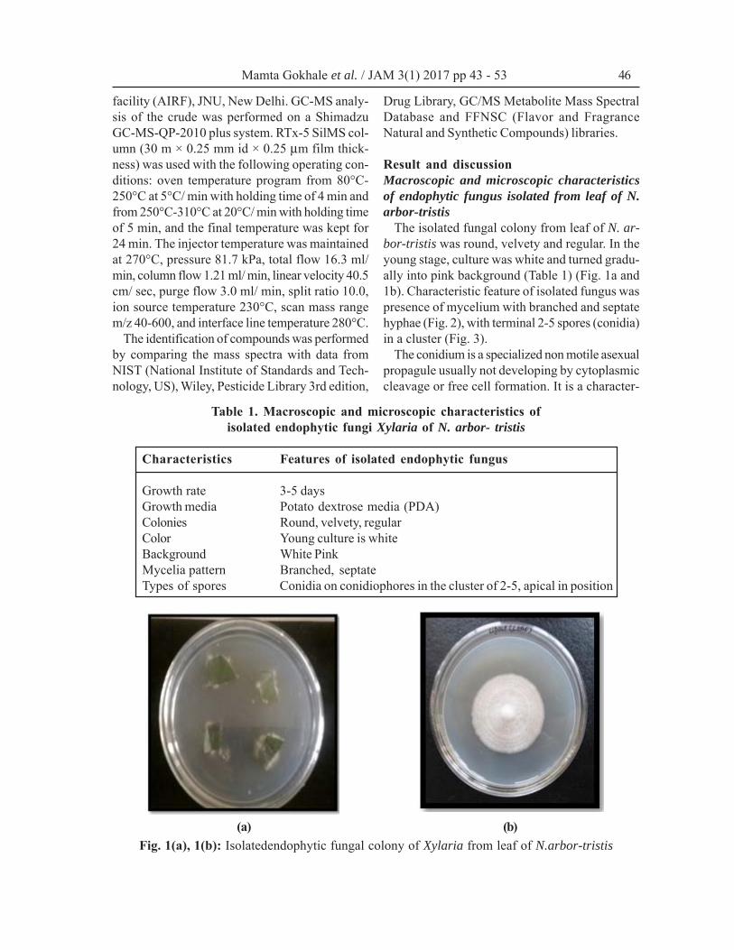
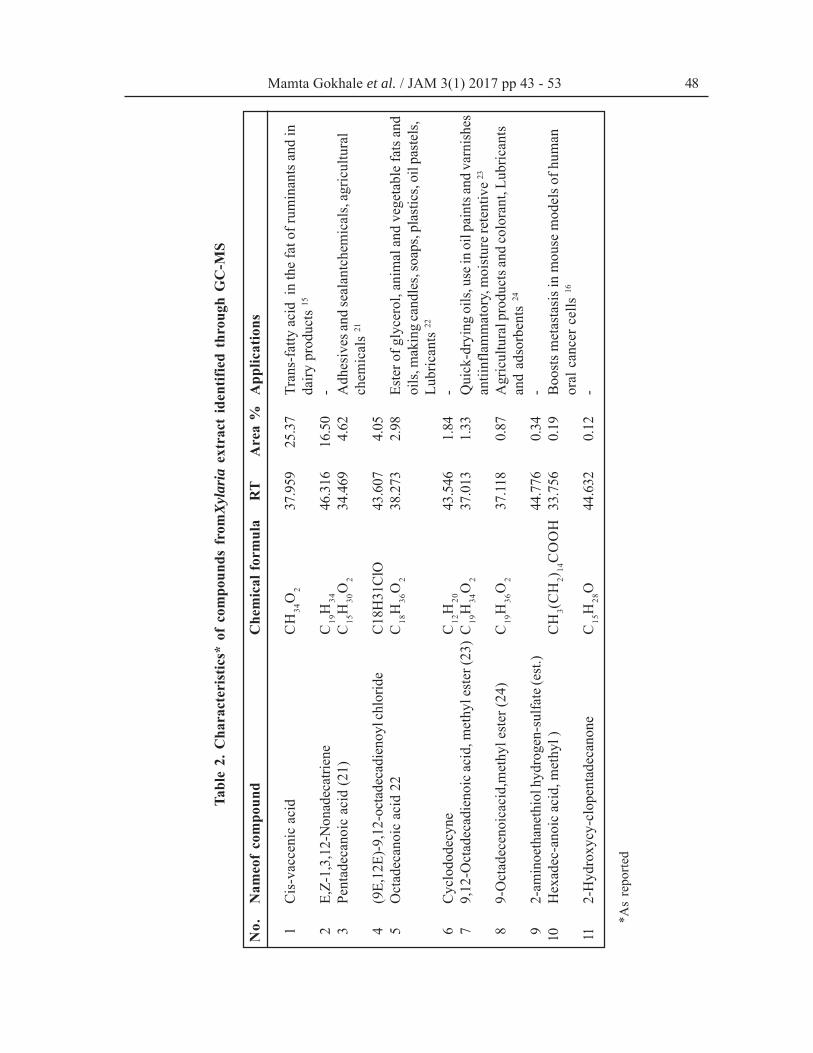
The isolated fungal colony from leaf of N. ar-bor-tristis was round, velvety and regular. In theyoung stage, culture was white and turned gradu-ally into pink background (Table 1) (Fig. 1a and1b). Characteristic feature of isolated fungus waspresence of mycelium with branched and septatehyphae (Fig. 2), with terminal 2-5 spores (conidia)in a cluster (Fig. 3).
The conidium is a specialized non motile asexualpropagule usually not developing by cytoplasmiccleavage or free cell formation. It is a character-
Table 1. Macroscopic and microscopic characteristics ofisolated endophytic fungi Xylaria of N. arbor- tristis
Characteristics Features of isolated endophytic fungus
Growth rate 3-5 daysGrowth media Potato dextrose media (PDA)Colonies Round, velvety, regularColor Young culture is whiteBackground White PinkMycelia pattern Branched, septateTypes of spores Conidia on conidiophores in the cluster of 2-5, apical in position
Fig. 1(a), 1(b): Isolatedendophytic fungal colony of Xylaria from leaf of N.arbor-tristis(a) (b)
Mamta Gokhale et al. / JAM 3(1) 2017 pp 43 - 53 46
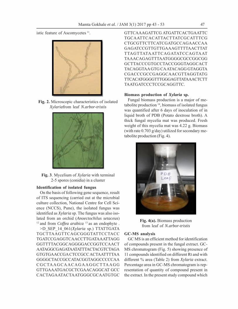
istic feature of Ascomycetes 11.
Identification of isolated fungusOn the basis of following gene sequence, result
of ITS sequencing (carried out at the microbialculture collection, National Centre for Cell Sci-ence (NCCS), Pune), the isolated fungus wasidentified as Xylaria sp. The fungus was also iso-lated from an orchid (Anoectochilus setaceus)12 and from Coffea arabica 13 as an endophyte .
>D_SEP_14_061(Xylaria sp.) TTATTGATATGCTTAAGTTCAGCGGGTATTCCTACCTGATCCGAGGTCAACCTTGATAAATTAGGGGTTTTACGGCAGGGGACCGGTCCAACTAATAGGCGAGATAATATTTACTACGTCTAGAGTGTGAACCGA CTCCGCC ACTAATTTTAAGGGGCTACCGCCATACGGTAGGCCCCCAACGCTAAGCAACAGAAGGCTTAAGGGTTGAAATGACGCTCGAACAGGCAT GCCCACTAGAATACTAATGGGCGCAATGTGC
GTTCAAAGATTCG ATGATTCACTGAATTCTGCAATTCACATTACTTATCGCATTTCGCTGCGTTCTTCATCGATGCCAGAACCAAGAGATCCGTTGTTGAAAGTTTTAACTTATTTAGTTATAATTCAGATATCCAGTAATTAAACAGAGTTTAATGGGGCGCCGGCGGGCTTACCCGTGCCTACCGGGTAGGCACTTACAGGTAA GTG CA ATACAGG GTAGGTACGACCCGCCGAGGCAACGTTAGGTATGTTCACATGGGGTTTGGGAGTTATAAACTCTTTAATGATCCCTCCGCAGGTTC.
Biomass production of Xylaria sp.Fungal biomass production is a major of me-
tabolite production 14, biomass of isolated funguswas quantified after 6 days of inoculation of inliquid broth of PDB (Potato dextrose broth). Athick fungal mycelia mat was produced. Freshweight of this mycelia mat was 4.22 g. Biomass(with rate 0.703 g/day) utilized for secondary me-tabolite production (Fig. 4).
GC-MS analysisGC MS is an efficient method for identification
of compounds present in the fungal extract. GC-MS chromatogram (Fig. 5) showing presence of11 compounds identified on different Rt and withdifferent % area (Table 2) from Xylaria extract.Percentage area in GC-MS chromatogram is rep-resentation of quantity of compound present inthe extract. In the present study compound which
Fig. 2. Microscopic characteristics of isolatedXylariafrom leaf N.arbor-tristis
Fig. 3. Mycelium of Xylaria with terminal2-5 spores (conidia) in a cluster
Fig. 4(a). Biomass productionfrom leaf of N.arbor-tristis
Mamta Gokhale et al. / JAM 3(1) 2017 pp 43 - 53 47
Tabl
e 2.
Cha
ract
eris
tics*
of
com
poun
ds f
rom
Xyl
aria
ext
ract
ide
ntifi
ed t
hrou
gh G
C-M
S
No.
Nam
eof
com
poun
dC
hem
ical
form
ula
RT
Are
a %
App
licat
ions
1C
is-v
acce
nic
acid
CH
34O
237
.959
25.3
7Tr
ans-
fatty
aci
d in
the
fat o
f rum
inan
ts a
nd in
da
iry p
rodu
cts
15
2E,
Z-1,
3,12
-Non
adec
atrie
neC
19H
3446
.316
16.5
0-
3Pe
ntad
ecan
oic
acid
(21)
C15
H30
O2
34.4
694.
62A
dhes
ives
and
seal
antc
hem
ical
s, ag
ricul
tura
lch
emic
als
21
4(9
E,12
E)-9
,12-
octa
deca
dien
oyl c
hlor
ide
C18H
31Cl
O43
.607
4.05
5O
ctad
ecan
oic
acid
22
C18
H36
O2
38.2
732.
98Es
ter o
f gly
cero
l, an
imal
and
veg
etab
le fa
ts a
ndoi
ls, m
akin
g ca
ndle
s, so
aps,
plas
tics,
oil p
aste
ls,
Lubr
ican
ts 22
6C
yclo
dode
cyne
C12
H20
43.5
461.
84-
79,
12-O
ctad
ecad
ieno
ic a
cid,
met
hyl e
ster
(23)
C19
H34
O2
37.0
131.
33Q
uick
-dry
ing
oils
, use
in o
il pa
ints
and
varn
ishe
san
tiinf
lam
mat
ory,
moi
stur
e ret
entiv
e 23
89-
Oct
adec
enoi
caci
d,m
ethy
l est
er (2
4)C
19H
36O
237
.118
0.87
Agr
icul
tura
l pro
duct
s and
colo
rant
, Lub
rican
tsan
d ad
sorb
ents
24
92-
amin
oeth
anet
hiol
hyd
roge
n-su
lfate
(est
.)44
.776
0.34
-10
Hex
adec
-ano
ic a
cid,
met
hyl )
CH
3(C
H2)
14C
OO
H33
.756
0.19
Boo
sts m
etas
tasi
s in
mou
se m
odel
s of h
uman
oral
can
cer c
ells
16
112-
Hyd
roxy
cy-c
lope
ntad
ecan
one
C15
H28
O44
.632
0.12
-
*As
repo
rted
Mamta Gokhale et al. / JAM 3(1) 2017 pp 43 - 53 48
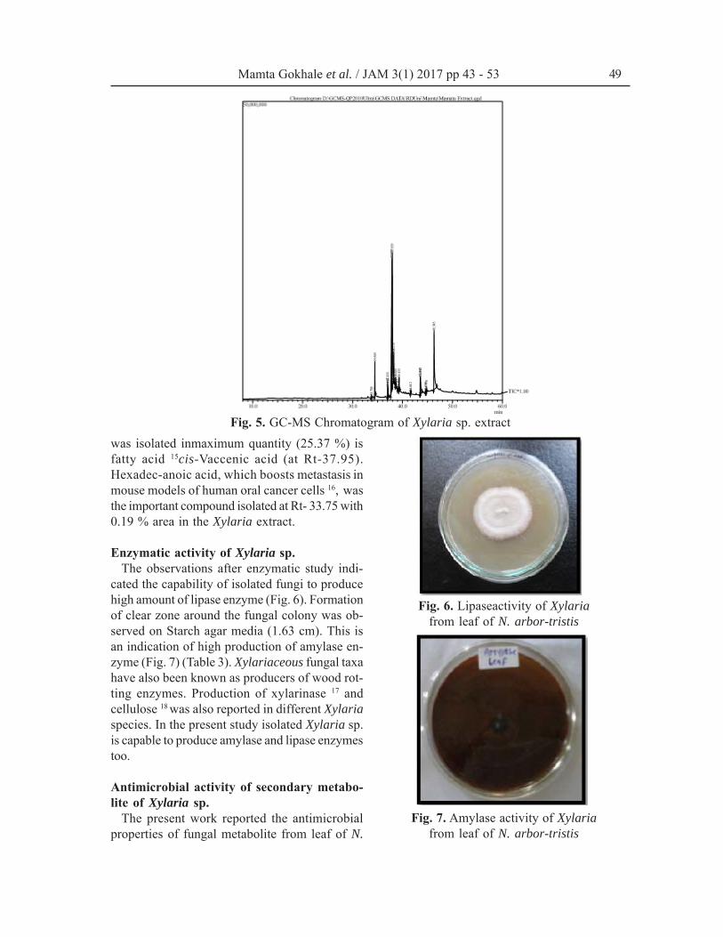
Fig. 5. GC-MS Chromatogram of Xylaria sp. extractwas isolated inmaximum quantity (25.37 %) isfatty acid 15cis-Vaccenic acid (at Rt-37.95).Hexadec-anoic acid, which boosts metastasis inmouse models of human oral cancer cells 16, wasthe important compound isolated at Rt- 33.75 with0.19 % area in the Xylaria extract.
Enzymatic activity of Xylaria sp.The observations after enzymatic study indi-
cated the capability of isolated fungi to producehigh amount of lipase enzyme (Fig. 6). Formationof clear zone around the fungal colony was ob-served on Starch agar media (1.63 cm). This isan indication of high production of amylase en-zyme (Fig. 7) (Table 3). Xylariaceous fungal taxahave also been known as producers of wood rot-ting enzymes. Production of xylarinase 17 andcellulose 18 was also reported in different Xylariaspecies. In the present study isolated Xylaria sp.is capable to produce amylase and lipase enzymestoo.
Antimicrobial activity of secondary metabo-lite of Xylaria sp.
The present work reported the antimicrobialproperties of fungal metabolite from leaf of N.
Fig. 6. Lipaseactivity of Xylariafrom leaf of N. arbor-tristis
Fig. 7. Amylase activity of Xylariafrom leaf of N. arbor-tristis
Mamta Gokhale et al. / JAM 3(1) 2017 pp 43 - 53 49
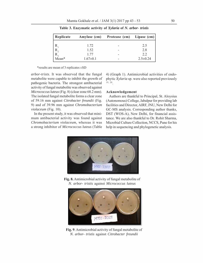
Table 3. Enzymatic activity of Xylaria of N. arbor- tristis
Replicate Amylase (cm) Protease (cm) Lipase (cm)
R1 1.72 - 2.5R2 1.52 - 2.8R3 1.77 - 2.2Mean* 1.67±0.1 - 2.5±0.24
*results are mean of 3 replicates ±SD
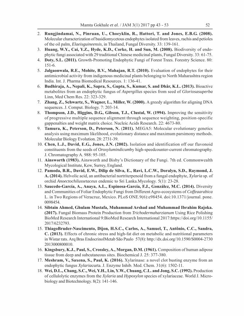
arbor-trists. It was observed that the fungalmetabolite were capable to inhibit the growth ofpathogenic bacteria. The strongest antibacterialactivity of fungal metabolite was observed againstMicrococcus luteus (Fig. 8) (clear zone 68.2 mm).The isolated fungal metabolite forms a clear zoneof 59.16 mm against Citrobacter freundii (Fig.9) and of 39.96 mm against Chromobacteriumviolaceum (Fig. 10).
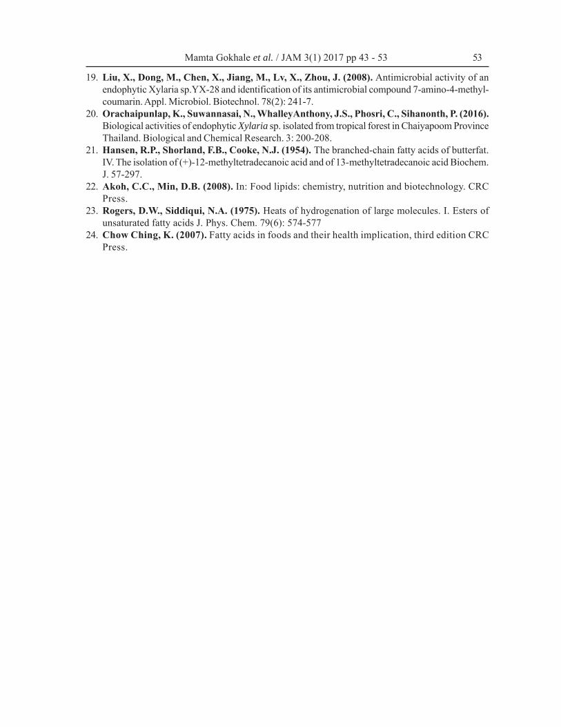
In the present study, it was observed that mini-mum antibacterial activity was found againstChromobacterium violaceum, whereas it wasa strong inhibitor of Micrococcus luteus (Table
4) (Graph 1). Antimicrobial activities of endo-phytic Xylaria sp. were also reported previously19, 20.
AcknowledgementAuthors are thankful to Principal, St. Aloysius
(Autonomous) College, Jabalpur for providing labfacilities and Director, AIRF, JNU, New Delhi forGC-MS analysis. Corresponding author thanks,DST (WOS-A), New Delhi, for financial assis-tance. We are also thankful to Dr. Rohit Sharma,Microbial Culture Collection, NCCS, Pune for hishelp in sequencing and phylogenetic analysis.
Fig. 8. Antimicrobial activity of fungal metabolite ofN. arbor- tristis against Micrococcus luteus
Fig. 9. Antimicrobial activity of fungal metabolite ofN. arbor- tristis against Citrobacter freundii
Mamta Gokhale et al. / JAM 3(1) 2017 pp 43 - 53 50
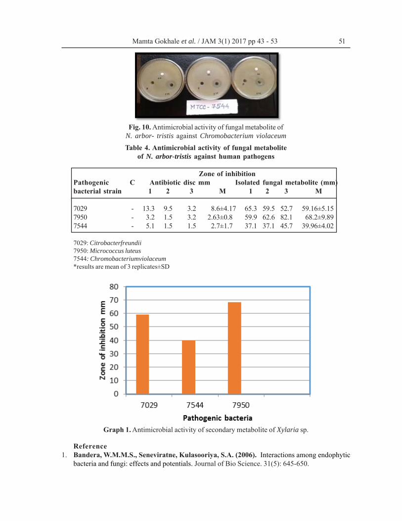
Fig. 10. Antimicrobial activity of fungal metabolite ofN. arbor- tristis against Chromobacterium violaceum
Table 4. Antimicrobial activity of fungal metaboliteof N. arbor-tristis against human pathogens
Zone of inhibitionPathogenic C Antibiotic disc mm Isolated fungal metabolite (mm)bacterial strain 1 2 3 M 1 2 3 M
7029 - 13.3 9.5 3.2 8.6±4.17 65.3 59.5 52.7 59.16±5.157950 - 3.2 1.5 3.2 2.63±0.8 59.9 62.6 82.1 68.2±9.897544 - 5.1 1.5 1.5 2.7±1.7 37.1 37.1 45.7 39.96±4.02
7029: Citrobacterfreundii7950: Micrococcus luteus7544: Chromobacteriumviolaceum*results are mean of 3 replicates±SD
Graph 1. Antimicrobial activity of secondary metabolite of Xylaria sp.
Reference1. Bandera, W.M.M.S., Seneviratne, Kulasooriya, S.A. (2006). Interactions among endophytic
bacteria and fungi: effects and potentials. Journal of Bio Science. 31(5): 645-650.
Mamta Gokhale et al. / JAM 3(1) 2017 pp 43 - 53 51
2. Rungjindamai, N., Pinruan, U., Choeyklin, R., Hattori, T. and Jones, E.B.G. (2008).Molecular characterization of basidiomycetous endophytes isolated from leaves, rachis and petiolesof the oil palm, Elaeisguineensis, in Thailand, Fungal Diversity. 33: 139-161.
3. Huang, W.Y., Cai, Y.Z., Hyde, K.D., Corke, H. and Sun, M. (2008). Biodiversity of endo-phytic fungi associated with 29 traditional Chinese medicinal plants, Fungal Diversity. 33: 61-75.
4. Doty, S.L. (2011). Growth-Promoting Endophytic Fungi of Forest Trees. Forestry Science. 80:151-6.
5. Jalgaonwala, R.E., Mohite, B.V., Mahajan, R.T. (2010). Evaluation of endophytes for theirantimicrobial activity from indigenous medicinal plants belonging to North Maharashtra regionIndia. Int. J. Pharma Biomedical Resources. 1: 136-41.
6. Budhiraja, A., Nepali, K., Sapra, S., Gupta, S., Kumar, S. and Dhâr, K.L. (2013). Bioactivemetabolites from an endophytic fungus of Aspergillus species from seed of GloriosasuperbaLinn, Med Chem Res. 22: 323-329.
7. Zhang, Z., Schwartz, S., Wagner, L., Miller, W. (2000). A greedy algorithm for aligning DNAsequences. J. Comput. Biology. 7: 203-14.
8. Thompson, J.D., Higgins, D.G., Gibson, T.J., Clustal, W. (1994). Improving the sensitivityof progressive multiple sequence alignment through sequence weighting, position-specificgappenalties and weight matrix choice. Nucleic Acids Research. 22: 4673-80.
9. Tamura, K., Peterson, D., Peterson, N. (2011). MEGA5: Molecular evolutionary geneticsanalysis using maximum likelihood, evolutionary distance and maximum parsimony methods.Molecular Biology Evolution. 28: 2731-39.
10. Chen, L.J., David, E.G., Jones, J.N. (2002). Isolation and identification off our flavonoidconstituents from the seeds of Oroxylumindicumby high-speedcounter-current chromatography.J. Chromatography A. 988: 95-105.
11. Ainsworth (1983). Ainsworth and Bisby’s Dictionary of the Fungi. 7th ed. CommonwealthMycological Institute, Kew, Surrey, England.
12. Pamoda, B.R., David, E.W., Dilip de Silva, E., Ravi, L.C.W., Doralyn, S.D., Raymond, J.A. (2014). Helvolic acid, an antibacterial nortriterpenoid from a fungal endophyte, Xylaria sp. oforchid Anoectochilussetaceus endemic to Sri Lanka.Mycology. 5(1): 23-28.
13. Saucedo-García, A., Anaya, A.L., Espinosa-García, F.J., González, M.C. (2014). Diversityand Communities of Foliar Endophytic Fungi from Different Agro-ecosystems of CoffeaarabicaL. in Two Regions of Veracruz, Mexico. PLoS ONE.9(6):e98454. doi:10.1371/journal. pone.0098454.
14. Sibtain Ahmed, Ghulam Mustafa, Muhammad Arshad and Muhammad Ibrahim Rajoka.(2017). Fungal Biomass Protein Production from Trichodermaharzianum Using Rice PolishingBioMed Research International 9:BioMed Research International 2017:https://doi.org/10.1155/2017/6232793.
15. ThiagoBruder-Nascimento, Dijon, H.S.C., Carlos, A., Samuel, T., Antônio, C.C., Sandra,C. (2013). Effects of chronic stress and high-fat diet on metabolic and nutritional parametersin Wistar rats. Arq Bras EndocrinolMetab São Paulo 57(8): http://dx.doi.org/10.1590/S0004-27302013000800010.
16. Kingsbury, K.J., Paul, S., Crossley, A., Morgan, D.M. (1961). Composition of human adiposetissue from deep and subcutaneous sites. Biochemical J. 25: 377-380.
17. Meshram, V., Saxena, S., Paul, K. (2016). Xylarinase: a novel clot busting enzyme from anendophytic fungus Xylariacurta. J. Enzyme Inhib. Med. Chem. 31(6): 1502-11.
18. Wei, D.L., Chang, S.C., Wei, Y.H., Lin, Y.W., Chuang, C.L. and Jong, S.C. (1992). Productionof cellulolytic enzymes from the Xylaria and Hypoxylon species of xylariaceae. World J. Micro-biology and Biotechnology. 8(2): 141-146.
Mamta Gokhale et al. / JAM 3(1) 2017 pp 43 - 53 52
19. Liu, X., Dong, M., Chen, X., Jiang, M., Lv, X., Zhou, J. (2008). Antimicrobial activity of anendophytic Xylaria sp.YX-28 and identification of its antimicrobial compound 7-amino-4-methyl-coumarin. Appl. Microbiol. Biotechnol. 78(2): 241-7.
20. Orachaipunlap, K., Suwannasai, N., WhalleyAnthony, J.S., Phosri, C., Sihanonth, P. (2016).Biological activities of endophytic Xylaria sp. isolated from tropical forest in Chaiyapoom ProvinceThailand. Biological and Chemical Research. 3: 200-208.
21. Hansen, R.P., Shorland, F.B., Cooke, N.J. (1954). The branched-chain fatty acids of butterfat.IV. The isolation of (+)-12-methyltetradecanoic acid and of 13-methyltetradecanoic acid Biochem.J. 57-297.
22. Akoh, C.C., Min, D.B. (2008). In: Food lipids: chemistry, nutrition and biotechnology. CRCPress.
23. Rogers, D.W., Siddiqui, N.A. (1975). Heats of hydrogenation of large molecules. I. Esters ofunsaturated fatty acids J. Phys. Chem. 79(6): 574-577
24. Chow Ching, K. (2007). Fatty acids in foods and their health implication, third edition CRCPress.
Mamta Gokhale et al. / JAM 3(1) 2017 pp 43 - 53 53
Top Related