Yang, Xin and Hamner, Margaret A. and Brown, Angus M....
32
Yang, Xin and Hamner, Margaret A. and Brown, Angus M. and Evans, Richard D. and Ye, Zu-Cheng and Chen, Shengdi and Ransom, Bruce R. (2014) Novel hypoglycemic injury mechanism: N-methyl-D-aspartate receptor-mediated white matter damage. Annals of Neurology, 75 (4). pp. 492-507. ISSN 0364-5134 Access from the University of Nottingham repository: http://eprints.nottingham.ac.uk/35413/1/Hypoglycemia%20ms%205-1-2013FINAL.pdf Copyright and reuse: The Nottingham ePrints service makes this work by researchers of the University of Nottingham available open access under the following conditions. This article is made available under the University of Nottingham End User licence and may be reused according to the conditions of the licence. For more details see: http://eprints.nottingham.ac.uk/end_user_agreement.pdf A note on versions: The version presented here may differ from the published version or from the version of record. If you wish to cite this item you are advised to consult the publisher’s version. Please see the repository url above for details on accessing the published version and note that access may require a subscription. For more information, please contact [email protected]
Transcript of Yang, Xin and Hamner, Margaret A. and Brown, Angus M....
Yang, Xin and Hamner, Margaret A. and Brown, Angus M. and Evans, Richard D. and Ye, Zu-Cheng and Chen, Shengdi and Ransom, Bruce R. (2014) Novel hypoglycemic injury mechanism: N-methyl-D-aspartate receptor-mediated white matter damage. Annals of Neurology, 75 (4). pp. 492-507. ISSN 0364-5134
Access from the University of Nottingham repository: http://eprints.nottingham.ac.uk/35413/1/Hypoglycemia%20ms%205-1-2013FINAL.pdf
Copyright and reuse:
The Nottingham ePrints service makes this work by researchers of the University of Nottingham available open access under the following conditions.
This article is made available under the University of Nottingham End User licence and may be reused according to the conditions of the licence. For more details see: http://eprints.nottingham.ac.uk/end_user_agreement.pdf
A note on versions:
The version presented here may differ from the published version or from the version of record. If you wish to cite this item you are advised to consult the publisher’s version. Please see the repository url above for details on accessing the published version and note that access may require a subscription.
For more information, please contact [email protected]
Novel Hypoglycemic Injury Mechanism: NMDA
Receptor-Mediated White Matter Damage
Running Head: NMDA receptors mediate WM hypoglycemic injury
Yang X1, Hamner MA2, Brown AM3, Evans R3, Ye Z2, Chen SD1 and Ransom
BR2*
1Department of Neurology
Ruijin Hospital affiliated to Shanghai Jiaotong University School of Medicine,
Shanghai 200025, China
2Department of Neurology
University of Washington
Seattle, WA 98195
3School of Biomedical Sciences
University of Nottingham
Nottingham, NG2 5HY, UK
*Corresponding author:
Bruce R. Ransom, MD, PhD
Department of Neurology

University of Washington, Rm RR650
1959 NE Pacific Street
Seattle, WA 98195
Email: [email protected]
Office: 206-543-2340
Number of characters in title: 79
Number of characters in Running Head: 40
Number of words in Abstract: 245
Number of Words in body of manuscript: 7532
Number of Figures: 8
Number of Color Figures: 1
Number of Tables: 0
Acknowledgment:
This work was supported by funding from the NIH/NINDS (R01
NS015589; B.R.R.), National Natural Science Foundation of China (NSFC
30900453 and 81070958; X.Y.) and the University of Nottingham (A.M.B.).

ABSTRACT
Objective: Hypoglycemia is a common adverse event and can injure central
nervous system (CNS) white matter (WM). We determined if glutamate receptors
were involved in hypoglycemic WM injury.
Methods: Mouse optic nerves (MON), CNS WM tracts, were maintained at 37°C
with oxygenated artificial cerebrospinal fluid (ACSF) containing 10 mM glucose.
Aglycemia was produced by switching to 0 glucose ACSF. Supra-maximal
compound action potentials (CAPs) were elicited using suction electrodes and
axon function was quantified as the area under the CAP. Amino acid release
was measured using HPLC. Extracellular [lactate] was measured using an
enzyme electrode.
Results: About 50% of MON axons were injured after 60 min of aglycemia (90%
after 90 min); injury was not affected by animal age. Blockade of NMDA-type
glutamate receptors improved recovery after 90 min of aglycemia by 250%.
Aglycemic injury was increased by reducing [Mg2+]o or increasing [glycine]o, and
decreased by lowering pHo, expected results for NMDA receptor-mediated injury.
Extracellular pH increased during aglycemia, due to a drop in [lactate-]o.
Aglycemic injury was dramatically reduced in the absence of [Ca2+]o. Extracellular
aspartate, a selective NMDA receptor agonist, increased during aglycemia.
Interpretation: Aglycemia injured WM by a unique excitotoxic mechanism
involving NMDA receptors (located primarily on oligodendrocytes). During WM
aglycemia, the selective NMDA agonist, aspartate, is released, probably from
astrocytes. Injury is mediated by Ca2+ influx through aspartate-activated NMDA
receptors made permeable by an accompanying alkaline shift in pHo caused by a
fall in [lactate-]o. These insights have important clinical implications.
INTRODUCTION
Hypoglycemia continues to be an important and common adverse clinical
event in patients with diabetes, and more rarely with other conditions 1-4. The
central nervous system (CNS) is extremely vulnerable to dysfunction and injury
with hypoglycemia 1, 5, 6. In addition, there is compelling evidence that CNS
dysfunction can develop slowly over years in patients with well controlled
diabetes, presumably related to subclinical episodes of hypoglycemia 7, 8. For
these reasons, it is important to understand how hypoglycemia produces
irreversible CNS injury. This knowledge will provide a basis for development of
effective therapies to minimize the extent of damage and improve clinical
outcome.
Recent observations have drawn attention to selective white matter (WM)
damage as a consequence of protracted hypoglycemia 9, 10; in fact, WM can be
the predominant site of injury 9, 10. However, experimental studies on the
mechanisms of hypoglycemic injury have used primarily rodents. As discussed
elsewhere, humans have roughly 4 to 5-fold more WM than rodents, a critical
difference because WM and gray matter (GM) are vastly different tissues with
unique mechanisms of injury 11, 12.
Fortunately, validated and practical models of CNS WM injury are now
available 13-16. Initial studies using the acutely isolated mouse optic nerve (MON),
a myelinated WM tract, found that 60 minutes of aglycemia produced substantial
irreversible injury. The mechanism of injury was found to be Ca2+-dependent and
to involve activation of L-type Ca2+ channels and reverse Na+/Ca2+ exchange 17.
These two pathways mediated toxic Ca2+ influx contributing to permanent loss of
axonal excitability 17. Subsequently, another form of energy disruption in WM,
ischemia, was found to produce injury mediated by excessive activation of
excitatory glutamate receptors. This pathogenesis, called excitotoxicity, was first
defined in CNS gray matter (GM)11. The characteristics of ‘excitotoxicity’ in WM
are distinct from GM; in other words, the toxic glutamate receptor activation seen
in WM during ischemia is a ‘special form’ of excitotoxicity 11, 16, 18-20. While both N-

methyl-D-aspartate (NMDA) and non-NMDA receptors have been found in WM,
only non-NMDA receptors appear to participate in ischemic (i.e., oxygen and
glucose deprivation (OGD)) WM injury 14, 18, 20-22, but see23). This was somewhat
unexpected because NMDA receptors are clearly expressed by myelinating
oligodendrocytes 18, 24 and these cells are unquestionably injured during energy
deprivation 18, 19, 21, 22, 25.
We sought to determine if excitotoxicity might also be involved in the WM
injury seen with aglycemia. Indeed, we found that excitotoxicity contributed
importantly to this special type of irreversible WM injury but unlike the situation
with OGD, in aglycemia NMDA receptors were prominently involved, in a Ca2+-
dependent manner. Our results also provided a plausible explanation for why
NMDA receptors were involved in aglycemia but not in ischemia. Finally, we
showed that WM aglycemic injury arose from NMDA receptors activated by
aspartate, not glutamate. These findings raise important questions about how to
optimally manage the clinical situation of severe hypoglycemic brain dysfunction.

METHODS AND MATERIALS
All experiments were done in accordance with the University of
Washington Institutional Animal Care and Use Committee.
Electrophysiology
Mouse optic nerves (MONs) were acutely obtained from C57BL/6 mice
that varied in age from one to 24 months of age; most experiments were done on
three month-old mice. As previously described 26, mice were deeply anesthetized
with CO2 and then decapitated. Optic nerves were dissected free and cut at the
optic chiasm and behind the orbit. The optic nerves were freed from their dural
sheaths and placed in an interface perfusion chamber (Medical Systems Corp.,
Greenvale, NY, USA) and maintained at 37°C. MONs were superfused with
artificial cerebrospinal fluid (ACSF) containing (in mmol/L): 125 NaCl, 3.0 KCl,
2.0 CaCl2, 2.0 MgSO4 ·7H2O, 1.25 NaH2PO4, 26 NaHCO3, and 10 glucose. The
ACSF was bubbled with an O2-free gas mixture (95% N2: 5%CO2) to maintain pH
at 7.45. A humidified gas mixture of 95% O2/5% CO2 continuously aerated the
chamber. Two sets of suction electrodes were placed in the bath to allow
recording from two optic nerves at the same time. Suction electrodes back-filled
with the appropriate ACSF were used for stimulating and recording. The
stimulating electrode was attached to the rostral end of the nerve, while the
proximal end was attached to a second electrode to record the compound action
potential (CAP), ensuring orthodromic stimulation. Stimulus pulse (30 µs
duration) strength (Isostim 520; WPI, Sarasota, FL) was adjusted to evoke the
maximum CAP and then increased another 25% (i.e., supramaximal
stimulation)13. During an experiment, the supramaximal CAP was elicited every
30 seconds. The recording electrode was connected to an amplifier (Standford
Research Systems, Model SR 560) and the signal was amplified 500 times,
filtered at 30 kHz and acquired at 20 kHz.
Nerves were allowed to equilibrate for at least 30 mins before recording
commenced. In glucose deprivation experiments, the solution in the stimulating

and recording electrodes was switched to glucose-free ACSF (i.e., aglycemia).
Osmotic compensation was achieved by adding 10mmol/L sucrose. Ca2+ free
ACSF was made by omitting CaCl2 and adding 0.5 mmol/L ethylene glycol-bis (ß-
aminoethylether)-N,N,N’,N’-tetraacetic acid (EGTA) with equimolar MgCl2. Zero
Mg2+ ACSF was made by omitting MgSO4 and replacing with equimolar Na2SO4.
In a few experiments, both oxygen and glucose were deleted to create an
ischemia-like condition, also referred to as oxygen-glucose deprivation or OGD.
Bath pH was changed from 7.4 to 7.0 by reducing the [HCO3-] using the
following ACSF recipe (in mmol/L): 141 NaCl, 3.0 KCl, 2.0 CaCl2, 2.0 MgSO4
·7H2O, 1.25 NaH2PO4, 10 NaHCO3 and 10 glucose. All changes in ACSF were
introduced 15 mins before the insult and continued until 15 mins after terminating
the insult (e.g., 18).
Pharmacological agents were applied for 15 mins before the insult and
continued until 15 mins after terminating the insult.. The following agents were
purchased from Tocris (Ellisville, Missouri): 2,3-Dioxo-6-nitro-1,2,3,4-
tetrahydrobenzo[f]quinoxaline-7-sulfonamide (NBQX) (30 µM; AMPA/Kainate
channel blocker; dissolved in DMSO as 30 mM stock solution), 7-chlorokynurenic
acid (7-CKA) (50 µM; glycine binding site blocker; dissolved in DMSO as 50 mM
stock solution) and (5S,10R)-(+)-5-Methyl-10,11-dihydro-5H-
dibenzo[a,d]cyclohepte n-5,10-imine maleate (MK801) (15 µM; NMDA receptor
blocker; dissolved in DMSO as 15 mM stock solution).
Lactate biosensor
Lactate and null biosensors were purchased from Sarissa Biomedical Ltd
(Coventry, UK). In these experiments, however, the lactate signals were
sufficiently large that subtraction of the null signal did not meaningfully alter the
lactate signal amplitude. The lactate biosensors (25 µmin diameter and 500 µm
in length) were pressed against the pial-glial membrane of the MON.
Experimental recordings began after an equilibration period of 30 to 60 minutes.
At the beginning and end of all experiments, lactate biosensors were calibrated
using lactate concentrations of 10, 100 and 1000 M. Results were considered
valid only if the pre- and post-calibrations deviated by no more than 5%.
pH-sensitive microelectrodes
Ion-sensitive microelectrodes for extracellular pH (pHo) measurements
were made according to the method of Borrelli et al.,27 with slight modifications 6.
Briefly, double-barrel microelectrodes were pulled and beveled to a tip diameter
of 2 to 5 µm. The ion-sensitive barrel was back-filled with a short column of H+-
sensitive sensor (Fluka pH ionophore). The indifferent barrel was back-filled with
140 mmole/L NaCl + 20 mmol/L HEPES adjusted to pH 7.0. Only electrodes that
showed near-Nernstian responses to 10 fold changes to [H+] calibrating solutions
were used in experiments.
Data analysis
Optic nerve function was quantitatively determined by integrating the area
under the CAP. Data were acquired online (Digidata 1440A; Molecular Devices,
Sunnyvale, CA) using proprietary software (Clampex, Molecular Devices). CAP
area was calculated using pClamp (Molecular Devices) and was normalized by
averaging the baseline CAP area over a period of 15 minutes, and setting this
value to 1.0. The normalized CAP area at any time is proportional to the relative
number of functioning axons 13. Data are presented as the mean, and standard
error of the mean (SEM), of normalized traces. Statistical significance was
determined by unpaired, two-tailed Student’s t-test. P values less than 0.05 were
considered significant.
Glutamate, aspartate and glycine measurements
Amino acid (i.e., glutamate, aspartate, glycine, etc.) release from MON
was determined by first collecting the superfusate during 60 or 90 min aglycemia
(or OGD in a few experiments) and then subjecting these samples to quantitative
amino acid measurement using HPLC 28. Experiments were designed to monitor
amino acid release simultaneously from 1 pair of MONs (this technique can be
modified to enhance sensitivity by using 5 pairs of MONs measured at one time).
Briefly, amino acids were pre-column derivatized with o-phthaldialdehyde
(Sigma, St Louis, MO), separated and measured using standard techniques.
Samples of extracellular perfusion fluid were collected continuously such that
every vial contained two min of superfusate. Glutamate content was measured in
every other vial (i.e., every 4 mins), except during the baseline when it was
measured every 2 mins. Collected samples were centrifuged at 16,000 g for 3
mins and supernatants transferred for HPLC analysis. Glutamate measurements,
normalized to baseline glutamate release were made from MONs treated
identically to those studied electrophysiologically and the results were plotted
against time. The rate and release pattern of glutamate was monitored for 30 min
before injury (60 mins and 90 mins aglycemia or 60 mins OGD), and was
continued for at least 30 min after the end of the insult. Actual amounts of
glutamate measured in mM were determined by comparing the experimental
measurements to standard samples.
Intracellular amino acid content of cells within the MON was determined
using a slightly modified established technique 29. Briefly, pairs of MONs were
sonicated in 0.2 ml of homogenization solution (distilled H2O supplemented with
0.4 mM DTT and 1 mM EDTA). The homogenate was diluted 10-fold in
homogenization solution prior to protein measurement and HPLC measurement
of amino acids. Aliquots for HPLC measurement of amino acids were then
acidified by HCl and centrifuged. The supernatant was harvested and
neutralized by NaOH prior to using HPLC to measure amino acid concentrations.
Protein content was measured in identical aliquots using Bio-Rad protein assay
reagent.
RESULTS
Aglycemia caused duration-dependent WM injury
As previously described 6, 30, aglycemia caused loss of WM excitability,
measured as failure of the CAP. The pattern of CAP loss with aglycemia was
unique because the CAP was largely unchanged for the first 10 to 15 min
following glucose deprivation (Fig. 1A, B) 30. Once the CAP began to fail, all three
peaks fell in unison (data not shown). When glucose was restored after
aglycemia, the CAP partially recovered to a stable new level (e.g., Fig. 1A). The
duration of aglycemia dictated the extent of CAP recovery (Fig. 1B). The area
under the CAP, which is proportional to the number of excited axons (see 13),
recovered to 72.2 ± 6.3%, 48.2 ± 3.7% or 5.0 ± 1.2% of control CAP area after
30, 60 or 90 min of aglycemia, respectively. The extent of CAP recovery following
a given period of aglycemia stabilized after about 30 min and remained stable
during observation periods as long as 4 hours. For quantification and
comparison, the area of CAP recovery was determined 60 min following the
conclusion of glucose deprivation 30.
Glycogen content affected the extent of aglycemic injury
It is known that tissue glycogen content affects the latency to CAP failure
after removing glucose; more glycogen prolonged and less glycogen shortened
latency to CAP failure 31. The present study focused on CAP recovery after
standard periods of aglycemia and the role of glycogen must be considered. As
shown in Figure 1C, the extent of recovery was significantly less if glycogen was
first depleted by pre-incubation for one hour in 2 mM glucose, rather than the
‘normal’ glucose of 10 mM 31. With this important variable in mind, all subsequent
experiments were done after one-hour incubation in 10 mM glucose to ensure a
consistent level of glycogen.
Aglycemic WM injury did not depend on animal age
The extent of WM injury due to ischemia is dependent on animal age; for a
given period of ischemia, tissue from older animals shows significantly greater
injury 18. Similar experiments were done to test for age-dependency of
hypoglycemic WM injury. The extent of injury after 60 min of aglycemia,
measured as CAP recovery, was determined in four groups of animals ranging in
age from 1 month to 24 months. The extent of irreversible injury was similar for
all age groups indicating that age did not affect aglycemia-induced injury extent
(Fig. 1D), as is the case with ischemia 18.
Aglycemic injury was mediated by NMDA receptors
The mechanism(s) underlying aglycemic WM injury were investigated.
AMPA/kainate-type glutamate receptors participate in ischemic WM injury 14, 22, 28
and we tested for involvement of these receptors in aglycemic injury. The dosage
of NBQX that was maximally protective for ischemic WM injury had no significant
effect on CAP recovery after 60 min of aglycemia (control vs. NBQX CAP: 49.2 ±
3.7% vs. 46.2 ± 5.8% , p > 0.05), and only weakly improved CAP recovery after
90 min of aglycemia (Fig. 2A; control vs. NBQX CAP recovery: 5.0 ± 1.2% vs.
12.2 ± 1.8%, p < 0.01).
Blocking NMDA-type glutamate receptors does not protect against
ischemic WM injury 18, 32, in fact it significantly worsens this injury 18. The NMDA
antagonist MK801 was tested and, surprisingly, this agent powerfully improved
CAP recovery after both 60 (see data summary below )and 90 min of aglycemia
(Fig. 2A; 30.8 ± 5.5%, p < 0.0005). In addition to dramatically improving CAP
recovery, MK801 delayed the onset of CAP decline (Fig. 2A). A second NMDA
antagonist, 7-CKA, was applied in the same manner as MK801 and similarly
improved CAP recovery after aglycemia (Fig. 2B; control recovery = 6.4% ± 1.3%
vs. 7-CKA recovery = 34.9 ± 4.1%, p < 0.001).
It was possible that the high dose of NBQX used in these experiments, 30
µM, might have weakly, and non-specifically, blocked NMDA receptors 33. If so,
no additional benefit would be expected if NBQX were added to MK801. We
found exactly that result (Fig. 2C). The amount of CAP recovery seen after 90

min of aglycemia was greatly improved by MK801, compared to control, but CAP
recovery did not improve beyond this level when NBQX was added (Fig. 2C).
These results strongly argue that NMDA receptors were the principal, probably
the only, type of glutamate receptor responsible for irreversible CAP loss
following aglycemia.
NMDA-type glutamate receptors (NMDAR) are modulated in a highly
characteristic manner by several factors, including extracellular [Mg2+] ([Mg2+]o),
extracellular [glycine] and extracellular pH (pHo) 34. Physiological [Mg2+]o blocks
the NMDAR pore at normal membrane potential. Reducing [Mg2+]o by application
of Mg2+-free ACSF would be expected to overcome this block and facilitate pore
opening and ion flux upon NMDAR activation. The effect of Mg2+-free ACSF on
aglycemia-induced WM injury was determined (Fig. 3A). In the presence of
greatly reduced [Mg2+]o, 60 min of aglycemia was much more damaging; CAP
recovery decreased from 48.05 ± 5.3% to 14.9 ± 4.4% (p < 4.1 X 10⁻⁵). The
worsened outcome with application of Mg2+-free ACSF was completely reversed
by MK801 (applied with 0 Mg2+), indicating that this effect depended entirely on
NMDAR activation (Fig. 3A).
Reduced [Mg2+]o, however, had no significant effect on the extent of CAP
recovery after 60 min of ischemia (ischemia recovery = 22.1 ± 4.7% vs. ischemia
+ 0 Mg2+ recovery = 28.2 ± 5.5%, p = 0.43; data not shown), consistent with the
fact that NMDARs are not involved in the pathophysiology of WM ischemic injury
18. This result eliminated concern about a non-specific effect of reducing [Mg2+]o.
The NMDAR is unique in requiring two agonists for activation, glutamate
and glycine (or d-serine) 34. We reasoned that glycine availability might limit the
extent of NMDAR activation during aglycemia, and tested this possibility by
providing glycine (1 mM) in ACSF during aglycemia (Fig. 3B). Increasing glycine
exogenously during aglycemia led to significantly less recovery (36.51 ± 7.00%
vs. 48.19 ± 3.70%, p < 0.03). This result supported the idea that NMDARs were
involved in aglycemia-induced WM injury and that [glycine] was ‘rate limiting’ in
terms of the extent of NMDAR activation.
Extracellular lactate fell rapidly during aglycemia accompanied by an
alkaline shift
Extracellular lactate was measured directly in the MON using a
commercially available ‘enzyme’ electrode specific for lactate (see methods; 35).
The size of the electrode necessitated measurement at the pial-glial boundary of
the nerve as shown diagrammatically in Figure 4A. In the presence of normal
bath glucose (i.e., 10 mM), extracellular [lactate-] ([lactate-]o) was zero in the bath
and averaged 0.46 ± 0.02 mM (n = 6) when the electrode was pressed against
the MON. This value reflected [lactate-]o within the optic nerve but will generally
be less than the ‘true’ nerve [lactate-]o because of an unknown amount of bath
dilution. The [lactate-]o in the nerve was stable over time and did not change
when bath glucose was lowered to 5 mM or increased to 20 mM (data not shown;
see discussion).
During aglycemia, [lactate-]o declined rapidly after about 5 min and fell to
near zero in about 15 min (Fig. 4B). Extracellular pH (pHo) within the optic nerve
was measured using pH-sensitive microelectrodes placed toward the center of
the nerve. As previously reported, pHo was about 0.2 pH units more acid than the
bath pH (~7.4) 6, 36. The fall in [lactate-]o during aglycemia was accompanied by a
temporally related increase in pHo which ultimately plateaued at about 7.4, near
the bath pH (Fig. 4C). The synchronous initiation of changes in [lactate-]o and pHo
occurred well before the CAP began to fall. When the CAP reached its minimum
during aglycemia, an inflection point was often seen in the alkaline shift of pHo
(arrow) suggesting that there were two stages to the alkaline shift (Fig 4C; see
discussion). After switching back to normal bath glucose, [lactate-]o and pHo
rapidly returned to their original values, although [lactate-]o transiently overshot its
baseline level.

The proton-binding site on the NMDAR renders it exquisitely sensitive to
pHo over the normal physiological range 37. Acidic shifts in pHo block, while
alkaline shifts facilitate, NMDAR-mediated ion fluxes. Aglycemia caused an
alkaline shift in pHo, due to the fall in [lactate-]o, which would enhance NMDAR-
mediated ion fluxes and, presumably, injury. This idea was tested by altering pHo
during aglycemia (Fig. 4D; see methods). The aglycemia-induced injury was
compared in bath solutions identical except for pHo (7.45 vs 7.20; Fig. 4D). CAP
recovery after 90 min of aglycemia was significantly greater in the more acidic
bath solution, as predicted if proton-sensitive NMDARs are a primary step in the
pathogenesis of aglycemic WM injury. Interestingly, the latencies to the start of
CAP decline and to complete CAP loss were both markedly delayed in the acidic
solution.
Aglycemic WM injury was dependent on extracellular [Ca2+]
An important feature of NMDARs is high permeability to Ca2+ and this
characteristic can lead to neural injury. If NMDARs are involved in mediating
aglycemic WM injury, a high degree of dependence on extracellular [Ca2+]
([Ca2+]o) is expected. In experiments on three month-old MONs, the extent of
CAP recovery after 90 min of aglycemia was enormously increased when the
insult occurred in the absence of extracellular Ca2+ (from 4.97 ± 1.2% to 86.93 ±
3.6%, p<0.00001; Fig. 5A). It was previously noted in older animals (i.e., >6
months of age) that exposure to Ca2+-free ACSF in conjunction with ischemia
caused a delayed CAP deterioration following the insult 18. This paradoxical
result, still unexplained, was not seen in older animals exposed to aglycemia in
the presence of Ca2+-free ACSF. In other words, Ca2+-free ACSF afforded
dramatic and persistent protection against aglycemic WM injury regardless of
age (Fig. 5B).
Experimental results from the above experiments are summarized in
figure 6. The results from 90 minutes and 60 minutes of aglycemia are shown in
panels A and B, respectively. Note that NBQX did not improve CAP recovery
from 60 minutes of aglycemia (Fig. 6B). Although NBQX had a small protective
effect against injury due to 90 minutes of aglycemia, this was not additive to the
much greater MK801 protection. Taken together, these results indicated that
AMPA/kainate receptors were not involved in mediating aglycemic injury, or at
most had a minimal effect compared to NMDARs.
Amino acid release during aglycemia
Glutamate is robustly released during ischemia in WM and activates
AMPA/Kainate receptors that mediate irreversible WM injury 18, 28. Aglycemic
injury, on the other hand, occurred through activation of NMDARs with little or no
involvement of AMPA/Kainate receptors (see above). Experiments to detect
glutamate release during aglycemia were performed. Using HPLC, glutamate
concentration was measured in sequential aliquots of bath solution passing over
the optic nerve 28. No apparent change in glutamate release was detected during
40 min of aglycemia (Fig. 8A). When aglycemia was switched to oxygen/glucose
deprivation (i.e., ischemia), however, glutamate release increased about 10-fold
over about 35 min, as previously reported (Fig 7A; 28). Aspartate can also
activate NMDARs and might be involved in hypoglycemic injury of gray matter
areas like the hippocampus 5. Like glutamate, no aspartate release could be
detected during aglycemia but it was clearly released by a following episode of
OGD (Fig 7B). Longer periods of aglycemia, up to 90 min, also failed to
demonstrate any obvious change in glutamate or aspartate release (not shown).
These results did not eliminate the possibility of subtle changes in
glutamate or aspartate release during aglycemia. The standard method
employed would not be able to detect very small changes in amino acid release
into the extracellular space of the optic nerve because tissue extracellular volume
is minute in comparison to bath volume, introducing a large dilution factor 28. To
increase the resolution for detecting small amounts of amino acid release, further
experiments were done using ten MONs, rather than two (see methods).
Because the volume of bath perfusion solution remained unchanged, or was

slightly less because of displacement by the eight additional nerves, this strategy
magnified the concentration of released amino acid by at least a factor of five.
Using this modification, aspartate release was clearly detected during aglycemia
(Fig 7D). It also became clear that glutamate release actually decreased
significantly during aglycemia (Fig 7C). The concentrations of the NMDAR co-
agonists, glycine and d-serine, were also measured and their release was not
significantly changed during aglycemia (data not shown).
A further analysis of amino acid changes during algycemia was carried out
by measuring amino acid content of MON tissue under control conditions and
after 30 min of aglycemia (Fig 7E). This provided a measure of intracellular
amino acid content. During aglycemia, intracellular aspartate concentration
increased about four-fold while intracellular glutamate concentration decreased
by at least four-fold. Intracellular glycine concentration did not change
significantly during aglycemia. These findings provide a rational explanation for
the measured changes in the release of glutamate and aspartate during
aglycemia (i.e., Figs 7C and 7D).
Taken together, these results indicated that aglycemia caused activation
of NMDARs, very likely due to aspartate released into the extracellular space.
Involvement of glutamate, the canonical agonist for NMDARs, seemed very
unlikely because its intracellular concentration and extracellular release both fell
during aglycemia. During ischemia, glutamate is released in a manner that is
sensitive to blockade of Na+-dependent glutamate transport 28. Aspartate is also
a substrate for this transporter. When Na+-dependent glutamate/aspartate
transport was blocked during algycemia by TBOA, CAP recovery was
significantly increased (Fig 7F). This supported the idea that the NMDAR agonist
released during aglycemia (i.e., aspartate) was likely released from an
intracellular pool via reversal of the glutamate/aspartate Na+-dependent
transporter.
DISCUSSION
Hypoglycemia is a common cause of neurologic symptoms 1-4, 38. If
severe and prolonged, hypoglycemia causes irreversible neural injury (5). Most
studies on the mechanisms of hypoglycemic brain dysfunction and/or injury have
focused on GM. This is unfortunate because WM also suffers from glucose
deprivation and damage to this region contributes to clinical deficits 9, 10, which is
not surprising in light of the fact that WM represents a major portion (about 55%)
of human forebrain volume 39. Moreover, the mechanisms of WM injury are
distinctive compared to GM (40; see below). Finally, recent clinical reports show
that WM can be selectively and severely injured by hypoglycemia 9, 10. Therapy
for severe hypoglycemia, therefore, must benefit both WM and GM in order to be
clinically effective.
We studied WM hypoglycemic injury and found that this injury was
caused, in part, by excitotoxicity mediated by NMDARs, most likely activated by
aspartate released into the extracellular space. This is surprising because a
related insult, ischemia (i.e., oxygen/glucose deprivation or OGD), injures WM via
AMPA/kainate receptors, and NMDARs are not involved 18, 22 (see below). Our
conclusion is supported by the following observations: 1) hypoglycemic injury
was significantly blocked by NMDAR antagonists (but minimally by
AMPA/kainate receptor antagonists), 2) aspartate release was detected during
aglycemia, and aspartate activates NMDARs but not other glutamate receptors,
3) glutamate release fell during aglycemia precluding activation of AMPA/ kainate
receptors, 4) conditions known to favor activation of, and ion permeation through,
NMDARs (reduced [Mg2+]o and increased [glycine]o) worsened injury, 5) during
aglycemia pHo increased (i.e., proton concentration fell) due to a fall in [lactate-]o,
relieving the ‘proton block’ that reduces ion fluxes through activated NMDARs,
and 6) algycemic injury was greatly mitigated by removal of [Ca2+]o as would be
expected if injury depended on NMDAR-mediated Ca2+ influx. These findings
have important implications for the management of severe hypoglycemia
because WM may be among the first brain regions affected10. They suggest that
early treatment with a suitable antagonist of NMDARs may be beneficial.

Beyond this, they clarify the importance of extracellular ionic environment as a
precondition for NMDAR involvement in neural injury.
Amino Acid Release During Hypoglycemia
One of the most striking differences between WM ischemia and WM
aglycemia is the pattern of amino acid release induced by the two conditions.
During ischemia, glutamate and aspartate are released into the extracellular
space after a latency period of about 20 min (18, 28; Fig 8 A, B). The latency is
directly related to the time necessary for astrocyte glycogen to be depleted 28, 30.
During aglycemia, in marked contrast, glutamate release into the extracellular
space actually decreased (e.g., Fig. 7C). Given that ischemia and aglycemia
both result in marked loss of tissue ATP 41, and that this is associated with failure
of extracellular glutamate homeostasis powered by ATP-dependent uptake, the
absence of increased glutamate release into the extracellular space during
aglycemia was unexpected.
The likely explanation is the well-known ability of brain tissue, including
WM, to utilize substrates other than glucose for energy metabolism (e.g., 42).
While glucose is the primary energy substrate transported across the blood-
brain-barrier, brain cells themselves can use a variety of substrates as
substitutes for glucose including fructose, glutamine and ketone bodies.
Glutamate may also be used as a fuel42. Glutamate, glutamine and ketone
bodies produce energy by entering the Krebs cycle, a metabolic sequence with
an absolute dependence on the presence of O2. This fact, the necessity of O2, is
the key for understanding why glutamate release is robust in ischemia, and
absent in aglycemia (actually, the level of extracellular glutamate fell during
aglycemia; Fig 7C). During aglycemia, glutamate and related metabolites are
consumed in a desperate effort to maintain a normal level of ATP. Indeed, we
found that intracellular [glutamate] fell to 25% of control level (Fig 7E). In
ischemia, however, the Krebs cycle is paralyzed due to the absence of O2 and
glutamate accumulates, available for export to the extracellular space (Fig. 7A)28.

The final piece of this metabolic puzzle relates to aspartate and why it
predominates during aglycemia. In the absence of glucose and the consequent
glycolytic production of pyruvate, the Kreb’s cycle intermediate, oxaloacetate,
accumulates and lacks its normal condensation partner, acetyl-CoA (derived
directly from pyruvate). Elevated intracellular oxaloacetate, in turn, drives
aspartate production via the aspartate-glutamate transaminase reaction5, 43. Our
data indicated that there was roughly a four-fold increase of intracellular
[aspartate] during aglycemia (Fig. 7E). Aspartate, as well as glutamate, is
transported by Na+-dependent glutamate transporters. During aglycemia, this
process can be expected to run in reverse and export aspartate to the
extracellular space (i.e., Fig. 7D)44 45. Evidence for this sequence of events has
been noted for more than a quarter century but all prior studies have been on GM
areas of the brain 44. Aspartate selectively activates NMDARs, and does so at
low concentrations34.
Model of Aglycemia-mediated Excitotoxic WM Injury
The observations reported here indicate that aglycemia sets in motion a
unique cascade of events producing irreversible CNS WM injury. Before
presenting this model in detail, however, an important loose end must be
addressed. The critical involvement of NMDARs in aglycemic WM injury begs the
question of where these receptors are located. Experimental evidence indicates
that oligodendrocytes are probably the main cell type in WM expressing
NMDARs 18, 24, 46;Dr. Frank Kirchhoff, personal communication). The critical
functional subunit of NMDARs, NR1, was expressed in rodent optic nerves of all
ages 18. A mouse has been created in which NR1 is selectively deleted in
oligodendrocytes (F. Kirchhoff, personal communication). Optic nerves from
animals lacking oligodendrocyte NR1 showed a 60% reduction in total NR1
content. The residual NR1 expression appeared to be located on astrocytes
and/or NG2 cells (F. Kirchhoff, personal communication). Based on these
findings, we may conclude that NMDARs are heavily expressed on

oligodendrocytes. Moreover, NMDA receptors on oligodendrocytes in the optic
nerve are clearly functional and mediate Ca2+ influx when activated24, 46. The
exact distribution of NMDARs on oligodendrocytes from older animals remains
unexplored but in young or developing cells of this lineage NMDARs are
expressed primarily on processes and myelin24, 46. The extent to which WM
astrocytes or NG2 cells might express functional NMDARs in WM is simply not
known at this time.
In normal WM, the supply of O2 and glucose is sufficient to maintain
adequate levels of ATP (Fig 8A). The function of WM is safeguarded by the
presence of glycogen in astrocytes which can serve as an emergency fuel supply
during intense neural activity when fuel demand might outstrip supply 31 or during
short periods of glucose deprivation 30. Extracellular glutamate is maintained at
very low levels that are below the threshold for activation of AMPA/kainate
receptors. Even if the high-affinity NMDARs on oligodendrocytes are partially
occupied by glutamate under physiological conditions, they would not conduct
ion fluxes without cell depolarization and relief of the proton block that exists at
physiological pH34.
Before considering the events put in motion by aglycemia, it is instructive
to look closely at the pathophysiology of WM injury due to ischemia, the essential
condition of stroke (Fig 8B). This form of metabolic deprivation forms a natural
counterpoint to the more selective metabolic disturbance caused by glucose
deprivation. In fact, the confusion has existed that these conditions, which both
reduce tissue ATP, might consequently damage cells in a similar fashion (e.g.,
47). This is most emphatically not the case in WM and the points of difference
between the injurious steps unleashed by these two pathologies highlight
remarkable differences of the upmost clinical importance. It is also germane to
note that the quality of information about WM ischemic injury is detailed and
robust, enjoying substantial cross validation by independent research groups (11,
12, 22).
Ischemia causes energy metabolism to fail in all the component cells of
WM, including axons, oligodendrocytes and astrocytes. A functional level of
energy metabolism persists in astrocytes until glycogen in exhausted (#1). In the
absence of O2, lactate levels rise and cause an extracellular acidosis (#2). A
consequence of acidosis is ‘proton block’ of NMDARs. With exhaustion of ATP,
[Na+]i increases (#3) causing glutamate release into the extracellular space via
reverse Na+-dependent glutamate transport (probably mediated by
astrocytes)(#4). In turn, glutamate activates AMPA/kainaite receptors on
oligodendrocytes and their processes, including myelin (#5), leading to
intracellular ionic derangements and irreversible injury (#6). The strong acidosis
blocks ion fluxes in NMDA receptors, preventing them from participating in the
injury cascade. Not shown in figure 8B are the ionic derangements in axons that
also participate in ischemic WM injury (e.g., 48)
With severe hypoglycemia, energy metabolism fails as astrocyte glycogen
is exhausted (#1). Because the Krebs cycle remains functional in the presence
of O2, glutamate can be consumed as an alternative fuel, driving down both
intracellular and extracellular [glutamate] (#2). As discussed above, glutamate
also interacts with accumulating oxaloacetate to form much higher
concentrations of intracellular aspartate (#3). As ATP falls, the Na+ pump fails,
[Na+]i increases and Na+-dependent glutamate/aspartate uptake reverses leading
to increasing extracellular [aspartate] (#4). Aspartate is an exclusive, and high-
affinity, agonist for NMDARs and activates these receptors that are located
primarily on oligodendrocyte processes and myelin. Both intracellular and
extracellular [lactate] fall due to lactate consumption in the Krebs cycle after
conversion to pyruvate (#5). This is associated with an increase in pHo causing
relief of the ‘proton’ block of NMDARs and leading to toxic ion fluxes, especially
Ca2+, that damage oligodendrocytes and myelin (#6 and 7). The damage may be
more focused on distal oligodendrocyte processes and myelin based on the
greater density of NMDARs in these areas46.
Clinical Implications
It is instructive to compare the mechanisms of hypoglycemic injury in WM
and GM, acknowledging at the outset that the pathophysiology of hypoglycemic
injury is not completely understood. In part this is due to the fact that most prior
experimental work on this topic has focused on GM5, 49. In GM, hypoglycemia
causes release of both glutamate and aspartate into the extracellular space.
Glutamate released in GM probably comes from glutamate-containing synaptic
vesicles because it is reduced by ablation of glutamatergic synaptic terminals49.
Because glutamate, but not aspartate, activates both non-NMDA, as well as
NMDA, glutamate receptors, it is immediately clear that the absence of
extracellular glutamate release in WM during hypoglycemia constitutes a major
difference in how GM and WM are injured by this deprivation. Antagonism of
NMDA receptors is essential to minimize WM damage but blockade of both
receptor subtypes appears to be important to protect GM 50.
In summary, our results coupled with recent clinical studies showing that
hypoglycemic encephalopathy is common in WM and may begin there9, 10,
suggest that patients presenting in hypoglycemic coma may benefit from
treatment with an NMDAR antagonist.
FIGURES
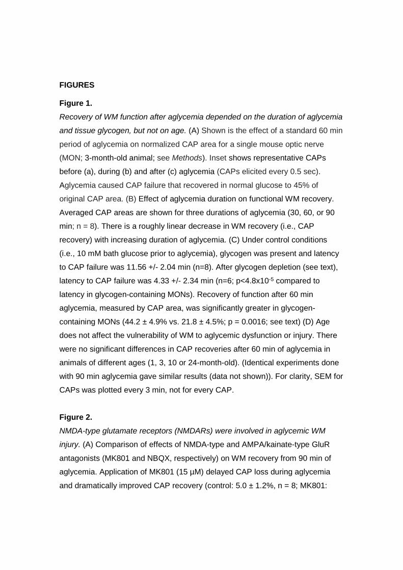
Figure 1.
Recovery of WM function after aglycemia depended on the duration of aglycemia
and tissue glycogen, but not on age. (A) Shown is the effect of a standard 60 min
period of aglycemia on normalized CAP area for a single mouse optic nerve
(MON; 3-month-old animal; see Methods). Inset shows representative CAPs
before (a), during (b) and after (c) aglycemia (CAPs elicited every 0.5 sec).
Aglycemia caused CAP failure that recovered in normal glucose to 45% of
original CAP area. (B) Effect of aglycemia duration on functional WM recovery.
Averaged CAP areas are shown for three durations of aglycemia (30, 60, or 90
min; n = 8). There is a roughly linear decrease in WM recovery (i.e., CAP
recovery) with increasing duration of aglycemia. (C) Under control conditions
(i.e., 10 mM bath glucose prior to aglycemia), glycogen was present and latency
to CAP failure was 11.56 +/- 2.04 min (n=8). After glycogen depletion (see text),
latency to CAP failure was 4.33 +/- 2.34 min (n=6; p<4.8x10⁻⁵ compared to
latency in glycogen-containing MONs). Recovery of function after 60 min
aglycemia, measured by CAP area, was significantly greater in glycogen-
containing MONs (44.2 ± 4.9% vs. 21.8 ± 4.5%; p = 0.0016; see text) (D) Age
does not affect the vulnerability of WM to aglycemic dysfunction or injury. There
were no significant differences in CAP recoveries after 60 min of aglycemia in
animals of different ages (1, 3, 10 or 24-month-old). (Identical experiments done
with 90 min aglycemia gave similar results (data not shown)). For clarity, SEM for
CAPs was plotted every 3 min, not for every CAP.
Figure 2.
NMDA-type glutamate receptors (NMDARs) were involved in aglycemic WM
injury. (A) Comparison of effects of NMDA-type and AMPA/kainate-type GluR
antagonists (MK801 and NBQX, respectively) on WM recovery from 90 min of
aglycemia. Application of MK801 (15 µM) delayed CAP loss during aglycemia
and dramatically improved CAP recovery (control: 5.0 ± 1.2%, n = 8; MK801:
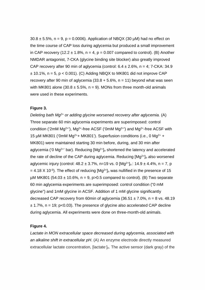
30.8 ± 5.5%, n = 9, p = 0.0006). Application of NBQX (30 µM) had no effect on
the time course of CAP loss during aglycemia but produced a small improvement
in CAP recovery (12.2 ± 1.8%, n = 4, p = 0.007 compared to control). (B) Another
NMDAR antagonist, 7-CKA (glycine binding site blocker) also greatly improved
CAP recovery after 90 min of aglycemia (control: 6.4 ± 2.6%, n = 4; 7-CKA: 34.9
± 10.1%, n = 5, p < 0.001). (C) Adding NBQX to MK801 did not improve CAP
recovery after 90 min of aglycemia (33.8 + 5.6%, n = 11) beyond what was seen
with MK801 alone (30.8 ± 5.5%, n = 9). MONs from three month-old animals
were used in these experiments.
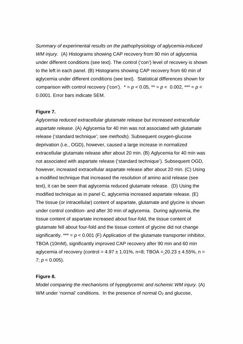
Figure 3.
Deleting bath Mg2+ or adding glycine worsened recovery after aglycemia. (A)
Three separate 60 min aglycemia experiments are superimposed: control
condition (‘2mM Mg2+’), Mg2+-free ACSF (‘0mM Mg2+’) and Mg2+-free ACSF with
15 µM MK801 (‘0mM Mg2++ MK801’). Superfusion conditions (i.e., 0 Mg2+ +
MK801) were maintained starting 30 min before, during, and 30 min after
aglycemia (‘0 Mg2+’ bar). Reducing [Mg2+]o shortened the latency and accelerated
the rate of decline of the CAP during aglycemia. Reducing [Mg2+]o also worsened
aglycemic injury (control: 48.2 ± 3.7%, n=19 vs. 0 [Mg2+]o : 14.9 ± 4.4%, n = 7, p
= 4.18 X 10⁻⁵). The effect of reducing [Mg2+]o was nullified in the presence of 15
µM MK801 (54.03 ± 10.6%, n = 9, p>0.5 compared to control). (B) Two separate
60 min aglycemia experiments are superimposed: control condition (“0 mM
glycine”) and 1mM glycine in ACSF. Addition of 1 mM glycine significantly
decreased CAP recovery from 60min of aglycemia (36.51 ± 7.0%, n = 8 vs. 48.19
± 1.7%, n = 19; p<0.03). The presence of glycine also accelerated CAP decline
during aglycemia. All experiments were done on three-month-old animals.
Figure 4.
Lactate in MON extracellular space decreased during aglycemia, associated with
an alkaline shift in extracellular pH. (A) An enzyme electrode directly measured
extracellular lactate concentration, [lactate-]o. The active sensor (dark gray) of the

enzyme electrode was pressed alongside the optic nerve. Stimulus-evoked CAPs
were simultaneously elicited and recorded via suction electrodes. (B) The
[lactate-]o was zero in the bath (not shown) but averaged about 0.5 mM at the
edge of the optic nerve. After about 5 minutes of aglycemia, [lactate-]o fell steeply
and reached nearly zero after about 10 to 15 min. When glucose is reintroduced,
[lactate-]o rapidly increases and briefly overshoots the original baseline level,
before returning to a level that is very close to the original value. (C) Extracellular
pH (pHo) within the nerve (~7.2), measured with a pH-sensitive microelectrode,
was about 0.2 pH unit lower than bath pH (7.4). During aglycemia, pHo
increased with a time course that mirrored the drop in [lactate-]o (a typical
[lactate-]o trace is superimposed for comparison). The pHo during aglycemia
plateaued at ~7.4. When glucose was reintroduced, pHo underwent an acid shift
with a similar time course to the increase in [lactate-]o. A typical CAP trace during
aglycemia is shown for comparison (see text). (D) Acidifying the ACSF (by
lowering the concentration of HCO3-) from pH 7.4 to 7.0 improved CAP recovery
after 90 min of aglycemia from 4.97 ± 1.23% (n = 8) to 13.38 ± 2.9% (n = 6; p =
0.012). The recovery segment of this experiment is expanded in the inset.
Figure 5.
Aglycemia-induced WM injury depended on extracellular Ca2+. MONs were
exposed to aglycemia in normal ACSF (containing 2mM Ca2+) or in ACSF with no
Ca2+ (Mg2+ was adjusted to 4mM to maintain constant divalent cation
concentration). The Ca2+-free condition was started 15 min before aglycemia and
discontinued 15 min after aglycemia. Note that the 0 Ca2+ ACSF caused a small
decrease in CAP area when introduced. (A) Perfusion with Ca2+-free ACSF led to
nearly complete CAP recovery from 90 min aglycemia in 3-month-old MONs
(control recovery: 4.97 ± 1.2%, n = 8 vs. Ca2+-free: 86.93 ± 3.6%, n = 4;
p<0.00001) and (B) in 10-month-old MONs (control: 6.86 ± 2.06%, n = 5 vs.
Ca2+-free: 84.54 ± 9.8%, n = 6; p< 0.00002).
Figure 6.
Summary of experimental results on the pathophysiology of aglycemia-induced
WM injury. (A) Histograms showing CAP recovery from 90 min of aglycemia
under different conditions (see text). The control (‘con’) level of recovery is shown
to the left in each panel. (B) Histograms showing CAP recovery from 60 min of
aglycemia under different conditions (see text). Statistical differences shown for
comparison with control recovery (‘con’). * = p < 0.05, ** = p < 0.002, *** = p <
0.0001. Error bars indicate SEM.
Figure 7.
Aglycemia reduced extracellular glutamate release but increased extracellular
aspartate release. (A) Aglycemia for 40 min was not associated with glutamate
release (‘standard technique’; see methods). Subsequent oxygen-glucose
deprivation (i.e., OGD), however, caused a large increase in normalized
extracellular glutamate release after about 20 min. (B) Aglycemia for 40 min was
not associated with aspartate release (‘standard technique’). Subsequent OGD,
however, increased extracellular aspartate release after about 20 min. (C) Using
a modified technique that increased the resolution of amino acid release (see
text), it can be seen that aglycemia reduced glutamate release. (D) Using the
modified technique as in panel C, aglycemia increased aspartate release. (E)
The tissue (or intracellular) content of aspartate, glutamate and glycine is shown
under control condition- and after 30 min of aglycemia. During aglycemia, the
tissue content of aspartate increased about four-fold, the tissue content of
glutamate fell about four-fold and the tissue content of glycine did not change
significantly. *** = p < 0.001 (F) Application of the glutamate transporter inhibitor,
TBOA (10mM), significantly improved CAP recovery after 90 min and 60 min
aglycemia of recovery (control = 4.97 ± 1.01%, n=8; TBOA = 20.23 ± 4.55%, n =
7; p < 0.005).
Figure 8.
Model comparing the mechanisms of hypoglycemic and ischemic WM injury. (A)
WM under ‘normal’ conditions. In the presence of normal O2 and glucose,
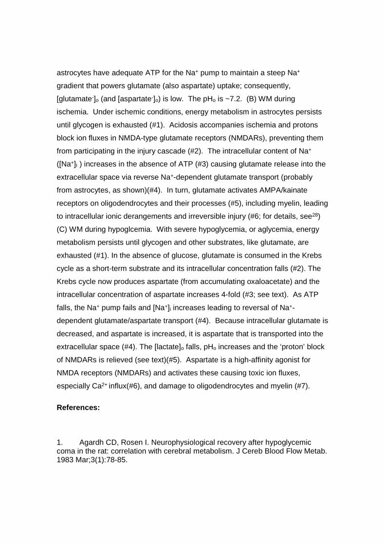
astrocytes have adequate ATP for the Na+ pump to maintain a steep Na+
gradient that powers glutamate (also aspartate) uptake; consequently,
[glutamate-]o (and [aspartate-]o) is low. The pHo is ~7.2. (B) WM during
ischemia. Under ischemic conditions, energy metabolism in astrocytes persists
until glycogen is exhausted (#1). Acidosis accompanies ischemia and protons
block ion fluxes in NMDA-type glutamate receptors (NMDARs), preventing them
from participating in the injury cascade (#2). The intracellular content of Na+
([Na+]I ) increases in the absence of ATP (#3) causing glutamate release into the
extracellular space via reverse Na+-dependent glutamate transport (probably
from astrocytes, as shown)(#4). In turn, glutamate activates AMPA/kainate
receptors on oligodendrocytes and their processes (#5), including myelin, leading
to intracellular ionic derangements and irreversible injury (#6; for details, see28)
(C) WM during hypoglcemia. With severe hypoglycemia, or aglycemia, energy
metabolism persists until glycogen and other substrates, like glutamate, are
exhausted (#1). In the absence of glucose, glutamate is consumed in the Krebs
cycle as a short-term substrate and its intracellular concentration falls (#2). The
Krebs cycle now produces aspartate (from accumulating oxaloacetate) and the
intracellular concentration of aspartate increases 4-fold (#3; see text). As ATP
falls, the Na+ pump fails and [Na+]i increases leading to reversal of Na+-
dependent glutamate/aspartate transport (#4). Because intracellular glutamate is
decreased, and aspartate is increased, it is aspartate that is transported into the
extracellular space (#4). The [lactate]o falls, pHo increases and the ‘proton’ block
of NMDARs is relieved (see text)(#5). Aspartate is a high-affinity agonist for
NMDA receptors (NMDARs) and activates these causing toxic ion fluxes,
especially Ca2+ influx(#6), and damage to oligodendrocytes and myelin (#7).
References:
1. Agardh CD, Rosen I. Neurophysiological recovery after hypoglycemiccoma in the rat: correlation with cerebral metabolism. J Cereb Blood Flow Metab.1983 Mar;3(1):78-85.
2. Arky RA. Hypoglycemia associated with liver disease and ethanol.Endocrinology and metabolism clinics of North America. 1989 Mar;18(1):75-90.3. Davis EA, Jones TW. Hypoglycemia in children with diabetes: incidence,counterregulation and cognitive dysfunction. Journal of pediatric endocrinology &metabolism : JPEM. 1998 Mar;11 Suppl 1:177-82.4. Lincoln NB, Faleiro RM, Kelly C, Kirk BA, Jeffcoate WJ. Effect of long-termglycemic control on cognitive function. Diabetes care. 1996 Jun;19(6):656-8.5. Auer RN. Hypoglycemic brain damage. Metab Brain Dis. 2004 Dec;19(3-4):169-75.6. Brown AM, Wender R, Ransom BR. Ionic mechanisms of aglycemic axoninjury in mammalian central white matter. J Cereb Blood Flow Metab.2001;21(4):385-95.7. Warren RE, Frier BM. Hypoglycaemia and cognitive function. Diabetes,obesity & metabolism. 2005 Sep;7(5):493-503.8. McCrimmon RJ, Ryan CM, Frier BM. Diabetes and cognitive dysfunction.Lancet. 2012 Jun 16;379(9833):2291-9.9. Ma JH, Kim YJ, Yoo WJ, et al. MR imaging of hypoglycemicencephalopathy: lesion distribution and prognosis prediction by diffusion-weighted imaging. Neuroradiology. 2009 Oct;51(10):641-9.10. Johkura K, Nakae Y, Kudo Y, Yoshida TN, Kuroiwa Y. Early diffusion MRimaging findings and short-term outcome in comatose patients withhypoglycemia. AJNR Am J Neuroradiol. 2012 May;33(5):904-9.11. Ransom BR, Baltan SB. Axons get excited to death. Ann Neurol. 2009Feb;65(2):120-1.12. Matute C, Domercq M, Perez-Samartin A, Ransom BR. Protecting WhiteMatter From Stroke Injury. Stroke. 2012 Dec 4.13. Stys PK, Ransom BR, Waxman SG. Compound action potential of nerverecorded by suction electrode: a theoretical and experimental analysis. BrainRes. 1991;546(1):18-32.14. Li S, Stys PK. Mechanisms of ionotropic glutamate receptor-mediatedexcitotoxicity in isolated spinal cord white matter. J Neurosci. 2000;20(3):1190-8.15. Peng W, Cotrina ML, Han X, et al. Systemic administration of anantagonist of the ATP-sensitive receptor P2X7 improves recovery after spinalcord injury. Proc Natl Acad Sci U S A. 2009 Jul 28;106(30):12489-93.16. Fern R, Moller T. Rapid ischemic cell death in immature oligodendrocytes:a fatal glutamate release feedback loop. J Neurosci. 2000 Jan 1;20(1):34-42.17. Brown AM, Westenbroek RE, Catterall WA, Ransom BR. Axonal L-typeCa2+ channels and anoxic injury in rat CNS white matter. J Neurophysiol. 2001Feb;85(2):900-11.18. Baltan S, Besancon EF, Mbow B, Ye Z, Hamner MA, Ransom BR. Whitematter vulnerability to ischemic injury increases with age because of enhancedexcitotoxicity. J Neurosci. 2008 Feb 6;28(6):1479-89.19. McDonald JW, Althomsons SP, Hyrc KL, Choi DW, Goldberg MP.Oligodendrocytes from forebrain are highly vulnerable to AMPA/kainate receptor-mediated excitotoxicity. Nat Med. 1998 Mar;4(3):291-7.
20. Sanchez-Gomez MV, Matute C. AMPA and kainate receptors eachmediate excitotoxicity in oligodendroglial cultures. Neurobiol Dis. 1999Dec;6(6):475-85.21. Alberdi E, Sanchez-Gomez MV, Marino A, Matute C. Ca(2+) influx throughAMPA or kainate receptors alone is sufficient to initiate excitotoxicity in culturedoligodendrocytes. Neurobiol Dis. 2002 Mar;9(2):234-43.22. Tekkok SB, Goldberg MP. AMPA/Kainate receptor activation mediateshypoxic oligodendrocyte death and axonal injury in cerebral white matter. JNeurosci. 2001 Jun 15;21(12):4237-48.23. Bakiri Y, Hamilton NB, Karadottir R, Attwell D. Testing NMDA receptorblock as a therapeutic strategy for reducing ischaemic damage to CNS whitematter. Glia. 2008 Jan 15;56(2):233-40.24. Micu I, Jiang Q, Coderre E, et al. NMDA receptors mediate calciumaccumulation in myelin during chemical ischaemia. Nature. 2006 Feb23;439(7079):988-92.25. Li S, Mealing GA, Morley P, Stys PK. Novel injury mechanism in anoxiaand trauma of spinal cord white matter: glutamate release via reverse Na(+)-dependent glutamate transport. J Neurosci. 1999;19(14):RC16.26. Hamner MA, Moller T, Ransom BR. Anaerobic function of CNS whitematter declines with age. Journal of cerebral blood flow and metabolism : officialjournal of the International Society of Cerebral Blood Flow and Metabolism. 2011Apr;31(4):996-1002.27. Borrelli MJ, Carlini WG, Dewey WC, Ransom BR. A simple method formaking ion-selective microelectrodes suitable for intracellular recording invertebrate cells. J Neurosci Methods. 1985;15(2):141-54.28. Tekkok SB, Ye Z, Ransom BR. Excitotoxic mechanisms of ischemic injuryin myelinated white matter. J Cereb Blood Flow Metab. 2007 Sep;27(9):1540-52.29. Ye ZC, Ransom BR, Sontheimer H. (1R,3S)-1-Aminocyclopentane-1,3-dicarboxylic acid (RS-ACPD) reduces intracellular glutamate levels in astrocytes.J Neurochem. 2001 Nov;79(4):756-66.30. Wender R, Brown AM, Fern R, Swanson RA, Farrell K, Ransom BR.Astrocytic glycogen influences axon function and survival during glucosedeprivation in central white matter. J Neurosci. 2000;20(18):6804-10.31. Brown AM, Tekkok SB, Ransom BR. Glycogen regulation and functionalrole in mouse white matter. J Physiol. 2003 Jun 1;549(Pt 2):501-12.32. Underhill SM, Goldberg MP. Hypoxic injury of isolated axons isindependent of ionotropic glutamate receptors. Neurobiol Dis. 2007Feb;25(2):284-90.33. Mathiesen C, Varming T, Jensen LH. In vivo and in vitro evaluation ofAMPA receptor antagonists in rat hippocampal neurones and cultured mousecortical neurones. Eur J Pharmacol. 1998 Jul 24;353(2-3):159-67.34. Traynelis SF, Wollmuth LP, McBain CJ, et al. Glutamate receptor ionchannels: structure, regulation, and function. Pharmacological reviews. 2010Sep;62(3):405-96.
35. Brown AM, Evans RD, Black J, Ransom BR. Schwann cell glycogenselectively supports myelinated axon function. Ann Neurol. 2012 Sep;72(3):406-18.36. Lee J, Taira T, Pihlaja P, Ransom BR, Kaila K. Effects of CO2 onexcitatory transmission apparently caused by changes in intracellular pH in therat hippocampal slice. Brain Res. 1996;706(2):210-6.37. Traynelis SF, Cull-Candy SG. Proton inhibition of N-methyl-D-aspartatereceptors in cerebellar neurons. Nature. 1990;345(6273):347-50.38. McCrimmon RJ, Frier BM. Hypoglycaemia, the most feared complicationof insulin therapy. Diabete Metab. 1994 Nov-Dec;20(6):503-12.39. Zhang K, Sejnowski TJ. A universal scaling law between gray matter andwhite matter of cerebral cortex. Proc Natl Acad Sci U S A. 2000 May9;97(10):5621-6.40. Ransom BR, Goldberg MP, Baltan SB. Molecular pathophysiology of whitematter anoxic-ischemic injury. In: Mohr JP, editor. Stroke: pathophysiology,diagnosis and management. 5th edition ed. Philadelphia, PA: Elsevier/Saunders;2011. p. 122-37.41. Hansen AJ. Effect of anoxia on ion distribution in the brain. Physiol Rev.1985;65(1):101-48.42. Brown AM, Wender R, Ransom BR. Metabolic substrates other thanglucose support axon function in central white matter. J Neurosci Res. 2001 Dec1;66(5):839-43.43. Agardh CD, Folbergrova J, Siesjo BK. Cerebral metabolic changes inprofound, insulin-induced hypoglycemia, and in the recovery period followingglucose administration. J Neurochem. 1978 Nov;31(5):1135-42.44. Pelligrino D, Siesjo BK. Regulation of extra- and intracellular pH in thebrain in severe hypoglycemia. Journal of cerebral blood flow and metabolism :official journal of the International Society of Cerebral Blood Flow andMetabolism. 1981;1(1):85-96.45. Sandberg M, Butcher SP, Hagberg H. Extracellular overflow ofneuroactive amino acids during severe insulin-induced hypoglycemia: in vivodialysis of the rat hippocampus. J Neurochem. 1986 Jul;47(1):178-84.46. Salter MG, Fern R. NMDA receptors are expressed in developingoligodendrocyte processes and mediate injury. Nature. 2005 Dec22;438(7071):1167-71.47. Rothman SM, Olney JW. Glutamate and the pathophysiology of hypoxic--ischemic brain damage. Ann Neurol. 1986;19(2):105-11.48. Stys PK, Waxman SG, Ransom BR. Na(+)-Ca2+ exchanger mediatesCa2+ influx during anoxia in mammalian central nervous system white matter.Ann Neurol. 1991;30(3):375-80.49. Suh SW, Hamby AM, Swanson RA. Hypoglycemia, brain energetics, andhypoglycemic neuronal death. Glia. 2007 Sep;55(12):1280-6.50. Nellgard B, Wieloch T. Cerebral protection by AMPA- and NMDA-receptorantagonists administered after severe insulin-induced hypoglycemia. Exp BrainRes. 1992;92(2):259-66.