X-linked myotubular myopathy in Rottweiler dogs is caused by a ...
X-linked myotubular myopathy in Rottweiler dogsis caused by a missense mutation in Exon 11 ofthe MTM1 geneShelton et al.
Shelton et al. Skeletal Muscle (2015) 5:1 DOI 10.1186/s13395-014-0025-3

Shelton et al. Skeletal Muscle (2015) 5:1 DOI 10.1186/s13395-014-0025-3
RESEARCH Open Access
X-linked myotubular myopathy in Rottweiler dogsis caused by a missense mutation in Exon 11 ofthe MTM1 geneG Diane Shelton1, Branden E Rider2, Georgina Child3, Sophia Tzannes3, Ling T Guo1, Behzad Moghadaszadeh2,Emily C Troiano2, Bianca Haase4, Claire M Wade4 and Alan H Beggs2*
Abstract
Background: Congenital and inherited myopathies in dogs are faithful models of human muscle diseases and arebeing recognized with increasing frequency. In fact, canine models of dystrophin deficient muscular dystrophy andX-linked myotubular myopathy are of tremendous value in the translation of new and promising therapies for thetreatment of these diseases. We have recently identified a family of Australian Rottweilers in which male puppieswere clinically affected with severe muscle weakness and atrophy that resulted in early euthanasia or death. X-linkedmyotubular myopathy was suspected based on the early and severe clinical presentation and histopathologicalchanges within muscle biopsies. The aim of this study was to determine the genetic basis for myopathy in these dogsand compare and contrast the clinical presentation, histopathology, ultrastructure, and mutation in this family ofRottweiler dogs with the previously described myotubular myopathy in Labrador retrievers.
Results: Histopathology, histochemistry, and ultrastructural examination of muscle biopsies from affected Rottweilerpuppies were consistent with an X-linked myotubular myopathy. An unusual finding that differed from the previouslyreported Labradors and similar human cases was the presence of excessive autophagy and prominent autophagicvacuoles. Molecular investigations confirmed a missense mutation in exon 11 of MTM1 that was predicted to result ina non-functional phosphatase activity. Although the clinical presentations and histopathology were similar, the MTM1p.(Q384P) mutation is different from the p.(N155K) mutation in exon 7 affecting Labrador retrievers with X-linkedmyotubular myopathy.
Conclusions: Here we describe a second pathogenic mutation in MTM1 causing X-linked myotubular myopathy indogs. Our findings suggest a variety of MTM1 mutations in dogs as seen in human patients. The number of MTM1mutations resulting in similar severe and progressive clinical myopathy and histopathological changes are likely toincrease as canine myopathies are further characterized.
Keywords: Congenital myopathy, Myotubularin, Canine myopathy, Animal model
BackgroundThe centronuclear myopathies (CNMs) are a group ofpathologically defined disorders that characteristicallyhave a high proportion of small myofibers with centrallyplaced nuclei [1-3]. Classical centronuclear myopathiesin humans have been associated with dominant mutationsin the large GTPase DNM2 gene [4] while recessive cases
* Correspondence: [email protected] of Genetics and Program in Genomics, The Manton Center forOrphan Disease Research, Boston Children’s Hospital and Harvard MedicalSchool, 300 Longwood Ave., Boston, MA 02115, USAFull list of author information is available at the end of the article
© 2015 Shelton et al.; licensee BioMed CentraCommons Attribution License (http://creativecreproduction in any medium, provided the orDedication waiver (http://creativecommons.orunless otherwise stated.
may be due to mutations of amphiphysin 2, encoded byBIN1 [5], and of the ryanodine receptor (RyR1) gene,RYR1 [6]. An important, well-defined sub-group of CNMsis X-linked myotubular myopathy (XLMTM) associatedwith mutations in the MTM1 gene [7]. The protein product,myotubularin, is a ubiquitously expressed 603 amino acidphosphoinositide phosphatase that is essential in a widevariety of cellular signaling pathways governing diversecellular functions including apoptosis and vesicle trafficking[8]. In skeletal muscle, myotubularin is localized at theterminal cisternae of the sarcoplasmic reticulum (SR) where
l. This is an Open Access article distributed under the terms of the Creativeommons.org/licenses/by/4.0), which permits unrestricted use, distribution, andiginal work is properly credited. The Creative Commons Public Domaing/publicdomain/zero/1.0/) applies to the data made available in this article,
Shelton et al. Skeletal Muscle (2015) 5:1 Page 2 of 12
its function is critical for regulating the lipid composition ofmembranes at the triads [9]. Abnormal membranetubulation in myotubularin-deficient muscle leads toaltered T-tubule and triad morphology and defectiveexcitation contraction coupling, which is thought toplay a major role in the initial pathophysiologicalevents leading to weakness [10,11]. Clinical onset ofXLMTM is at or near birth with males born withsevere generalized hypotonia and weakness with respiratoryinsufficiency [12]. Affected infants are typically areflexicand hypotonic with little or no anti-gravity movements.The condition is not dystrophic as myofibers remain largelyintact and serum creatine kinase (CK) activities are withinthe reference range or only mildly elevated.In veterinary medicine, the first mutation associated
with classical CNM was identified in young Labradorretrievers. A widely distributed exonic SINE insertion inthe PTPLA gene segregated with autosomal recessiveCNM in these dogs [13]. Mutations in the PTPLA genehave not been reported to date in human cases of CNM,however, mutation in the BIN1 exon 11 acceptor splicesite was recently shown to be the cause of a rapidlyprogressive and fatal centronuclear myopathy in both aconsanguineous human family and in young Great Danedogs with a progressive CNM [14]. While specific mutationshave not yet been identified, CNM has also been reportedin a family of young Manchester Terriers (G.D. Shelton,unpublished observations), in a young Border Collie [15],and in an Arabian foal [16].In 2008, a form of CNM resembling XLMTM was
described in a family of Labrador Retrievers fromCanada [17]. Molecular investigation confirmed that thesedogs indeed did have an MTM1 mutation and representeda faithful model of human XLMTM [18]. Male puppiesfrom two kindreds were presented for evaluation ofprogressive weakness and muscle atrophy beginningin the first few months of life. Pathological changesin muscle biopsies were consistent with a myotubularmyopathy and a missense mutation, p.(N155K), wasidentified in exon 7 of canine MTM1. Here we de-scribe, compare, and contrast the clinical presentation,histopathology, muscle ultrastructure, and a second inde-pendent mutation of MTM1 in a family of Rottweiler dogsfrom Australia.
MethodsAnimalsAll dogs reported in this communication were privatelyowned pets receiving clinical veterinary care and noexperimental procedures were carried out on live animals.All research on tissue biopsies and blood samples wasperformed on specimens collected and submitted for clin-ical diagnostic purposes and the owners and breederswere informed and gave consent to study tissues for
the welfare of the breed. Four male puppies, aged 8to 13 weeks, were presented to the Small AnimalSpecialist Hospital, North Ryde, Australia (GC and ST) forevaluation of progressive muscle weakness. The affectedpuppies were from two different litters with the same damand sire (Figure 1). The sire of both litters had sired twoother litters from different dams with no reportedabnormalities in the offspring. DNA for genetic testingwas collected from all four affected puppies and 10unaffected first-degree relatives. Additional unaffectedcontrol samples were obtained from The RottweilerBlood Bank, The University of Manchester, Manchester,UK, The Faculty of Veterinary Science, University ofSydney, Australia, and the Animal Specialist Hospital,North Ryde, Australia. None of the dogs in the unaffectedcontrol groups showed clinical signs of a neuromusculardisease.
Histopathology, histochemistry, and immunofluorescencestainingImmediately after collection, muscle biopsy specimenswere either wrapped in a saline-dampened gauze spongeand immediately refrigerated or immersion-fixed in 10%neutral buffered formalin. All specimens were shipped bya courier service under refrigeration to the ComparativeNeuromuscular Laboratory, University of California SanDiego. Immediately on receipt, unfixed specimens wereflash-frozen in isopentane precooled in liquid nitrogenand stored at -80°C until further processing. Control mus-cles were all histologically normal specimens from the fro-zen tissue archive of the Shelton laboratory. Cryosections(8 μm) were cut and processed by a standard panel ofhistochemical stains and reactions [19]. Mouse monoclonalantibodies against slow (1:10, NCL-MHCs, Novocastra,Newcastle, England) and fast (1:10, NCL-MHCf, Novocastra)myosin heavy chains were used for fiber typing. Peroxidasesubstrate kits (DAB, Vector SK-4100, Vector Laboratories,Burlingame, CA, USA) and Vector red substrate kits (Vector,SK-5100) were used for color development according topackage instructions.Additional cryosections (8 μm) were cut and processed
by indirect immunofluorescence. Sections were fixed inchilled acetone for 5 min, then incubated with a rabbitpolyclonal antibody against the T-tubule marker, dihy-dropyridine receptor (DHPR), DHPRα1 (ab58552, 1:100dilution, Abcam, Cambridge, UK) and a mouse mono-clonal antibody against the SR marker, RyR1 (ab2827,1:100 dilution; Abcam, Cambridge, UK). Rabbit poly-clonal antibodies against LC3 N terminal (1:100,Abgent AP1802a, Abgent, San Diego, CA, USA) andubiquitin (1:250, Dako 2015-02, Dako Denmark A/S,Glostrup, Denmark) were used as markers for au-tophagy. Details of further processing are as previouslydescribed [18].
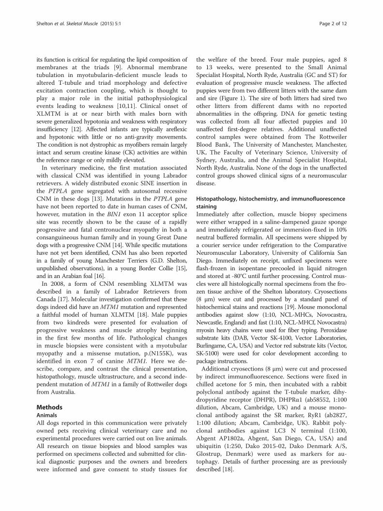
Figure 1 Clinical presentations and family history of affected dogs. Photographs of the healthy dam II-2 (A), sire II-3 (B), and two affectedmale littermates III-12 (C) and III-13 (D) at 12 weeks of age. (E) Pedigree of the two affected Rottweiler litters with male puppies that presentedwith progressive muscle weakness. The 15 members whose DNA was available for analysis are indicated by an asterisk. Squares =males,circles = females, SB = still born. Filled symbols indicate clinically affected. The dot in the center of II-2 indicates her genetically confirmedstatus as a carrier for the MTM1 mutation illustrated below.
Shelton et al. Skeletal Muscle (2015) 5:1 Page 3 of 12
Electron microscopy and quantification of triad structuresFollowing collection, muscles were immersed in ice cold5% glutaraldehyde for 4 h, cut into 2 to 3 mm3 blocks,and fixed for an additional 2 h before post-fixation in1% osmium tetroxide for 1 h. The samples weredehydrated in increasing ethanol concentrations andfinally propylene oxide, and then embedded in ara-ldite resin. Thick sections (1 μm) were prepared andexamined by light microscopy after staining withtoluidine blue-basic fuchsin. Ultra-thin sections (60 to65 nm) were cut and collected on 100 or 200 squaremesh grids, stained with uranyl acetate and lead cit-rate, and examined with a Zeiss 10 electron micro-scope. For quantification of triad structures in arepresentative affected dog and an age-matched con-trol dog, longitudinal sections were evaluated at 9,900magnification. A triad structure was defined as thetypical T-tubule density and at least one adjacent cis-tern. The first 10 fields encountered with no artifactwere photographed and the number of triads per fieldcounted.
DNA extraction and MTM1 gene sequencing and analysisGenomic DNA was isolated from muscle biopsies andEDTA anticoagulated peripheral blood by standard proce-dures. The 15 transcribed exons, plus 81 to 278 bp offlanking intronic sequences, of the canine MTM1 genewere polymerase chain reaction (PCR) amplified andSanger sequenced using primer set 2 as described [18].The canine MTM1 c.1151A > C variant was screened forin relatives of the proband and unaffected control popula-tions using a Custom TaqMan® SNP Genotyping Assay(Life Technologies custom assay AH89RBZ, ThermoFisher Scientific, Waltham, MA USA) on an ABI 7300 realtime PCR platform (Thermo Fisher Scientific). CanineDNA and protein sequence variants are specified relativeto reference sequences XM_850116.3 and XP_855209.1where cDNA numbering is based on designating the first‘A’ of the initiating methionine as position 1.
Variant interpretation and mutational modelingPotential pathogenic consequences of theMTM1 c.1151A >C variant were assessed using SIFT (http://sift.jcvi.org/) [20],

Shelton et al. Skeletal Muscle (2015) 5:1 Page 4 of 12
PolyPhen 2 v2.2.2 (http://genetics.bwh.harvard.edu/pph2/)[21], and SuSPect (http://www.sbg.bio.ic.ac.uk/suspect/) [22]and effects on protein structure were modeled using Phyre2(http://www.sbg.bio.ic.ac.uk/phyre2/) [23] and visualizedusing Jmol, an open-source JAVA viewer for chemicalstructures in 3D (http://www.jmol.org/). Amino acidalignments were performed using the ClustalW algorithmin MacVector version 13.04 (MacVector Inc., Cary NC,USA), using the following reference sequences for com-parison. For myotubularins: human (Homo sapiens),NP_000243.1; monkey (Macaca mulatta), NP_001248647;dog (Canis familiaris), XP_005641991; cow (Bos taurus),NP_001193354; chicken (Gallus gallus), XP_004940916;xenopus (Xenopus tropicalis), AAH94184.1; zebra fish(Danio rerio), NP_001032773.1; drosophila (Drosophilamelanogaster), AAF52327.1. For myotubularin-relatedproteins: MTM1, NP_000243.1; MTMR1, NP_003819.1;MTMR2, Q13614.4; MTMR3, CAG30411.1; MTMR4,NP_004678.3; MTMR5, O95248.3; MTMR6, NP_004676.3;MTMR7, EAW63816.1; MTMR8, Q96EF0.1; MTMR9,Q96QG7.1; MTMR10, Q9NXD2.3; MTMR11, A4FU01.2;MTMR12, Q9C0I1.2; MTMR13, Q86WG5.1.
ResultsSevere and progressive myopathy in related litters ofRottweiler puppiesA 13-week-old male Rottweiler (III-2 from Figure 1)from a litter of 10 puppies (6 females and 4 males) wasreferred for progressive weakness. Two pups from thisfirst litter were previously lost; the first was a female thatdied within a few weeks of birth (unknown cause), andthe second a male pup (III-1) euthanized by the attendingveterinarian at 9 weeks of age for an initial alteration infacial expression and exercise intolerance progressing toan inability to bear weight. In the affected pup evaluated(III-2), clinical signs of weakness had been apparent sinceacquisition from the breeder at 8 weeks of age. Clinicalexamination at referral revealed normal mentation, gener-alized mild to moderate reduction in muscle mass withoutmyalgia, and profound tetraparesis with an inability tosupport weight. Cervical ventroflexion occurred in sternalrecumbency. Spinal pain was absent. Hyporeflexia andhypotonia were noted in all limbs, and were symmetricalin severity. Sensory abnormalities were not detected.There were no cranial nerve deficits. Conscious proprio-ception (proprioceptive paw positioning) was intact. CKwas mildly elevated (2119 U/l, reference 0-400). Multiplemuscle and peripheral nerve biopsies were obtained viaopen surgical biopsy under general anesthesia. Aftersample collection, the puppy was euthanized for humani-tarian reasons and at the owners’ request due to the severenature of the disease.Subsequently, three male Rottweiler puppies (Figure 1E,
puppies III-12, 13, and 14) were presented with a history
of rapidly progressive generalized weakness and poormuscle development. The puppies were from a secondlitter of eight surviving puppies (6 male and 2 female, 1additional male stillborn) produced after the secondmating of the dam and sire of puppy III-2 (Figure 1E).Two of the affected puppies (III-12 and III-13) were pre-sented for evaluation at 11 weeks of age. Both wereregarded as normal with respect to their littermates inactivity level and muscle development until 7 weeks of agewhen the owner noted both puppies showed difficulty inholding their heads up and tired easily. The puppiesremained bright, alert, and normally responsive, were keento play with their littermates but were increasingly unableto do so. Muscle development was poor compared to theirlittermates despite comparable size, a normal appetite,and food intake. Weakness was exacerbated by any stressand cold. The third puppy (III-14) was regarded as normaluntil 9 weeks of age and was reported to be less active at11 weeks of age. This puppy presented 2 weeks after hislittermates (at 13 weeks of age).Clinical findings were similar for all three affected
puppies. All puppies were bright, friendly, and normallyresponsive. Bent or crinkled vibrissae were identified at anearly age, and not apparent in the normal littermates. Poordevelopment or reduced muscle mass compared to normallittermates was noted in the limb muscles, paraspinal mus-cles, and masticatory muscles. The most severely affectedpuppy (Figure 1C, puppy III-12) was unable to stand formore than 10 s. All affected puppies had marked cervicalweakness, were unable to lift their heads when standing(Figure 1D) and were unable to raise the head when ster-nally recumbent after any exertion. The puppies adopted ahunched thoracolumbar posture with a stiff limbed stanceand narrow base when standing. A stiff, short-stride gaitwas evident before sitting or lying down. In a recumbentposition the puppies would follow the activities of otherpuppies or people with their eyes rather than moving theirhead or neck. When ambulatory no evidence of ataxia orproprioceptive deficit was evident. Voluntary motor move-ment was present in all limbs and limb positioning appro-priate, but all showed reduced muscle strength. Whenstanding or encouraged to walk the respiratory rate of allthe affected puppies would increase, they would pant andthe heart rate would increase significantly (>200 bpm).Postural reactions including proprioceptive paw
positioning were normal if the puppies’ weight wassupported. Patellar reflexes were absent in the twomost severely affected puppies (Figure 1E, puppiesIII-12 and III-13) and reduced in the third affectedpuppy (III-14). Flexion reflexes were present in allfour limbs in all puppies but weak in puppies III-12and III-13. Cranial nerve examination was normal in allthree puppies. CK was normal or very mildly increased(104, 348, 277 U/L; reference 0 to 200).

Shelton et al. Skeletal Muscle (2015) 5:1 Page 5 of 12
In view of the severe and rapidly progressive clinicalsigns, and diagnosis of myopathy in a related dog, all threepuppies were euthanized at 11 weeks of age (III-12),12 weeks of age (III-13), and 13 weeks of age (III-14),respectively. Skeletal and cardiac muscle and blood sampleswere collected at necropsy for histology, immunohisto-chemistry, and genetic analysis.
Histopathology, histochemistry, andimmunohistochemical findings suggest affected puppieshave a form of centronuclear myopathy with excessiveautophagyCryosections from the triceps, extensor carpi radialis,tensor fascia lata, cranial tibial, and biceps femoris muscleswere evaluated from puppy III-2 (Figure 2). There was amarked variability in myofiber size in all musclesexamined with fiber diameters in the range of 12 to60 μm (Figure 2A). Normal canine biceps femoriscontains 41% type 1, 58% type 2, and 1% type 2Cfibers [24]. Biceps femoris from the affected dogsdisplayed type 1 fiber predominance (73% type 1 fibers and27% type 2 fibers), which was evident by antibody stainingfor fast and slow myosin heavy chains (Figure 2B). Mosttype 1 fibers were hypotrophic with prominent central
Figure 2 Pathological findings in skeletal muscle from anaffected puppy are suggestive of a diagnosis of XLMTM orother form of CNM. Cryosections of the biceps femoris musclefrom the affected puppy III-2 were evaluated with a standard panelof histochemical stains and reactions. Marked variability in myofibersize was noted (A) with numerous hypotrophic fibers containingcentral nuclei. Most hypotrophic fibers were of type 1 as determinedby staining with antibodies against slow (brown stain, type 1 fibers)and fast (pink stain, type 2 fibers) myosin heavy chains (B). Densecentral and subsarcolemmal deposits were noted with the oxidativereactions SDH (C) and COX (D).
nuclei. The hypotrophic fibers contained central accumula-tions and subsarcolemmal rings, the so-called ‘necklacefibers’, that stained dark blue with the oxidative reactionsnicotinamide adenine dinucleotide - tetrazolium reductase(not shown), succinate dehydrogenase (SDH, Figure 2C)and cytochrome C oxidase (COX, Figure 2D). Necroticfibers were rare. No abnormalities were detected in theradial, femoral, and sciatic nerve sections, and gross andmicroscopic examination of the hearts from several affecteddogs revealed no evidence of cardiomyopathy or otherrecognizable lesions. Based on these pathological changes, acongenital myopathy including centronuclear myopathy ormyotubular myopathy was diagnosed. Cryosections werealso evaluated from the affected male puppies III-12 andIII-14 with similar morphologic findings.Immunofluorescence staining of muscle cryosections
from affected puppies, using antibodies against markersfor T-tubules (DHPRα1) and SR (RyR1), revealed anabnormal localization of both structures compared tocontrol muscle (Figure 3). T-tubules and surroundingSR were concentrated in irregular densities within numer-ous myofibers, but not in the control muscle (Figure 3A).Antibodies against LC3 and ubiquitin, markers forautophagy, showed prominent dense staining in mostmyofibers from the affected puppies but not in controlmuscle (Figure 3B).
Abnormal triads, autophagic vacuoles, and mitochondrialaccumulations were found in affected muscles fromRotweillers with XLMTMUltrastructural examinations (Figure 4) were performedon the biceps femoris muscles of puppies III-12 and III-14.Representative images in Figure 4 are from puppy III-12.Normal appearing triads were not observed in the sectionsfrom the affected puppies, while 18 to 20 triads per fieldwere present in the control dog sections (not shown). Largeareas of myofibrillar disarray were prominent containingdegenerating organelles, and lysosomal and tubular struc-tures (Figure 4A). Centrally located nuclei were confirmed(Figure 4B). Dilated tubular structures (T-tubules and SRprofiles) were present throughout the sections (Figure 4C)and clusters of mitochondria with interspersed glycogengranules were found (Figure 4D). Remarkably, prominentabundant autophagic vacuoles (Figure 4E,F) containingheterogeneous contents including small dense bodiesstudded with glycogen particles, membrane fragments,myeloid structures, and debris were noted.
Identification of a novel MTM1 mutation in affected dogsAll 15 exons and between 81 and 278 bp of flankingintronic sequences of the canine MTM1 gene were PCRamplified from the genomic DNA of one of the affectedpuppies and subjected to Sanger sequencing. The puppy’sMTM1 coding sequence completely matched the published

Figure 3 Abnormal staining patterns for triad proteins and markers of autophagy in skeletal muscles from affected dogs. (A)Immunofluorescence staining of cryosections from the biceps femoris muscles of Rottweiler puppies affected with XLMTM (puppies III-12 andIII-14). Irregular staining patterns were noted in affected puppies using antibodies against DHPRα1 (T-tubule marker) and RyR1 (SR marker)compared to control muscle. (B) Compared to control muscle, dense and irregular staining patterns were also noted following staining withthe autophagy markers LC3 and ubiquitin.
Shelton et al. Skeletal Muscle (2015) 5:1 Page 6 of 12
reference sequence XM_850116.3 with the exception of asingle base substitution of C for A in exon 11 at position1151 on the coding portion of the cDNA, designatedMTM1 c.1151A >C (Figure 5A). This variant is predicted tointroduce a non-conservative missense mutation replacing aglutamine at position 384 with a proline, designatedp.(Q384P). Further PCR amplification and sequencing ofexon 11 in the DNAs from 13 additional first degree rela-tives of the affected puppy, and one unrelated unaffectedcontrol revealed that all four affected males were hemizy-gous for this variant and the dam of these two affected lit-ters was a heterozygous carrier, while all the remaining dogscarried only the wild type A allele at this position.
To investigate the occurrence and potential distribu-tion of the MTM1 c.1151A > C variant among healthyRottweilers, a Custom TaqMan® SNP Genotyping Assay wasdeveloped to distinguish the two alternate alleles. Applica-tion of this assay to the genomic DNAs of 57 Rottweilers(30 male and 27 female) previously tested for rabies titersthrough the United Kingdom’s Pet Travel Scheme [25], aswell as 19 additional Rottweilers from Australia, includingfour females, 14 males, and one of uncertain gender,revealed that all of the 107+ X chromosomes representedby these cohorts carried only the wild type A allele.To assess the likelihood that the non-conservative
replacement of the acidic amino acid glutamine with the
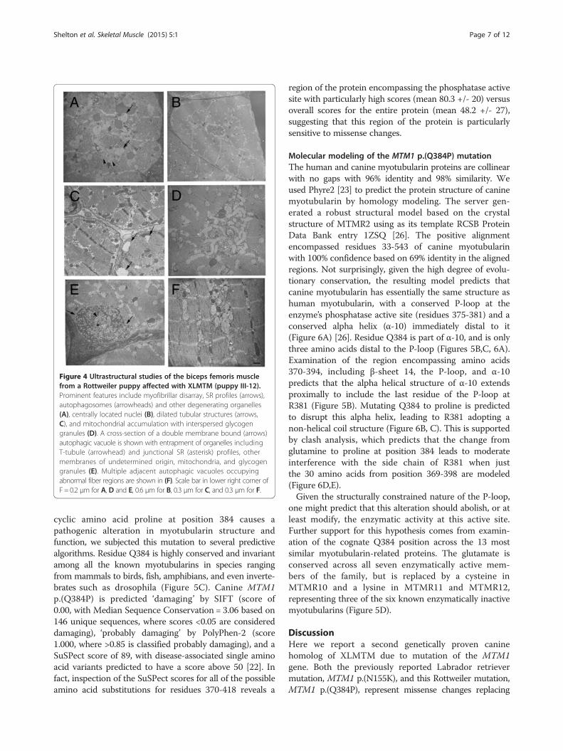
Figure 4 Ultrastructural studies of the biceps femoris musclefrom a Rottweiler puppy affected with XLMTM (puppy III-12).Prominent features include myofibrillar disarray, SR profiles (arrows),autophagosomes (arrowheads) and other degenerating organelles(A), centrally located nuclei (B), dilated tubular structures (arrows,C), and mitochondrial accumulation with interspersed glycogengranules (D). A cross-section of a double membrane bound (arrows)autophagic vacuole is shown with entrapment of organelles includingT-tubule (arrowhead) and junctional SR (asterisk) profiles, othermembranes of undetermined origin, mitochondria, and glycogengranules (E). Multiple adjacent autophagic vacuoles occupyingabnormal fiber regions are shown in (F). Scale bar in lower right corner ofF = 0.2 μm for A, D and E, 0.6 μm for B, 0.3 μm for C, and 0.3 μm for F.
Shelton et al. Skeletal Muscle (2015) 5:1 Page 7 of 12
cyclic amino acid proline at position 384 causes apathogenic alteration in myotubularin structure andfunction, we subjected this mutation to several predictivealgorithms. Residue Q384 is highly conserved and invariantamong all the known myotubularins in species rangingfrom mammals to birds, fish, amphibians, and even inverte-brates such as drosophila (Figure 5C). Canine MTM1p.(Q384P) is predicted ‘damaging’ by SIFT (score of0.00, with Median Sequence Conservation = 3.06 based on146 unique sequences, where scores <0.05 are considereddamaging), ‘probably damaging’ by PolyPhen-2 (score1.000, where >0.85 is classified probably damaging), and aSuSPect score of 89, with disease-associated single aminoacid variants predicted to have a score above 50 [22]. Infact, inspection of the SuSPect scores for all of the possibleamino acid substitutions for residues 370-418 reveals a
region of the protein encompassing the phosphatase activesite with particularly high scores (mean 80.3 +/- 20) versusoverall scores for the entire protein (mean 48.2 +/- 27),suggesting that this region of the protein is particularlysensitive to missense changes.
Molecular modeling of the MTM1 p.(Q384P) mutationThe human and canine myotubularin proteins are collinearwith no gaps with 96% identity and 98% similarity. Weused Phyre2 [23] to predict the protein structure of caninemyotubularin by homology modeling. The server gen-erated a robust structural model based on the crystalstructure of MTMR2 using as its template RCSB ProteinData Bank entry 1ZSQ [26]. The positive alignmentencompassed residues 33-543 of canine myotubularinwith 100% confidence based on 69% identity in the alignedregions. Not surprisingly, given the high degree of evolu-tionary conservation, the resulting model predicts thatcanine myotubularin has essentially the same structure ashuman myotubularin, with a conserved P-loop at theenzyme’s phosphatase active site (residues 375-381) and aconserved alpha helix (α-10) immediately distal to it(Figure 6A) [26]. Residue Q384 is part of α-10, and is onlythree amino acids distal to the P-loop (Figures 5B,C, 6A).Examination of the region encompassing amino acids370-394, including β-sheet 14, the P-loop, and α-10predicts that the alpha helical structure of α-10 extendsproximally to include the last residue of the P-loop atR381 (Figure 5B). Mutating Q384 to proline is predictedto disrupt this alpha helix, leading to R381 adopting anon-helical coil structure (Figure 6B, C). This is supportedby clash analysis, which predicts that the change fromglutamine to proline at position 384 leads to moderateinterference with the side chain of R381 when justthe 30 amino acids from position 369-398 are modeled(Figure 6D,E).Given the structurally constrained nature of the P-loop,
one might predict that this alteration should abolish, or atleast modify, the enzymatic activity at this active site.Further support for this hypothesis comes from examin-ation of the cognate Q384 position across the 13 mostsimilar myotubularin-related proteins. The glutamate isconserved across all seven enzymatically active mem-bers of the family, but is replaced by a cysteine inMTMR10 and a lysine in MTMR11 and MTMR12,representing three of the six known enzymatically inactivemyotubularins (Figure 5D).
DiscussionHere we report a second genetically proven caninehomolog of XLMTM due to mutation of the MTM1gene. Both the previously reported Labrador retrievermutation, MTM1 p.(N155K), and this Rottweiler mutation,MTM1 p.(Q384P), represent missense changes replacing
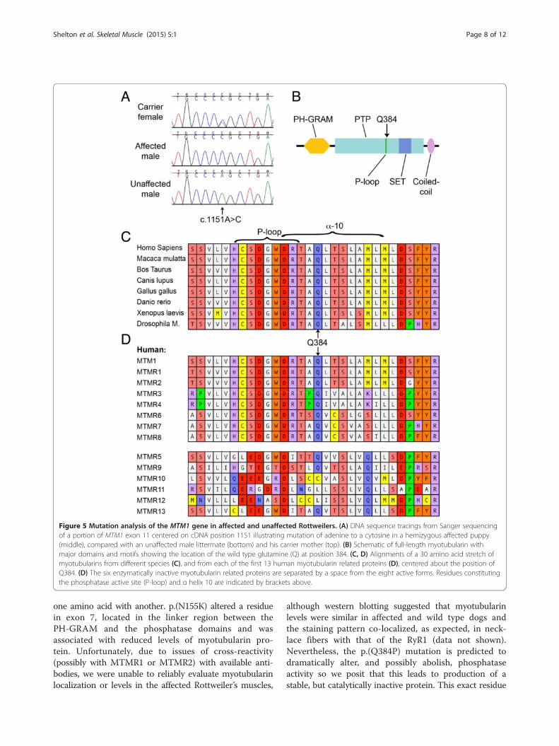
Figure 5 Mutation analysis of the MTM1 gene in affected and unaffected Rottweilers. (A) DNA sequence tracings from Sanger sequencingof a portion of MTM1 exon 11 centered on cDNA position 1151 illustrating mutation of adenine to a cytosine in a hemizygous affected puppy(middle), compared with an unaffected male littermate (bottom) and his carrier mother (top). (B) Schematic of full-length myotubularin withmajor domains and motifs showing the location of the wild type glutamine (Q) at position 384. (C, D) Alignments of a 30 amino acid stretch ofmyotubularins from different species (C), and from each of the first 13 human myotubularin related proteins (D), centered about the position ofQ384. (D) The six enzymatically inactive myotubularin related proteins are separated by a space from the eight active forms. Residues constitutingthe phosphatase active site (P-loop) and α helix 10 are indicated by brackets above.
Shelton et al. Skeletal Muscle (2015) 5:1 Page 8 of 12
one amino acid with another. p.(N155K) altered a residuein exon 7, located in the linker region between thePH-GRAM and the phosphatase domains and wasassociated with reduced levels of myotubularin pro-tein. Unfortunately, due to issues of cross-reactivity(possibly with MTMR1 or MTMR2) with available anti-bodies, we were unable to reliably evaluate myotubularinlocalization or levels in the affected Rottweiler’s muscles,
although western blotting suggested that myotubularinlevels were similar in affected and wild type dogs andthe staining pattern co-localized, as expected, in neck-lace fibers with that of the RyR1 (data not shown).Nevertheless, the p.(Q384P) mutation is predicted todramatically alter, and possibly abolish, phosphataseactivity so we posit that this leads to production of astable, but catalytically inactive protein. This exact residue

Figure 6 Structural predictions of the effects of the MTM1 p.(Q384P) mutation. Jmol visualizations of predicted structural models offull-length canine myotubularin generated by Phyre2 (A) show the PH-GRAM domain (yellow) oriented at the top and the phosphatase domain(light blue) below. The SET and coiled coil domains are shown in blue and purple, respectively (as in Figure 5B). The P-loop is colored green andthe position of Q384 is shown in red. The region encompassing β-sheet 14, the P-loop, and α-10 (residues 370-394), is shown in isolation for thewild type protein (B), and for the p.(Q384P) mutation (C). Note loss of alpha helical structure at arginine 381 (indicated). Repeating the Phyre2 structuralpredictions for just the 30 residue peptide including positions 369-398 and modeling space-filling atoms for positions 381 and 384 illustrates predictedsteric interference between R381 and mutated P394 (E), but not wild type Q384 (D) (arrowheads).
Shelton et al. Skeletal Muscle (2015) 5:1 Page 9 of 12
has not been reported mutated in human patients withXLMTM, however, missense mutations of S376 and G378(both within the P-loop), and S387, A389, and L391(all within the adjacent α-10 helix), have been reported. Of13 patients in the MTM1 Leiden Open Variation database[27] with missense mutation of one of these five residues,10 were reported to have a severe phenotype similar tothat seen in patients with MTM1 null mutations, whilethree were of unknown severity.Although a proven canine MTM1 null mutation has
not yet been reported, it is worth noting that both theLabrador retrievers and Rottweilers exhibited similarsevere clinical phenotypes, with onset of a generalized,progressive, and fatal myopathy in the second to thirdmonth of life. Breeding of the Labrador MTM1 p.(N155K)mutation onto a Beagle genetic background (that is,‘Labbes’) has had no discernable impact on severity ofthe phenotype or natural history of the disease [28],suggesting that clinical expression of these mutationsmay be largely independent of breed.Histopathology in the CNMs, including XLMTM, may
be similar and include excessive variability in myofibersize with numerous hypotrophic fibers, central nuclei in
myofibers reminiscent of myotubes, and the presence of‘necklace fibers’. The necklace fibers, which are indicativeof XLMTM, may appear as a ring of increased stainingaround the periphery of H&E stained myofibers, but aremost apparent upon reaction with oxidative stains [29].The necklaces are positive for SDH or COX (Figure 2C,D) and stain positively for components of the triad suchas DHPR, and RyR1 (Figure 3A). These, and other abnor-mal staining intensities using antibodies against DHPRand RyR1, are typically found in XLMTM in the Labradorretrievers [18] and the Rottweilers presented with similarfindings. However, a striking finding unique to theRottweiler dogs was the presence of intensely stainingmyofibers with the autophagy markers LC3 and ubiquitin(Figure 3B) and this correlated with the presence ofabundant autophagosomes and autophagic vacuoles uponultrastructural examination (Figure 4E,F).Autophagic vacuoles are not found in normal muscle
fibers but are prominent in some muscle diseases wherethere is a deficiency of lysosomal enzymes as in acidmaltase deficiency and Danon disease [30], impaireddegradation of lysosomal contents due to an increase inlysosomal pH as in chloroquine myopathy [31], or when

Shelton et al. Skeletal Muscle (2015) 5:1 Page 10 of 12
there is inhibition of phospholipid catabolism as occurs invincristine myopathy [32]. Phosphatidylinositol 3-phosphateand phosphatidylinositol 3,5-bisphosphate, the substratesof myotubularins, are known to play important roles invesicular trafficking and autophagy, and in recent yearsseveral myotubularin related proteins have been implicatedas negative regulators of autophagy [33-37]. Althoughprominent upregulation of markers of autophagy has notyet been described as a general feature of XLMTM inhumans, recent studies on two myotubularin-deficientmouse models have revealed significant abnormalities ofthe autophagy-lysosomal pathway [38,39]. Fetalvero et al.identified a profound increase of ubiquitin aggregates, LC3levels, and activation of mTORC1 signaling associated withdefective autophagy. Al-Qusairi et al. have shown theautophagic pathway is hyperactivated, however, late stagesinvolving fusion of autophagosomes to lysosomes areblocked, leading to abnormal accumulations of autophago-somes similar to those seen in the affected Rottweiler’smuscles. Whether the particularly prominent accumula-tions in these dogs represents a general phenomenon, orperhaps reflects the effects of a genetic variant affectingsome autophagy-related protein or other breed-specificfinding, remains to be determined.The clinical presentation of young dogs with either of the
autosomal forms of CNM or of XLMTM is myopathic innature (generalized weakness, muscle atrophy, short-stridedgait) but these general signs of muscle disease are largelynon-specific and typically cannot be used to diagnose thespecific condition. If the serum CK activity is markedlyelevated, a dystrophy rather than a congenital myopathywould be most likely. In either case, interpretation ofmuscle biopsies processed at a laboratory experienced inthe diagnosis of neuromuscular diseases is of utmostimportance as the congenital myopathies are defined by thepresence of specific anatomic changes in frozen musclebiopsy sections [19]. Identification of an excessive variabilityin myofiber size, numerous myofibers containing centrallylocated nuclei resembling myotubes and the identificationof ‘necklace fibers’ in the absence of dystrophic changesshould alert the muscle pathologist to the diagnosis ofeither autosomal CNM or XLMTM. Then, differences inthe ages of onset and rates of progression may provideclinical clues to allow differentiation of XLMTM from theknown autosomal CNMs and may be used to guide theprioritization of genetic testing. XLMTM is restricted tomales, and affected puppies present with a severe progres-sive weakness typically beginning in the first 2 months oflife and requiring euthanasia by 4 to 5 months of age. Incontrast, both Labrador retrievers with the PTPLA muta-tion [13] and Great Danes with BIN1 mutation [14] havesimilar clinical phenotypes but tend to present at older agesand often survive well past 1 year of age [40,41]. Thus, thediagnosis cannot be made by physical examination
alone but requires combined clinical and histopathologicalevaluations leading to selection of the proper genetic testto confirm the etiology.The MTM1 p.(Q384P) mutation appears to be restricted
to this one kindred of Australian Rottweilers. Enquiries asto the history of any similar abnormalities in the dam’sand sire’s lines have not revealed any additional cases ofcongenital myopathy. Extensive pedigrees reveal ancestorsincluding US, UK, German, and Swedish bred dogs. Thegrand dam on the dam side of the affected litters (I-3) wasimported from Germany and has not produced anypuppies with known abnormalities. DNA testing of thedam (II-2) of the two affected litters revealed that she wasa heterozygous carrier for the MTM1 p.(Q384P) mutation,yet testing of her dam’s (I-3) DNA demonstrated that shewas not a carrier for the mutation. Given that the dam’ssire (I-2) was unaffected and able to breed, it is most likelythat the MTM1 mutation arose as a de novo event, eitherin the germ line of either grandparent I-2 or I-3, or in thedam II-2 during her embryonic development. Of the littersiblings of affected dogs, the males available for follow-uphave not shown any abnormalities. The female littermateshave either been neutered or not produced any knownoffspring. Nevertheless, any remaining unneutered femalelittermates in the affected litters are at 50% risk to carrythe mutation and should either be neutered, or at leasttested for the mutation. Regardless, unlike the situationwith regard to the autosomal recessive PTPLA mutationthat is distributed widely among geographically separatedLabrador retrievers [42], it is highly unlikely that thisparticular MTM1 mutation would be found in otherRottweilers, apart from direct lineal descendants of thissingle kindred.
ConclusionsA progressive, non-dystrophic myopathy in male puppiesfrom two litters of Rottweilers born in Australia repre-sents the second genetically confirmed instance ofXLMTM in dogs. The pathogenic mutation, MTM1 p.(Q384P), lies three amino acids distal to the P-loop atthe phosphatase active site and likely alters its structurethrough disruption of an adjacent alpha helix. Affectedpuppies have a similar clinical presentation and progres-sive course as previously reported Labrador retrieverswith MTM1 p.(N155K), suggesting that both models arerepresentative of a variety of MTM1 mutations as seenin human patients. Possible or probable XLMTM indogs can be predicted for litters containing exclusivelyaffected males with a skeletal myopathy of early onsetand rapidly fatal course, together with characteristicskeletal muscle pathological findings, and should lead todiagnostic sequencing of the entire MTM1 gene as thiscondition may be due to multiple independent mutationalevents regardless of breed or geographic origin.
Shelton et al. Skeletal Muscle (2015) 5:1 Page 11 of 12
AbbreviationsCK: Creatine kinase; CNM: Centronuclear myopathy; COX: Cytochrome Coxidase; DHPR: Dihydropyridine receptor; PCR: Polymerase chain reaction;RyR1: Ryanodine receptor 1; SDH: Succinate dehydrogenase; SR: Sarcoplasmicreticulum; XLMTM: X-linked myotubular myopathy.
Competing interestsAHB is a member of the scientific advisory board of Audentes Therapeutics,Inc. The remaining authors declare that they have no competing interests.
Authors’ contributionsGC and ST ascertained and examined the affected dogs and collectedblood samples from kindred and non-kindred dogs. LTG conducted thepathological, morphometric, immunohistochemical, and ultrastructuralstudies. GDS interpreted the results and determined the clinicopathologicaldiagnosis. BER and BM conducted the MTM1 mutation analysis in affectedlitters. BER and ECT screened for the MTM1 mutation in control populations.BM and AHB analyzed the structural and functional consequences of themutation. BH and CMW provided DNA samples from unaffected AustralianRottweilers. GDS and AHB drafted the manuscript. All authors read andapproved the final manuscript.
AcknowledgementsThe authors wish to thank the breeders and owners of the affected andkindred dogs for bringing them for care and attention and for informationon their pedigrees and also Dr Blair Kurtz, Dr Mark Weingarth, and Dr AineSeavers for their help in referring affected puppies to SASH (BK) and thecollection of blood samples from two kindred dogs. Thanks also to Dr LornaKennedy for making control DNA samples available through the RottweilerBlood Bank at The University of Manchester. This work was supported byNational Institutes of Health Grant R01 AR044345, the Muscular DystrophyAssociation (USA) grant MDA201302, the Joshua Frase Foundation, andWhere There’s A Will There’s A Cure Foundation for Myotubular Myopathy.
Author details1Department of Pathology, School of Medicine, University of California SanDiego, La Jolla, CA, USA. 2Division of Genetics and Program in Genomics, TheManton Center for Orphan Disease Research, Boston Children’s Hospital andHarvard Medical School, 300 Longwood Ave., Boston, MA 02115, USA. 3SmallAnimal Specialist Hospital, North Ryde, NSW, Australia. 4Faculty of VeterinaryScience, The University of Sydney, Sydney, NSW 2006, Australia.
Received: 2 October 2014 Accepted: 9 December 2014
References1. Pierson CR, Tomczak K, Agrawal P, Moghadaszadeh B, Beggs AH. X-linked
myotubular and centronuclear myopathies. J Neuropathol Exp Neurol.2005;64:555–64.
2. Biancalana V, Beggs AH, Das S, Jungbluth H, Kress W, Nishino I, et al. Clinicalutility gene card for: Centronuclear and myotubular myopathies. Eur J HumGenet. 2012; 20. doi: 10.1038/ejhg.2012.91.
3. North KN, Wang CH, Clarke N, Jungbluth H, Vainzof M, Dowling JJ, et al.Approach to the diagnosis of congenital myopathies. Neuromuscul Disord.2014;24:97–116.
4. Bohm J, Biancalana V, Dechene ET, Bitoun M, Pierson CR, Schaefer E, et al.Mutation spectrum in the large GTPase dynamin 2, and genotype-phenotype correlation in autosomal dominant centronuclear myopathy.Hum Mutat. 2012;33:949–59.
5. Prokic I, Cowling BS, Laporte J. Amphiphysin 2 (BIN1) in physiology anddiseases. J Mol Med (Berl). 2014;92:453–63.
6. Wilmshurst JM, Lillis S, Zhou H, Pillay K, Henderson H, Kress W, et al. RYR1mutations are a common cause of congenital myopathies with centralnuclei. Ann Neurol. 2010;68:717–26.
7. Laporte J, Hu LJ, Kretz C, Mandel JL, Kioschis P, Coy JF, et al. A genemutated in X-linked myotubular myopathy defines a new putative tyrosinephosphatase family conserved in yeast. Nat Genet. 1996;13:175–82.
8. Hnia K, Vaccari I, Bolino A, Laporte J. Myotubularin phosphoinositidephosphatases: cellular functions and disease pathophysiology. Trends MolMed. 2012;18:317–27.
9. Amoasii L, Hnia K, Chicanne G, Brech A, Cowling BS, Muller MM, et al.Myotubularin and PtdIns3P remodel the sarcoplasmic reticulum in musclein vivo. J Cell Sci. 2013;126:1806–19.
10. Al-Qusairi L, Weiss N, Toussaint A, Berbey C, Messaddeq N, Kretz C, et al.T-tubule disorganization and defective excitation-contraction coupling inmuscle fibers lacking myotubularin lipid phosphatase. Proc Natl Acad Sci US A. 2009;106:18763–8.
11. Dowling JJ, Vreede AP, Low SE, Gibbs EM, Kuwada JY, Bonnemann CG, et al.Loss of myotubularin function results in T-tubule disorganization inzebrafish and human myotubular myopathy. PLoS Genet. 2009;5:e1000372.
12. McEntagart M, Parsons G, Buj-Bello A, Biancalana V, Fenton I, Little M, et al.Genotype-phenotype correlations in X-linked myotubular myopathy.Neuromuscul Disord. 2002;12:939–46.
13. Pele M, Tiret L, Kessler JL, Blot S, Panthier JJ. SINE exonic insertion in thePTPLA gene leads to multiple splicing defects and segregates with theautosomal recessive centronuclear myopathy in dogs. Hum Mol Genet.2005;14:1417–27.
14. Bohm J, Vasli N, Maurer M, Cowling BS, Shelton GD, Kress W, et al. Alteredsplicing of the BIN1 muscle-specific exon in humans and dogs with highlyprogressive centronuclear myopathy. PLoS Genet. 2013;9:e1003430.
15. Eminaga S, Cherubini GB, Shelton GD. Centronuclear myopathy in a Bordercollie dog. J Small Anim Pract. 2012;53:608–12.
16. Polle F, Andrews FM, Gillon T, Eades SC, McConnico RS, Strain GM, et al.Suspected congenital centronuclear myopathy in an Arabian-cross foal. JVet Intern Med. 2014;28:1886–91.
17. Cosford KL, Taylor SM, Thompson L, Shelton GD. A possible new inheritedmyopathy in a young Labrador retriever. Can Vet J. 2008;49:393–7.
18. Beggs AH, Bohm J, Snead E, Kozlowski M, Maurer M, Minor K, et al. MTM1mutation associated with X-linked myotubular myopathy in Labradorretrievers. Proc Natl Acad Sci U S A. 2010;107:14697–702.
19. Dubowitz V, Sewry CA. Histological and histochemical stains and reactions.In: Dubowitz V, Sewry CA, editors. Muscle Biopsy: A Practical Approach. 3rded. St. Louis, MO: Elsevier; 2007. p. 21–39.
20. Kumar P, Henikoff S, Ng PC. Predicting the effects of coding non-synonymous variants on protein function using the SIFT algorithm. NatProtoc. 2009;4:1073–81.
21. Adzhubei IA, Schmidt S, Peshkin L, Ramensky VE, Gerasimova A, Bork P, et al.A method and server for predicting damaging missense mutations.Nat Methods. 2010;7:248–9.
22. Yates CM, Filippis I, Kelley LA, Sternberg MJ. SuSPect: enhanced predictionof single amino acid variant (SAV) phenotype using network features. J MolBiol. 2014;426:2692–701.
23. Kelley LA, Sternberg MJ. Protein structure prediction on the Web: a casestudy using the Phyre server. Nat Protoc. 2009;4:363–71.
24. Braund KG, McGuire JA, Lincoln CE. Observations on normal skeletal muscleof mature dogs: a cytochemical, histochemical, and morphometric study.Vet Pathol. 1982;19:577–95.
25. Kennedy LJ, Lunt M, Barnes A, McElhinney L, Fooks AR, Baxter DN, et al.Factors influencing the antibody response of dogs vaccinated againstrabies. Vaccine. 2007;25:8500–7.
26. Begley MJ, Taylor GS, Kim SA, Veine DM, Dixon JE, Stuckey JA. Crystal structureof a phosphoinositide phosphatase, MTMR2: insights into myotubularmyopathy and Charcot-Marie-Tooth syndrome. Mol Cell. 2003;12:1391–402.
27. Oliveira J, Oliveira ME, Kress W, Taipa R, Pires MM, Hilbert P, et al. Expandingthe MTM1 mutational spectrum: novel variants including the first multi-exonic duplication and development of a locus-specific database. Eur JHum Genet. 2013;21:540–9.
28. Childers MK, Joubert R, Poulard K, Moal C, Grange RW, Doering JA, et al.Gene therapy prolongs survival and restores function in murine and caninemodels of myotubular myopathy. Sci Transl Med. 2014;6:220ra210.
29. Bevilacqua JA, Bitoun M, Biancalana V, Oldfors A, Stoltenburg G, Claeys KG, et al.“Necklace” fibers, a new histological marker of late-onset MTM1-relatedcentronuclear myopathy. Acta Neuropathol. 2009;117:283–91.
30. Malicdan MC, Noguchi S, Nonaka I, Saftig P, Nishino I. Lysosomalmyopathies: an excessive build-up in autophagosomes is too much tohandle. Neuromuscul Disord. 2008;18:521–9.
31. Wattiaux R, Wattiaux-De Coninck S. Effects of ischemia on lysosomes. IntRev Exp Pathol. 1984;26:85–106.
32. Clarke JT, Karpati G, Carpenter S, Wolfe LS. The effect of vincristine onskeletal muscle in the rat. A correlative histochemical, ultrastructural andchemical study. J Neuropathol Exp Neurol. 1972;31:247–66.

Shelton et al. Skeletal Muscle (2015) 5:1 Page 12 of 12
33. Vergne I, Roberts E, Elmaoued RA, Tosch V, Delgado MA, Proikas-Cezanne T,et al. Control of autophagy initiation by phosphoinositide 3-phosphataseJumpy. Embo J. 2009;28:2244–58.
34. Taguchi-Atarashi N, Hamasaki M, Matsunaga K, Omori H, Ktistakis NT,Yoshimori T, et al. Modulation of local PtdIns3P levels by the PI phosphataseMTMR3 regulates constitutive autophagy. Traffic. 2010;11:468–78.
35. Zou J, Zhang C, Marjanovic J, Kisseleva MV, Majerus PW, Wilson MP.Myotubularin-related protein (MTMR) 9 determines the enzymatic activity,substrate specificity, and role in autophagy of MTMR8. Proc Natl Acad SciU S A. 2012;109:9539–44.
36. Zou J, Majerus PW, Wilson DB, Schrade A, Chang SC, Wilson MP. The role ofmyotubularin-related phosphatases in the control of autophagy andprogrammed cell death. Adv Biol Regul. 2012;52:282–9.
37. Hnia K, Kretz C, Amoasii L, Bohm J, Liu X, Messaddeq N, et al. Primary T-tubuleand autophagy defects in the phosphoinositide phosphatase Jumpy/MTMR14knockout mice muscle. Adv Biol Regul. 2012;52:98–107.
38. Fetalvero KM, Yu Y, Goetschkes M, Liang G, Valdez RA, Gould T, et al.Defective autophagy and mTORC1 signaling in myotubularin null mice. MolCell Biol. 2013;33:98–110.
39. Al-Qusairi L, Prokic I, Amoasii L, Kretz C, Messaddeq N, Mandel JL, et al. Lackof myotubularin (MTM1) leads to muscle hypotrophy through unbalancedregulation of the autophagy and ubiquitin-proteasome pathways. Faseb J.2013;27:3384–94.
40. McKerrell RE, Braund KG. Hereditary myopathy in Labrador retrievers: amorphologic study. Vet Pathol. 1986;23:411–7.
41. Lujan Feliu-Pascual A, Shelton GD, Targett MP, Long SN, Comerford EJ,McMillan C, et al. Inherited myopathy of great Danes. J Small Anim Pract.2006;47:249–54.
42. Maurer M, Mary J, Guillaud L, Fender M, Pele M, Bilzer T, et al. Centronuclearmyopathy in Labrador retrievers: a recent founder mutation in the PTPLAgene has rapidly disseminated worldwide. PLoS One. 2012;7:e46408.
Submit your next manuscript to BioMed Centraland take full advantage of:
• Convenient online submission
• Thorough peer review
• No space constraints or color figure charges
• Immediate publication on acceptance
• Inclusion in PubMed, CAS, Scopus and Google Scholar
• Research which is freely available for redistribution
Submit your manuscript at www.biomedcentral.com/submit