Wound management complicated by cellulitis: a patient’s ...
7
78 Wounds UK | Vol 13 | No 4 | 2017 PRODUCT CASE STUDY Wound management complicated by cellulitis: a patient’s experience W ound care (including wounds with comorbidities) costs the UK NHS £10.1 billion according to 2013/14 figures (Guest et al, 2015). That places wound care fourth in the illness cost league table, behind the high-profile diseases of diabetes (£21.8 billion), cardiovascular disease (£20.7 billion) and cancer (£20.0 billion). Yet wound care has a lower profile than less-costly illnesses such as stroke (£3.8 billion), alcoholism (£3.8 billion) and dementia (£1.5 billion). Wound care is, therefore, a significant but often hidden burden on healthcare systems around the globe, and is expected to increase further with rising life expectancies. If awareness of the common acute and post-acute conditions of leg ulcers, pressure ulcers, diabetic foot ulcers, post-surgical wounds and their management can be raised in the general and healthcare professional populations, then patients and healthcare systems alike may benefit. Today, the wound care industry may complicate the problem by offering too many potential solutions, some of which may lack evidence for clinical- or cost-effectiveness. A drive by government and healthcare authorities towards the lowest cost — but not necessarily most effective — options will likely only exacerbate the growing wound care problem. It is therefore the duty of wound care companies and, indeed, key healthcare influencers to generate cost-effectiveness data to help healthcare institutions implement effective care protocols. A dressing that is demonstrated to facilitate wound healing in clinical studies (Kammerlander et al, 2015; Harding et al, 2016) and case study evaluations (Woo, 2014; Walker et al 2015; Metcalf et al 2016a; Metcalf et al, 2017), as exemplified in this case study, may form part of effective protocols of care. This case study is thought to be unique in that one of the authors (DM) is the patient case. DM is a PhD microbiologist with 13 years’ experience in the wound care industry, 10 of which have been spent in Research and Development at ConvaTec Ltd. DM’s focus has been on the science of wound biofilm and infection (Metcalf and Bowler, 2013; Metcalf et al, 2014), and the development of infection prevention products, including the first specifically-designed anti-biofilm wound dressing (Bowler and Parsons, 2016; Metcalf et al, 2016b). Acute and chronic wounds place a huge burden on patients and healthcare settings, so the timely resolution of complications arising from infections such as cellulitis is important. The patient (an employee of ConvaTec Ltd) suffered a left shin insect bite that developed into cellulitis with systemic symptoms. Despite successful treatment of the cellulitis with intravenous antibiotics, a circumferential wound developed with local blistering and heavy colonisation suspected. Wound dressings were applied in the acute and community setting, but debridement was not conducted, and the wound deteriorated to necrosis. Excision and skin grafts were planned, but a protocol of care was initiated comprising sharp debridement and moistened AQUACEL® Ag+ Extra™ dressing. The dressing helped to manage bioburden, aided debridement and facilitated wound healing. Surgery and additional treatment costs were avoided, and the patient was discharged. This case study highlights the need for improved access to wound care technologies for all healthcare staff. Such access can improve patient experiences and outcomes while controlling the significant costs associated with wound care. KEY WORDS Antibiotics Biofilm Cellulitis Costs Debridement Dressings Infection Tissue viability DANIEL METCALF Associate Director, Science and Technology, Research and Development, ConvaTec Ltd, Deeside, Flintshire RACHEL TORKINGTON- STOKES Associate Director, Medical Affairs, Podiatrist, ConvaTec Ltd, Deeside, Flintshire
Transcript of Wound management complicated by cellulitis: a patient’s ...

78 Wounds UK | Vol 13 | No 4 | 2017
PRODUCT CASE STUDY
Wound management complicated by cellulitis: a patient’s experience
Wound care (including wounds with comorbidities) costs the UK NHS £10.1 billion according to 2013/14
figures (Guest et al, 2015). That places wound care fourth in the illness cost league table, behind the high-profile diseases of diabetes (£21.8 billion), cardiovascular disease (£20.7 billion) and cancer (£20.0 billion). Yet wound care has a lower profile than less-costly illnesses such as stroke (£3.8 billion), alcoholism (£3.8 billion) and dementia (£1.5 billion). Wound care is, therefore, a significant but often hidden burden on healthcare systems around the globe, and is expected to increase further with rising life expectancies. If awareness of the common acute and post-acute conditions of leg ulcers, pressure ulcers, diabetic foot ulcers, post-surgical wounds and their management can be raised in the general and healthcare professional populations, then patients and healthcare systems alike may benefit.
Today, the wound care industry may complicate the problem by offering too many potential solutions, some of which may lack evidence for clinical- or cost-effectiveness. A drive by government and healthcare authorities towards the
lowest cost — but not necessarily most effective — options will likely only exacerbate the growing wound care problem. It is therefore the duty of wound care companies and, indeed, key healthcare influencers to generate cost-effectiveness data to help healthcare institutions implement effective care protocols. A dressing that is demonstrated to facilitate wound healing in clinical studies (Kammerlander et al, 2015; Harding et al, 2016) and case study evaluations (Woo, 2014; Walker et al 2015; Metcalf et al 2016a; Metcalf et al, 2017), as exemplified in this case study, may form part of effective protocols of care.
This case study is thought to be unique in that one of the authors (DM) is the patient case. DM is a PhD microbiologist with 13 years’ experience in the wound care industry, 10 of which have been spent in Research and Development at ConvaTec Ltd. DM’s focus has been on the science of wound biofilm and infection (Metcalf and Bowler, 2013; Metcalf et al, 2014), and the development of infection prevention products, including the first specifically-designed anti-biofilm wound dressing (Bowler and Parsons, 2016; Metcalf et al, 2016b).
Acute and chronic wounds place a huge burden on patients and healthcare settings, so the timely resolution of complications arising from infections such as cellulitis is important. The patient (an employee of ConvaTec Ltd) suffered a left shin insect bite that developed into cellulitis with systemic symptoms. Despite successful treatment of the cellulitis with intravenous antibiotics, a circumferential wound developed with local blistering and heavy colonisation suspected. Wound dressings were applied in the acute and community setting, but debridement was not conducted, and the wound deteriorated to necrosis. Excision and skin grafts were planned, but a protocol of care was initiated comprising sharp debridement and moistened AQUACEL® Ag+ Extra™ dressing. The dressing helped to manage bioburden, aided debridement and facilitated wound healing. Surgery and additional treatment costs were avoided, and the patient was discharged. This case study highlights the need for improved access to wound care technologies for all healthcare staff. Such access can improve patient experiences and outcomes while controlling the significant costs associated with wound care.
KEY WORDS��Antibiotics��Biofilm��Cellulitis��Costs��Debridement ��Dressings ��Infection ��Tissue viability
DANIEL METCALFAssociate Director, Science and Technology, Research and Development, ConvaTec Ltd, Deeside, Flintshire
RACHEL TORKINGTON-STOKESAssociate Director, Medical Affairs, Podiatrist, ConvaTec Ltd, Deeside, Flintshire
80 Wounds UK | Vol 13 | No 4 | 2017
PRODUCT CASE STUDY
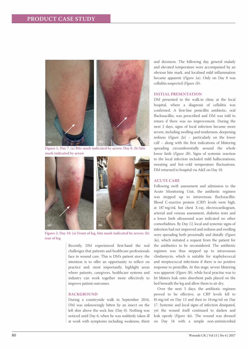
and dizziness. The following day, general malady and elevated temperature were accompanied by an obvious bite mark, and localised mild inflammation became apparent (Figure 1a). Only on Day 8 was cellulitis suspected (Figure 1b). INITIAL PRESENTATIONDM presented to the walk-in clinic at the local hospital, where a diagnosis of cellulitis was confirmed. A first-line penicillin antibiotic, oral flucloxacillin, was prescribed and DM was told to return if there was no improvement. During the next 2 days, signs of local infection became more severe, including swelling and tenderness, deepening redness (Figure 2a) – particularly on the lower calf – along with the first indications of blistering spreading circumferentially around the whole lower limb (Figure 2b). Signs of systemic reaction to the local infection included mild hallucinations, sweating and hot–cold temperature fluctuations. DM returned to hospital via A&E on Day 10.
ACUTE CAREFollowing swift assessment and admission to the Acute Monitoring Unit, the antibiotic regimen was stepped up to intravenous flucloxacillin. Blood C-reactive protein (CRP) levels were high, at 187 mg/ml, but chest X-ray, electrocardiogram, arterial and venous assessment, diabetes tests and a lower limb ultrasound scan indicated no other comorbidities. By Day 12, local and systemic signs of infection had not improved and redness and swelling were spreading both proximally and distally (Figure 3a), which initiated a request from the patient for the antibiotics to be reconsidered. The antibiotic regimen was thus stepped up to intravenous clindamycin, which is suitable for staphylococcal and streptococcal infections if there is no positive response to penicillin. At this stage, severe blistering was apparent (Figure 3b), while local practise was to let blisters leak onto absorbent pads placed on the bed beneath the leg and allow them to air dry.
Over the next 5 days, the antibiotic regimen proved to be effective, as CRP levels fell to 81 mg/ml on Day 13 and then to 16 mg/ml on Day 17. Systemic and local signs of infection dissipated, yet the wound itself continued to darken and leak openly (Figure 4a). The wound was dressed on Day 16 with a simple non-antimicrobial
Figure 1. Day 7. (a) Bite mark indicated by arrow. Day 8. (b) bite mark indicated by arrow
Figure 2. Day 10. (a) Front of leg, bite mark indicated by arrow; (b) rear of leg
Recently, DM experienced first-hand the real challenges that patients and healthcare professionals face in wound care. This is DM’s patient story: the intention is to offer an opportunity to reflect on practice and, most importantly, highlight areas where patients, caregivers, healthcare systems and industry can work together more effectively to improve patient outcomes.
BACKGROUNDDuring a countryside walk in September 2016, DM was unknowingly bitten by an insect on the left shin above the sock line (Day 0). Nothing was noticed until Day 6, when he was suddenly taken ill at work with symptoms including weakness, thirst
(a)
(a) (b)
(b)

Wounds UK | Vol 13 | No 4 | 2017 81
PRODUCT CASE STUDY
gauze, absorptive pads and hosiery. By Day 18, the wound was very dark red or black and was producing high levels of serous, sanguineous and purulent exudate (Figure 4b). When the leg was inspected, the characteristic odour of Pseudomonas aeruginosa was evident, which was reflected in the green-blue pigmented exudate on the gauze dressing. As the patient deemed fit for discharge, intravenous clindamycin was stepped down to oral therapy.
COMMUNITY CAREOnce at home, DM was tasked with making appointments at his local clinic for dressing changes. At the first community dressing change, the wound was in a poor state. On inspection of the removed dressing and periwound skin, it was evident that the gauze dressing had inadequately managed the local wound symptoms (Figure 5a). The wound was sloughy, with areas of necrosis, and fibrin, with poor quality and friable granulation tissue (Figure 5b), and was likely heavily colonised with biofilm involvement based on these clinical signs (Metcalf et al, 2014). Despite this presentation, debridement was not possible. At this time, wound cleansing was carried out with a saline-moistened gauze and a silver gauze dressing applied.
A day later (Day 20), during a visit to a different
local clinic, due to the presenting wound symptoms DM queried the change to a foam dressing as opposed to an antimicrobial dressing, along with options for debridement. The dressing choice remained unchanged, and there was no treatment plan for sharp or mechanical debridement, nor a referral on.
Three days later, DM’s leg had become increasingly odorous with the dressings clearly saturated and leaking. The leg now required constant elevation to alleviate pain. At a third visit to a different wound clinic, it was apparent that both the anterior and posterior aspects of the wound had become almost completely necrotic (Figure 6a), while oozing purulent exudate with areas of poor-quality and manifestly colonised granulation tissue (Figure 6b). After redressing in simple gauze, DM immediately tried to make a GP appointment for a referral for specialist tissue viability assessment, but was told to go to a local walk-in clinic again. Via the walk-in clinic and local A&E, DM was transferred by ambulance to a larger hospital, where he was admitted, assessed and referred to an orthopaedic surgeon.
ORTHOPAEDIC ASSESSMENTOn assessment by the orthopaedic team, the patient was informed that he would require wound excisions and skin grafts from his seat and buttocks.
Figure 3. Day 12. (a) Signs of localised infection; (b) blistering
Figure 4. Day 16. (a) Day 17. (b) green-blue pigment on dressing indicated by arrows
Figure 5. Day 19. (a) Dressing with non-antimicrobial gauze; (b) poor quality granulation, necrotic and devitalised tissue
(a)
(b)
(a) (b)
(b)
(a)

82 Wounds UK | Vol 13 | No 4 | 2017
PRODUCT CASE STUDY
retention technologies in the dressing (Parsons et al, 2016) had seemingly removed some of these key barriers to wound healing and facilitated dramatic wound progression. The wound was dressed again with moistened AQUACEL Ag+ Extra dressing. Ironically, that same evening, DM, who should have been in Florence at the World Union of Wound Healing Societies Congress, was announced co-winner of the ‘Infection and Biofilm’ award for work on wound biofilm science, while the AQUACEL Ag+ Extra dressing received the ‘Most Innovative Dressing’ award.
TISSUE VIABILITY ASSESSMENTOn assessment (Day 25), DM presented with necrosis to the anterior and posterior aspects of the left leg. On review of the medical notes, it was disclosed there had been no history of cellulitis or tissue damage prior to the current episode. The orthopaedic team documented that a skin graft was the best option to promote a positive wound healing environment. The wound area had varying consistencies of necrotic tissue present, with some areas starting to become demarcated (Figure 7a and b). The dorsalis pedis and anterior tibial pulse, along with other key venous identifiers, confirmed that there was no reduced arterial or venous involvement.
Due to the demarcated area of the necrosis, it became apparent that sharp debridement was a viable option (Figure 8a). The demarcation of the necrosis was aided by the application of the moistened AQUACEL Ag+ Extra dressing. During sharp debridement, when some of the necrosis was removed, there was healthy tissue present that negated the need for a skin graft (Figure 8b). This was documented in the medical notes and relayed to the orthopaedic team.
Due to the benefits of the primary dressing (Figure 9a–c), it was decided to continue with AQUACEL Ag+ Extra dressing followed by toe-to-knee bandaging as a figure-of-eight to apply a reduced amount of compression. After a second sharp debridement and re-application of moistened AQUACEL Ag+ Extra dressing on Day 27 (Figure 10a), the patient was discharged. Following district nurse visits for dressing changes, oral clindamycin was stopped on Day 34, and the wound went on to heal completely (Figure 10b and c).
Figure 6. Day 23. (a) Front of leg; (b) rear of leg, oozing purulence indicated by arrows; poor quality, colonised granulation tissue indicated by arrow heads
Figure 7. Day 24. (a) Rear of leg during AQUACELAg+ Extra dressing removal; (b) rear of leg after AQUACEL Ag+ Extra dressing removal
DM was placed on nil by mouth and admitted to the pre-operative ward to stay overnight. At this stage, due to areas of hard necrotic eschar combined with moist areas (Figure 6a and b), the wound was dressed with saline-moistened AQUACEL Ag+ Extra dressing with the aim of controlling bioburden and managing exudate.
The next day, around 18 hours after the initial dressing application, the orthopaedic consultant saw an improvement in the leg and agreed to postpone the operation for 24 hours. Beneath the moistened dressing, the necrotic tissue had started to lift off, while the dressing itself had absorbed exudate, pus and some of the devitalised tissue (Figure 7a). There were clear areas of pink healthy tissue at the wound edges (Figure 7b) that had not been seen the day before (Figure 6a and b). Within hours of application, the combination of anti-biofilm, antimicrobial and absorption-
(a) (b)
(a) (b)

Wounds UK | Vol 13 | No 4 | 2017 83
PRODUCT CASE STUDY
DISCUSSIONThis case study highlights the challenges that healthcare professionals can face in caring for people with wounds in various clinical settings. Despite the wide range of wound care products available, often access to those with the stronger clinical- or cost-effectiveness data is restricted, while there is a need for training before surgical debridement can be conducted.
CARE COSTSThe types of care settings and wound management strategies experienced during this case, and associated cost estimates, are given in Table 1. Although these cost estimates are based on approximations, the trend of high and ongoing costs associated with received wound care, versus
Figure 8. Day 25. (a) Sharp debridement with plastic tweezers; (b) pink and healing tissue beneath necrosis
Figure 9. Day 25. (a) A necrotic area that could not be sharp debrided, indicated by arrow. Day 27. (b) AQUACEL Ag+ Extra dressing facilitating debridement of previously strongly adhered necrosis, indicated by arrow on dressing; (c) Day 27. AQUACEL Ag+ Extra dressing facilitating debridement
smaller investments required once optimised protocols of care are established, is clear. The cost-effectiveness of wound dressings is increasingly under the spotlight, and efforts are being made to develop health economics models assessing the use of advanced dressing technologies (Harding et al, 2013; Walker et al, 2015; Nherera et al, 2016; Jiménez et al, 2017).
ANTIBIOTIC STEWARDSHIPIn this case study, the antibiotic regimen of intravenous clindamycin was eventually successful in dealing with the patient’s local and systemic signs of infection. Despite the resolution of infection, oral clindamycin continued to be prescribed for a further 3 weeks. This dose did not improve local signs of colonisation during the week spent in community care; it was not until the AQUACEL Ag+ Extra dressing was applied that the wound started to progress. In this case, topical antisepsis, by the application of a silver-containing Hydrofiber™ dressing with anti-biofilm technology, was shown to be far more effective at resolving a heavily-colonised, post-acute wound than antibiotics.
DRESSING SELECTIONThis case study highlights the fact that not all antimicrobial dressings are the same; indeed, not all silver dressings are the same. The dramatic difference in performance of a silver gauze compared to a silver-containing Hydrofiber dressing with anti-biofilm technology is a reminder that dressing design and how the antimicrobial agent is formulated is as important to a dressing’s effectiveness as the antimicrobial agent itself. Gauzes are still widely used as a primary wound contact layer, even though gauze is an ideal substrate for biofilm development (Bowler and Parsons, 2016). Foam dressings are often marketed as being absorbent, but unless they contain some additional moisture-retentive technology, skin integrity and associated wound outcomes could be impacted, as the patient experienced in this case (Figure 6a and b).
Dressings account for a fraction of the overall costs of wound care (Harding et al, 2013; Guest et al, 2015), yet there remains a perception that antimicrobial dressings are expensive and ineffective, based on some meta-analyses (O’Meara et al, 2014; National Institute for Health
(a) (b)
(a) (b) (c)

84 Wounds UK | Vol 13 | No 4 | 2017
PRODUCT CASE STUDY
and Care Excellence [NICE], 2016) and clinical studies (Michaels et al, 2009) and there have been challenges about their appropriateness (White et al, 2010; Leaper and Drake, 2011). Although some wound care technologies are priced at a marginally higher premium than others, this might reflect the cost of their advanced development and manufacture. As this case study shows, and as previous case study analyses have demonstrated (Woo 2014; Walker et al, 2015; Metcalf et al, 2016a); Metcalf et al, 2017), a modest and timely investment in an advanced wound care technology can kick-start wound healing by removing and reducing barriers to healing. Not only can this facilitate healing, but it can reduce antibiotic usage, avoid costly and painful operations, eliminate future wound care costs, and take patients out of acute and community healthcare settings. Effective wound care protocols can not only save time and money, but can save patients from pain, life-long disfigurement or even limb loss, and dramatically improve quality of life.
CONCLUSIONNursing and care staff, along with midwifery, form the largest proportion of the healthcare workforce, so they must be equipped with knowledge and technologies to lead the delivery of a positive patient experience and outcomes with the efficient use of resources. In a recent initiative, the theoretical ‘Betty’s story’ has been developed by the NHS to highlight the disparity in experience, outcome and cost, between sub-optimal and optimal leg ulcer care pathways (NHS RightCare, 2017). This present case study is a real-life example of the cost of certain
pathways (Table 1), eventually resolved, in part, due to a unique antimicrobial dressing technology. DM was fortunate enough to have more than a decade of wound care knowledge; something that most of our patients and some caregivers will not have.
This case study highlights the social and economic impact through patient wellbeing and cost drivers incurred in both the community and hospital care setting. It demonstrates a required shift of the wound management journey from a reactive approach to a proactive, evidence-based approach. Improved wound care including effective assessment, diagnosis, treatment and prevention of wound care complications can minimise treatment costs (Guest et al, 2015) and, importantly, improve outcomes and experience for people with a wound. Wuk
REFERENCESBowler PG, Parsons D (2016) Combatting wound biofilm and recalcitrance
with a novel anti-biofilm Hydrofiber® wound dressing. Wound Medicine 14: 6–11
Guest JF, Ayoub N, McIlwraith T et al (2015) Health economic burden that wounds impose on the National Health Service in the UK. BMJ Open 5(12): e009283
Harding K, Posnett J, Vowden K (2013). A new methodology for costing wound care. Int Wound J 10(6): 623–29
Harding KG, Szczepkowski M, Mikosiński J et al (2016) Safety and performance evaluation of a next-generation antimicrobial dressing in patients with chronic venous leg ulcers. Int Wound J 13(4): 442–8
Jiménez AC, Ortiz CN, Galisteo MG et al (2017) Cost efficiency of the choice of dressings in chronic wounds with biofilm based on a theoretical model. Gerokomos 28(2): 98–102
Kammerlander G, Lantin A, Geyrhofer C et al (2015) Kritisch kolonisierte und ortsständig infizierte wunden: effektivität einer neuen Hydrofiber mit silber, EDTA und BeCl bei der stabilisation. Procare 20(9): 14–16
Leaper D, Drake R (2011) Should one size fit all? An overview and critique of the VULCAN study on silver dressings. Int Wound J 8(1): 1–4
Metcalf DG, Bowler PG (2013). Biofilm delays wound healing: a review of the evidence. Burns Trauma 1(1): 5-12
Figure 10. Day 27. (a) on discharge, rear of leg. Day 38. (b) front of leg. Day 38 (c) rear of leg
(a) (b) (c)

Wounds UK | Vol 13 | No 4 | 2017 85
PRODUCT CASE STUDY
Metcalf DG, Bowler PG, Hurlow J (2014) A clinical algorithm for wound biofilm identification. J Wound Care 23: 137-142.
Metcalf DG, Parsons D, Bowler PG (2016a) A next-generation antimicrobial wound dressing: a real-life clinical evaluation in the UK and Ireland. J Wound Care 25(3): 132–38
Metcalf D, Bowler P, Parsons D (2016b) Wound Biofilm and Therapeutic Strategies, Microbial Biofilms. Available at: https://www.intechopen.com/books/microbial-biofilms-importance-and-applications/wound-biofilm-and-therapeutic-strategies (accessed 7.10. 2017)
Metcalf DG, Parsons D, Bowler PG (2017). Clinical safety and effectiveness evaluation of a new antimicrobial wound dressing designed to manage exudate, infection and biofilm. Int Wound J 14(1): 203–13
Michaels JA, Campbell WB, King BM et al (2009) A prospective randomised controlled trial and economic modelling of antimicrobial silver dressings versus non-adherent control dressings for venous leg ulcers: the VULCAN trial. Health Technol Assess 13(56) : 1–114
National Institute for Health and Care Excellence (2016) Chronic Wounds: Advanced Wound Dressings and Antimicrobial Dressings. Available at: https://www.nice.org.uk/advice/esmpb2/chapter/Key-points-from-the-evidence (accessed 7.10. 2017)
National Institute of Health and Care Excellence (2017) BNF. Available at: https://bnf.nice.org.uk/ (accessed 19.10.2017)
Nherera LM, Woodmansey E, Trueman P, Gibbons GW (2016) Estimating the clinical outcomes and cost differences between standard care with and without cadexomer iodine in the management of chronic venous leg ulcers using a Markov model. Ostomy Wound Manage 62(6): 26–40
NHS RightCare (2017) RightCare Scenario: The Variation Between Sub-Optimal and Optimal Pathways. Betty’s Story: Leg Ulcer Wound Care. Available at: www.england.nhs.uk/rightcare/wp-content/uploads/sites/40/2017/01/nhs-rightcare-bettys-story-narrative-full.pdf (accessed 7.10. 2017)
O’Meara S, Al-Kurdi D, Ologun Y et al (2014) Antibiotics and antiseptics for venous leg ulcers. Cochrane Database Syst Rev 10(1): CD003557
Parsons D, Meredith K, Rowlands VJ et al (2016). Enhanced performance and mode of action of a novel antibiofilm Hydrofiber® wound dressing. Biomed Res Int 7616471.
Regional Drug and Therapeutics Centre (2017) Cost Comparison Charts. Available at: http://gmmmg.nhs.uk/docs/cost_comparison_charts.pdf (accessed 7.10. 2017)
Shenvi C (2015) The Clindamycin Fact Sheet. Available at: http://www.medpagetoday.com/blogs/epmonthly/53645 (accessed 7.10. 2017)
Walker M, Metcalf D, Parsons D, Bowler P (2015). A real-life clinical evaluation of a next-generation antimicrobial dressing on acute and chronic wounds. J Wound Care 24(1): 11–22
White R, Cutting K, Ousey K et al (2010) Randomized controlled trial and cost-effectiveness analysis of silver-donating antimicrobial dressings for venous leg ulcers (VULCAN trial) (Br J Surg 2009; 96: 1147-1156) Br J Surg 97(3): 459–60
Woo K (2014) AQUACEL Ag+ dressings in practice. In: Next-Generation Antimicrobial Dressings: AQUACEL™ Ag+ Extra™ and Ribbon. Available at: http://bit.ly/2xnEJtM (accessed
Care type Occurrence Approximate cost Reference
Walk-in visits 2 £250 Guest et al, 2015
A&E visits 2 £120 Guest et al, 2015
Overnight hospital stays 12 £15,000 Guest et al, 2015
Microbiological swabbing 2 £50 Personal communication (Austrian and Belgian physicians, 2012)
Ambulance transfer 1 £250 Guest et al, 2015
Antibiotics (oral) 5 days £5 Regional Drug and Therapeutics Centre, 2017
Antibiotics (intravenous) 10 days £100 Shenvi, 2015
Dressings, acute (gauze, pads, hosiery)
2 £10 Estimates using British National Formulary
Dressings, community (3 × silver gauze, 3 × foam dressings, pads, hosiery)
3 £39 Estimates using British National Formulary
Approximate total before AQUACEL Ag+ Extra and tissue viability assessment: £15,824
Antibiotics (oral) 13 days £13 Regional Drug and Therapeutics Centre, 2017
Dressings, acute (6x AQUACEL Ag+ Extra 10 × 10, pads, hosiery)
3 £90 Estimates using British National Formulary
Dressings, community (6 × AQUACEL Ag+ Extra 10 × 10, pads, hosiery)
3 £90 Estimates using British National Formulary
Tissue viability nurse visit (1 hour)
2 £100 Guest et al, 2015
District nurse home visits (30 minutes)
3 £150 Guest et al, 2015
Approximate total after AQUACEL Ag+ Extra and tissue viability assessment: £443
Table 1. Cost estimates of the care settings and wound management strategies experienced (National Institute of Health and Care Excellence [NICE], 2017)