Women and Babies: Neonatal Early Assessment Program (NEAP)content/pdf/guidelines... · Compliance...
16
Compliance with this Guideline is recommended Page 1 of 16 Guideline Women and Babies: Neonatal Early Assessment Program (NEAP) Document No: RPAH_GL2016_xxx Functional Sub-Group: Clinical Governance Corporate Governance Summary: The Neonatal Early Assessment Program (NEAP) consists of a risk factor assessment and four sets of measurements within the first 6 hours after birth: 1. The first physical examination. 2. The first lower limb oxygen saturation. 3. Anthropometry 4. Body fat (percentage) by air displacement plethysmography National Standard: National Standard 1 Policy Author: Original policy – Prof Heather Jeffery Updated policy – Dr Tracey Lutz (Neonatologist) Approved by: RPA Women and Babies Service Improvement Committee Publication (Issue) Date: July 2016 Next Review Date: July 2019 Replaces Existing Policy: N/A Previous Review Dates: N/A Note: Sydney Local Health District (LHD) and South Western Sydney LHD were established on 1 July 2011, with the dissolution of the former Sydney South West Area Health Service (SSWAHS) in January 2011. The former SSWAHS was established on 1 January 2005 with the amalgamation of the former Central Sydney Area Health Service (CSAHS) and the former South Western Sydney Area Health Service (SWSAHS). In the interim period between 1 January 2011 and the release of specific LHN policies (dated after 1 January 2011) and SLHD (dated after July 2011), the former SSWAHS, CSAHS and SWSAHS policies are applicable to the LHDs as follows: Where there is a relevant SSWAHS policy, that policy will apply Where there is no relevant SSWAHS policy, relevant CSAHS policies will apply to Sydney LHD; and relevant SWSAHS policies will apply to South Western Sydney LHD.
Transcript of Women and Babies: Neonatal Early Assessment Program (NEAP)content/pdf/guidelines... · Compliance...
Compliance with this Guideline is recommended Page 1 of 16
Guideline
Women and Babies: Neonatal Early Assessment Program (NEAP)
Document No: RPAH_GL2016_xxx
Functional Sub-Group: Clinical Governance
Corporate Governance
Summary: The Neonatal Early Assessment Program (NEAP)
consists of a risk factor assessment and four sets of
measurements within the first 6 hours after birth:
1. The first physical examination.
2. The first lower limb oxygen saturation.
3. Anthropometry
4. Body fat (percentage) by air displacement
plethysmography
National Standard: National Standard 1
Policy Author: Original policy – Prof Heather Jeffery
Updated policy – Dr Tracey Lutz (Neonatologist)
Approved by: RPA Women and Babies Service Improvement Committee
Publication (Issue) Date: July 2016
Next Review Date: July 2019
Replaces Existing Policy: N/A
Previous Review Dates: N/A
Note: Sydney Local Health District (LHD) and South Western Sydney LHD were established
on 1 July 2011, with the dissolution of the former Sydney South West Area Health Service
(SSWAHS) in January 2011. The former SSWAHS was established on 1 January 2005 with
the amalgamation of the former Central Sydney Area Health Service (CSAHS) and the former
South Western Sydney Area Health Service (SWSAHS).
In the interim period between 1 January 2011 and the release of specific LHN policies (dated
after 1 January 2011) and SLHD (dated after July 2011), the former SSWAHS, CSAHS and
SWSAHS policies are applicable to the LHDs as follows:
Where there is a relevant SSWAHS policy, that policy will apply
Where there is no relevant SSWAHS policy, relevant CSAHS policies will apply to Sydney
LHD; and relevant SWSAHS policies will apply to South Western Sydney LHD.
Compliance with this Guideline is recommended Page 2 of 16
Neonatal Early Assessment Program (NEAP)
Contents
1. Introduction 3
2. Policy Statement 3
3. Guidelines
3.1 Early Risk Factor assessment and clinical examination 3
3.2 Oxygen saturation screening 4
3.3 Anthropometry 4
3.4 Body composition - Fat measurement (fat mass, fat free mass and body fat %)
4. Figures
Figure 1: Overview on how to perform NEAP 7
Figure 2: Early physical examination of the newborn baby 8
Figure 3: Physical examination referral pathway 9
Figure 4: PeaPod quick reference guide 10
Figure 5: Anthropometric referral pathway 11
Figure 6: Oxygen saturation referral pathway 12
Figure 7: Body Fat percentage referral pathway 12
5. Key points 13
6. References 14

Sydney Local Health District Guideline No: RPAH_GL2013_019 Royal Prince Alfred Hospital September 2016
Compliance with this Guideline is recommended Page 3 of 16
Neonatal Early Assessment Program (NEAP)
1. Introduction
The risks addressed by this policy:
Clinical risk of neurological damage or death of babies as a result of physical
abnormalities, cardiac defects, or neonatal hypoglycaemia going undetected and
untreated.
The aims / expected outcome of this policy
Babies with physical abnormalities, cardiac defects, or increased risk of hypoglycaemia
will be identified and appropriately managed.
2. Policy Statement
The goal of this guideline is to familiarise medical staff with the evidence, indications and
practical management of a neonate undergoing the NEAP (neonatal early assessment
program).
3. Guidelines
3.1 Early Risk Factor Assessment and Physical Examination
The overall goal of the early assessment is to identify risk factors for early neonatal problems
that will require regular postnatal observations, as well as identifying congenital and acquired
abnormalities which may have an immediate impact on the baby’s care and/or wellbeing.
Early Risk Factor Assessment
All babies should be reviewed for risk factors for the five main problem areas as identified on
the ‘Women and Babies Newborn Care Plan and Observations Chart (NCPOC – MR504) and
‘The Standard Newborn Observation Chart (SNOC – SMR 110.014)’. These include
Risk of hypoglycaemia
Respiratory distress
Risk of subgaleal haemorrhage - trauma from instrumental delivery
Risk of sepsis
Risk of jaundice
Assessment of risk of hypoglycaemia will include the anthropometric measurements and
percentage body fat as outlined below. Babies identified with risk factors will need
observations as outlined in the relevant RPAH guideline and on the NCPOC.
Early physical examination
The goal of this examination is to identify obvious external anomalies, as well as other
congenital and acquired problems that may have an immediate impact on the baby’s care or
well-being. This would include (but not be confined to) problems such as respiratory distress,
consequences of birth trauma (e.g bruising, sub-galeal haemorrhage, nerve palsy), cleft palate,
imperforate anus, abnormal genitalia and other dysmorphology. The examination should

Sydney Local Health District Guideline No: RPAH_GL2013_019 Royal Prince Alfred Hospital September 2016
Compliance with this Guideline is recommended Page 4 of 16
include a complete ‘head to toe to back’ inspection of the baby as detailed below in Figure 2.
The findings should be documented in the relevant section of the NCPOC.
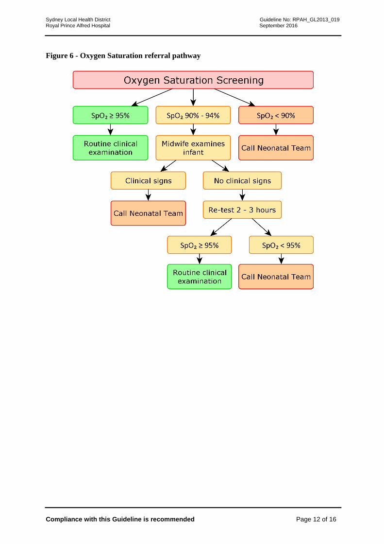
3.2 Oxygen saturation screening:
In the first minutes of life, there is a transition from the placenta being the main organ of gas
exchange to the newborn lungs. Toth1 et al demonstrated in 50 healthy newborns that it takes
between 12 and 14 minutes to achieve saturations > 95%. Routine oxygenation saturation
screening of well newborns has been shown to improve early diagnosis of congenital heart
disease (CHD)2 with a low false positive rate and minimal impact on resources. About half of
the babies with a low saturation screen (<95%) will have either congenital heart disease
(~1/3rd
) or other significant pathologies which include respiratory disease, delayed transition
to extra-uterine life (persistent pulmonary hypertension of the newborn), sepsis or metabolic
conditions (~2/3rd
)3. Saturation measurement within the first 4 to 6 hours of birth has a lower
accuracy for detection of CHD but greater sensitivity for non-cardiac pathology while
screening after 24 hours (late screen) has greater sensitivity for detection of CHD, especially
left heart ductal dependent lesions. At RPAH, both early and late oxygen saturation screening
will be performed in accordance with the existing guideline.
3.2.1 How to perform oxygen saturation screening:
Early oxygen saturation screening will be performed in accordance with the existing
guideline
Perform screening when the infant is quiet.
Place the probe around one foot with light source and receiver on each side of foot.
Secure with Coban® tape. To ensure good blood flow to the foot, do not secure too
tightly and do not hold the probe around the foot.
Switch on oximeter and allow signal to stabilise. Read when stabilised and there is a
good plethysmographic light pulse.
3.3 Anthropometry
Newborn measurement of weight, length, and head circumference reflects fetal nutrition and
forms the basis on which future growth measurements. 4,5,7,8
3.3.1 How to measure weight:
The scales on the Pea Pod are used for accurate measurement of weight to the nearest
gram.
The newborn is bare weighed.
The weight percentile is calculated using the Beeby electronic calculator on the
computer in the NEAP room, or if unavailable the weight is plotted on New South
Wales population-based birthweight percentile charts (less accurate)9.
3.3.2 How to measure length:
The length-board measurement, infantometer, has been shown to be the most reliable
and accurate measurement of neonatal length 6,10,11
and more recent designs have
improved ease of use such as the Easy-Glide Bearing Infantometer (Perspective
Enterprises, Portage, MI, USA).
The neonate is placed supine and unclothed on the board and held gently with his or
her body aligned and head in a neutral position. One person stands at the top of the
length board and holds the baby’s head in contact with the headboard while another
Sydney Local Health District Guideline No: RPAH_GL2013_019 Royal Prince Alfred Hospital September 2016
Compliance with this Guideline is recommended Page 5 of 16
extends the left leg by placing the hand over the left knee, depressing the knee,
straightening the leg and moving the footboard to touch the plantar surface of the foot
at a right angle to the leg. Recheck that the head has not moved from the headboard
before taking the measurement. The actual reading is marked by an arrow as there is an
offset for greater ease of reading and accuracy.
The length percentile is calculated using the Beeby electronic calculator on the
computer in the NEAP room or plotted on New South Wales population-based birth
length percentile charts9
(less accurate).
.
3.3.3 How to measure head circumference:
Head circumference is measured using disposable paper circumference tapes at the
maximum fronto-occipital circumference.
Two reproducible measurements are required.
The head circumference percentile is calculated using the Beeby electronic calculator
on the computer in the NEAP room or plotted on New South Wales population-based
birth head circumference percentile charts9 (less accurate).
3.4 Body composition - Fat measurement (fat mass, fat free mass and body fat %)
Accurately determining the nutritional status of newborns is a major public health problem.
Furthermore undernourished neonates that survive are at risk of long term health outcomes,
including hypertension, stroke, type 2 diabetes, obesity and cardiovascular disease. 12,13
Neonatal undernutrition or ‘wasting’ is a clinical diagnosis often characterised by diminished
subcutaneous tissues and underlying muscles with loose wrinkled skin of the arms, thighs,
elbows and knees. However this clinical sign is, in our experience at RPAH, not well
recognised by a range of health providers.
Thus defining who is undernourished is problematic. Conventional approaches include the use
of population based percentiles (< 3rd
, 5th
or 10th
percentiles) or customised growth charts.
Population based charts rely on a large cross section of neonates and use weight for gestational
age by sex. Customised charts account for more maternal variables, however there is not
strong evidence to support their use at present. 14,15
An alternative to birth weight is the use of
body composition or body fat % (BF%).
The wasted, undernourished newborn is characterised by loss of the normal fat accretion in the
last 4-6 weeks of pregnancy. As fat is used by the newborn as an alternative substrate to
glucose for brain metabolism, BF% is a potentially very useful measure indicating degree of
undernutrition and directly related to neonatal metabolic outcomes. The additional advantage
is that it can distinguish the normal fat SGA (weight less than 10th
percentile) from the low fat,
undernourished SGA newborn and define the low fat AGA newborn at high risk of
hypoglycaemia, currently not possible with any other methods.
Recently a new technology using air displacement plethysmography (ADP) has become
available to non-invasively, accurately and quickly measure BF% in infants from birth to 6
months of age. ADP has been validated against the four-compartment model and biological
and physical phantoms16,17
and is considered the criterion method for determining BF% in
neonates. 18-22
Several studies have investigated the BF% as an indicator of neonatal nutritional status using
ADP. 20-25
Body fat is a better measure than customised charts for assessing neonatal
morbidity and as good as population based charts.26
The advantage of this method is it is

Sydney Local Health District Guideline No: RPAH_GL2013_019 Royal Prince Alfred Hospital September 2016
Compliance with this Guideline is recommended Page 6 of 16
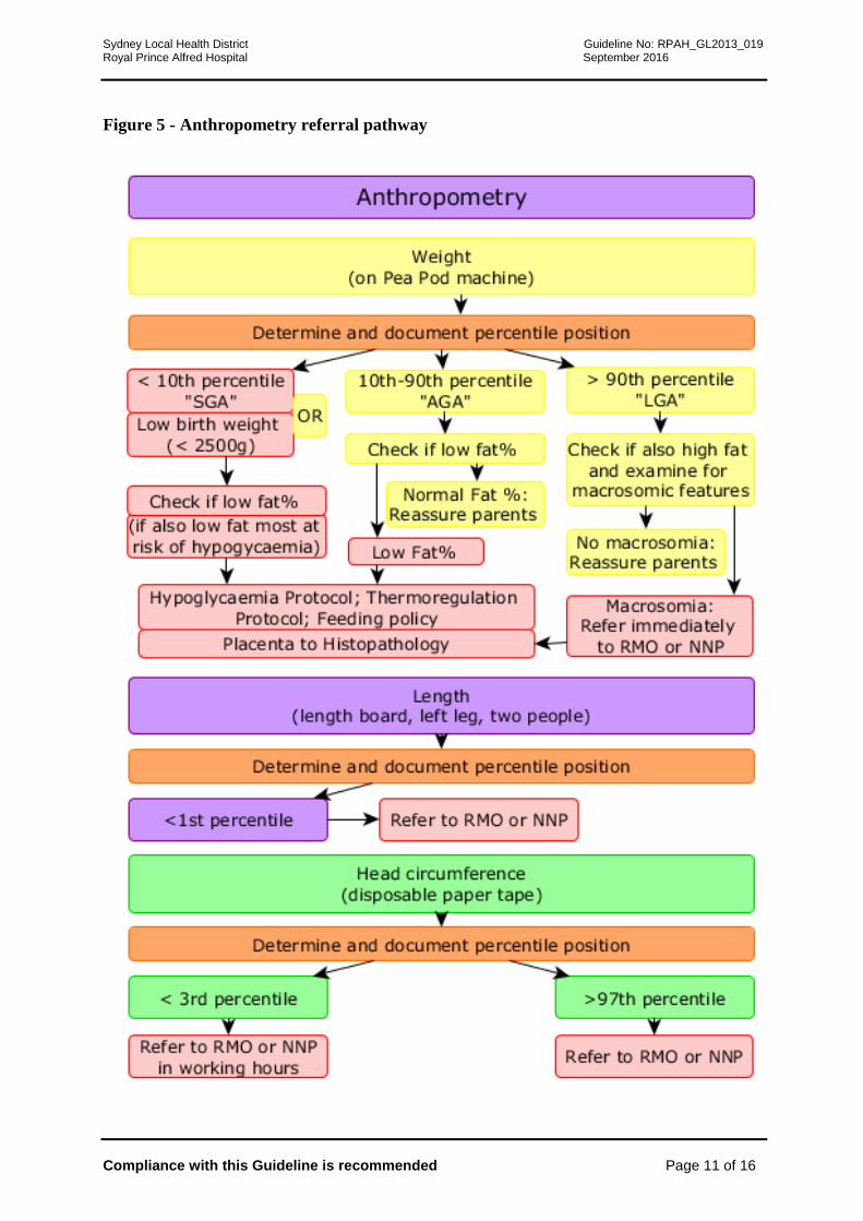
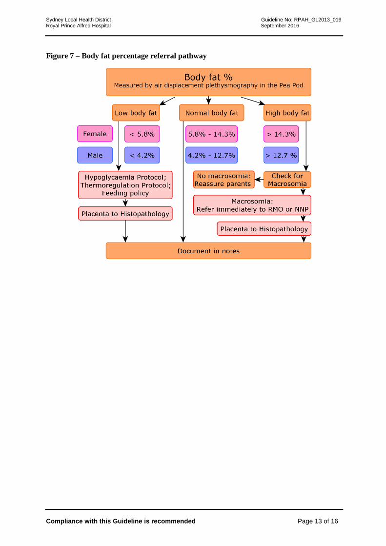
accurate, easy, reliable, non-invasive and acceptable to parents. The cut offs for low and high
fat are shown in the flow chart. These were determined by significantly better Receiver
Operator Curves (ROCs) for combined morbidity assessed by 3 pre-specified outcomes (temp
<36.5C, prolonged hospital stay, poor feeding (2 of 3 objective criteria). The cut off was
determined by the highest sensitivity balanced by the best specificity. 26
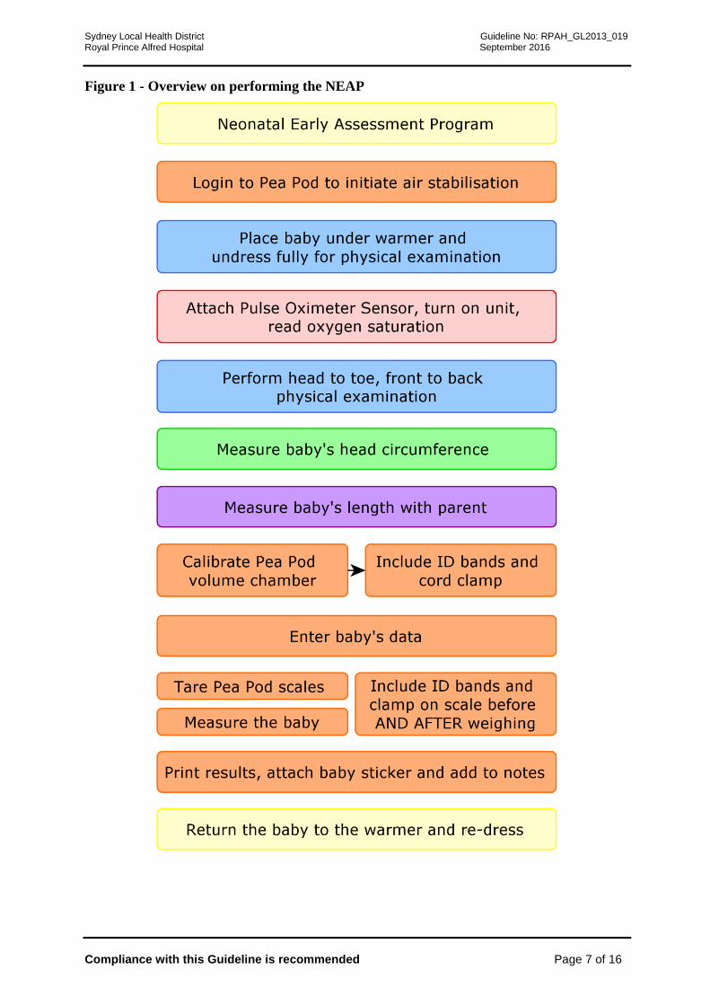
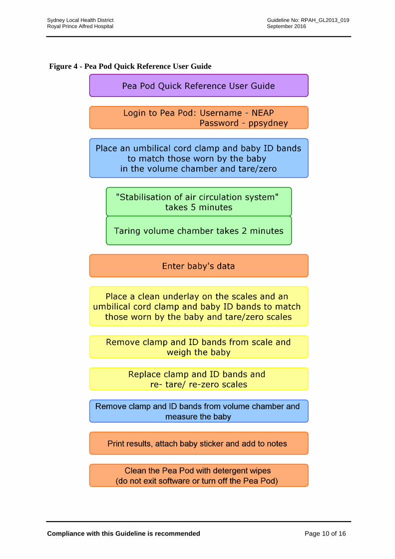
3.4.1 How to measure body composition:
This is performed in the NEAP room just after the measurement of length and weight
using the PeaPod (CosMed, USA).
Sydney Local Health District Guideline No: RPAH_GL2013_019 Royal Prince Alfred Hospital September 2016
Compliance with this Guideline is recommended Page 7 of 16
Figure 1 - Overview on performing the NEAP
Sydney Local Health District Guideline No: RPAH_GL2013_019 Royal Prince Alfred Hospital September 2016
Compliance with this Guideline is recommended Page 8 of 16
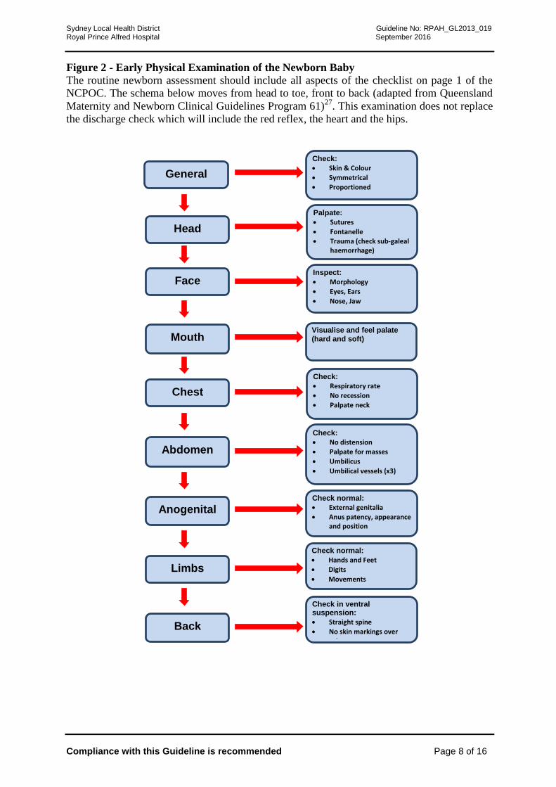
Figure 2 - Early Physical Examination of the Newborn Baby
The routine newborn assessment should include all aspects of the checklist on page 1 of the
NCPOC. The schema below moves from head to toe, front to back (adapted from Queensland
Maternity and Newborn Clinical Guidelines Program 61)27
. This examination does not replace
the discharge check which will include the red reflex, the heart and the hips.
Head
Palpate:
Sutures
Fontanelle
Trauma (check sub-galeal haemorrhage)
Face Inspect:
Morphology
Eyes, Ears
Nose, Jaw
Mouth Visualise and feel palate
(hard and soft)
Chest
Check:
Respiratory rate
No recession
Palpate neck
Abdomen
Check:
No distension
Palpate for masses
Umbilicus
Umbilical vessels (x3)
Anogenital
Check normal:
External genitalia
Anus patency, appearance and position
Limbs
Check normal:
Hands and Feet
Digits
Movements
Back
Check in ventral suspension:
Straight spine
No skin markings over spine
General
Check:
Skin & Colour
Symmetrical
Proportioned

Sydney Local Health District Guideline No: RPAH_GL2013_019 Royal Prince Alfred Hospital September 2016
Compliance with this Guideline is recommended Page 9 of 16
Figure 3 - Physical examination referral pathway
Sydney Local Health District Guideline No: RPAH_GL2013_019 Royal Prince Alfred Hospital September 2016
Compliance with this Guideline is recommended Page 10 of 16
Figure 4 - Pea Pod Quick Reference User Guide
Sydney Local Health District Guideline No: RPAH_GL2013_019 Royal Prince Alfred Hospital September 2016
Compliance with this Guideline is recommended Page 11 of 16
Figure 5 - Anthropometry referral pathway
Sydney Local Health District Guideline No: RPAH_GL2013_019 Royal Prince Alfred Hospital September 2016
Compliance with this Guideline is recommended Page 12 of 16
Figure 6 - Oxygen Saturation referral pathway
Sydney Local Health District Guideline No: RPAH_GL2013_019 Royal Prince Alfred Hospital September 2016
Compliance with this Guideline is recommended Page 13 of 16
Figure 7 – Body fat percentage referral pathway

Sydney Local Health District Guideline No: RPAH_GL2013_019 Royal Prince Alfred Hospital September 2016
Compliance with this Guideline is recommended Page 14 of 16
4. Key Points
Key point Level of Evidence &
Recommendation (NHMRC)25
Air displacement plethysmography (ADP) non-
invasively, accurately and quickly measures
BF% in infants from birth to 6 months of age.
Level of evidence: 3
Strength of recommendation: A
Body fat is a better measure than customised
charts for assessing neonatal morbidity
Level of evidence: 3
Strength of recommendation: A
5. References
1. Toth B, Becker A, Seelbach-Gobel B. Oxygen saturation in healthy newborn infants
immediately after birth measured by pulse oximetry. Arch Gynecol Obstet 2002 April:
266 (2)105-107
2. A. Meberg, A. Andreassen, L. Brunvand, T. Markestad, D. Moster, L. Nietsch, I. E.
Silberg, and J. E. Skålevik, 'Pulse Oximetry Screening as a Complementary Strategy to
Detect Critical Congenital Heart Defects', Acta Pædiatrica, 98 (2009), 682-86.
3. A. Meberg, S. Brugmann-Pieper, R. Due, Jr., L. Eskedal, I. Fagerli, T. Farstad, D. H.
Froisland, C. H. Sannes, O. J. Johansen, J. Keljalic, T. Markestad, E. A. Nygaard, A.
Rosvik, and I. E. Silberg, 'First Day of Life Pulse Oximetry Screening to Detect
Congenital Heart Defects', J Pediatr, 152 (2008), 761-5.
4. T. S. Johnson, J. L. Engstrom, and D. K. Gelhar, 'Intra- and Interexaminer Reliability
of Anthropometric Measurements of Term Infants', J Pediatr Gastroenterol Nutr, 24
(1997), 497-505.
5. T. S. Johnson, J. L. Engstrom, J. A. Warda, M. Kabat, and B. Peters, 'Reliability of
Length Measurements in Full-Term Neonates', J Obstet Gynecol Neonatal Nurs, 27
(1998), 270-6.
6. A. J. Wood, C. H. Raynes-Greenow, A. E. Carberry, and H. E. Jeffery, 'Neonatal
Length Inaccuracies in Clinical Practice and Related Percentile Discrepancies Detected
by a Simple Length-Board', J Paediatr Child Health, 49 (2013), 199-203.
7. T. H. Lipman, K. D. Hench, T. Benyi, J. Delaune, K. A. Gilluly, L. Johnson, M. G.
Johnson, H. McKnight-Menci, D. Shorkey, J. Shults, F. L. Waite, and C. Weber, 'A
Multicentre Randomised Controlled Trial of an Intervention to Improve the Accuracy
of Linear Growth Measurement', Arch Dis Child, 89 (2004), 342-6.
8. T. H. Lipman, K. D. Hench, J. D. Logan, D. A. DiFazio, P. M. Hale, and C. Singer-
Granick, 'Assessment of Growth by Primary Health Care Providers', Journal of
Pediatric Health Care, 14 (2000), 166-71.
Sydney Local Health District Guideline No: RPAH_GL2013_019 Royal Prince Alfred Hospital September 2016
Compliance with this Guideline is recommended Page 15 of 16
9. P. J. Beeby, T. Bhutap, and L. K. Taylor, 'New South Wales Population-Based
Birthweight Percentile Charts', J Paediatr Child Health, 32 (1996), 512-8.
10. D. P. Davies, and R. E. Holding, 'Neonatometer: A New Infant Length Measurer', Arch
Dis Child, 47 (1972), 938-40.
11. A. F. Roche T. G. Lohman, & R. Martorell (eds.), Anthropometric Standardization
Reference Manual (Champaign, IL: Human Kinetics Books, 1988).
12. D. J. Barker, J. G. Eriksson, T. Forsen, and C. Osmond, 'Fetal Origins of Adult
Disease: Strength of Effects and Biological Basis', Int J Epidemiol, 31 (2002), 1235-9.
13. J. C. Wells, S. Chomtho, and M. S. Fewtrell, 'Programming of Body Composition by
Early Growth and Nutrition', Proc Nutr Soc, 66 (2007), 423-34.
14. A. E. Carberry, A. Gordon, D. M. Bond, J. Hyett, C. H. Raynes-Greenow, and H. E.
Jeffery, 'Customised Versus Population-Based Growth Charts as a Screening Tool for
Detecting Small for Gestational Age Infants in Low-Risk Pregnant Women', in
Cochrane Database of Systematic Reviews John Wiley & Sons, Ltd 2011 ).
15. A. E. Carberry, C. H. Raynes-Greenow, R. M. Turner, and H. E. Jeffery, 'Customized
Versus Population-Based Birth Weight Charts for the Detection of Neonatal Growth
and Perinatal Morbidity in a Cross-Sectional Study of Term Neonates', American
Journal of Epidemiology, August 21 (2013)..
16. A. Frondas-Chauty, I. Louveau, I. Le Huerou-Luron, J. C. Roze, and D. Darmaun, 'Air-
Displacement Plethysmography for Determining Body Composition in Neonates:
Validation Using Live Piglets', Pediatric research, 72 (2012), 26-31.
17. K. J. Ellis, M. Yao, R. J. Shypailo, A. Urlando, W. W. Wong, and W. C. Heird, 'Body-
Composition Assessment in Infancy: Air-Displacement Plethysmography Compared
with a Reference 4-Compartment Model', Am J Clin Nutr, 85 (2007), 90-5.
18. G. Ma, M. Yao, Y. Liu, A. Lin, H. Zou, A. Urlando, W. W. Wong, L. Nommsen-
Rivers, and K. G. Dewey, 'Validation of a New Pediatric Air-Displacement
Plethysmograph for Assessing Body Composition in Infants', Am J Clin Nutr, 79
(2004), 653-60.
19. A. Urlando, P. Dempster, and S. Aitkens, 'A New Air Displacement Plethysmograph
for the Measurement of Body Composition in Infants', Pediatr Res, 53 (2003), 486-92.
20. M. Yao, L. Nommsen-Rivers, K. Dewey, and A. Urlando, 'Preliminary Evaluation of a
New Pediatric Air Displacement Plethysmograph for Body Composition Assessment in
Infants', Acta Diabetol, 40 Suppl 1 (2003), S55-8.
21. G.S. Andersen, T. Girma, J.C.K. Wells, P. Kæstel, K.F. Michaelsen, and H. Friis, 'Fat
and Fat-Free Mass at Birth: Air Displacement Plethysmography Measurements on 350
Ethiopian Newborns', Pediatr Res, 70 (2011), 501-06.
22. D.A. Fields, J.M. Gilchrist, P.M. Catalano, M.L. Giannì, P.M. Roggero, and F. Mosca,
'Longitudinal Body Composition Data in Exclusively Breast-Fed Infants: A
Multicenter Study', Obesity, 19 (2011), 1887-91.
23. B. E. Lingwood, A. M. Storm van Leeuwen, A. E. Carberry, E. C. Fitzgerald, L. K.
Callaway, P. B. Colditz, and L. C. Ward, 'Prediction of Fat-Free Mass and Percentage
of Body Fat in Neonates Using Bioelectrical Impedance Analysis and Anthropometric
Measures: Validation against the Pea Pod', Br J Nutr, 107 (2012), 1545-52.
Sydney Local Health District Guideline No: RPAH_GL2013_019 Royal Prince Alfred Hospital September 2016
Compliance with this Guideline is recommended Page 16 of 16
24. P. Roggero, M. L. Gianni, O. Amato, P. Piemontese, D. Morniroli, W. W. Wong, and
F. Mosca, 'Evaluation of Air-Displacement Plethysmography for Body Composition
Assessment in Preterm Infants', Pediatric research, 72 (2012), 316-20.
25. A. Fields, P. B. Higgins, and D. Radley, 'Air-Displacement Plethysmography: Here to
Stay', Curr Opin Clin Nutr Metab Care, 8 (2005), 624-9.
26. A. E. Carberry, C. H. Raynes-Greenow, R. M. Turner, L. M. Askie, and H. E. Jeffery,
'Is Body Fat Percentage a Better Measure of Undernutrition in Newborns Than Birth
Weight Percentiles', Pediatric Research, Advance online publication 02 October
(2013).
27. Queensland Maternity and Neonatal Clinical Guidelines Program, 'Examination of the
Newborn Baby', ed. by Queensland Health (Queensland Queensland Health, 2009).