Williams syndrome: an exploration of neurocognitive …lcn.salk.edu/publications/2001/Bellugi -...
13
Williams syndrome: an exploration of neurocognitive and genetic features Ursula Bellugi a, * , Julie R. Korenberg b , Edward S. Klima a a Laboratory for Cognitive Neuroscience, The Salk Institute for Biological Studies, 10010 N. Torrey Pines Road, La Jolla, CA 92037, USA b Cedars-Sinai Medical Center and University of California, Los Angeles, CA, USA Abstract We report here on significant attempts to forge links between neurodevelopmental disorders, development of specific neuropsychological abilities, and the functional establishment of patterns of brain organization. Such research programs are providing converging evidence for the coherence or dissociability of components of cognition (e.g. language, spatial cognition) and will allow development of theoretical explanations for the underlying architecture of human cognition. Williams syndrome involves focal rather than generalized cognitive deficits, and offers an important opportunity for linking brain findings to specific atypical cognitive profiles. The unusual neurocognitive profile of Williams syndrome makes it a compelling model of the pathways between genes and human cognition. It is becoming clear that the syndrome’s unique genomic organization may also make it an important model of human chromosomal evolution and disease. These studies with a specific neurodevelopmental disorder that presents a rare dissociation of higher cortical functioning may provide opportunities to explore some of the central issues of cognitive neuroscience that tie cognitive functions to brain organization and ultimately to the human genome. q 2001 Association for Research in Nervous and Mental Disease. Published by Elsevier Science B.V. All rights reserved. Keywords: Language; Spatial cognition; Molecular genetics; Williams syndrome; Dissociations in cognitive functions; Neural systems 1. Introduction: Williams syndrome as a model for linking gene, brain and cognition This paper is about a particular syndrome, Williams syndrome, which typically involves mental retardation, a specific heart defect and a constellation of other medical features. It is also about the ways we think a broad biologi- cal perspective on conditions such as Williams could inform the field of cognitive neuroscience, a perspective accommo- dating findings from behavior, brain physiology, brain struc- ture and brain cytoarchitectonics. We provide a behavioral and neurological profile of a relatively large group of indi- viduals, comparing Williams syndrome (WMS) with another form of mental retardation, Down syndrome (DNS). We found striking contrasts in the behavioral profiles of the two syndromes. In Williams syndrome, language abilities tended to be a relative strength, compared to age- and IQ-matched Down syndrome subjects, where language abilities represented a definite weakness. We also have uncovered apparent dissociations in Williams syndrome subjects between aspects of visual-based cogni- tion. Performance on face processing tasks appeared remarkably ‘spared’ but other aspects of visual based cogni- tion showed signs of marked impairment in Williams syndrome subjects, below the level of the Down syndrome subjects. What has emerged is an unusual profile of cogni- tive dissociations in two different genetically based syndromes. We explore these contrasting behavioral profiles and complement them by studies of neuroanatomy and neurophysiology, suggesting specific abnormalities in brain structure and function. We also probe the molecular genetic basis of Williams syndrome. In these studies, we apply the same probes across all levels from cognition to brain to gene in a large group of subjects [1–4]. Cognitive neuroscience is inherently multidisciplinary, examining processes of development from diverse perspec- tives, all of which converge on the central issue of the development of higher cognitive functions in man. In our studies, we examine the cascade of events from the cogni- tive to the neurobiological level, using different modes of brain imaging that would not have been possible a decade ago. We also take advantage of exciting advances in mole- cular genetics. This permits coordinated studies of the links among higher cognitive functions, brain structure, brain function and brain cytoarchitectonics. We feel that some of the central issues of cognitive neuroscience may be further illuminated by extending such inquiries to the level of molecular genetics. In this paper we contrast chil- dren with different genetically-based disorders such as Clinical Neuroscience Research 1 (2001) 217–229 1566-2772/01/$ - see front matter q 2001 Association for Research in Nervous and Mental Disease. Published by Elsevier Science B.V. All rights reserved. PII: S1566-2772(01)00008-1 www.elsevier.nl/locate/clires * Corresponding author. Tel.: 11-858-453-4100 ext. 1222; fax: 11-858- 452-7052. E-mail address: [email protected] (U. Bellugi).
Transcript of Williams syndrome: an exploration of neurocognitive …lcn.salk.edu/publications/2001/Bellugi -...

Williams syndrome: an exploration of neurocognitive and genetic features
Ursula Bellugia,*, Julie R. Korenbergb, Edward S. Klimaa
aLaboratory for Cognitive Neuroscience, The Salk Institute for Biological Studies, 10010 N. Torrey Pines Road, La Jolla, CA 92037, USAbCedars-Sinai Medical Center and University of California, Los Angeles, CA, USA
Abstract
We report here on signi®cant attempts to forge links between neurodevelopmental disorders, development of speci®c neuropsychological
abilities, and the functional establishment of patterns of brain organization. Such research programs are providing converging evidence for
the coherence or dissociability of components of cognition (e.g. language, spatial cognition) and will allow development of theoretical
explanations for the underlying architecture of human cognition. Williams syndrome involves focal rather than generalized cognitive de®cits,
and offers an important opportunity for linking brain ®ndings to speci®c atypical cognitive pro®les. The unusual neurocognitive pro®le of
Williams syndrome makes it a compelling model of the pathways between genes and human cognition. It is becoming clear that the
syndrome's unique genomic organization may also make it an important model of human chromosomal evolution and disease. These studies
with a speci®c neurodevelopmental disorder that presents a rare dissociation of higher cortical functioning may provide opportunities to
explore some of the central issues of cognitive neuroscience that tie cognitive functions to brain organization and ultimately to the human
genome. q 2001 Association for Research in Nervous and Mental Disease. Published by Elsevier Science B.V. All rights reserved.
Keywords: Language; Spatial cognition; Molecular genetics; Williams syndrome; Dissociations in cognitive functions; Neural systems
1. Introduction: Williams syndrome as a model forlinking gene, brain and cognition
This paper is about a particular syndrome, Williams
syndrome, which typically involves mental retardation, a
speci®c heart defect and a constellation of other medical
features. It is also about the ways we think a broad biologi-
cal perspective on conditions such as Williams could inform
the ®eld of cognitive neuroscience, a perspective accommo-
dating ®ndings from behavior, brain physiology, brain struc-
ture and brain cytoarchitectonics. We provide a behavioral
and neurological pro®le of a relatively large group of indi-
viduals, comparing Williams syndrome (WMS) with
another form of mental retardation, Down syndrome
(DNS). We found striking contrasts in the behavioral
pro®les of the two syndromes. In Williams syndrome,
language abilities tended to be a relative strength, compared
to age- and IQ-matched Down syndrome subjects, where
language abilities represented a de®nite weakness. We
also have uncovered apparent dissociations in Williams
syndrome subjects between aspects of visual-based cogni-
tion. Performance on face processing tasks appeared
remarkably `spared' but other aspects of visual based cogni-
tion showed signs of marked impairment in Williams
syndrome subjects, below the level of the Down syndrome
subjects. What has emerged is an unusual pro®le of cogni-
tive dissociations in two different genetically based
syndromes. We explore these contrasting behavioral pro®les
and complement them by studies of neuroanatomy and
neurophysiology, suggesting speci®c abnormalities in
brain structure and function. We also probe the molecular
genetic basis of Williams syndrome. In these studies, we
apply the same probes across all levels from cognition to
brain to gene in a large group of subjects [1±4].
Cognitive neuroscience is inherently multidisciplinary,
examining processes of development from diverse perspec-
tives, all of which converge on the central issue of the
development of higher cognitive functions in man. In our
studies, we examine the cascade of events from the cogni-
tive to the neurobiological level, using different modes of
brain imaging that would not have been possible a decade
ago. We also take advantage of exciting advances in mole-
cular genetics. This permits coordinated studies of the links
among higher cognitive functions, brain structure, brain
function and brain cytoarchitectonics. We feel that some
of the central issues of cognitive neuroscience may be
further illuminated by extending such inquiries to the
level of molecular genetics. In this paper we contrast chil-
dren with different genetically-based disorders such as
Clinical Neuroscience Research 1 (2001) 217±229
1566-2772/01/$ - see front matter q 2001 Association for Research in Nervous and Mental Disease. Published by Elsevier Science B.V. All rights reserved.
PII: S1566-2772(01)00008-1
www.elsevier.nl/locate/clires
* Corresponding author. Tel.: 11-858-453-4100 ext. 1222; fax: 11-858-
452-7052.
E-mail address: [email protected] (U. Bellugi).

Williams syndrome and Down syndrome at these multiple
levels.
1.1. Comparison of different genetically based disorders
1.1.1. Why Williams syndrome?
Williams syndrome is a rare genetic disorder of
previously unknown etiology that typically results in a char-
acteristic heart defect (supravalvular aortic stenosis) and
other medical characteristics. It is associated with mental
retardation and a speci®c facial appearance (see Fig. 1) and
affects behavior in highly speci®c ways. The British cardi-
ologist J.C.P. Williams and his colleagues [5] labeled the
syndrome following a clinical study of four patients with
supravalvular aortic stenosis associated with mental retarda-
tion and a peculiar facial appearance. Our studies are also
®nding a characteristic cognitive and neuroanatomical
pro®le for Williams. This sporadic disorder occurs approxi-
mately 1 in 25 000 live births and has been identi®ed in
many different countries all around the world. Molecular
genetic studies have found that Williams syndrome has as
its genetic basis a hemizygous deletion encompassing the
elastin gene locus on chromosome 7 (i.e. one copy of a small
set of genes, including elastin, LIM1-kinase, syntaxin 1a,
and other surrounding genes) and this same deletion is
present in 98% or more of clinically diagnosed WMS indi-
viduals [1,4,6±11]. Although medical characteristics of chil-
dren with Williams syndrome had been well described, the
neuropsychological characteristics of this distinctive popu-
lation had been little studied until relatively recently, and
have now given rise to a lively ®eld of studies, more than we
can review here [3,12±17]. Although there are some contro-
versies, the unusual peaks and valleys of cognitive abilities
we have found in Williams syndrome are generally agreed
upon across investigators.
We have been engaged in a major program for investigat-
ing Williams syndrome subjects over the past decade. These
studies now encompass multiple levels of investigation, and
include a broad range of linguistic and cognitive capacities,
as well as studies of underlying neural systems and mole-
cular genetics [1,18±22]. Studies are underway of the func-
tional and structural organization of the brain in subjects
with Williams syndrome [3,23±25]. We are also investigat-
ing the genetic basis for the disorder in the same group of
subjects.
1.1.2. Why Down syndrome?
In order to examine the neuropsychological pro®le of
subjects with Williams syndrome, our studies compare
Williams syndrome adolescents with age- and IQ-matched
subjects with Down syndrome (trisomy 21), as well as with
mental age matched normal controls. Subjects with Down
syndrome were chosen as a contrast group for Williams
syndrome because Down syndrome is a genetically identi®-
able chromosomal anomaly and has been relatively-well
characterized in the literature. Down syndrome provides a
relatively homogeneous and well-de®ned contrast group
from the larger population of adolescents with mental retar-
dation, and active research on the neurobiology and genetic
basis of Down syndrome makes it another exciting area for
investigations of the biological basis of cognition [4,26].
Since subjects with Down syndrome are generally readily
available, Down and Williams syndrome subjects can be
individually matched on the basis of chronological age
and mental age (IQ), and form the focus of the subset of
studies included here.
Individuals with Williams syndrome and Down
syndrome exhibit differing linguistic, cognitive, neurodeve-
lopmental and genetic patterns of abnormalities
[1,4,9,10,17,25,27]. Molecular genetics has recently made
major discoveries with respect to the pathogenesis of
Williams syndrome in the identi®cation of a major part of
the speci®c defect responsible for Williams syndrome: a
loss of one copy of a small stretch of genes including the
elastin gene on chromosome 7; other genes in this stretch
are Lim1Kinase, Frizzled 3, Syntaxin 1a (see Section 4).
Consequently, in general, the genetic diagnosis by ¯uores-
cent in situ hybridization (FISH) is now straightforward
[4,6,11].
We therefore undertook systematic sets of studies
across matched subjects with Williams syndrome and
Down syndrome who are contrasted with normal controls,
and with children with language impairment, with early
onset focal lesions, and with autism In a series of
research studies, Williams syndrome and Down syndrome
subjects were matched for age, full-scale intelligence
quotient (IQ), and educational background. Each of the
subjects was studied using a comprehensive battery of
neuropsychological, linguistic, neurobiological, neuroana-
tomic, neurophysiological and molecular genetic probes
[17].
2. Contrasting cognitive pro®les in two syndromes
2.1. Equal impairment of general intellectual ability in
Williams and Down
Both the Williams syndrome subjects and the comparison
U. Bellugi et al. / Clinical Neuroscience Research 1 (2001) 217±229218
Fig. 1. Photos of individuals with Williams syndrome. Note the similarity in
facial features.

cohort of Down syndrome subjects are classi®ed as mentally
retarded, as de®ned by the American Association on Mental
De®ciency. In our subject pool, Williams syndrome subjects
in our samples have a mean full scale IQ score of 55, stan-
dard deviation of 11, and range between 40 and 90 (see Fig.
2). It is important to note the variability of intellectual func-
tion within this population, as well as the relatively narrow
range of scores. While one survey found that Williams
syndrome adults lived or studied in sheltered environments
[28], there are also, in fact, examples of individuals living
autonomously or with minimal support from family
members. In general, daily living problems are consistent
with the continued marked impairment of general cognitive
abilities observed in Williams syndrome and Down
syndrome. This equal intellectual impairment forms the
background context for comparative studies of language
and spatial abilities in the two genetically based disorders.
On other probes of general intelligence, subjects in our
studies with Williams syndrome and Down syndrome are
also equally impaired, such as Piagetian conservation tasks,
the Halstead Reitan Battery, a cognitive estimation task,
studies of biological knowledge, etc. Across the array of
conceptual and problem-solving tasks, both groups demon-
strate an equivalent impairment in general intellectual func-
tioning. For example, on Piagetian tests of conservation,
including number, weight and substance, both Williams
syndrome and Down syndrome adolescents fail consistently
on conceptual tasks that are easily mastered by the age of
eight [19,20]. In contrast, Williams subjects score at ceiling
on a test of comprehension of reversible passive sentences
(e.g. `The horse is chased by the girl'), whereas Down
subjects are close to chance. In general, language abilities
in the two groups are dramatically different. Whereas
adolescents with Williams can readily master, exhibit and
use complex grammatical constructions, typically the IQ
and age-matched adolescents with Down syndrome have
far more dif®culty with syntactic probes and expressive
language tasks.
2.2. Expressive language ability in Williams
The precise relationship between language structure
(grammar) and other aspects of cognitive functions is a
strongly debated theoretical issue. Major theoretical models
of language acquisition present alternative views on the
relationship between cognitive and linguistic domains.
The study of normal development sheds little light on this
issue in that linguistic and nonlinguistic cognitive functions
are so intimately intertwined that it is dif®cult to separate
these functions. Studies with atypical populations such as
Williams syndrome and Down syndrome can be critical in
addressing these issues that pertain to the domains of higher
cognitive capacities and their underlying neural substrate
[1,18].
In the context of overall cognitive impairment, the
expressive language of adolescent subjects with Williams
syndrome is distinct from the language of matched Down
syndrome subjects. Indeed, one of the hallmarks of
Williams syndrome subjects may be their relatively compe-
tent language processing, given their level of cognitive
impairment. We have investigated many aspects of their
linguistic abilities (phonological, morphological, syntactic,
semantic, and lexical semantic, as well as discourse and
narrative capacities). Our studies are also examining the
interplay between language and affect [18,29,30].
2.2.1. Relative strength in grammar in Williams
The grammatical facility of adolescents with Williams
syndrome, as compared to IQ- and age-matched Down
syndrome subjects, is apparent on formal tests of compre-
hension and production as well as on their expressive
language. The Williams syndrome adolescents perform
much better than their Down syndrome counterparts on
tests of comprehension of passive sentences, negation and
conditionals [19,31]. The ability to detect and correct
anomalies in the syntax of a sentence depends on knowledge
of syntactic constraints and the ability to re¯ect upon gram-
matical form. These are metalinguistic abilities that are
mastered considerably after the acquisition of grammar
and may never fully develop in certain at-risk populations.
We ®nd that the Williams syndrome subjects' advantage in
linguistic pro®ciency extends to some tests of metalinguistic
abilities as well [18,22,29]. Moreover, analysis of the spon-
taneous expressive language of adolescent Williams
syndrome subjects shows that they generally produce gram-
matically correct sentences. These individuals characteris-
tically employ a variety of grammatically complex forms,
including passive sentences, conditional clauses and
U. Bellugi et al. / Clinical Neuroscience Research 1 (2001) 217±229 219
Fig. 2. Distribution of full-scale IQ in Williams syndrome sample. Wechsler
full-scale IQs in the Williams syndrome subjects in our sample range from
40 to 90, and are fairly normally distributed, with a mean IQ of approxi-
mately 55 (SD � 11).

embedded relative clauses, although there are occasional
errors, and even some systematic ones, e.g. spatial preposi-
tions [30,32±34]. By contrast, the language of the matched
IQ Down syndrome subjects is simpli®ed and less varied in
construction, often with errors and omissions in both
morphology and syntax. These differences in linguistic
competence, on both production and comprehension tasks,
suggest a remarkable strength in linguistic ability in
Williams syndrome, in the context of their overall cognitive
impairment.
2.2.2. Unusual vocabulary: a characteristic of Williams
syndrome?
Across several studies, Williams syndrome adolescents
and adults show a proclivity for unusual words, not typical
of normal or Down syndrome subjects. Despite their low IQ
scores, adolescents with Williams syndrome were usually
able to match such words as `canine', `abrasive', and
`solemn' with a picture on the Peabody Picture Vocabulary
Test. In a task of semantic organization (`¯uency'), subjects
were asked to name all the animals they could think of in a
minute. The Williams syndrome adolescents produced far
more responses than the Down syndrome adolescents, in
fact, as many as IQ-matched normal controls. The Down
syndrome group gave fewer responses in different cate-
gories and sometimes strayed from the category altogether
(`ice cream' for animal). Williams syndrome subjects
produced many animal names, and not just typical category
members but also low frequency, non-prototypical choices
(see Fig. 3). Adolescent and adult subjects included choices
such as `yak', `Chihuahua', `ibex', `condor', `vulture',
`unicorn', `saber-tooth tiger', far more often than controls
matched for mental age. Thus, it appears that unusual word
knowledge, processing and choice may turn out to be char-
acteristic of adolescent and adult Williams syndrome
subjects [29]. Note that this is unlike the semantic distur-
bances that accompany such clinical disorders (as aphasias
and dementias), unlike performance errors occasionally
made by normal subjects (slips of the tongue), and espe-
cially unlike the semantic limitations characteristic of
other mentally retarded groups [18,35].
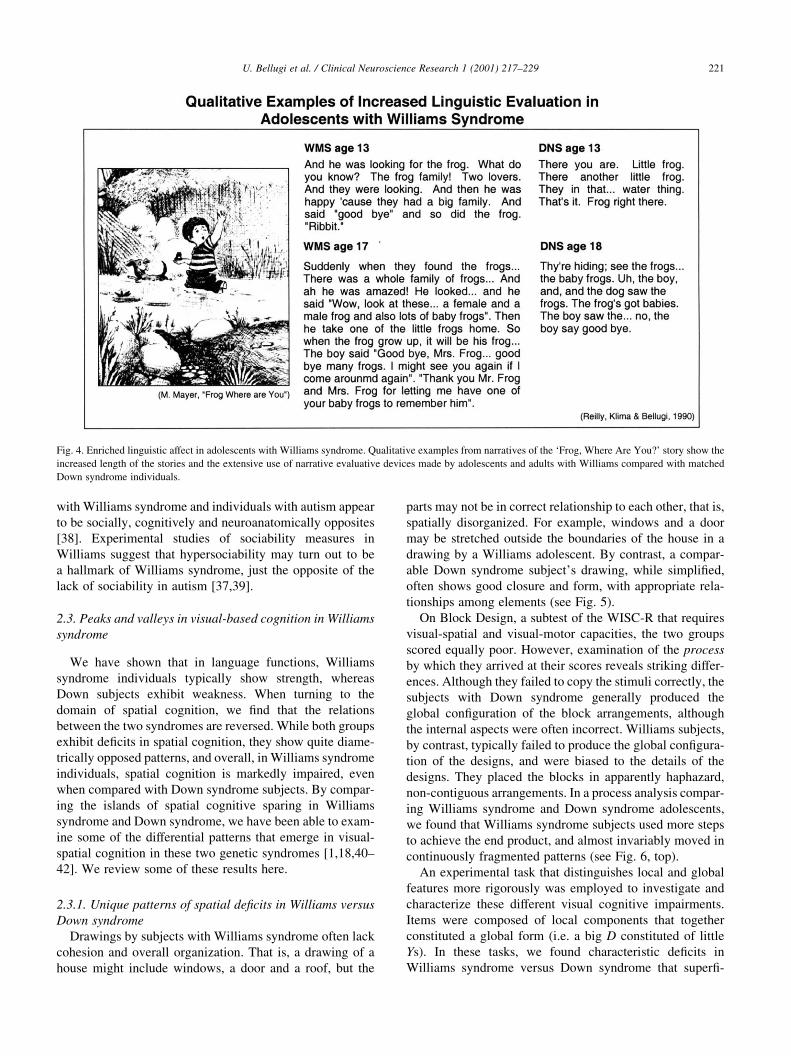
2.2.3. Enrichment of linguistic affect in Williams syndrome
Language may be emotionally enriched by affective
prosody as well as through the use of lexically-encoded
affective devices. In their narrations, Williams syndrome
subjects were found to use affective prosody (pitch change,
vocalic lengthening, modi®cations in volume) far more
frequently than either Down syndrome matches, or even
normal children. The affective richness of the Williams
syndrome subjects' narratives was also re¯ected in their
lexical choices. Their narratives included frequent
comments on the affective state of the characters in the
stories (e.g. `And ah! He was amazed' or `The dog gets
worried and the boy gets mad'), as well as the use of
dramatic devices such as character speech and sound effects
(`And boom, millions of bees came out and tried to sting
him'). Their use of exclamatory phrases and other audience
engagement devices is evident throughout many of the stor-
ies, for example `Suddenly splash! The water came up'; `Lo
and behold, they found him with a lady'; and `Gadzooks!
The boy and the dog start ¯ipping over'. These devices were
far less frequently utilized by normal subjects and were
notably absent in the Down syndrome subjects' stories. In
sum, not only are the Williams syndrome adolescents' stor-
ies replete with narrative enrichment devices, they use
proportionately more affective prosody and make greater
use of linguistic affective devices than do Down syndrome,
or even matched normal children (see Fig. 4) [36].
Despite their intellectual impairments, subjects with
Williams syndrome are not only sociable and affectively
sensitive, but they also appear to be able to manipulate
affective linguistic devices for purposes of story-telling.
However, these subjects appear to use the same level of
expressivity regardless of how many times they have told
the story and irrespective of their audience. This suggests
that their extreme expressivity may represent a deviation
from the norm [30,37]. Research suggests that the abun-
dance of affectivity, both in prosody and in linguistic
devices, may be characteristic of most subjects with
Williams syndrome, distinctly different from subjects with
right hemisphere damage, and markedly different from clas-
sic autistic subjects. Indeed, in some respects, individuals
U. Bellugi et al. / Clinical Neuroscience Research 1 (2001) 217±229220
Fig. 3. Unusual vocabulary in Williams syndrome. The ®gure shows that there is a large proportion of uncommon names in Williams subjects' responses to a
¯uency probe (a); that the total number of words produced is signi®cantly larger in Williams than in Down syndrome, regardless of category (b); and presents
sample responses from a matched individual with Williams syndrome and one with Down syndrome (c).
with Williams syndrome and individuals with autism appear
to be socially, cognitively and neuroanatomically opposites
[38]. Experimental studies of sociability measures in
Williams suggest that hypersociability may turn out to be
a hallmark of Williams syndrome, just the opposite of the
lack of sociability in autism [37,39].
2.3. Peaks and valleys in visual-based cognition in Williams
syndrome
We have shown that in language functions, Williams
syndrome individuals typically show strength, whereas
Down subjects exhibit weakness. When turning to the
domain of spatial cognition, we ®nd that the relations
between the two syndromes are reversed. While both groups
exhibit de®cits in spatial cognition, they show quite diame-
trically opposed patterns, and overall, in Williams syndrome
individuals, spatial cognition is markedly impaired, even
when compared with Down syndrome subjects. By compar-
ing the islands of spatial cognitive sparing in Williams
syndrome and Down syndrome, we have been able to exam-
ine some of the differential patterns that emerge in visual-
spatial cognition in these two genetic syndromes [1,18,40±
42]. We review some of these results here.
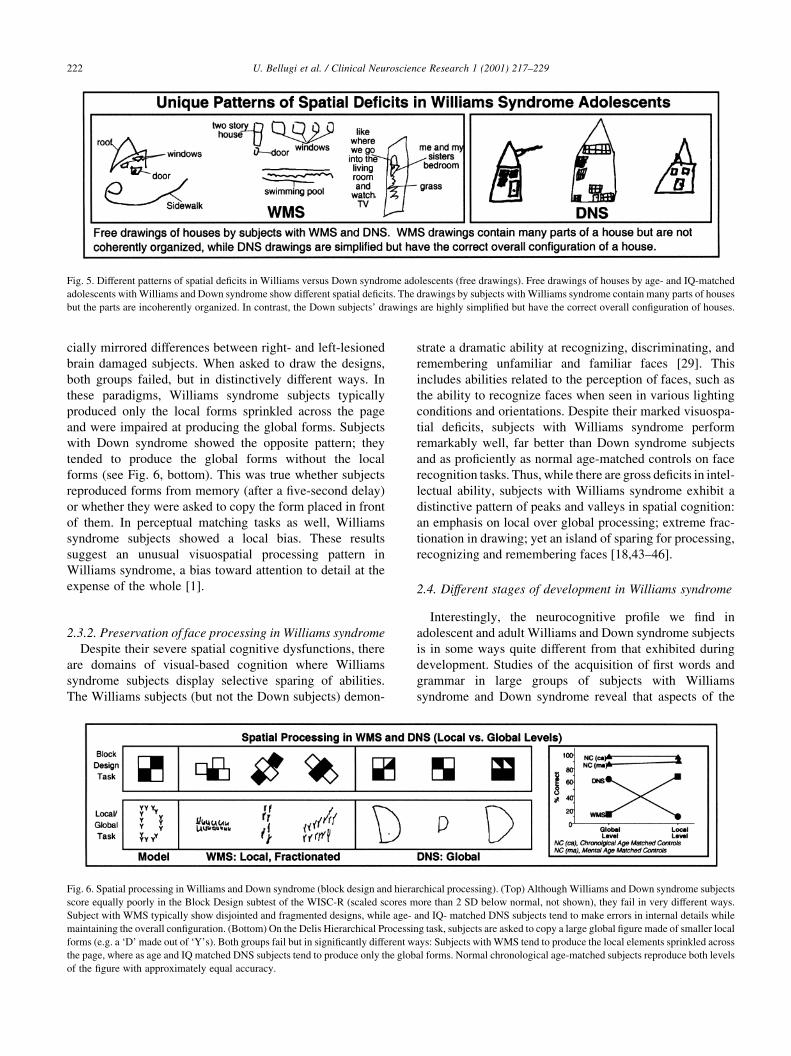
2.3.1. Unique patterns of spatial de®cits in Williams versus
Down syndrome
Drawings by subjects with Williams syndrome often lack
cohesion and overall organization. That is, a drawing of a
house might include windows, a door and a roof, but the
parts may not be in correct relationship to each other, that is,
spatially disorganized. For example, windows and a door
may be stretched outside the boundaries of the house in a
drawing by a Williams adolescent. By contrast, a compar-
able Down syndrome subject's drawing, while simpli®ed,
often shows good closure and form, with appropriate rela-
tionships among elements (see Fig. 5).
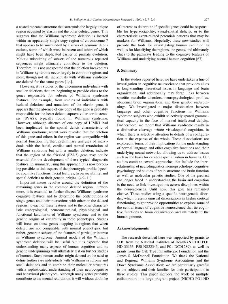
On Block Design, a subtest of the WISC-R that requires
visual-spatial and visual-motor capacities, the two groups
scored equally poor. However, examination of the process
by which they arrived at their scores reveals striking differ-
ences. Although they failed to copy the stimuli correctly, the
subjects with Down syndrome generally produced the
global con®guration of the block arrangements, although
the internal aspects were often incorrect. Williams subjects,
by contrast, typically failed to produce the global con®gura-
tion of the designs, and were biased to the details of the
designs. They placed the blocks in apparently haphazard,
non-contiguous arrangements. In a process analysis compar-
ing Williams syndrome and Down syndrome adolescents,
we found that Williams syndrome subjects used more steps
to achieve the end product, and almost invariably moved in
continuously fragmented patterns (see Fig. 6, top).
An experimental task that distinguishes local and global
features more rigorously was employed to investigate and
characterize these different visual cognitive impairments.
Items were composed of local components that together
constituted a global form (i.e. a big D constituted of little
Ys). In these tasks, we found characteristic de®cits in
Williams syndrome versus Down syndrome that super®-
U. Bellugi et al. / Clinical Neuroscience Research 1 (2001) 217±229 221
Fig. 4. Enriched linguistic affect in adolescents with Williams syndrome. Qualitative examples from narratives of the `Frog, Where Are You?' story show the
increased length of the stories and the extensive use of narrative evaluative devices made by adolescents and adults with Williams compared with matched
Down syndrome individuals.
cially mirrored differences between right- and left-lesioned
brain damaged subjects. When asked to draw the designs,
both groups failed, but in distinctively different ways. In
these paradigms, Williams syndrome subjects typically
produced only the local forms sprinkled across the page
and were impaired at producing the global forms. Subjects
with Down syndrome showed the opposite pattern; they
tended to produce the global forms without the local
forms (see Fig. 6, bottom). This was true whether subjects
reproduced forms from memory (after a ®ve-second delay)
or whether they were asked to copy the form placed in front
of them. In perceptual matching tasks as well, Williams
syndrome subjects showed a local bias. These results
suggest an unusual visuospatial processing pattern in
Williams syndrome, a bias toward attention to detail at the
expense of the whole [1].
2.3.2. Preservation of face processing in Williams syndrome
Despite their severe spatial cognitive dysfunctions, there
are domains of visual-based cognition where Williams
syndrome subjects display selective sparing of abilities.
The Williams subjects (but not the Down subjects) demon-
strate a dramatic ability at recognizing, discriminating, and
remembering unfamiliar and familiar faces [29]. This
includes abilities related to the perception of faces, such as
the ability to recognize faces when seen in various lighting
conditions and orientations. Despite their marked visuospa-
tial de®cits, subjects with Williams syndrome perform
remarkably well, far better than Down syndrome subjects
and as pro®ciently as normal age-matched controls on face
recognition tasks. Thus, while there are gross de®cits in intel-
lectual ability, subjects with Williams syndrome exhibit a
distinctive pattern of peaks and valleys in spatial cognition:
an emphasis on local over global processing; extreme frac-
tionation in drawing; yet an island of sparing for processing,
recognizing and remembering faces [18,43±46].
2.4. Different stages of development in Williams syndrome
Interestingly, the neurocognitive pro®le we ®nd in
adolescent and adult Williams and Down syndrome subjects
is in some ways quite different from that exhibited during
development. Studies of the acquisition of ®rst words and
grammar in large groups of subjects with Williams
syndrome and Down syndrome reveal that aspects of the
U. Bellugi et al. / Clinical Neuroscience Research 1 (2001) 217±229222
Fig. 6. Spatial processing in Williams and Down syndrome (block design and hierarchical processing). (Top) Although Williams and Down syndrome subjects
score equally poorly in the Block Design subtest of the WISC-R (scaled scores more than 2 SD below normal, not shown), they fail in very different ways.
Subject with WMS typically show disjointed and fragmented designs, while age- and IQ- matched DNS subjects tend to make errors in internal details while
maintaining the overall con®guration. (Bottom) On the Delis Hierarchical Processing task, subjects are asked to copy a large global ®gure made of smaller local
forms (e.g. a `D' made out of `Y's). Both groups fail but in signi®cantly different ways: Subjects with WMS tend to produce the local elements sprinkled across
the page, where as age and IQ matched DNS subjects tend to produce only the global forms. Normal chronological age-matched subjects reproduce both levels
of the ®gure with approximately equal accuracy.
Fig. 5. Different patterns of spatial de®cits in Williams versus Down syndrome adolescents (free drawings). Free drawings of houses by age- and IQ-matched
adolescents with Williams and Down syndrome show different spatial de®cits. The drawings by subjects with Williams syndrome contain many parts of houses
but the parts are incoherently organized. In contrast, the Down subjects' drawings are highly simpli®ed but have the correct overall con®guration of houses.

acquisition of ®rst words are quite delayed in both cohorts.
However, we note that children with Down syndrome exhi-
bit an early advantage for communicative gestures, while
children with Williams syndrome display an advantage for
grammar later in development [47]. Other differences
emerge in a comparison of three domains across develop-
mental ages (vocabulary, visuospatial abilities, and face
processing). Down syndrome children showed similar low
scores across the three domains. In contrast, the Williams
syndrome developmental pro®le is different across the three
domains: visuospatial functions are signi®cantly below the
Down syndrome level at all ages and seldom develop
beyond the normal ®ve-year level. Face processing is strong
from very early on, with Williams syndrome subjects tend-
ing to score above their mental age regardless of chronolo-
gical age. In language development there is an initial delay
in development of words in Williams syndrome subjects
equivalent to that of the Down syndrome subjects, and
followed by a later continuing rise in linguistic processing
as grammar emerges [48,49]. Thus the pro®le of linguistic
preservation found in older children is not evident initially.
Table 1 provides a summary of developmental neurocogni-
tive features of Williams syndrome, contrasting Williams
and Down syndromes. Performances on neurocognitive
measures suggest that individuals with Williams syndrome,
but not Down syndrome, show an uneven neurobehavioral
pro®le of speci®c de®cits, with preservations and anomalies
both within and across domains of higher cognitive func-
tioning. Furthermore, the early stages in Williams syndrome
do not necessarily predict the later stages. Williams
syndrome thus presents a rare pattern of dissociations
providing an unusual opportunity to forge links to neural
substrates and to the genetic basis of the syndrome, and we
turn to these issues next.
3. The neurobiological pro®le of Williams syndrome
The neurobiological pro®le of individuals with Williams
syndrome is being revealed through studies of brain func-
tion (event-related potentials, or ERPs), brain structure
(thre-dimensional computer-graphic analyses of magnetic
resonance images, or MRI) and brain cytoarchitectonics in
autopsy brains. Initial proposals about how the cognitive
and brain pro®les might be linked are presented here.
Studies using event-related potential (ERP) techniques are
useful in assessing the timing and organization of the neural
systems that are active during sensory, cognitive and
linguistic processing in subjects with Williams syndrome.
Event-related potentials provide information about the
timing and temporal sequence of neural events and, to
some extent, the location of neural activity. Electrodes are
placed on the scalp over speci®c brain areas while subjects
are processing information, thus allowing the monitoring of
the time course of neural activation on a ms-to-ms basis.
Studies of brain wave activity during language and face
processing paradigms in individuals with Williams
syndrome and normal individuals are reported here. The
characterization of these neurophysiological results for
Williams syndrome individuals represents one of the most
enticing ®ndings to date, and the ®rst to provide `brain
markers' for Williams syndrome as they are not seen in
other studied populations including children with language
impairments, focal lesions, Down syndrome, and normal
controls.
3.1. Neurophysiological characteristics of Williams
syndrome
3.1.1. A neurophysiological marker for language processing
The morphology of ERP components to auditory words
was dramatically different in individuals with Williams
syndrome compared to normal controls. ERPs were
recorded as subjects listened to sentences presented one
word at a time. The ®nal word in each sentence provided
good closure or was semantically anomalous (for example,
`I have ®ve ®ngers on my moon'). The results revealed that
the morphology of Williams syndrome individuals' ERP
components to auditory words were different from that of
normal controls. The unique pattern of ERPs in Williams
includes prominent positivities at 50 and 200 ms, and a
smaller than normal negativity at 100 ms which was most
striking over temporal brain regions. This pattern of compo-
nents (see Fig. 7a) was not evident in normal school-age
children or adults-or any other group examined, which
suggests that this might emerge as a marker for Williams
syndrome.
In age-matched normal controls, there are differences in
ERPs to open and closed class words. In normals, open class
words which typically convey speci®c referential meaning
(e.g. nouns, verbs, and adjectives), elicit a negativity at 400
ms that tends to be larger from posterior regions of the right
hemisphere. Closed class words, which typically convey
information about grammatical relations (e.g. articles,
prepositions, conjunctions), elicit a negativity that peaks
U. Bellugi et al. / Clinical Neuroscience Research 1 (2001) 217±229 223
Table 1
Developmental neuropsychological pro®les in Williams and Down
syndrome
Williams syndrome Down syndrome
Preschoolers
Vocabulary acquisition Delayed Delayed
Motor Milestone Delayed Delayed
Adolescents/young adults
Grammar Correct, complex Poor, simple
Semantics Larger vocabulary,
uncommon word
choices
Small vocabulary
Linguistic affect Rich Diminished
Visuomotor ability Poor, fragmented Simple, cohesive
Hierarchical processing Local Global
Processing of faces Remarkably strong Impaired

somewhat earlier and is largest over anterior regions of the
left hemisphere. Unlike normals, individuals with Williams
syndrome do not show ERP differences to open and closed
class words, nor do they show the normal right and left
hemispheres asymmetries. In normal controls, the semanti-
cally anomalous ®nal word elicits an N400 component
(negativity that peaks at 400 ms) that is larger from the
left than the right hemisphere. The N400 effect is larger in
individuals with Williams syndrome than in normal control
individuals over the left hemisphere. This larger semantic
anomaly may be related to the unusual semantic proclivities
shown by subjects with Williams syndrome in lexical tasks.
Thus, the results showing ERP differences between subjects
with Williams and normal subjects in language processing
suggest that the neural organization of these aspects of
language might be different in subjects with Williams
syndrome compared to normal controls. [1,2,50,51].
3.1.2. A neurophysiological marker for face processing
ERP recordings were made as subjects watched photo-
graphic pairs of faces presented sequentially on the compu-
ter monitor. The subjects' task was to indicate whether or
not the pairs of faces matched. Face processing ERP data on
subjects with Williams syndrome and normal controls were
obtained; the results showed that both individuals with
Williams syndrome and normal control groups showed
ERP differences to matched versus mismatched upright
faces. The normal subjects showed the largest component
over anterior regions which was greater over the right hemi-
sphere than the left; however, the subjects with Williams
syndrome did not show this right greater than left asymme-
try. In contrast to the normal adults, the subjects with
Williams syndrome also displayed an abnormally large
negativity at 200 ms to upright faces (see Fig. 7b), but not
to pictures of objects. These results appear to be speci®c to
individuals with Williams syndrome and might well be
related to their increased attention to faces. The abnormally
large negativity at 200 ms, which occurred in all subjects
with Williams syndrome subjects but not in any other
groups studied, is suggestive of a brain activity marker
that is linked to the noted strength in face processing abil-
ities found in individuals with Williams syndrome. Neuro-
physiological indices that relate brain and behavior and that
might be phenotypic markers for Williams syndrome are
suggested by these neurophysiological studies. The distinc-
tive brain wave markers, one found during face processing
and a different one found during language processing, could
be characteristic of individuals with Williams syndrome but
not of other groups. Taken together, these ®ndings suggest
that the neural systems subserving higher cognitive func-
tions such as language and face processing are different in
individuals with Williams syndrome than in normal indivi-
duals [1,2].
3.2. Neuromorphological characteristics of Williams
syndrome
New techniques of brain imaging permit visualization
and analysis of structures within the brain that were not
possible in the past. Techniques developed by Reiss, by
Damasio, and others [3,52,53], now permit an unprece-
dented visualization and three-dimensional analysis of the
living brain of subjects. Initial studies revealed that both
Williams and Down syndrome leave a distinctive morpho-
logical stamp on speci®c brain regions. MRI studies of brain
volumes were performed on a group of matched adolescents
and young adults with Williams and Down syndrome
[24,54,55]. Neuromorphological characterization of
Williams and Down syndrome subjects by magnetic reso-
nance imaging showed that the cerebral volume in both
groups was smaller than that of age-matched normal
controls. Analyses revealed important regional differences
in brain volume between the two groups of subjects. First,
anterior-brain volume was found to be disproportionally
reduced in Down syndrome subjects but proportionately
U. Bellugi et al. / Clinical Neuroscience Research 1 (2001) 217±229224
Fig. 7. Neurophysiological markers for Williams syndrome. Shown on the left is the unusual wave form to language (auditory words) exhibited by all subjects
with Williams and none of the normal controls. This is a candidate neurophysiological marker for Williams. Shown on the right is the abnormally large
negativity at 200 ms in subjects with Williams but not in normal controls or other groups tested, occurring over multiple brain regions. This is another candidate
neurophysiological marker for Williams.

preserved in subjects with Williams. Secondly, limbic struc-
tures in the temporal lobe showed essentially equal volumes
in Williams and control subjects, but were signi®cantly
reduced in Down subjects. On the other hand, the volume
of the thalamus and lenticular nuclei were seen to be much
better preserved in subjects with Down syndrome than those
with Williams. We also found that the anterior parts of the
corpus callosum, like the anterior hemispheres, were
preserved in Williams subjects, but diminished in Down
subjects [56].
Quantitative analysis of cerebellar volumes also suggested
differences, with cerebellar volume well preserved in
Williams subjects but diminished in Down subjects. Closer
regional analyses were enlightening: Jernigan and Bellugi
[24] found that the locus of preservation in Williams was
the neocerebellum. Of the two parts of the neocerebellum
that were subjected to analysis, the neocerebellar tonsils
and the neocerebellar vermis both showed volumetric preser-
vation or even increases in Williams as compared to controls,
whereas both were found to be volumetrically diminished in
Down syndrome. Importantly, the speci®c regions of the
neocerebellum that may be enlarged in Williams were
shown to be dysplagic in autism [24,38,57].
More recently, Reiss and his brain imaging group
[3,25,53,58] carried out MRI studies with higher resolution
techniques. In 14 young adult subjects with Williams and an
aged-matched control group, the decrease in total brain
volume was con®rmed, as well as the relative preservation
of the cerebellum. The superior temporal gyrus was also
found to be relatively preserved, an area that contains the
auditory system and those auditory association areas that
form part of language networks. There was also a signi®cant
curtailment of the volume of the brainstem. A greater ratio
of frontal to parieto-occipital forebrain volume was also
found, and there was reduction of the forebrain white
matter, with relative preservation of the cerebrocortical
volume. Nonetheless, regionally, the right-occipital lobe
showed excessive volume loss.
Results of related research suggest that the expansive
prefrontal cortex and the neocerebellum, both selectively
(relatively) preserved in Williams, are thought to be closely
related. These two regions of the brain are most highly
developed in Homo sapiens, and are thought to have
evolved contemporaneously [59]. Furthermore, the neocer-
ebellum has more extensive connections to prefrontal and
other association areas of the cortex than do the older parts
of the cerebellum. On the other hand, the reduction in the
forebrain white matter may explain the curtailment of the
brainstem, but it may be relevant to note that FZD3, which is
one of the deleted genes, is associated with hindbrain
segmentation, which could also explain, in part, the brain-
stem changes in Williams. The neuroanatomic pro®le of
Williams emerging from neuroimaging is beginning to
contribute to the understanding of the brain's organization
by exhibiting a morphological pattern that can result from
genetic bias. The ®nding that frontal and neocerebellar
regions are selectively preserved in Williams suggests that
they all may come under the in¯uence of a single genetic
developmental factor, or that their development is mutually
interactive, or both. These issues bearing on the relationship
of brain to behavior are fundamental to central questions of
cognitive neuroscience.
3.3. Brain cytoarchitectonic characteristics of Williams
syndrome
Anatomy is the logical link between genes and behavior.
The purpose of our research on the neuroanatomy of
Williams is to help link the anatomical ®ndings to the
genetic/molecular disorder on the one hand and to the beha-
vior disorder on the other, thus helping to link genes to
cognition and emotion. Speci®cally, an anatomical research
program in Williams must ultimately be able to explain the
relationship between the deleted genes in region 7q11.23
[4,9,11,23,60±62] and the building and maintenance of
brain structures, on the one hand, and, on the other hand,
the abnormal behaviors, consisting of mental retardation,
visuo-spatial de®cits, relatively good linguistic abilities,
an unusual personality, and good facial recognition and
musical abilities [7,18±20,37,63±65].
Williams syndrome involves selective rather than gener-
alized cognitive de®cits, and offers an important opportunity
for linking brain ®ndings to speci®c atypical cognitive
pro®les. Four autopsy brains of individuals with Williams
syndrome have been studied by Galaburda and colleagues
[23,60,62]. Microencephaly and the relative curtailment of
the occipital and posterior-parietal areas were evident in
three of the brains. One of the four brains showed a marked
reduction in the size of the parietal, posterior-temporal and
occipital regions in comparison with the more rostral
portions of the hemispheres. These abrupt and dramatic
reductions led to the brain appearing as if a band had
constricted its posterior portions. MRI data also corrobo-
rated the general ®nding of a reduction in posterior areas.
Curtailment of the dorsal-parietal regions and posterior-
temporal areas might indeed be relevant to the extreme
visuospatial de®cits seen in individuals with Williams
syndrome (see Fig. 8). One brain showed dramatic reduction
in the size of the amygdala, which could be associated with
the hypersocial behavior that occurs in subjects with
Williams syndrome. The four brains show largely normal
overall sulcal patterns, except for some simpli®cation of
tertiary sulcation and a consistently non-opercularized
dorsal central sulcus. The central sulci in normal brains
reach all the way to the interhemispheric ®ssure and then
a short distance further onto the medial surfaces of the hemi-
spheres, but in all the available Williams syndrome cases
the central sulcus ends no less than a centimeter lateral to
the interhemispheric ®ssure. This ®nding could indicate
abnormal development of the medio-dorsal cortices, which
have been associated with visuospatial functions (see Fig.
8b) [60,62].
U. Bellugi et al. / Clinical Neuroscience Research 1 (2001) 217±229 225

The observed cell numbers and cell-packing densities
suggest early developmental arrest (for example, prenatally
or before the second year of age), or regressive events
occurring postnatally into the middle of the ®rst decade of
life. Galaburda and colleagues are currently examining gene
expression in comparative Williams and normal brains with
respect to elastin, syntaxin 1A and other genes in the
Williams region [60]. Research that involves links between
genomic changes, messenger and product expression lead-
ing to the unusual development of the Williams syndrome
brain, will shed light on normal brain and behavioral devel-
opment. The results may relate to the peaks and valleys of
cognitive abilities in Williams syndrome. These analyses
provide opportunities for linking brain ®ndings to cognitive
de®cits and their genetic underpinnings [66].
4. The molecular genetic pro®le of Williams
The unusual neurocognitive pro®le of Williams
syndrome makes it a compelling model of the pathways
between genes and human cognition. It is becoming clear
that the syndrome's unique genomic organization may also
make it an important model of human chromosomal evolu-
tion and disease [4,9]. Williams syndrome is known to be
caused by a deletion that includes the gene encoding elastin
(ELN), Frizzled, Syntaxin 1a, Lim1 Kinase and other genes
on chromosome 7 [4]. Studies are underway seeking to
identify the genes and elucidate the chromosomal mechan-
isms responsible for Williams syndrome, in order to relate
these to the cognitive and neural characteristics of the popu-
lation [4]. Fig. 9 shows the molecular genetic basis for
Williams, speci®cally, the area of the hemideletion in
Williams on chromosome 7 affecting approximately 20
genes, some of which are indicated on the genetic map.
Molecular genetic studies [4] are involved in constructing
a physical map of the deleted region of chromosome 7 band
q11.23 by using multi-color ¯uorescence in situ hybridiza-
tion (FISH) of bacterial arti®cial chromosomes (BACs) on
metaphase and interphase chromosomes, large-fragment
library screening, genomic Southern blot and pulsed ®eld
gel analyses, STS (sequence tagged site) and polymorphic
marker analyses. BACs were chosen to construct the physi-
cal map because they are cloned in a stable vector and
contain large genomic fragments of up to 300 kb that are
stable and readily manipulated and are therefore suitable for
gene isolation and DNA sequencing. These map reagents
were used to investigate the size and extent of the deletions
in individuals with Williams syndrome in whom subsets of
features including neurocognitive pro®les, brain structures
and functions were simultaneously determined [1,9±11].
A working model of the genome organization character-
izing chromosome band 7q11.2 that incorporates other maps
was developed, which suggested that the region includes
highly homologous chromosomal duplications which are
also characterized by a number of repeat sequence families,
genes and pseudogenes, the totality of which is organized as
U. Bellugi et al. / Clinical Neuroscience Research 1 (2001) 217±229226
Fig. 9. Genome organization of the Williams region. A genetic marker for
WMS is the deletion of one copy of a small set of genes on chromosome 7,
band 7q1123, shown in the ideogram. This region is expanded to the right to
illustrate genes that are missing one copy in WMS, including the gene for
elastin. The region involving the common breakpoints in WMS are also
illustrated.
Fig. 8. Brain cytoarchitectonic ®ndings in Williams syndrome. The arrows point to a marked indentation of the temporoparietal region and to posterior
curtailment of the hemispheres in Williams' brains, consistent with their spatial de®cit. (left). Note the difference in the medial reach (arrows) of the central
sulci between the Williams and controls subjects/brains, particularly in the dorsomedial portions of the hemispheres, again consistent with the visuo-spatial
de®cit in Williams syndrome (right).
a nested repeated structure that surrounds the largely unique
region occupied by elastin and the other deleted genes. This
suggests that the Williams syndrome deletion is located
within an apparently single copy region of chromosome 7
that appears to be surrounded by a series of genomic dupli-
cations, some of which must be recent and others of which
might have been duplicated earlier in primate evolution.
Meiotic mispairing of subsets of the numerous repeated
sequences might ultimately contribute to the deletion.
Therefore, it is not unexpected that the deletion breakpoints
in Williams syndrome occur largely in common regions and
most, though not all, individuals with Williams syndrome
are deleted for the same genes [1,4].
However, it is studies of the uncommon individuals with
smaller deletions that are beginning to provide clues to the
genes responsible for subsets of Williams syndrome
features. For example, from studies of individuals with
isolated deletions and mutations of the elastin gene, it
appears that the absence of one copy of the gene is probably
responsible for the heart defect, supravalvular aortic steno-
sis (SVAS), typically found in Williams syndrome.
However, although absence of one copy of LIMK1 had
been implicated in the spatial de®cit characteristic of
Williams syndrome, recent work revealed that the deletion
of this gene and others in the region was compatible with
normal function. Further, preliminary analyses of indivi-
duals with the facial, cardiac and mental retardation of
Williams syndrome but with a smaller deletion, indicate
that the region of the frizzled (FZD3) gene may not be
essential for the development of these typical diagnostic
features. In summary, using this approach, it is now becom-
ing possible to link aspects of the phenotypic pro®le (speci-
®c cognitive functions, facial features, hypersociability, and
spatial de®cits) to their genetic origins. [4,9±11].
Important issues revolve around the de®nition of the
remaining genes in the common deleted region. Further-
more, it is essential to further dissect Williams syndrome
cognitive features and to determine the contributions of
single genes and their interactions with others in the deleted
regions, to each of these features and to the other character-
istic embryological, neuroanatomical, physiological and
functional landmarks of Williams syndrome and to the
genetic origins of variability in these phenotypes. Studies
will focus on those genes mapping in regions that when
deleted are not compatible with normal phenotypes, but
rather, generate subsets of the features of particular interest
in Williams syndrome. Animal models of the Williams
syndrome deletion will be useful but it is expected that
understanding many aspects of human cognition and its
genetic underpinnings will ultimately rest on further studies
of humans. Such human studies might depend on the need to
de®ne further rare individuals with Williams syndrome and
small deletions and to combine their molecular structures
with a sophisticated understanding of their neurocognitive
and behavioral phenotypes. Although many genes probably
contribute to the mental retardation, it will without doubt be
of interest to determine if speci®c genes could be responsi-
ble for hypersociability, visual-spatial de®cits, or to the
characteristic event-related potentials patterns that may be
markers for Williams. Hopefully, these new studies will
provide the tools for investigating human evolution as
well as for identifying the regions, the genes, and ultimately
clues to the pathways leading to the cognitive features of
Williams and underlying normal human cognition [67].
5. Summary
In the studies reported here, we have undertaken a line of
investigation in cognitive neuroscience that provides clues
to long-standing theoretical issues in language and brain
organization, and additionally may forge links between
speci®c metabolic disorders, neuropsychological pro®les,
abnormal brain organization, and their genetic underpin-
nings. We investigated a major dissociation between
language and other cognitive functions in Williams
syndrome subjects who exhibit selectively spared gramma-
tical capacity in the face of marked intellectual de®cits.
Furthermore, we report that Williams syndrome results in
a distinctive cleavage within visual/spatial cognition, in
which there is selective attention to details of a con®gura-
tion at the expense of the whole. These dissociations are
explored in terms of their implications for the understanding
of normal language and other cognitive functions and their
underlying neural networks, allowing us to address issues
such as the basis for cerebral specialization in humans. Our
studies combine several approaches that include the inter-
relationship of neurolinguistics, neuropsychology, cognitive
psychology and studies of brain structure and brain function
as well as molecular genetic studies. One of the greatest
challenges faced in understanding the brain and cognition
is the need to link investigations across disciplines within
the neurosciences. Until now, this goal has remained
elusive. These studies using a speci®c neurogenetic disor-
der, which presents unusual dissociations in higher cortical
functioning, might provide opportunities to explore some of
the central issues of cognitive neuroscience that tie cogni-
tive functions to brain organization and ultimately to the
human genome.
Acknowledgements
The research described here was supported by grants to
U.B. from the National Institutes of Health (NICHD PO1
HD 33133, P50 NS22343, and P01 DC01289), as well as
grants from the Oak Tree Philanthropic Foundation and the
James S. McDonnell Foundation. We thank the National
and Regional Williams Syndrome Associations and the
Down Syndrome Association; we are particularly grateful
to the subjects and their families for their participation in
these studies. This paper includes the work of multiple
collaborators in a large program project (NICHD PO1 HD
U. Bellugi et al. / Clinical Neuroscience Research 1 (2001) 217±229 227
33133): Drs. Ursula Bellugi, Debra Mills, Julie R. Koren-
berg, Albert Galaburda, and Allan Reiss. We are also grate-
ful to Diana Kikuchi for her help in preparation of the
manuscript. Illustrations, copyright Dr Ursula Bellugi,
Laboratory for Cognitive Neuroscience, The Salk Institute
for Biological Studies, La Jolla, CA, 92037.
References
[1] Bellugi U, Lichtenberger L, Mills D, Galaburda A, Korenberg JR.
Bridging cognition, brain and molecular genetics: evidence from
Williams syndrome. Trends Neurosci 1999;5:197±208.
[2] Mills DL, Alvarez T, St George M, Applebaum L, Bellugi U, Neville
H, et al. Electrophysiological studies of face processing. Special issue:
Linking cognitive neuroscience and molecular genetics: new perspec-
tives from Williams syndrome. Bellugi U, St George M, editors. J
Cogn Neurosci 2000:47±64.
[3] Reiss AL, Eliez S, Schmitt JE, et al. Neuroanatomy of Williams
syndrome: a high-resolution MRI study. Special issue: Linking cogni-
tive neuroscience and molecular genetics: new perspectives from
Williams syndrome. Bellugi U, St George M, editors. J Cogn
Neurosci 2000:65±73.
[4] Korenberg JR, Chen X-N, Hirota H, et al. Genome structure and
cognitive map of Williams syndrome. Special issue: Linking cogni-
tive neuroscience and molecular genetics: new perspectives from
Williams syndrome. Bellugi U, St George M, editors. J Cogn
Neurosci 2000:89±107.
[5] Williams J, Barratt-Boyes B, Lowe J. Supravalvular aortic stenosis.
Circulation 1961;24:1311±1318.
[6] Bellugi U, Morris CA, editors. Williams syndrome: from cognition to
gene. Abstracts from the Williams Syndrome Association Profes-
sional Conference. Genet Couns 1995;95(1):131±192.
[7] Bellugi U, Lai ZC, Korenberg J. Genes, brains and behavior: what
genetic disorders reveal about behavior. In: Bizzi E, Calissano P,
Volterra V, editors. The brain of Homo sapiens, Frontiere della Biolo-
gia, vol 4. Rome, Italy: Istituto della Enciclopedia Italiana, 1999, pp.
127±135.
[8] Ewart AK, Jin W, Atkinson DL, Morris CA, Keating MT. Supravalv-
ular aortic stenosis associated with a deletion disrupting the elastin
gene. J Clin Invest 1994;83:1071±1077.
[9] Korenberg JR, Chen X-N, Mitchell S, et al. The genomic organization
of Williams syndrome. Am J Hum Genet Suppl 1996;59(4):A306.
[10] Korenberg JR, Chen X-N, Mitchell S, et al. The genomic organization
of Williams syndrome. In poster symposium `Bridging cognition,
brain and gene'. Int Behav Neurosci Soc Abstr 1997;6(59):P252.
[11] Korenberg JR, Hirota H, Chen X-N, et al. The molecular genetic basis
of Williams syndrome. Presentation in symposium, `Bridging cogni-
tion, brain and gene: evidence from Williams syndrome'. Cogn
Neurosci Soc Progr Abstr 1998:11.
[12] Clahsen H, Almazan M. Syntax and morphology in Williams
syndrome. Cognition 1998;68:167±198.
[13] Karmiloff-Smith A. Development itself is the key to understanding
developmental disorders. Trends Cogn Sci 1998;2(10):389±398.
[14] Jarrold C, Baddley AD, Hewes AKJ. Genetically dissociated compo-
nents of working memory: evidence from Down's and Williams
syndrome. Child Psychol Psychiatry Allied Discipl 1998;39:511±552.
[15] Pinker S. The language instinct. Harmondsworth: Penguin, 1994.
[16] Volterra V, Caprici O, Pezzini G, Sabbadini L. Linguistic abilities in
Italian children with Williams syndrome. Cortex 1996;32:663±677.
[17] Bellugi U, St George M, editors. Special Issue: Linking cognitive
neuroscience and molecular genetics: new perspectives from
Williams syndrome. Cogn Neurosci 2000:1±107.
[18] Bellugi U, Lichtenberger L, Jones H, Lai Z, St George M. The neuro-
cognitive pro®le of Williams syndrome: a complex pattern of
strengths and weaknesses. Special issue: Linking cognitive
neuroscience and molecular genetics: new perspectives from
Williams syndrome. Bellugi U, St George M, editors. J Cogn
Neurosci 2000:7±29.
[19] Bellugi U, Klima ES, Wang PP. Cognitive and neural development:
clues from genetically based syndromes. In: Magnussen D, editor.
The life-span development of individuals: behavioral, neurobiologi-
cal, and psychosocial perspectives. The Nobel Symposium, New
York: Cambridge University Press, 1996, pp. 223±243.
[20] Bellugi U, Mills D, Jernigan T, Hickok G, Galaburda A. Linking
cognition, brain structure and brain function in Williams syndrome.
In: Tager-Flusberg H, editor. Neurodevelopmental disorders: contri-
butions to a new framework from the cognitive neurosciences,
Cambridge, MA: MIT Press, 1999, pp. 111±136.
[21] Bellugi U, Wang P, Jernigan TL. Williams syndrome: an unusual
neuropsychological pro®le. In: Broman S, Grafman J, editors. Atypi-
cal cognitive de®cits in developmental disorders: implications for
brain function, Hillsdale, NJ: Lawrence Erlbaum, 1994, pp. 23±56.
[22] Bellugi U, Wang PP. Williams syndrome: from cognition to brain to
gene. In: Edelman G, Smith BH, editors. Encyclopedia of
neuroscience, Amsterdam: Elsevier Science, 1998.
[23] Galaburda AM, Wang PP, Bellugi U, Rossen M. Cytoarchitectonic
®ndings in a genetically based disorder: Williams syndrome. Neurore-
port 1994;5:753±757.
[24] Jernigan TL, Bellugi U. Neuroanatomical distinctions between
Williams and Down syndromes. In: Broman S, Grafman J, editors.
Atypical cognitive de®cits in developmental disorders: implications
for brain function, Hillsdale, NJ: Lawrence Erlbaum, 1994, pp. 57±66.
[25] Schmitt JE, Eliez S, Bellugi U, Reiss AL. Analysis of cerebral shape
in Williams syndrome. Arch Neurol 2001;58(2):283±287.
[26] Korenberg J. Down syndrome phenotypes: The consequences of chro-
mosomal imbalance. Proc Natl Acad Sci USA 1994;91:4997±5001.
[27] Korenberg J. Down syndrome: a molecular understanding of the
origin of phenotypes. In: Epstein C, editor. The phenotypic mapping
of Down syndrome and other aneuploid conditions, New York:
Wiley-Liss, 1993, pp. 87±115.
[28] Udwin O. A survey of adults with Williams syndrome and idiopathic
infantile hypercalcemia. Dev Med Child Neurol 1990;32:129±141.
[29] Rossen ML, Klima ES, Bellugi U, Bihrle A, Jones W. Interaction
between language and cognition: evidence from Williams syndrome.
In: Beitchman JH, Cohen N, Konstantareas M, Tannock R, editors.
Language, learning, and behavior disorders: developmental, biologi-
cal, and clinical perspectives, New York: Cambridge University
Press, 1996, pp. 367±392.
[30] Losh M, Bellugi U, Reilly J, Anderson D. The integrity and indepen-
dence of evaluation in narratives: evidence from children with
Williams Syndrome. Narrative Inquiry 2001 in press.
[31] Bellugi U, Bihrle A, Neville H, Jernigan T, Doherty S. Language,
cognition, and brain organization in a neurodevelopmental disorder.
In: Gunnar M, Nelson C, editors. Developmental behavioral
neuroscience, Hillsdale, NJ: Lawrence Erlbaum, 1992, pp. 201±232.
[32] Rubba J, Klima ES. Preposition use in speakers with Williams
syndrome: Some cognitive grammar proposals. Center for Research
in Language Newsletter, University of California at San Diego
1991:3±12.
[33] Losh M, Reilly J, Bellugi U, Cassady C, Klima ES. Linguistically
encoded affect is abnormally high in Williams syndrome children. In
poster symposium `Bridging cognition, brain and gene'. Int Behav
Neurosci Soc Abstr 1997;6:P253.
[34] Lichtenberger L, Bellugi U. The intersection of spatial cognition and
language in Williams syndrome. Cogn Neurosci Soci Abstr Progr
1998;80:68.
[35] Wang PP, Bellugi U. Williams syndrome, Down syndrome, and
cognitive neuroscience. Am J Dis Child 1993;147:1246±1251.
[36] Bellugi U, Losh M, Reilly J, Anderson D. Excessive use of linguis-
tically encoded affect: Stories from young children with Williams
syndrome (Technical Report CND-9801). University of California,
U. Bellugi et al. / Clinical Neuroscience Research 1 (2001) 217±229228

San Diego, Center for Research in Language, Project in Cognitive and
Neural Development. San Diego, CA: University of California, 1998.
[37] Jones W, Bellugi U, St George M, et al. Hypersociability in Williams
syndrome. Special issue: Linking cognitive neuroscience and mole-
cular genetics: new perspectives from Williams syndrome. Bellugi U,
St George M, editors. J Cogn Neurosci 2000:30±46.
[38] Courchesne E, Bellugi U, Singer N. Infantile autism and Williams
syndrome: social and neural worlds apart. Genet Couns
1995;6(1):144±145.
[39] Jones W, Lincoln A, Reilly J, Grafstein S, Beret N, Bellugi U. The use
of social engagement techniques in young children with Williams
syndrome. Williams Syndrome Association Professional Conference,
King of Prussia, PA, 1996.
[40] Wang PP, Doherty S, Rourke SB, Bellugi U. Unique pro®le of visuo-
perceptual skills in a genetic syndrome. Brain Cogn 1995;29:54±65.
[41] Bihrle AM, Bellugi U, Delis D, Marks S. Seeing either the forest or
the trees: dissociation in visuospatial processing. Brain Cogn
1989;11:37±49.
[42] Bihrle AM. Visuospatial processing in Williams and Down
syndromes. Doctoral dissertation. San Diego, CA: University of Cali-
fornia at San Diego and San Diego State University, 1990.
[43] Jones W, Rossen M, Hickok G, Jernigan T, Bellugi U. Links between
behavior and brain: brain morphological correlates of language, face,
and auditory processing in Williams syndrome. Soc Neurosci Abstr
1996;21(3):757/18.
[44] Jones W, Lai ZC. The relationship between intact face processing and
impaired spatial cognition in Williams syndrome. In poster sympo-
sium `Bridging cognition, brain and gene'. Int Behav Neurosci Soc
Abstr 1997;6(59):P251.
[45] Rossen ML, Jones W, Wang PP, Klima ES. Face processing: remark-
able sparing in Williams syndrome. Genet Couns 1995;6(1):138±140.
[46] Rossen ML, Smith D, Jones W, Bellugi U, Korenberg JR. Spared face
processing in Williams syndrome: new perspectives on brain-beha-
vior links in a genetically-based syndrome. Soc Neurosci Abstr
1995;121(3):1926.
[47] Singer-Harris NG, Bellugi U, Bates E, Jones W, Rossen ML.
Contrasting pro®les of language development in children with
Williams and Down syndromes. Dev Neuropsychol
1997;13(3):345±370.
[48] Bellugi U, Lai ZC, Wang P. Language, communication and neural
systems in Williams syndrome. Ment Retard Dev Disabil Res Rev
1997;4(3):334±342.
[49] Jones W, Hickok G, Rossen M, Bellugi U. Dissociations in cognitive
development: differential effects from two genetically based
syndrome (Technical Report CND-9805). University of California,
San Diego: Center for Research in Language, Project in Cognitive
and Neural Development, 1998.
[50] Mills D, Neville H, Appelbaum G, Prat C, Bellugi U. Electrophysio-
logical markers of Williams syndrome. In poster symposium `Brid-
ging cognition, brain and gene'. Int Behav Neurosci Soc Abstr
1997;6:P2±P50.
[51] Neville HJ, Mills DL, Bellugi U. Effects of altered auditory sensitivity
and age of language acquisition on the development of language-
relevant neural systems: preliminary studies of Williams syndrome.
In: Broman S, Grafman J, editors. Atypical cognitive de®cits in devel-
opmental disorders: implications for brain function, Hillsdale, NJ:
Lawrence Erlbaum, 1994, pp. 67±83.
[52] Frank RJ, Damasio H, Grabowski TJ. Brainvox: an interactive, multi-
modal visualization and analysis system for neuroanatomical
imaging. Neuroimage 1997;5(1):13±30.
[53] Schmitt JE, Eliez S, Warsofsky IS, Bellugi U, Reiss AL. Corpus
callosum morphology of Williams syndrome: relationships to genet-
ics and behavior. Dev Med Child Neurol 2001;43(3):155±159.
[54] Jernigan TL, Bellugi U. Anomalous brain morphology on magnetic
resonance images in Williams syndrome and Down syndrome. Arch
Neurol 1990;47:529±533.
[55] Jernigan TL, Bellugi U, Sowell E, Doherty S, Hesselink JR. Cerebral
morphological distinctions between Williams and Down syndromes.
Arch Neurol 1993;50:186±191.
[56] Wang PP, Hesselink JR, Jernigan TL, Doherty S, Bellugi U. Callosal
morphology concurs with neurobehavioral and neuropathological
®ndings in two neurodevelopmental disorder. Neurology
1992;42:1999±2002.
[57] Courchesne E. New evidence of cerebellar and brainstem hypoplasia
in autistic infants, children and adolescents: the MR imaging study by
Hashimoto and colleagues. J Autism Dev Disord 1995;25(1):19±22.
[58] Schmitt E, Eliez S, Warsofsky I, Bellugi U, Reiss A. Cerebellar
vermis preservation in Williams syndrome. J Psychiatric Res 2001
submitted.
[59] Deacon TW. Rethinking mammalian brain evolution. Am Zool
1990;30:629±705.
[60] Galaburda AM, Bellugi U. Multi-level analysis of cortical neuroanat-
omy in williams syndrome. Special issue: Linking cognitive
neuroscience and molecular genetics: new perspectives from
Williams syndrome. Bellugi U, St George M, editors. J Cogn
Neurosci 2000:74±88.
[61] Osborne LR, Herbick JA, Greavette T, Heng HH, Tsui L-C, Sherer
SW. PMS2-related genes ¯ank the rearrangement breakpoints asso-
ciated with Williams syndrome and other diseases on human chromo-
some 7. Genomics 1997;45:402±406.
[62] Galaburda AM, Wang PP, Rossen ML, Bellugi U. Cytoarchitectonic
and immunohistochemical ®ndings in Williams syndrome. Genet
Couns 1995;6(1):142±144.
[63] Lenhoff HM, Wang PP, Greenberg F, Bellugi U. Williams syndrome
and the brain. Sci Am 1997;277(6):68±73.
[64] Bellugi U, Adolphs R, Cassady C, Chiles M. Towards the neural basis
for hypersociability in a genetic syndrome. NeuroReport
1999;10:1653±1657.
[65] Levitin DJ, Bellugi U. Musical abilities in individuals with Williams
syndrome. Music Percept 1998;15(4):357±389.
[66] Sawchenko P, Dargusch R, Arias C, Bellugi U. Evidence for elastin
expression in cerebellar Purkinje cells: implications for Williams
syndrome. Int Behav Neurosci Soc Abstr 1997;6:P249.
[67] Korenberg JR, Chen X-N, Shi Z-Y, Schmitt E, Lai Z, Bellugi U, Reiss
A, Mills D, Galaburda A. Williams syndrome: genes and pathways
responsible for human cognition. Abstract from the American Society
of Human Genetics Annual Conference, 2000.
U. Bellugi et al. / Clinical Neuroscience Research 1 (2001) 217±229 229