Human Memory and the Medial Temporal Lobe Yael Shrager Squire Lab December 1, 2005.

Neurobiology of Learning and Memory 82 (2004) 178–198
www.elsevier.com/locate/ynlme
What, if anything, is the medial temporal lobe, and how canthe amygdala be part of it if there is no such thing?
Elisabeth A. Murraya,* and Steven P. Wiseb
a Laboratory of Neuropsychology, National Institute of Mental Health, Building 49, Room 1B80, MSC 4415, 49 Convent Drive,
Bethesda, MD 20892-4415, USAb Laboratory of Systems Neuroscience, National Institute of Mental Health, Building 49, Room B1EE17, MSC 4401, 49 Convent Drive,
Bethesda, MD 20892-4401, USA
Received 28 March 2004; revised 6 May 2004; accepted 7 May 2004
Available online 17 June 2004
Abstract
Should the medial temporal lobe (MTL) of primates—which includes allocortical structures such as the hippocampus, neocortical
structures such as the parahippocampal cortex, and nuclear structures such as the basolateral amygdala—be considered a single
‘‘thing’’? According to the prevailing view, here termed the reification theory, the answer is yes. According to this theory, the MTL
functions as an amalgamated entity that provides the neuronal mechanisms for declarative memory; the greater the damage to the
MTL or any of its components, the greater the deleterious effects on declarative memory. A countervailing view, here called the
balkanization theory, holds that the various components of the MTL process and store different kinds of information. According to
this theory, damage to each part of the MTL causes a unique set of behavioral deficits—some involving memory, others involving
perception, and yet others involving response selection. The empirical neuropsychological evidence favors the balkanization theory,
as do some new concepts in theoretical neuroanatomy.
Published by Elsevier Inc.
Keywords: Recognition memory; Discrimination learning; Perirhinal cortex; Hippocampus; Basal ganglia; Declarative memory
1. Introduction
The questions posed in our title are admittedly am-
biguous and somewhat strange, but they follow Swan-
son and Petrovich (1998) who asked: ‘‘What is the
amygdala?’’ They answered that the amygdala is not any
‘‘thing,’’ at all. Instead, they described the amygdala as a
composite of other things, namely parts of the cortex,
striatum, and claustrum. Our title also pays homage to
the 1957 classic by Wood, ‘‘What, if anything, is arabbit?’’ He explored a question posed early in the 20th
century: Are rabbits a separate thing—in contemporary
terms, a clade—or, as some experts thought at the time,
various kinds of rodent? We, too, explore ideas about
‘‘things’’ that date to the early 20th century. We ask
whether the medial temporal lobe (MTL) might be
* Corresponding author. Fax: 1-301-402-0046.
E-mail address: [email protected] (E.A. Murray).
1074-7427/$ - see front matter. Published by Elsevier Inc.
doi:10.1016/j.nlm.2004.05.005
better understood if we imagined that there is no such
thing. Of course, the structures in the MTL will notdisappear as a result; they will continue to be where they
are and do what they do. We hope, however, that it will
prove helpful to change the question from ‘‘What does
the MTL do?’’, which seems to assume a solitary an-
swer, to the questions posed in our title, which recognize
the structural diversity of the MTL.
To anticipate our presentation, we offer two take-
home messages. First, according to a new view of fore-brain organization, most parts of the MTL—including
the hippocampus and much of the amygdala—function
in recurrent, neural loops that include striatal and pal-
lidal structures. In this context, theories that seek to
contrast the functions of one entity called the MTL with
other entities called ‘‘the striatum,’’ ‘‘the basal ganglia,’’
or ‘‘the corticostriatal system’’ need fundamental re-
consideration or, at the very least, reformulation. Sec-ond, the MTL is not a ‘‘thing’’ at all, but a diverse
collection of structures, each with its own embryological

E.A. Murray, S.P. Wise / Neurobiology of Learning and Memory 82 (2004) 178–198 179
and evolutionary history. Each part of the MTL con-tributes to perception, memory, and response selection
in its own way. This conclusion stands in stark contrast
with the prevailing view of the MTL, which holds that
‘‘it’’ functions as an amalgamated declarative memory
system. We begin with a brief survey of the prevailing
view and its history.
2. The reification theory
The prevailing view of MTL function began to take
shape roughly 50 years ago, when a patient—famously
designated H.M.—underwent a bilateral excision of
large parts of his MTL. As intended, the operation
lessened the frequency and severity of his epileptic sei-zures. As one unintended side effect of the operation,
however, H.M. became profoundly amnesic. Another
unintended consequence of that surgery was five decades
of neuropsychological research, which led to the pre-
vailing view of the MTL and its functions, called here
the reification theory.
The reification theory holds that the MTL is a single
‘‘thing,’’ one that performs a unitary function as a de-clarative memory system. For example, Zola-Morgan,
Squire, and Ramus (1994, p. 493) concluded that
The severity of memory impairment increases as additional
components of the medial temporal lobe memory system are
damaged.
As one measure of declarative memory in monkeys,
they evaluated performance on the delayed non-
matching-to-sample task (DNMS). In this task, eachtrial comprises two parts: a sample presentation and a
choice test. In a typical DNMS experiment, monkeys
first see and displace a single object, called the sample,
to obtain a food reward hidden underneath. On the
second part of the trial, monkeys are presented with
two objects, the sample and a novel object, and they
must choose the novel object to earn another food
reward. Memory for the sample object is taxed by in-creasing the delay interval between the sample presen-
tation and choice test. In the view of Zola, Squire, and
their colleagues, damage to almost any part of the
MTL leads to an impairment in the ability to learn the
DNMS rule and to perform the DNMS task over a
variety of delay intervals. It should be noted that these
authorities hold that the amygdala is not part of the
MTL memory system, but, with respect to the MTL�sremaining structures, the basic proposition is that they
work together as a single functional entity. According
to the reification theory, damage to each component of
the MTL should cause similar behavioral deficits
(Zola-Morgan et al., 1994). We acknowledge that some
might view this formulation as one that oversimplifies a
more nuanced theory (S. Zola, personal communica-
tion), but we rely here on a fair reading of the pub-lished record, one that leads to testable and falsifiable
hypotheses.
Although the reification theory dominates the field,
not all experts share a high level of enthusiasm for it.
For example, Murray and her colleagues have argued
that the MTL consists of multiple functional subdivi-
sions (Murray, 1996; Murray, Bussey, Hampton, &
Saksida, 2000). In addition, Gaffan (2002) has arguedagainst the very idea of ‘‘memory systems.’’ According
to his analysis, the ‘‘dense amnesia’’ that follows damage
to the MTL results from interrupting inputs to that re-
gion from the basal forebrain, as well as disconnecting
the MTL from the prefrontal cortex. He reasoned that
the former disconnection, perhaps involving cholinergic
inputs, disables plasticity mechanisms in the MTL and
that the latter eliminates the contribution of somethingakin to a global workspace (Baars, Ramsoy, & Laureys,
2003), an information-processing system transcending
domain specificity. In other words, according to Gaffan
(2002), the MTL is nothing special in the pantheon of
telencephalic structures, some parts of it process certain
kinds of information, other parts process different in-
formation, and damage to each of these parts produces a
different set of behavioral deficits (see also Gaffan, 2001;O�Keefe, 1999). In Gaffan�s view, the susceptibility to
global anterograde amnesia after MTL damage arises
from an accident of telencephalic geometry in certain
primates. We call this idea—one positing different
functions for each of the MTL�s many components—the
balkanization theory.
In view of such doubts, how did the reification theory
gain such wide acceptance? This process began with theunfortunate neurosurgical outcome mentioned above
for the patient H.M.: the unintentional production of a
severe and relatively selective amnesia, mainly antero-
grade in nature, through surgical ablation of large parts
of his MTL. Attempts to mimic H.M.�s amnesia in
monkeys met, at first, with little success. In early studies,
monkeys with bilateral removals of the amygdala, hip-
pocampus, and underlying cortex—a lesion designed tomimic the MTL damage in H.M.—could learn and re-
member as well, or nearly as well, as intact monkeys
(Correll & Scoville, 1965; Orbach, Milner, & Rasmus-
sen, 1960). Monkeys with such lesions could remember
cued places for both short and long periods of time and
could learn and remember which of two objects to
choose to obtain food. These and other early investi-
gators did not know whether their negative results oc-curred because of differences between humans and
monkeys, differences in the structures damaged, or
weaknesses of the methods used to assess learning and
memory in monkeys. Although early thinking focused
on the first of these possibilities, the third is now widely
acknowledged to have played the largest role in these
early failures.

180 E.A. Murray, S.P. Wise / Neurobiology of Learning and Memory 82 (2004) 178–198
By the mid-1970s, both Gaffan and Mishkin beganusing various matching tasks, such as the DNMS task,
to evaluate memory in monkeys with MTL damage.
Although matching tasks had been used previously, the
versions used by Gaffan and Mishkin employed novel
stimuli on each trial (Mishkin & Delacour, 1975), and,
in addition, taxed memory by requiring the monkeys to
remember lists of items or single items over increasingly
longer delay intervals (Gaffan, 1974). Gaffan (1974)transected the fornix in a group of monkeys and re-
ported that they showed a memory deficit as assessed by
the delayed matching-to-sample task. This finding ap-
peared to support the initial speculation about the cause
of H.M.�s anterograde amnesia, which focused on a
potential contribution of the hippocampus. But in 1978,
Mishkin reported that combined—but not separate—
removals of the amygdala and hippocampus led to se-vere impairments in visual recognition memory as
measured by the DNMS task. The monkeys with the
combined lesions could relearn the rule and could per-
form well when required to remember items over short
(�10 s) delay intervals, but scored near chance levels
when required to remember items for 60 s or more
(Mishkin, 1978). Accordingly, Mishkin concluded that
combined damage to the amygdala and hippocampuscaused the global anterograde amnesia in H.M. Several
additional studies provided evidence that seemed to
verify Mishkin�s idea (Bachevalier, Parkinson, & Mish-
kin, 1985; Murray & Mishkin, 1984; Saunders, Murray,
& Mishkin, 1984; Zola-Morgan & Squire, 1984, 1985;
Zola-Morgan, Squire, & Mishkin, 1982), but his con-
clusion did not hold up in the long run (see Section 4).
Nevertheless, it was highly influential in forging the re-ification theory of MTL function: The idea that damage
to one part of the MTL did little, but damage to two or
more parts did much, soon became the prevailing view.
There are two additional pillars of the reification
theory. If the MTL is a ‘‘thing’’ that provides the
mechanisms for a declarative memory system, then there
must be other ‘‘things’’ that provide the mechanisms for
other ‘‘systems.’’ These other ‘‘things’’ and ‘‘systems’’involve perception and procedural memory. Regarding
perception, Zola-Morgan et al. (1994) reviewed data
obtained from tasks which they thought tested either
declarative memory or perception in relative isolation
from the other. For example, removals of the perirhinal
cortex and hippocampus, two major parts of the MTL,
were said to disrupt performance on two ‘‘memory’’
tasks but not on a ‘‘perception’’ task. Conversely,damage to sensory areas of cortex, outside the bound-
aries of the MTL, were said to cause a selective deficit on
the same ‘‘perception’’ task (Buffalo, Stefanacci, Squire,
& Zola, 1998b). Furthermore, in accord with the reifi-
cation theory, Zola-Morgan et al. (1994) concluded that
lesions of the hippocampus and of the perirhinal cortex
yield the same behavioral effect. Regarding procedural
memory, the evidence taken as support for the idea thatyet another ‘‘thing,’’ the basal ganglia, mediates those
forms of memory has been recited so often that its
principal tenets need not be recapitulated here (see, for
example, Packard & Knowlton, 2002).
We turn to evidence that casts doubt on the reifica-
tion theory of MTL function in Section 4, but first we
present some new ideas from theoretical neuroanatomy
that put the MTL in a new perspective.
3. Beyond textbook anatomy
What makes up the MTL of primates? This question
can be answered at several levels. At one level, the an-
swer could be the hippocampus, amygdala, entorhinal
cortex, perirhinal cortex, and parahippocampal cortex.Fig. 1 shows this view of the MTL in schematic form,
and Fig. 2 shows the configuration of some of these
structures in the primate brain. At a finer level, a
number of additional components could be mentioned:
the many subdivisions of the hippocampal complex,
including the subiculum proper, presubiculum, parasu-
biculum, prosubiculum, dentate gyrus, and cortical
fields CA1–CA4; the many subdivisions of the amyg-dala; and so forth. The hippocampus and subiculum can
both be divided into septal and temporal parts, often
termed dorsal and ventral, respectively, in rodents, and
typically called posterior and anterior, respectively, in
humans. So the MTL contains a lot of ‘‘things.’’ This
fact, alone, seems at odds with the reification theory of
MTL function. But is there some other way to look at
all of these ‘‘things’’?Recent neuroanatomical work, most closely associ-
ated with Swanson and his colleagues, leads to a new
view of the MTL and many of its components. Fig. 1
color codes the major components of the MTL in terms
of their cytoarchitecture, in part following Swanson�sviews. Some parts of the MTL have the three-layered
structure of allocortex, which evolved very early in
vertebrate history (Aboitiz, Montiel, Morales, & Con-cha, 2002; Neary, 1990; Northcutt, 1996, 2001; Striedter,
1997; Ulinski, Larson-Prior, & Slater, 1991; Wicht &
Northcutt, 1992, 1998). Other parts of the MTL have
the structure of neocortex, which evolved only recently,
with the advent of mammals (see Aboitiz et al., 2002;
Striedter, 1997; Ulinski et al., 1991). There is a great deal
of confusion associated with these cytoarchitectonic
classifications, much of it stemming from the writings ofSanides and his followers. Vaguely defined terms such as
juxtallocortex, periallocortex, and proisocortex have
been used to support a majestically uninformed theory
of brain evolution. Another source of confusion is the
fact that the term isocortex is synonymous with neo-
cortex, but this usage is not always explained. Most
pertinent to our presentation is that fact that perirhinal
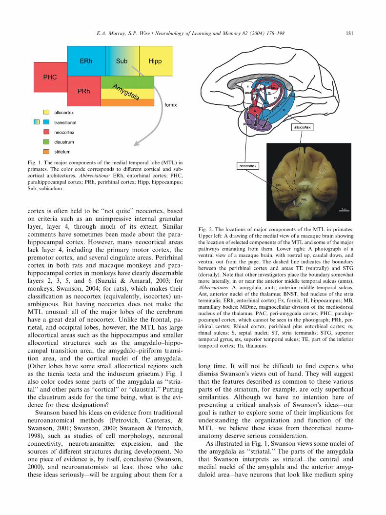
Fig. 1. The major components of the medial temporal lobe (MTL) in
primates. The color code corresponds to different cortical and sub-
cortical architectures. Abbreviations: ERh, entorhinal cortex; PHC,
parahippocampal cortex; PRh, perirhinal cortex; Hipp, hippocampus;
Sub, subiculum.
Fig. 2. The locations of major components of the MTL in primates.
Upper left: A drawing of the medial view of a macaque brain showing
the location of selected components of the MTL and some of the major
pathways emanating from them. Lower right: A photograph of a
ventral view of a macaque brain, with rostral up, caudal down, and
ventral out from the page. The dashed line indicates the boundary
between the perirhinal cortex and areas TE (ventrally) and STG
(dorsally). Note that other investigators place the boundary somewhat
more laterally, in or near the anterior middle temporal sulcus (amts).
Abbreviations: A, amygdala; amts, anterior middle temporal sulcus;
Ant, anterior nuclei of the thalamus; BNST, bed nucleus of the stria
terminalis; ERh, entorhinal cortex; Fx, fornix; H, hippocampus; MB,
mamillary bodies; MDmc, magnocellular division of the mediodorsal
nucleus of the thalamus; PAC, peri-amygdala cortex; PHC, parahip-
pocampal cortex, which cannot be seen in the photograph; PRh, per-
irhinal cortex; Rhinal cortex, perirhinal plus entorhinal cortex; rs,
rhinal sulcus; S, septal nuclei; ST, stria terminalis; STG, superior
temporal gyrus, sts, superior temporal sulcus; TE, part of the inferior
temporal cortex; Th, thalamus.
E.A. Murray, S.P. Wise / Neurobiology of Learning and Memory 82 (2004) 178–198 181
cortex is often held to be ‘‘not quite’’ neocortex, based
on criteria such as an unimpressive internal granular
layer, layer 4, through much of its extent. Similar
comments have sometimes been made about the para-
hippocampal cortex. However, many neocortical areas
lack layer 4, including the primary motor cortex, the
premotor cortex, and several cingulate areas. Perirhinal
cortex in both rats and macaque monkeys and para-hippocampal cortex in monkeys have clearly discernable
layers 2, 3, 5, and 6 (Suzuki & Amaral, 2003; for
monkeys, Swanson, 2004; for rats), which makes their
classification as neocortex (equivalently, isocortex) un-
ambiguous. But having neocortex does not make the
MTL unusual: all of the major lobes of the cerebrum
have a great deal of neocortex. Unlike the frontal, pa-
rietal, and occipital lobes, however, the MTL has largeallocortical areas such as the hippocampus and smaller
allocortical structures such as the amgydalo–hippo-
campal transition area, the amygdalo–piriform transi-
tion area, and the cortical nuclei of the amygdala.
(Other lobes have some small allocortical regions such
as the taenia tecta and the induseum griseum.) Fig. 1
also color codes some parts of the amygdala as ‘‘stria-
tal’’ and other parts as ‘‘cortical’’ or ‘‘claustral.’’ Puttingthe claustrum aside for the time being, what is the evi-
dence for these designations?
Swanson based his ideas on evidence from traditional
neuroanatomical methods (Petrovich, Canteras, &
Swanson, 2001; Swanson, 2000; Swanson & Petrovich,
1998), such as studies of cell morphology, neuronal
connectivity, neurotransmitter expression, and the
sources of different structures during development. Noone piece of evidence is, by itself, conclusive (Swanson,
2000), and neuroanatomists—at least those who take
these ideas seriously—will be arguing about them for a
long time. It will not be difficult to find experts who
dismiss Swanson�s views out of hand. They will suggest
that the features described as common to these various
parts of the striatum, for example, are only superficial
similarities. Although we have no intention here of
presenting a critical analysis of Swanson�s ideas—our
goal is rather to explore some of their implications for
understanding the organization and function of theMTL—we believe these ideas from theoretical neuro-
anatomy deserve serious consideration.
As illustrated in Fig. 1, Swanson views some nuclei of
the amygdala as ‘‘striatal.’’ The parts of the amygdala
that Swanson interprets as striatal—the central and
medial nuclei of the amygdala and the anterior amyg-
daloid area—have neurons that look like medium spiny

182 E.A. Murray, S.P. Wise / Neurobiology of Learning and Memory 82 (2004) 178–198
neurons in the traditionally recognized parts of thestriatum. In addition, these cells express GABA and co-
transmitters typical of medium spiny neurons, project to
Fig. 3. The convergence of traditional anatomy and embryology with the exp
of a 4- to 5-week-old human embryo, from Swanson (2000). The telencephalic
including the structures collectively called the amygdala (designated by the re
various parts of the cerebral cortex are noted, along with other structures of i
to a characteristic pattern in the expression of developmental regulatory g
striatum and pallidum; the septal nuclei (red S) consist of the rostral parts of
basal forebrain with the temporal lobe. Abbreviations: A, amygdala; C, cerebr
of piriform cortex); MC, medial cortex (hippocampus); OB, olfactory bulb; V
the lateral olfactory tract); N, basal ganglia; Pal, pallidum; S, septal nuclei; S
permission from Elsevier.
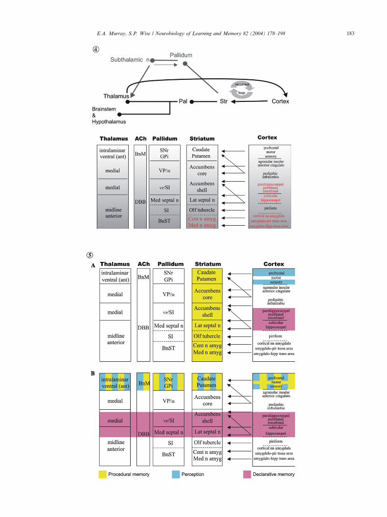
Fig. 4. Parallel architecture of cortical–basal ganglionic loops, including th
components of the MTL. Abbreviations: ant, anterior; BnM, basal nucleus
central nucleus of the amygdala; DBB, diagonal band of Broca; GPi, internal
septal nuclei; Med n amyg, medial nucleus of the amygdala; Med septal n,
pallidum; pir, piriform; SI, substantia innominata; SNr, reticular part of the
Note that the arrows evoke only a fraction of the relevant connectivity.
Fig. 5. Cortical–basal ganglionic modules in the context of the reification t
nonical reification theory. (B) Putative functions of forebrain structures if r
roanatomy of the forebrain according to Swanson and his colleagues. Note th
of the view depicted; rather, it provides a framework for reconsideration. A
other GABAergic neurons that, like traditionally rec-ognized pallidal neurons, project both to the thalamus
and brainstem. These nontraditional parts of the stria-
ression domains of developmental regulatory genes. (A) The forebrain
vesicle, in pink, gives rise to the entire cortex (C) and basal ganglia (N),
d A) and the septal nuclei (designated by the red S). The sources of the
nterest. (B) Modified from Puelles et al. (2000). Each color corresponds
enes. The amygdala (red A) consists of the caudal aspects of cortex,
the striatum and pallidum. Green circle is located at the junction of the
al cortex; DC, dorsal cortex; LC, lateral cortex (largest and lateral part
C, ventral cortex (medial part of piriform cortex, directly adjacent to
tr, striatum; *, basal forebrain. Reprinted from Swanson (2000), with
ose involving allocortex. Structures in red text indicate that they are
of Meynert; BnST, bed nucleus of the stria terminalis; Cent n amyg,
segment of the globus pallidus; hipp, hippocampal; Lat septal n, lateral
medial septal nuclei; nn, nuclei; Olf tubercle, olfactory tubercle; Pal,
substantia nigra; Str, striatum; trans, transition; VP, ventral pallidum.
heory. (A) Putative functions of forebrain structures according to ca-
eification theory is modified to take into account the theoretical neu-
at the presentation of this figure should not be taken as an endorsement
bbreviations as in Fig. 4.
c
E.A. Murray, S.P. Wise / Neurobiology of Learning and Memory 82 (2004) 178–198 183

184 E.A. Murray, S.P. Wise / Neurobiology of Learning and Memory 82 (2004) 178–198
tum, like the traditionally recognized parts, receive in-puts from dopamine neurons in the brainstem and from
both the dorsal thalamus and the cerebral cortex. The
same observations hold for the lateral septal nucleus,
which Swanson also believes to be part of the striatum.
Furthermore, both the striatal parts of the amygdala
and the lateral septal nucleus derive from a part of the
embryonic telencephalon—the region between the cor-
ticostriatal sulcus and the striatopallidal sulcus—thatgives rise to the traditionally recognized parts of the
striatum (Fig. 3A). The lateral septal nucleus (S) derives
from its rostral part and the striatal parts of the amyg-
dala (A) derive from its caudal part. Like the tradi-
tionally recognized parts of the striatum, these
nontraditional parts participate in recurrent modules,
commonly known as ‘‘loops,’’ which include cortical,
striatal, pallidal, and thalamic structures. We take upthese concepts below, but first we introduce some in-
dependent support for Swanson�s ideas.Puelles et al. (2000, 2001) have studied the expression
of developmental regulatory genes in embryonic mice.
Fig. 3B illustrates their conclusions. Each of four
cortical regions is indicated in a different color, as is the
striatum and pallidum, based on patterns of gene ex-
pression. Parts of the amygdala express the same regu-latory genes in development as do the traditionally
recognized parts of the striatum. Others express the
cortical or pallidal patterns. Puelles et al. concluded that
the parts of the telencephalon traditionally considered to
be the amygdala (A) are the caudal aspects of cortex,
striatum, and pallidum. These conclusions closely re-
semble those of Swanson and his colleagues and provide
independent support. This analysis also supportsSwanson�s view that the lateral septal nucleus (S) is a
part of the striatum: its most rostral part. Importantly,
the cortical nuclei of the amygdala have gene-expression
patterns like those of the lateral and ventral cortex (LC
and VC, respectively) and unlike those of the dorsal
cortex (DC) or the medial cortex (MC). The lateral and
ventral cortex compose the piriform cortex, and, in the
view of Puelles et al., the cortical nuclei of the amygdalamake up their caudal extension. The medial cortex
corresponds to the hippocampus. Note the fundamental
similarity between these two analyses (Figs. 3A and B)
regarding the amygdala (A) and the septal nuclei (S). On
both of these views, the amygdala arises from the caudal
(and lateral) aspect of the telencephalon, the septal nu-
clei from a largely rostral (and medial) aspect.
Although the idea that the lateral septal nucleus ispart of the striatum seems jarring to those of us who
learned textbook neuroanatomy, Swanson (2000) has
pointed out that this idea dates to the work of Ramon y
Cajal in 1911. In a sense, the caudate and putamen in
primates can be considered to be ‘‘striatum of the neo-
cortex,’’ and the lateral septal nucleus can be thought of
as ‘‘striatum of the hippocampus.’’ Further, as Puelles
et al. (2000) noted, the idea that the medial and centralnuclei of the amygdala are striatal dates to the early 20th
century work of Holmgren.
Thus, the MTL in primates seems to be a complex
admixture of different cortical and subcortical architec-
tures, some of which evolved very early in vertebrate
history, such as the hippocampus (called the medial
cortex in Fig. 3B) and parts of amygdala, and others of
which evolved relatively recently, such as the parahip-pocampal and perirhinal neocortex. In the primate
brain, these various structures get pushed, during de-
velopment, into the MTL. So what makes up the MTL?
In large part, it consists of the most medial and caudal
parts of the cerebral cortex and striatum, distorted by
evolutionary and embryological development into a
nearly unrecognizable configuration. Their concentra-
tion in a region called the MTL in primates is, on thisview, more an accident of history than a coherent ana-
tomical construct.
One implication of Swanson�s new view of the telen-
cephalon involves what are commonly known as recur-
rent, basal ganglia loops (DeLong & Georgopoulos,
1981). These loops involve neurons in several different
anatomical structures, which interact to function as
distributed neural networks, sometimes called distrib-uted modules (Houk & Wise, 1995). As illustrated in the
top part of Fig. 4, the prototypical cortical–basal gan-
glionic loop involves connections from cortex to stria-
tum, from striatum to pallidum (via both the direct and
indirect striatal-output pathways), from pallidum to
thalamus, and from thalamus to cortex. Previous work
has focused almost exclusively on the loops that involve
neocortex. Accordingly, many of the functions attrib-uted to ‘‘the striatum’’ depend on the traditional view
that the striatum consists of the caudate and putamen,
which excludes the lateral septal nucleus and the striatal
parts of the amygdala, and sometimes even excludes well
accepted parts of the striatum such as the nucleus ac-
cumbens and other parts of the ventral striatum.
According to the view presented in Fig. 4, the cere-
bral cortex includes not only the neocortex, but also thehippocampus, the piriform cortex, the cortical ‘‘nuclei’’
of the amygdala, and transitional allocortical areas near
the piriform cortex and hippocampus. The associated
parts of the striatum include the lateral septal nucleus,
the olfactory tubercle, the medial and central nuclei of
the amygdala, and the anterior amygdala area. The
bottom part of Fig. 4 also captures the idea that each
part of the striatum has an associated part of the palli-dum, including the medial septal nucleus, the substantia
innominata, and the bed nucleus of the stria terminalis
(sometimes considered part of the ‘‘extended amyg-
dala’’). To each cortical–basal ganglionic loop there is
also a source of cholinergic (and other) inputs from the
basal forebrain, specifically from the diagonal band (of
Broca) and the basal nucleus (of Meynert).

E.A. Murray, S.P. Wise / Neurobiology of Learning and Memory 82 (2004) 178–198 185
In the next section, we will review several lines ofempirical evidence that cast doubt on the reification
theory (see Section 2), at least in its canonical form.
Before we do that, let us examine some of the implica-
tions of the ideas sketched above from theoretical neu-
roanatomy. The reification theory holds that theMTL, as
a whole, is a declarative memory system, that the lateral
temporal lobe, along with other parts of the neocortex,
subserves perception, and that an entity sometimestermed the ‘‘basal ganglia,’’ sometimes termed the ‘‘stri-
atum,’’ and sometimes termed the ‘‘corticostriatal sys-
tem’’ provides the mechanism for procedural memory (or
equivalently, with varying degrees of rigor, habits).
However, by the term striatum, the proponents of the
reification theory mean to refer to the caudate and pu-
tamen, only, not to the other parts of the striatum men-
tioned above.This traditional view of the basal ganglia has not
always been explicitly stated, and has often instead been
taken for granted. Packard and Knowlton (2002) are
among only a few proponents of the reification theory to
acknowledge explicitly that functional attributes usually
ascribed to the ‘‘basal ganglia’’ or ‘‘striatum’’ are in-
tended to apply only to the most dorsal part of it.
However, by limiting their extended concept of thestriatum to a less inclusive view than that sketched here,
they still seek to find a functional dissociation between
entities called the hippocampus and the basal ganglia.
Fig. 5 illustrates a problem with that line of thought. It
shows that the hippocampus and other allocortical parts
of the MTL have their associated parts of the striatum
and pallidum, much as the neocortical areas have theirs.
Fig. 5A shows how the reification theory maps ontothe main structures of the forebrain according to ca-
nonical reification theory. Fig. 5B shows how the same
ideas can be modified to bring them more into line with
the new ideas about forebrain organization outlined in
this section. We emphasize that this exercise should not
be construed as an endorsement of the scheme presented
Table 1
Effects of lesions on stimulus memory
Tasks H
Trial-unique DNMS � (IBO)�
Visual-visual paired association learning )(ASP)
Crossmodal DNMS )(ASP)
The data come from several sources (Buckley & Gaffan, 1998b; Buckley,
Meunier, Bachevalier, Mishkin, & Murray, 1993; Mishkin, 1978; Murray,
Alvarado, & Bachevalier, 2004). Tests of stimulus memory are listed in the le
whereas the two other tasks, visual–visual paired-associate learning and cross
shows the effects of either selective removals of the perirhinal cortex or com
A(ASP), aspiration lesions of the amygdala, which include nearby areas of
amygdala; ASP, aspiration lesions of the hippocampus; DNMS, the delay
hippocampal lesions; IBO, ibotenic acid lesions of the hippocampus; PRh, pe
�, deficits in some conditions, but not in others, �some findings (Beason-Hel
conclusion that selective hippocampal lesions and combined lesions of the h
DNMS.
in Fig. 5B. We note only that this way of structuring thereification theory avoids the problem of assuming that
an entity called the ‘‘basal ganglia’’ or the ‘‘striatum’’
subserves any particular kind of memory, knowledge, or
system. Instead, Fig. 5B recognizes that different parts
of the basal ganglia have different functions, depending
on the cortical–basal ganglionic loops to which they
contribute. The difference between Figs. 5A and B re-
flects a change from traditional ‘‘column-wise thinking’’in the former to ‘‘row-wise thinking’’ in the latter. It is
just another way of looking at the reification theory. But
the question remains: Does the reification theory hold
up to empirical testing? The next section answers that
question.
4. Falsifying the reification theory
4.1. Recognition memory
Despite its popularity, evidence against the reification
theory has accumulated rapidly. The history of these
studies is elaborated elsewhere (Buckley & Gaffan, 2000;
Murray, 1996) and will not be repeated here. Table 1
shows the results of certain experiments assessing stim-ulus memory. According to the reification theory, the
greater the damage to the MTL, the greater should be
the impairment in recognition memory as measured by
DNMS (Zola-Morgan et al., 1994). Contrary to the
prediction of the reification theory, however, combined
lesions of the amygdala and hippocampus made with the
fiber-sparing excitotoxin ibotenic acid fail to produce
impairments on DNMS (Murray & Mishkin, 1998).Furthermore, damage to the entorhinal and perirhinal
cortex (also known collectively as the ‘‘rhinal cortex’’)
produces a severe impairment (Baxter & Murray, 2001a;
Eacott, Gaffan, & Murray, 1994; Meunier et al., 1993;
Table 1, Fig. 6). Aspiration lesions of the amygdala do
causemild deficits, but this finding ismisleading because it
A(IBO) A(ASP) PRh PRh+ERh
) + + + +
) + + +
) + + + + + +
Gaffan, & Murray, 1997; Buffalo et al., 1999; Goulet & Murray, 2001;
Gaffan, & Mishkin, 1993; Murray & Mishkin, 1985, 1998; Nemanic,
ft column. DNMS tests recognition memory of items for up to 40min,
modal DNMS, assess long-term associative memory. The right column
bined removals of the perirhinal and entorhinal areas. Abbreviations:
cortex and fiber tracts; A(IBO), selective ibotenic acid lesions of the
ed nonmatching-to-sample task; ERh, entorhinal cortex lesions; H,
rirhinal cortex lesions; +, mild deficit; +++, severe deficit; ), no deficit;
d, Rosene, Killiany, & Moss, 1999; Zola et al., 2000) disagree with the
ippocampus and the amygdala (Fig. 6) have no effect on trial-unique

Fig. 6. Lesions of the perirhinal and entorhinal cortex are sufficient to
cause a deficit in visual recognition memory, as assessed by the delayed
nonmatching-to-sample task (DNMS). Curves on the left show the
effects of imposing increasingly longer delays between sample presen-
tation and choice test. Curves on the right show the effects of in-
creasing list length, requiring the memory of multiple objects. After
combined, selective excitotoxic lesions of the amygdala and hippo-
campus (dashed line, circles), performance remains the same as for an
unoperated control group (solid line, triangles). In other tests, these
animals can perform as well as controls even with delays of about
40min (Murray & Mishkin, 1998). Monkeys with entorhinal plus
perirhinal cortex lesions were severely impaired in object recognition
memory (solid line, squares). Data from Meunier et al. (1993) and
Murray and Mishkin (1998). Abbreviations: Amygdala+Hipp (Ibote-
nate), monkeys with selective excitotoxic lesions of both the amygdala
and the hippocampus, bilaterally (N ¼ 7); PRh+ERh, monkeys with
combined bilateral aspiration lesions of the perirhinal cortex (PRh)
and entorhinal cortex (ERh) (N ¼ 7); Controls, unoperated control
monkeys (N ¼ 8).
186 E.A. Murray, S.P. Wise / Neurobiology of Learning and Memory 82 (2004) 178–198
fails to take into account additional damage to the cortex
of theMTLandfiber pathways passingnear the amygdala
(Goulet, Dor�e, &Murray, 2001). Since the original report
of Murray and Mishkin (1998), four additional studieshave assessed the effect on DNMS of selective hippo-
campal lesions, two reporting impairment (Beason-Held
et al., 1999; Zola et al., 2000) and two reporting no im-
pairment (Buckmaster, Eichenbaum, Amaral, & Rapp,
1999;Nemanic et al., 2004). Although there remains some
controversy regarding whether the hippocampus makes
any contribution to recognition memory as measured by
DNMS (as noted by the asterisk in Table 1), there iswidespread agreement that the entorhinal and perirhinal
cortex are the main substrates of visual recognition as
measured byDNMS. In sum, damage to just two parts of
theMTL, the perirhinal cortex and the entorhinal cortex,
is sufficient to produce nearly the same degree of deficit as
would removal of the entire MTL.
There are additional findings involving recognition
memory that argue against the reification theory. First,
the small effect of hippocampal lesions on tasks such asDNMS may be accounted for by other factors. For
example, Baxter and Murray (2001b) conducted a meta-
analysis of the studies examining the effects of selective
hippocampal damage on the DNMS task in monkeys.
They found an inverse relationship between the extent of
hippocampal damage and the magnitude of memory
loss. That is, paradoxically, the smaller the hippocampal
lesion, the greater the impairment in recognition mem-ory as measured by the DNMS task. This finding is the
opposite of that predicted by the reification theory, and
it could account for the reports of an impairment on the
DNMS task after selective hippocampal damage: On
this view, small deficits are an artifact of an incomplete
lesion, and even more incomplete lesions would lead to
yet greater deficits. Although Zola and Squire (2001)
have argued that several factors other than this ‘‘inverserelationship’’ are the likely cause of Murray and Mish-
kin�s (1998) failure to detect an impairment on DNMS
in monkeys with selective hippocampal lesions, most of
these possibilities (e.g., one vs. two stages of surgery;
preoperative vs. postoperative training) have been ruled
out by the findings of Nemanic et al. (2004) and the
preliminary findings of Buckmaster et al. (1999). Spe-
cifically, Buckmaster et al. showed that monkeys trainedusing the same methods as those used by Zola and
Squire and their colleagues (including postoperative
acquisition of the DNMS task and one-stage surgery)
were unimpaired on the DNMS task. Although the ex-
tent of the lesions in these monkeys has not yet been
assessed quantitatively, the results add weight to the
negative evidence reported by Murray and Mishkin
(1998). And the recent report by Nemanic et al. (2004),who likewise found no impairment on DNMS after se-
lective hippocampal lesions in monkeys that sustained
one-stage surgery, provides additional support. Evi-
dently, monkeys with selective, excitotoxic hippocampal
lesions can be unimpaired on DNMS. Yet another
problem for the reification theory is the report by
Meunier, Hadfield, Bachevalier, and Murray (1996),
who found that hippocampal lesions, when added torhinal cortex lesions, yielded a slightly (and significantly)
smaller impairment in recognition memory than rhinal
cortex lesions alone. This unexpected finding suggests
that structures in the MTL interact in memory in a
complex way, voiding the simple predictions of the re-
ification theory. Finally, it appears that removals of the
parahippocampal cortex, another component of the
putative MTL memory system, fail to yield deficits onDNMS (Nemanic et al., 2004).
If damage to the perirhinal and entorhinal cortex can
account for almost the entire recognition memory im-
pairment that follows MTL lesions (Hadfield, Baxter, &
Murray, 2003), and if neither the hippocampus nor
parahippocampal cortex is necessary for DNMS, what
does this imply for the reification theory? One position
E.A. Murray, S.P. Wise / Neurobiology of Learning and Memory 82 (2004) 178–198 187
would be to reject DNMS as a measure of declarativememory, thereby preserving reification theory. But if
this stance were adopted, then the only remaining
measures of declarative memory in monkeys would be
single-pair object discrimination learning (Zola-Morgan
et al., 1994), and perhaps tests of preferential viewing.
Thus, although Zola-Morgan and Squire (1985) started
with the idea that a battery of four tasks (including
delayed retention of single object discriminations, con-current object discrimination learning, delayed response,
and DNMS) ‘‘tapped’’ declarative memory, it is now
clear that it is difficult to be certain which, if any, of
these tasks are specific measures of declarative memory
in monkeys. For example, concurrent object-discrimi-
nation learning was found to be more sensitive to
damage to area TE (part of the inferior temporal cortex)
than to damage to the MTL, and this task was thereforediscarded as a test of declarative memory (Buffalo,
Reber, & Squire, 1998a). In addition, the evidence for
involvement of MTL structures in single-pair discrimi-
nation learning, a topic taken up in detail below, is
equivocal, as is the status of this task as an unambiguous
measure of declarative memory. And the preferential
viewing task suffers from similar problems. Indeed, we
argue that all supposed tests of ‘‘declarative’’ memoryshould be treated with skepticism. Also, consider the
following scenario: What if the relevant findings had
been reported in reverse order, with the dramatic effects
of rhinal cortex damage on performance of the DNMS
task being reported first? In this counterfactual, it is
doubtful whether the present controversy concerning the
hippocampal contributions to recognition memory—
which mainly amounts to an argument over whetherthat contribution is small or none—would even exist.
In sum, it appears that the perirhinal cortex, in con-
junction with the entorhinal cortex, plays a central role
in recognizing objects. The weight of evidence suggests
that the other parts of the entity called the MTL have
little or no role in this function. The next section also
presents evidence contrary to the reification theory. The
perirhinal cortex appears to play an important role inboth the perception and memory of objects and in es-
tablishing associations among objects, including ab-
stractions such as progress toward a goal (Liu, Murray,
and Richmond, 2000).
4.2. Perception vs. memory
Another tenet of the reification theory is that theMTL is not involved in the perceptual processing of
complex visual stimuli. This idea is based, in part, on the
findings that lesions of two major parts of the MTL (the
hippocampus and the perirhinal cortex), disrupt per-
formance on two ‘‘memory’’ tasks, visual DNMS and
single-pair visual discriminations, but not on a ‘‘per-
ception’’ task, concurrent visual discrimination. By
contrast, lesions of lateral temporal cortical area TE, inaddition to yielding impairment on the DNMS task,
yield the opposite pattern of results for the two types of
discrimination learning, that is, impairment on the ac-
quisition of concurrent discriminations (the so-called
‘‘perception’’ task), but not on learning single-pair sen-
sory discrimination, the task regarded as memory spe-
cific (Buffalo et al., 1999). These and other findings led
these investigators (Buffalo et al., 1998a, p. 388) to theconclusion that ‘‘the perirhinal cortex is not involved in
the perceptual processing of complex visual stimuli.’’ In
short, the extra-MTL sensory cortex subserves a ‘‘per-
ception system,’’ whereas the MTL subserves a declar-
ative memory system. But what if the cognitive labels
that these authors give to their tasks fail to correspond
with the underlying cognitive processes? Such labels can
be misleading and always demand a high level of skep-ticism and scrutiny, an admonition that applies to all of
the cognitive labels used in this paper, as well.
There is now strong evidence that at least one part of
the MTL, the perirhinal cortex, plays a role in percep-
tion as well as memory, in distinct conflict with the re-
ification theory. When sensory discrimination requires
the disambiguation of complex, conflicting stimulus
features, perirhinal cortex becomes essential to perfor-mance. On this view, the perirhinal cortex participates
both in visual memory, as one subdivision of the MTL,
and in visual perception, as part of the ventral stream
object analyzer pathway (Buckley & Gaffan, 1998a,
1998b, 1998c, 2000; Bussey & Saksida, 2002; Bussey,
Saksida, & Murray, 2002; Eacott et al., 1994; Murray &
Bussey, 1999).
Here we discuss two sets of experiments that havebeen carried out to test this idea. In one, the results of
which are illustrated in Fig. 7, control monkeys and
monkeys with aspiration lesions of perirhinal cortex
were assessed for their rate of acquisition of a series of
concurrent visual discriminations. In these experiments,
the number of object pairs was held constant, but the
degree of feature ambiguity was varied systematically.
Feature ambiguity is a property of visual discrimina-tions that emerges when features appear as part of both
rewarded and unrewarded objects. Monkeys were tested
in three conditions. In the first of these, called Maximum
Feature Ambiguity, all features were explicitly ambigu-
ous (AB+, CD+, BC), AD), also known as the bi-
conditional problem). In the biconditional problem, the
correct choice depends on the combination of features.
For example, it is correct to choose A in the presence ofB, but not in the presence of D. All features are ‘‘bal-
anced’’ in this way, so that correct choices cannot be
guided by any single feature, but only by the combina-
tion of features. In another condition, called Minimum
Feature Ambiguity, no features were explicitly ambig-
uous (AB+, CD+, EF), GH)). In a third condition,
called Medium Feature Ambiguity, half the features

Fig. 7. Lesions of the perirhinal cortex cause deficits dependent on feature ambiguity: discriminating compound stimuli. The left side of the figure
shows some of the stimuli used in this experiment. In the Minimum Feature Ambiguity condition, each component of a given, two-part stimulus was
either always rewarded when chosen (+) or never rewarded ()). That is, every part of each compound stimulus that was not rewarded (arrows) was
never rewarded and every part that was rewarded was always rewarded. In the Maximum Feature Ambiguity condition, every part of each com-
pound stimulus was both rewarded (for one part, arrows to the left of the pictures) and not rewarded (arrows to the right of the pictures) equally. The
right side of the figure shows errors to criterion. Data are averaged over several problems of each type. With minimum feature ambiguity, monkeys
with perirhinal cortex lesions are unimpaired relative to controls, but as feature ambiguity increased, the monkeys with perirhinal cortex lesions were
increasingly and significantly (*, **) impaired. Data from Bussey et al. (2002). Abbreviations: PRh, monkeys with bilateral aspiration lesions of
perirhinal cortex (N ¼ 4); Control, unoperated control monkeys (N ¼ 4); Minimum, Medium, and Maximum, different levels of feature ambiguity.
188 E.A. Murray, S.P. Wise / Neurobiology of Learning and Memory 82 (2004) 178–198
were explicitly ambiguous (AB+, CD+, CE), AF)). Itwas found that perirhinal cortex lesions yield an in-
creasingly greater impairment on discrimination learn-ing as the degree of feature ambiguity is increased. This
result cannot be accounted for by task difficulty because
the same monkeys are unimpaired relative to controls on
very difficult color discriminations, and other controls
for difficulty were carried out as well. In addition, the
Maximum Feature Ambiguity condition is not any more
difficult than the Intermediate condition for the control
group of monkeys.These results suggest that a function of perirhinal
cortex is to resolve feature ambiguity in complex vi-
sual discriminations. The proposal is that the peri-
rhinal cortex performs this function by representing
conjunctions of features. Lesions in this region com-
promise the representation of visual stimuli, and be-
cause both accurate perception and accurate memory
require accurate representation, both memory andperception are affected by damage to the perirhinal
cortex.
If this interpretation is valid, then perirhinal cortex
lesions should also cause an impairment on simple,
single-pair visual discriminations that exhibit feature
ambiguity. To test this idea, in a second experiment
Bussey, Saksida, and Murray (2003) manipulated fea-
ture ambiguity by morphing (or blending) two pictures.
Monkeys were required to learn visual discriminations
under conditions of high or low feature ambiguity. The
low feature ambiguity condition used two randomlyselected pictures, which were not morphed together. The
high feature ambiguity condition used morphed pictures
consisting of a stimulus that was roughly 65% of the
rewarded stimulus and 35% of the foil vs. one that was
35% of the rewarded stimulus and 65% of the foil.
Monkeys were tested for their acquisition of several
problems of each type. Fig. 8 shows the result. With low
feature ambiguity, the monkeys with perirhinal cortexlesions learned single-pair discrimination problems just
as fast as the controls. In the high feature ambiguity
condition, the monkeys with perirhinal cortex lesions
were impaired relative to controls.
Thus, the two experiments by Bussey et al. (2002,
2003), taken together, support the idea that perirhinal
cortex operates as part of the ventral visual stream, or
object-analyzer pathway, and that it helps process,represent and store conjunctions of object features.
Fig. 9 depicts a model that accounts for the results re-
lated here. Of course, the perirhinal cortex does not
operate in a vacuum. It interacts with other structures in
the brain, and depending on the requirements of the
task, it may interact with the hippocampus, the amyg-
dala or the prefrontal cortex in storing information.
Gaffan and his colleagues (Buckley & Gaffan, 1998a,

Fig. 8. Lesions of the perirhinal cortex cause deficits dependent on feature ambiguity: discriminating a single pair of morphed stimuli. Left: Percent
correct choices as a function of blocks of 8 trials each for low feature ambiguity (top) and high feature ambiguity (bottom) conditions. Significant
differences between the groups are marked with asterisks (*, **). Right: The bars show the group data averaged over the total of 96 trials, and
symbols indicate the scores of individual monkeys. Results are averaged over several problems of the same type. In acquiring low feature ambiguity
discrimination problems, monkeys with perirhinal cortex lesions are unimpaired relative to controls, but in acquiring high feature ambiguity dis-
crimination problems, the monkeys with perirhinal cortex lesions were significantly (**) impaired. Data from Bussey et al. (2003). Abbreviations:
PRh, monkeys with bilateral aspiration lesions of the perirhinal cortex; Control, unoperated control monkeys.
E.A. Murray, S.P. Wise / Neurobiology of Learning and Memory 82 (2004) 178–198 189
1998b, 1998c, 2000; Eacott et al., 1994) have used a very
different set of tasks and a very different experimental
approach to reach much the same conclusion: Perirhinal
cortex plays an important role perceptual processing,contrary to the reification theory.
There is another important aspect of the results from
the study of single-pair discrimination learning de-
scribed above. As we have seen, the reification theory
depends on the assignment of discrimination learning, in
the single-pair format, to declarative memory, in con-
trast to the concurrent version of visual discrimination
learning, which is held to depend on perceptual pro-cesses. Indeed, rapid learning is thought to be a defining
characteristic of declarative memory (Bachevalier &
Mishkin, 1984; Buffalo et al., 1999; Squire, Knowlton, &
Musen, 1993; Squire & Zola-Morgan, 1983; Teng, Ste-
fanacci, Squire, & Zola, 2000; Zola et al., 2000). As Zola
and Squire (2000, p. 492) expressed this view in a sem-
inal paper on the subject:
simple two-choice discrimination tasks—that is, ones that are
learned quickly by normal animals—are dependent on the [me-
dial] temporal lobe. More difficult two-choice discrimination
tasks. . .are independent of the medial temporal lobe.
As illustrated in Fig. 8, the results of Bussey et al.
(2003) were inconsistent with the prediction of an im-
pairment on rapidly learned visual discrimination
problems, but not slowly learned ones. Indeed, this
study yielded the opposite pattern of results. These
findings accorded with the countervailing predictionthat impairments would occur only on perceptually
difficult, slowly learned visual discriminations, those
with high levels of feature ambiguity (Bussey & Saksida,
2002; Murray & Bussey, 1999). Thus, in contrast to the
idea of Zola and Squire articulated above, the empirical
evidence shows that the impairments in single-pair vi-
sual discrimination caused by lesions of the perirhinal
cortex are related more to perceptual factors than to thespeed of learning.
So how can we account for the impairment on single-
pair discriminations after perirhinal cortex damage re-
ported by Buffalo and her colleagues (Buffalo et al.,
1999; Buffalo, Ramus, Squire, & Zola, 2000) and as

Fig. 9. Model accounting for feature ambiguity effects. According to
this model, networks of neurons (ellipses) in caudal regions of the
ventral visual stream represent elemental features (Element layer) or
simple conjunctions of features (Feature layer). Each element is de-
noted by a capital letter (A, B, and C). More rostral regions, such as
perirhinal cortex, represent more complex conjunctions of these fea-
tures (ABC). Abbreviations: IT, inferotemporal cortex; PRh, perirhinal
cortex. Adapted from Bussey and Saksida (2002).
190 E.A. Murray, S.P. Wise / Neurobiology of Learning and Memory 82 (2004) 178–198
confirmed by Hampton and Murray (2002)? One pos-sibility is that the objects being discriminated had en-
ough feature overlap to yield an impairment on the
discrimination task. Although the idea of feature am-
biguity was not assessed, Buffalo et al. (1999) used four
different pairs of objects, administered serially, to assess
single-pair discrimination learning. At least 3 of the 4
object discriminations appear to involve relatively high
levels of feature ambiguity. For example, one pair con-sisted of red and green peanut shells, another of white
and black rectangles, and yet another of yellow and pink
plastic eggshells. In each case, although the items dif-
fered in color, they had the same shape and size, thereby
exhibiting feature overlap for these dimensions. Thus,
feature ambiguity might account for the impairment on
single-pair discrimination of monkeys with perirhinal
cortex lesions.More recently, in an attempt to extend our analysis of
perirhinal cortical contributions to perception, we have
employed the transverse patterning task. In this study,
to assess the contributions to learning by various MTL
structures, we compared directly the effects of selective
hippocampal lesions with those of perirhinal cortex le-
sions (Saksida, Bussey, Buckmaster, & Murray, 2003).
This task is created by taking three objects, A, B, and C,and pairing them in such a way that when A is paired
with B, A is rewarded; when B occurs with C, B is re-
warded; and when C occurs with A, C is rewarded. In
the notation used above, the task involves three prob-
lems: A+ vs. B), B+ vs. C), and A) vs. C+. These three
problems are mixed together within sessions, and the
measure of interest, as before, is the rate of acquisition.
Note that the transverse patterning problem has exactlythe same characteristics as the foregoing discrimination
problems that we arranged for the high feature ambi-
guity condition, only now at an object level. That is,
across the three individual discrimination problems,
every object is both rewarded and unrewarded.
The ‘‘objects’’ in this task, as in our earlier experi-
ment, were complex grayscale pictures. On each trial,
monkeys were presented with a single pair of objects,one object on the left and one on the right side of the
screen, and they were required to touch one stimulus on
the screen to indicate their choice. The monkeys learned
through trial and error which stimulus, when touched,
resulted in delivery of a food reward. Because the
monkeys with hippocampal lesions were tested later
than those with perirhinal cortex lesions, they were ac-
companied by their own group of unoperated controls.As predicted, monkeys with perirhinal cortex lesions
were impaired relative to the controls. By contrast,
monkeys with selective hippocampal lesions were actu-
ally facilitated on this task, relative to their controls
(Saksida et al., 2003). These data support the idea that
the perirhinal cortex, but not the hippocampus, is es-
sential for representing conjunctions of features. In ad-
dition, these findings provide the strongest evidence todate against the reification theory; the opposing effects
of lesions of the hippocampus, on the one hand, and of
perirhinal cortex, on the other, are inconsistent with the
idea that these structures are working together as a
single functional entity.
Not only does the pattern of data contradict the idea
that these structures are working together to support
memory, they suggest there is a competition betweenhippocampal and perirhinal processing in storing in-
formation. The facilitation is reminiscent of findings
reported by McDonald and White (1995), who found
that fornix transection in rats facilitates acquisition of
conditioned cue preference. Whereas we found that
hippocampal damage facilitates acquisition of a peri-
rhinal-dependent task, transverse patterning, they found
that hippocampal damage facilitated acquisition of anamygdala-dependent task, conditioned cue preference.
The work cited here adds to the ‘‘overwhelming evi-
dence,’’ in the words of Buckley and Gaffan, that the
perirhinal cortex is important for both memory and
perception (e.g., Buckley & Gaffan, 1998a, 1998b, 1998c,
2000; Bussey & Saksida, 2002; Bussey et al., 2002, 2003;
Eacott et al., 1994; Murray & Bussey, 1999). Thus, we
can reject that aspect of the reification theory whichholds that the MTL is a thing that subserves declarative
memory, but not visual analysis or perception (Buffalo
et al., 1998a, 1999; Sakai & Miyashita, 1993; Squire,
1992). It is important to emphasize that these data do
not argue against an important role for the perirhinal
cortex in memory, only that it does not function exclu-
sively in a mnemonic role. It seems likely that the peri-

Table 3
Excitotoxic vs. aspiration lesions of the amygdala
Task A(IBO) A(ASP) PRh
E.A. Murray, S.P. Wise / Neurobiology of Learning and Memory 82 (2004) 178–198 191
rhinal cortex is important for object memory, if for noother reason than its pivotal anatomical position. The
perirhinal cortex is reciprocally related to several high-
order, modality-specific sensory cortical processing re-
gions and can interact with the hippocampus (indirectly
via the entorhinal cortex) and other brain regions such
as the prefrontal cortex and dorsal thalamus (Aggleton
& Brown, 1999; Bussey, Duck, Muir, & Aggleton, 2000;
Bussey, Muir, & Aggleton, 2001; Gaffan & Parker, 1996;Murray, 2000). Rather than considering the perirhinal
cortex exclusively as part of a thing called the MTL, it
can be considered to be a rostral component of the
ventral visual stream, as well. It is not surprising, in this
context, that the perirhinal cortex functions in both
perception and memory: it processes and stores repre-
sentations of complex visual stimuli (Bussey & Saksida,
2002; Bussey et al., 2002; Murray & Bussey, 1999).Finally, it is worth noting that complete, selective,
excitotoxic amygdala lesions do not disrupt the learning
of rapidly acquired discrimination problems (Malkova,
Gaffan, & Murray, 1997). There are other examples of
intact instrumental learning in rats with amygdala
damage (Blundell, Hall, & Killcross, 2001; Burns,
Robbins, & Everitt, 1993). Of course, these findings do
not imply that, in the absence of an amygdala, animalslearn discrimination problems in an entirely normal
manner, but it does indicate that there is a mechanism
outside the amygdala that can mediate this type
of learning. Thus, single-pair sensory discrimination
learning is not a function of the MTL per se, but rather
depends on the contribution of a particular subset of its
components.
4.3. Stimulus–response associations and spatial tasks
As we have seen, the reification theory fails to ac-
count for the feature-ambiguity results. What about
other tasks? Table 2 shows some of the effects of hip-
pocampal lesions on tests of spatial memory. It has long
Table 2
Excitotoxic vs. aspiration lesions of the hippocampus
H(IBO) H(ASP) PHC
Spatial reversals ) ++
Spatial DNMS )� +++
Object–place association ) +++ +++
The data come from several sources (Jones & Mishkin, 1972;
Mahut, 1971; Mahut & Moss, 1986; Malkova & Mishkin, 2003;
Murray & Mishkin, 1998; Murray, Baxter, & Gaffan, 1998; Parkinson,
Murray, & Mishkin, 1988). Abbreviations: DNMS, the delayed non-
matching-to-sample task; H(IBO), selective, ibotenic acid lesions of the
hippocampus; H(ASP), aspiration lesions of the hippocampus, which
include the parahippocampal cortex; PHC, parahippocampal cortex
lesions; ++, moderate deficit; +++, severe deficit; ), no deficit.*Although testing in a manual test apparatus yields no impairment,
open-field testing does yield impairment on a delayed spatial matching-
to-sample task (Hampton, Hampstead, & Murray, 2004).
been thought, based on studies using aspiration lesions,that hippocampal damage produced a severe impair-
ment on tasks such as spatial reversal learning (Jones &
Mishkin, 1972; Mahut, 1971), spatial DNMS (Mahut &
Moss, 1986), and object–place association (Parkinson
et al., 1988). Indeed, these are some of the classic neu-
ropsychological findings cited in support of a role for
the hippocampus in spatial memory in monkeys. More
recently, however, studies using selective excitotoxiclesions have failed to find the same impairments. Re-
markably, selective excitotoxic lesions of the hippo-
campus in monkeys have been reported to yield no
impairment on spatial reversal learning (Murray et al.,
1998), no impairment on spatial DNMS (Murray &
Mishkin, 1998), and no impairment on object–place
association (Malkova & Mishkin, 2003). For one task,
object–place association, impairments appear to followdamage to the parahippocampal cortex, the cortical re-
gion directly underlying the hippocampus, rather than
the hippocampus (Malkova & Mishkin, 2003). This
finding presents yet another argument against the reifi-
cation hypothesis. As with the results on recognition
memory using the DNMS task, and as with the results
on visual discrimination using the feature-ambiguity
tasks, these spatial tasks depend on particular compo-nents of the MTL. Damage to those components pro-
duces impairments comparable to much larger lesions
that include the key area, rather than producing larger
deficits as predicted by the reification theory. Table 3
shows similar data for other tasks. The rule seems to be
that different parts of the MTL perform different func-
tions, not that they all contribute to the same functions.
In addition to the results summarized in Tables 2 and3, there is additional evidence for a division of labor
among the various parts of the MTL. According to
PRh+ERh
Object reversals ) + + + + + +
Visual discrimination
Aud. 2� reinforcement
) + + + +
Visual discrimination
learning set
+ +
Win-stay, lose-shift � +
Data are from several sources (Baxter, Hadfield, & Murray, 1999;
Baxter & Murray, 2001a; Gaffan & Harrison, 1987; Gaffan & Murray,
1990; Izquierdo, Suda, & Murray, 2003; Jones & Mishkin, 1972;
Malkova et al., 1997; Murray et al., 1998; Spiegler & Mishkin, 1981;
Stefanacci, Clark, & Zola, 2003). The right column shows the effects of
either selective removals of the perirhinal cortex or combined removals
of the perirhinal and entorhinal areas. Abbreviations: A(ASP), aspi-
ration lesions of the amygdala, which include nearby areas of cortex
and fiber tracts; A(IBO), selective ibotenic acid lesions of the amyg-
dala; Aud. 2� reinforcement, auditory secondary reinforcement. ERh,
entorhinal cortex lesions; PRh, perirhinal cortex lesions; +, mild defi-
cit; +++, severe deficit; ), no deficit; �, mild, transient deficit.

192 E.A. Murray, S.P. Wise / Neurobiology of Learning and Memory 82 (2004) 178–198
reification theory, stimulus–response associations arenot the province of the MTL, but of the basal ganglia.
And, of course, the ‘‘basal ganglia’’ in this view refers to
the portion of the basal ganglia that receives projections
from neocortex. One kind of stimulus–response (S–R)
learning that has been used in monkeys is called con-
ditional motor learning or the arbitrary visuomotor as-
sociation task.
In the arbitrary visuomotor association task, themonkeys learned, by trial and error, that a visual stim-
ulus instructs a given action (Murray & Wise, 1996). For
the results illustrated in Fig. 10, responses consisted of
forelimb movements made with a joystick. Monkeys
were seated in a primate chair in front of a color mon-
itor and a single visual stimulus on the screen instructed
the monkey what actions to make on each trial. Mon-
keys always learned a set of three problems simulta-neously; each of three stimuli in a set instructed one and
only one action. For example, stimulus A might instruct
Fig. 10. Lesions of the hippocampus cause deficits in S–R learning (top left),
right). Lesions of the amygdala cause deficits in neither learning (bottom lef
acquisition trials. Data were averaged over 40 three-problem sets. Right side: m
of preoperatively learned problems. Bars show the group mean and symbols
(1996). Abbreviations: Preop, data gathered before surgery; Postop, data gat
lesions of the hippocampus plus underlying parahippocampal cortex; Amyg
underlying cortex.
the monkey to make a forelimb movement to the left;stimulus B to the right, and stimulus C towards the
monkey. The two panels on the left side of Fig. 10 show
the reduction in percent error as monkeys learn these
kinds of problems. The solid lines show the mean per-
formance before bilateral aspiration removal of either
the hippocampal formation (top) or amygdala (bottom)
and the dashed lines show performance after the sur-
gery. Before the surgery, the monkeys learned the arbi-trary visuomotor associations impressively fast; after �8
trials per problem (roughly 25 trials total), they had
mastered all three of the concurrently presented associ-
ations. Whereas removal of the hippocampal formation
severely disrupted the acquisition of arbitrary mapping
problems, amygdala lesions had no effect. Similar to the
results mentioned earlier in this section, addition of an
amygdala removal to a pre-existing hippocampal lesionhad no further effect. The two panels on the right side of
Fig. 10 show that the same surgeries had no effect on the
but not in retention or retrieval of well learned S–R associations (top
t) nor retention (bottom right). Left side: mean percent error over 50
ean percent error averaged over a block of 50 trials assessing retention
show the scores of individual monkeys. Data from Murray and Wise
hered after surgery. Hippocampus+, monkeys with bilateral aspiration
dala+, monkeys with bilateral aspiration lesions of the amygdala plus

Fig. 11. The effects of brain lesions on reinforcer devaluation effects.
The amygdala, not the hippocampus or rhinal cortex, is essential for
linking objects with the current value of food reinforcers. Abbrevia-
tions: Amygdala, monkeys with selective, bilateral excitotoxic amyg-
dala lesions; Control: unoperated control monkeys; Hipp, monkeys
with selective, bilateral, excitotoxic hippocampal lesions; PRh, mon-
keys with bilateral aspiration lesions of the perirhinal cortex (N ¼ 4);
PRh+ERh, monkeys with combined, bilateral aspiration (N ¼ 2) or
excitotoxic (N ¼ 3) lesions of the perirhinal and entorhinal cortex.
E.A. Murray, S.P. Wise / Neurobiology of Learning and Memory 82 (2004) 178–198 193
retention and retrieval of preoperatively learned prob-lems. More recent work has shown that the fornix
contributes to normal rates of learning both spatial
(Rupniak & Gaffan, 1987) and nonspatial (Brasted,
Bussey, Murray, & Wise, 2003) versions of this S–R
learning task. Although the contribution of perirhinal
cortex has not yet been assessed, these results provide
further evidence for a division of labor among MTL
structures.
4.4. Stimulus–reward associations
A further division of labor within the MTL can be
demonstrated with the reinforcer devaluation task, a
measure of stimulus–reward association. The reinforcer
devaluation task requires monkeys to discriminate ob-
jects, to link objects with a representation of the currentvalue of the food, and to use those associations to guide
response selection. It is thought that this task probes the
ability of monkeys to use objects to predict the outcome
of an action and thereby choose appropriately. There
are two phases of these experiments. In the first, mon-
keys are familiarized with a large number of objects,
some of which are associated with one type of food, say,
a fruit snack, and others with another type of food, say,a peanut. In the second phase of the experiment, con-
sisting of four critical sessions, the monkeys are required
to choose between these two classes of objects. On these
trials, the two types of objects are pitted against one
another, and both overlie the appropriate food. In two
critical sessions, monkeys� baseline choices of objects arescored. Two other sessions are preceded by pre-feeding
with one of these two foods in a procedure intended todevalue that food. Monkeys choices of either food-1 or
food-2 associated objects are again scored. Under these
conditions, normal monkeys shift their responding away
from baseline: They avoid choosing objects that cover
the now devalued food.
The measure of interest is the ‘‘difference score.’’ As
shown in Fig. 11, control monkeys do a good job of
adjusting their responses; they generally avoid displac-ing objects that overlie the sated food. By contrast,
monkeys with bilateral excitotoxic lesions of the amyg-
dala show much less of this tendency (Malkova et al.,
1997), as do monkey with disconnections between the
orbitofrontal cortex and the amygdala (Baxter, Parker,
Lindner, Izquierdo, & Murray, 2000). Preliminary data
(Chudasama & Murray, 2004) indicate that neither
perirhinal cortex lesions nor selective hippocampal le-sions impair performance on this task (see also Thorn-
ton, Malkova, & Murray, 1998). Thus, the amygdala,
but not the perirhinal cortex or the hippocampus, is
essential for linking objects with the current value of
food reinforcers. This finding is not what one would
expect from viewing the MTL as a unitary functional
system.
5. Beyond balkanization
Not only is the view that the MTL is a ‘‘thing’’
difficult to maintain, but the functional homogeneity of
some of its components is also in doubt. Some studies
report dissociations of function within the hippocam-
pus. For example, lesions of the dorsal (septal) but not
the ventral (temporal) hippocampus disrupt spatial
memory tasks and, conversely, lesions of the ventralbut not the dorsal portion of the hippocampus disrupt
rats ability to respond appropriately in conditions of
increased anxiety (Bannerman et al., 2002, 2003;
Kjelstrup et al., 2002). There are also a handful of
reports showing a dissociation of function within the
amygdala, which, given the structure of the amygdala,
should come as no surprise. For example, the baso-
lateral nuclei of the amygdala, but not the centralnucleus, mediate reinforcer devaluation effects in rats
(Hatfield, Han, Conley, Gallagher, & Holland, 1996).
In addition, the central nucleus of the amygdala, but
not the basolateral nuclei, contribute importantly to
certain kinds of Pavlovian conditioning, and, con-
versely, the basolateral amygdala but not the central
amygdala contribute to certain kinds of instrumental
responses (Killcross, Robbins, & Everitt, 1997; Par-kinson, Robbins, & Everitt, 2000). These data also
demonstrate the diversity of function within the MTL
and violate the reification theory at a yet more fun-
damental level than the data reviewed in Section 4.
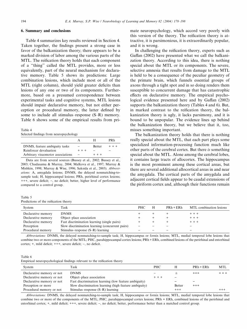
194 E.A. Murray, S.P. Wise / Neurobiology of Learning and Memory 82 (2004) 178–198
6. Summary and conclusions
Table 4 summarizes key results reviewed in Section 4.
Taken together, the findings present a strong case in
favor of the balkanization theory; there appears to be a
marked division of labor among the various parts of the
MTL. The reification theory holds that each component
of a ‘‘thing’’ called the MTL provides, more or less
equivalently, part of the neural mechanism for declara-tive memory. Table 5 shows its predictions: Large
combination lesions, which include most or all of the
MTL (right column), should yield greater deficits than
lesions of any one or two of its components. Further-
more, based on a presumed correspondence between
experimental tasks and cognitive systems, MTL lesions
should impair declarative memory, but not either per-
ception or procedural memory, the latter defined bysome to include all stimulus–response (S–R) memory.
Table 6 shows some of the empirical results from pri-
Table 4
Selected findings from neuropsychology
A H PRh
DNMS, feature ambiguity tasks ) Better + + +
Reinforcer devaluation + + + ) )Arbitrary visuomotor associations ) + + +
Data are from several sources (Bussey et al., 2002; Bussey et al.,
2003; Chudasama & Murray, 2004; Malkova et al., 1997; Murray &
Mishkin, 1998; Murray & Wise, 1996; Saksida et al., 2003). Abbrevi-
ations: A, amygdala lesions; DNMS, the delayed nonmatching-to-
sample task; H, hippocampal lesions; PRh, perirhinal cortex lesions;
+++, severe deficit; ), no deficit; better, higher level of performance
compared to a control group.
Table 6
Empirical neuropsychological findings relevant to the reification theory
System Task
Declarative memory or not DNMS
Declarative memory or not Object–place association
Declarative memory or not Fast discrimination learning (low feature
Perception or more Slow discrimination learning (high feature
Procedural memory or not Stimulus–response (S–R) learning
Abbreviations: DNMS, the delayed nonmatching-to-sample task; H, hip
combine two or more of the components of the MTL; PHC, parahippocamp
entorhinal cortex; +, mild deficit; +++, severe deficit; ), no deficit; better, p
Table 5
Predictions of the reification theory
System Task
Declarative memory DNMS
Declarative memory Object–place association
Declarative memory Fast discrimination learning (single pairs)
Perception Slow discrimination learning (concurrent pairs)
Procedural memory Stimulus–response (S–R) learning
Abbreviations: DNMS, the delayed nonmatching-to-sample task; H, hip
combine two or more components of the MTL; PHC, parahippocampal corte
cortex; +, mild deficit; +++, severe deficit; ), no deficit.
mate neuropsychology, which accord very poorly withthis version of the theory. The reification theory is at-
tractive, it is parsimonious, it is extraordinarily popular,
and it is wrong.
In challenging the reification theory, experts such as
Gaffan (2002) have presented what we call the balkani-
zation theory. According to this idea, there is nothing
special about the MTL or its components. The severe,
selective amnesia that results from damage to the MTLis held to be a consequence of the peculiar geometry of
the primate brain, which funnels essential groups of
axons through a tight spot and in so doing renders them
susceptible to concurrent damage that has catastrophic
effects on declarative memory. The empirical psycho-
logical evidence presented here and by Gaffan (2002)
supports the balkanization theory (Tables 4 and 6). But,
in marked contrast to the reification theory, the bal-kanization theory is ugly, it lacks parsimony, and it is
bound to be unpopular. The evidence lines up behind
the balkanization theory, but we believe that it, too,
misses something important.
The balkanization theory holds that there is nothing
really special about the MTL, that each part plays some
specialized information-processing function much like
other parts of the cerebral cortex. But there is somethingspecial about the MTL. Alone among the cerebral lobes,
it contains large tracts of allocortex. The hippocampus
is the most prominent among these cortical areas, but
there are several additional allocortical areas in and near
the amygdala. The cortical parts of the amygdala and
adjacent cortical fields appear to be caudal extensions of
the piriform cortex and, although their functions remain
PHC H PRh+ERh MTL
) � +++ + + +
+ + + )ambiguity) ) )ambiguity) Better +++
+++ +++
pocampus or fornix lesions; MTL, medial temporal lobe lesions that
al cortex lesions; PRh + ERh, combined lesions of the perirhinal and
erformance better than a matched control group.
PHC H PRh+ERh MTL combination lesions
+ + + + + +
+ + + + + +
+ + + + + +
) ) ) )) ) ) )
pocampus or fornix lesions; MTL, medial temporal lobe lesions that
x lesions; PRh+ERh, combined lesions of the perirhinal and entorhinal

E.A. Murray, S.P. Wise / Neurobiology of Learning and Memory 82 (2004) 178–198 195
enigmatic, some neuroanatomical work suggests thatthey share a close association with the autonomic ner-
vous system (Westerhaus & Loewy, 2001) and the hy-
pothalamus (Swanson, 2000). If one accepts the
conclusions of Swanson and Puelles and their col-
leagues, outlined in Section 3, the MTL also contains
small, extremely caudal parts of the striatum and, for
what it is worth, the claustrum, too. Perhaps these ideas
will contribute to an improved understanding of the‘‘thing’’ called the MTL in primate brains—some day.
That day has not come, at least not for us, and we make
no attempt to develop a grand synthesis or alternative
theory here.
Were we to attempt to go farther, which we will not,
we might point out that all of these allocortical areas
and their associated parts of the striatum and pallidum
have long evolutionary histories, perhaps stretchingback to some of our most distant vertebrate ancestors.
We might speculate that the MTL is special because of
the long history of its allocortical components, because
of some role in the behaviors most fundamental to the
life of vertebrates, or because of a role in certain
instincts and the behaviors built most directly on
instincts.
We feel, however, that such speculation would breakthe pledge that we made at the beginning of this pre-
sentation. We began this review with the promise that
we would attempt to convey two (and only two) take-
home messages: (1) Any theory that attempts to distin-
guish the functions of one ‘‘thing’’ called the MTL and
another ‘‘thing’’ called the basal ganglia should take
into account all of the latter�s components (as illustrated
in Fig. 4), not merely those that have become mostprominent in mammalian brains because of their rela-
tionship with the neocortex. In the context of Fig. 5, we
believe that row-wise thinking (Fig. 5B) will prove to be
more useful than column-wise thinking (Fig. 5A). And
(2) the MTL is not a ‘‘thing’’ at all. ‘‘It’’ is a collection of
functionally disparate structures that each contribute to
memory, perception, and response selection in a spe-
cialized way (Table 4):• the amygdala (also not a thing)—not the hippocam-
pus and not the perirhinal cortex—links objects with
the current value of food reinforcers and thus con-
tributes to response selection;
• the perirhinal cortex processes, represents, and stores
object features, it associates inputs within and across
sensory modalities, and it does so in the service of
both perception and memory; the perirhinal and en-torhinal cortex—not the hippocampus and not the
amygdala—underlie the memory of objects as mea-
sured by the DNMS task; and
• the hippocampal system (including the fornix)—not
the amygdala—contributes to the acquisition of arbi-
trary S–R associations (‘‘habits,’’ according to some
authorities), whether spatial or nonspatial in nature.
7. Epilogue
We stated in Section 1 that we hoped it would be
helpful to change the question from ‘‘What does the
MTL do?’’ to those posed in our title. But we have made
no attempt to answer those questions yet. So: What, if
anything, is the MTL? How can the amygdala be part of
the MTL if there is no such ‘‘thing’’ as the MTL or the
amygdala? These questions are difficult to answer be-cause the phylogenetic and ontogenetic processes un-
derlying the fantastic expansion of the neocortex have
distorted the relationships among structures in the
forebrain almost beyond recognition. It is easy to think
of the MTL�s components and the basal forebrain as
separate building blocks that abut each other in some
arbitrary way, as if they were boxes like those in Fig. 1
that could be rearranged to suit the heuristic require-ments of a given theory, a course of instruction, or some
other cultural construct. But the anatomical arrange-
ment of structures in the forebrain results from the
evolutionary and embryological processes that put them
there: The structures that end up in the MTL of pri-
mates result from that history. Notwithstanding some
interesting patterns of cell migration during develop-
ment, the telencephalon cannot stray far from the geo-metric relationships established in the embryonic
telencephalic vesicle (Fig. 3A). The ends of the hippo-
campus do not detach and reconnect in some haphazard
way during development. Nor do the components of the
telencephalon rearrange themselves willy–nilly. The tel-
encephalic vesicle can expand, balloon, fold, and contort
beyond credible comprehension, but fundamental geo-
metrical relationships remain. These considerations leadto our answer: The MTL of primates is what it is be-
cause a part of each of its components is located near the
junction of the basal forebrain with the temporal lobe
(green circle in Fig. 3A). The nuclei and cortical struc-
tures collectively called the amygdala are located there,
too, and so it is reasonable to include them.
Acknowledgment
We thank Dr. Richard Saunders for providing the
brain photograph and architectonic boundaries shown
in Fig. 2.
References
Aboitiz, F., Montiel, J., Morales, D., & Concha, M. (2002). Evolu-
tionary divergence of the reptilian and the mammalian brains:
Considerations on connectivity and development. Brain Research
Reviews, 39, 141–153.
Aggleton, J. P., & Brown, M. W. (1999). Episodic memory, amnesia,
and the hippocampal–anterior thalamic axis. Behavioral and Brain
Sciences, 22, 425–444.

196 E.A. Murray, S.P. Wise / Neurobiology of Learning and Memory 82 (2004) 178–198
Baars, B. J., Ramsoy, T. Z., & Laureys, S. (2003). Brain, conscious
experience, and the observing self. Trends in Neurosciences, 26,
671–675.
Bachevalier, J., & Mishkin, M. (1984). An early and a late developing
system for learning and retention in infant monkeys. Behavioral
Neuroscience, 98, 770–778.
Bachevalier, J., Parkinson, J. K., & Mishkin, M. (1985). Visual
recognition in monkeys: Effects of separate vs. combined transec-
tion of fornix and amygdalofugal pathways. Experimental Brain
Research, 57, 554–561.
Bannerman, D. M., Deacon, R. M. J., Offen, S., Friswell, J., Grubb,
M., & Rawlins, J. N. P. (2002). Double dissociation of function
within the hippocampus: Spatial memory and hyponeophagia.
Behavioral Neuroscience, 116, 884–901.
Bannerman,D.M.,Grubb,M.,Deacon,R.M. J., Yee, B.K., Feldon, J.,
& Rawlins, J. N. P. (2003). Ventral hippocampal lesions affect
anxiety but not spatial learning. Behavioural Brain Research, 139,
197–213.
Baxter, M. G., Hadfield, W. S., & Murray, E. A. (1999). Rhinal cortex
lesions produce mild deficits in visual discrimination learning for an
auditory secondary reinforcer in rhesus monkeys. Behavioral
Neuroscience, 113, 243–252.
Baxter, M. G., & Murray, E. A. (2001a). Impairments in visual
discrimination learning and recognition memory produced by
neurotoxic lesions of rhinal cortex in rhesus monkeys. European
Journal of Neuroscience, 13, 1228–1238.
Baxter, M. G., & Murray, E. A. (2001b). Opposite relationship of
hippocampal and rhinal cortex damage to delayed nonmatching-
to-sample deficits in monkeys. Hippocampus, 11, 61–71.
Baxter, M. G., Parker, A., Lindner, C. C., Izquierdo, A. D., &Murray,
E. A. (2000). Control of response selection by reinforcer value
requires interaction of amygdala and orbital prefrontal cortex.
Journal of Neuroscience, 20, 4311–4319.
Beason-Held, L. L., Rosene, D. L., Killiany, R. J., & Moss, M. B.
(1999). Hippocampal formation lesions produce memory impair-
ment in the rhesus monkey. Hippocampus, 9, 562–574.
Blundell, P., Hall, G., & Killcross, S. (2001). Lesions of the basolateral
amygdala disrupt selective aspects of reinforcer representation in
rats. Journal of Neuroscience, 21, 9018–9026.
Brasted, P. J., Bussey, T. J., Murray, E. A., & Wise, S. P. (2003). Role
of the hippocampal system in associative learning beyond the
spatial domain. Brain, 126, 1202–1223.
Buckley, M. J., & Gaffan, D. (1998a). Learning and transfer of object–
reward associations and the role of the perirhinal cortex. Behavioral
Neuroscience, 112, 15–23.
Buckley, M. J., & Gaffan, D. (1998b). Perirhinal cortex ablation
impairs configural learning and paired-associate learning equally.
Neuropsychologia, 36, 535–546.
Buckley, M. J., & Gaffan, D. (1998c). Perirhinal cortex ablation
impairs visual object identification. Journal of Neuroscience, 18,
2268–2275.
Buckley, M. J., & Gaffan, D. (2000). The hippocampus, perirhinal
cortex, and memory in the monkey. In J. J. Bolhuis (Ed.), Brain,
perception, memory. Advances in cognitive neuroscience. Oxford:
Oxford University Press.
Buckley, M. J., Gaffan, D., & Murray, E. A. (1997). Functional double
dissociation between two inferior temporal cortical areas: Perirhi-
nal cortex versus middle temporal gyrus. Journal of Neurophysiol-
ogy, 77, 587–598.
Buckmaster, C. A., Eichenbaum, H., Amaral, D. G., & Rapp, P. R.
(1999). Unimpaired object recognition and discrimination perfor-
mance in monkeys with excitotoxic lesions of the hippocampus.
Society for Neuroscience Abstracts, 25, 88.
Buffalo, E. A., Ramus, S. J., Clark, R. E., Teng, E., Squire, L. R.,
& Zola, S. M. (1999). Dissociation between the effects of damage
to perirhinal cortex and area TE. Learning & Memory, 6, 572–
599.
Buffalo, E. A., Ramus, S. J., Squire, L. R., & Zola, S. M. (2000).
Perception and recognition memory in monkeys following lesions
of area TE and perirhinal cortex. Learning & Memory, 7, 375–382.
Buffalo, E. A., Reber, P. J., & Squire, L. R. (1998a). The human
perirhinal cortex and recognition memory. Hippocampus, 8, 330–
339.
Buffalo, E. A., Stefanacci, L., Squire, L., & Zola, S. M. (1998b). A
reexamination of the concurrent discrimination learning task: The
importance of anterior inferotemporal cortex, area TE. Behavioral
Neuroscience, 112, 3–14.
Burns, L. H., Robbins, T. W., & Everitt, B. J. (1993). Differential
effects of excitotoxic lesions of the basolateral amygdala, ventral
subiculum and medial prefrontal cortex on responding with
conditioned reinforcement and locomotor activity potentiated by
intra-accumbens infusions of DD-amphetamine. Behavioural Brain
Research, 55, 167–183.
Bussey, T. J., Duck, J., Muir, J. L., & Aggleton, J. P. (2000). Distinct
patterns of behavioral impairments resulting from fornix transec-
tion or neurotoxic lesions of the perirhinal and postrhinal cortices
in the rat. Behavioural Brain Research, 111, 187–202.
Bussey, T. J., Muir, J. L., & Aggleton, J. P. (2001). Functionally
dissociating aspects of event memory: The effects of combined
perirhinal and postrhinal cortex lesions on object and place
memory in the rat. Journal of Neuroscience, 19, 495–502.
Bussey, T. J., & Saksida, L. M. (2002). The organization of visual
object representations: A connectionist model of effects of lesions
in perirhinal cortex. European Journal of Neuroscience, 15, 355–
364.
Bussey, T. J., Saksida, L. M., & Murray, E. A. (2002). Perirhinal
cortex resolves feature ambiguity in complex visual discrimina-
tions. European Journal of Neuroscience, 15, 365–374.
Bussey, T. J., Saksida, L. M., & Murray, E. A. (2003). Impairments in
visual discrimination after perirhinal cortex lesions: Testing
�declarative� vs. �perceptual–mnemonic� views of perirhinal cortex
function. European Journal of Neuroscience, 17, 649–660.
Chudasama, Y., & Murray E. A. (2004). Hippocampal lesions disrupt
emotional responses but not reinforcer devaluation. Federated
European Neuroscience Societies Abstract, July, 2004.
Correll, R. E., & Scoville, W. B. (1965). Performance on delayed match
following lesions of medial temporal lobe structures. Journal of
Comparative and Physiological Psychology, 60, 360–367.
DeLong, M. R., & Georgopoulos, A. P. (1981). Motor functions of the
basal ganglia. In J. M. Brookhart, V. B. Mountcastle, & V. B.
Brooks (Eds.), Handbook of physiology (pp. 1017–1061). Bethesda:
American Physiological Society.
Eacott, M. J., Gaffan, D., & Murray, E. A. (1994). Preserved
recognition memory for small sets and impaired stimulus identi-
fication for large sets, following rhinal cortex ablations in monkeys.
European Journal of Neuroscience, 6, 1466–1478.
Gaffan, D. (1974). Recognition impaired and association intact in the
memory of monkeys after transection of the fornix. Journal of
Comparative and Physiological Psychology, 86, 1100–1109.
Gaffan, D. (2001). What is a memory system? Horel�s critique revisited.Behavioural Brain Research, 127, 5–11.
Gaffan, D. (2002). Against memory systems. Philosophical Transac-
tions of the Royal Society of London, Series B, Biological Sciences,
357, 1111–1121.
Gaffan, D., & Harrison, S. (1987). Amygdalectomy and disconnection
in visual learning for auditory secondary reinforcement by mon-
keys. Journal of Neuroscience, 7, 2285–2292.
Gaffan, D., & Murray, E. A. (1990). Amygdalar interaction with the
mediodorsal nucleus of the thalamus and the ventromedial
prefrontal cortex in stimulus–reward associative learning in the
monkey. Journal of Neuroscience, 10, 3479–3493.
Gaffan, D., & Parker, A. (1996). Interaction of perirhinal cortex with
the fornix-fimbria: Memory for objects and ‘‘object-in-place’’
memory. Journal of Neuroscience, 16, 5864–5869.

E.A. Murray, S.P. Wise / Neurobiology of Learning and Memory 82 (2004) 178–198 197
Goulet, S., Dor�e, F. Y., & Murray, E. A. (2001). Aspiration lesions of
the amygdala disrupt the rhinal corticothalamic projection system
in rhesus monkeys. Experimental Brain Research, 119, 131–140.
Goulet, S., & Murray, E. A. (2001). Neural substrates of crossmodal
association memory in monkeys: The amygdala versus the anterior
rhinal cortex. Behavioral Neuroscience, 115, 271–284.
Hadfield, W. S., Baxter, M. G., & Murray, E. A. (2003). Effects of
combined and separate removals of rostral dorsal superior temporal
sulcus cortex and perirhinal cortex on visual recognition memory in
rhesus monkeys. Journal of Neurophysiology, 90, 2419–2427.
Hampton, R. R., Hampstead, B., & Murray, E. A. (2004). Selective
hippocampal damage in rhesus monkeys impairs spatial memory in
an open-field test. Hippocampus, Published Online: 15 Apr 2004,
DOI:10.1002/hipo.10217.
Hampton, R. R., & Murray, E. A. (2002). Learning of discriminations
is impaired, but generalization to altered views is intact, in
monkeys (Macaca mulatta) with perirhinal cortex removal. Behav-
ioral Neuroscience, 116, 363–377.
Hatfield, T., Han, J., Conley, M., Gallagher, M., & Holland, P. (1996).
Neurotoxic lesions of basolateral, but not central, amygdala
interfere with Pavlovian second-order conditioning and reinforcer
devaluation effects. Journal of Neuroscience, 16, 5256–5265.
Houk, J. C., & Wise, S. P. (1995). Distributed modular architectures
linking basal ganglia, cerebellum, and cerebral cortex: Their role in
planning and controlling action. Cerebral Cortex, 5, 95–110.
Izquierdo, A., Suda, R. K., & Murray, E. A. (2003). Effects of selective
amygdala and orbital prefrontal cortex lesions on object reversal
learning in monkeys. 2003 Abstract viewer/itinerary planner.
Washington, DC, Society for Neuroscience, Program no. 90.2.
Jones, B., & Mishkin, M. (1972). Limbic lesions and the problem of
stimulus–reinforcement associations. Experimental Neurology, 36,
362–377.
Killcross, S., Robbins, T. W., & Everitt, B. J. (1997). Different types of
fear-conditioned behaviour mediated by separate nuclei within
amygdala. Nature, 388, 377–380.
Kjelstrup, K. G., Tuvnes, F. A., Steffenach, H. A., Murison, R.,
Moser, E. I., & Moser, M. B. (2002). Reduced fear expression after
lesions of the ventral hippocampus. Proceedings of the National
Academy of Sciences, USA, 99, 10825–10830.
Liu, Z., Murray, E. A., & Richmond, B. J. (2000). Learning
motivational significance of visual cues for reward schedules
requires rhinal cortex. Nature Neuroscience, 3, 1307–1315.
Mahut, H. (1971). Spatial and object reversal learning in monkeys with
partial temporal lobe ablations. Neuropsychologia, 9, 409–424.
Mahut, H., & Moss, M. (1986). The monkey and the sea horse. In R.
L. Isaacson & K. H. Pribram (Eds.), The Hippocampus (pp. 241–
279). New York: Plenum Press.
Malkova, L., Gaffan, D., & Murray, E. A. (1997). Excitotoxic lesions
of the amygdala fail to produce impairment in visual learning for
auditory secondary reinforcement but interfere with reinforcer
devaluation effects in rhesus monkeys. Journal of Neuroscience, 17,
6011–6020.
Malkova, L., & Mishkin, M. (2003). One-trial memory for object–
place associations after separate lesions of hippocampus and
posterior parahippocampal region in the monkey. Journal of
Neuroscience, 23, 1956–1965.
McDonald, R. J., & White, N. M. (1995). Information acquired by the
hippocampus interferes with acquisition of the amygdala-based
conditioned-cue preference in the rat. Hippocampus, 5, 189–197.
Meunier, M., Bachevalier, J., Mishkin, M., & Murray, E. A. (1993).
Effects on visual recognition of combined and separate ablations of
the entorhinal and perirhinal cortex in rhesus monkeys. Journal of
Neuroscience, 13, 5418–5432.
Meunier, M., Hadfield, W., Bachevalier, J., & Murray, E. A. (1996).
Effects of rhinal cortex lesions combined with hippocampectomy
on visual recognition memory in rhesus monkeys. Journal of
Neurophysiology, 75, 1190–1205.
Mishkin, M. (1978). Memory in monkeys severely impaired by
combined but not by separate removal of amygdala and hippo-
campus. Nature, 273, 297–298.
Mishkin, M., & Delacour, J. (1975). An analysis of short-term visual
memory in the monkey. Journal of Experimental Psychology,
Animal Behaviour, 326–334.
Murray, E. A. (1996). What have ablation studies told us about the
neural substrates of stimulus memory? Seminars in the Neurosci-
ences, 8, 13–22.
Murray, E. A. (2000). Memory for objects in nonhuman primates. In
M. S. Gazzaniga (Ed.), The new cognitive neurosciences (pp. 753–
763). Cambridge, MA: MIT Press.
Murray, E. A., Baxter, M. G., & Gaffan, D. (1998). Monkeys with
rhinal cortex damage or neurotoxic hippocampal lesions are
impaired on spatial scene learning and object reversals. Behavioral
Neuroscience, 112, 1291–1303.
Murray, E. A., & Bussey, T. J. (1999). Perceptual–mnemonic functions
of the perirhinal cortex. Trends in Cognitive Sciences, 3, 142–151.
Murray, E. A., Bussey, T. J., Hampton, R. R., & Saksida, L. M.
(2000). The parahippocampal region and object identification.
Annals of the New York Academy of Sciences, 911, 166–174.
Murray, E. A., Gaffan, D., & Mishkin, M. (1993). Neural substrates of
visual stimulus–stimulus association in rhesus monkeys. Journal of
Neuroscience, 13, 4549–4561.
Murray, E. A., & Mishkin, M. (1984). Severe tactual as well as visual
memory deficits follow combined removal of the amygdala and
hippocampus in monkeys. Journal of Neuroscience, 4, 2565–2580.
Murray, E. A., & Mishkin, M. (1985). Amygdalectomy impairs
crossmodal association in monkeys. Science, 228, 604–606.
Murray, E. A., & Mishkin, M. (1998). Object recognition and location
memory in monkeys with excitotoxic lesions of the amygdala and
hippocampus. Journal of Neuroscience, 16, 6568–6582.
Murray, E. A., & Wise, S. P. (1996). Role of the hippocampus plus
subjacent cortex but not amygdala in visuomotor conditional
learning in rhesus monkeys. Behavioral Neuroscience, 110, 1261–
1270.
Neary, T. J. (1990). The pallium of anuran amphibians. In E. G. Jones
& A. Peters (Eds.), Cerebral cortex: Comparative structure and
evolution of cerebral cortex, Part I (pp. 107–138). New York:
Plenum.
Nemanic, S., Alvarado, M. C., & Bachevalier, J. (2004). The
hippocampal/parahippocampal regions and recognition memory:
Insights from visual paired comparison versus object-delayed
nonmatching in monkeys. Journal of Neuroscience, 24, 2013–2026.
Northcutt, R. G. (1996). The agnathan ark: The origin of craniate
brains. Brain, Behavior, and Evolution, 48, 237–247.
Northcutt, R. G. (2001). Changing views of brain evolution. Brain
Research Bulletin, 55, 663–674.
O�Keefe, J. (1999). Do hippocampal pyramidal cells signal non-spatial
as well as spatial information? Hippocampus, 9, 352–364.
Orbach, J., Milner, B., & Rasmussen, T. (1960). Learning and
retention in monkeys after amygdala–hippocampus resection.
Archives of Neurology, 3, 230–251.
Packard, M. G., & Knowlton, B. J. (2002). Learning and memory
functions of the basal ganglia. Annual Review of Neuroscience, 25,
563–593.
Parkinson, J. A., Robbins, T. W., & Everitt, B. J. (2000). Dissociable
roles of the central and basolateral amygdala in appetitive
emotional learning. European Journal of Neuroscience, 12, 405–413.
Parkinson, J. K., Murray, E. A., & Mishkin, M. (1988). A selective
mnemonic role for hippocampus in monkeys: Memory for the
location of objects. Journal of Neuroscience, 8, 4159–4167.
Petrovich, G. D., Canteras, N. S., & Swanson, L. W. (2001). Combi-
natorial amygdalar inputs to hippocampal domains and hypotha-
lamic behavior systems. Brain Research Reviews, 38, 247–289.
Puelles, L. (2001). Thoughts on the development, structure and
evolution of the mammalian and avian telencephalic pallium.

198 E.A. Murray, S.P. Wise / Neurobiology of Learning and Memory 82 (2004) 178–198
Philosophical Transactions of the Royal Society of London, Series B
Biological Sciences, 356, 1583–1598.
Puelles, L., Kuwana, E., Puelles, E., Bulfone, A., Shimamura, K.,
Keleher, J., Smiga, S., & Rubenstein, J. L. R. (2000). Pallial and
subpallial derivatives in the embryonic chick and mouse telenceph-
alon, traced by the expression of the genes dlx-2, Emx-1, Nkx-2.1,
Pax-6, and Tbr-1. Journal of Comparative Neurology, 424, 409–438.
Rupniak, N. M. J., & Gaffan, D. (1987). Monkey hippocampus and
learning about spatially directed movements. Journal of Neurosci-
ence, 7, 2331–2337.
Sakai, K., & Miyashita, Y. (1993). Memory and imagery in the
temporal lobe. Current Opinion in Neurobiology, 3, 166–170.
Saksida, L. M., Bussey, T. J., Buckmaster, C. A., & Murray, E. A.
(2003). Perirhinal cortex lesions can impair, and hippocampal can
facilitate, transverse patterning in rhesus monkeys. 2003 Abstract
viewer/itinerary planner. Washington, DC, Society for Neuroscience,
Program no. 939.12.
Saunders, R. C., Murray, E. A., & Mishkin, M. (1984). Further
evidence that amygdala and hippocampus contribute equally to
recognition memory. Neuropsychologia, 22, 785–796.
Spiegler, B. J., & Mishkin, M. (1981). Evidence for the sequential
participation of inferior temporal cortex and amygdala in the
acquisition of stimulus–reward associations. Behavioural Brain
Research, 3, 303–317.
Squire, L. R. (1992). Memory and the hippocampus: a synthesis from
findings with rats, monkeys and humans. Psychological Reviews,
99, 195–231.
Squire, L. R., Knowlton, B., & Musen, G. (1993). The structure and
organization of memory. Annual Review of Psychology, 44, 453–
495.
Squire, L. R., & Zola-Morgan, S. (1983). The neurology of memory:
The case for correspondence between the findings for human and
nonhuman primate. In J. A. Deutsch (Ed.), The physiological basis
of memory (pp. 199–268). New York: Academic.
Stefanacci, L., Clark, R. E., & Zola, S. M. (2003). Selective neurotoxic
amygdala lesions in monkeys disrupt reactivity to food and object
stimuli and have limited effects on memory. Behavioral Neurosci-
ence, 117, 1029–1043.
Striedter, G. F. (1997). The telencephalon of tetrapods in evolution.
Brain, Behavior, and Evolution, 49, 179–213.
Suzuki, W. A., & Amaral, D. G. (2003). Perirhinal and parahippo-
campal cortices of the macaque monkey: cytoarchitectonic and
chemoarchitectonic organization. Journal of Comparative Neurol-
ogy, 463, 67–91.
Swanson, L. W. (2000). Cerebral hemisphere regulation of motivated
behavior. Brain Research, 886, 113–164.
Swanson, L. W. (2004). Brain maps: Structure of the rat brain (3rd ed.).
Amsterdam: Elsevier.
Swanson, L. W., & Petrovich, G. D. (1998). What is the amygdala?
Trends in Neurosciences, 21, 323–331.
Teng, E., Stefanacci, L., Squire, L. R., & Zola, S. M. (2000).
Contrasting effects on discrimination learning after hippocampal
lesions and conjoint hippocampal–caudate lesions in monkeys.
Journal of Neuroscience, 20, 3853–3863.
Thornton, J. A., Malkova, L., & Murray, E. A. (1998). Rhinal cortex
lesions fail to disrupt reinforcer devaluation effects in rhesus
monkeys (Macaca mulatta). Behavioral Neuroscience, 112, 1020–
1025.
Ulinski, P. S., Larson-Prior, L. J., & Slater, N. T. (1991). Cortical
circuitry underlying visual motion analysis in turtles. In M. Arbib
(Ed.), Visual structures and integrated functions. Berlin: Springer-
Verlag.
Westerhaus, M. J., & Loewy, A. D. (2001). Central representation of
the sympathetic nervous system in the cerebral cortex. Brain
Research, 903, 117–127.
Wicht, H., & Northcutt, R. G. (1992). The forebrain of the Pacific
hagfish: A cladistic reconstruction of the ancestral craniate
forebrain. Brain, Behavior, and Evolution, 40, 25–64.
Wicht, H., & Northcutt, R. G. (1998). Telencephalic connections in the
Pacific hagfish (Eptatretus stouti), with special reference to the
thalamopallial system. Journal of Comparative Neurology, 395,
245–260.
Zola, S. M., & Squire, L. R. (2000). The medial temporal lobe and the
hippocampus. In E. Tulving & F. I. M. Craik (Eds.), The Oxford
handbook of memory (pp. 485–500). Oxford: Oxford University
Press.
Zola, S. M., & Squire, L. R. (2001). Relationship between magnitude
of damage to the hippocampus and impaired recognition memory
in monkeys. Hippocampus, 11, 92–98.
Zola, S. M., Squire, L. R., Teng, E., Stefanacci, L., Buffalo, E. A., &
Clark, R. E. (2000). Impaired recognition memory in monkeys
after damage limited to the hippocampal region. Journal of
Neuroscience, 20, 451–463.
Zola-Morgan, S., & Squire, L. R. (1984). Preserved learning in
monkeys with medial temporal lesions: Sparing of motor and
cognitive skills. Journal of Neuroscience, 4, 1072–1085.
Zola-Morgan, S., & Squire, L. R. (1985). Medial temporal lesions in
monkeys impair memory on a variety of tasks sensitive to human
amnesia. Behavioral Neuroscience, 99, 22–34.
Zola-Morgan, S., Squire, L. R., & Mishkin, M. (1982). The
neuroanatomy of amnesia: Amygdala–hippocampus versus tem-
poral stem. Science, 218, 1337–1339.
Zola-Morgan, S., Squire, L. R., & Ramus, S. J. (1994). Severity of
memory impairment in monkeys as a function of locus and extent
of damage within the medial temporal lobe memory system.
Hippocampus, 4, 483–495.