220349 IGCSE Biology 4325 Specimen Papers and Mark Schemes UG013058 (1)

9/24/2014
1
Eugene A. Lewis, D.C.,M.P.H.
for the North Carolina Chiropractic Association
The Essentials of the Chiropractic, Orthopedic and
Neurologic Examination
What Follows is a Summary of
Common Exam Procedures
Seen in a Chiropractic Office d
Many Other Tests Exist That Aren’t Included in This Module
Learning About the Patient
Involves Several Steps
• Consultation with patient
• History-taking
• Examination
• Diagnostic testing options
The Examination Often Has
More Than One Component
• General physical examination*
• Chiropractic examination
• Orthopedic examination
• Neurologic examination
It depends on the patient and condition
*Covered in a previous module
PART Formula Identifies the
Minimum Requirements to Report
• Pain reported by patient / areas of tenderness found on examination
• Asymmetry / misalignment of an anatomical structure
• Range of motion findings
• Tissue changes / muscle tone abnormalities discovered
Used in documentation for Medicare and others
I. The Chiropractic Examination
• A variety of methods are used to determine what spinal or other body areas may or may not benefit from chiropractic treatment
• Is not limited to but significantly depends on palpation
• Includes bony structural, soft-tissue, postural and range of motion components

9/24/2014
2
May use observation, a plumb line or a posture grid
to assess recent or long-standing postural issues
Postural Examination
Plumb line and patient with antalgia (laterally flexed posture to avoid pain)
Examining for Spinal Scoliosis • It is a lateral curvature of the spine of 10 or more
• Under 10 is considered a normal variant
• May be more than one spinal curve
• 65% idiopathic (from an unidentifiable cause); 15%
congenital; 10% due to neuromuscular disease
• Affects 3-5% of the population
• Generally isn’t the source of pain
• Graded in four categories; 95% are are are
in the mildest category (Grade I)
• Male / female incidence is equal, but
females are 8 times more likely to
develop a larger curvature
• Age of onset usually 10-15 years
Grade IV Spinal Scoliosis
Examining for Spinal Scoliosis
• Patient flexes at waist to 90
• Asymmetrical rib cage “hump” often
appears if spinal scoliosis
• Often also one low shoulder and hip
Palpation
The process of using one's hands to
examine the body
Palpating the spine Palpating swelling in the knee
Joint Palpation
The chiropractor feels for mobility (or
lack of it) in bony structures, as well as
issues with alignment and symmetry

9/24/2014
3
Joint Palpation • Static palpation - patient is not moving
during the exam
• Motion palpation - patient is asked to
move while contact is
held on two or more
bony structures
Static palpation
Motion palpation
Examining for Joint Mobility • Hypermobility (more mobility than expected) can
involve
• 2 adjacent segments
• Multiple joints in one area
• Entire regions of the spine or
other structures
• Hypomobility (reduced mobility) can involve
• 2 adjacent segments
• Multiple joints in one area
• Entire regions of the spine or other structures
• Often referred to by chiropractors as subluxation,
joint fixation or “locked joints”
Soft-Tissue Examination For muscle tone, swelling, masses,
temperature changes
Percussion
• Tapping an area to listen for sounds
and to elicit complaints
• Conditions that inflame bone can
cause pain if the structure is
percussed (and sometimes when
palpated)
• Examples are fracture,
infections, and
malignancy
Leg Length Examination
Scanogram of the lower
extremities
• A difference in length between the lower
extremities can contribute to lower back
and lower extremity symptoms
• Examination by observation (at right)
• Examination by measurement
• A measuring device (below left)
• With radiography and a scanogram
ruler (the most accurate method, but often
not worth the radiation)
Tape measure
Scanogram ruler
II. The Orthopedic Examination • Mechanical tests to isolate area(s) of
involvement, frequently performed with the chiropractic examination
• Tests vary from one anatomical area to another
• Dozens of orthopedic tests exist, but some are more routinely used than others
• Tests in a chiropractic office for the spine, shoulder, knee, elbow, wrist, ankle, hip, foot
• Tests are usually done in the order of patient’s position

9/24/2014
4
Common Cervical Spine Orthopedic Tests
• Jackson Compression
Test: examiner exerts
downward pressure on top of
the patient’s head; positive if
this exacerbates cervical and
or radiating pain or other
symptoms to the arm,
indicating nerve root
compression
• Soto-Hall Test: the examiner
flexes the head and neck upon
the sternum and is mainly
used to diagnose and localize
vertebral bony disease and
injuries
• Kemp’s Test: the patient is
placed into extension and
rotation of the lumbar spine and,
if productive of pain and or
numbness or tingling radiating
from the lower back to the
buttocks or legs, indicates disc
or facet joint involvement
• Straight-Leg Raise(SLR): is
typically used to determine if a
low back spinal nerve is under
tension after the leg is raised;
also localizes source of pain
Common Lumbar Spine Orthopedic Tests
Common Shoulder Orthopedic Tests • Impingement test: pain upon this action may
indicate tendon entrapment
• Drop arm test: if patient can’t hold the arm
up, the topmost rotator cuff tendon
(supraspinatus) may be torn
The Most Common Hip
Orthopedic Test Fabere (Patrick’s)Test: if pain is produced in
the movement seen here, arthritis or other
inflammation of the hip or sacroiliac
involvement is usually indicated
Common Knee Orthopedic Tests • Abduction and adduction stress tests: pain
upon compressing or distracting the knee
may indicate damage to the exterior
(collateral) ligaments
• Drawer tests: unusual play in the knee joint
may indicate damage to the internal
(cruciate) ligaments
Range of Motion (ROM) • Helps to localize problem areas
• Compare patient ROM with normal values
• Demonstrates what
movements cause symptoms
• Can be done manually or
with ROM devices

9/24/2014
5
Range of Motion (ROM) • Spine and extremities can be
measured
• Extremity ROM measurement
uses devices seen here
(goniometers)
Manual goniometer Electronic goniometer
III. The Neurologic Examination
Frequently performed with the
chiropractic examination to confirm or
rule out
• Specific nerve involvement
• Neurologic disease
Observation is a Major Component
of the Neurologic Examination
Asymmetry of the face in a stroke patient
Hand tremors are present in a number of neurologic conditions
Cranial Nerve (CN) Evaluation • Testing function of the 12 pairs of
nerves that originate in the brain
• One or more of the CN can be affected
by conditions such as tumors,
aneurysm and neurologic disease
Testing cranial nerves III, IV and VI
(eye movement)
Cranial Nerve Evaluation • I (olfactory n.) - smell
• II (optic n.) - vision
• III (oculomotor n.) - light response, eye movement
• IV (trochlear n.) - eye movement
• V (trigeminal n.) - facial sensation
• VI (abducens n.) eye movement
• VII (facial n.) - facial muscles, taste
• VIII (auditory n.) – hearing, balance
• IX (glossopharangeal n.) - taste, gag reflex
• X (vagus n.) – speaking, swallowing
• XI (spinal accessory n.) - shoulder shrug
• XII (hypoglossal n.) - tongue movement
9/24/2014
6
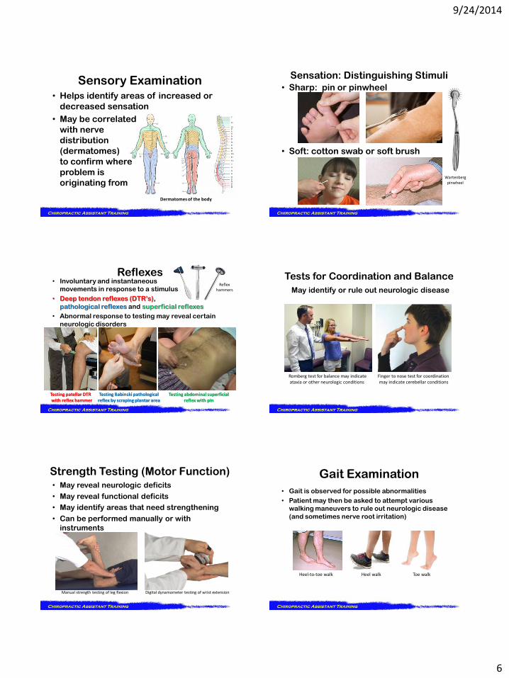
Sensory Examination • Helps identify areas of increased or
decreased sensation
• May be correlated
with nerve
distribution
(dermatomes)
to confirm where
problem is
originating from
Dermatomes of the body
• Sharp: pin or pinwheel
• Soft: cotton swab or soft brush
Sensation: Distinguishing Stimuli
Wartenberg pinwheel
Reflexes
Testing patellar DTR Testing Babinski pathological Testing abdominal superficial with reflex hammer reflex by scraping plantar area reflex with pin
• Involuntary and instantaneous
movements in response to a stimulus
• Deep tendon reflexes (DTR’s),
pathological reflexes and superficial reflexes
• Abnormal response to testing may reveal certain
neurologic disorders
Reflex hammers
Tests for Coordination and Balance
May identify or rule out neurologic disease
Romberg test for balance may indicate Finger to nose test for coordination ataxia or other neurologic conditions may indicate cerebellar conditions
Strength Testing (Motor Function) • May reveal neurologic deficits
• May reveal functional deficits
• May identify areas that need strengthening
• Can be performed manually or with
instruments
Manual strength testing of leg flexion Digital dynamometer testing of wrist extension
• Gait is observed for possible abnormalities
• Patient may then be asked to attempt various
walking maneuvers to rule out neurologic disease
(and sometimes nerve root irritation)
Gait Examination
Heel-to-toe walk Heel walk Toe walk

9/24/2014
7
Examples of Abnormal Gait
All raise the possibility of neurologic disease
A condition of the wrist, hand and fingers caused by
irritation of one or more
nerves at the wrist, often
causing pain, tingling
or numbness
• Tinel Sign: gentle tapping over the
anterior wrist may cause pain and
or tingling there and or into the hand
• Phalen’s Sign: holding wrists
as shown for 30 to 60 seconds
may cause pain and/or tingling
there and or into the hand
Testing for Carpal Tunnel Syndrome
Tinel’s
Phalen’s
IV. Diagnostic Testing
Exam may identify a need
for diagnostic testing
• Blood laboratory studies
• Imaging
• Neurodiagnostic studies
• Other, as needed
Lumbar spine MRI
Blood Laboratory Studies
(“Labwork,” “Labs”) • To confirm or rule out other health care
conditions that might either be the cause of the patient’s symptoms or might be a factor
• To confirm or rule out certain inflammatory joint conditions such as rheumatoid arthritis, ankylosing spondylitis
• To confirm or rule out connective tissue diseases such as systemic lupus erythematosus (SLE)
“Imaging” Relates to any diagnostic procedure that
produces an image of a body part
• Radiography (includes mammography)
• MRI
• CT
• Diagnostic ultrasound
• Nuclear medicine
• PET
• Echocardiography
• Bone mineral densitometry
Common Imaging Studies
Ordered by
Chiropractic Doctors
9/24/2014
8
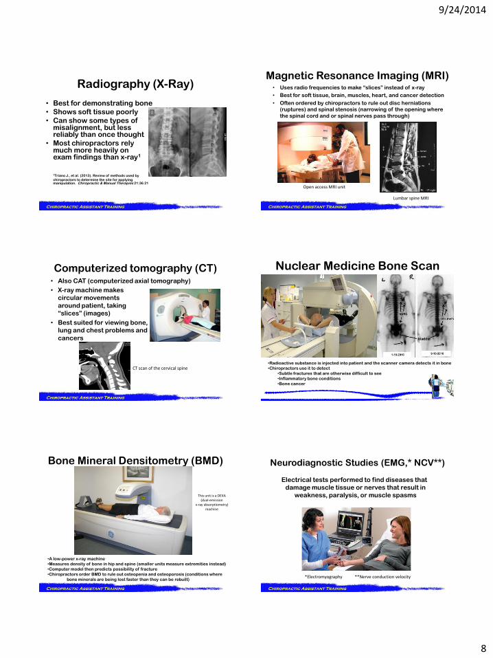
Radiography (X-Ray)
• Best for demonstrating bone
• Shows soft tissue poorly
• Can show some types of misalignment, but less reliably than once thought
• Most chiropractors rely much more heavily on exam findings than x-ray1
1Triano J., et al. (2013). Review of methods used by
chiropractors to determine the site for applying manipulation. Chiropractic & Manual Therapies 21:36 21
• Uses radio frequencies to make “slices” instead of x-ray
• Best for soft tissue, brain, muscles, heart, and cancer detection
• Often ordered by chiropractors to rule out disc herniations
(ruptures) and spinal stenosis (narrowing of the opening where
the spinal cord and or spinal nerves pass through)
Magnetic Resonance Imaging (MRI)
Open access MRI unit
Lumbar spine MRI
Computerized tomography (CT)
CT scan of the cervical spine
• Also CAT (computerized axial tomography)
• X-ray machine makes
circular movements
around patient, taking
“slices” (images)
• Best suited for viewing bone,
lung and chest problems and
cancers
Nuclear Medicine Bone Scan
•Radioactive substance is injected into patient and the scanner camera detects it in bone
•Chiropractors use it to detect
•Subtle fractures that are otherwise difficult to see
•Inflammatory bone conditions
•Bone cancer
Bone Mineral Densitometry (BMD)
•A low-power x-ray machine
•Measures density of bone in hip and spine (smaller units measure extremities instead)
•Computer model then predicts possibility of fracture
•Chiropractors order BMD to rule out osteopenia and osteoporosis (conditions where
bone minerals are being lost faster than they can be rebuilt)
This unit is a DEXA (dual-emission
x-ray absorptiometry) machine
Neurodiagnostic Studies (EMG,* NCV**)
Electrical tests performed to find diseases that
damage muscle tissue or nerves that result in
weakness, paralysis, or muscle spasms
*Electromyography **Nerve conduction velocity