€¦ · Web viewThe Pupil Reflex ….. The amount of light entering the eye is controlled by a ....
28
The human circulatory system is a double circulatory system. It has two separate circuits and blood passes through the heart twice - The Pulmonary Circuit is between the heart and the lungs - The Systemic Circuit is between the heart and the other organs The Pulmonary Circuit transports blood TO the lungs. The blood is oxygenated there and then carried back to the heart. Gaseous exchange happens in the lungs - Carbon dioxide diffuses from the blood into the air in the alveoli - Oxygen diffuses from the air in the alveoli and into the blood, and is absorbed by haemoglobin in the red blood cells - Unlike other arteries and veins, the pulmonary artery carries deoxygenated blood and the pulmonary vein carries oxygenated blood The Systemic Circuit transports blood around the body. It transports oxygen and nutrients to the body tissues and carries away deoxygenated blood containing carbon dioxide and other waste materials
Transcript of €¦ · Web viewThe Pupil Reflex ….. The amount of light entering the eye is controlled by a ....

The human circulatory system is a double circulatory system. It has two separate circuits and blood passes through the heart twice- The Pulmonary Circuit is between the heart and the lungs- The Systemic Circuit is between the heart and the other
organs The Pulmonary Circuit transports blood TO the lungs. The
blood is oxygenated there and then carried back to the heart. Gaseous exchange happens in the lungs- Carbon dioxide diffuses from the blood into the air in the
alveoli- Oxygen diffuses from the air in the alveoli and into the
blood, and is absorbed by haemoglobin in the red blood cells
- Unlike other arteries and veins, the pulmonary artery carries deoxygenated blood and the pulmonary vein carries oxygenated blood
The Systemic Circuit transports blood around the body. It transports oxygen and nutrients to the body tissues and carries away deoxygenated blood containing carbon dioxide and other waste materials
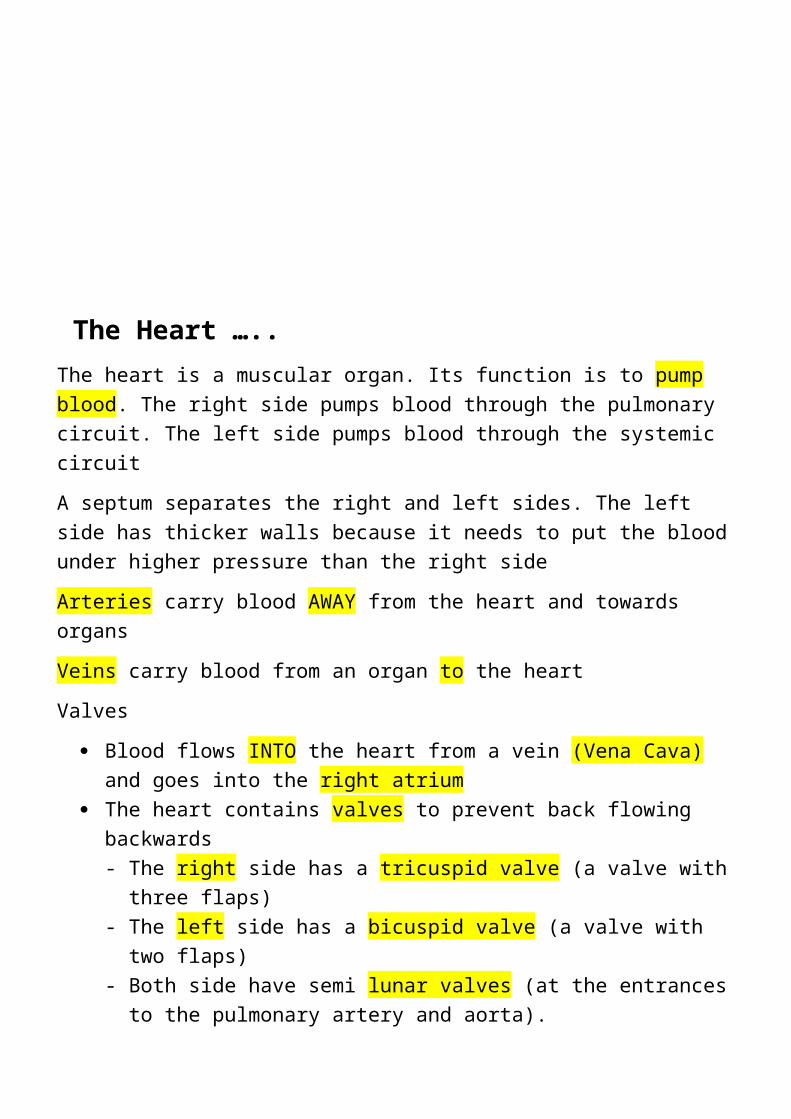
The Heart …..The heart is a muscular organ. Its function is to pump blood. The right side pumps blood through the pulmonary circuit. The left side pumps blood through the systemic circuitA septum separates the right and left sides. The left side has thicker walls because it needs to put the blood under higher pressure than the right sideArteries carry blood AWAY from the heart and towards organsVeins carry blood from an organ to the heartValves
Blood flows INTO the heart from a vein (Vena Cava) and goes into the right atrium
The heart contains valves to prevent back flowing backwards- The right side has a tricuspid valve (a valve with three
flaps)- The left side has a bicuspid valve (a valve with two flaps)- Both side have semi lunar valves (at the entrances to the
pulmonary artery and aorta).
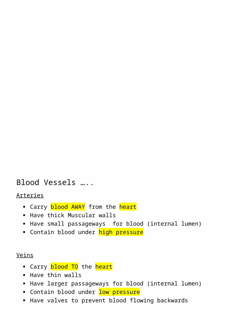
Blood Vessels …..Arteries
Carry blood AWAY from the heart Have thick Muscular walls Have small passageways for blood (internal lumen) Contain blood under high pressure
Veins Carry blood TO the heart Have thin walls Have larger passageways for blood (internal lumen) Contain blood under low pressure Have valves to prevent blood flowing backwards
Capillaries Found in the muscles and lungs Microscopic- One cell thick Very low blood pressure Where gas exchange takes place

Coronary Heart Disease….. The coronary arteries supply blood to the heart muscle. These
may become blocked by a build-up of fatty plaques containing cholesterol, resulting in Coronary Heart Disease
If a coronary artery is blocked, the bloody supply to part of the heart muscle is cut off. That part of the heart can’t continue to contract, causing a heart attack
Possible causes of coronary heart disease- - Poor diet- eating more saturated fat tends to increase
cholesterol levels- Stress and smoking- increases blood pressure - Lack of exercise- Being overweight

The trachea branches into two bronchi (one to each lung) Pleural membranes surround each lung Cartilage rings in the walls of the trachea help to keep it open The bronchi split into smaller and smaller tubes called
bronchioles. These end in microscopic air sacs called alveoli. There is a muscular diaphragm below the lungs
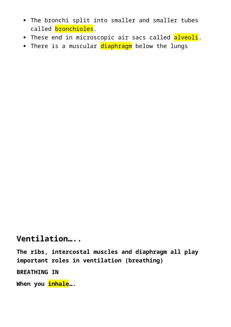
Ventilation…..The ribs, intercostal muscles and diaphragm all play important roles in ventilation (breathing)BREATHING IN When you inhale….
The internal intercostal muscles relax and the external intercostal muscles contract, pulling the ribcage upwards and outwards
The diaphragm contracts, pulling downwards Lung Volume increases and the air pressure inside
decreases Air is pushed into the lungs
BREATHING OUTWhen you exhale….
The external intercostal muscles relax and internal intercostal muscles contract, pulling the ribcage downwards and inwards
The diaphragm relaxes, moving back and upwards Lung Volume decreases and the air pressure inside
increases Air is pushed out of the lungs
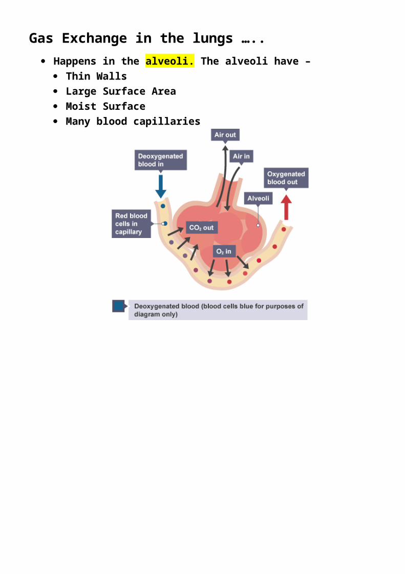
Gas Exchange in the lungs ….. Happens in the alveoli. The alveoli have –
Thin Walls Large Surface Area Moist Surface Many blood capillaries
Blood is a complex liquid tissue. It transports oxygen, dissolved substances and hear around the body, as well as being involved in the body’s immune response.
The blood is composed off PLASMA- Transports CO2, digested food, urea,
hormones and heat RED BLOOD CELLS – Transports oxygen WHITE BLOOD CELLS- Ingests pathogens and
produces antibodies PLATELETS- Involved in blood clotting
Red Blood Cells…. Transport oxygen for aerobic respiration Red blood cells have adaptations that make them
suitable for their role - They contain haemoglobin- a red protein that
combines with oxygen to form oxyhaemoglobin They have no nucleus- so they can contain more
haemoglobin They are small and flexible so that they can fit
through narrow blood vessels They have a biconcave shape (Flattened disc) to
maximise their surface area for oxygen absorption

About 25% of the white blood cells are LYMPHOCYTES
They are part of the body’s immune system and produce soluble proteins called antibodies. Antibodies can recognise particular types of pathogen
ANTIBODIES attach to ANTIGENS Antigens are substances found on the surface of
cells, including bacteria and pathogens Different antibodies attach to different antigens.
Meaning the body’s immune system can’t recognise foreign antigens (antigens that aren’t normally produced by the body but by pathogens
Antibodies can neutralise toxins produced by pathogens. They can cause the destruction of pathogens by-- Causing bacteria to burst open and die- Labelling the pathogen so that it is recognised
more easily by phagocytes- Sticking pathogens together so they can be
engulfed by phagocytes more easily
About 70% of the white blood cells are PHAGOCYTES Phagocytes ingest and destroy pathogens such as
bacteria- The phagocyte surrounds the bacterial cell,
enclosing it in a vacuole- Enzymes are secreted into the vacuole to
destroy the bacterial cell The process of ingesting the pathogen is called
phagocytosis

People can be immunised against a pathogen through vaccination
Different vaccines are needed for different pathogens Vaccination involved putting a small amount of an
inactive form of a pathogen into the body Vaccines contain
- Live pathogens treated to make them harmless- Harmless fragments of the pathogen- Toxins produced by the pathogen- Dead Pathogens
These all contain antigens. When injected into the body they stimulate lymphocytes to produce antibodies that can recognise the pathogen
Some lymphocytes develop into memory cells If the vaccinated person later becomes infected with
the same pathogen, the immune system is prepared and the required lymphocytes are able to reproduce rapidly and destroy it. Meaning the person is unlikely to become ill

If the skin is cut the wound must be closed to prevent blood loss and the entry of pathogens. The formation of a scab does that
Blood contains tiny fragments of cells called platelets. There platelets are involved in blood clotting and scab forming
When the skin is wounded platelets are able to – - Release chemicals that cause soluble fibrinogen
proteins to form a mesh of insoluble fibrin fibres across the wound
- Stick together to form clumps that get stuck in the fibrin mesh
Red Blood cells get stuck in the fibrin mesh forming a clot, which develops into a scab and protects the
wound as it heals
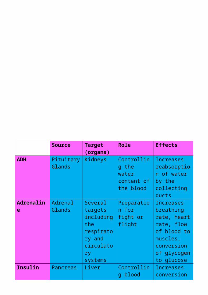
A hormone is a chemical substance produced by a gland and carried by the blood which alters the activity of more specific target organs and then destroyed by the liver
Like the nervous system, hormone can control the body Different hormones affect different organs or cells

Source Target (organs)
Role Effects
ADH Pituitary Glands
Kidneys Controlling the water content of the blood
Increases reabsorption of water by the collecting ducts
Adrenaline
Adrenal Glands
Several targets including the respiratory and circulatory systems
Preparation for fight or flight
Increases breathing rate, heart rate, flow of blood to muscles, conversion of glycogen to glucose
Insulin Pancreas Liver Controlling blood glucose levels
Increases conversion of glucose into glycogen

The Human Nervous System consists of- THE CENTRAL NERVOUS SYSTEM (CNS) - The brain and spinal cord
THE PERIPHERAL NERVOUS SYSTEM - nerve cells that carry information to or from the CNS
Nerve Cells ….. Nerve cells are also called neurones. They are
adapted to carry electrical impulses from one place to another- They have a long fibre (axon) which is insulated
by a fatty sheath - They have tiny branches (Dendron’s) which
branch further as dendrites at each end
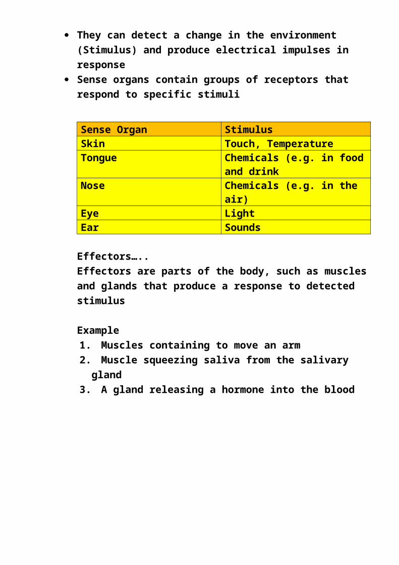
Receptors ….. Receptors are groups of specialised cells. They can detect a change in the environment
(Stimulus) and produce electrical impulses in response
Sense organs contain groups of receptors that respond to specific stimuli
Sense Organ StimulusSkin Touch, Temperature Tongue Chemicals (e.g. in food
and drinkNose Chemicals (e.g. in the
air)Eye LightEar Sounds

Effectors…..Effectors are parts of the body, such as muscles and glands that produce a response to detected stimulus
Example 1.Muscles containing to move an arm2.Muscle squeezing saliva from the salivary gland 3.A gland releasing a hormone into the blood
Reflex Actions ….. A reflex action is a way for the body to
automatically and rapidly respond to a stimulus to minimise any further damage to the body
It follows this general sequence and does not involve the brain

The nerve pathway followed by a reflex action is called a reflex arc, e.g. a simple reflex arc happens if we accidentally touch something hot
1. Receptor in the skin detects a stimulus (the change in temperature)2. Sensory Neurone sends impulses to relay neurone 3. Motor neurone sends impulses to effector4. Effector produces a response (muscles contracts to move hand away)
Antagonistic pairs ….. Muscles work in antagonistic pairs
This ensures when a part of the body is moved, it can move back to its original position, e.g
- The biceps and triceps in the arm- The quadriceps and hamstrings in the leg
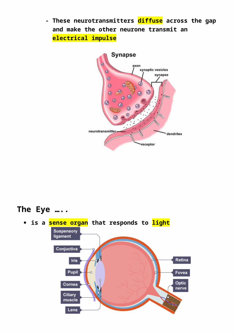
The Synapse …..- Where two neurones meet there is a tiny gap
called a synapse- Information crosses this gap using
neurotransmitters- One neuron releases neurotransmitters into the
synapse- These neurotransmitters diffuse across the gap
and make the other neurone transmit an electrical impulse

The Eye ….. is a sense organ that responds to light
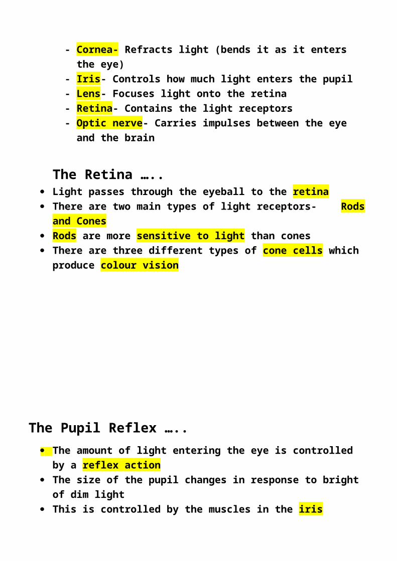
- Cornea- Refracts light (bends it as it enters the eye)
- Iris- Controls how much light enters the pupil- Lens- Focuses light onto the retina- Retina- Contains the light receptors- Optic nerve- Carries impulses between the eye and
the brain
The Retina ….. Light passes through the eyeball to the retina There are two main types of light receptors- Rods
and Cones Rods are more sensitive to light than cones There are three different types of cone cells which
produce colour vision

The Pupil Reflex ….. The amount of light entering the eye is controlled by a
reflex action The size of the pupil changes in response to bright of
dim light This is controlled by the muscles in the iris The ability of the lens to change shape to focus near
and distant objects is called accommodation
A stent is a small mesh tube that's used to treat narrow or weak arteries.
A stent is placed in an artery as part of a procedure called percutaneous coronary intervention (PCI), also known as coronary angioplasty
Stents help keep coronary arteries open and reduce the chance of a heart attack
A stent is inserted into the clogged artery with a balloon catheter
The balloon is inflated and the stent expands and locks into place. This holds the artery open and allows blood to flow more freely
Cholesterol….. Is a substance found in the blood It is made in the liver and is needed for healthy cell
membranes However too much cholesterol in the blood increases
the risk of heart disease and of diseased arteries The bloodstream transports cholesterol around the
body attached to proteins. The combination of cholesterol and protein is called lipoprotein. There are two types of lipoprotein
- Low Density Lipoproteins- LDLs- Carry Cholesterol from the liver to the cells
- High Density Lipoproteins- HDL’s- Carry excess cholesterol back to the liver
LDLs are often called bad cholesterol because they lead to fat building up on artery walls, which causes heart disease
HDLs are often called good cholesterol because they help stop fat building up in the arteries
Statins are a drug sometimes used to help people with high cholesterol lower their cholesterol. They work by blocking a step in the body’s production of cholesterol
A pacemaker is a small device that is placed in the chest or abdomen to help control abnormal heart rhythms
This device uses low energy electrical pulses to prompt the heart heat at a normal rate
Pacemakers are used to treat arrhythmias (problems with the rate or rhythm of the heart

Effects of Smoking ….. Smoking can cause lung disease, heart disease and
certain cancers

Nicotine is the addictive substance in tobacco. It reaches the brain and creates a dependency so that smokers become addictive
Sticky mucus in the lungs traps pathogens. The mucus is normally swept out the lungs by cilia on the epithelial cells lining the trachea, bronchi and bronchioles
Cigarette smoke contains harmful chemicals that damage these cells, leading to a build-up of mucus and a smokers cough
Smoke irritates the bronchi, causing bronchitis’s Smoking damages the walls of the alveoli. The alveoli
walls break down and join together forming larger air spaces than normal. This reduces the efficiency of gas exchange, so people with the lung disease emphysema carry less oxygen in their blood and find even mild exercise difficult
Carbon Monoxide combines with haemoglobin in red blood cells. This reduces the ability of the blood to carry oxygen, putting a strain on the circulatory system and increasing the risk of Coronary heart disease and strokes
Carcinogens are substances that cause cancer. Tobacco smoke contains many carcinogens, including tar. Smoking increase the risk of lung cancer and cancer of the mouth throat and oesophagus