Differential Diagnoses Symptoms...Anisocoria Anisocoria –Which Pupil is Abnormal? Dilated Pupil(s)...
195
Differential Diagnoses and other Useful Lists and Tables For Ophthalmologists Kenn Freedman MD PhD Department of Ophthalmology and Visual Sciences Texas Tech University Health Sciences Center Lubbock, Texas USA Symptoms Signs Case Presentations
Transcript of Differential Diagnoses Symptoms...Anisocoria Anisocoria –Which Pupil is Abnormal? Dilated Pupil(s)...
Differential Diagnoses and other Useful Lists and Tables
For Ophthalmologists
Kenn Freedman MD PhDDepartment of Ophthalmology and Visual SciencesTexas Tech University Health Sciences CenterLubbock, Texas USA
SymptomsSigns
Case Presentations
Acknowledgments and Disclaimer
The differential diagnoses and lists contained herein are not meant to be exhaustive, but are to give in most cases the most common causes of many ocular / visual symptoms, signs and situations. Included also in these lists are also some less common, but serious conditions that must be “ruled-out”. These lists have been based on years of experience, and I am grateful for God’s help in developing them. I also owe gratitude to several sources* including Roy’s classic text on Ocular Differential Diagnosis.
This presentation, of course, will continue to be a work in progress and any concerns or suggestions as to errors or omissions or picture copyrights will be considered. Please feel free to contact me at [email protected]
Kenn Freedman
Lubbock, Texas - October 2018
* Please see
references at
end of document
Disclaimer: The diagnostic algorithm for the diagnosis and management of Ocular or Neurological Conditions contained in this presentation is not intended to replace the independent medical or professional judgment of the physician or other health care providers in the context of individual clinical circumstances to determine a patient’s care.

Use of this PresentationThe lists are divided into three main areas
1. Symptoms
2. Signs from the Eight Point Eye Exam
3. Common Situations and Case Presentations
The index for all of the lists is given on the following 3 pages. The lists follow in the presentation in the order shown in the index. Each entry in the index (blue) is also a link and when clicked will take to you that specific list in the document.
If you want to go to another list within the document you will find a link on each page (shown below) which will take you back to the original index of links
At the end is also a list of abbreviations used in the presentation
Click to
Return To Links
Index o
f all L
ists
with
Lin
ks
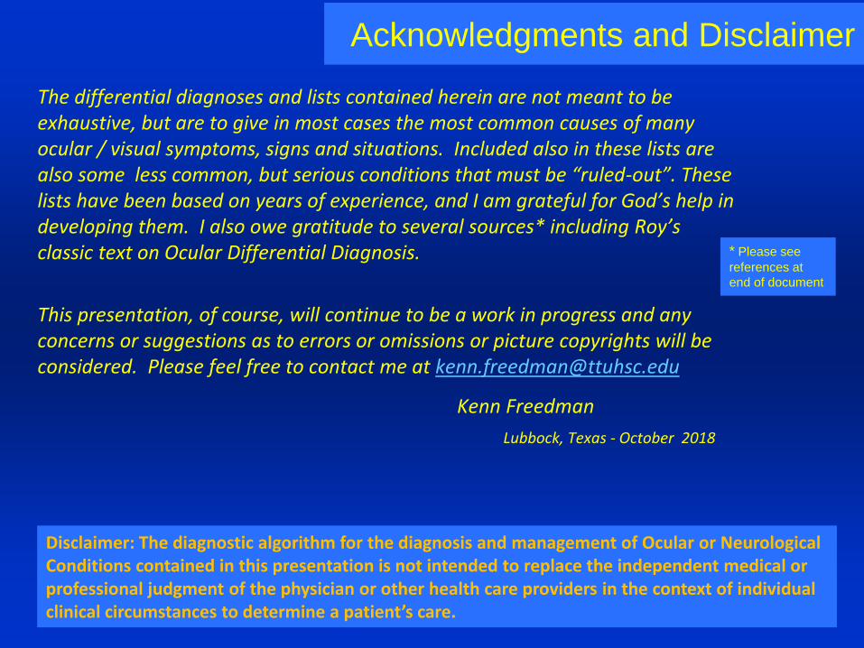
Symptoms
Loss of Vision
Transient Visual Phenomena
Floaters
Flashes, Photopsias
Diplopia
Monocular Diplopia
Binocular Diplopia – Ocular Misalignment
Transient Diplopia
Oscillopsia
Night Blindness
Transient Visual Loss
Photophobia
Headache
Eye and Face Pain
Epiphora
Foreign Body Sensation and Itching
Problems Opening Eyes
Chronic Red Eye
Loss of Visual Acuity and Refractive Issues
Decreased Distance Visual Acuity
Refractive Shift – Myopic
Refractive Shift – Hyperopic
Refractive Shift – Astigmatic
Asymmetric and Irregular Astigmatism
Dull or Abnormal Retinoscope Reflex
Poor Near Visual Acuity
Problems with Glasses
Loss of Visual Field (VF)
Visual Field Defects and Localizing Lesions
VF Defects Respecting the Horizontal Midline
VF Defects Respecting the Vertical Midline
Bitemporal Hemianopsia
Homonymous Hemianopsia
Central, Centrocecal and Cecal VF Defects
Severe Constriction of VF, Tunnel VF
Eyelids and Orbit
Blepharospasm
Loss of Sensation, Numbness of Face around Eye
Ptosis
Eyelashes and Eyelid Margin
Eyelid Malpositions – Entropion and Trichiasis
Eyelid Malpositions – Ectropion
Eyelid Retraction
Lagophthalmos and Lid Lag
Seventh Nerve Palsy
Eyelid Mass / Lesion
Signs Suggesting Orbital Disease
Eyelid Edema
Conjunctival Hemorrhage and Peri-Orbital Ecchymosis
Proptosis
Orbital Tumors
Enophthalmos
Orbital Inflammation
Visible and Palpable Orbital Masses
Orbital and Facial Distortions
Distortions of the Globe

Index o
f all L
ists
with
Lin
ks
Motility and Alignment Problems
Types and Causes of Motility Problems
Abduction Deficit
Adduction Deficit
Acquired Esotropia
Acquired Exotropia
Apparent Horizontal Strabismus
Hypertropia / Hypotropia
Elevation and Upgaze Deficits
Depression and Downgaze Deficits
Convergence and Divergence
Ophthalmoplegia – One Eye
Ophthalmoplegia – Both Eyes
Head Turns and Tilts / Torticollis
Causes of Nystagmus
Acquired Nystagmus
Downbeat Nystagmus
Pupils
Leukocoria
Corectopia
Poor Pupil Mobility
Anisocoria
Anisocoria – Which Pupil is Abnormal?
Dilated Pupil(s)
Transient Pupil Dilation
Constricted Pupil(s)
Light Near Dissociation
Relative Afferent Pupillary Defect
Bilateral Miotic or Mydriatic Pupils
Anterior Segment
Conjunctival Bumps - Papillae and Follicles
Chemosis
Injected, Congested or Prominent Conjunctival Vessels
Symblepharon
Conjunctival or Epibulbar Mass
Spot on the White of Eye, Scleral Lesion
Scleral Thinning, Episcleritis, and Scleritis
Corneal Fluorescein Staining or Pooling
Corneal Epithelial Defects – Chronic or Non-Healing
Corneal Haze or Opacification
Acute Corneal Edema
Chronic Corneal Edema
Punctate Corneal Staining
Corneal Infiltrates
Corneal Ulcer
Corneal Vessels and Pannus
Decreased Corneal Sensation
Keratic Precipitates (KP)
Anterior Uveitis
Cells in the Anterior Chamber
Hypopion
Hyphema
Neovascularization of the Iris
Lesions of the Iris
Defects of the Iris
Lens Opacification
Lens Abnormalities
Index o
f all L
ists
with
Lin
ks
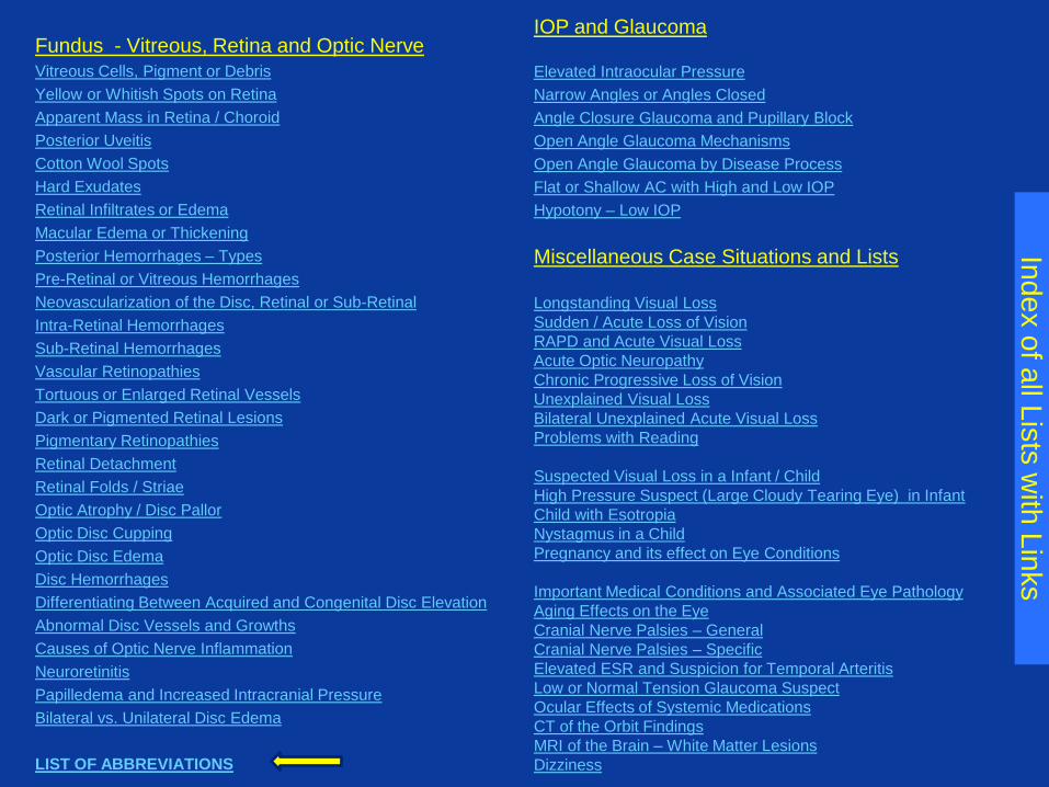
Fundus - Vitreous, Retina and Optic Nerve
Vitreous Cells, Pigment or Debris
Yellow or Whitish Spots on Retina
Apparent Mass in Retina / Choroid
Posterior Uveitis
Cotton Wool Spots
Hard Exudates
Retinal Infiltrates or Edema
Macular Edema or Thickening
Posterior Hemorrhages – Types
Pre-Retinal or Vitreous Hemorrhages
Neovascularization of the Disc, Retinal or Sub-Retinal
Intra-Retinal Hemorrhages
Sub-Retinal Hemorrhages
Vascular Retinopathies
Tortuous or Enlarged Retinal Vessels
Dark or Pigmented Retinal Lesions
Pigmentary Retinopathies
Retinal Detachment
Retinal Folds / Striae
Optic Atrophy / Disc Pallor
Optic Disc Cupping
Optic Disc Edema
Disc Hemorrhages
Differentiating Between Acquired and Congenital Disc Elevation
Abnormal Disc Vessels and Growths
Causes of Optic Nerve Inflammation
Neuroretinitis
Papilledema and Increased Intracranial Pressure
Bilateral vs. Unilateral Disc Edema
LIST OF ABBREVIATIONS
IOP and Glaucoma
Elevated Intraocular Pressure
Narrow Angles or Angles Closed
Angle Closure Glaucoma and Pupillary Block
Open Angle Glaucoma Mechanisms
Open Angle Glaucoma by Disease Process
Flat or Shallow AC with High and Low IOP
Hypotony – Low IOP
Miscellaneous Case Situations and Lists
Longstanding Visual Loss
Sudden / Acute Loss of Vision
RAPD and Acute Visual Loss
Acute Optic Neuropathy
Chronic Progressive Loss of Vision
Unexplained Visual Loss
Bilateral Unexplained Acute Visual Loss
Problems with Reading
Suspected Visual Loss in a Infant / Child
High Pressure Suspect (Large Cloudy Tearing Eye) in Infant
Child with Esotropia
Nystagmus in a Child
Pregnancy and its effect on Eye Conditions
Important Medical Conditions and Associated Eye Pathology
Aging Effects on the Eye
Cranial Nerve Palsies – General
Cranial Nerve Palsies – Specific
Elevated ESR and Suspicion for Temporal Arteritis
Low or Normal Tension Glaucoma Suspect
Ocular Effects of Systemic Medications
CT of the Orbit Findings
MRI of the Brain – White Matter Lesions
Dizziness
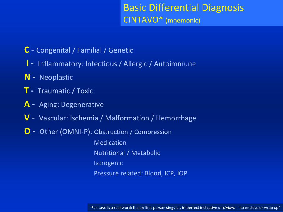
Basic Differential DiagnosisCINTAVO* (mnemonic)
C - Congenital / Familial / Genetic
I - Inflammatory: Infectious / Allergic / Autoimmune
N - Neoplastic
T - Traumatic / Toxic
A - Aging: Degenerative
V - Vascular: Ischemia / Malformation / Hemorrhage
O - Other (OMNI-P): Obstruction / Compression
Medication
Nutritional / Metabolic
Iatrogenic
Pressure related: Blood, ICP, IOP
*cintavo is a real word: Italian first-person singular, imperfect indicative of cintare - “to enclose or wrap up”
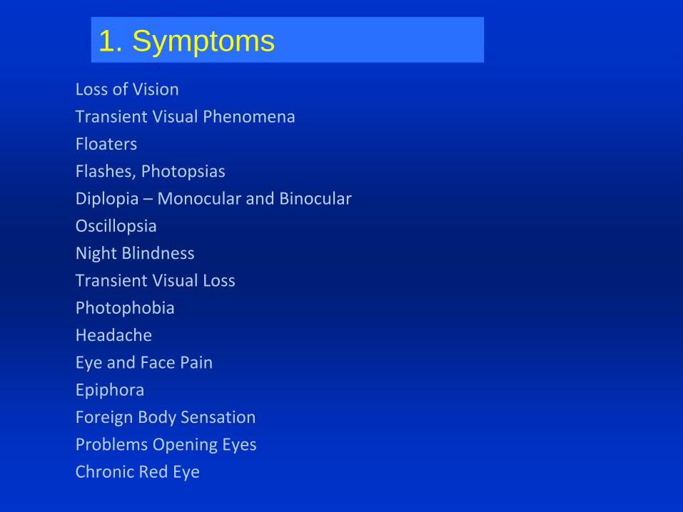
1. Symptoms
Loss of Vision
Transient Visual Phenomena
Floaters
Flashes, Photopsias
Diplopia – Monocular and Binocular
Oscillopsia
Night Blindness
Transient Visual Loss
Photophobia
Headache
Eye and Face Pain
Epiphora
Foreign Body Sensation
Problems Opening Eyes
Chronic Red Eye
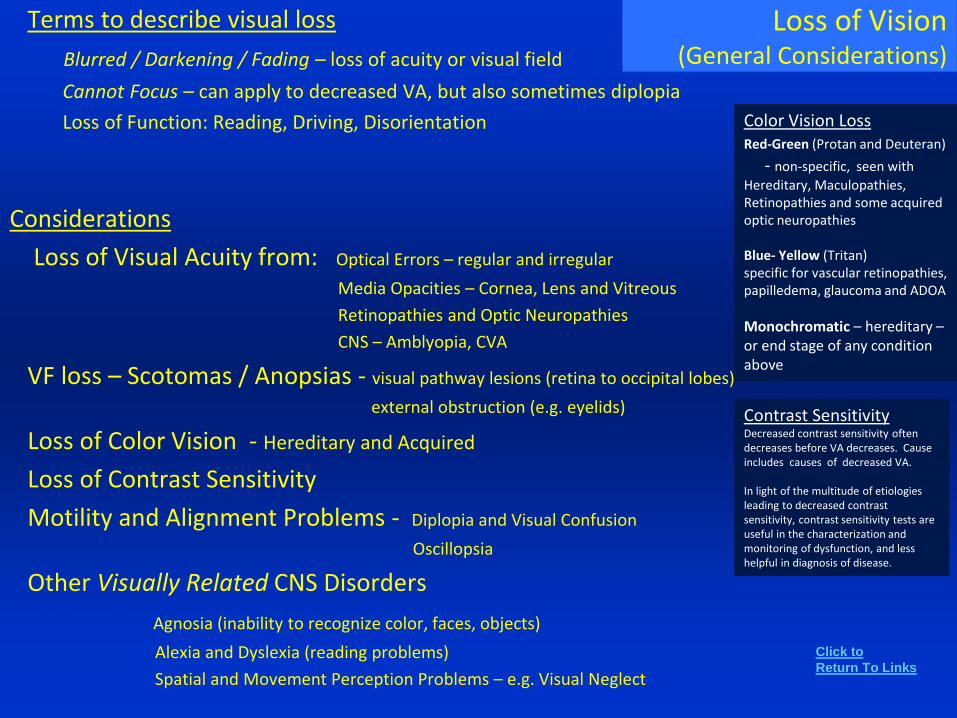
Loss of Vision(General Considerations)
Terms to describe visual loss
Blurred / Darkening / Fading – loss of acuity or visual field
Cannot Focus – can apply to decreased VA, but also sometimes diplopia
Loss of Function: Reading, Driving, Disorientation
Considerations
Loss of Visual Acuity from: Optical Errors – regular and irregular
Media Opacities – Cornea, Lens and Vitreous
Retinopathies and Optic Neuropathies
CNS – Amblyopia, CVA
VF loss – Scotomas / Anopsias - visual pathway lesions (retina to occipital lobes)
external obstruction (e.g. eyelids)
Loss of Color Vision - Hereditary and Acquired
Loss of Contrast Sensitivity
Motility and Alignment Problems - Diplopia and Visual Confusion
Oscillopsia
Other Visually Related CNS Disorders
Agnosia (inability to recognize color, faces, objects)
Alexia and Dyslexia (reading problems)
Spatial and Movement Perception Problems – e.g. Visual Neglect
Color Vision LossRed-Green (Protan and Deuteran)
- non-specific, seen with
Hereditary, Maculopathies, Retinopathies and some acquired optic neuropathies
Blue- Yellow (Tritan) specific for vascular retinopathies, papilledema, glaucoma and ADOA
Monochromatic – hereditary –or end stage of any condition above
Contrast SensitivityDecreased contrast sensitivity often decreases before VA decreases. Cause includes causes of decreased VA.
In light of the multitude of etiologies leading to decreased contrast sensitivity, contrast sensitivity tests are useful in the characterization and monitoring of dysfunction, and less helpful in diagnosis of disease.
Click to
Return To Links
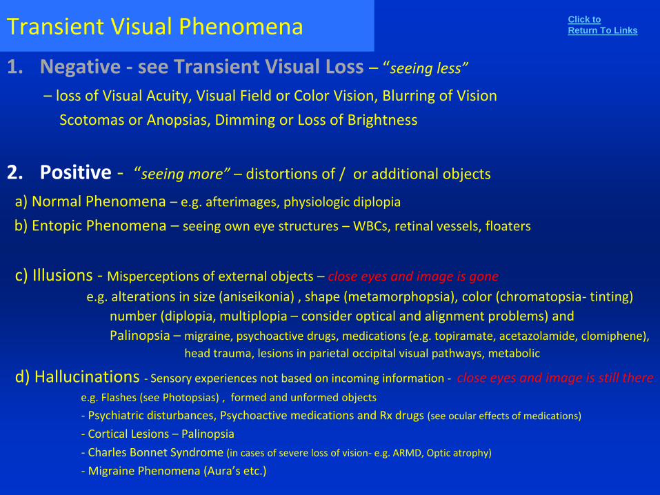
Transient Visual Phenomena
1. Negative - see Transient Visual Loss – “seeing less”
– loss of Visual Acuity, Visual Field or Color Vision, Blurring of Vision
Scotomas or Anopsias, Dimming or Loss of Brightness
2. Positive - “seeing more” – distortions of / or additional objects
a) Normal Phenomena – e.g. afterimages, physiologic diplopia
b) Entopic Phenomena – seeing own eye structures – WBCs, retinal vessels, floaters
c) Illusions - Misperceptions of external objects – close eyes and image is gone
e.g. alterations in size (aniseikonia) , shape (metamorphopsia), color (chromatopsia- tinting)
number (diplopia, multiplopia – consider optical and alignment problems) and
Palinopsia – migraine, psychoactive drugs, medications (e.g. topiramate, acetazolamide, clomiphene),
head trauma, lesions in parietal occipital visual pathways, metabolic
d) Hallucinations - Sensory experiences not based on incoming information - close eyes and image is still there
e.g. Flashes (see Photopsias) , formed and unformed objects
- Psychiatric disturbances, Psychoactive medications and Rx drugs (see ocular effects of medications)
- Cortical Lesions – Palinopsia
- Charles Bonnet Syndrome (in cases of severe loss of vision- e.g. ARMD, Optic atrophy)
- Migraine Phenomena (Aura’s etc.)
Click to
Return To Links

Floaters
• Vitreous Syneresis
• R/O Retinal Detachment
Especially in the case of new floaters!
• Vitreous Detachment (e.g. PVD)
• Vitreous Hemorrhage
• Posterior or Intermediate Uveitis
• Other sources of Vitreous Cells
e.g. Masquerade Syndrome for Uveitis: Lymphoma or Tumor (RB, Melanoma)
• Other Unusual Causes (in Vitreous)
Asteroid Hyalosis, Amyloidosis, Cholesterol Crystals- Synchysis scintillans
Click to
Return To Links
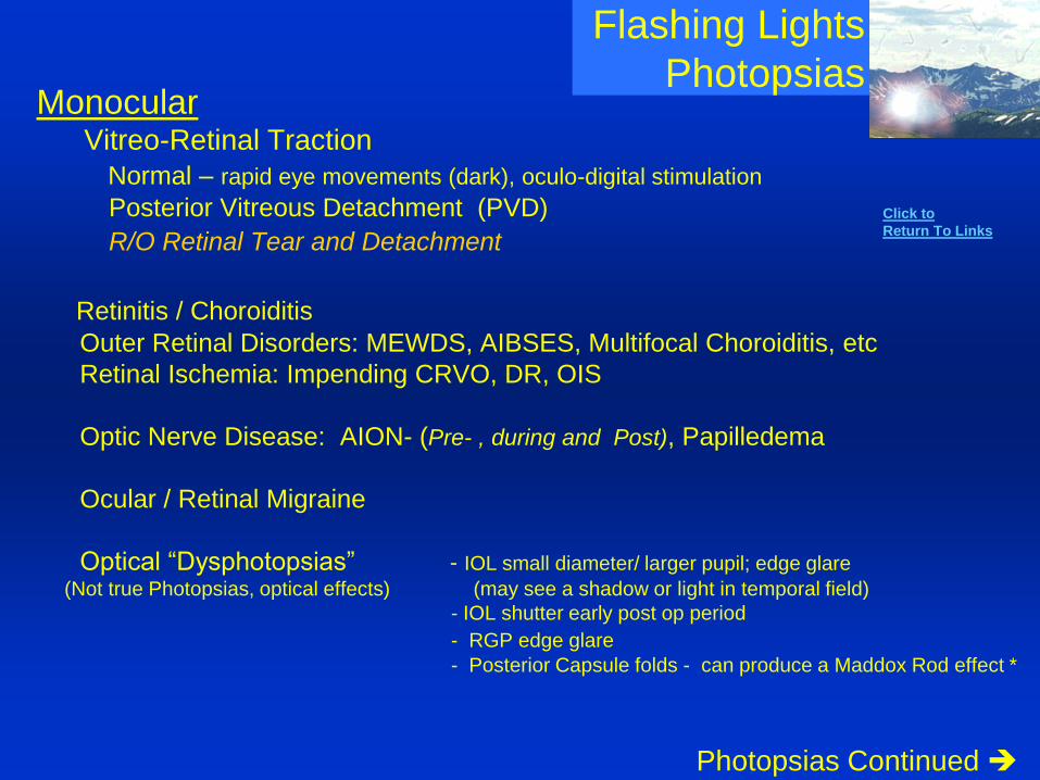
Flashing Lights
PhotopsiasMonocular
Vitreo-Retinal Traction
Normal – rapid eye movements (dark), oculo-digital stimulation
Posterior Vitreous Detachment (PVD)
R/O Retinal Tear and Detachment
Retinitis / Choroiditis
Outer Retinal Disorders: MEWDS, AIBSES, Multifocal Choroiditis, etc
Retinal Ischemia: Impending CRVO, DR, OIS
Optic Nerve Disease: AION- (Pre- , during and Post), Papilledema
Ocular / Retinal Migraine
Optical “Dysphotopsias” - IOL small diameter/ larger pupil; edge glare
(Not true Photopsias, optical effects) (may see a shadow or light in temporal field)
- IOL shutter early post op period
- RGP edge glare
- Posterior Capsule folds - can produce a Maddox Rod effect *
Photopsias Continued
Click to
Return To Links
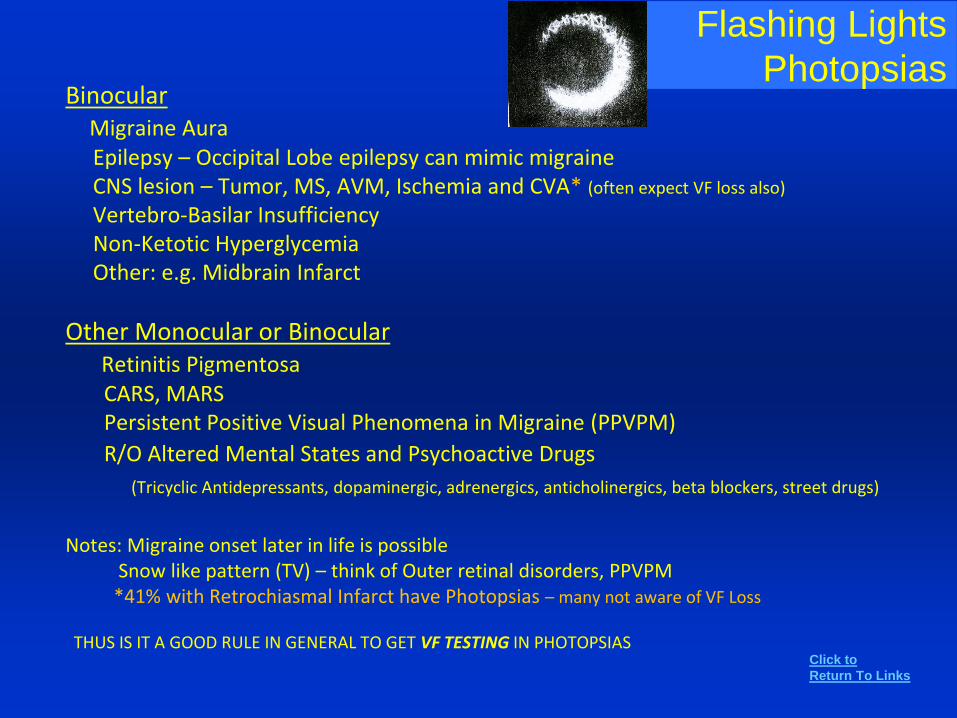
Flashing Lights
PhotopsiasBinocular
Migraine AuraEpilepsy – Occipital Lobe epilepsy can mimic migraineCNS lesion – Tumor, MS, AVM, Ischemia and CVA* (often expect VF loss also)
Vertebro-Basilar InsufficiencyNon-Ketotic HyperglycemiaOther: e.g. Midbrain Infarct
Other Monocular or BinocularRetinitis PigmentosaCARS, MARSPersistent Positive Visual Phenomena in Migraine (PPVPM)
R/O Altered Mental States and Psychoactive Drugs
(Tricyclic Antidepressants, dopaminergic, adrenergics, anticholinergics, beta blockers, street drugs)
Notes: Migraine onset later in life is possibleSnow like pattern (TV) – think of Outer retinal disorders, PPVPM*41% with Retrochiasmal Infarct have Photopsias – many not aware of VF Loss
THUS IS IT A GOOD RULE IN GENERAL TO GET VF TESTING IN PHOTOPSIASClick to
Return To Links
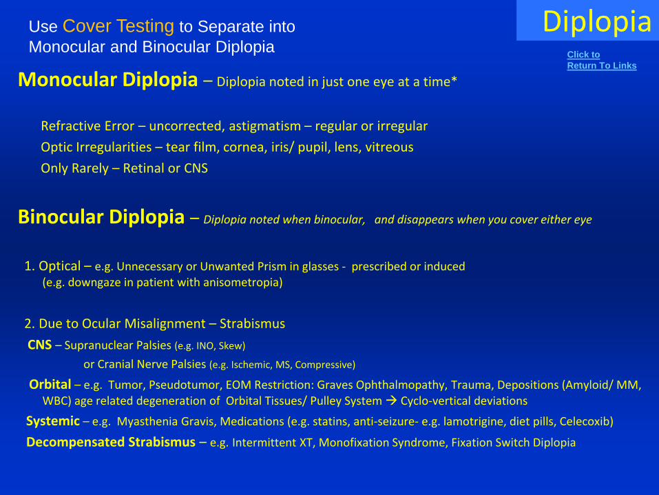
Diplopia
Monocular Diplopia – Diplopia noted in just one eye at a time*
Refractive Error – uncorrected, astigmatism – regular or irregular
Optic Irregularities – tear film, cornea, iris/ pupil, lens, vitreous
Only Rarely – Retinal or CNS
Binocular Diplopia – Diplopia noted when binocular, and disappears when you cover either eye
1. Optical – e.g. Unnecessary or Unwanted Prism in glasses - prescribed or induced (e.g. downgaze in patient with anisometropia)
2. Due to Ocular Misalignment – Strabismus
CNS – Supranuclear Palsies (e.g. INO, Skew)
or Cranial Nerve Palsies (e.g. Ischemic, MS, Compressive)
Orbital – e.g. Tumor, Pseudotumor, EOM Restriction: Graves Ophthalmopathy, Trauma, Depositions (Amyloid/ MM, WBC) age related degeneration of Orbital Tissues/ Pulley System Cyclo-vertical deviations
Systemic – e.g. Myasthenia Gravis, Medications (e.g. statins, anti-seizure- e.g. lamotrigine, diet pills, Celecoxib)
Decompensated Strabismus – e.g. Intermittent XT, Monofixation Syndrome, Fixation Switch Diplopia
Use Cover Testing to Separate into
Monocular and Binocular DiplopiaClick to
Return To Links
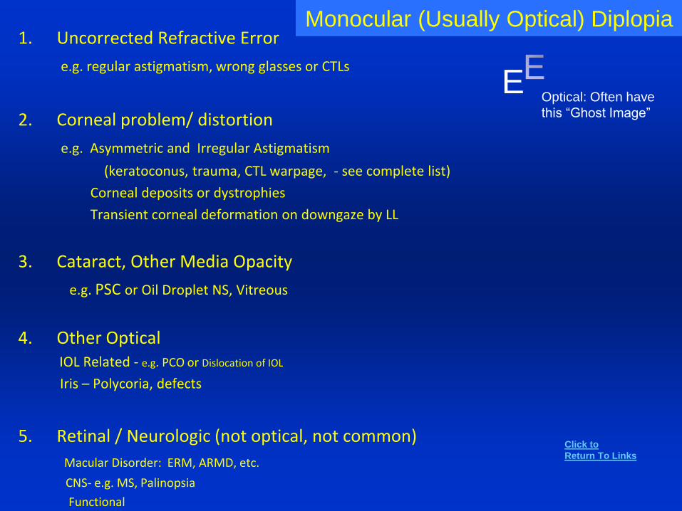
Monocular (Usually Optical) Diplopia1. Uncorrected Refractive Error
e.g. regular astigmatism, wrong glasses or CTLs
2. Corneal problem/ distortion
e.g. Asymmetric and Irregular Astigmatism
(keratoconus, trauma, CTL warpage, - see complete list)
Corneal deposits or dystrophies
Transient corneal deformation on downgaze by LL
3. Cataract, Other Media Opacity
e.g. PSC or Oil Droplet NS, Vitreous
4. Other OpticalIOL Related - e.g. PCO or Dislocation of IOL
Iris – Polycoria, defects
5. Retinal / Neurologic (not optical, not common)
Macular Disorder: ERM, ARMD, etc.
CNS- e.g. MS, Palinopsia
Functional
EOptical: Often have
this “Ghost Image”
Click to
Return To Links
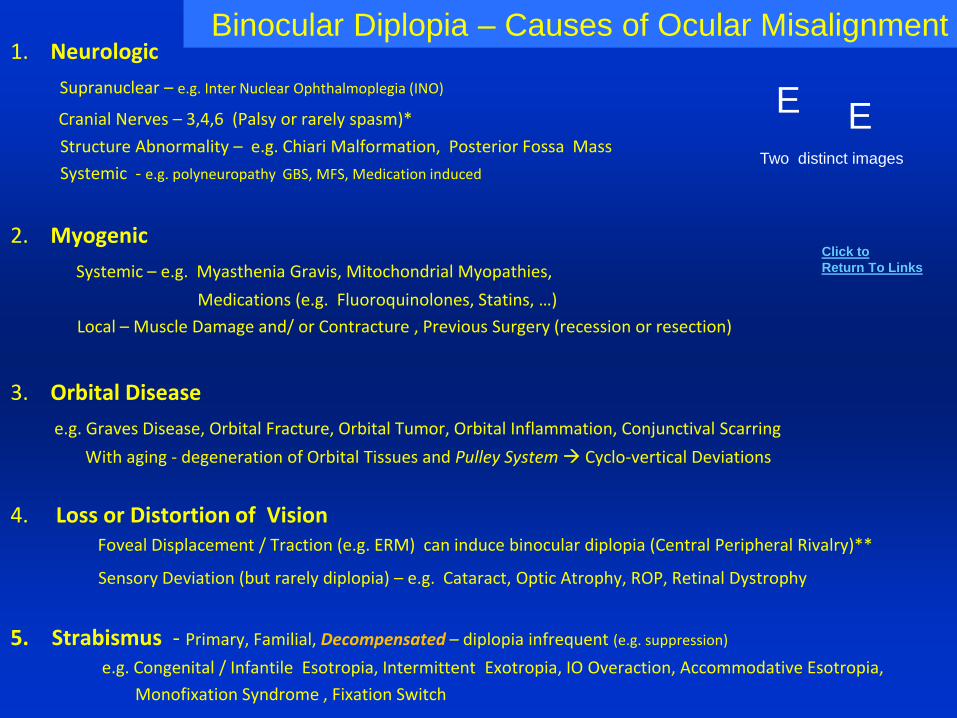
Binocular Diplopia – Causes of Ocular Misalignment1. Neurologic
Supranuclear – e.g. Inter Nuclear Ophthalmoplegia (INO)
Cranial Nerves – 3,4,6 (Palsy or rarely spasm)*
Structure Abnormality – e.g. Chiari Malformation, Posterior Fossa Mass
Systemic - e.g. polyneuropathy GBS, MFS, Medication induced
2. Myogenic
Systemic – e.g. Myasthenia Gravis, Mitochondrial Myopathies,
Medications (e.g. Fluoroquinolones, Statins, …)
Local – Muscle Damage and/ or Contracture , Previous Surgery (recession or resection)
3. Orbital Disease
e.g. Graves Disease, Orbital Fracture, Orbital Tumor, Orbital Inflammation, Conjunctival Scarring
With aging - degeneration of Orbital Tissues and Pulley System Cyclo-vertical Deviations
4. Loss or Distortion of Vision Foveal Displacement / Traction (e.g. ERM) can induce binocular diplopia (Central Peripheral Rivalry)**
Sensory Deviation (but rarely diplopia) – e.g. Cataract, Optic Atrophy, ROP, Retinal Dystrophy
5. Strabismus - Primary, Familial, Decompensated – diplopia infrequent (e.g. suppression)
e.g. Congenital / Infantile Esotropia, Intermittent Exotropia, IO Overaction, Accommodative Esotropia,
Monofixation Syndrome , Fixation Switch
E ETwo distinct images
Click to
Return To Links
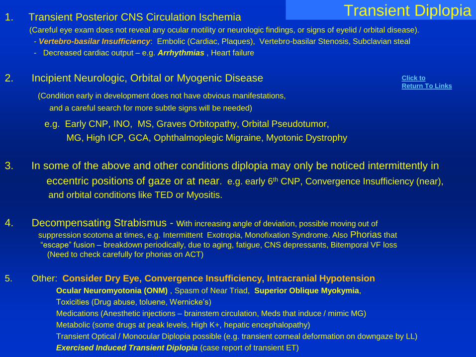
Transient Diplopia1. Transient Posterior CNS Circulation Ischemia(Careful eye exam does not reveal any ocular motility or neurologic findings, or signs of eyelid / orbital disease).
- Vertebro-basilar Insufficiency: Embolic (Cardiac, Plaques), Vertebro-basilar Stenosis, Subclavian steal
- Decreased cardiac output – e.g. Arrhythmias , Heart failure
2. Incipient Neurologic, Orbital or Myogenic Disease
(Condition early in development does not have obvious manifestations,
and a careful search for more subtle signs will be needed)
e.g. Early CNP, INO, MS, Graves Orbitopathy, Orbital Pseudotumor,
MG, High ICP, GCA, Ophthalmoplegic Migraine, Myotonic Dystrophy
3. In some of the above and other conditions diplopia may only be noticed intermittently in
eccentric positions of gaze or at near. e.g. early 6th CNP, Convergence Insufficiency (near),
and orbital conditions like TED or Myositis.
4. Decompensating Strabismus - with increasing angle of deviation, possible moving out of
suppression scotoma at times, e.g. Intermittent Exotropia, Monofixation Syndrome. Also Phorias that
“escape” fusion – breakdown periodically, due to aging, fatigue, CNS depressants, Bitemporal VF loss
(Need to check carefully for phorias on ACT)
5. Other: Consider Dry Eye, Convergence Insufficiency, Intracranial Hypotension
Ocular Neuromyotonia (ONM) , Spasm of Near Triad, Superior Oblique Myokymia,
Toxicities (Drug abuse, toluene, Wernicke’s)
Medications (Anesthetic injections – brainstem circulation, Meds that induce / mimic MG)
Metabolic (some drugs at peak levels, High K+, hepatic encephalopathy)
Transient Optical / Monocular Diplopia possible (e.g. transient corneal deformation on downgaze by LL)
Exercised Induced Transient Diplopia (case report of transient ET)
Click to
Return To Links
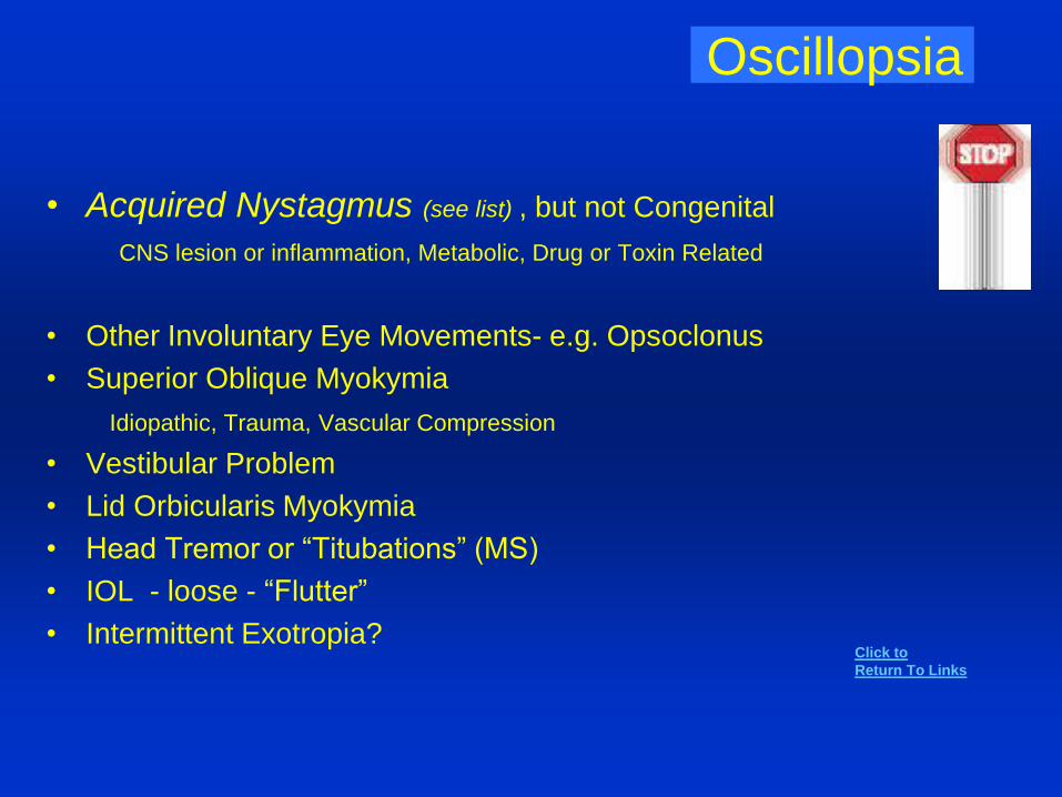
Oscillopsia
• Acquired Nystagmus (see list) , but not Congenital
CNS lesion or inflammation, Metabolic, Drug or Toxin Related
• Other Involuntary Eye Movements- e.g. Opsoclonus
• Superior Oblique Myokymia
Idiopathic, Trauma, Vascular Compression
• Vestibular Problem
• Lid Orbicularis Myokymia
• Head Tremor or “Titubations” (MS)
• IOL - loose - “Flutter”
• Intermittent Exotropia?Click to
Return To Links
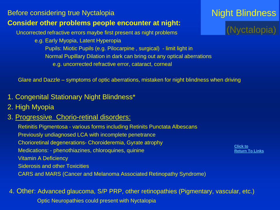
Night Blindness
(Nyctalopia)
Before considering true Nyctalopia
Consider other problems people encounter at night:
Uncorrected refractive errors maybe first present as night problems
e.g. Early Myopia, Latent Hyperopia
Pupils: Miotic Pupils (e.g. Pilocarpine , surgical) - limit light in
Normal Pupillary Dilation in dark can bring out any optical aberrations
e.g. uncorrected refractive error, cataract, corneal
Glare and Dazzle – symptoms of optic aberrations, mistaken for night blindness when driving
1. Congenital Stationary Night Blindness*
2. High Myopia
3. Progressive Chorio-retinal disorders:
Retinitis Pigmentosa - various forms including Retinits Punctata Albescans
Previously undiagnosed LCA with incomplete penetrance
Chorioretinal degenerations- Choroideremia, Gyrate atrophy
Medications: - phenothiazines, chloroquines, quinine
Vitamin A Deficiency
Siderosis and other Toxicities
CARS and MARS (Cancer and Melanoma Associated Retinopathy Syndrome)
4. Other: Advanced glaucoma, S/P PRP, other retinopathies (Pigmentary, vascular, etc.)
Optic Neuropathies could present with Nyctalopia
Click to
Return To Links
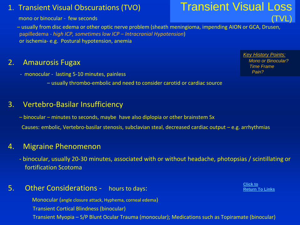
Transient Visual Loss(TVL)
1. Transient Visual Obscurations (TVO)mono or binocular - few seconds
– usually from disc edema or other optic nerve problem (sheath meningioma, impending AION or GCA, Drusen, papilledema - high ICP, sometimes low ICP – Intracranial Hypotension) or ischemia- e.g. Postural hypotension, anemia
2. Amaurosis Fugax
- monocular - lasting 5-10 minutes, painless
– usually thrombo-embolic and need to consider carotid or cardiac source
3. Vertebro-Basilar Insufficiency
– binocular – minutes to seconds, maybe have also diplopia or other brainstem Sx
Causes: embolic, Vertebro-basilar stenosis, subclavian steal, decreased cardiac output – e.g. arrhythmias
4. Migraine Phenomenon
- binocular, usually 20-30 minutes, associated with or without headache, photopsias / scintillating or
fortification Scotoma
5. Other Considerations - hours to days:
Monocular (angle closure attack, Hyphema, corneal edema)
Transient Cortical Blindness (binocular)
Transient Myopia – S/P Blunt Ocular Trauma (monocular); Medications such as Topiramate (binocular)
Key History Points:
Mono or Binocular?
Time Frame
Pain?
Click to
Return To Links
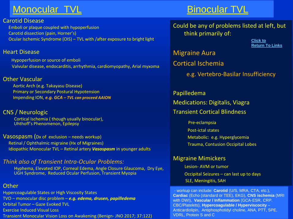
Monocular TVL Binocular TVLCarotid Disease
Emboli or plaque coupled with hypoperfusionCarotid dissection (pain, Horner’s)Ocular Ischemic Syndrome (OIS) – TVL with /after exposure to bright light
Heart DiseaseHypoperfusion or source of emboli
Valvular disease, endocarditis, arrhythmia, cardiomyopathy, Arial myxoma
Other VascularAortic Arch (e.g. Takayasu Disease)Primary or Secondary Postural HypotensionImpending ION, e.g. GCA – TVL can proceed AAION
CNS / NeurologicCortical Ischemia ( though usually binocular), Uhthoff’s Phenomenon, Epilepsy
Vasospasm (Dx of exclusion – needs workup)
Retinal / Ophthalmic migraine (Hx of Migraines)Idiopathic Monocular TVL – Retinal artery Vasospasm in younger adults
Think also of Transient Intra-Ocular Problems:Hyphema, Elevated IOP, Corneal Edema, Angle Closure Glaucoma, Dry Eye, UGH Syndrome, Reduced Ocular Perfusion, Transient Myopia
OtherHypercoagulable States or High Viscosity StatesTVO – monocular disc problem – e.g. edema, drusen, papilledemaOrbital Tumor – Gaze Evoked TVLExercise Induced Visual LossTransient Monocular Vision Loss on Awakening (Benign- JNO 2017; 37:122)
Could be any of problems listed at left, but think primarily of:
Migraine Aura
Cortical Ischemia
e.g. Vertebro-Basilar Insufficiency
Papilledema
Medications: Digitalis, Viagra
Transient Cortical Blindness
Pre-eclampsia
Post-ictal states
Metabolic: e.g. Hyperglycemia
Trauma, Contusion Occipital Lobes
Migraine MimickersLesion- AVM or tumor
Occipital Seizures – can last up to days
SLE, Meningitis, SAH
- workup can include: Carotid (U/S, MRA, CTA, etc.),
Cardiac (Echo (standard or TEE), EKG), CNS ischemia (MRI
with DWI), Vascular / Inflammation (GCA-ESR, CRP.
CBC/Platelets), Hypercoagulable / Hyperviscocity –
Anticardiolipin, Antiphosphotidyl choline, ANA, PTT, SPE,
VDRL, Protein S and C
Click to
Return To Links
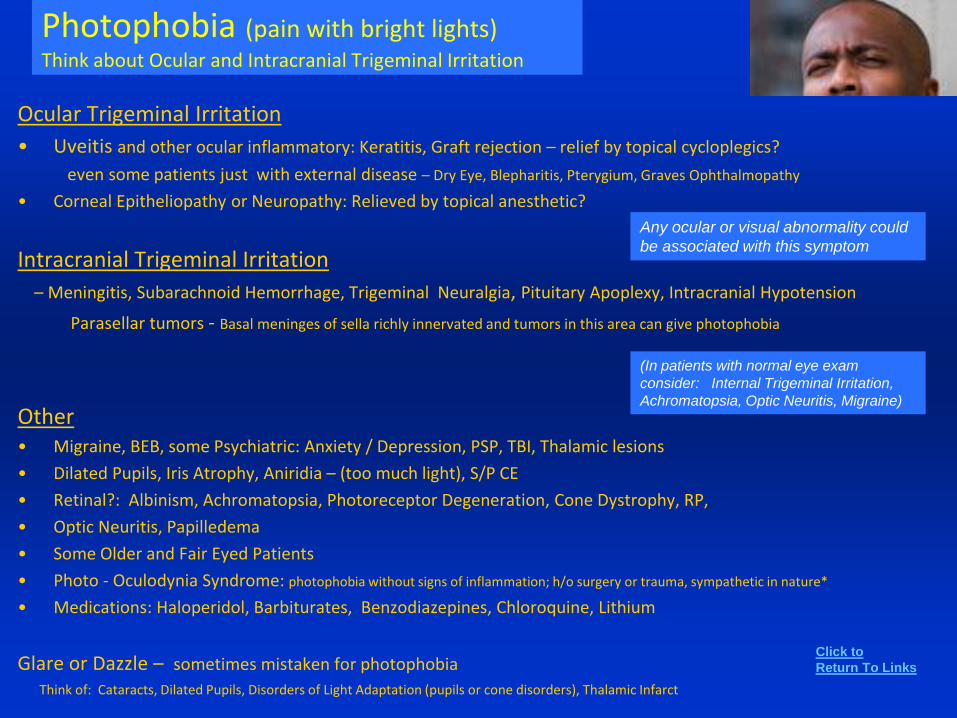
Photophobia (pain with bright lights)Think about Ocular and Intracranial Trigeminal Irritation
Ocular Trigeminal Irritation
• Uveitis and other ocular inflammatory: Keratitis, Graft rejection – relief by topical cycloplegics?
even some patients just with external disease – Dry Eye, Blepharitis, Pterygium, Graves Ophthalmopathy
• Corneal Epitheliopathy or Neuropathy: Relieved by topical anesthetic?
Intracranial Trigeminal Irritation
– Meningitis, Subarachnoid Hemorrhage, Trigeminal Neuralgia, Pituitary Apoplexy, Intracranial Hypotension
Parasellar tumors - Basal meninges of sella richly innervated and tumors in this area can give photophobia
Other• Migraine, BEB, some Psychiatric: Anxiety / Depression, PSP, TBI, Thalamic lesions
• Dilated Pupils, Iris Atrophy, Aniridia – (too much light), S/P CE
• Retinal?: Albinism, Achromatopsia, Photoreceptor Degeneration, Cone Dystrophy, RP,
• Optic Neuritis, Papilledema
• Some Older and Fair Eyed Patients
• Photo - Oculodynia Syndrome: photophobia without signs of inflammation; h/o surgery or trauma, sympathetic in nature*
• Medications: Haloperidol, Barbiturates, Benzodiazepines, Chloroquine, Lithium
Glare or Dazzle – sometimes mistaken for photophobia
Think of: Cataracts, Dilated Pupils, Disorders of Light Adaptation (pupils or cone disorders), Thalamic Infarct
(In patients with normal eye exam
consider: Internal Trigeminal Irritation,
Achromatopsia, Optic Neuritis, Migraine)
Any ocular or visual abnormality could
be associated with this symptom
Click to
Return To Links
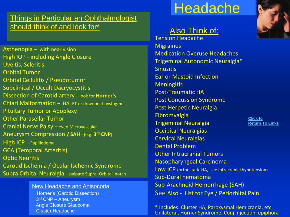
Headache
Tension HeadacheMigrainesMedication Overuse HeadachesTrigeminal Autonomic Neuralgia*SinusitisEar or Mastoid InfectionMeningitisPost-Traumatic HAPost Concussion SyndromePost Herpetic NeuralgiaFibromyalgiaTrigeminal NeuralgiaOccipital NeuralgiasCervical NeuralgiasDental ProblemOther Intracranial TumorsNasopharyngeal CarcinomaLow ICP (orthostatic HA, see intracranial hypotension)
Sub-Dural hematomaSub-Arachnoid Hemorrhage (SAH)
See Also - List for Eye / Periorbital Pain
* Includes: Cluster HA, Paroxysmal Hemicrania, etc. Unilateral, Horner Syndrome, Conj injection, epiphora
Asthenopia – with near vision
High IOP - including Angle ClosureUveitis, ScleritisOrbital TumorOrbital Cellulitis / PseudotumorSubclinical / Occult DacryocystitisDissection of Carotid artery – look for Horner’s
Chiari Malformation – HA, ET or downbeat nystagmus
Pituitary Tumor or ApoplexyOther Parasellar TumorCranial Nerve Palsy – even Microvascular
Aneurysm Compression / SAH - (e.g. 3rd CNP)
High ICP - Papilledema
GCA (Temporal Arteritis) Optic NeuritisCarotid Ischemia / Ocular Ischemic SyndromeSupra Orbital Neuralgia – palpate Supra -Orbital notch
Things in Particular an Ophthalmologist
should think of and look for*Also Think of:
New Headache and Anisocoria:
Horner’s (Carotid Dissection)
3rd CNP – Aneurysm
Angle Closure Glaucoma
Cluster Headache
Click to
Return To Links
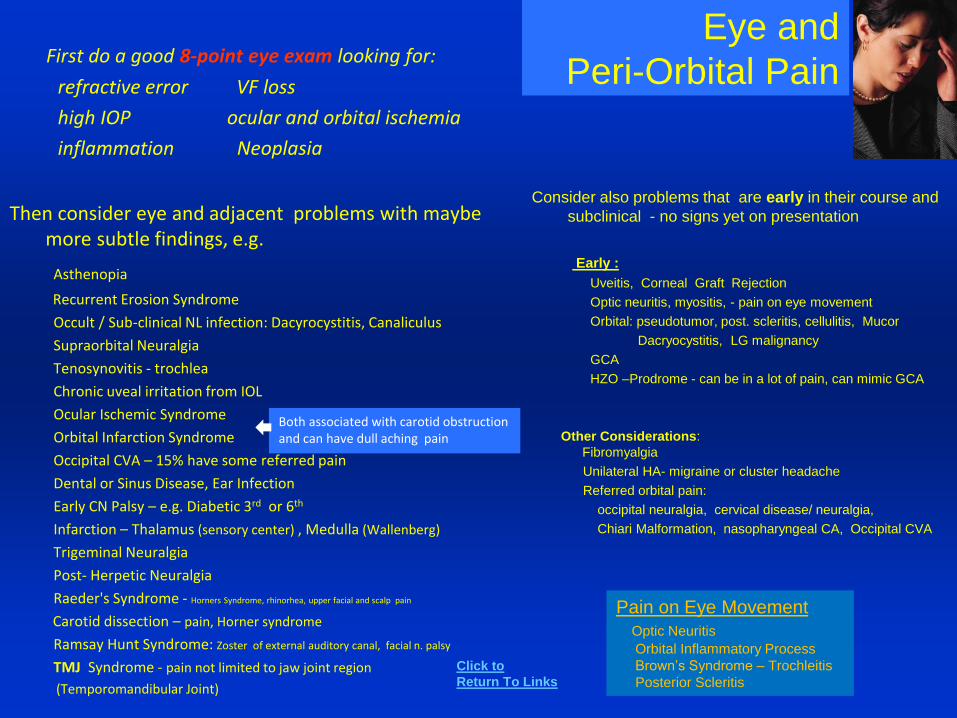
Eye and
Peri-Orbital PainFirst do a good 8-point eye exam looking for:
refractive error VF loss
high IOP ocular and orbital ischemia
inflammation Neoplasia
Then consider eye and adjacent problems with maybe more subtle findings, e.g.
Asthenopia
Recurrent Erosion Syndrome
Occult / Sub-clinical NL infection: Dacyrocystitis, Canaliculus
Supraorbital Neuralgia
Tenosynovitis - trochlea
Chronic uveal irritation from IOL
Ocular Ischemic Syndrome
Orbital Infarction Syndrome
Occipital CVA – 15% have some referred pain
Dental or Sinus Disease, Ear Infection
Early CN Palsy – e.g. Diabetic 3rd or 6th
Infarction – Thalamus (sensory center) , Medulla (Wallenberg)
Trigeminal Neuralgia
Post- Herpetic Neuralgia
Raeder's Syndrome - Horners Syndrome, rhinorhea, upper facial and scalp pain
Carotid dissection – pain, Horner syndrome
Ramsay Hunt Syndrome: Zoster of external auditory canal, facial n. palsy
TMJ Syndrome - pain not limited to jaw joint region
(Temporomandibular Joint)
Consider also problems that are early in their course and
subclinical - no signs yet on presentation
Early :
Uveitis, Corneal Graft Rejection
Optic neuritis, myositis, - pain on eye movement
Orbital: pseudotumor, post. scleritis, cellulitis, Mucor
Dacryocystitis, LG malignancy
GCA
HZO –Prodrome - can be in a lot of pain, can mimic GCA
Other Considerations:
Fibromyalgia
Unilateral HA- migraine or cluster headache
Referred orbital pain:
occipital neuralgia, cervical disease/ neuralgia,
Chiari Malformation, nasopharyngeal CA, Occipital CVA
Pain on Eye Movement
Optic Neuritis
Orbital Inflammatory Process
Brown’s Syndrome – Trochleitis
Posterior Scleritis
Both associated with carotid obstruction and can have dull aching pain
Click to
Return To Links

Epiphora (Tearing)Epiphora is a very non-specific SymptomBefore Direct Assessment of the Nasolacrimal Drainage System
Consider External Conditions or other irritants that can evoke tearing
e.g. Uncorrected Refractive Error
Ocular Allergies
Chronic Blepharoconjunctivitis, Dry Eye, Corneal FB or Abrasion, RES, Keratitis, Uveitis, etc.
Glaucoma – High IOP
Trichiasis, Lid Foreign Body (e.g. concretion)
Lid Malpositions (Entropion, Ectropion , Punctal Eversion, Retraction , Centurion Syndrome)
Lower Lid Laxity (sometimes lid tightening procedures can stop Epiphora)
Dermatochalasis – “Upper Eyelid Wick Syndrome” – JAMA Oph 2012;130:1007
7th Nerve Palsy (poor pump function and lid laxity)
Jaw winking
Crocodile tearing (e.g. after Bell’s Palsy)
Lacrimal Gland inflammation, mass
Nasolacrimal obstructions
- punctal stenosis or conjunctiva blockage or megalocaruncle
- canalicular stenosis (e.g. canaliculitis, HSV, Radioactive Iodine, Docetaxel )
- relative obstruction- with edema of epithelium, chronic allergic, mucous fishing syndrome
- sac (stone, tumor, recurrent dacryocystitis - scarring)
- duct - previous sinus disease or surgery, nose trauma, acquired NLDO
- Congenital malformation – punctal atresia, Canalicular dysgenesis, NLD
NL probing and irrigation – if system is patent, consider punctal stenosis and see if the punctal dilation provides relief for even a few days – if so then punctoplasty maybe helpful
Click to
Return To Links
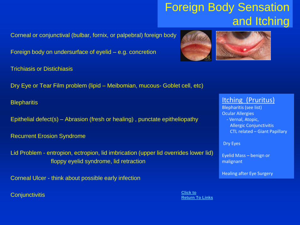
Foreign Body Sensation
and ItchingCorneal or conjunctival (bulbar, fornix, or palpebral) foreign body
Foreign body on undersurface of eyelid – e.g. concretion
Trichiasis or Distichiasis
Dry Eye or Tear Film problem (lipid – Meibomian, mucous- Goblet cell, etc)
Blepharitis
Epithelial defect(s) – Abrasion (fresh or healing) , punctate epitheliopathy
Recurrent Erosion Syndrome
Lid Problem - entropion, ectropion, lid imbrication (upper lid overrides lower lid)
floppy eyelid syndrome, lid retraction
Corneal Ulcer - think about possible early infection
ConjunctivitisClick to
Return To Links
Itching (Pruritus) Blepharitis (see list)Ocular Allergies
- Vernal, Atopic, Allergic ConjunctivitisCTL related – Giant Papillary
Dry Eyes
Eyelid Mass – benign or malignant
Healing after Eye Surgery
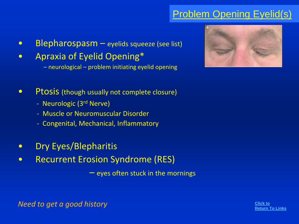
Problem Opening Eyelid(s)
• Blepharospasm – eyelids squeeze (see list)
• Apraxia of Eyelid Opening* – neurological – problem initiating eyelid opening
• Ptosis (though usually not complete closure)
- Neurologic (3rd Nerve)
- Muscle or Neuromuscular Disorder
- Congenital, Mechanical, Inflammatory
• Dry Eyes/Blepharitis
• Recurrent Erosion Syndrome (RES)
– eyes often stuck in the mornings
Need to get a good history Click to
Return To Links
Chronic Red EyeOrbital Disease
– TED - Congestive Stage
- IOIS – including posterior scleritis, dacryoadenitis, myositis
- Orbital Tumors including LG, Lymphoma,…
- Carotid Cavernous Fistula / Dural AV Shunts
Lacrimal - NLO, Dacryocystitis, Canaliculitis (chronic)
Lacrimal Gland Ductulitis (look at palpebral lobe)
Eyelid Problems - Malpositions, Trichiasis,
Lid imbrication (upper lid overrides lower lid),
Floppy Eyelid syndrome
Autoimmune Disease w/ related episcleritis, scleritis
Reiter's, Wegener’s, Relapsing Polychondritis, SLE,
Sjogrens Syndrome
OCP , Epidermolysis Bulosa (Symblephara)
Graft versus Host Disease
Loss of 7th and 5th CN Function- e.g. skull based tumor
Sign of Systemic Conditions: Polycythemia, Sickle Cell,
Fabry’s Disease, Telangiectasia, Alcohol, Cannabis
Chronic Conjunctivitis
- Allergic, mucous fishing syndrome
Irritation from smoke, chemical, topical meds
- Chronic use of any eye drop - medicamentosa
topical anesthetics, preservatives even in ATs
“red out drops”, atropine, antivirals, alpha agonists
- Chlamydial – AIC, Trachoma
- Molluscum Contagiosum
- Parinaud's Ocular glandular syndrome – cat scratch, tularemia, mycobacterial
- Blepharoconjunctivitis, Acne Rosacea
- Superior Limbic Keratoconjunctivitis (SLK)
Conjunctival Mass, Tumor – Pingueculae, Pterygium Papilloma, OSSN, infiltrative malignancy
(e.g. sebaceous cell CA, Lymphoma)
Chronic Ocular Inflammation:
Corneal: Stromal Keratitis, Neurotrophic keratopathy
Uveitis – Ciliary Flush
Dry Eye
Two important points:1. Don't just think infection as most are self-limited and
need to think about some other process.
2. Don't let corneal signs focus you too much on the cornea, e.g. chronic epithelial defects can be a sign of chronic dry eyes, chronic allergic disease, chronic eyelid problems, Neurotrophic (CN 5 and or 7 dysfunction), etc.
Click to
Return To Links
2. Signs (Based on the 8-point eye exam)
Loss of Visual Acuity and Refractive Issues
Loss of Visual Field
Eyelids and Orbit
Motility
Pupils
Anterior Segment
IOP
Fundus
Loss of Visual Acuity
and Refractive Issues
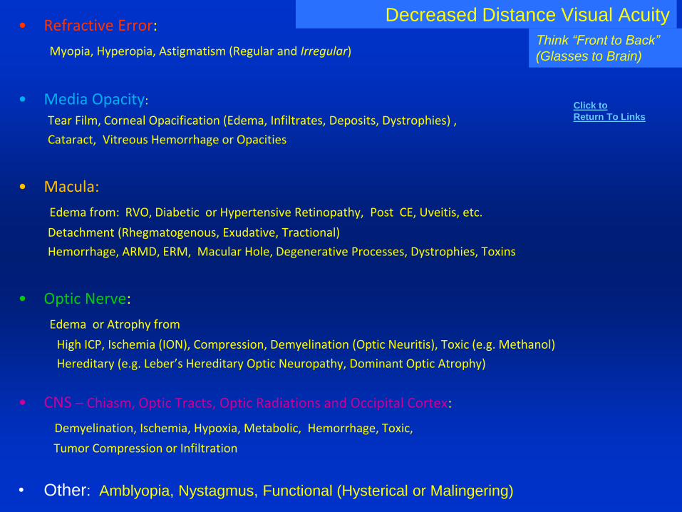
Decreased Distance Visual Acuity• Refractive Error:
Myopia, Hyperopia, Astigmatism (Regular and Irregular)
• Media Opacity:
Tear Film, Corneal Opacification (Edema, Infiltrates, Deposits, Dystrophies) ,
Cataract, Vitreous Hemorrhage or Opacities
• Macula:
Edema from: RVO, Diabetic or Hypertensive Retinopathy, Post CE, Uveitis, etc.
Detachment (Rhegmatogenous, Exudative, Tractional)
Hemorrhage, ARMD, ERM, Macular Hole, Degenerative Processes, Dystrophies, Toxins
• Optic Nerve:
Edema or Atrophy from
High ICP, Ischemia (ION), Compression, Demyelination (Optic Neuritis), Toxic (e.g. Methanol)
Hereditary (e.g. Leber’s Hereditary Optic Neuropathy, Dominant Optic Atrophy)
• CNS – Chiasm, Optic Tracts, Optic Radiations and Occipital Cortex:
Demyelination, Ischemia, Hypoxia, Metabolic, Hemorrhage, Toxic,
Tumor Compression or Infiltration
• Other: Amblyopia, Nystagmus, Functional (Hysterical or Malingering)
Think “Front to Back”
(Glasses to Brain)
Click to
Return To Links
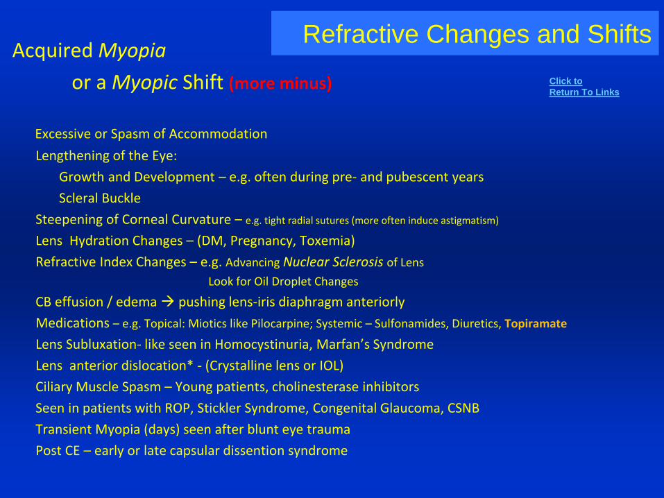
Refractive Changes and ShiftsAcquired Myopia
or a Myopic Shift (more minus)
Excessive or Spasm of Accommodation
Lengthening of the Eye:
Growth and Development – e.g. often during pre- and pubescent years
Scleral Buckle
Steepening of Corneal Curvature – e.g. tight radial sutures (more often induce astigmatism)
Lens Hydration Changes – (DM, Pregnancy, Toxemia)
Refractive Index Changes – e.g. Advancing Nuclear Sclerosis of Lens
Look for Oil Droplet Changes
CB effusion / edema pushing lens-iris diaphragm anteriorly
Medications – e.g. Topical: Miotics like Pilocarpine; Systemic – Sulfonamides, Diuretics, Topiramate
Lens Subluxation- like seen in Homocystinuria, Marfan’s Syndrome
Lens anterior dislocation* - (Crystalline lens or IOL)
Ciliary Muscle Spasm – Young patients, cholinesterase inhibitors
Seen in patients with ROP, Stickler Syndrome, Congenital Glaucoma, CSNB
Transient Myopia (days) seen after blunt eye trauma
Post CE – early or late capsular dissention syndrome
Click to
Return To Links
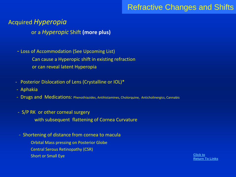
Refractive Changes and Shifts
Acquired Hyperopia or a Hyperopic Shift (more plus)
- Loss of Accommodation (See Upcoming List)
Can cause a Hyperopic shift in existing refraction
or can reveal latent Hyperopia
- Posterior Dislocation of Lens (Crystalline or IOL)*
- Aphakia
- Drugs and Medications: Phenothiazides, Antihistamines, Cholorquine, Anticholinergics, Cannabis
- S/P RK or other corneal surgery
with subsequent flattening of Cornea Curvature
- Shortening of distance from cornea to macula
Orbital Mass pressing on Posterior Globe
Central Serous Retinopathy (CSR)
Short or Small Eye Click to
Return To Links
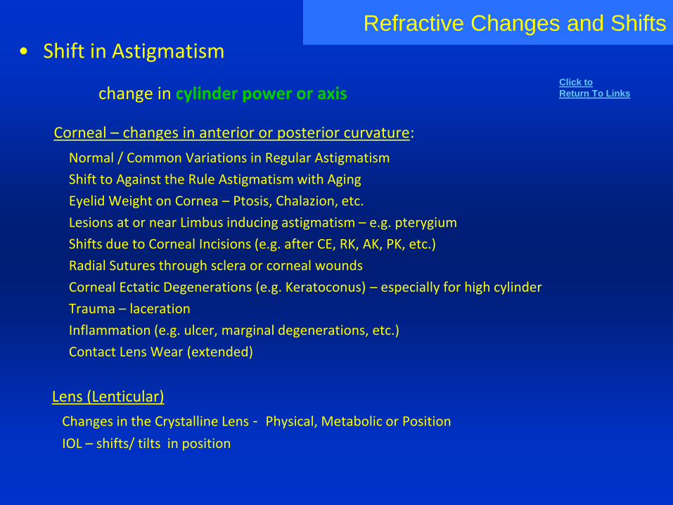
Refractive Changes and Shifts
• Shift in Astigmatism
change in cylinder power or axis
Corneal – changes in anterior or posterior curvature:
Normal / Common Variations in Regular Astigmatism
Shift to Against the Rule Astigmatism with Aging
Eyelid Weight on Cornea – Ptosis, Chalazion, etc.
Lesions at or near Limbus inducing astigmatism – e.g. pterygium
Shifts due to Corneal Incisions (e.g. after CE, RK, AK, PK, etc.)
Radial Sutures through sclera or corneal wounds
Corneal Ectatic Degenerations (e.g. Keratoconus) – especially for high cylinder
Trauma – laceration
Inflammation (e.g. ulcer, marginal degenerations, etc.)
Contact Lens Wear (extended)
Lens (Lenticular)
Changes in the Crystalline Lens - Physical, Metabolic or Position
IOL – shifts/ tilts in position
Click to
Return To Links
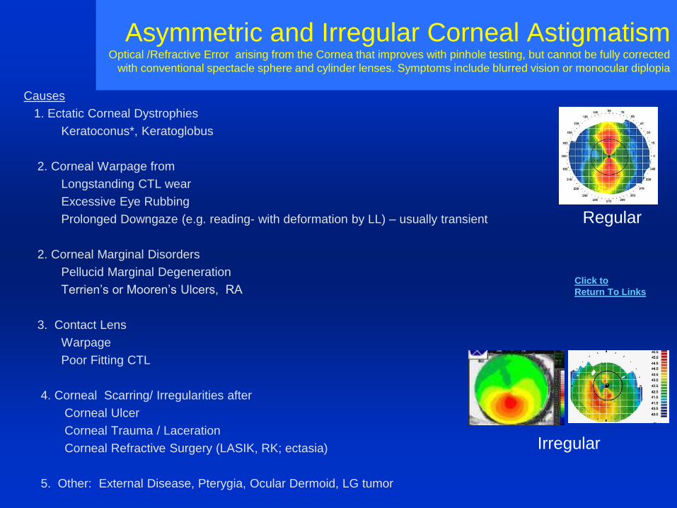
Asymmetric and Irregular Corneal AstigmatismOptical /Refractive Error arising from the Cornea that improves with pinhole testing, but cannot be fully corrected
with conventional spectacle sphere and cylinder lenses. Symptoms include blurred vision or monocular diplopia
Causes
1. Ectatic Corneal Dystrophies
Keratoconus*, Keratoglobus
2. Corneal Warpage from
Longstanding CTL wear
Excessive Eye Rubbing
Prolonged Downgaze (e.g. reading- with deformation by LL) – usually transient
2. Corneal Marginal Disorders
Pellucid Marginal Degeneration
Terrien’s or Mooren’s Ulcers, RA
3. Contact Lens
Warpage
Poor Fitting CTL
4. Corneal Scarring/ Irregularities after
Corneal Ulcer
Corneal Trauma / Laceration
Corneal Refractive Surgery (LASIK, RK; ectasia)
5. Other: External Disease, Pterygia, Ocular Dermoid, LG tumor
Regular
Irregular
Click to
Return To Links
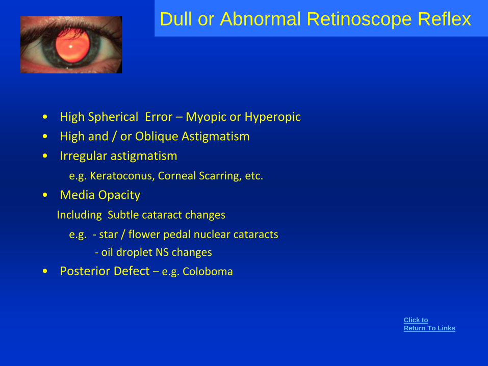
Dull or Abnormal Retinoscope Reflex
• High Spherical Error – Myopic or Hyperopic
• High and / or Oblique Astigmatism
• Irregular astigmatism
e.g. Keratoconus, Corneal Scarring, etc.
• Media Opacity
Including Subtle cataract changes
e.g. - star / flower pedal nuclear cataracts
- oil droplet NS changes
• Posterior Defect – e.g. Coloboma
Click to
Return To Links
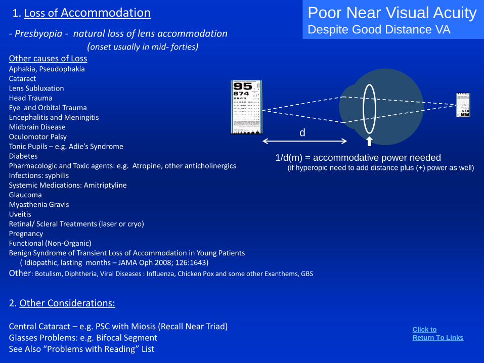
1. Loss of Accommodation
- Presbyopia - natural loss of lens accommodation(onset usually in mid- forties)
Other causes of LossAphakia, PseudophakiaCataractLens SubluxationHead TraumaEye and Orbital TraumaEncephalitis and MeningitisMidbrain DiseaseOculomotor PalsyTonic Pupils – e.g. Adie’s SyndromeDiabetesPharmacologic and Toxic agents: e.g. Atropine, other anticholinergicsInfections: syphilisSystemic Medications: AmitriptylineGlaucomaMyasthenia GravisUveitisRetinal/ Scleral Treatments (laser or cryo)PregnancyFunctional (Non-Organic)Benign Syndrome of Transient Loss of Accommodation in Young Patients
( Idiopathic, lasting months – JAMA Oph 2008; 126:1643)
Other: Botulism, Diphtheria, Viral Diseases : Influenza, Chicken Pox and some other Exanthems, GBS
2. Other Considerations:
Central Cataract – e.g. PSC with Miosis (Recall Near Triad)Glasses Problems: e.g. Bifocal SegmentSee Also “Problems with Reading” List
E
Poor Near Visual AcuityDespite Good Distance VA
1/d(m) = accommodative power needed(if hyperopic need to add distance plus (+) power as well)
d
Click to
Return To Links

Problems with GlassesPatients Complaint’s
Consider
1. Was Refraction / Prescription (Rx) Correct?
2. Were glasses made correctly to Rx?
3. High Refractive Error – Vertex Distance Issues
(Try over-refraction over old glasses)
4. Over- Minused Correction – can happen in younger accommodating patients
5. Astigmatism – was there a significant change in axis from last Rx? Often not tolerated
6. Optical Center (OC) – check with respect to the pupil, PD and bifocal segment
7. Pantoscopic Tilt – e.g. minus lenses (tilt can induce cylinder)
8. Optical Aberrations – “waves” in lens sometimes happen when grinding
9. Induced Prism – causing Hypertropia and Diplopia (recall Prentice’s Rule P=hD)
Problems with Near
1. Bifocal Segment – not enough or too much add power
- position: top should be a lower lid level. Some are too low
2. Progressive Bifocals - too narrow or patient has to look too far down to get full add
3. Anisometropia – with large differences in vertical induced prism Diplopia - may need SLAB OFF
OC- Optic Center
PD- Pupillary Distance
P- Prism Power
h – displacement from center
D- Diopters of Lens Power in the
axis of concern
Click to
Return To Links
Loss of Visual Field
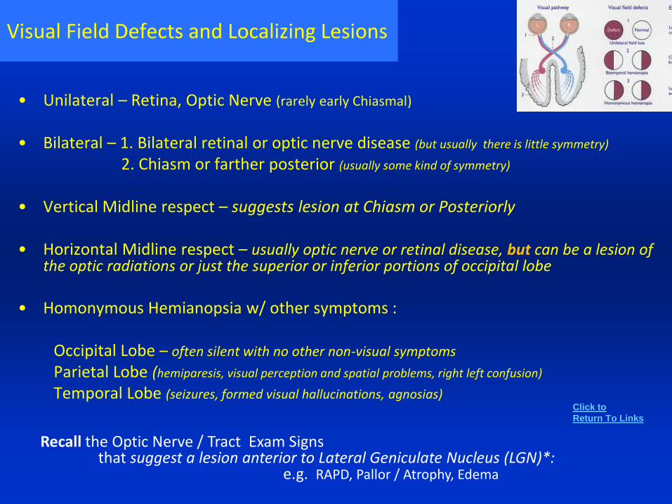
Visual Field Defects and Localizing Lesions
• Unilateral – Retina, Optic Nerve (rarely early Chiasmal)
• Bilateral – 1. Bilateral retinal or optic nerve disease (but usually there is little symmetry)
2. Chiasm or farther posterior (usually some kind of symmetry)
• Vertical Midline respect – suggests lesion at Chiasm or Posteriorly
• Horizontal Midline respect – usually optic nerve or retinal disease, but can be a lesion of the optic radiations or just the superior or inferior portions of occipital lobe
• Homonymous Hemianopsia w/ other symptoms :
Occipital Lobe – often silent with no other non-visual symptoms
Parietal Lobe (hemiparesis, visual perception and spatial problems, right left confusion)
Temporal Lobe (seizures, formed visual hallucinations, agnosias)
Recall the Optic Nerve / Tract Exam Signsthat suggest a lesion anterior to Lateral Geniculate Nucleus (LGN)*:
e.g. RAPD, Pallor / Atrophy, Edema
Click to
Return To Links
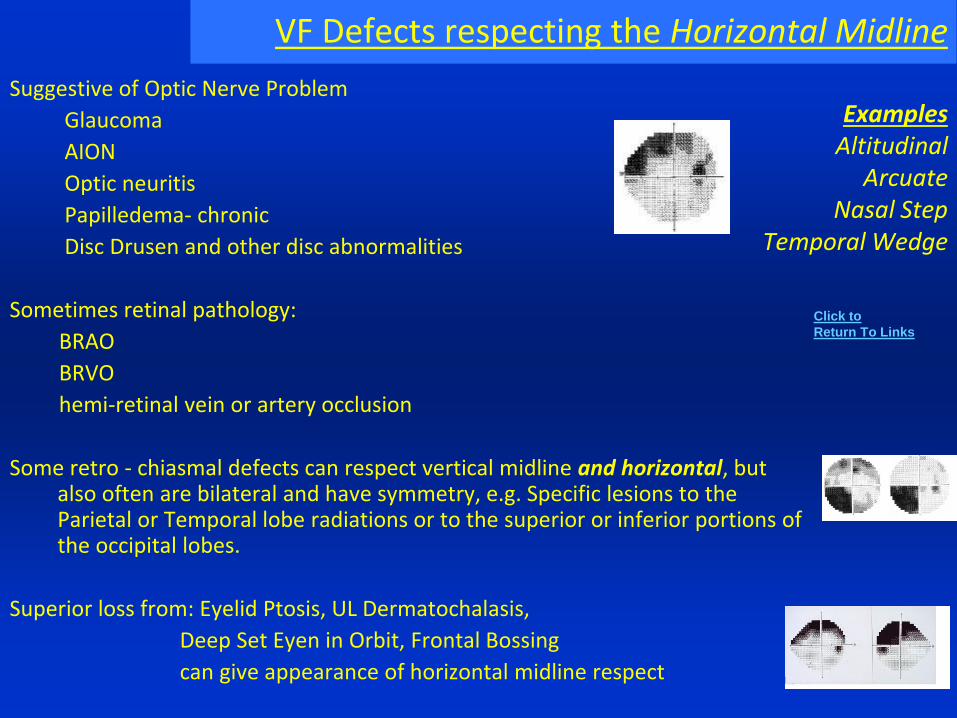
VF Defects respecting the Horizontal Midline
ExamplesAltitudinal
ArcuateNasal Step
Temporal Wedge
Suggestive of Optic Nerve Problem
Glaucoma
AION
Optic neuritis
Papilledema- chronic
Disc Drusen and other disc abnormalities
Sometimes retinal pathology:
BRAO
BRVO
hemi-retinal vein or artery occlusion
Some retro - chiasmal defects can respect vertical midline and horizontal, but also often are bilateral and have symmetry, e.g. Specific lesions to the Parietal or Temporal lobe radiations or to the superior or inferior portions of the occipital lobes.
Superior loss from: Eyelid Ptosis, UL Dermatochalasis,
Deep Set Eyen in Orbit, Frontal Bossing
can give appearance of horizontal midline respect
Click to
Return To Links
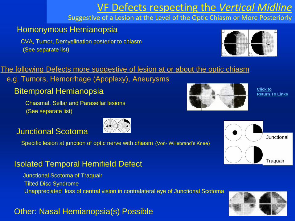
VF Defects respecting the Vertical MidlineSuggestive of a Lesion at the Level of the Optic Chiasm or More Posteriorly
Homonymous Hemianopsia
CVA, Tumor, Demyelination posterior to chiasm
(See separate list)
The following Defects more suggestive of lesion at or about the optic chiasm
e.g. Tumors, Hemorrhage (Apoplexy), Aneurysms
Bitemporal Hemianopsia
Chiasmal, Sellar and Parasellar lesions
(See separate list)
Junctional Scotoma
Specific lesion at junction of optic nerve with chiasm (Von- Willebrand’s Knee)
Isolated Temporal Hemifield Defect
Junctional Scotoma of Traquair
Tilted Disc Syndrome
Unappreciated loss of central vision in contralateral eye of Junctional Scotoma
Other: Nasal Hemianopsia(s) Possible
Junctional
Traquair
Click to
Return To Links
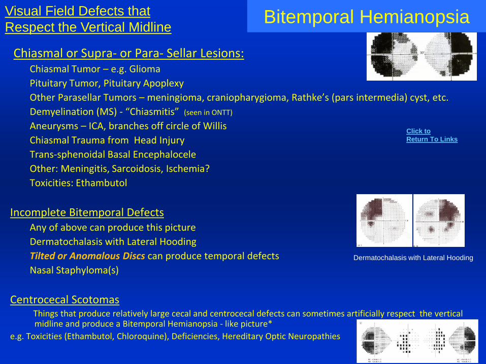
Bitemporal Hemianopsia
Chiasmal or Supra- or Para- Sellar Lesions:Chiasmal Tumor – e.g. Glioma
Pituitary Tumor, Pituitary Apoplexy
Other Parasellar Tumors – meningioma, craniopharygioma, Rathke’s (pars intermedia) cyst, etc.
Demyelination (MS) - “Chiasmitis” (seen in ONTT)
Aneurysms – ICA, branches off circle of Willis
Chiasmal Trauma from Head Injury
Trans-sphenoidal Basal Encephalocele
Other: Meningitis, Sarcoidosis, Ischemia?
Toxicities: Ethambutol
Incomplete Bitemporal DefectsAny of above can produce this picture
Dermatochalasis with Lateral Hooding
Tilted or Anomalous Discs can produce temporal defects
Nasal Staphyloma(s)
Centrocecal ScotomasThings that produce relatively large cecal and centrocecal defects can sometimes artificially respect the vertical midline and produce a Bitemporal Hemianopsia - like picture*
e.g. Toxicities (Ethambutol, Chloroquine), Deficiencies, Hereditary Optic Neuropathies
Visual Field Defects that
Respect the Vertical Midline
Dermatochalasis with Lateral Hooding
Click to
Return To Links
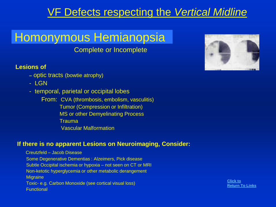
VF Defects respecting the Vertical Midline
Homonymous HemianopsiaComplete or Incomplete
Lesions of
– optic tracts (bowtie atrophy)
- LGN
- temporal, parietal or occipital lobes
From: CVA (thrombosis, embolism, vasculitis)
Tumor (Compression or Infiltration)
MS or other Demyelinating Process
Trauma
Vascular Malformation
If there is no apparent Lesions on Neuroimaging, Consider:
Creutzfeld – Jacob Disease
Some Degenerative Dementias : Alzeimers, Pick disease
Subtle Occipital ischemia or hypoxia – not seen on CT or MRI
Non-ketotic hyperglycemia or other metabolic derangement
Migraine
Toxic- e.g. Carbon Monoxide (see cortical visual loss)
Functional
Click to
Return To Links
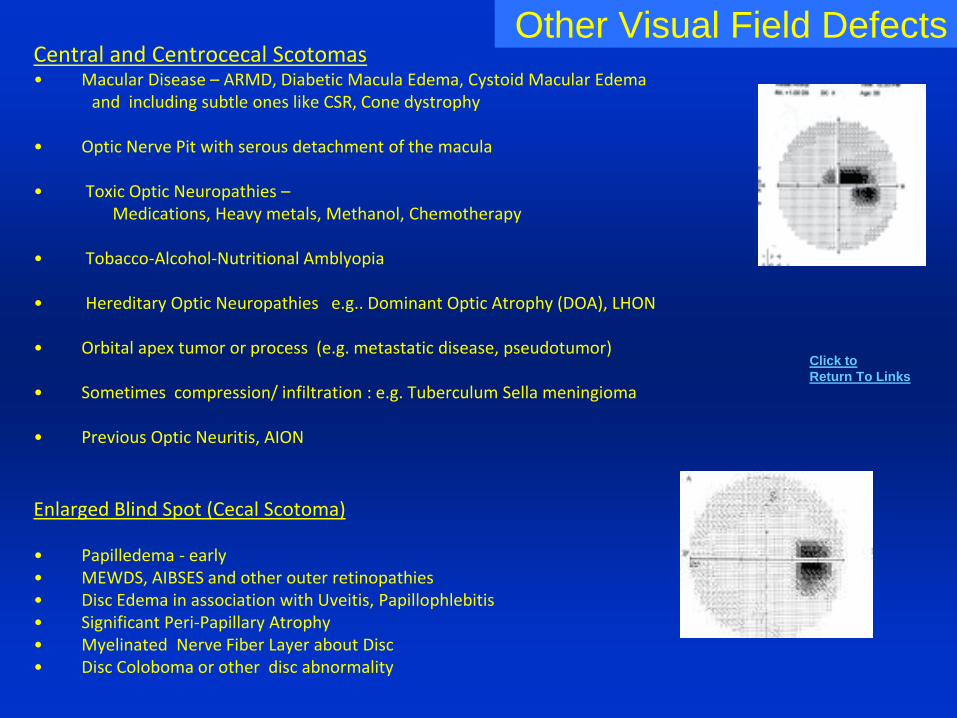
Other Visual Field DefectsCentral and Centrocecal Scotomas • Macular Disease – ARMD, Diabetic Macula Edema, Cystoid Macular Edema
and including subtle ones like CSR, Cone dystrophy
• Optic Nerve Pit with serous detachment of the macula
• Toxic Optic Neuropathies –Medications, Heavy metals, Methanol, Chemotherapy
• Tobacco-Alcohol-Nutritional Amblyopia
• Hereditary Optic Neuropathies e.g.. Dominant Optic Atrophy (DOA), LHON
• Orbital apex tumor or process (e.g. metastatic disease, pseudotumor)
• Sometimes compression/ infiltration : e.g. Tuberculum Sella meningioma
• Previous Optic Neuritis, AION
Enlarged Blind Spot (Cecal Scotoma)
• Papilledema - early• MEWDS, AIBSES and other outer retinopathies• Disc Edema in association with Uveitis, Papillophlebitis• Significant Peri-Papillary Atrophy• Myelinated Nerve Fiber Layer about Disc• Disc Coloboma or other disc abnormality
Click to
Return To Links
Other Visual Field Defects
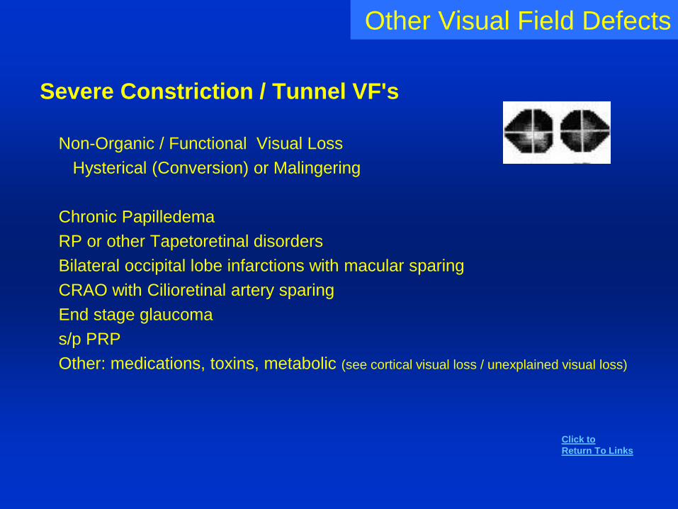
Severe Constriction / Tunnel VF's
Non-Organic / Functional Visual Loss
Hysterical (Conversion) or Malingering
Chronic Papilledema
RP or other Tapetoretinal disorders
Bilateral occipital lobe infarctions with macular sparing
CRAO with Cilioretinal artery sparing
End stage glaucoma
s/p PRP
Other: medications, toxins, metabolic (see cortical visual loss / unexplained visual loss)
Click to
Return To Links
Eyelids and Orbit
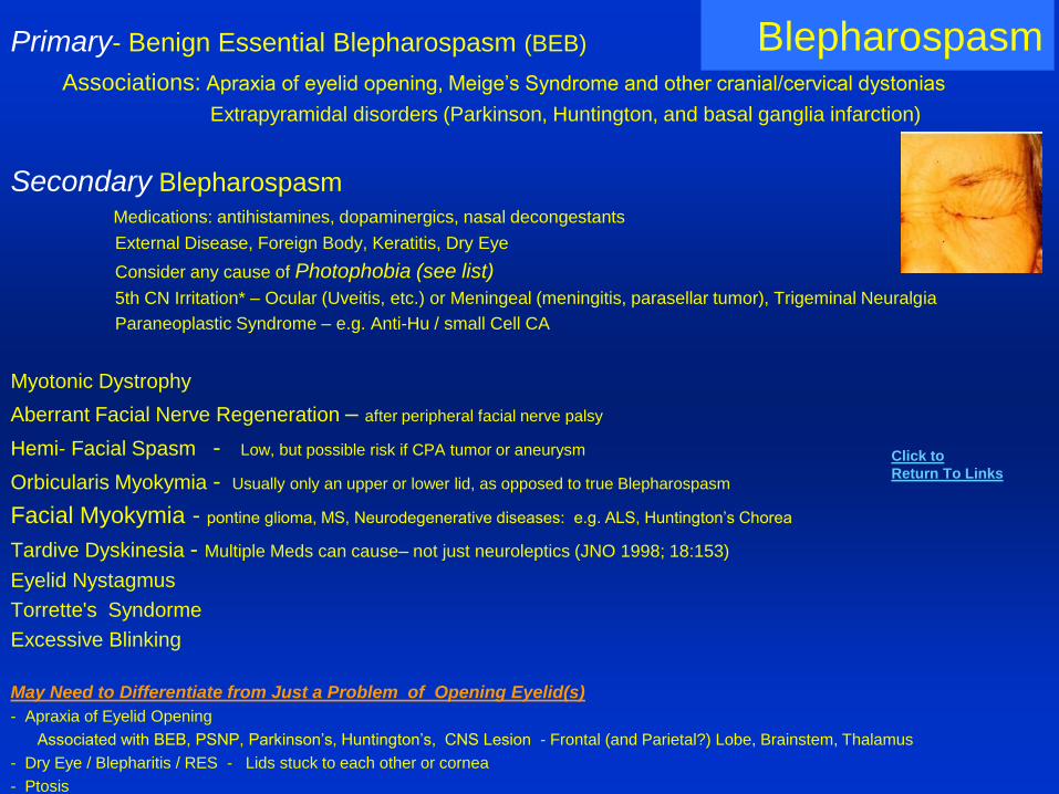
BlepharospasmPrimary- Benign Essential Blepharospasm (BEB)
Associations: Apraxia of eyelid opening, Meige’s Syndrome and other cranial/cervical dystonias
Extrapyramidal disorders (Parkinson, Huntington, and basal ganglia infarction)
Secondary Blepharospasm
Medications: antihistamines, dopaminergics, nasal decongestants
External Disease, Foreign Body, Keratitis, Dry Eye
Consider any cause of Photophobia (see list)
5th CN Irritation* – Ocular (Uveitis, etc.) or Meningeal (meningitis, parasellar tumor), Trigeminal Neuralgia
Paraneoplastic Syndrome – e.g. Anti-Hu / small Cell CA
Myotonic Dystrophy
Aberrant Facial Nerve Regeneration – after peripheral facial nerve palsy
Hemi- Facial Spasm - Low, but possible risk if CPA tumor or aneurysm
Orbicularis Myokymia - Usually only an upper or lower lid, as opposed to true Blepharospasm
Facial Myokymia - pontine glioma, MS, Neurodegenerative diseases: e.g. ALS, Huntington’s Chorea
Tardive Dyskinesia - Multiple Meds can cause– not just neuroleptics (JNO 1998; 18:153)
Eyelid Nystagmus
Torrette's Syndorme
Excessive Blinking
May Need to Differentiate from Just a Problem of Opening Eyelid(s)
- Apraxia of Eyelid Opening
Associated with BEB, PSNP, Parkinson’s, Huntington’s, CNS Lesion - Frontal (and Parietal?) Lobe, Brainstem, Thalamus
- Dry Eye / Blepharitis / RES - Lids stuck to each other or cornea
- Ptosis
Click to
Return To Links
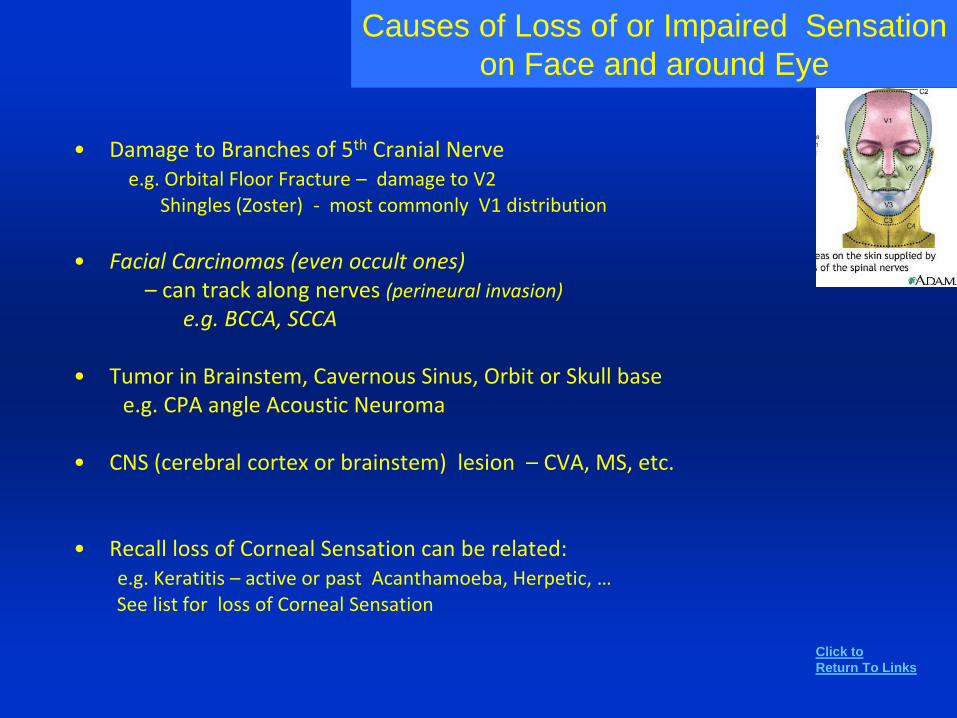
Causes of Loss of or Impaired Sensation
on Face and around Eye
• Damage to Branches of 5th Cranial Nervee.g. Orbital Floor Fracture – damage to V2
Shingles (Zoster) - most commonly V1 distribution
• Facial Carcinomas (even occult ones) – can track along nerves (perineural invasion)
e.g. BCCA, SCCA
• Tumor in Brainstem, Cavernous Sinus, Orbit or Skull basee.g. CPA angle Acoustic Neuroma
• CNS (cerebral cortex or brainstem) lesion – CVA, MS, etc.
• Recall loss of Corneal Sensation can be related:e.g. Keratitis – active or past Acanthamoeba, Herpetic, … See list for loss of Corneal Sensation
Click to
Return To Links
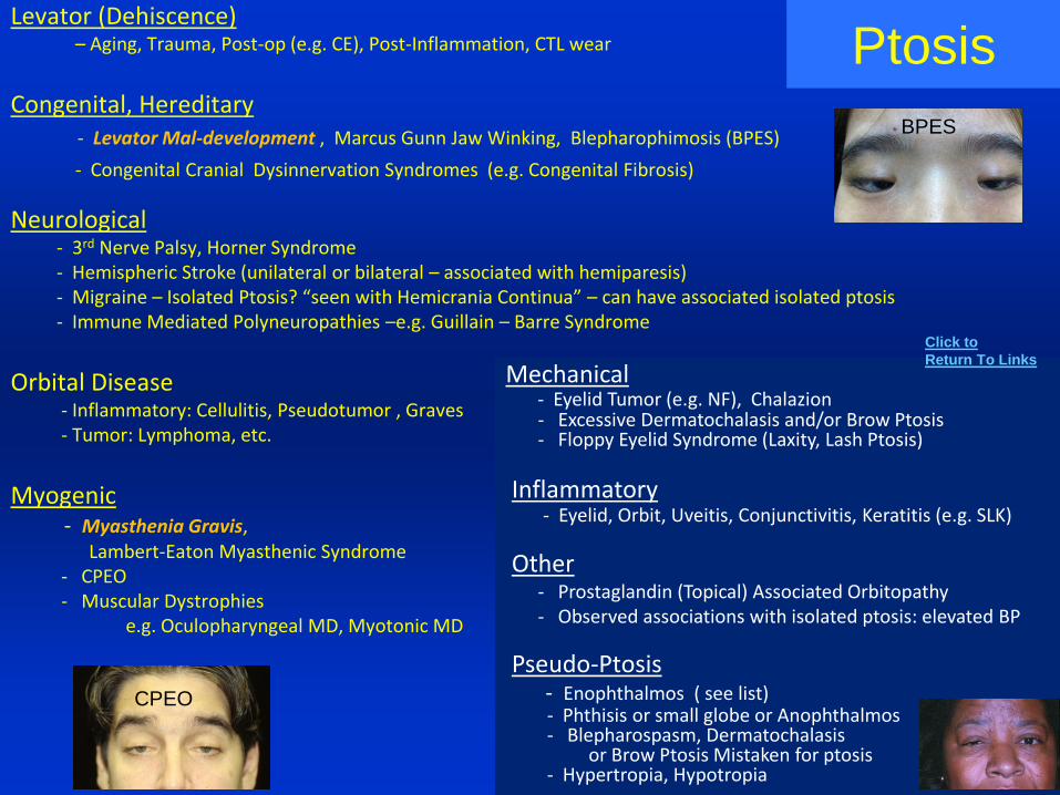
PtosisLevator (Dehiscence)
– Aging, Trauma, Post-op (e.g. CE), Post-Inflammation, CTL wear
Congenital, Hereditary- Levator Mal-development , Marcus Gunn Jaw Winking, Blepharophimosis (BPES)
- Congenital Cranial Dysinnervation Syndromes (e.g. Congenital Fibrosis)
Neurological- 3rd Nerve Palsy, Horner Syndrome- Hemispheric Stroke (unilateral or bilateral – associated with hemiparesis) - Migraine – Isolated Ptosis? “seen with Hemicrania Continua” – can have associated isolated ptosis- Immune Mediated Polyneuropathies –e.g. Guillain – Barre Syndrome
Orbital Disease- Inflammatory: Cellulitis, Pseudotumor , Graves- Tumor: Lymphoma, etc.
Myogenic - Myasthenia Gravis,
Lambert-Eaton Myasthenic Syndrome- CPEO- Muscular Dystrophies
e.g. Oculopharyngeal MD, Myotonic MD
CPEO
Mechanical - Eyelid Tumor (e.g. NF), Chalazion- Excessive Dermatochalasis and/or Brow Ptosis- Floppy Eyelid Syndrome (Laxity, Lash Ptosis)
Inflammatory- Eyelid, Orbit, Uveitis, Conjunctivitis, Keratitis (e.g. SLK)
Other- Prostaglandin (Topical) Associated Orbitopathy- Observed associations with isolated ptosis: elevated BP
Pseudo-Ptosis- Enophthalmos ( see list) - Phthisis or small globe or Anophthalmos- Blepharospasm, Dermatochalasis
or Brow Ptosis Mistaken for ptosis- Hypertropia, Hypotropia
BPES
Click to
Return To Links
Eyelashes and Eyelid Margin
Madarosis (Loss of Lashes)
- R/O Carcinoma – e.g. BCCA, Sebaceous Cell CA
- Chronic Blepharitis – e.g. Herpetic, Staph, Fungal, Mites …
- Endocrine – e.g. Hyper and hypo parathyroid and thyroid, hypopituitism
- Dermatoses - Dermatitis ( atopic, contact), ichthyosis, lichen planus,…
- Trauma – radiation, chemical, Thermal, tattooing, surgery, cryo
- Congenital disorders - multiple
- Drugs and Toxins - e.g. Arsenic, Chemotherapy, Botulinum, …
- Systemic Conditions – e.g. Parry-Rhomberg, VKH, Lupus, Sarcoidosis,…
Hypertrichosis (Excess Lashes = Trichomegaly)
- multiple congenital / genetic causes
- frequent manipulation
- Paraneoplastic syndrome
- malnutrition, anorexia, pregnancy, thyroid problems, lupus, uveitis
- Drugs: prostaglandin analogs (e.g. bimatoprost)
* Comprehensive Listing : Survey 2006; 51:550
Click to
Return To Links
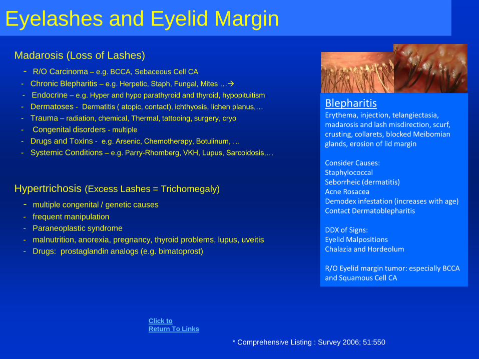
BlepharitisErythema, injection, telangiectasia, madarosis and lash misdirection, scurf, crusting, collarets, blocked Meibomian glands, erosion of lid margin
Consider Causes:StaphylococcalSeborrheic (dermatitis) Acne RosaceaDemodex infestation (increases with age)Contact Dermatoblepharitis
DDX of Signs:Eyelid MalpositionsChalazia and Hordeolum
R/O Eyelid margin tumor: especially BCCA and Squamous Cell CA
Eyelid Malpositions
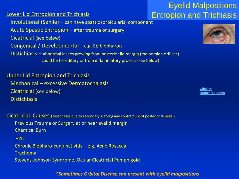
Entropion and TrichiasisLower Lid Entropion and Trichiasis
Involutional (Senile) – can have spastic (orbicularis) component
Acute Spastic Entropion – after trauma or surgery
Cicatricial (see below)
Congenital / Developmental – e.g. Epiblepharon
Distichiasis – abnormal lashes growing from posterior lid margin (meibomian orifices)
could be hereditary or from inflammatory process (see below)
Upper Lid Entropion and Trichiasis
Mechanical – excessive Dermatochalasis
Cicatricial (see below)
Distichiasis
Cicatricial Causes (Most cases due to secondary scarring and contracture of posterior lamella )
Previous Trauma or Surgery at or near eyelid margin
Chemical Burn
HZO
Chronic Blepharo-conjunctivitis - e.g. Acne Rosacea
Trachoma
Stevens-Johnson Syndrome, Ocular Cicatricial Pemphigoid
*Sometimes Orbital Disease can present with eyelid malpositions
Click to
Return To Links
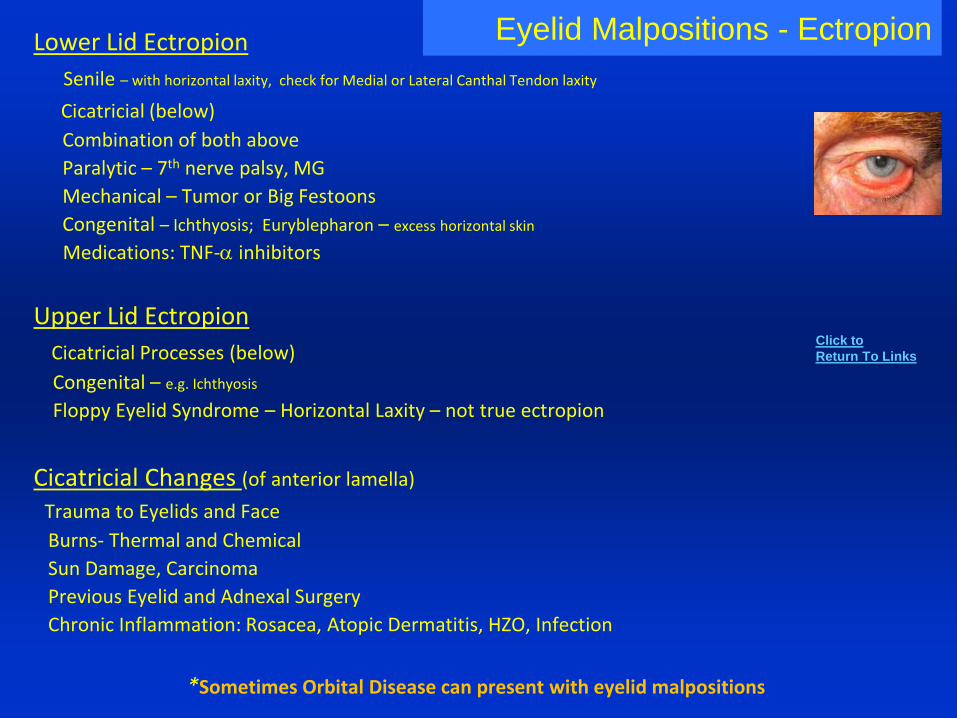
Eyelid Malpositions - EctropionLower Lid Ectropion
Senile – with horizontal laxity, check for Medial or Lateral Canthal Tendon laxity
Cicatricial (below)
Combination of both above
Paralytic – 7th nerve palsy, MG
Mechanical – Tumor or Big Festoons
Congenital – Ichthyosis; Euryblepharon – excess horizontal skin
Medications: TNF-a inhibitors
Upper Lid Ectropion
Cicatricial Processes (below)
Congenital – e.g. Ichthyosis
Floppy Eyelid Syndrome – Horizontal Laxity – not true ectropion
Cicatricial Changes (of anterior lamella)
Trauma to Eyelids and Face
Burns- Thermal and Chemical
Sun Damage, Carcinoma
Previous Eyelid and Adnexal Surgery
Chronic Inflammation: Rosacea, Atopic Dermatitis, HZO, Infection
*Sometimes Orbital Disease can present with eyelid malpositions
Click to
Return To Links
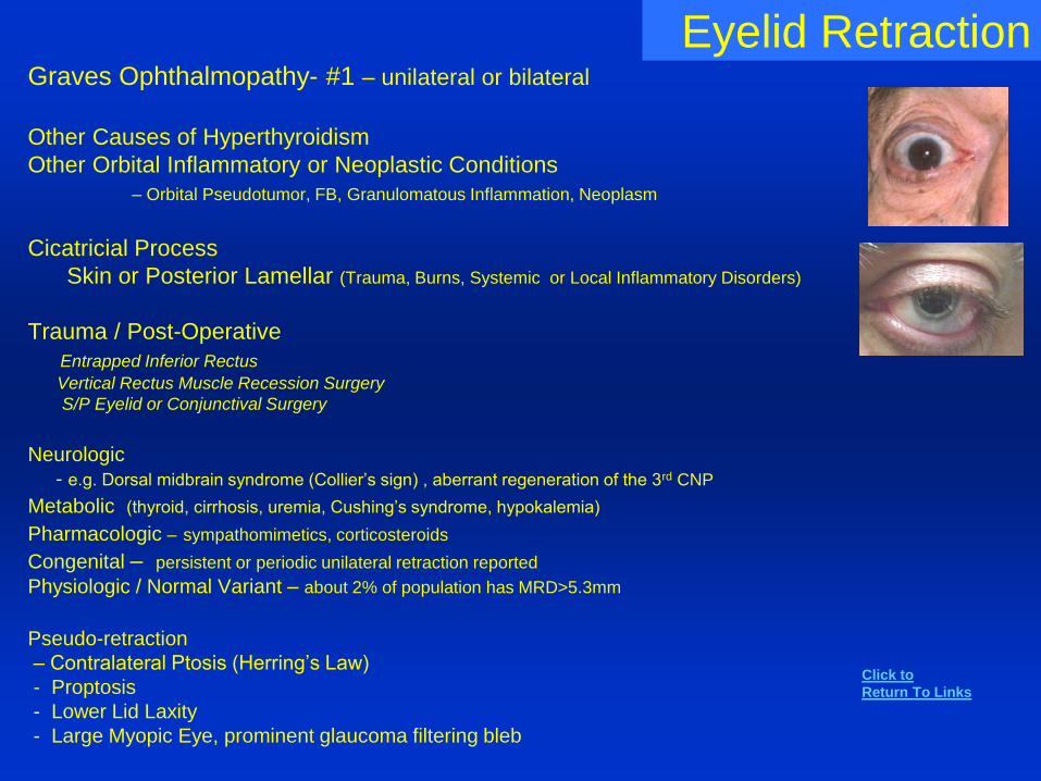
Eyelid RetractionGraves Ophthalmopathy- #1 – unilateral or bilateral
Other Causes of Hyperthyroidism
Other Orbital Inflammatory or Neoplastic Conditions
– Orbital Pseudotumor, FB, Granulomatous Inflammation, Neoplasm
Cicatricial Process
Skin or Posterior Lamellar (Trauma, Burns, Systemic or Local Inflammatory Disorders)
Trauma / Post-Operative
Entrapped Inferior Rectus
Vertical Rectus Muscle Recession Surgery
S/P Eyelid or Conjunctival Surgery
Neurologic
- e.g. Dorsal midbrain syndrome (Collier’s sign) , aberrant regeneration of the 3rd CNP
Metabolic (thyroid, cirrhosis, uremia, Cushing’s syndrome, hypokalemia)
Pharmacologic – sympathomimetics, corticosteroids
Congenital – persistent or periodic unilateral retraction reported
Physiologic / Normal Variant – about 2% of population has MRD>5.3mm
Pseudo-retraction
– Contralateral Ptosis (Herring’s Law)
- Proptosis
- Lower Lid Laxity
- Large Myopic Eye, prominent glaucoma filtering bleb
Click to
Return To Links
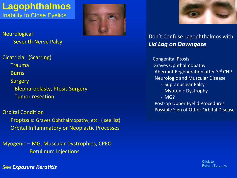
LagophthalmosInability to Close Eyelids
Neurological
Seventh Nerve Palsy
Cicatricial (Scarring)
Trauma
Burns
Surgery
Blepharoplasty, Ptosis Surgery
Tumor resection
Orbital Condition
Proptosis: Graves Ophthalmopathy, etc. ( see list)
Orbital Inflammatory or Neoplastic Processes
Myogenic – MG, Muscular Dystrophies, CPEO
Botulinum Injections
See Exposure Keratitis
Don’t Confuse Lagophthalmos with
Lid Lag on Downgaze
Congenital PtosisGraves OphthalmopathyAberrant Regeneration after 3rd CNPNeurologic and Muscular Disease
- Supranuclear Palsy- Myotonic Dystrophy- MG?
Post-op Upper Eyelid ProceduresPossible Sign of Other Orbital Disease
Click to
Return To Links
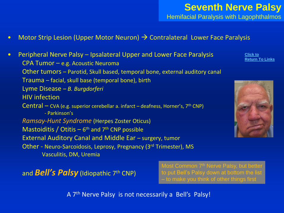
Seventh Nerve PalsyHemifacial Paralysis with Lagophthalmos
• Motor Strip Lesion (Upper Motor Neuron) Contralateral Lower Face Paralysis
• Peripheral Nerve Palsy – Ipsalateral Upper and Lower Face ParalysisCPA Tumor – e.g. Acoustic Neuroma
Other tumors – Parotid, Skull based, temporal bone, external auditory canal
Trauma – facial, skull base (temporal bone), birth
Lyme Disease – B. Burgdorferi
HIV infectionCentral – CVA (e.g. superior cerebellar a. infarct – deafness, Horner’s, 7th CNP)
- Parkinson’s
Ramsay-Hunt Syndrome (Herpes Zoster Oticus)
Mastoiditis / Otitis – 6th and 7th CNP possible
External Auditory Canal and Middle Ear – surgery, tumor
Other - Neuro-Sarcoidosis, Leprosy, Pregnancy (3rd Trimester), MSVasculitis, DM, Uremia
and Bell’s Palsy (Idiopathic 7th CNP) Most Common 7th Nerve Palsy, but better
to put Bell’s Palsy down at bottom the list
– to make you think of other things first
A 7th Nerve Palsy is not necessarily a Bell’s Palsy!
Click to
Return To Links
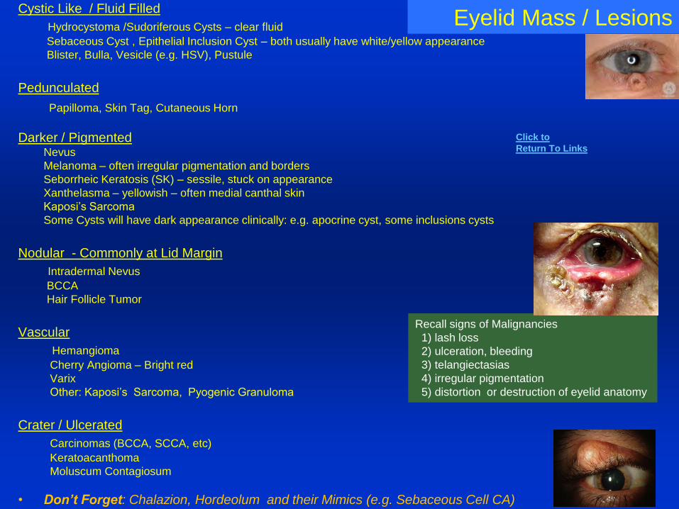
Eyelid Mass / LesionsCystic Like / Fluid Filled
Hydrocystoma /Sudoriferous Cysts – clear fluid
Sebaceous Cyst , Epithelial Inclusion Cyst – both usually have white/yellow appearance
Blister, Bulla, Vesicle (e.g. HSV), Pustule
Pedunculated
Papilloma, Skin Tag, Cutaneous Horn
Darker / PigmentedNevus
Melanoma – often irregular pigmentation and borders
Seborrheic Keratosis (SK) – sessile, stuck on appearance
Xanthelasma – yellowish – often medial canthal skin
Kaposi’s Sarcoma
Some Cysts will have dark appearance clinically: e.g. apocrine cyst, some inclusions cysts
Nodular - Commonly at Lid Margin
Intradermal Nevus
BCCA
Hair Follicle Tumor
Vascular
Hemangioma
Cherry Angioma – Bright red
Varix
Other: Kaposi’s Sarcoma, Pyogenic Granuloma
Crater / Ulcerated
Carcinomas (BCCA, SCCA, etc)
Keratoacanthoma
Moluscum Contagiosum
• Don’t Forget: Chalazion, Hordeolum and their Mimics (e.g. Sebaceous Cell CA)
Recall signs of Malignancies
1) lash loss
2) ulceration, bleeding
3) telangiectasias
4) irregular pigmentation
5) distortion or destruction of eyelid anatomy
Click to
Return To Links
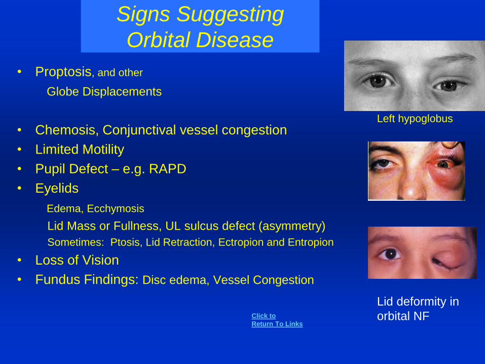
Signs Suggesting
Orbital Disease
• Proptosis, and other
Globe Displacements
• Chemosis, Conjunctival vessel congestion
• Limited Motility
• Pupil Defect – e.g. RAPD
• Eyelids
Edema, Ecchymosis
Lid Mass or Fullness, UL sulcus defect (asymmetry)
Sometimes: Ptosis, Lid Retraction, Ectropion and Entropion
• Loss of Vision
• Fundus Findings: Disc edema, Vessel Congestion
Left hypoglobus
Lid deformity in
orbital NFClick to
Return To Links
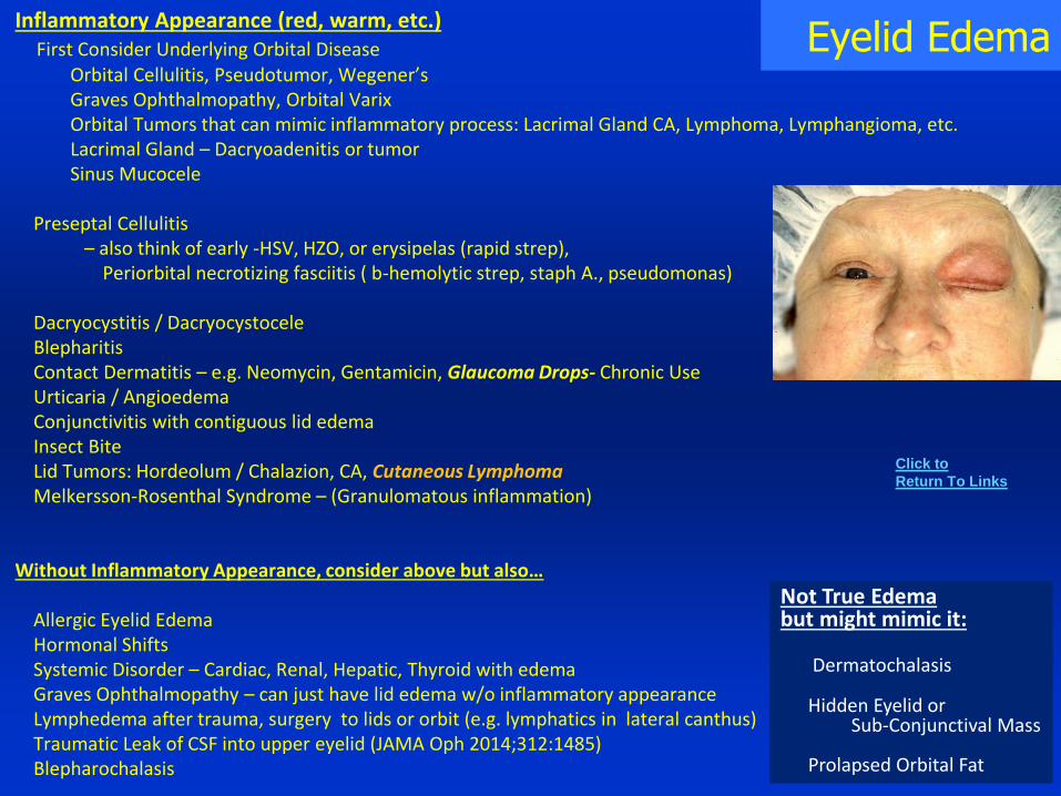
Eyelid EdemaInflammatory Appearance (red, warm, etc.)
First Consider Underlying Orbital DiseaseOrbital Cellulitis, Pseudotumor, Wegener’sGraves Ophthalmopathy, Orbital Varix Orbital Tumors that can mimic inflammatory process: Lacrimal Gland CA, Lymphoma, Lymphangioma, etc.Lacrimal Gland – Dacryoadenitis or tumorSinus Mucocele
Preseptal Cellulitis– also think of early -HSV, HZO, or erysipelas (rapid strep),
Periorbital necrotizing fasciitis ( b-hemolytic strep, staph A., pseudomonas)
Dacryocystitis / DacryocystoceleBlepharitisContact Dermatitis – e.g. Neomycin, Gentamicin, Glaucoma Drops- Chronic UseUrticaria / AngioedemaConjunctivitis with contiguous lid edemaInsect BiteLid Tumors: Hordeolum / Chalazion, CA, Cutaneous LymphomaMelkersson-Rosenthal Syndrome – (Granulomatous inflammation)
Without Inflammatory Appearance, consider above but also…
Allergic Eyelid EdemaHormonal ShiftsSystemic Disorder – Cardiac, Renal, Hepatic, Thyroid with edemaGraves Ophthalmopathy – can just have lid edema w/o inflammatory appearanceLymphedema after trauma, surgery to lids or orbit (e.g. lymphatics in lateral canthus)Traumatic Leak of CSF into upper eyelid (JAMA Oph 2014;312:1485)Blepharochalasis
Not True Edemabut might mimic it:
Dermatochalasis
Hidden Eyelid or Sub-Conjunctival Mass
Prolapsed Orbital Fat
Click to
Return To Links
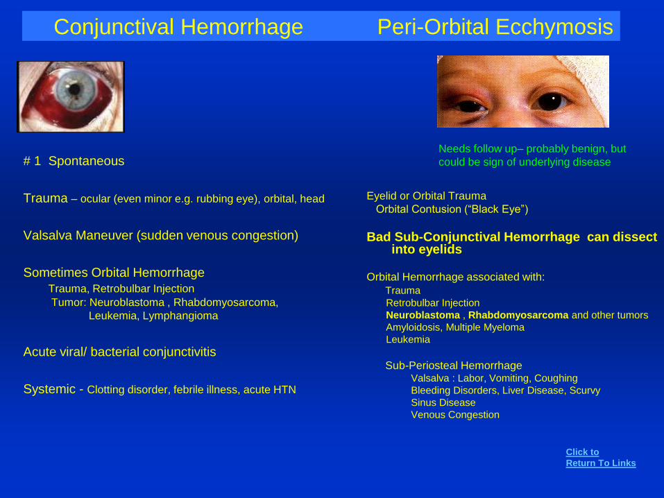
Conjunctival Hemorrhage Peri-Orbital Ecchymosis
Eyelid or Orbital Trauma
Orbital Contusion (“Black Eye”)
Bad Sub-Conjunctival Hemorrhage can dissect into eyelids
Orbital Hemorrhage associated with:
Trauma
Retrobulbar Injection
Neuroblastoma , Rhabdomyosarcoma and other tumors
Amyloidosis, Multiple Myeloma
Leukemia
Sub-Periosteal HemorrhageValsalva : Labor, Vomiting, Coughing
Bleeding Disorders, Liver Disease, Scurvy
Sinus Disease
Venous Congestion
# 1 Spontaneous
Trauma – ocular (even minor e.g. rubbing eye), orbital, head
Valsalva Maneuver (sudden venous congestion)
Sometimes Orbital Hemorrhage
Trauma, Retrobulbar Injection
Tumor: Neuroblastoma , Rhabdomyosarcoma,
Leukemia, Lymphangioma
Acute viral/ bacterial conjunctivitis
Systemic - Clotting disorder, febrile illness, acute HTN
Needs follow up– probably benign, but
could be sign of underlying disease
Click to
Return To Links
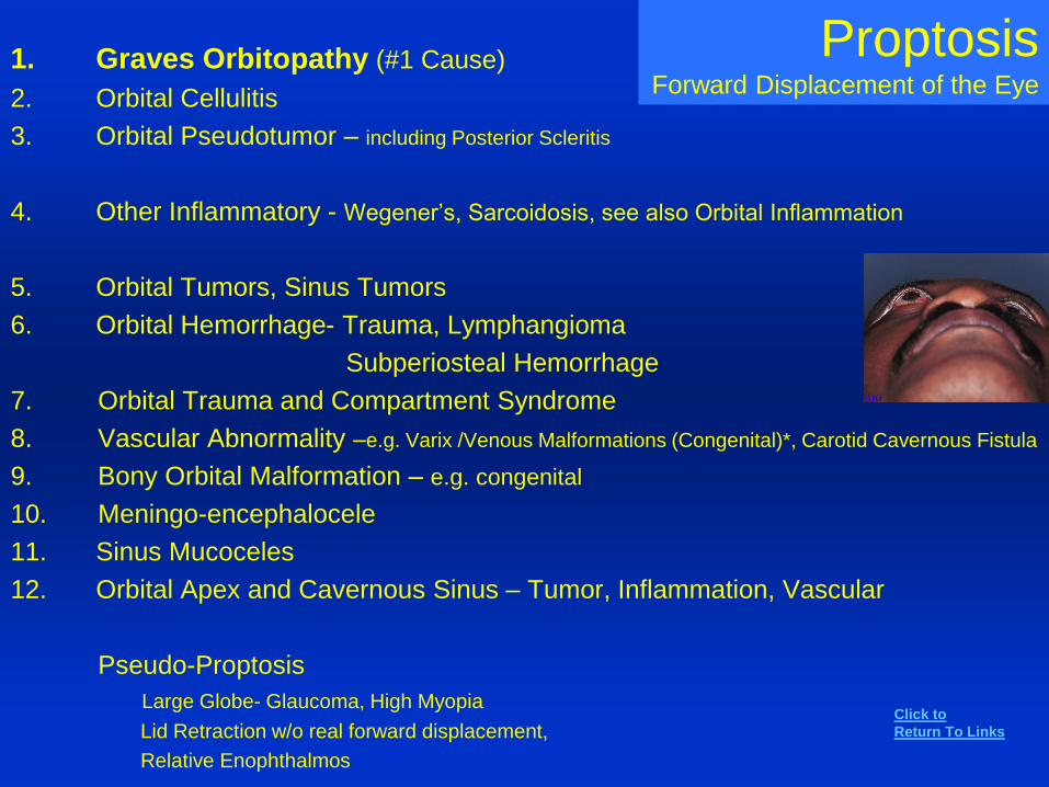
ProptosisForward Displacement of the Eye
1. Graves Orbitopathy (#1 Cause)
2. Orbital Cellulitis
3. Orbital Pseudotumor – including Posterior Scleritis
4. Other Inflammatory - Wegener’s, Sarcoidosis, see also Orbital Inflammation
5. Orbital Tumors, Sinus Tumors
6. Orbital Hemorrhage- Trauma, Lymphangioma
Subperiosteal Hemorrhage
7. Orbital Trauma and Compartment Syndrome
8. Vascular Abnormality –e.g. Varix /Venous Malformations (Congenital)*, Carotid Cavernous Fistula
9. Bony Orbital Malformation – e.g. congenital
10. Meningo-encephalocele
11. Sinus Mucoceles
12. Orbital Apex and Cavernous Sinus – Tumor, Inflammation, Vascular
Pseudo-Proptosis
Large Globe- Glaucoma, High Myopia
Lid Retraction w/o real forward displacement,
Relative Enophthalmos
Click to
Return To Links
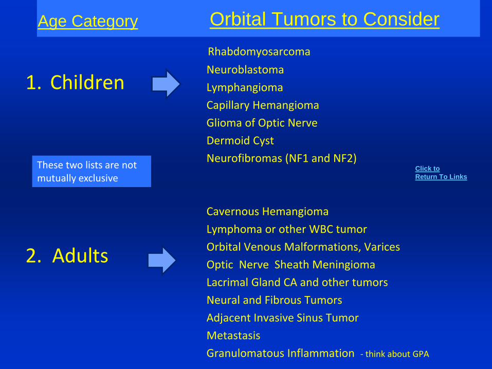
Orbital Tumors to ConsiderAge Category
1. Children
2. Adults
Rhabdomyosarcoma
Neuroblastoma
Lymphangioma
Capillary Hemangioma
Glioma of Optic Nerve
Dermoid Cyst
Neurofibromas (NF1 and NF2)
Cavernous Hemangioma
Lymphoma or other WBC tumor
Orbital Venous Malformations, Varices
Optic Nerve Sheath Meningioma
Lacrimal Gland CA and other tumors
Neural and Fibrous Tumors
Adjacent Invasive Sinus Tumor
Metastasis
Granulomatous Inflammation - think about GPA
Click to
Return To Links
These two lists are not mutually exclusive
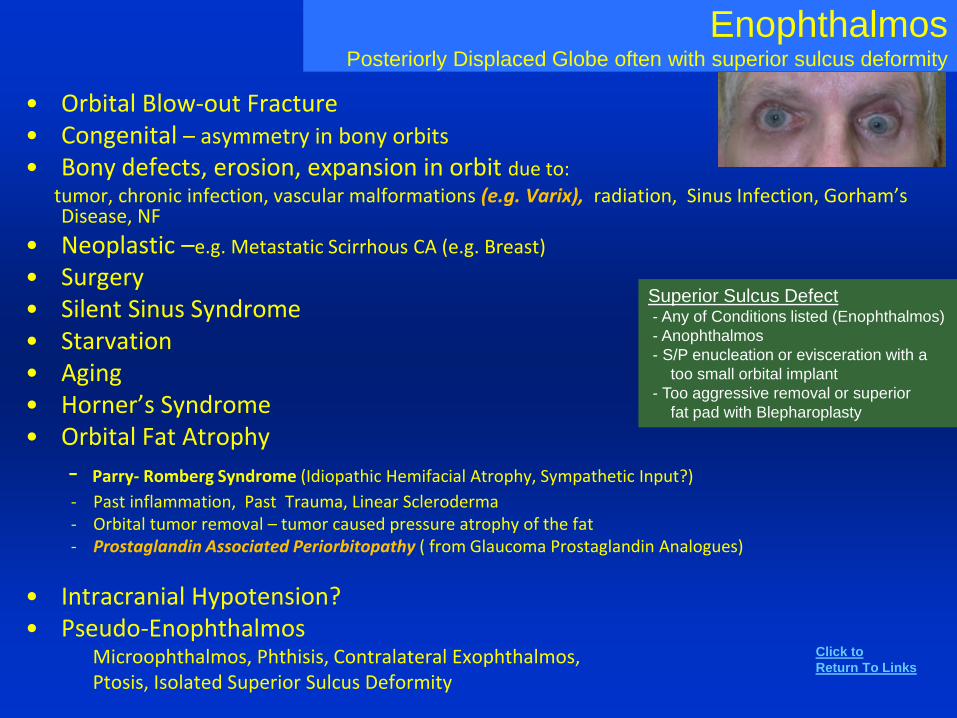
EnophthalmosPosteriorly Displaced Globe often with superior sulcus deformity
• Orbital Blow-out Fracture• Congenital – asymmetry in bony orbits
• Bony defects, erosion, expansion in orbit due to:
tumor, chronic infection, vascular malformations (e.g. Varix), radiation, Sinus Infection, Gorham’s Disease, NF
• Neoplastic –e.g. Metastatic Scirrhous CA (e.g. Breast)
• Surgery• Silent Sinus Syndrome• Starvation• Aging• Horner’s Syndrome• Orbital Fat Atrophy
- Parry- Romberg Syndrome (Idiopathic Hemifacial Atrophy, Sympathetic Input?)
- Past inflammation, Past Trauma, Linear Scleroderma- Orbital tumor removal – tumor caused pressure atrophy of the fat- Prostaglandin Associated Periorbitopathy ( from Glaucoma Prostaglandin Analogues)
• Intracranial Hypotension?• Pseudo-Enophthalmos
Microophthalmos, Phthisis, Contralateral Exophthalmos,Ptosis, Isolated Superior Sulcus Deformity
Superior Sulcus Defect- Any of Conditions listed (Enophthalmos)
- Anophthalmos
- S/P enucleation or evisceration with a
too small orbital implant
- Too aggressive removal or superior
fat pad with Blepharoplasty
Click to
Return To Links
Hypotony
Causes and Associations
Low IOP
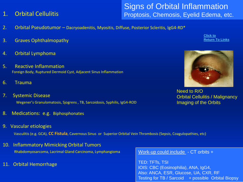
1. Orbital Cellulitis
2. Orbital Pseudotumor – Dacryoadenitis, Myositis, Diffuse, Posterior Scleritis, IgG4-RD*
3. Graves Ophthalmopathy
4. Orbital Lymphoma
5. Reactive InflammationForeign Body, Ruptured Dermoid Cyst, Adjacent Sinus Inflammation
6. Trauma
7. Systemic DiseaseWegener’s Granulomatosis, Sjogrens , TB, Sarcoidosis, Syphilis, IgG4-ROD
8. Medications: e.g. Biphosphonates
9. Vascular etiologies
Vasculitis (e.g. GCA), CC Fistula, Cavernous Sinus or Superior Orbital Vein Thrombosis (Sepsis, Coagulopathies, etc)
10. Inflammatory Mimicking Orbital TumorsRhabdomyosarcoma, Lacrimal Gland Carcinoma, Lymphangioma
11. Orbital Hemorrhage
Need to R/O
Orbital Cellulitis / Malignancy
Imaging of the Orbits
Signs of Orbital InflammationProptosis, Chemosis, Eyelid Edema, etc.
Work-up could include - CT orbits +
TED: TFTs, TSI
IOIS: CBC (Eosinophilia), ANA, IgG4,
Also: ANCA, ESR, Glucose, UA, CXR, RF
Testing for TB / Sarcoid + possible Orbital Biopsy
Click to
Return To Links
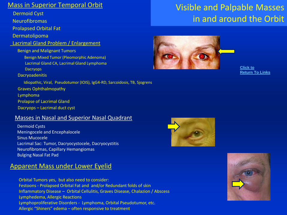
Mass in Superior Temporal OrbitDermoid Cyst
Neurofibromas
Prolapsed Orbital Fat
Dermatolipoma
Lacrimal Gland Problem / Enlargement
Benign and Malignant Tumors
Benign Mixed Tumor (Pleomorphic Adenoma)
Lacrimal Gland CA, Lacrimal Gland Lymphoma
Dacryops
Dacryoadenitis
Idiopathic, Viral, Pseudotumor (IOIS), IgG4-RD, Sarcoidosis, TB, Sjogrens
Graves Ophthalmopathy
Lymphoma
Prolapse of Lacrimal Gland
Dacryops – Lacrimal duct cyst
Masses in Nasal and Superior Nasal Quadrant
Dermoid CystsMeningocele and EncephaloceleSinus MucoceleLacrimal Sac: Tumor, Dacryocystocele, DacryocystitisNeurofibromas, Capillary HemangiomasBulging Nasal Fat Pad
Visible and Palpable Massesin and around the Orbit
Apparent Mass under Lower Eyelid
Orbital Tumors yes, but also need to consider:Festoons - Prolapsed Orbital Fat and and/or Redundant folds of skinInflammatory Disease – Orbital Cellulitis, Graves Disease, Chalazion / AbscessLymphedema, Allergic ReactionsLymphoproliferative Disorders - Lymphoma, Orbital Pseudotumor, etc.Allergic “Shiners” edema – often responsive to treatment
Click to
Return To Links
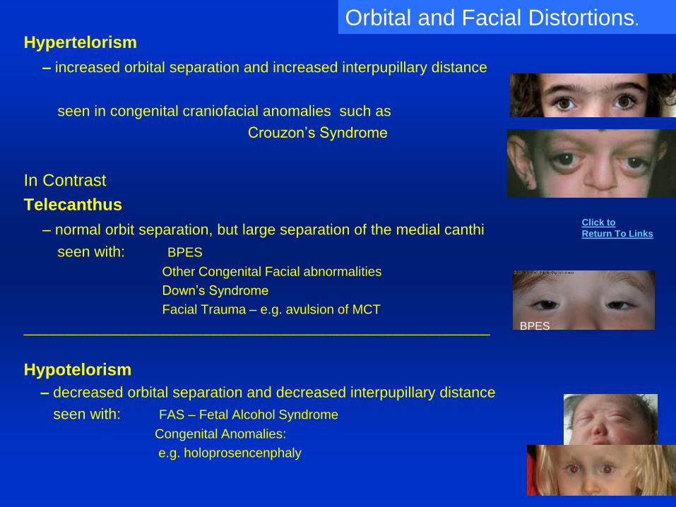
Hypertelorism
– increased orbital separation and increased interpupillary distance
seen in congenital craniofacial anomalies such as
Crouzon’s Syndrome
In Contrast
Telecanthus
– normal orbit separation, but large separation of the medial canthi
seen with: BPES
Other Congenital Facial abnormalities
Down’s Syndrome
Facial Trauma – e.g. avulsion of MCT
________________________________________________________________
Hypotelorism
– decreased orbital separation and decreased interpupillary distance
seen with: FAS – Fetal Alcohol Syndrome
Congenital Anomalies:
e.g. holoprosencenphaly
Orbital and Facial Distortions.
BPES
Click to
Return To Links
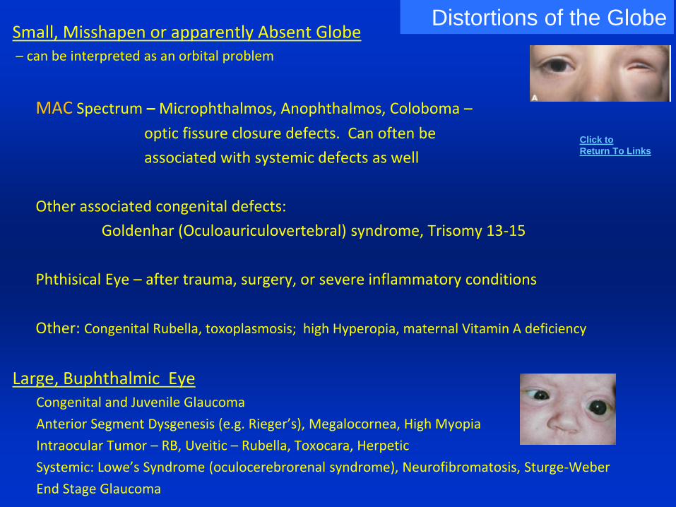
Small, Misshapen or apparently Absent Globe– can be interpreted as an orbital problem
MAC Spectrum – Microphthalmos, Anophthalmos, Coloboma –
optic fissure closure defects. Can often be
associated with systemic defects as well
Other associated congenital defects:
Goldenhar (Oculoauriculovertebral) syndrome, Trisomy 13-15
Phthisical Eye – after trauma, surgery, or severe inflammatory conditions
Other: Congenital Rubella, toxoplasmosis; high Hyperopia, maternal Vitamin A deficiency
Large, Buphthalmic EyeCongenital and Juvenile Glaucoma
Anterior Segment Dysgenesis (e.g. Rieger’s), Megalocornea, High Myopia
Intraocular Tumor – RB, Uveitic – Rubella, Toxocara, Herpetic
Systemic: Lowe’s Syndrome (oculocerebrorenal syndrome), Neurofibromatosis, Sturge-Weber
End Stage Glaucoma
Distortions of the Globe
Click to
Return To Links
Motility and Alignment
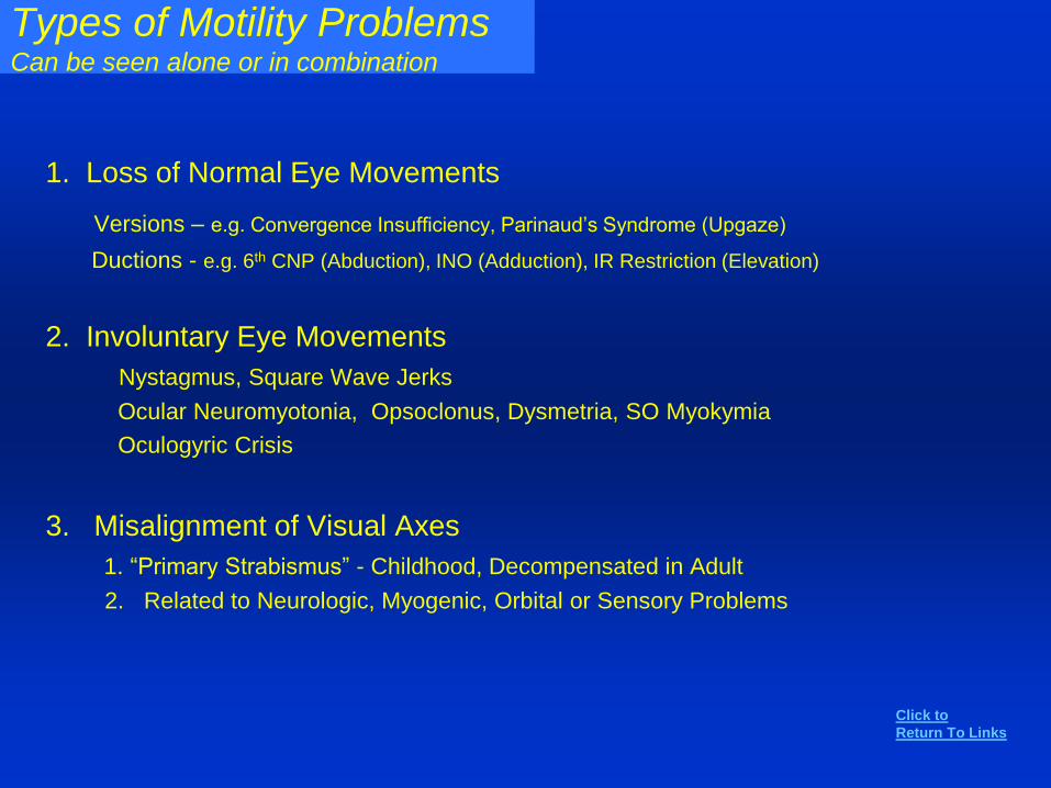
Types of Motility ProblemsCan be seen alone or in combination
1. Loss of Normal Eye Movements
Versions – e.g. Convergence Insufficiency, Parinaud’s Syndrome (Upgaze)
Ductions - e.g. 6th CNP (Abduction), INO (Adduction), IR Restriction (Elevation)
2. Involuntary Eye Movements
Nystagmus, Square Wave Jerks
Ocular Neuromyotonia, Opsoclonus, Dysmetria, SO Myokymia
Oculogyric Crisis
3. Misalignment of Visual Axes
1. “Primary Strabismus” - Childhood, Decompensated in Adult
2. Related to Neurologic, Myogenic, Orbital or Sensory Problems
Click to
Return To Links
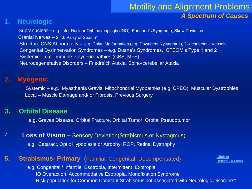
Motility and Alignment Problems
1. Neurologic
Supranuclear – e.g. Inter Nuclear Ophthalmoplegia (INO), Parinaud’s Syndrome, Skew Deviation
Cranial Nerves – 3,4,6 Palsy or Spasm*
Structure CNS Abnormality – e.g. Chiari Malformation (e.g. Downbeat Nystagmus), Dolichoectatic Vessels
Congenital Dysinnervation Syndromes – e.g. Duane’s Syndromes, CFEOM’s Type 1 and 2
Systemic – e.g. Immune Polyneuropathies (GBS, MFS)
Neurodegenerative Disorders – Friedreich Ataxia, Spino-cerebellar Ataxia
2. Myogenic
Systemic – e.g. Myasthenia Gravis, Mitochondrial Myopathies (e.g. CPEO), Muscular Dystrophies
Local – Muscle Damage and/ or Fibrosis, Previous Surgery
3. Orbital Disease
e.g. Graves Disease, Orbital Fracture, Orbital Tumor, Orbital Pseudotumor
4. Loss of Vision – Sensory Deviation(Strabismus or Nystagmus)
e.g. Cataract, Optic Hypoplasia or Atrophy, ROP, Retinal Dystrophy
5. Strabismus- Primary (Familial, Congenital, Decompensated)
e.g. Congenital / Infantile Esotropia, Intermittent Exotropia,
IO Overaction, Accommodative Esotropia, Monofixation Syndrome
Risk population for Common Comitant Strabismus not associated with Neurologic Disorders*
A Spectrum of Causes
Click to
Return To Links
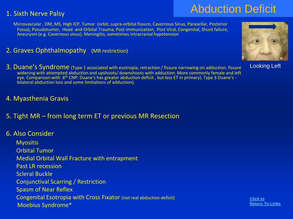
Abduction Deficit1. Sixth Nerve Palsy
Microvascular , DM, MS, High ICP, Tumor (orbit, supra-orbital fissure, Cavernous Sinus, Parasellar, Posterior Fossa), Pseudotumor, Head and Orbital Trauma, Post-immunization, Post Viral, Congenital, Shunt failure, Aneurysm (e.g. Cavernous sinus), Meningitis, sometimes intracranial hypotension
2. Graves Ophthalmopathy (MR restriction)
3. Duane’s Syndrome (Type 1 associated with esotropia, retraction / fissure narrowing on adduction, fissure widening with attempted abduction and upshoots/ downshoots with adduction. More commonly female and left eye. Comparison with 6th CNP: Duane's has greater abduction deficit , but less ET in primary). Type 3 Duane’s -bilateral abduction loss and some limitations of adduction).
4. Myasthenia Gravis
5. Tight MR – from long term ET or previous MR Resection
6. Also ConsiderMyositisOrbital Tumor Medial Orbital Wall Fracture with entrapment Past LR recessionScleral BuckleConjunctival Scarring / RestrictionSpasm of Near ReflexCongenital Esotropia with Cross Fixator (not real abduction deficit)
Moebius Syndrome*
Looking Left
Click to
Return To Links
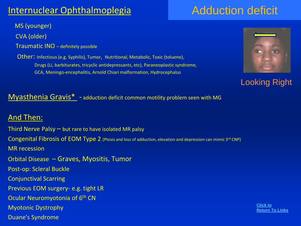
Adduction deficitInternuclear Ophthalmoplegia
MS (younger)
CVA (older)
Traumatic INO – definitely possible
Other: Infectious (e.g. Syphilis), Tumor, Nutritional, Metabolic, Toxic (toluene),
Drugs (Li, barbiturates, tricyclic antidepressants, etc), Paraneoplastic syndrome,
GCA, Meningo-encephalitis, Arnold Chiari malformation, Hydrocephalus
Myasthenia Gravis* - adduction deficit common motility problem seen with MG
And Then:
Third Nerve Palsy – but rare to have isolated MR palsy
Congenital Fibrosis of EOM Type 2 (Ptosis and loss of adduction, elevation and depression can mimic 3rd CNP)
MR recession
Orbital Disease – Graves, Myositis, Tumor
Post-op: Scleral Buckle
Conjunctival Scarring
Previous EOM surgery- e.g. tight LR
Ocular Neuromyotonia of 6th CN
Myotonic Dystrophy
Duane's Syndrome
Looking Right
Click to
Return To Links
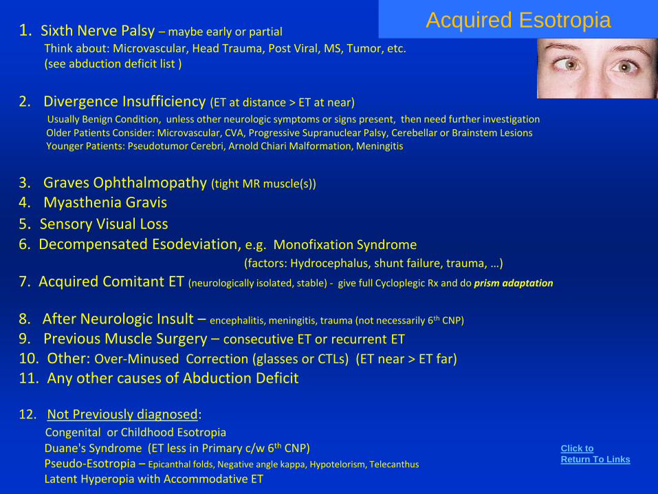
Acquired Esotropia1. Sixth Nerve Palsy – maybe early or partial
Think about: Microvascular, Head Trauma, Post Viral, MS, Tumor, etc. (see abduction deficit list )
2. Divergence Insufficiency (ET at distance > ET at near)
Usually Benign Condition, unless other neurologic symptoms or signs present, then need further investigationOlder Patients Consider: Microvascular, CVA, Progressive Supranuclear Palsy, Cerebellar or Brainstem Lesions Younger Patients: Pseudotumor Cerebri, Arnold Chiari Malformation, Meningitis
3. Graves Ophthalmopathy (tight MR muscle(s))
4. Myasthenia Gravis
5. Sensory Visual Loss6. Decompensated Esodeviation, e.g. Monofixation Syndrome
(factors: Hydrocephalus, shunt failure, trauma, …)
7. Acquired Comitant ET (neurologically isolated, stable) - give full Cycloplegic Rx and do prism adaptation
8. After Neurologic Insult – encephalitis, meningitis, trauma (not necessarily 6th CNP)
9. Previous Muscle Surgery – consecutive ET or recurrent ET
10. Other: Over-Minused Correction (glasses or CTLs) (ET near > ET far)
11. Any other causes of Abduction Deficit
12. Not Previously diagnosed:Congenital or Childhood EsotropiaDuane's Syndrome (ET less in Primary c/w 6th CNP)Pseudo-Esotropia – Epicanthal folds, Negative angle kappa, Hypotelorism, Telecanthus
Latent Hyperopia with Accommodative ET
Click to
Return To Links
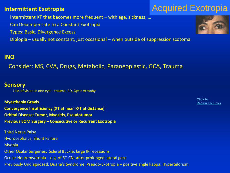
Acquired ExotropiaIntermittent ExotropiaIntermittent XT that becomes more frequent – with age, sickness, …
Can Decompensate to a Constant Exotropia
Types: Basic, Divergence Excess
Diplopia – usually not constant, just occasional – when outside of suppression scotoma
INO
Consider: MS, CVA, Drugs, Metabolic, Paraneoplastic, GCA, Trauma
Sensory Loss of vision in one eye – trauma, RD, Optic Atrophy
Myasthenia Gravis
Convergence Insufficiency (XT at near >XT at distance)
Orbital Disease: Tumor, Myositis, Pseudotumor
Previous EOM Surgery – Consecutive or Recurrent Exotropia
Third Nerve Palsy
Hydrocephalus, Shunt Failure
Myopia
Other Ocular Surgeries: Scleral Buckle, large IR recessions
Ocular Neuromyotonia – e.g. of 6th CN- after prolonged lateral gaze
Previously Undiagnosed: Duane’s Syndrome, Pseudo-Exotropia – positive angle kappa, Hypertelorism
Click to
Return To Links
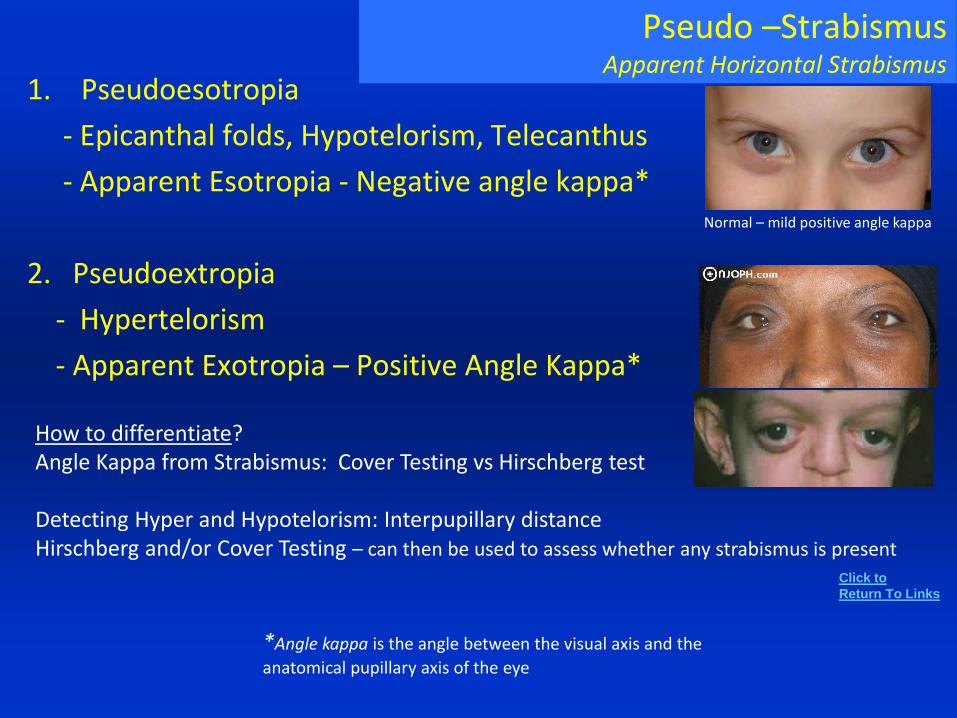
Pseudo –StrabismusApparent Horizontal Strabismus
1. Pseudoesotropia
- Epicanthal folds, Hypotelorism, Telecanthus
- Apparent Esotropia - Negative angle kappa*
2. Pseudoextropia
- Hypertelorism
- Apparent Exotropia – Positive Angle Kappa*
Normal – mild positive angle kappa
How to differentiate?Angle Kappa from Strabismus: Cover Testing vs Hirschberg test
Detecting Hyper and Hypotelorism: Interpupillary distanceHirschberg and/or Cover Testing – can then be used to assess whether any strabismus is present
Click to
Return To Links
*Angle kappa is the angle between the visual axis and the
anatomical pupillary axis of the eye
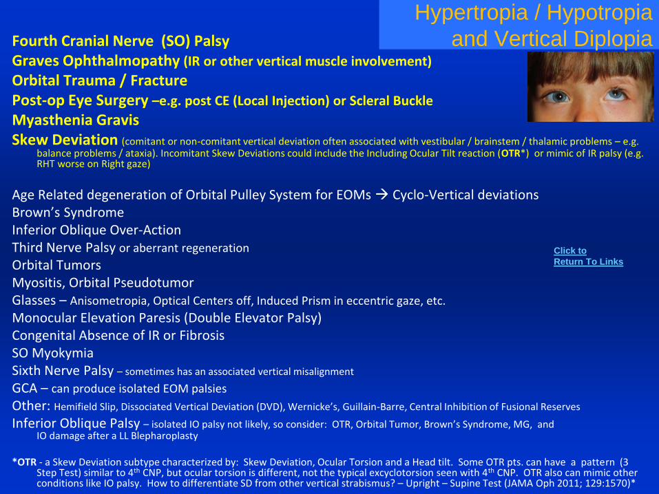
Hypertropia / Hypotropia
and Vertical DiplopiaFourth Cranial Nerve (SO) PalsyGraves Ophthalmopathy (IR or other vertical muscle involvement)
Orbital Trauma / FracturePost-op Eye Surgery –e.g. post CE (Local Injection) or Scleral Buckle
Myasthenia GravisSkew Deviation (comitant or non-comitant vertical deviation often associated with vestibular / brainstem / thalamic problems – e.g.
balance problems / ataxia). Incomitant Skew Deviations could include the Including Ocular Tilt reaction (OTR*) or mimic of IR palsy (e.g. RHT worse on Right gaze)
Age Related degeneration of Orbital Pulley System for EOMs Cyclo-Vertical deviationsBrown’s SyndromeInferior Oblique Over-Action Third Nerve Palsy or aberrant regeneration
Orbital TumorsMyositis, Orbital PseudotumorGlasses – Anisometropia, Optical Centers off, Induced Prism in eccentric gaze, etc.
Monocular Elevation Paresis (Double Elevator Palsy)Congenital Absence of IR or FibrosisSO MyokymiaSixth Nerve Palsy – sometimes has an associated vertical misalignment
GCA – can produce isolated EOM palsies
Other: Hemifield Slip, Dissociated Vertical Deviation (DVD), Wernicke’s, Guillain-Barre, Central Inhibition of Fusional Reserves
Inferior Oblique Palsy – isolated IO palsy not likely, so consider: OTR, Orbital Tumor, Brown’s Syndrome, MG, and IO damage after a LL Blepharoplasty
*OTR - a Skew Deviation subtype characterized by: Skew Deviation, Ocular Torsion and a Head tilt. Some OTR pts. can have a pattern (3 Step Test) similar to 4th CNP, but ocular torsion is different, not the typical excyclotorsion seen with 4th CNP. OTR also can mimic other conditions like IO palsy. How to differentiate SD from other vertical strabismus? – Upright – Supine Test (JAMA Oph 2011; 129:1570)*
Click to
Return To Links
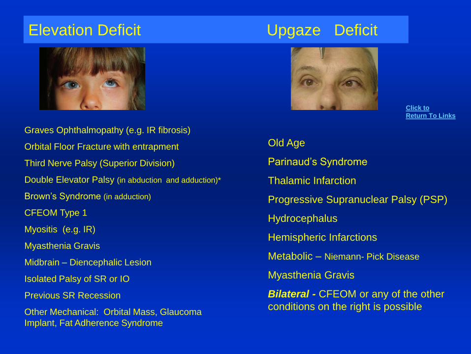
Elevation Deficit Upgaze Deficit
Old Age
Parinaud’s Syndrome
Thalamic Infarction
Progressive Supranuclear Palsy (PSP)
Hydrocephalus
Hemispheric Infarctions
Metabolic – Niemann- Pick Disease
Myasthenia Gravis
Bilateral - CFEOM or any of the other
conditions on the right is possible
Graves Ophthalmopathy (e.g. IR fibrosis)
Orbital Floor Fracture with entrapment
Third Nerve Palsy (Superior Division)
Double Elevator Palsy (in abduction and adduction)*
Brown’s Syndrome (in adduction)
CFEOM Type 1
Myositis (e.g. IR)
Myasthenia Gravis
Midbrain – Diencephalic Lesion
Isolated Palsy of SR or IO
Previous SR Recession
Other Mechanical: Orbital Mass, Glaucoma
Implant, Fat Adherence Syndrome
Click to
Return To Links
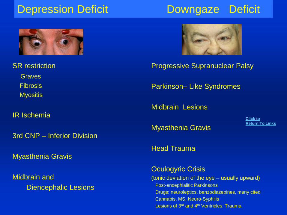
Depression Deficit Downgaze Deficit
SR restriction
Graves
Fibrosis
Myositis
IR Ischemia
3rd CNP – Inferior Division
Myasthenia Gravis
Midbrain and
Diencephalic Lesions
Progressive Supranuclear Palsy
Parkinson– Like Syndromes
Midbrain Lesions
Myasthenia Gravis
Head Trauma
Oculogyric Crisis(tonic deviation of the eye – usually upward)
Post-encephlalitic Parkinsons
Drugs: neuroleptics, benzodiazepines, many cited
Cannabis, MS, Neuro-Syphilis
Lesions of 3rd and 4th Ventricles, Trauma
Click to
Return To Links
Convergence and Divergence
Divergence InsufficiencyComitant ET at distance, less or none at
near and full ductions
• Primary Divergence Insufficiency
(Neurologically Isolated*, usually older patients)
• Secondary Divergence Insufficiency
(Usually not Neurologically Isolated)
Cerebellar Ischemia / Stroke
Chiari Malformation
Demyelinating Disease
Idiopathic Cerebellar Degeneration
Progressive Supranuclear Palsy
Temporal Arteritis
High ICP / Pseudotumor Cerebri
Consider also:
6th CNP with spread of comitance over time
MG
Convergence InsufficiencyComitant Exotropia greater at near that
distance. Decreased Near Point of
Convergence (NPC)
• Primary
• Secondary
- After Head Injury
- Parkinson’s Disease
and other CNS degenerative
disorders
Click to
Return To Links

Ophthalmoplegia – One Eye(More likely Local Lesions)
Multiple Cranial Nerve PalsiesOrbital apex, Superior Orbital Fissure, Cavernous Sinus Lesions:
Tumor (e.g. meningioma, extension of pituitary tumor)Aneurysm, AVMOrbital Pseudotumor / IOIS (e.g. Myositis, Tolosa - Hunt Syndrome)Orbital Cellulitis
Consider Fungal including Mucormycosis, AspergillosisOther Inflammatory:
Wegener, Sarcoidosis, TB, Syphilis, GCA, etc.HZO (can see Ophthalmoplegia up to 3 months after onset)
Cavernous Sinus Fistula or Thrombosis
Other Causes: Mechanical, Orbital Infiltration of Tumor, Ischemia, etc.
Trauma- edema and or orbital hemorrhagePeri-neural Spread of Cutaneous CarcinomaIschemia – Saturday Night Ophthalmoplegia, Orbital Infarction Syndrome*Graves Ophthalmopathy – multiple musclesHigh ICP, DM – could potentially cause multiple CNP (e.g. 3rd and 6th) at same timeThird Nerve Palsy – mistake for total Ophthalmoplegia
*Systemic Conditions could initially present unilaterally – e.g. MG, Guillain-Barre, …SEE NEXT LISTING
Click to
Return To Links
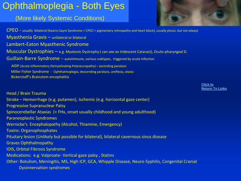
Ophthalmoplegia - Both Eyes
(More likely Systemic Conditions)
CPEO – usually bilateral (Kearns Sayre Syndrome = CPEO + pigmentary retinopathy and heart block), usually ptosis, but not always
Myasthenia Gravis – unilateral or bilateral
Lambert-Eaton Myasthenic Syndrome
Muscular Dystrophies – e.g. Myotonic Dystrophy ( can see an Iridescent Cataract), Oculo-pharyngeal D.
Guillain-Barre Syndrome – autoimmune, various subtypes, triggered by acute infection
AIDP (Acute inflammatory Demyelinating Polyneuropathy) – ascending paralysis
Miller Fisher Syndrome - Ophthalmoplegia, descending paralysis, areflexia, ataxia
Bickerstaff’s Brainstem encephalitis
Head / Brain Trauma
Stroke – Hemorrhage (e.g. putamen), Ischemic (e.g. horizontal gaze center)
Progressive Supranuclear Palsy
Spinocerebellar Ataxias (+ FHx, onset usually childhood and young adulthood)
Paraneoplastic Syndromes
Wernicke’s Encephalopathy (Alcohol, Thiamine, Emergency)
Toxins: Organophosphates
Pituitary lesion (Unlikely but possible for bilateral), bilateral cavernous sinus disease
Graves Ophthalmopathy
IOIS, Orbital Fibrosis Syndrome
Medications: e.g. Valproate- Vertical gaze palsy , Statins
Other: Botulism, Meningitis, MS, High ICP, GCA, Whipple Disease, Neuro-Syphilis, Congenital Cranial
Dysinnervation syndromes
Click to
Return To Links
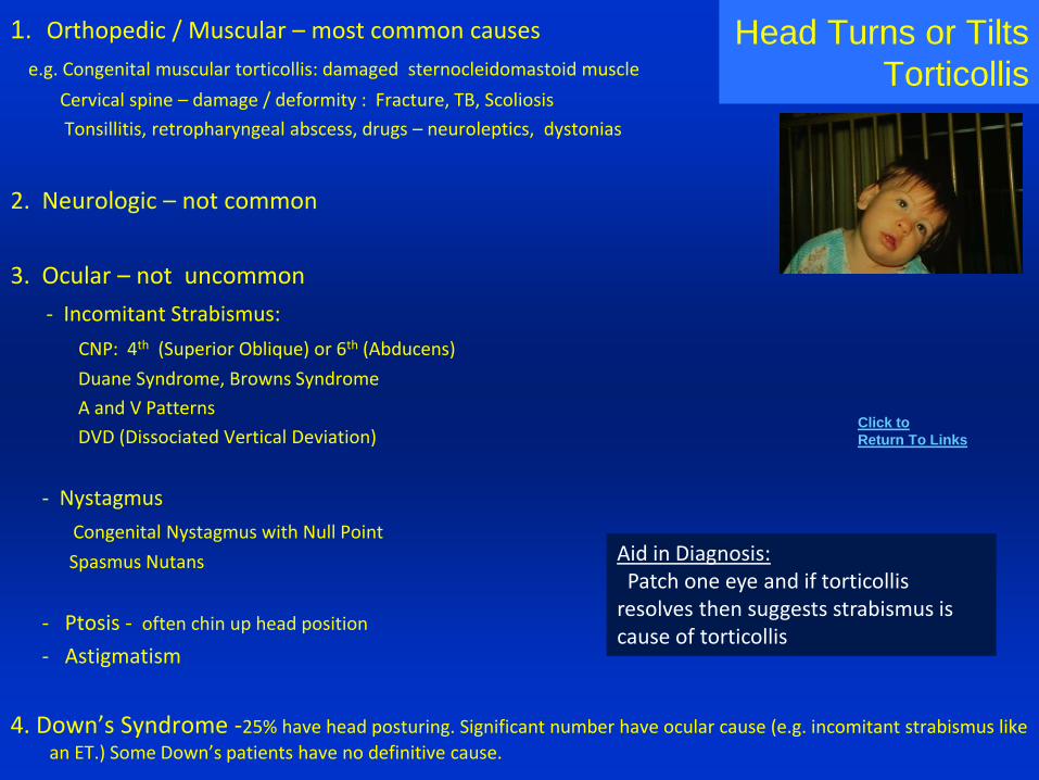
Head Turns or Tilts
Torticollis
1. Orthopedic / Muscular – most common causes
e.g. Congenital muscular torticollis: damaged sternocleidomastoid muscle
Cervical spine – damage / deformity : Fracture, TB, Scoliosis
Tonsillitis, retropharyngeal abscess, drugs – neuroleptics, dystonias
2. Neurologic – not common
3. Ocular – not uncommon
- Incomitant Strabismus:
CNP: 4th (Superior Oblique) or 6th (Abducens)
Duane Syndrome, Browns Syndrome
A and V Patterns
DVD (Dissociated Vertical Deviation)
- Nystagmus
Congenital Nystagmus with Null Point
Spasmus Nutans
- Ptosis - often chin up head position
- Astigmatism
4. Down’s Syndrome -25% have head posturing. Significant number have ocular cause (e.g. incomitant strabismus like
an ET.) Some Down’s patients have no definitive cause.
Aid in Diagnosis:Patch one eye and if torticollis
resolves then suggests strabismus is cause of torticollis
Click to
Return To Links
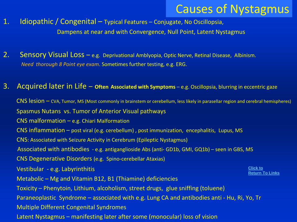
Causes of Nystagmus1. Idiopathic / Congenital – Typical Features – Conjugate, No Oscillopsia,
Dampens at near and with Convergence, Null Point, Latent Nystagmus
2. Sensory Visual Loss – e.g. Deprivational Amblyopia, Optic Nerve, Retinal Disease, Albinism.
Need thorough 8 Point eye exam. Sometimes further testing, e.g. ERG.
3. Acquired later in Life – Often Associated with Symptoms – e.g. Oscillopsia, blurring in eccentric gaze
CNS lesion – CVA, Tumor, MS (Most commonly in brainstem or cerebellum, less likely in parasellar region and cerebral hemispheres)
Spasmus Nutans vs. Tumor of Anterior Visual pathways
CNS malformation – e.g. Chiari Malformation
CNS inflammation – post viral (e.g. cerebellum) , post immunization, encephalitis, Lupus, MS
CNS: Associated with Seizure Activity in Cerebrum (Epileptic Nystagmus)
Associated with antibodies - e.g. antiganglioside Abs (anti- GD1b, GMI, GQ1b) – seen in GBS, MS
CNS Degenerative Disorders (e.g. Spino-cerebellar Ataxias)
Vestibular - e.g. Labyrinthitis
Metabolic – Mg and Vitamin B12, B1 (Thiamine) deficiencies
Toxicity – Phenytoin, Lithium, alcoholism, street drugs, glue sniffing (toluene)
Paraneoplastic Syndrome – associated with e.g. Lung CA and antibodies anti - Hu, Ri, Yo, Tr
Multiple Different Congenital Syndromes
Latent Nystagmus – manifesting later after some (monocular) loss of vision
Click to
Return To Links
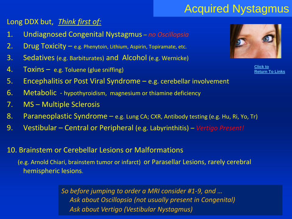
Hypotony
Causes and Associations
Low IOP
Long DDX but, Think first of:
1. Undiagnosed Congenital Nystagmus – no Oscillopsia
2. Drug Toxicity – e.g. Phenytoin, Lithium, Aspirin, Topiramate, etc.
3. Sedatives (e.g. Barbiturates) and Alcohol (e.g. Wernicke)
4. Toxins – e.g. Toluene (glue sniffing)
5. Encephalitis or Post Viral Syndrome – e.g. cerebellar involvement
6. Metabolic - hypothyroidism, magnesium or thiamine deficiency
7. MS – Multiple Sclerosis
8. Paraneoplastic Syndrome – e.g. Lung CA; CXR, Antibody testing (e.g. Hu, Ri, Yo, Tr)
9. Vestibular – Central or Peripheral (e.g. Labyrinthitis) – Vertigo Present!
10. Brainstem or Cerebellar Lesions or Malformations
(e.g. Arnold Chiari, brainstem tumor or infarct) or Parasellar Lesions, rarely cerebral hemispheric lesions.
Acquired Nystagmus
So before jumping to order a MRI consider #1-9, and … Ask about Oscillopsia (not usually present in Congenital)
Ask about Vertigo (Vestibular Nystagmus)
Click to
Return To Links
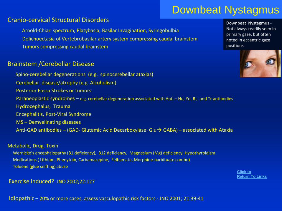
Downbeat NystagmusCranio-cervical Structural Disorders
Arnold-Chiari spectrum, Platybasia, Basilar Invagination, Syringobulbia
Dolichoectasia of Vertebrobasilar artery system compressing caudal brainstem
Tumors compressing caudal brainstem
Brainstem /Cerebellar Disease
Spino-cerebellar degenerations (e.g. spinocerebellar ataxias)
Cerebellar disease/atrophy (e.g. Alcoholism)
Posterior Fossa Strokes or tumors
Paraneoplastic syndromes – e.g. cerebellar degeneration associated with Anti – Hu, Yo, Ri, and Tr antibodies
Hydrocephalus, Trauma
Encephalitis, Post-Viral Syndrome
MS – Demyelinating diseases
Anti-GAD antibodies – (GAD- Glutamic Acid Decarboxylase: Glu GABA) – associated with Ataxia
Metabolic, Drug, Toxin
Wernicke’s encephalopathy (B1 deficiency), B12 deficiency, Magnesium (Mg) deficiency, Hypothyroidism
Medications ( Lithium, Phenytoin, Carbamazepine, Felbamate, Morphine-barbituate combo)
Toluene (glue sniffing) abuse
Exercise induced? JNO 2002;22:127
Idiopathic – 20% or more cases, assess vasculopathic risk factors - JNO 2001; 21:39-41
Downbeat Nystagmus -Not always readily seen in primary gaze, but often noted in eccentric gaze positions
Click to
Return To Links

Pupils
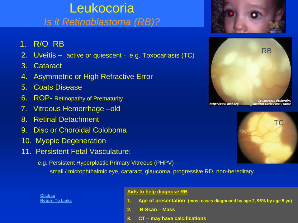
LeukocoriaIs it Retinoblastoma (RB)?
1. R/O RB
2. Uveitis – active or quiescent - e.g. Toxocariasis (TC)
3. Cataract
4. Asymmetric or High Refractive Error
5. Coats Disease
6. ROP- Retinopathy of Prematurity
7. Vitreous Hemorrhage –old
8. Retinal Detachment
9. Disc or Choroidal Coloboma
10. Myopic Degeneration
11. Persistent Fetal Vasculature:
e.g. Persistent Hyperplastic Primary Vitreous (PHPV) –
small / microphthalmic eye, cataract, glaucoma, progressive RD, non-hereditary
Aids to help diagnose RB
1. Age of presentation (most cases diagnosed by age 2, 95% by age 5 yo)
2. B-Scan – Mass
3. CT – may have calcifications
RB
TC
Click to
Return To Links
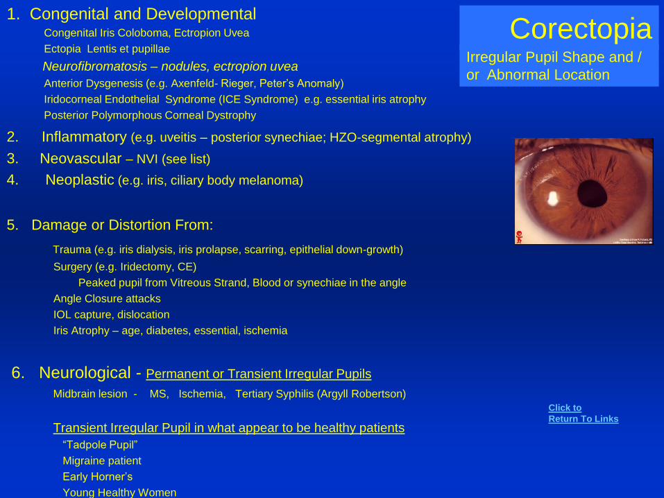
Corectopia1. Congenital and Developmental
Congenital Iris Coloboma, Ectropion Uvea
Ectopia Lentis et pupillae
Neurofibromatosis – nodules, ectropion uvea
Anterior Dysgenesis (e.g. Axenfeld- Rieger, Peter’s Anomaly)
Iridocorneal Endothelial Syndrome (ICE Syndrome) e.g. essential iris atrophy
Posterior Polymorphous Corneal Dystrophy
2. Inflammatory (e.g. uveitis – posterior synechiae; HZO-segmental atrophy)
3. Neovascular – NVI (see list)
4. Neoplastic (e.g. iris, ciliary body melanoma)
5. Damage or Distortion From:
Trauma (e.g. iris dialysis, iris prolapse, scarring, epithelial down-growth)
Surgery (e.g. Iridectomy, CE)
Peaked pupil from Vitreous Strand, Blood or synechiae in the angle
Angle Closure attacks
IOL capture, dislocation
Iris Atrophy – age, diabetes, essential, ischemia
6. Neurological - Permanent or Transient Irregular Pupils
Midbrain lesion - MS, Ischemia, Tertiary Syphilis (Argyll Robertson)
Transient Irregular Pupil in what appear to be healthy patients
“Tadpole Pupil”
Migraine patient
Early Horner’s
Young Healthy Women
Irregular Pupil Shape and /
or Abnormal Location
Click to
Return To Links
Anisocoria (Unequal pupils)
• Age
• Diabetes Mellitus
• Damaged or Paralyzed Pupil
Uveitis, Angle Closure, Surgery, Trauma
Topical: Mydriatics (e.g. atropine), Miotics (Pilocarpine), Natural Products
• Afferent or Efferent Input Defect
Damage to any part of Pupillary Light Reflex Pathways
RAPD
Parasympathetic or Sympathetic Defect: Third Nerve Palsy, Adie’s, Horner's Syndromes
• Systemic
Medications or Toxins
Mydriatic - Anticholinergics , Adrenergics (Methamphetamine),
Miotic - Narcotics
Systemic / Neuro-Muscular Conditions
Lesions at level of Tectum (dilated), Midbrain (mid-dilated) , or Pons (miotic)
Myotonic Dystrophy – miotic
Fisher variant of G/B Syndrome or Riley-Day Syndrome - mydriatic
Poor Pupil MobilityPoor Reactions to Light, Dark or other stimuli
Click to
Return To Links
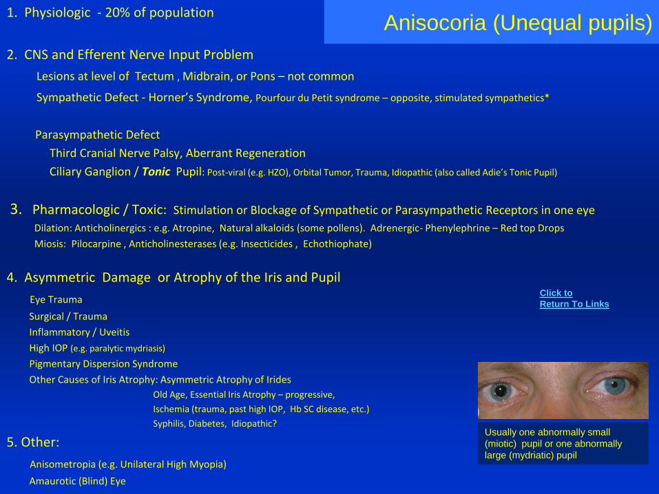
Anisocoria (Unequal pupils)1. Physiologic - 20% of population
2. CNS and Efferent Nerve Input Problem
Lesions at level of Tectum , Midbrain, or Pons – not common
Sympathetic Defect - Horner’s Syndrome, Pourfour du Petit syndrome – opposite, stimulated sympathetics*
Parasympathetic Defect
Third Cranial Nerve Palsy, Aberrant Regeneration
Ciliary Ganglion / Tonic Pupil: Post-viral (e.g. HZO), Orbital Tumor, Trauma, Idiopathic (also called Adie’s Tonic Pupil)
3. Pharmacologic / Toxic: Stimulation or Blockage of Sympathetic or Parasympathetic Receptors in one eye
Dilation: Anticholinergics : e.g. Atropine, Natural alkaloids (some pollens). Adrenergic- Phenylephrine – Red top Drops
Miosis: Pilocarpine , Anticholinesterases (e.g. Insecticides , Echothiophate)
4. Asymmetric Damage or Atrophy of the Iris and Pupil
Eye Trauma
Surgical / Trauma
Inflammatory / Uveitis
High IOP (e.g. paralytic mydriasis)
Pigmentary Dispersion Syndrome
Other Causes of Iris Atrophy: Asymmetric Atrophy of Irides
Old Age, Essential Iris Atrophy – progressive,
Ischemia (trauma, past high IOP, Hb SC disease, etc.)
Syphilis, Diabetes, Idiopathic?
5. Other:
Anisometropia (e.g. Unilateral High Myopia)
Amaurotic (Blind) Eye
Usually one abnormally small
(miotic) pupil or one abnormally
large (mydriatic) pupil
Click to
Return To Links
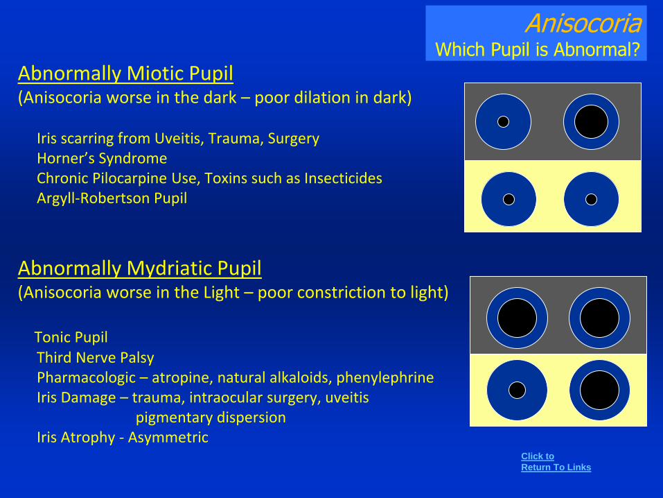
AnisocoriaWhich Pupil is Abnormal?
Abnormally Miotic Pupil(Anisocoria worse in the dark – poor dilation in dark)
Iris scarring from Uveitis, Trauma, SurgeryHorner’s SyndromeChronic Pilocarpine Use, Toxins such as Insecticides Argyll-Robertson Pupil
Abnormally Mydriatic Pupil (Anisocoria worse in the Light – poor constriction to light)
Tonic PupilThird Nerve PalsyPharmacologic – atropine, natural alkaloids, phenylephrineIris Damage – trauma, intraocular surgery, uveitis
pigmentary dispersionIris Atrophy - Asymmetric
Click to
Return To Links
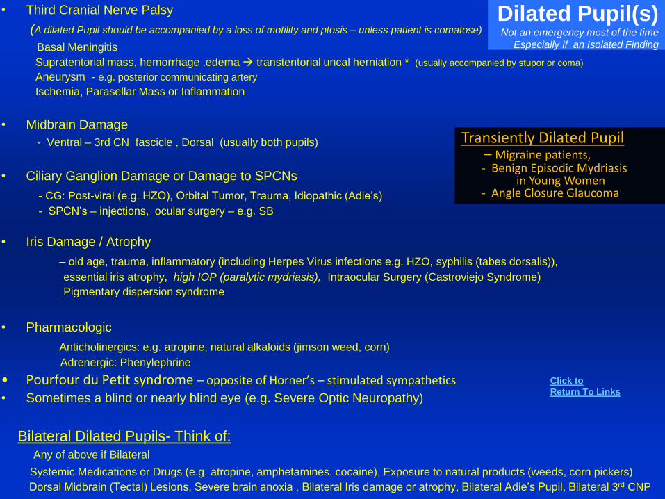
Dilated Pupil(s) Not an emergency most of the time
Especially if an Isolated Finding
• Third Cranial Nerve Palsy
(A dilated Pupil should be accompanied by a loss of motility and ptosis – unless patient is comatose)
Basal Meningitis
Supratentorial mass, hemorrhage ,edema transtentorial uncal herniation * (usually accompanied by stupor or coma)
Aneurysm - e.g. posterior communicating artery
Ischemia, Parasellar Mass or Inflammation
• Midbrain Damage
- Ventral – 3rd CN fascicle , Dorsal (usually both pupils)
• Ciliary Ganglion Damage or Damage to SPCNs
- CG: Post-viral (e.g. HZO), Orbital Tumor, Trauma, Idiopathic (Adie’s)
- SPCN’s – injections, ocular surgery – e.g. SB
• Iris Damage / Atrophy
– old age, trauma, inflammatory (including Herpes Virus infections e.g. HZO, syphilis (tabes dorsalis)),
essential iris atrophy, high IOP (paralytic mydriasis), Intraocular Surgery (Castroviejo Syndrome)
Pigmentary dispersion syndrome
• Pharmacologic
Anticholinergics: e.g. atropine, natural alkaloids (jimson weed, corn)
Adrenergic: Phenylephrine
• Pourfour du Petit syndrome – opposite of Horner’s – stimulated sympathetics
• Sometimes a blind or nearly blind eye (e.g. Severe Optic Neuropathy)
Bilateral Dilated Pupils- Think of:
Any of above if Bilateral
Systemic Medications or Drugs (e.g. atropine, amphetamines, cocaine), Exposure to natural products (weeds, corn pickers)
Dorsal Midbrain (Tectal) Lesions, Severe brain anoxia , Bilateral Iris damage or atrophy, Bilateral Adie’s Pupil, Bilateral 3rd CNP
Transiently Dilated Pupil– Migraine patients,- Benign Episodic Mydriasis
in Young Women- Angle Closure Glaucoma
Click to
Return To Links
Transient Pupillary Dilation
• Migraine Patients (EPDYW)
• Benign Episodic Mydriasis in Young Women
• Tadpole pupils
• Pourfour de Petit Syndrome
• Neck-carotid trauma
• Seizure disorder, postictal state
• Episodic angle closure
• First sign of early third?
Midbrain Corectopia
Click to
Return To Links
Constricted Pupil(s) Unilateral Miotic Pupil
Horner’s Syndrome
Idiopathic, Traumatic (head, neck, shoulder), Cluster Headache, Carotid Dissection
Lesion-sympathetic pathway – including Brainstem, Cervical Spinal Cord, Apical Lungs, Neck, Thyroid,
Local Anesthetic Blocks – Head, neck, brachial plexus; Central lines / Jugular Venipuncture
Pharmacologic – Topical Medications, Toxins
Pilocarpine, Cholinesterase Inhibitor (e.g. Echothiophate, Organophosphates)
Damaged Pupil
Uveitis , Trauma, Surgery (e.g. Posterior synechiae)
Rare: Lesion in Midbrain, Unilateral Argyll Robertson Pupil*
Bilateral Miotic PupilsOpioids (e.g. Morphine)
Drugs with Cholinergic Activity - antipsychotics, antidepressants, antihistamines
Cholinesterase Inhibitors:
Snake Venom, Biochemical Warfare Agents
Systemic Medications - e.g. For MG like Pyridostigmine (Mestinon)
Organophosphate Poisoning ( Insecticides like Malathion, Herbicides)
Pontine Hemorrhage
Lesions in Hypothalamus , Dorsal Midbrain
Argyll Robertson Pupils (syphilis)
Click to
Return To Links
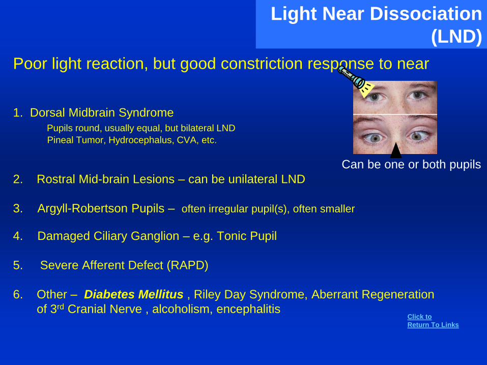
Light Near Dissociation
(LND)
Poor light reaction, but good constriction response to near
1. Dorsal Midbrain Syndrome
Pupils round, usually equal, but bilateral LND
Pineal Tumor, Hydrocephalus, CVA, etc.
2. Rostral Mid-brain Lesions – can be unilateral LND
3. Argyll-Robertson Pupils – often irregular pupil(s), often smaller
4. Damaged Ciliary Ganglion – e.g. Tonic Pupil
5. Severe Afferent Defect (RAPD)
6. Other – Diabetes Mellitus , Riley Day Syndrome, Aberrant Regeneration
of 3rd Cranial Nerve , alcoholism, encephalitis
Can be one or both pupils
Click to
Return To Links
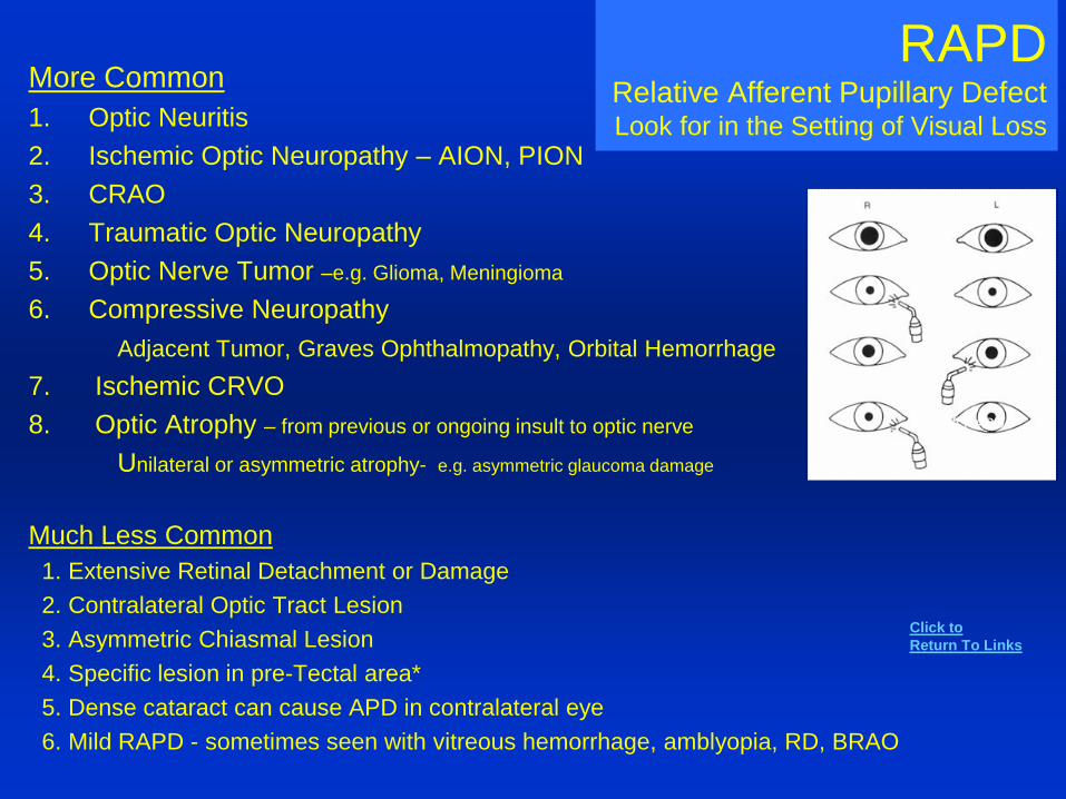
RAPDRelative Afferent Pupillary DefectLook for in the Setting of Visual Loss
More Common
1. Optic Neuritis
2. Ischemic Optic Neuropathy – AION, PION
3. CRAO
4. Traumatic Optic Neuropathy
5. Optic Nerve Tumor –e.g. Glioma, Meningioma
6. Compressive Neuropathy
Adjacent Tumor, Graves Ophthalmopathy, Orbital Hemorrhage
7. Ischemic CRVO
8. Optic Atrophy – from previous or ongoing insult to optic nerve
Unilateral or asymmetric atrophy- e.g. asymmetric glaucoma damage
Much Less Common
1. Extensive Retinal Detachment or Damage
2. Contralateral Optic Tract Lesion
3. Asymmetric Chiasmal Lesion
4. Specific lesion in pre-Tectal area*
5. Dense cataract can cause APD in contralateral eye
6. Mild RAPD - sometimes seen with vitreous hemorrhage, amblyopia, RD, BRAO
Left RAPD
Click to
Return To Links
Bilateral Miosis or Mydriasis1. Bilateral MiosisSystemic Medications: e.g. Narcotics (e.g. Morphine),
Cholinesterase Inhibitors (Snake venom, biochemical warfare, pesticides, MG treatments)
– early effects excessive salivation and tearing
Topical Medications: e.g. Pilocarpine
Pontine Lesions – pinpoint pupils
Damage to Sympathetic Pathways in Hypothalamus
Diencephalon Lesions – small but reactive
Metabolic or Medical Encephalopathy / Coma – small pupils but still reactive
2. Bilateral MydriasisSystemic Medications: anticholinergic (atropine)
sympathomimetics (amphetamines, cocaine, etc.)
Topical Medications or Natural Products – atropine, cyclopentolate, Jimson Weed pollen
Familial Bilateral Congenital Mydriasis
Tectal and Midbrain Lesions – inflammatory , neoplastic
Severe Anoxia of Brain (e.g. cardiac arrest) – sympathetic effect
Can be seen in Coma from: Alcoholism, Uremia, Epilepsy, Meningitis, Apoplexy
There are many other causes such as Intraocular Inflammation, Trauma and Surgery, Congenital and Developmental defects, and Lesions that cause disrupted Sympathetic or Parasympathetic input - but usually these do not produce symmetrically bilaterally large or small fixed pupils.
*note some
confusion and
possible variations
of pupils in
medical coma.
Click to
Return To Links

Anterior Segment and IOP
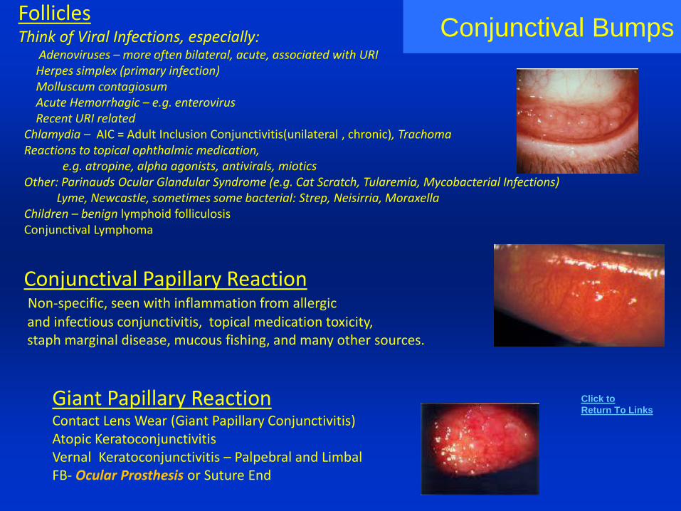
Conjunctival BumpsFolliclesThink of Viral Infections, especially:
Adenoviruses – more often bilateral, acute, associated with URIHerpes simplex (primary infection)Molluscum contagiosumAcute Hemorrhagic – e.g. enterovirusRecent URI related
Chlamydia – AIC = Adult Inclusion Conjunctivitis(unilateral , chronic), TrachomaReactions to topical ophthalmic medication,
e.g. atropine, alpha agonists, antivirals, mioticsOther: Parinauds Ocular Glandular Syndrome (e.g. Cat Scratch, Tularemia, Mycobacterial Infections)
Lyme, Newcastle, sometimes some bacterial: Strep, Neisirria, MoraxellaChildren – benign lymphoid folliculosisConjunctival Lymphoma
Conjunctival Papillary ReactionNon-specific, seen with inflammation from allergicand infectious conjunctivitis, topical medication toxicity,staph marginal disease, mucous fishing, and many other sources.
Giant Papillary ReactionContact Lens Wear (Giant Papillary Conjunctivitis)Atopic KeratoconjunctivitisVernal Keratoconjunctivitis – Palpebral and LimbalFB- Ocular Prosthesis or Suture End
Click to
Return To Links
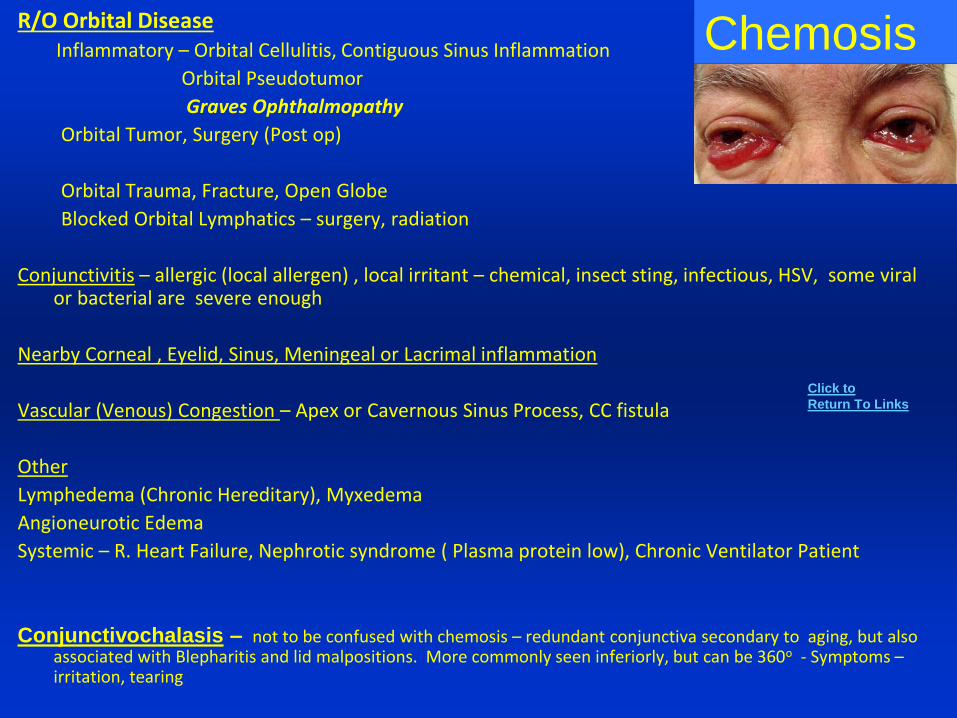
ChemosisR/O Orbital DiseaseInflammatory – Orbital Cellulitis, Contiguous Sinus Inflammation
Orbital Pseudotumor
Graves Ophthalmopathy
Orbital Tumor, Surgery (Post op)
Orbital Trauma, Fracture, Open Globe
Blocked Orbital Lymphatics – surgery, radiation
Conjunctivitis – allergic (local allergen) , local irritant – chemical, insect sting, infectious, HSV, some viral or bacterial are severe enough
Nearby Corneal , Eyelid, Sinus, Meningeal or Lacrimal inflammation
Vascular (Venous) Congestion – Apex or Cavernous Sinus Process, CC fistula
Other
Lymphedema (Chronic Hereditary), Myxedema
Angioneurotic Edema
Systemic – R. Heart Failure, Nephrotic syndrome ( Plasma protein low), Chronic Ventilator Patient
Conjunctivochalasis – not to be confused with chemosis – redundant conjunctiva secondary to aging, but also associated with Blepharitis and lid malpositions. More commonly seen inferiorly, but can be 360o - Symptoms –irritation, tearing
Click to
Return To Links
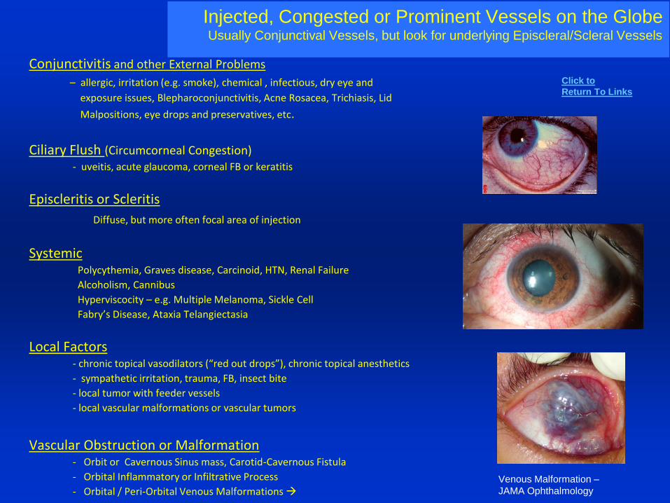
Conjunctivitis and other External Problems
– allergic, irritation (e.g. smoke), chemical , infectious, dry eye and
exposure issues, Blepharoconjunctivitis, Acne Rosacea, Trichiasis, Lid
Malpositions, eye drops and preservatives, etc.
Ciliary Flush (Circumcorneal Congestion) - uveitis, acute glaucoma, corneal FB or keratitis
Episcleritis or Scleritis
Diffuse, but more often focal area of injection
SystemicPolycythemia, Graves disease, Carcinoid, HTN, Renal Failure
Alcoholism, Cannibus
Hyperviscocity – e.g. Multiple Melanoma, Sickle Cell
Fabry’s Disease, Ataxia Telangiectasia
Local Factors- chronic topical vasodilators (“red out drops”), chronic topical anesthetics
- sympathetic irritation, trauma, FB, insect bite
- local tumor with feeder vessels
- local vascular malformations or vascular tumors
Vascular Obstruction or Malformation- Orbit or Cavernous Sinus mass, Carotid-Cavernous Fistula
- Orbital Inflammatory or Infiltrative Process
- Orbital / Peri-Orbital Venous Malformations
Injected, Congested or Prominent Vessels on the GlobeUsually Conjunctival Vessels, but look for underlying Episcleral/Scleral Vessels
Venous Malformation –
JAMA Ophthalmology
Click to
Return To Links
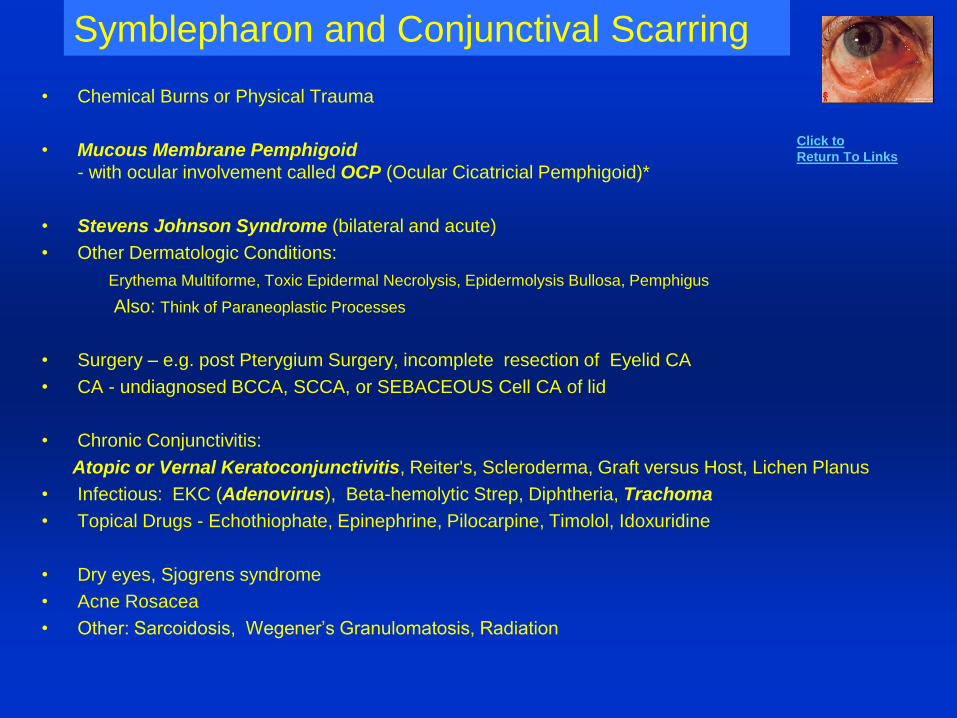
Symblepharon and Conjunctival Scarring
• Chemical Burns or Physical Trauma
• Mucous Membrane Pemphigoid
- with ocular involvement called OCP (Ocular Cicatricial Pemphigoid)*
• Stevens Johnson Syndrome (bilateral and acute)
• Other Dermatologic Conditions:
Erythema Multiforme, Toxic Epidermal Necrolysis, Epidermolysis Bullosa, Pemphigus
Also: Think of Paraneoplastic Processes
• Surgery – e.g. post Pterygium Surgery, incomplete resection of Eyelid CA
• CA - undiagnosed BCCA, SCCA, or SEBACEOUS Cell CA of lid
• Chronic Conjunctivitis:
Atopic or Vernal Keratoconjunctivitis, Reiter's, Scleroderma, Graft versus Host, Lichen Planus
• Infectious: EKC (Adenovirus), Beta-hemolytic Strep, Diphtheria, Trachoma
• Topical Drugs - Echothiophate, Epinephrine, Pilocarpine, Timolol, Idoxuridine
• Dry eyes, Sjogrens syndrome
• Acne Rosacea
• Other: Sarcoidosis, Wegener’s Granulomatosis, Radiation
Click to
Return To Links
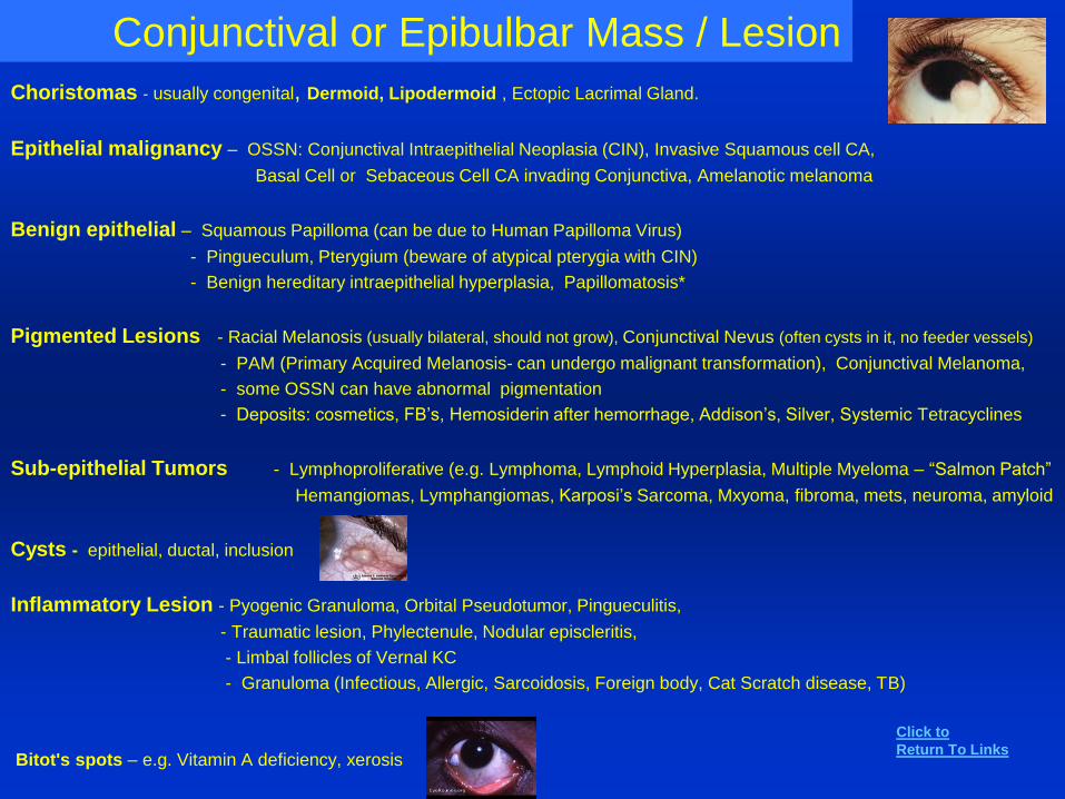
Conjunctival or Epibulbar Mass / Lesion
Choristomas - usually congenital, Dermoid, Lipodermoid , Ectopic Lacrimal Gland.
Epithelial malignancy – OSSN: Conjunctival Intraepithelial Neoplasia (CIN), Invasive Squamous cell CA,
Basal Cell or Sebaceous Cell CA invading Conjunctiva, Amelanotic melanoma
Benign epithelial – Squamous Papilloma (can be due to Human Papilloma Virus)
- Pingueculum, Pterygium (beware of atypical pterygia with CIN)
- Benign hereditary intraepithelial hyperplasia, Papillomatosis*
Pigmented Lesions - Racial Melanosis (usually bilateral, should not grow), Conjunctival Nevus (often cysts in it, no feeder vessels)
- PAM (Primary Acquired Melanosis- can undergo malignant transformation), Conjunctival Melanoma,
- some OSSN can have abnormal pigmentation
- Deposits: cosmetics, FB’s, Hemosiderin after hemorrhage, Addison’s, Silver, Systemic Tetracyclines
Sub-epithelial Tumors - Lymphoproliferative (e.g. Lymphoma, Lymphoid Hyperplasia, Multiple Myeloma – “Salmon Patch”
Hemangiomas, Lymphangiomas, Karposi’s Sarcoma, Mxyoma, fibroma, mets, neuroma, amyloid
Cysts - epithelial, ductal, inclusion
Inflammatory Lesion - Pyogenic Granuloma, Orbital Pseudotumor, Pingueculitis,
- Traumatic lesion, Phylectenule, Nodular episcleritis,
- Limbal follicles of Vernal KC
- Granuloma (Infectious, Allergic, Sarcoidosis, Foreign body, Cat Scratch disease, TB)
Bitot's spots – e.g. Vitamin A deficiency, xerosis
Don’t Confuse with Scleral Lesions (next list)
Click to
Return To Links

Spots on the “White of the Eye”
Scleral Lesions:
• Scleral Dellen – depression or dimple, loss of epithelium over defect in sclera. Seen after
surgery (e.g. Pterygium, EOM surgery), Cautery Trauma, medication toxicity, autoimmune disorders
• Scleral thinning - e.g. aging- typically in front of rectus muscle insertions,
areas of dehydration, previous surgery
• Staphyloma – thinning, bulge in sclera
• Axenfeld Loops
• Medication Deposits
• Congenital Melanosis Oculi (CMO) increased melanocytes in episclera, sclera or
uvea. Bluish/gray patches suggest increased underlying uveal pigment. Can undergo malignant transformation later in life.
• Nevus of Ota (CMO +lid nevus)
• Scleral discoloration – also periocular tissues, nails, teeth, auricles - from
tetracyclines (e.g. minocycline)
Click to
Return To Links
Scleral Thinning:
- Area of dehydration
- Dell (depression with loss of overlying epithelium)
- Staphyloma
- Age Related Changes – e.g. anterior to EOM insertions
- Episcleritis, Scleritis
Episcleritis
Mild pain, diffuse or nodular, rare uveitis or peripheral keratitis or thinning
Causes include:
Idiopathic
Related to autoimmune disease
Associated with toxic effects of external diseases such as staph Blepharitis –local effect –Episcleritis in region of lid disease
Scleritis
Fire engine red, not blanching with Neo2.5%. – Possible uveitis and peripheral corneal effects
- Local or diffuse, Nodular, -- usually moderately painful, tender
- Posterior Scleritis – thickening seen on B-scan, moderate to severe pain
- Necrotizing Scleritis – extreme pain, vasculitis, thinning , perforation
Consider:
- Post-Surgical Necrotizing/Infectious Scleritis – e.g. pterygium surgery
(e.g. pseudomonas, actinomyces, fungal, mycobacterial, gram + cocci)
- Idiopathic, but…
- 50% of time associated with a systemic disease, see list
Scleral Thinning, Episcleritis and Scleritis
Diseases associated with
Episcleritis and Scleritis:
Rheumatoid Arthritis
Herpes Viruses (e.g. Simplex, Zoster)
Other Infectious: Staph, Strep, Syphilis,
Pseudomonas, Mycobacterium,
Acanthamoeba
Relapsing Polychondritis (check ears, nose)
Orbital Pseudotumor
Wegener’s Granulomatosis
Sarcoidosis
Medications: Fosamax, Aredia
Lupus (SLE)
Inflammatory Bowel Disease
Spondyloarthropathies
Possible Testing:
ANCA Panel ,
Rheumatoid Factor, FTAbS, SSA/SSB,
ESR(GCA), PPD, ANA
Urinalysis, CXR, ACE, HLA testing
Scleral Biopsy or Trial of Oral Acyclovir
Click to
Return To Links
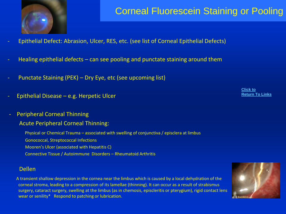
Corneal Fluorescein Staining or Pooling
- Epithelial Defect: Abrasion, Ulcer, RES, etc. (see list of Corneal Epithelial Defects)
- Healing epithelial defects – can see pooling and punctate staining around them
- Punctate Staining (PEK) – Dry Eye, etc (see upcoming list)
- Epithelial Disease – e.g. Herpetic Ulcer
- Peripheral Corneal Thinning
Acute Peripheral Corneal Thinning:
Physical or Chemical Trauma – associated with swelling of conjunctiva / episclera at limbus
Gonococcal, Streptococcal Infections
Mooren’s Ulcer (associated with Hepatitis C)
Connective Tissue / Autoimmune Disorders – Rheumatoid Arthritis
Dellen
A transient shallow depression in the cornea near the limbus which is caused by a local dehydration of the
corneal stroma, leading to a compression of its lamellae (thinning). It can occur as a result of strabismus surgery, cataract surgery, swelling at the limbus (as in chemosis, episcleritis or pterygium), rigid contact lens wear or senility* Respond to patching or lubrication.
Click to
Return To Links
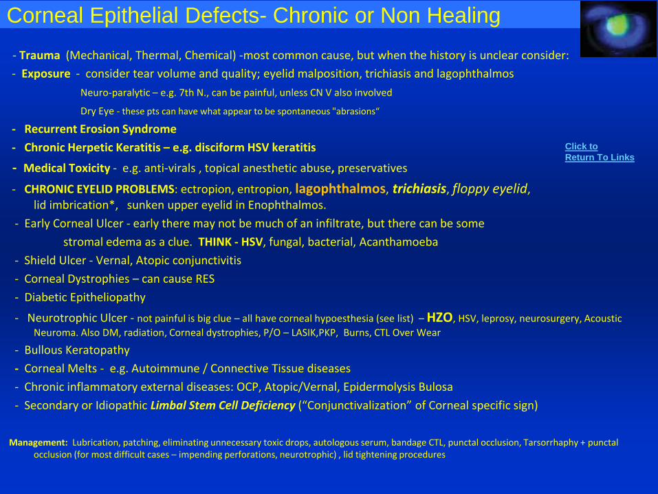
Corneal Epithelial Defects- Chronic or Non Healing
- Trauma (Mechanical, Thermal, Chemical) -most common cause, but when the history is unclear consider:
- Exposure - consider tear volume and quality; eyelid malposition, trichiasis and lagophthalmos
Neuro-paralytic – e.g. 7th N., can be painful, unless CN V also involved
Dry Eye - these pts can have what appear to be spontaneous "abrasions“
- Recurrent Erosion Syndrome
- Chronic Herpetic Keratitis – e.g. disciform HSV keratitis
- Medical Toxicity - e.g. anti-virals , topical anesthetic abuse, preservatives
- CHRONIC EYELID PROBLEMS: ectropion, entropion, lagophthalmos, trichiasis, floppy eyelid, lid imbrication*, sunken upper eyelid in Enophthalmos.
- Early Corneal Ulcer - early there may not be much of an infiltrate, but there can be some
stromal edema as a clue. THINK - HSV, fungal, bacterial, Acanthamoeba
- Shield Ulcer - Vernal, Atopic conjunctivitis
- Corneal Dystrophies – can cause RES
- Diabetic Epitheliopathy
- Neurotrophic Ulcer - not painful is big clue – all have corneal hypoesthesia (see list) – HZO, HSV, leprosy, neurosurgery, Acoustic
Neuroma. Also DM, radiation, Corneal dystrophies, P/O – LASIK,PKP, Burns, CTL Over Wear
- Bullous Keratopathy
- Corneal Melts - e.g. Autoimmune / Connective Tissue diseases
- Chronic inflammatory external diseases: OCP, Atopic/Vernal, Epidermolysis Bulosa
- Secondary or Idiopathic Limbal Stem Cell Deficiency (“Conjunctivalization” of Corneal specific sign)
Management: Lubrication, patching, eliminating unnecessary toxic drops, autologous serum, bandage CTL, punctal occlusion, Tarsorrhaphy + punctal occlusion (for most difficult cases – impending perforations, neurotrophic) , lid tightening procedures
Click to
Return To Links
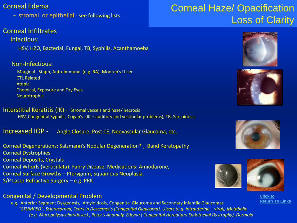
Corneal Haze/ Opacification
Loss of Clarity
Corneal Edema– stromal or epithelial - see following lists
Corneal Infiltrates Infectious:
HSV, HZO, Bacterial, Fungal, TB, Syphilis, Acanthamoeba
Non-Infectious:Marginal –Staph, Auto-immune (e.g. RA), Mooren’s UlcerCTL RelatedAtopicChemical, Exposure and Dry EyesNeurotrophic
Interstitial Keratitis (IK) - Stromal vessels and haze/ necrosis
HSV, Congenital Syphilis, Cogan’s (IK + auditory and vestibular problems), TB, Sarcoidosis
Increased IOP - Angle Closure, Post CE, Neovascular Glaucoma, etc.
Corneal Degenerations: Salzmann’s Nodular Degeneration* , Band KeratopathyCorneal DystrophiesCorneal Deposits, CrystalsCorneal Whorls (Verticillata): Fabry Disease, Medications: Amiodarone, Corneal Surface Growths – Pterygium, Squamous Neoplasia, S/P Laser Refractive Surgery – e.g. PRK
Congenital / Developmental Probleme.g. Anterior Segment Dysgenesis, Amyloidosis, Congenital Glaucoma and Secondary Infantile Glaucomas
“STUMPED”: Sclerocornea, Tears in Descemet’s (Congenital Glaucoma), Ulcers (e.g. intrauterine – viral), Metabolic(e.g. Mucopolysaccharidoses) , Peter’s Anomaly, Edema ( Congenital Hereditary Endothelial Dystrophy), Dermoid
Click to
Return To Links
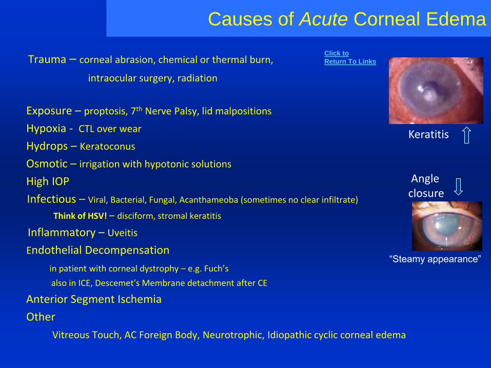
Causes of Acute Corneal Edema
Trauma – corneal abrasion, chemical or thermal burn,
intraocular surgery, radiation
Exposure – proptosis, 7th Nerve Palsy, lid malpositions
Hypoxia - CTL over wear
Hydrops – Keratoconus
Osmotic – irrigation with hypotonic solutions
High IOP
Infectious – Viral, Bacterial, Fungal, Acanthameoba (sometimes no clear infiltrate)
Think of HSV! – disciform, stromal keratitis
Inflammatory – Uveitis
Endothelial Decompensation
in patient with corneal dystrophy – e.g. Fuch’s
also in ICE, Descemet's Membrane detachment after CE
Anterior Segment Ischemia
Other
Vitreous Touch, AC Foreign Body, Neurotrophic, Idiopathic cyclic corneal edema
Keratitis
Angle closure
“Steamy appearance”
Click to
Return To Links
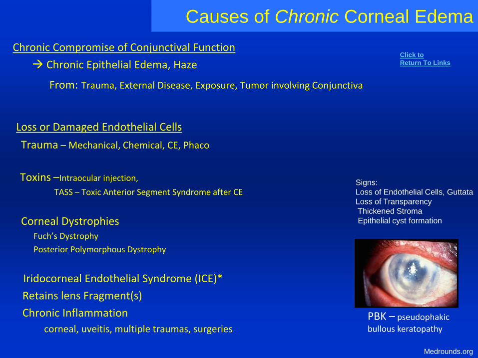
Causes of Chronic Corneal Edema
Chronic Compromise of Conjunctival Function
Chronic Epithelial Edema, Haze
From: Trauma, External Disease, Exposure, Tumor involving Conjunctiva
Loss or Damaged Endothelial Cells
Trauma – Mechanical, Chemical, CE, Phaco
Toxins –Intraocular injection,
TASS – Toxic Anterior Segment Syndrome after CE
Corneal Dystrophies Fuch’s Dystrophy
Posterior Polymorphous Dystrophy
Iridocorneal Endothelial Syndrome (ICE)*
Retains lens Fragment(s)
Chronic Inflammation
corneal, uveitis, multiple traumas, surgeriesPBK – pseudophakic
bullous keratopathy
Signs:
Loss of Endothelial Cells, Guttata
Loss of Transparency
Thickened Stroma
Epithelial cyst formation
Medrounds.org
Click to
Return To Links
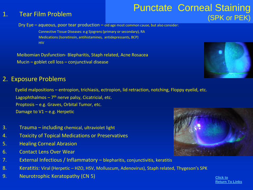
Punctate Corneal Staining(SPK or PEK)
1. Tear Film Problem
Dry Eye – aqueous, poor tear production – old age most common cause, but also consider:
Connective Tissue Diseases: e.g Sjogrens (primary or secondary), RA
Medications (Isoretinoin, antihistamines, antidepressants, BCP)
HIV
Meibomian Dysfunction- Blepharitis, Staph related, Acne Rosacea
Mucin – goblet cell loss – conjunctival disease
2. Exposure Problems
Eyelid malpositions – entropion, trichiasis, ectropion, lid retraction, notching, Floppy eyelid, etc.
Lagophthalmos – 7th nerve palsy, Cicatricial, etc.
Proptosis – e.g. Graves, Orbital Tumor, etc.
Damage to V1 – e.g. Herpetic
3. Trauma – including chemical, ultraviolet light
4. Toxicity of Topical Medications or Preservatives
5. Healing Corneal Abrasion
6. Contact Lens Over Wear
7. External Infectious / Inflammatory – blepharitis, conjunctivitis, keratitis
8. Keratitis: Viral (Herpetic – HZO, HSV, Molluscum, Adenovirus), Staph related, Thygeson’s SPK
9. Neurotrophic Keratopathy (CN 5) Click to
Return To Links
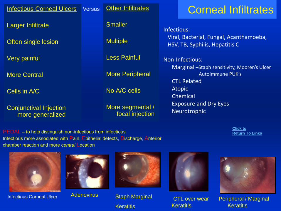
Corneal InfiltratesInfectious Corneal Ulcers
Larger Infiltrate
Often single lesion
Very painful
More Central
Cells in A/C
Conjunctival Injection more generalized
Other Infiltrates
Smaller
Multiple
Less Painful
More Peripheral
No A/C cells
More segmental / focal injection
CTL over wear Peripheral / Marginal
Keratitis Keratitis
Staph Marginal
Keratitis
Adenovirus
Infectious:Viral, Bacterial, Fungal, Acanthamoeba,HSV, TB, Syphilis, Hepatitis C
Non-Infectious:Marginal –Staph sensitivity, Mooren’s Ulcer
Autoimmune PUK’s
CTL RelatedAtopicChemicalExposure and Dry EyesNeurotrophic
PEDAL – to help distinguish non-infectious from infectious
Infectious more associated with Pain, Epithelial defects, Discharge, Anterior
chamber reaction and more central Location
Infectious Corneal Ulcer
Click to
Return To Links
Versus
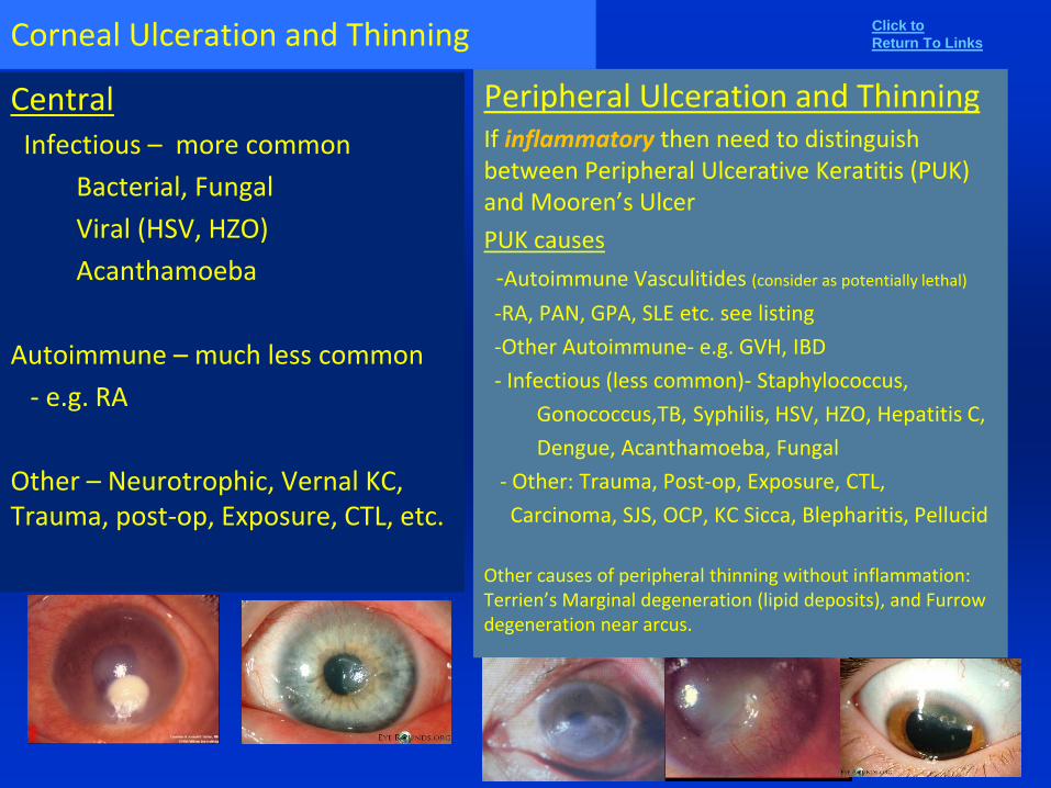
Corneal Ulceration and Thinning
CentralInfectious – more common
Bacterial, Fungal
Viral (HSV, HZO)
Acanthamoeba
Autoimmune – much less common
- e.g. RA
Other – Neurotrophic, Vernal KC, Trauma, post-op, Exposure, CTL, etc.
Peripheral Ulceration and ThinningIf inflammatory then need to distinguish between Peripheral Ulcerative Keratitis (PUK) and Mooren’s Ulcer
PUK causes
-Autoimmune Vasculitides (consider as potentially lethal)
-RA, PAN, GPA, SLE etc. see listing
-Other Autoimmune- e.g. GVH, IBD
- Infectious (less common)- Staphylococcus,
Gonococcus,TB, Syphilis, HSV, HZO, Hepatitis C,
Dengue, Acanthamoeba, Fungal
- Other: Trauma, Post-op, Exposure, CTL,
Carcinoma, SJS, OCP, KC Sicca, Blepharitis, Pellucid
Other causes of peripheral thinning without inflammation: Terrien’s Marginal degeneration (lipid deposits), and Furrow degeneration near arcus.
Click to
Return To Links
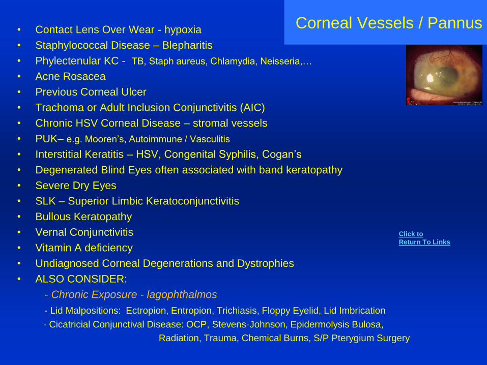
Corneal Vessels / Pannus• Contact Lens Over Wear - hypoxia
• Staphylococcal Disease – Blepharitis
• Phylectenular KC - TB, Staph aureus, Chlamydia, Neisseria,…
• Acne Rosacea
• Previous Corneal Ulcer
• Trachoma or Adult Inclusion Conjunctivitis (AIC)
• Chronic HSV Corneal Disease – stromal vessels
• PUK– e.g. Mooren’s, Autoimmune / Vasculitis
• Interstitial Keratitis – HSV, Congenital Syphilis, Cogan’s
• Degenerated Blind Eyes often associated with band keratopathy
• Severe Dry Eyes
• SLK – Superior Limbic Keratoconjunctivitis
• Bullous Keratopathy
• Vernal Conjunctivitis
• Vitamin A deficiency
• Undiagnosed Corneal Degenerations and Dystrophies
• ALSO CONSIDER:
- Chronic Exposure - lagophthalmos
- Lid Malpositions: Ectropion, Entropion, Trichiasis, Floppy Eyelid, Lid Imbrication
- Cicatricial Conjunctival Disease: OCP, Stevens-Johnson, Epidermolysis Bulosa,
Radiation, Trauma, Chemical Burns, S/P Pterygium Surgery
Click to
Return To Links
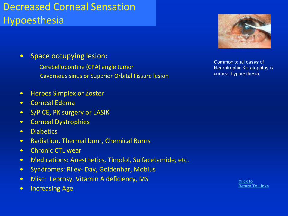
Decreased Corneal SensationHypoesthesia
• Space occupying lesion:
Cerebellopontine (CPA) angle tumor
Cavernous sinus or Superior Orbital Fissure lesion
• Herpes Simplex or Zoster
• Corneal Edema
• S/P CE, PK surgery or LASIK
• Corneal Dystrophies
• Diabetics
• Radiation, Thermal burn, Chemical Burns
• Chronic CTL wear
• Medications: Anesthetics, Timolol, Sulfacetamide, etc.
• Syndromes: Riley- Day, Goldenhar, Mobius
• Misc: Leprosy, Vitamin A deficiency, MS
• Increasing Age
Common to all cases of
Neurotrophic Keratopathy is
corneal hypoesthesia
Click to
Return To Links
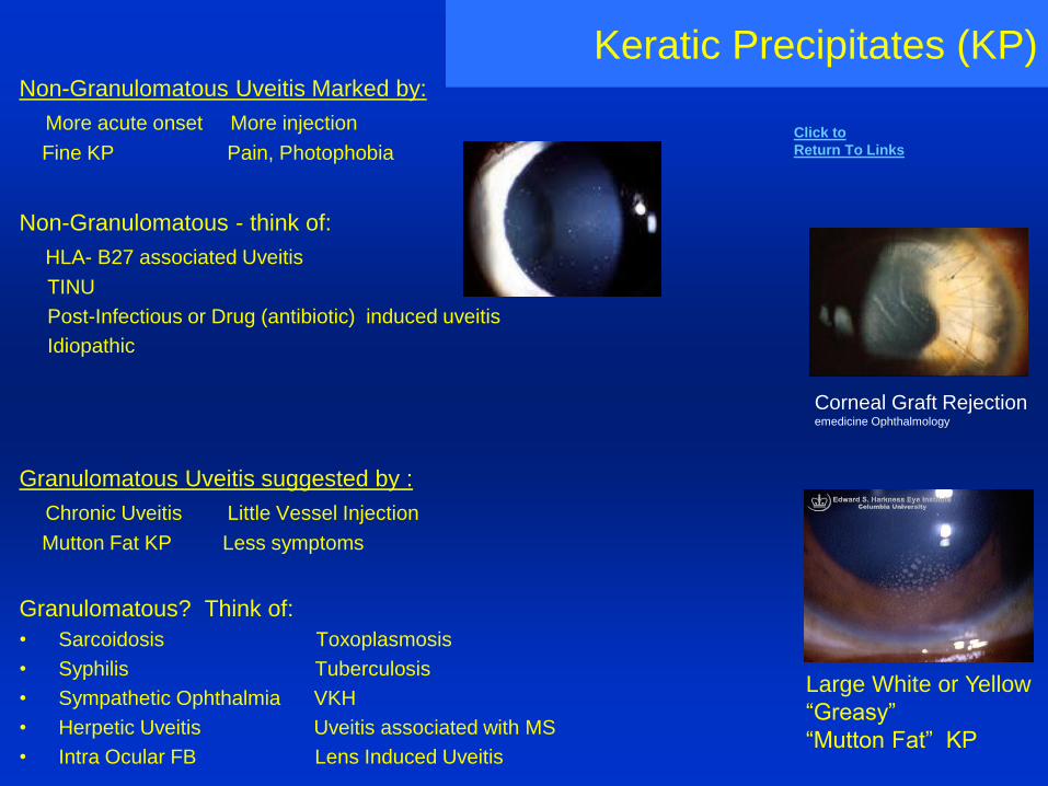
Keratic Precipitates (KP)Non-Granulomatous Uveitis Marked by:
More acute onset More injection
Fine KP Pain, Photophobia
Non-Granulomatous - think of:
HLA- B27 associated Uveitis
TINU
Post-Infectious or Drug (antibiotic) induced uveitis
Idiopathic
Granulomatous Uveitis suggested by :
Chronic Uveitis Little Vessel Injection
Mutton Fat KP Less symptoms
Granulomatous? Think of:
• Sarcoidosis Toxoplasmosis
• Syphilis Tuberculosis
• Sympathetic Ophthalmia VKH
• Herpetic Uveitis Uveitis associated with MS
• Intra Ocular FB Lens Induced Uveitis
Large White or Yellow
“Greasy”
“Mutton Fat” KP
Corneal Graft Rejectionemedicine Ophthalmology
Click to
Return To Links
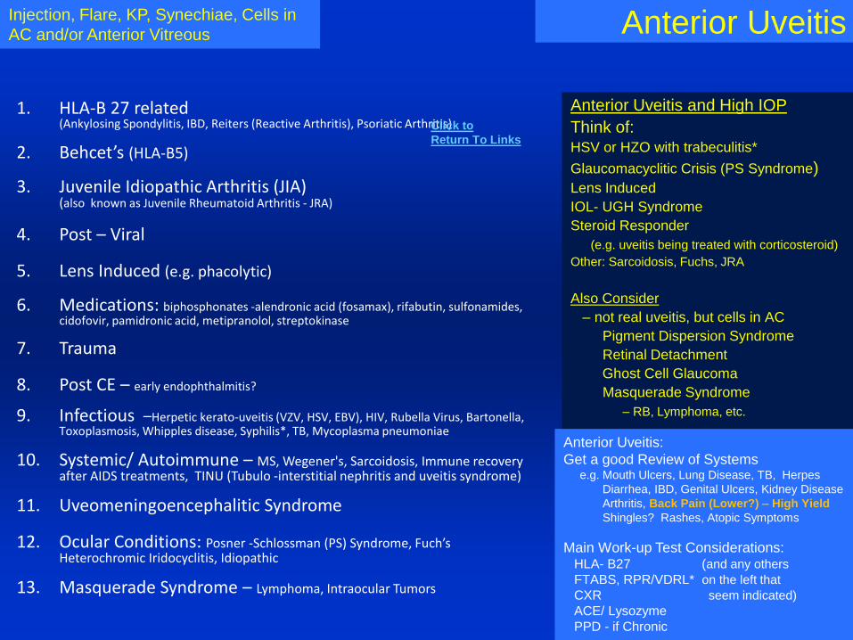
Anterior Uveitis
1. HLA-B 27 related (Ankylosing Spondylitis, IBD, Reiters (Reactive Arthritis), Psoriatic Arthritis)
2. Behcet’s (HLA-B5)
3. Juvenile Idiopathic Arthritis (JIA) (also known as Juvenile Rheumatoid Arthritis - JRA)
4. Post – Viral
5. Lens Induced (e.g. phacolytic)
6. Medications: biphosphonates -alendronic acid (fosamax), rifabutin, sulfonamides, cidofovir, pamidronic acid, metipranolol, streptokinase
7. Trauma
8. Post CE – early endophthalmitis?
9. Infectious –Herpetic kerato-uveitis (VZV, HSV, EBV), HIV, Rubella Virus, Bartonella, Toxoplasmosis, Whipples disease, Syphilis*, TB, Mycoplasma pneumoniae
10. Systemic/ Autoimmune – MS, Wegener's, Sarcoidosis, Immune recovery after AIDS treatments, TINU (Tubulo -interstitial nephritis and uveitis syndrome)
11. Uveomeningoencephalitic Syndrome
12. Ocular Conditions: Posner -Schlossman (PS) Syndrome, Fuch’s Heterochromic Iridocyclitis, Idiopathic
13. Masquerade Syndrome – Lymphoma, Intraocular Tumors
Injection, Flare, KP, Synechiae, Cells in
AC and/or Anterior Vitreous
Anterior Uveitis and High IOP
Think of:HSV or HZO with trabeculitis*
Glaucomacyclitic Crisis (PS Syndrome)Lens Induced
IOL- UGH Syndrome
Steroid Responder
(e.g. uveitis being treated with corticosteroid)
Other: Sarcoidosis, Fuchs, JRA
Also Consider
– not real uveitis, but cells in AC
Pigment Dispersion Syndrome
Retinal Detachment
Ghost Cell Glaucoma
Masquerade Syndrome
– RB, Lymphoma, etc.
Anterior Uveitis:
Get a good Review of Systemse.g. Mouth Ulcers, Lung Disease, TB, Herpes
Diarrhea, IBD, Genital Ulcers, Kidney Disease
Arthritis, Back Pain (Lower?) – High Yield
Shingles? Rashes, Atopic Symptoms
Main Work-up Test Considerations:HLA- B27 (and any others
FTABS, RPR/VDRL* on the left that
CXR seem indicated)
ACE/ Lysozyme
PPD - if Chronic
Click to
Return To Links
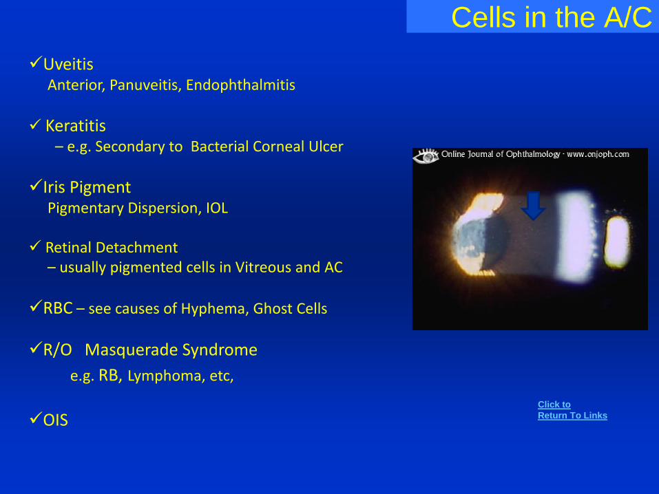
Cells in the A/C
UveitisAnterior, Panuveitis, Endophthalmitis
Keratitis– e.g. Secondary to Bacterial Corneal Ulcer
Iris PigmentPigmentary Dispersion, IOL
Retinal Detachment – usually pigmented cells in Vitreous and AC
RBC – see causes of Hyphema, Ghost Cells
R/O Masquerade Syndrome
e.g. RB, Lymphoma, etc,
OISClick to
Return To Links
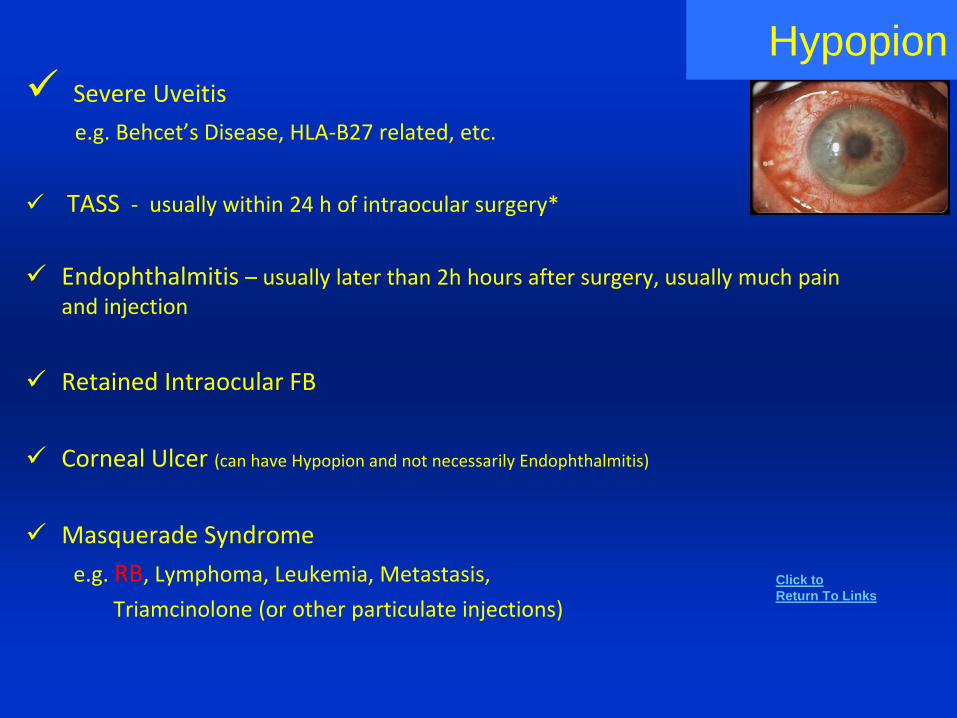
Hypopion Severe Uveitis
e.g. Behcet’s Disease, HLA-B27 related, etc.
TASS - usually within 24 h of intraocular surgery*
Endophthalmitis – usually later than 2h hours after surgery, usually much pain and injection
Retained Intraocular FB
Corneal Ulcer (can have Hypopion and not necessarily Endophthalmitis)
Masquerade Syndrome
e.g. RB, Lymphoma, Leukemia, Metastasis,
Triamcinolone (or other particulate injections)
Click to
Return To Links
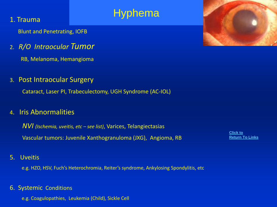
1. Trauma
Blunt and Penetrating, IOFB
2. R/O Intraocular Tumor
RB, Melanoma, Hemangioma
3. Post Intraocular Surgery
Cataract, Laser PI, Trabeculectomy, UGH Syndrome (AC-IOL)
4. Iris Abnormalities
NVI (Ischemia, uveitis, etc – see list), Varices, Telangiectasias
Vascular tumors: Juvenile Xanthogranuloma (JXG), Angioma, RB
5. Uveitis
e.g. HZO, HSV, Fuch’s Heterochromia, Reiter’s syndrome, Ankylosing Spondylitis, etc
6. Systemic Conditions
e.g. Coagulopathies, Leukemia (Child), Sickle Cell
Hyphema
Click to
Return To Links
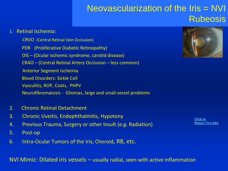
Neovascularization of the Iris = NVI
Rubeosis
1. Retinal Ischemia:
CRVO (Central Retinal Vein Occlusion)
PDR (Proliferative Diabetic Retinopathy)
OIS – (Ocular ischemic syndrome, carotid disease)
CRAO – (Central Retinal Artery Occlusion – less common)
Anterior Segment Ischemia
Blood Disorders: Sickle Cell
Vasculitis, ROP, Coats, PHPV
Neurofibromatosis - Gliomas, large and small vessel problems
2. Chronic Retinal Detachment
3. Chronic Uveitis, Endophthalmitis, Hypotony
4. Previous Trauma, Surgery or other Insult (e.g. Radiation)
5. Post-op
6. Intra-Ocular Tumors of the Iris, Choroid, RB, etc.
NVI Mimic: Dilated iris vessels – usually radial, seen with active inflammation
Click to
Return To Links
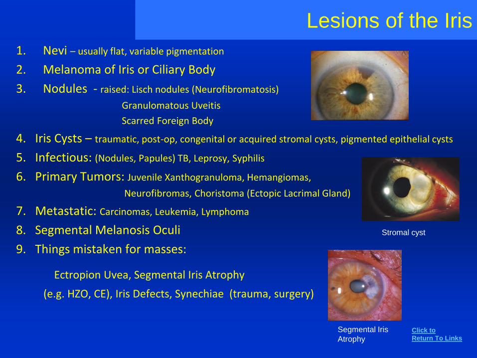
Lesions of the Iris
1. Nevi – usually flat, variable pigmentation
2. Melanoma of Iris or Ciliary Body
3. Nodules - raised: Lisch nodules (Neurofibromatosis)
Granulomatous Uveitis
Scarred Foreign Body
4. Iris Cysts – traumatic, post-op, congenital or acquired stromal cysts, pigmented epithelial cysts
5. Infectious: (Nodules, Papules) TB, Leprosy, Syphilis
6. Primary Tumors: Juvenile Xanthogranuloma, Hemangiomas,
Neurofibromas, Choristoma (Ectopic Lacrimal Gland)
7. Metastatic: Carcinomas, Leukemia, Lymphoma
8. Segmental Melanosis Oculi
9. Things mistaken for masses:
Ectropion Uvea, Segmental Iris Atrophy
(e.g. HZO, CE), Iris Defects, Synechiae (trauma, surgery)
Stromal cyst
Segmental Iris
AtrophyClick to
Return To Links
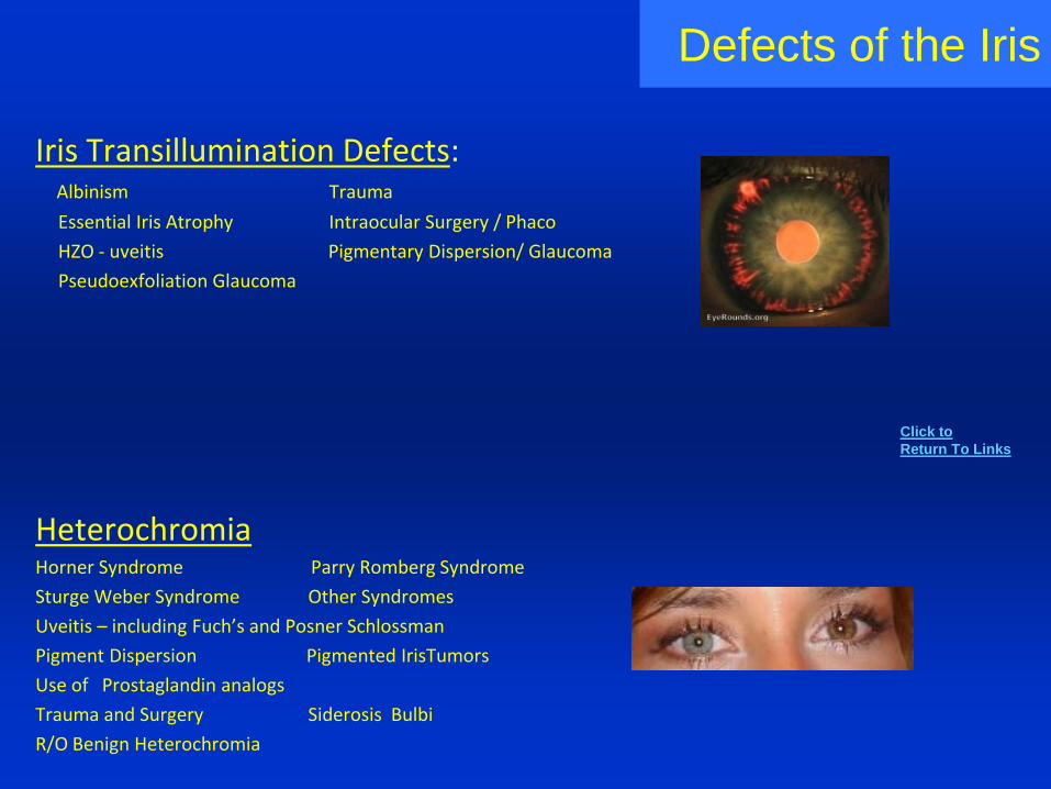
Defects of the Iris
Iris Transillumination Defects:Albinism Trauma
Essential Iris Atrophy Intraocular Surgery / Phaco
HZO - uveitis Pigmentary Dispersion/ Glaucoma
Pseudoexfoliation Glaucoma
HeterochromiaHorner Syndrome Parry Romberg Syndrome
Sturge Weber Syndrome Other Syndromes
Uveitis – including Fuch’s and Posner Schlossman
Pigment Dispersion Pigmented IrisTumors
Use of Prostaglandin analogs
Trauma and Surgery Siderosis Bulbi
R/O Benign Heterochromia
Click to
Return To Links
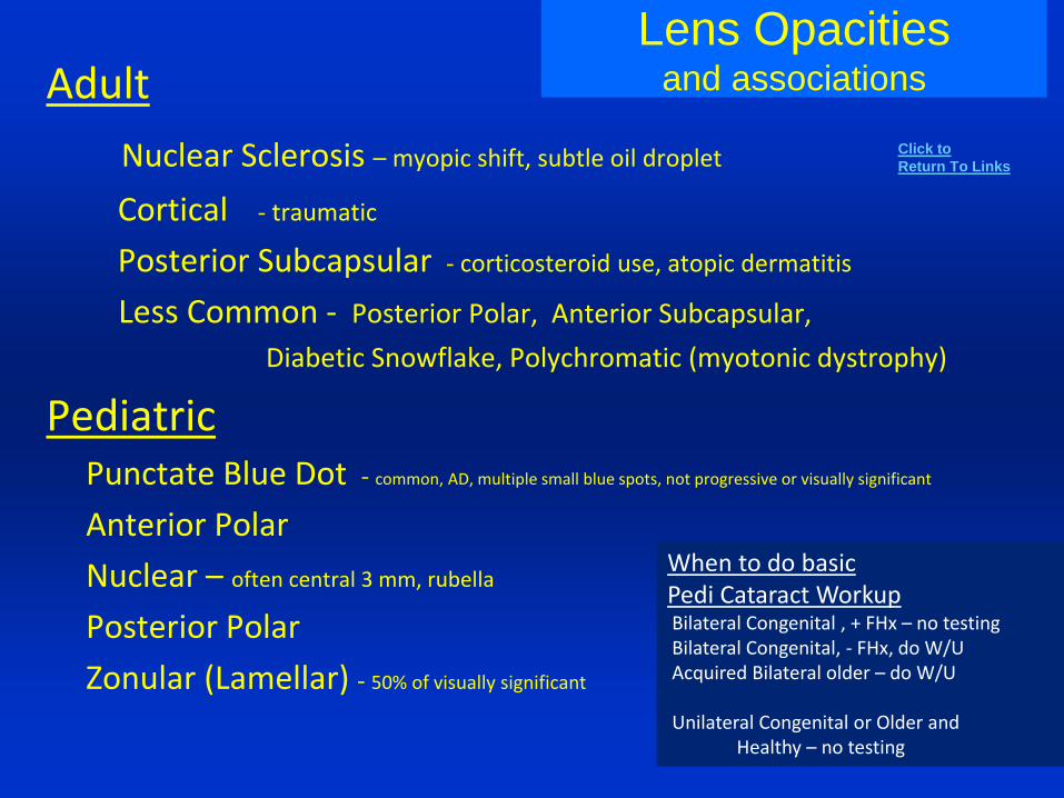
Lens Opacities and associationsAdult
Nuclear Sclerosis – myopic shift, subtle oil droplet
Cortical - traumatic
Posterior Subcapsular - corticosteroid use, atopic dermatitis
Less Common - Posterior Polar, Anterior Subcapsular,
Diabetic Snowflake, Polychromatic (myotonic dystrophy)
PediatricPunctate Blue Dot - common, AD, multiple small blue spots, not progressive or visually significant
Anterior Polar
Nuclear – often central 3 mm, rubella
Posterior Polar
Zonular (Lamellar) - 50% of visually significant
When to do basicPedi Cataract WorkupBilateral Congenital , + FHx – no testingBilateral Congenital, - FHx, do W/UAcquired Bilateral older – do W/U
Unilateral Congenital or Older and Healthy – no testing
Click to
Return To Links
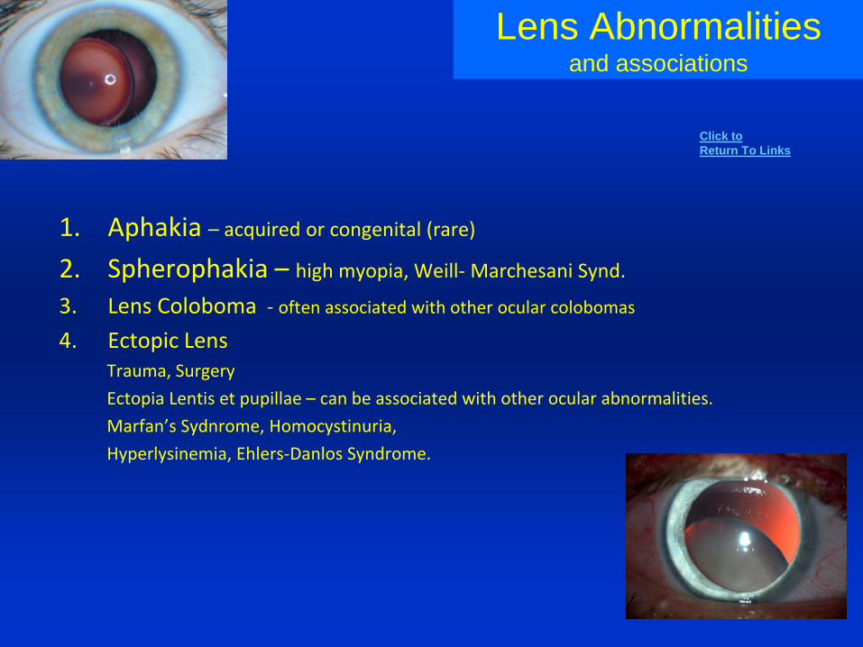
Lens Abnormalitiesand associations
1. Aphakia – acquired or congenital (rare)
2. Spherophakia – high myopia, Weill- Marchesani Synd.
3. Lens Coloboma - often associated with other ocular colobomas
4. Ectopic Lens Trauma, Surgery
Ectopia Lentis et pupillae – can be associated with other ocular abnormalities.
Marfan’s Sydnrome, Homocystinuria,
Hyperlysinemia, Ehlers-Danlos Syndrome.
Click to
Return To Links
High Intraocular Pressure
Any form of Glaucoma can result in High Pressures
but the More Common Causes Include:
• Acute Angle Closure Attack or other causes of Pupillary Block (See upcoming lists)
• Open Angle Glaucoma (POAG, secondary forms – see upcoming lists)
• Post Intraocular surgery – CE, PK, DMEK/DSEK*, Vitrectomy, Glaucoma
Acute: Viscoelastic, Air Bubble, Pupillary Block, Aqueous Misdirection
Later: Non-pupillary block Pseudophakic /Aphakic Glaucoma,
UGH Syndrome (IOL, especially AC IOLs)
• Uveitic Glaucoma
– HSV, HZO, Glaucomatocyclitic crisis (PS Syndrome) , UGH, Phacolytic,
Fuchs Heterochromic Uveitis, JIA, Sarcoidosis
• Steroid Induced High Pressure
• NVG – Neovascular Glaucoma
• Trauma Related – after Hyphema, Angle Recession, etc.
• Orbital Trauma- Hemorrhage, Compartment Syndrome
• Trabecular Outflow Obstruction – Lens Material, RBC, Tumor Cells
• Venous Outflow Problem – Orbital Apex Mass, CC Fistula
* DMEK / DSEK = Descemet’s Membrane/ Stripping Endothelial Keratoplasty
Click to
Return To Links
Acute IOP ElevationWith sudden onset of symptoms
(HA, N/V, Eye Pain, Decreased VA) Think of:
Acute ACGPost op –
Viscoelastic, AirAqueous Misdirection
Uveitic – Lens, HerpeticHyphemaOrbital Causes – Hemorrhage, CCF
Narrow Angles or Angle Closure
1. Primary Angle Closure Glaucoma (ACG)
2. Secondary Angle Closure or Narrow AnglesAnterior Pulling
Peripheral Anterior Synechiae - Inflammation, Trauma
Neovascularization of the Iris and angle
ICE Syndrome
Epithelial Downgrowth
Posterior Pushing
Posterior Synechiae (secluded pupil, iris Bombay)
Phacomorphic
Tumors
Choroidal Effusions – after surgery, PRP, CRVO
Malignant Glaucoma
Click to
Return To Links
Angle Closure Glaucomaand Pupillary Block
With Pupillary Block
Primary ACG
Secondary ACGPhacomorphicEctopia lentisPseudophakic (IOL) Aphakic (anterior vitreous face)Posterior Synechiae (with secluded pupil, iris bombe)
Without Pupillary Block
Neovascular GlaucomaICE – abnormal corneal endothelial growthIntra-ocular tumorsUveitis – Peripheral Anterior SynechiaePlateau Iris
Click to
Return To Links
Open Angle GlaucomaMechanisms
Primary - (POAG)* - usually symmetric
Secondary – more often unilateral
1. Clogging of TM - RBC, lens protein and macrophages,
pigment cells, tumor cells, viscoelastics
2. Alteration of TM - Inflammation (Trabeculitis),
Trauma (physical and chemical)
FB – siderosis, Corticosteroids
3. Blockage of drainage after the TM
- Sickle Cells in Schlemm’s Canal (even in Sickle trait)
- Increased episcleral pressure – Carotid Cavernous (CC) Fistula,
Orbital tumor /inflammation, venous backup
Click to
Return To Links
Open angle GlaucomaBy Disease Process
1. Primary Open Angle (POAG)
2. Secondary Open Angle (SPLIT mnemonic)
Substances: Steroids, Viscoelastics, Siderosis (Iron IOFB)
Pigmentary
Pseudoexfoliation
Lens – natural – phacolytic
Lens – IOL (UGH)
Inflammatory – Uveitic Glaucoma. e.g. PG syndrome, HZO
Increased episcleral pressure – e.g. CC fistula
Tumor – e.g. melanoma
Trauma – angle recession, Hyphema
Click to
Return To Links
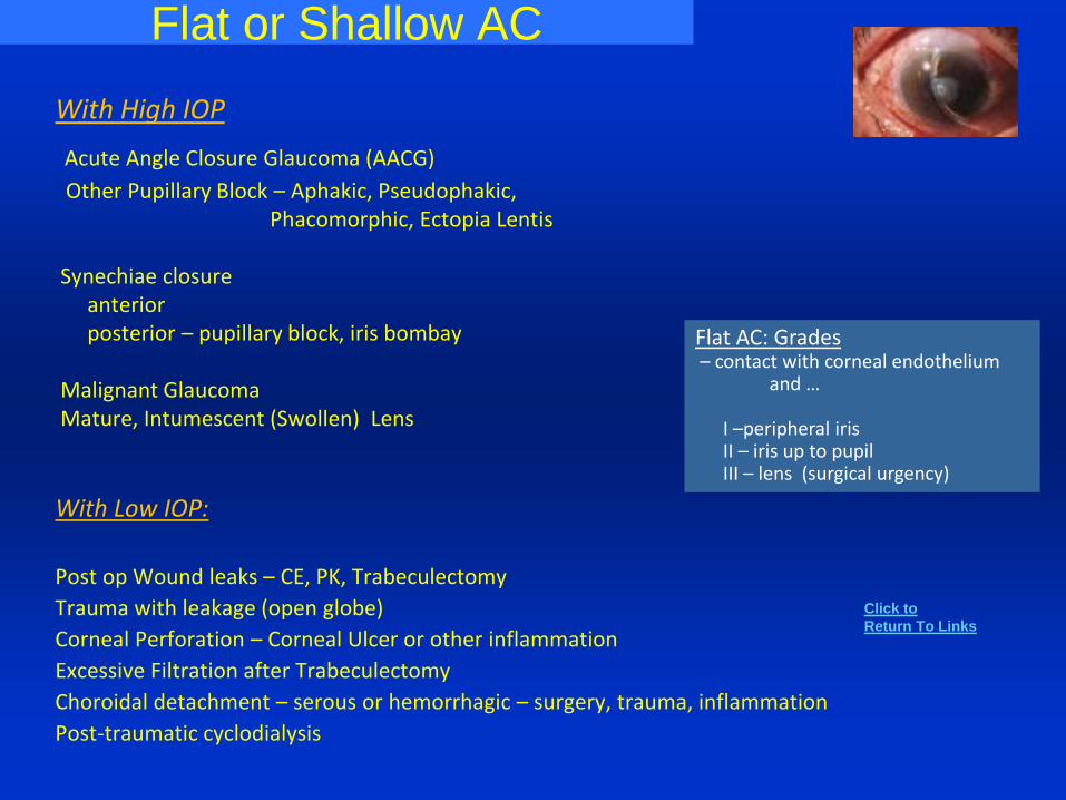
Flat or Shallow AC
With High IOP
Acute Angle Closure Glaucoma (AACG)
Other Pupillary Block – Aphakic, Pseudophakic,Phacomorphic, Ectopia Lentis
Synechiae closureanteriorposterior – pupillary block, iris bombay
Malignant GlaucomaMature, Intumescent (Swollen) Lens
With Low IOP:
Post op Wound leaks – CE, PK, Trabeculectomy
Trauma with leakage (open globe)
Corneal Perforation – Corneal Ulcer or other inflammation
Excessive Filtration after Trabeculectomy
Choroidal detachment – serous or hemorrhagic – surgery, trauma, inflammation
Post-traumatic cyclodialysis
Flat AC: Grades– contact with corneal endothelium
and …
I –peripheral irisII – iris up to pupilIII – lens (surgical urgency)
Click to
Return To Links
Hypotony (Low IOP)Causes and Associations
- Post-op (CE, Trabeculectomy, Scleral suture) Wound Leak
- Excessive Filtration after Trabeculectomy, Drainage Devices/Implants
- Choroidal Detachment
- Ciliary Body Detachment (e.g. traction), sometimes large Retinal Detachment
- Uveitis (Ciliary Body Shutdown)
- Trauma with leakage (open globe), Post-traumatic cyclodialysis
- Corneal Perforation – Corneal Ulcer or other inflammation
- Ocular Ischemia
- S/ P Cyclodestructive Procedures, Phthisis Bulbi
- Systemic: Hypertonicity, Acidosis, Uremia, Hyperglycemia,Osmotic Agents – like Mannitol
- Myotonic Dystrophy
- Congenital Anomalies: microphthalmos, aniridia , coloboma
Click to
Return To Links

Fundus – Vitreous, Retina and Disc
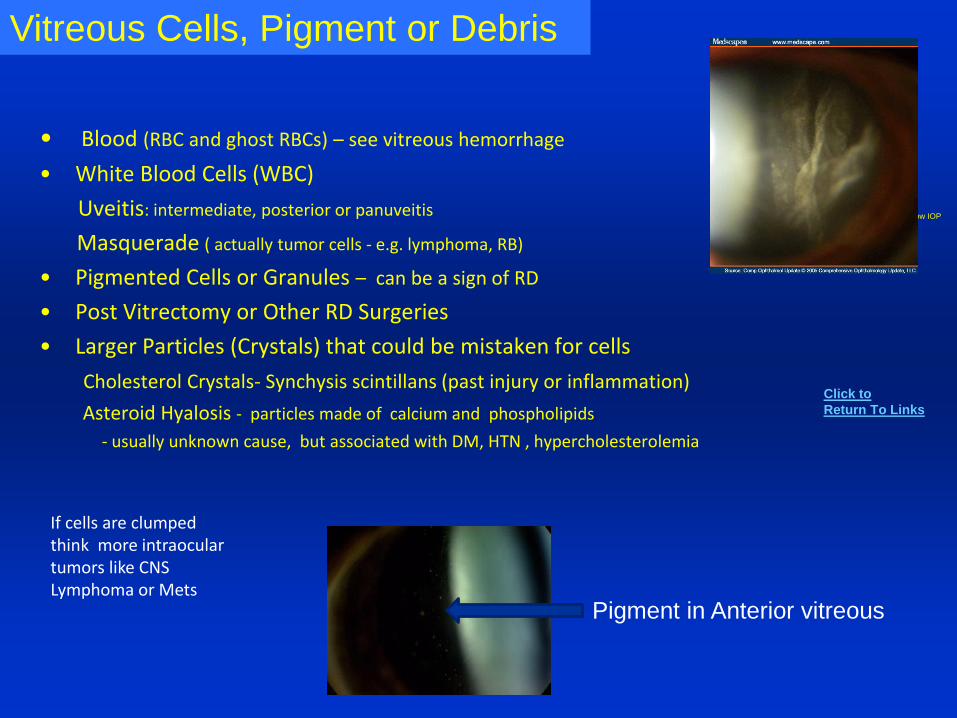
Hypotony
Causes and Associations Low IOP
• Blood (RBC and ghost RBCs) – see vitreous hemorrhage
• White Blood Cells (WBC)
Uveitis: intermediate, posterior or panuveitis
Masquerade ( actually tumor cells - e.g. lymphoma, RB)
• Pigmented Cells or Granules – can be a sign of RD
• Post Vitrectomy or Other RD Surgeries
• Larger Particles (Crystals) that could be mistaken for cells
Cholesterol Crystals- Synchysis scintillans (past injury or inflammation)
Asteroid Hyalosis - particles made of calcium and phospholipids
- usually unknown cause, but associated with DM, HTN , hypercholesterolemia
Vitreous Cells, Pigment or Debris
Pigment in Anterior vitreous
Click to
Return To Links
If cells are clumped think more intraocular tumors like CNS Lymphoma or Mets
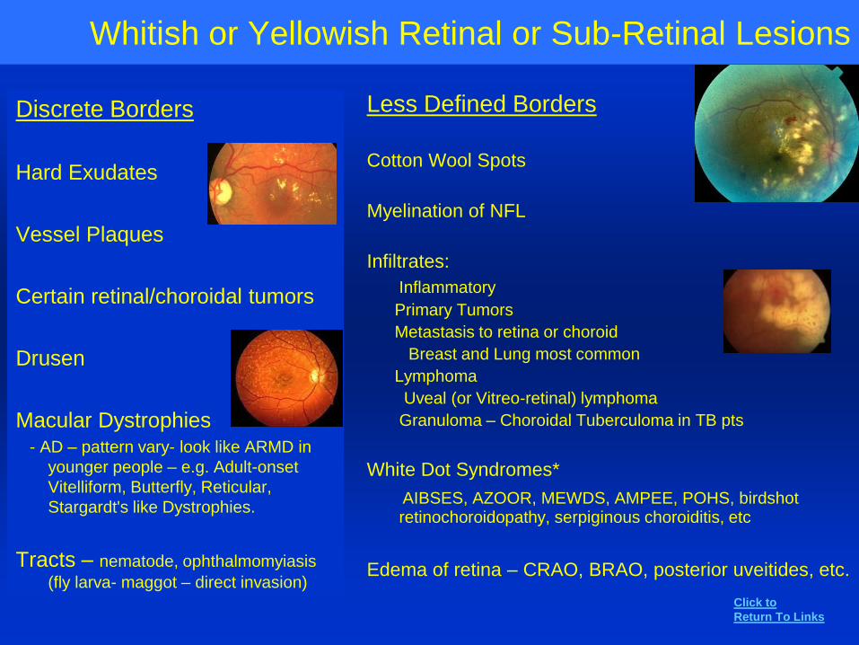
Whitish or Yellowish Retinal or Sub-Retinal Lesions
Discrete Borders
Hard Exudates
Vessel Plaques
Certain retinal/choroidal tumors
Drusen
Macular Dystrophies- AD – pattern vary- look like ARMD in
younger people – e.g. Adult-onset
Vitelliform, Butterfly, Reticular,
Stargardt's like Dystrophies.
Tracts – nematode, ophthalmomyiasis
(fly larva- maggot – direct invasion)
Less Defined Borders
Cotton Wool Spots
Myelination of NFL
Infiltrates:
Inflammatory
Primary Tumors
Metastasis to retina or choroid
Breast and Lung most common
Lymphoma
Uveal (or Vitreo-retinal) lymphoma
Granuloma – Choroidal Tuberculoma in TB pts
White Dot Syndromes*
AIBSES, AZOOR, MEWDS, AMPEE, POHS, birdshot retinochoroidopathy, serpiginous choroiditis, etc
Edema of retina – CRAO, BRAO, posterior uveitides, etc.
Click to
Return To Links
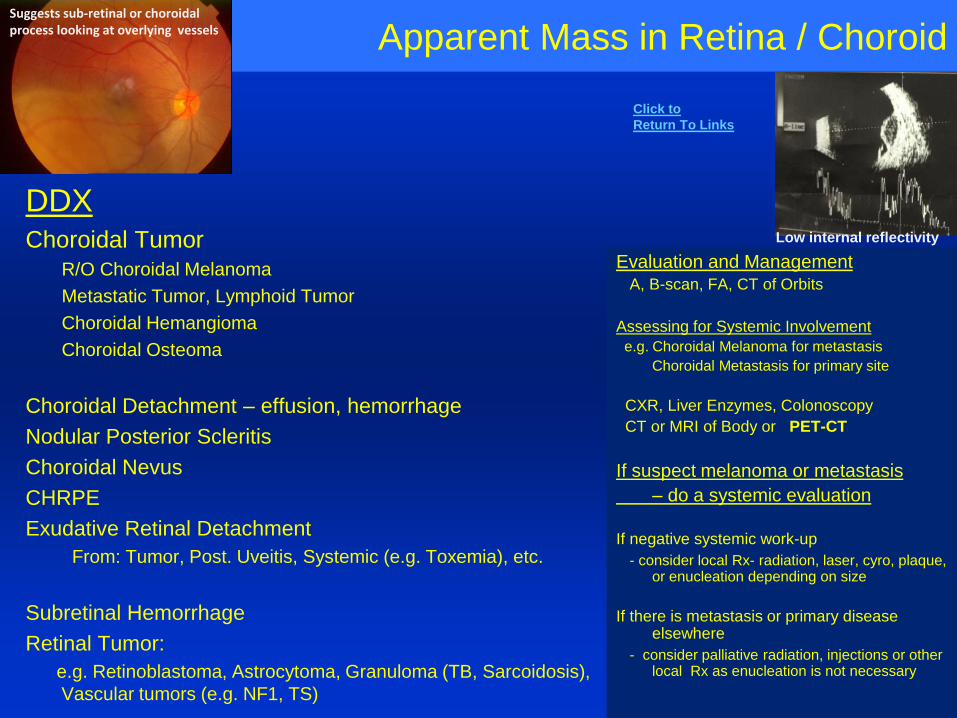
Apparent Mass in Retina / Choroid
DDXChoroidal Tumor
R/O Choroidal Melanoma
Metastatic Tumor, Lymphoid Tumor
Choroidal Hemangioma
Choroidal Osteoma
Choroidal Detachment – effusion, hemorrhage
Nodular Posterior Scleritis
Choroidal Nevus
CHRPE
Exudative Retinal Detachment
From: Tumor, Post. Uveitis, Systemic (e.g. Toxemia), etc.
Subretinal Hemorrhage
Retinal Tumor:
e.g. Retinoblastoma, Astrocytoma, Granuloma (TB, Sarcoidosis),
Vascular tumors (e.g. NF1, TS)
Evaluation and ManagementA, B-scan, FA, CT of Orbits
Assessing for Systemic Involvement
e.g. Choroidal Melanoma for metastasis
Choroidal Metastasis for primary site
CXR, Liver Enzymes, Colonoscopy
CT or MRI of Body or PET-CT
If suspect melanoma or metastasis
– do a systemic evaluation
If negative systemic work-up
- consider local Rx- radiation, laser, cyro, plaque, or enucleation depending on size
If there is metastasis or primary disease elsewhere
- consider palliative radiation, injections or other local Rx as enucleation is not necessary
Low internal reflectivity
Suggests sub-retinal or choroidal process looking at overlying vessels
Click to
Return To Links
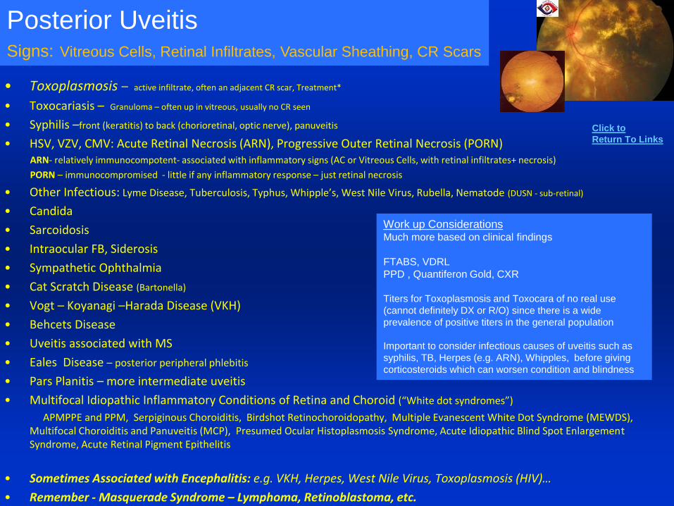
Hypotony
Causes and Associations
Low IOP• Toxoplasmosis – active infiltrate, often an adjacent CR scar, Treatment*
• Toxocariasis – Granuloma – often up in vitreous, usually no CR seen
• Syphilis –front (keratitis) to back (chorioretinal, optic nerve), panuveitis
• HSV, VZV, CMV: Acute Retinal Necrosis (ARN), Progressive Outer Retinal Necrosis (PORN)ARN- relatively immunocompotent- associated with inflammatory signs (AC or Vitreous Cells, with retinal infiltrates+ necrosis)
PORN – immunocompromised - little if any inflammatory response – just retinal necrosis
• Other Infectious: Lyme Disease, Tuberculosis, Typhus, Whipple’s, West Nile Virus, Rubella, Nematode (DUSN - sub-retinal)
• Candida
• Sarcoidosis
• Intraocular FB, Siderosis
• Sympathetic Ophthalmia
• Cat Scratch Disease (Bartonella)
• Vogt – Koyanagi –Harada Disease (VKH)
• Behcets Disease
• Uveitis associated with MS
• Eales Disease – posterior peripheral phlebitis
• Pars Planitis – more intermediate uveitis
• Multifocal Idiopathic Inflammatory Conditions of Retina and Choroid (“White dot syndromes”)
APMPPE and PPM, Serpiginous Choroiditis, Birdshot Retinochoroidopathy, Multiple Evanescent White Dot Syndrome (MEWDS), Multifocal Choroiditis and Panuveitis (MCP), Presumed Ocular Histoplasmosis Syndrome, Acute Idiopathic Blind Spot Enlargement Syndrome, Acute Retinal Pigment Epithelitis
• Sometimes Associated with Encephalitis: e.g. VKH, Herpes, West Nile Virus, Toxoplasmosis (HIV)…
• Remember - Masquerade Syndrome – Lymphoma, Retinoblastoma, etc.
Posterior UveitisSigns: Vitreous Cells, Retinal Infiltrates, Vascular Sheathing, CR Scars
Work up ConsiderationsMuch more based on clinical findings
FTABS, VDRL
PPD , Quantiferon Gold, CXR
Titers for Toxoplasmosis and Toxocara of no real use
(cannot definitely DX or R/O) since there is a wide
prevalence of positive titers in the general population
Important to consider infectious causes of uveitis such as
syphilis, TB, Herpes (e.g. ARN), Whipples, before giving
corticosteroids which can worsen condition and blindness
Click to
Return To Links
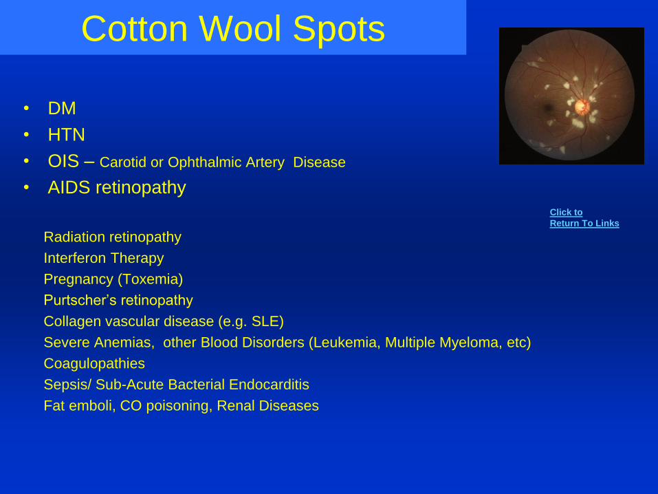
Cotton Wool Spots
• DM
• HTN
• OIS – Carotid or Ophthalmic Artery Disease
• AIDS retinopathy
Radiation retinopathy
Interferon Therapy
Pregnancy (Toxemia)
Purtscher’s retinopathy
Collagen vascular disease (e.g. SLE)
Severe Anemias, other Blood Disorders (Leukemia, Multiple Myeloma, etc)
Coagulopathies
Sepsis/ Sub-Acute Bacterial Endocarditis
Fat emboli, CO poisoning, Renal Diseases
Click to
Return To Links
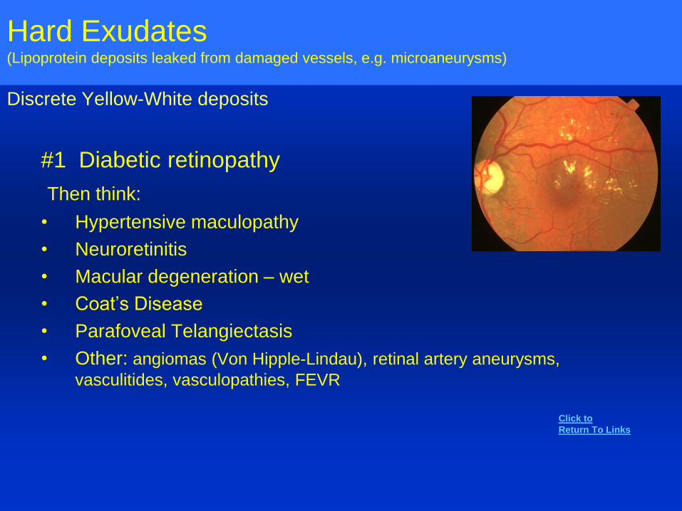
Hard Exudates(Lipoprotein deposits leaked from damaged vessels, e.g. microaneurysms)
Discrete Yellow-White deposits
#1 Diabetic retinopathy
Then think:
• Hypertensive maculopathy
• Neuroretinitis
• Macular degeneration – wet
• Coat’s Disease
• Parafoveal Telangiectasis
• Other: angiomas (Von Hipple-Lindau), retinal artery aneurysms,
vasculitides, vasculopathies, FEVR
Click to
Return To Links
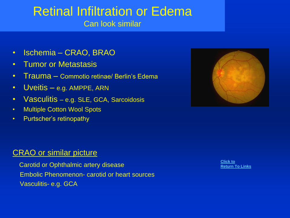
Retinal Infiltration or EdemaCan look similar
• Ischemia – CRAO, BRAO
• Tumor or Metastasis
• Trauma – Commotio retinae/ Berlin’s Edema
• Uveitis – e.g. AMPPE, ARN
• Vasculitis – e.g. SLE, GCA, Sarcoidosis
• Multiple Cotton Wool Spots
• Purtscher’s retinopathy
CRAO or similar picture
Carotid or Ophthalmic artery disease
Embolic Phenomenon- carotid or heart sources
Vasculitis- e.g. GCA
Click to
Return To Links
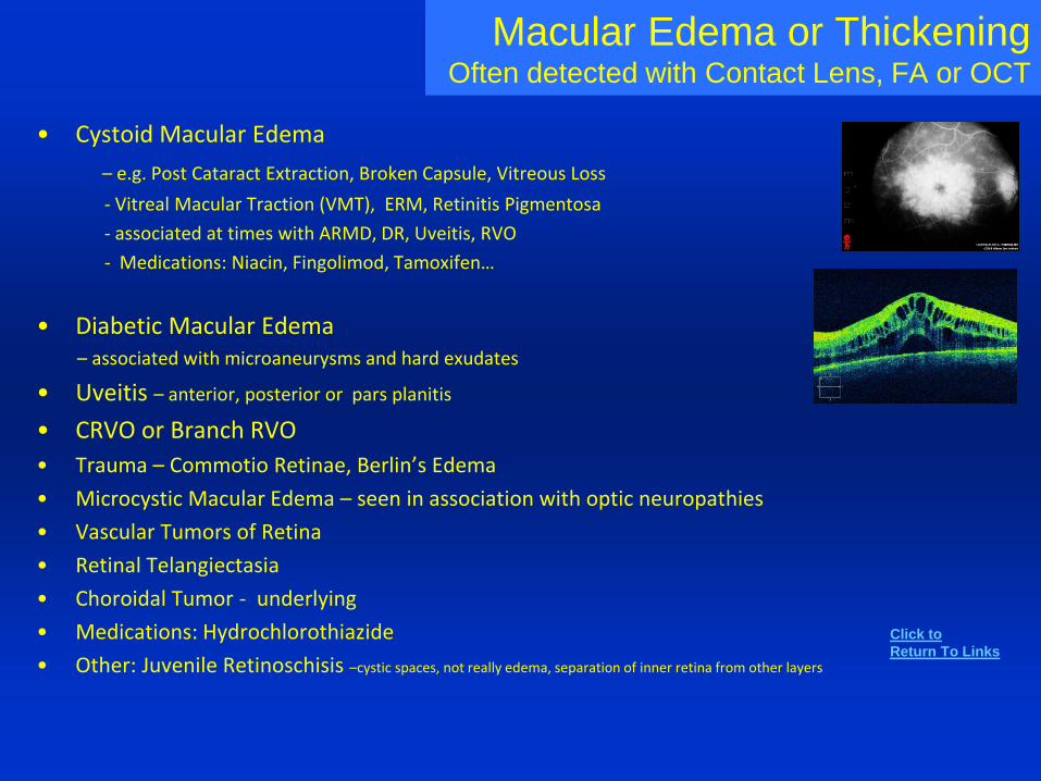
Macular Edema or ThickeningOften detected with Contact Lens, FA or OCT
• Cystoid Macular Edema
– e.g. Post Cataract Extraction, Broken Capsule, Vitreous Loss
- Vitreal Macular Traction (VMT), ERM, Retinitis Pigmentosa
- associated at times with ARMD, DR, Uveitis, RVO
- Medications: Niacin, Fingolimod, Tamoxifen…
• Diabetic Macular Edema– associated with microaneurysms and hard exudates
• Uveitis – anterior, posterior or pars planitis
• CRVO or Branch RVO
• Trauma – Commotio Retinae, Berlin’s Edema
• Microcystic Macular Edema – seen in association with optic neuropathies
• Vascular Tumors of Retina
• Retinal Telangiectasia
• Choroidal Tumor - underlying
• Medications: Hydrochlorothiazide
• Other: Juvenile Retinoschisis –cystic spaces, not really edema, separation of inner retina from other layers
Click to
Return To Links
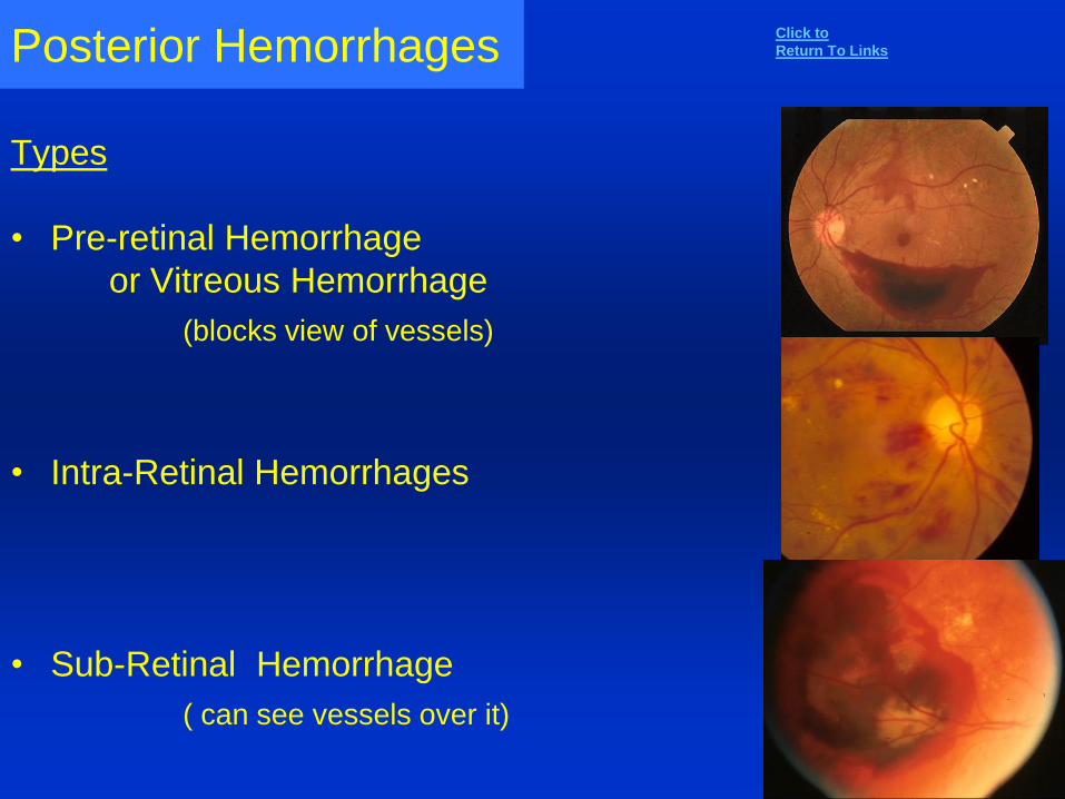
Posterior Hemorrhages
Types
• Pre-retinal Hemorrhage
or Vitreous Hemorrhage
(blocks view of vessels)
• Intra-Retinal Hemorrhages
• Sub-Retinal Hemorrhage
( can see vessels over it)
Click to
Return To Links
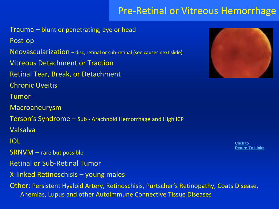
Pre-Retinal or Vitreous Hemorrhage
Trauma – blunt or penetrating, eye or head
Post-op
Neovascularization – disc, retinal or sub-retinal (see causes next slide)
Vitreous Detachment or Traction
Retinal Tear, Break, or Detachment
Chronic Uveitis
Tumor
Macroaneurysm
Terson’s Syndrome – Sub - Arachnoid Hemorrhage and High ICP
Valsalva
IOL
SRNVM – rare but possible
Retinal or Sub-Retinal Tumor
X-linked Retinoschisis – young males
Other: Persistent Hyaloid Artery, Retinoschisis, Purtscher’s Retinopathy, Coats Disease, Anemias, Lupus and other Autoimmune Connective Tissue Diseases
Click to
Return To Links
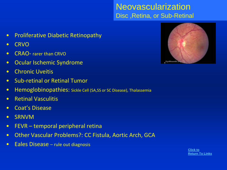
Hypotony
Causes and Associations Low IOP
• Proliferative Diabetic Retinopathy
• CRVO
• CRAO- rarer than CRVO
• Ocular Ischemic Syndrome
• Chronic Uveitis
• Sub-retinal or Retinal Tumor
• Hemoglobinopathies: Sickle Cell (SA,SS or SC Disease), Thalassemia
• Retinal Vasculitis
• Coat’s Disease
• SRNVM
• FEVR – temporal peripheral retina
• Other Vascular Problems?: CC Fistula, Aortic Arch, GCA
• Eales Disease – rule out diagnosis
Neovascularization Disc ,Retina, or Sub-Retinal
Click to
Return To Links
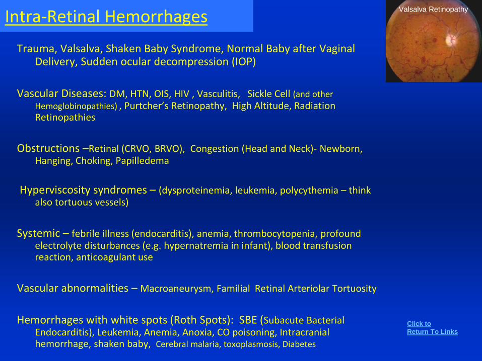
Intra-Retinal Hemorrhages
Trauma, Valsalva, Shaken Baby Syndrome, Normal Baby after Vaginal Delivery, Sudden ocular decompression (IOP)
Vascular Diseases: DM, HTN, OIS, HIV , Vasculitis, Sickle Cell (and other
Hemoglobinopathies) , Purtcher’s Retinopathy, High Altitude, Radiation Retinopathies
Obstructions –Retinal (CRVO, BRVO), Congestion (Head and Neck)- Newborn, Hanging, Choking, Papilledema
Hyperviscosity syndromes – (dysproteinemia, leukemia, polycythemia – think also tortuous vessels)
Systemic – febrile illness (endocarditis), anemia, thrombocytopenia, profound electrolyte disturbances (e.g. hypernatremia in infant), blood transfusion reaction, anticoagulant use
Vascular abnormalities – Macroaneurysm, Familial Retinal Arteriolar Tortuosity
Hemorrhages with white spots (Roth Spots): SBE (Subacute Bacterial Endocarditis), Leukemia, Anemia, Anoxia, CO poisoning, Intracranial hemorrhage, shaken baby, Cerebral malaria, toxoplasmosis, Diabetes
Valsalva Retinopathy
Click to
Return To Links
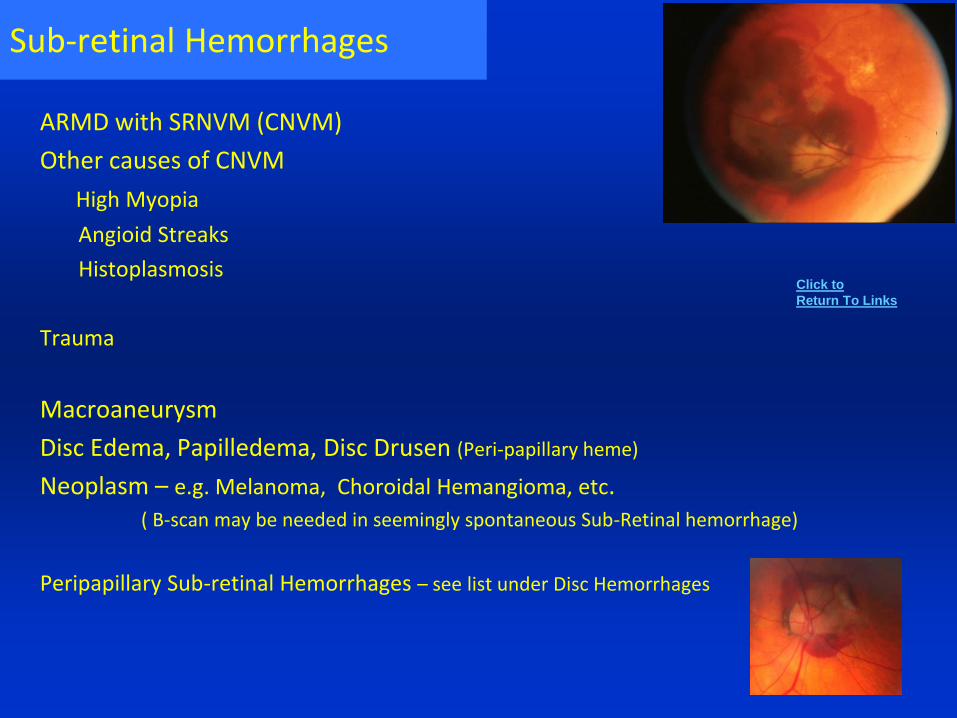
Sub-retinal Hemorrhages
ARMD with SRNVM (CNVM)
Other causes of CNVM
High Myopia
Angioid Streaks
Histoplasmosis
Trauma
Macroaneurysm
Disc Edema, Papilledema, Disc Drusen (Peri-papillary heme)
Neoplasm – e.g. Melanoma, Choroidal Hemangioma, etc.( B-scan may be needed in seemingly spontaneous Sub-Retinal hemorrhage)
Peripapillary Sub-retinal Hemorrhages – see list under Disc Hemorrhages
Click to
Return To Links
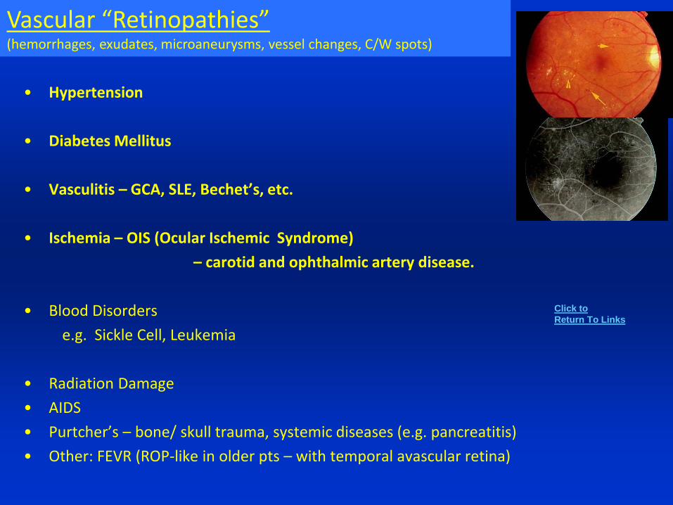
Sub-retinal Hemorrhages
• Hypertension
• Diabetes Mellitus
• Vasculitis – GCA, SLE, Bechet’s, etc.
• Ischemia – OIS (Ocular Ischemic Syndrome)
– carotid and ophthalmic artery disease.
• Blood Disorders
e.g. Sickle Cell, Leukemia
• Radiation Damage
• AIDS
• Purtcher’s – bone/ skull trauma, systemic diseases (e.g. pancreatitis)
• Other: FEVR (ROP-like in older pts – with temporal avascular retina)
Vascular “Retinopathies”(hemorrhages, exudates, microaneurysms, vessel changes, C/W spots)
Click to
Return To Links
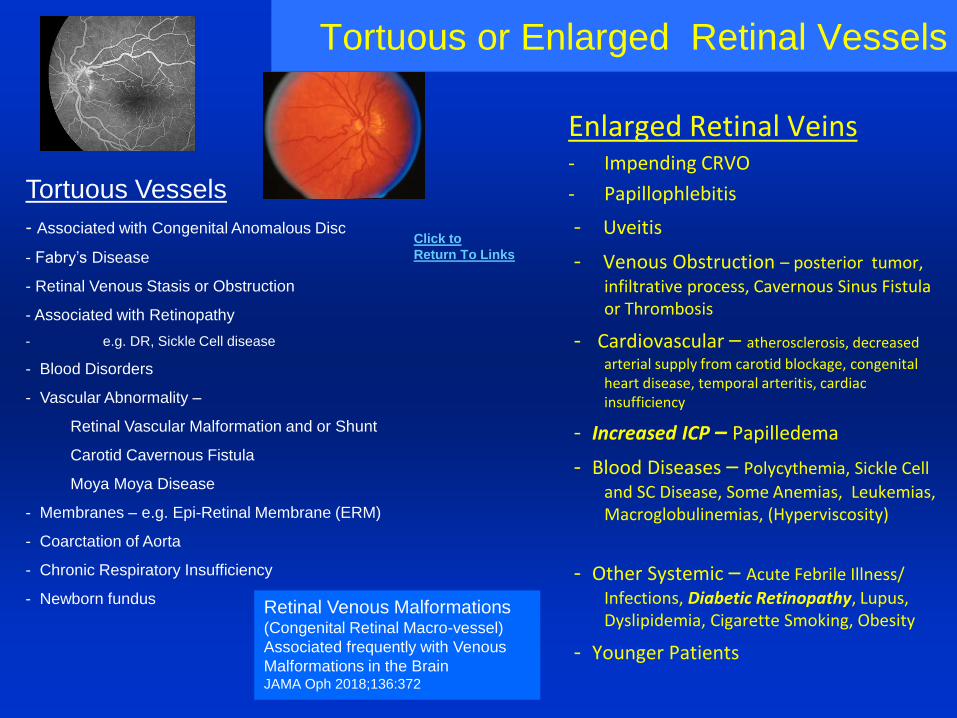
Tortuous or Enlarged Retinal Vessels
Enlarged Retinal Veins- Impending CRVO
- Papillophlebitis
- Uveitis
- Venous Obstruction – posterior tumor,
infiltrative process, Cavernous Sinus Fistula or Thrombosis
- Cardiovascular – atherosclerosis, decreased
arterial supply from carotid blockage, congenital heart disease, temporal arteritis, cardiac insufficiency
- Increased ICP – Papilledema
- Blood Diseases – Polycythemia, Sickle Cell
and SC Disease, Some Anemias, Leukemias, Macroglobulinemias, (Hyperviscosity)
- Other Systemic – Acute Febrile Illness/
Infections, Diabetic Retinopathy, Lupus, Dyslipidemia, Cigarette Smoking, Obesity
- Younger Patients
Tortuous Vessels
- Associated with Congenital Anomalous Disc
- Fabry’s Disease
- Retinal Venous Stasis or Obstruction
- Associated with Retinopathy
- e.g. DR, Sickle Cell disease
- Blood Disorders
- Vascular Abnormality –
Retinal Vascular Malformation and or Shunt
Carotid Cavernous Fistula
Moya Moya Disease
- Membranes – e.g. Epi-Retinal Membrane (ERM)
- Coarctation of Aorta
- Chronic Respiratory Insufficiency
- Newborn fundus
Click to
Return To Links
Retinal Venous Malformations(Congenital Retinal Macro-vessel)
Associated frequently with Venous
Malformations in the BrainJAMA Oph 2018;136:372
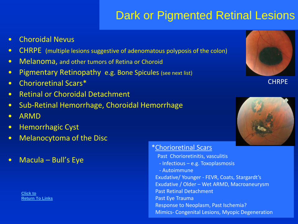
Dark or Pigmented Retinal Lesions
• Choroidal Nevus
• CHRPE (multiple lesions suggestive of adenomatous polyposis of the colon)
• Melanoma, and other tumors of Retina or Choroid
• Pigmentary Retinopathy e.g. Bone Spicules (see next list)
• Chorioretinal Scars*
• Retinal or Choroidal Detachment
• Sub-Retinal Hemorrhage, Choroidal Hemorrhage
• ARMD
• Hemorrhagic Cyst
• Melanocytoma of the Disc
• Macula – Bull’s Eye
*Chorioretinal ScarsPast Chorioretinitis, vasculitis- Infectious – e.g. Toxoplasmosis- Autoimmune
Exudative/ Younger - FEVR, Coats, Stargardt’sExudative / Older – Wet ARMD, MacroaneurysmPast Retinal DetachmentPast Eye TraumaResponse to Neoplasm, Past Ischemia?Mimics- Congenital Lesions, Myopic Degeneration
CHRPE
Click to
Return To Links
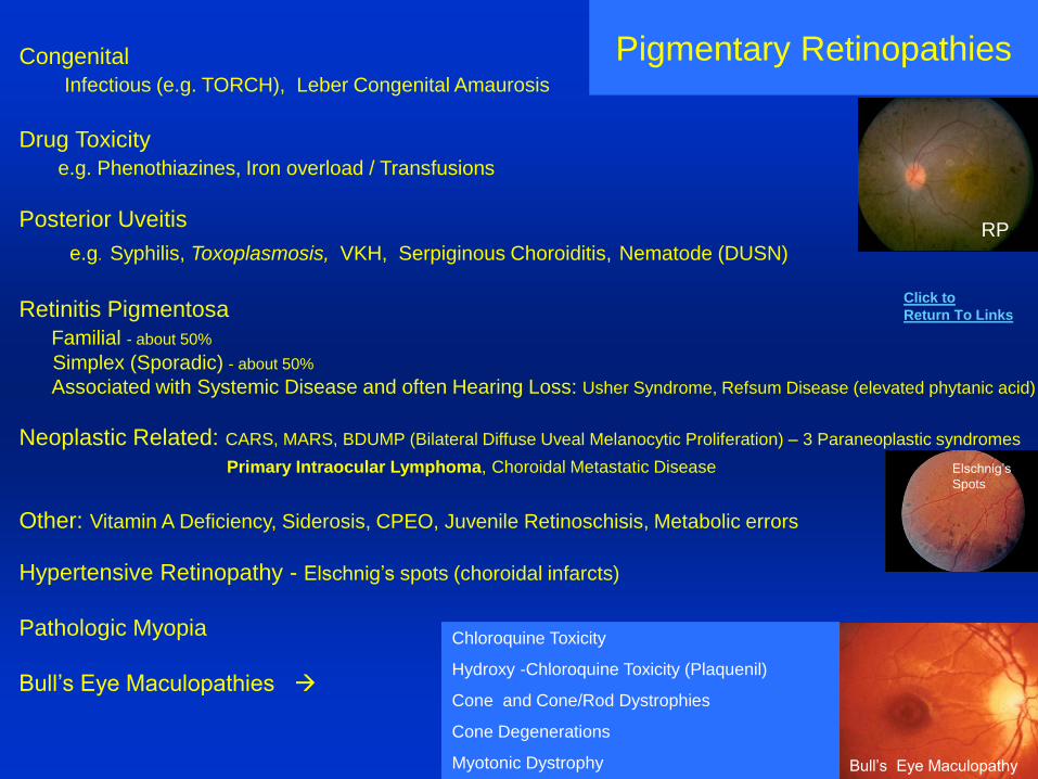
Pigmentary RetinopathiesCongenital
Infectious (e.g. TORCH), Leber Congenital Amaurosis
Drug Toxicity
e.g. Phenothiazines, Iron overload / Transfusions
Posterior Uveitis
e.g. Syphilis, Toxoplasmosis, VKH, Serpiginous Choroiditis, Nematode (DUSN)
Retinitis Pigmentosa
Familial - about 50%
Simplex (Sporadic) - about 50%
Associated with Systemic Disease and often Hearing Loss: Usher Syndrome, Refsum Disease (elevated phytanic acid)
Neoplastic Related: CARS, MARS, BDUMP (Bilateral Diffuse Uveal Melanocytic Proliferation) – 3 Paraneoplastic syndromes
Primary Intraocular Lymphoma, Choroidal Metastatic Disease
Other: Vitamin A Deficiency, Siderosis, CPEO, Juvenile Retinoschisis, Metabolic errors
Hypertensive Retinopathy - Elschnig’s spots (choroidal infarcts)
Pathologic Myopia
Bull’s Eye Maculopathies
Bull’s Eye Maculopathy
Chloroquine Toxicity
Hydroxy -Chloroquine Toxicity (Plaquenil)
Cone and Cone/Rod Dystrophies
Cone Degenerations
Myotonic Dystrophy
RP
Elschnig’s
Spots
Click to
Return To Links
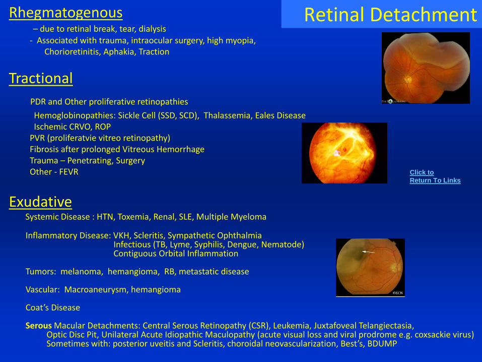
Retinal DetachmentRhegmatogenous– due to retinal break, tear, dialysis
- Associated with trauma, intraocular surgery, high myopia, Chorioretinitis, Aphakia, Traction
Tractional
PDR and Other proliferative retinopathies
Hemoglobinopathies: Sickle Cell (SSD, SCD), Thalassemia, Eales DiseaseIschemic CRVO, ROP
PVR (proliferatvie vitreo retinopathy)Fibrosis after prolonged Vitreous HemorrhageTrauma – Penetrating, SurgeryOther - FEVR
ExudativeSystemic Disease : HTN, Toxemia, Renal, SLE, Multiple Myeloma
Inflammatory Disease: VKH, Scleritis, Sympathetic OphthalmiaInfectious (TB, Lyme, Syphilis, Dengue, Nematode) Contiguous Orbital Inflammation
Tumors: melanoma, hemangioma, RB, metastatic disease
Vascular: Macroaneurysm, hemangioma
Coat’s Disease
Serous Macular Detachments: Central Serous Retinopathy (CSR), Leukemia, Juxtafoveal Telangiectasia, Optic Disc Pit, Unilateral Acute Idiopathic Maculopathy (acute visual loss and viral prodrome e.g. coxsackie virus)Sometimes with: posterior uveitis and Scleritis, choroidal neovascularization, Best’s, BDUMP
Click to
Return To Links
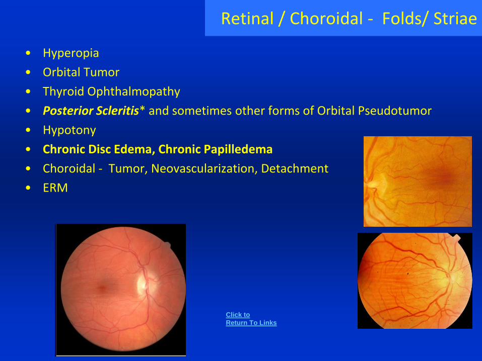
Retinal / Choroidal - Folds/ Striae
• Hyperopia
• Orbital Tumor
• Thyroid Ophthalmopathy
• Posterior Scleritis* and sometimes other forms of Orbital Pseudotumor
• Hypotony
• Chronic Disc Edema, Chronic Papilledema
• Choroidal - Tumor, Neovascularization, Detachment
• ERM
Click to
Return To Links
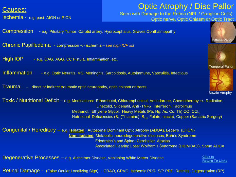
Optic Atrophy / Disc PallorSeen with Damage to the Retina (NFL / Ganglion Cells),
Optic nerve, Optic Chiasm or Optic Tract
Causes:Ischemia - e.g. past AION or PION
Compression - e.g. Pituitary Tumor, Carotid artery, Hydrocephalus, Graves Ophthalmopathy
Chronic Papilledema - compression +/- ischemia – see high ICP list
High IOP - e.g. OAG, AGG, CC Fistula, Inflammation, etc.
Inflammation - e.g. Optic Neuritis, MS, Meningitis, Sarcoidosis, Autoimmune, Vasculitis, Infectious
Trauma – direct or indirect traumatic optic neuropathy, optic chiasm or tracts
Toxic / Nutritional Deficit – e.g. Medications: Ethambutol, Chloramphenicol, Amiodarone, Chemotherapy +/- Radiation,
Linezolid, Sildenafil, Anti -TNFa, Interferon, Tacrolimus
Methanol, Ethylene Glycol, Heavy Metals (Pb, Hg, As, Co, Th),CO, CCl4Nutritional Deficiencies (B1 (Thiamine), B12, Folate, niacin), Copper (Bariatric Surgery)
Congenital / Hereditary – e.g. Isolated : Autosomal Dominant Optic Atrophy (ADOA), Leber’s (LHON)
Non–isolated: Metabolic, neurodegenerative diseases, Behr’s Syndrome
Friedreich’s and Spino- Cerebellar Ataxias
Associated Hearing Loss: Wolfram’s Syndrome (DIDMOAD), Some ADOA
Degenerative Processes – e.g. Alzheimer Disease, Vanishing White Matter Disease
Retinal Damage - (False Ocular Localizing Sign) - CRAO, CRVO, Ischemic PDR, S/P PRP, Retinitis; Degeneration (RP)
Temporal Pallor
Click to
Return To Links
Bowtie Atrophy
Diffuse Pallor
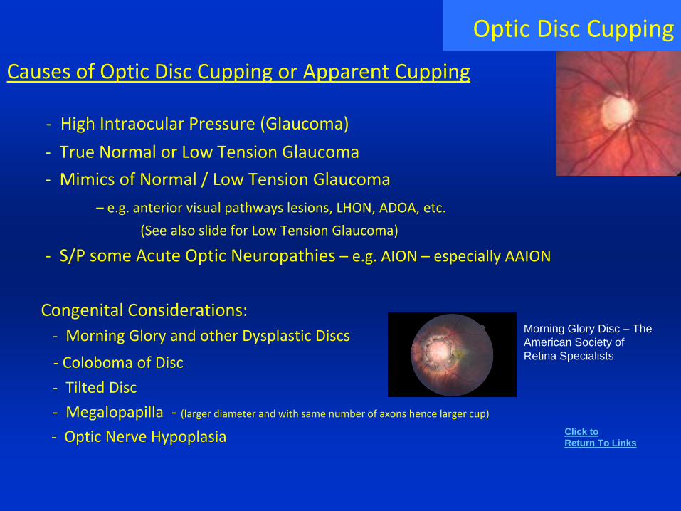
Causes of Optic Disc Cupping or Apparent Cupping
- High Intraocular Pressure (Glaucoma)
- True Normal or Low Tension Glaucoma
- Mimics of Normal / Low Tension Glaucoma
– e.g. anterior visual pathways lesions, LHON, ADOA, etc.
(See also slide for Low Tension Glaucoma)
- S/P some Acute Optic Neuropathies – e.g. AION – especially AAION
Congenital Considerations:
- Morning Glory and other Dysplastic Discs
- Coloboma of Disc
- Tilted Disc
- Megalopapilla - (larger diameter and with same number of axons hence larger cup)
- Optic Nerve Hypoplasia
Optic Disc Cupping
Morning Glory Disc – The
American Society of
Retina Specialists
Click to
Return To Links
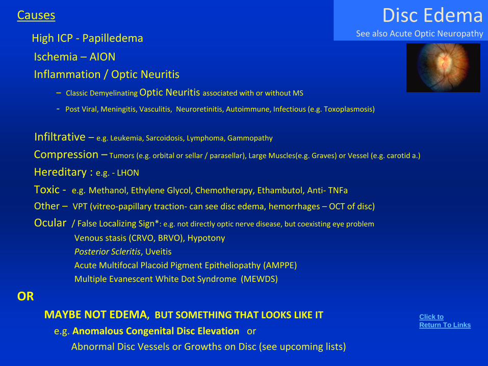
Disc EdemaSee also Acute Optic Neuropathy
Causes
High ICP - Papilledema
Ischemia – AION
Inflammation / Optic Neuritis
– Classic Demyelinating Optic Neuritis associated with or without MS
- Post Viral, Meningitis, Vasculitis, Neuroretinitis, Autoimmune, Infectious (e.g. Toxoplasmosis)
Infiltrative – e.g. Leukemia, Sarcoidosis, Lymphoma, Gammopathy
Compression – Tumors (e.g. orbital or sellar / parasellar), Large Muscles(e.g. Graves) or Vessel (e.g. carotid a.)
Hereditary : e.g. - LHON
Toxic - e.g. Methanol, Ethylene Glycol, Chemotherapy, Ethambutol, Anti- TNFa
Other – VPT (vitreo-papillary traction- can see disc edema, hemorrhages – OCT of disc)
Ocular / False Localizing Sign*: e.g. not directly optic nerve disease, but coexisting eye problem
Venous stasis (CRVO, BRVO), Hypotony
Posterior Scleritis, Uveitis
Acute Multifocal Placoid Pigment Epitheliopathy (AMPPE)
Multiple Evanescent White Dot Syndrome (MEWDS)
OR
MAYBE NOT EDEMA, BUT SOMETHING THAT LOOKS LIKE IT
e.g. Anomalous Congenital Disc Elevation or
Abnormal Disc Vessels or Growths on Disc (see upcoming lists)
Click to
Return To Links
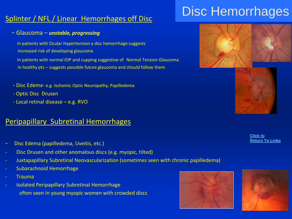
Disc HemorrhagesSplinter / NFL / Linear Hemorrhages off Disc
- Glaucoma – unstable, progressing
In patients with Ocular Hypertension a disc hemorrhage suggests
increased risk of developing glaucoma
In patients with normal IOP and cupping suggestive of Normal Tension Glaucoma
In healthy pts – suggests possible future glaucoma and should follow them
- Disc Edema- e.g. Ischemic Optic Neuropathy, Papilledema
- Optic Disc Drusen
- Local retinal disease – e.g. RVO
Peripapillary Subretinal Hemorrhages
- Disc Edema (papilledema, Uveitis, etc.)
- Disc Drusen and other anomalous discs (e.g. myopic, tilted)
- Juxtapapillary Subretinal Neovascularization (sometimes seen with chronic papilledema)
- Subarachnoid Hemorrhage
- Trauma
- Isolated Peripapillary Subretinal Hemorrhage
often seen in young myopic women with crowded discs
Click to
Return To Links
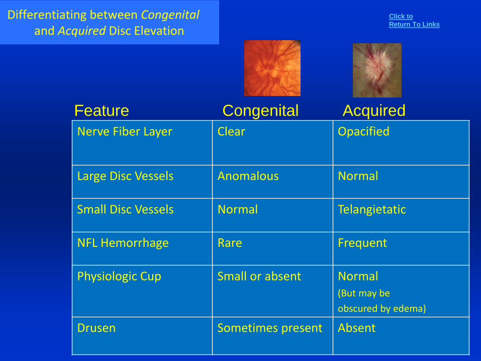
Differentiating between Congenital and Acquired Disc Elevation
Feature Congenital Acquired
Nerve Fiber Layer Clear Opacified
Large Disc Vessels Anomalous Normal
Small Disc Vessels Normal Telangietatic
NFL Hemorrhage Rare Frequent
Physiologic Cup Small or absent Normal(But may be
obscured by edema)
Drusen Sometimes present Absent
Click to
Return To Links
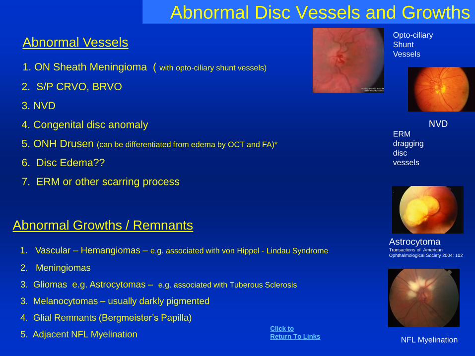
Abnormal Disc Vessels and Growths
Abnormal Vessels
1. ON Sheath Meningioma ( with opto-ciliary shunt vessels)
2. S/P CRVO, BRVO
3. NVD
4. Congenital disc anomaly
5. ONH Drusen (can be differentiated from edema by OCT and FA)*
6. Disc Edema??
7. ERM or other scarring process
Abnormal Growths / Remnants
1. Vascular – Hemangiomas – e.g. associated with von Hippel - Lindau Syndrome
2. Meningiomas
3. Gliomas e.g. Astrocytomas – e.g. associated with Tuberous Sclerosis
3. Melanocytomas – usually darkly pigmented
4. Glial Remnants (Bergmeister’s Papilla)
5. Adjacent NFL Myelination
NVD
Opto-ciliary
Shunt
Vessels
NFL Myelination
AstrocytomaTransactions of American
Ophthalmological Society 2004; 102
ERM
dragging
disc
vessels
Click to
Return To Links
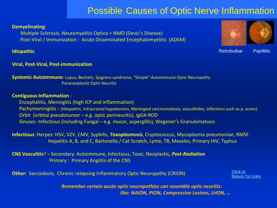
Possible Causes of Optic Nerve Inflammation
Demyelinating: Multiple Sclerosis, Neuromyelitis Optica = NMO (Devic’s Disease)Post-Viral / Immunization : Acute Disseminated Encephalomyelitis (ADEM)
Idiopathic
Viral, Post-Viral, Post-immunization
Systemic Autoimmune: Lupus, Bechets, Sjogrens syndrome, “Simple” Autoimmune Optic NeuropathyParaneoplastic Optic Neuritis
Contiguous Inflammation : Encephalitis, Meningitis (high ICP and inflammation)Pachymeninigitis – (Idiopathic, intracranial hypotension, Meningeal carcinomatosis, vasculitides, infections such as p. acnes)
Orbit (orbital pseudotumor – e.g. optic perineuritis), IgG4-RODSinuses -Infectious (including Fungal – e.g. mucor, aspergillis), Wegener’s Granulomatosis
Infectious: Herpes: HSV, VZV, CMV, Syphilis, Toxoplasmosis, Cryptococcus, Mycoplasma pneumoniae, RMSFHepatitis A, B, and C, Bartonella / Cat Scratch, Lyme, TB, Measles, Primary HIV, Typhus
CNS Vasculitis? – Secondary: Autoimmune, Infectious, Toxic, Neoplastic, Post-Radiation Primary : Primary Angiitis of the CNS
Other: Sarcoidosis, Chronic relapsing Inflammatory Optic Neuropathy (CRION)
Remember certain acute optic neuropathies can resemble optic neuritis: like: NAION, PION, Compressive Lesions, LHON, …
Retrobulbar Papillitis
Click to
Return To Links
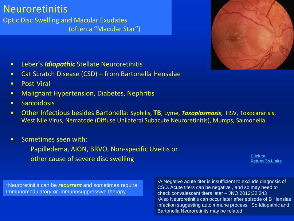
NeuroretinitisOptic Disc Swelling and Macular Exudates
(often a “Macular Star”)
• Leber’s Idiopathic Stellate Neuroretinitis
• Cat Scratch Disease (CSD) – from Bartonella Hensalae
• Post-Viral
• Malignant Hypertension, Diabetes, Nephritis
• Sarcoidosis
• Other Infectious besides Bartonella: Syphilis, TB, Lyme, Toxoplasmosis, HSV, Toxocararisis, West Nile Virus, Nematode (Diffuse Unilateral Subacute Neuroretinitis), Mumps, Salmonella
• Sometimes seen with:
Papilledema, AION, BRVO, Non-specific Uveitis or
other cause of severe disc swelling
•A Negative acute titer is insufficient to exclude diagnosis of
CSD. Acute titers can be negative , and so may need to
check convalescent titers later – JNO 2012;32:243
•Also Neuroretinitis can occur later after episode of B Henslae
infection suggesting autoimmune process. So Idiopathic and
Bartonella Neuroretinits may be related.
*Neuroretinitis can be recurrent and sometimes require
Immunomodulatory or immunosuppressive therapy
Click to
Return To Links
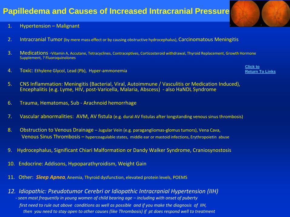
Papilledema and Causes of Increased Intracranial Pressure
1. Hypertension – Malignant
2. Intracranial Tumor (by mere mass effect or by causing obstructive hydrocephalus), Carcinomatous Meningitis
3. Medications -Vitamin A, Accutane, Tetracyclines, Contraceptives, Corticosteroid withdrawal, Thyroid Replacement, Growth Hormone Supplement, ? Fluoroquinolones
4. Toxic: Ethylene Glycol, Lead (Pb), Hyper-ammonemia
5. CNS Inflammation: Meningitis (Bacterial, Viral, Autoimmune / Vasculitis or Medication Induced), Encephalitis (e.g. Lyme, HIV, post-Varicella, Malaria, Abscess) - also HaNDL Syndrome
6. Trauma, Hematomas, Sub - Arachnoid hemorrhage
7. Vascular abnormalities: AVM, AV fistula (e.g. dural AV fistulas after longstanding venous sinus thrombosis)
8. Obstruction to Venous Drainage – Jugular Vein (e.g. paragangliomas-glomus tumors), Vena Cava,
Venous Sinus Thrombosis – hypercoagulable states, middle ear or mastoid infections, Erythropoietin abuse
9. Hydrocephalus, Significant Chiari Malformation or Dandy Walker Syndrome, Craniosynostosis
10. Endocrine: Addisons, Hypoparathyroidism, Weight Gain
11. Other: Sleep Apnea, Anemia, Thyroid dysfunction, elevated protein levels, POEMS
12. Idiopathic: Pseudotumor Cerebri or Idiopathic Intracranial Hypertension (IIH)- seen most frequently in young women of child bearing age – including with onset of puberty
first need to rule out above conditions as well as possible and if you make the diagnosis of IIH,
then you need to stay open to other causes (like Thrombosis) if pt does respond well to treatment
Click to
Return To Links
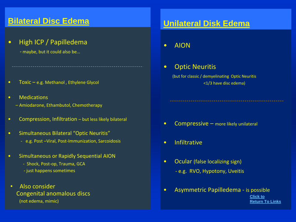
Bilateral Disc Edema
• High ICP / Papilledema- maybe, but it could also be…
• Toxic – e.g. Methanol , Ethylene Glycol
• Medications
– Amiodarone, Ethambutol, Chemotherapy
• Compression, Infiltration – but less likely bilateral
• Simultaneous Bilateral “Optic Neuritis”
- e.g. Post –Viral, Post-Immunization, Sarcoidosis
• Simultaneous or Rapidly Sequential AION
- Shock, Post-op, Trauma, GCA
- just happens sometimes
Unilateral Disk Edema
• AION
• Optic Neuritis (but for classic / demyelinating Optic Neuritis
<1/3 have disc edema)
• Compressive – more likely unilateral
• Infiltrative
• Ocular (false localizing sign)
- e.g. RVO, Hypotony, Uveitis
• Asymmetric Papilledema - is possibleClick to
Return To Links
• Also considerCongenital anomalous discs
(not edema, mimic)
3. Miscellaneous Case Situations
and other Lists
Visual Loss
Reading Problems
Pediatric Presentations: Visual Loss, ET, Glaucoma, Nystagmus
Pregnancy
Systemic Medical Conditions
CN Palsies
Elevated ESR and Concern for Temporal Arteritis
Low Tension Glaucoma
Ocular Effects of Systemic Medications
Imaging Findings- CT, MRI
Dizziness
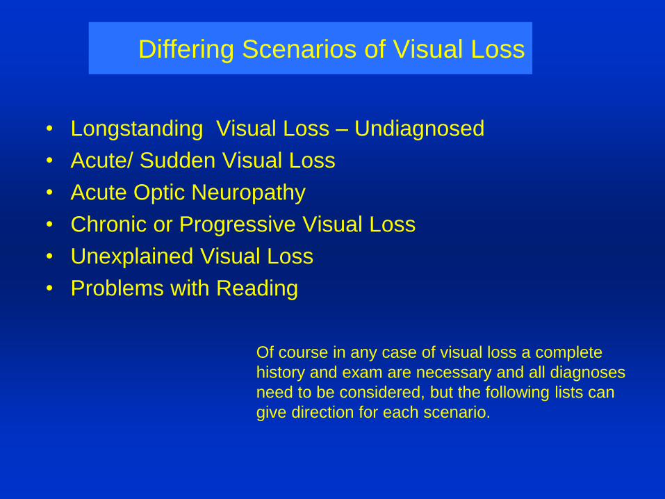
Differing Scenarios of Visual Loss
• Longstanding Visual Loss – Undiagnosed
• Acute/ Sudden Visual Loss
• Acute Optic Neuropathy
• Chronic or Progressive Visual Loss
• Unexplained Visual Loss
• Problems with Reading
Of course in any case of visual loss a complete
history and exam are necessary and all diagnoses
need to be considered, but the following lists can
give direction for each scenario.
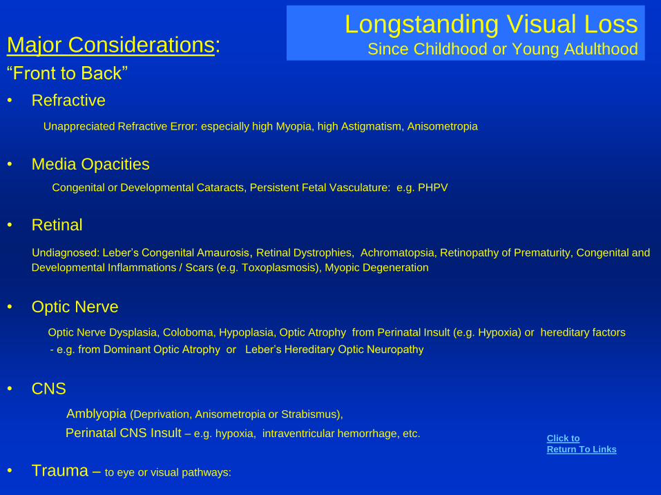
Longstanding Visual LossSince Childhood or Young AdulthoodMajor Considerations:
“Front to Back”
• Refractive
Unappreciated Refractive Error: especially high Myopia, high Astigmatism, Anisometropia
• Media Opacities
Congenital or Developmental Cataracts, Persistent Fetal Vasculature: e.g. PHPV
• Retinal
Undiagnosed: Leber’s Congenital Amaurosis, Retinal Dystrophies, Achromatopsia, Retinopathy of Prematurity, Congenital and
Developmental Inflammations / Scars (e.g. Toxoplasmosis), Myopic Degeneration
• Optic Nerve
Optic Nerve Dysplasia, Coloboma, Hypoplasia, Optic Atrophy from Perinatal Insult (e.g. Hypoxia) or hereditary factors
- e.g. from Dominant Optic Atrophy or Leber’s Hereditary Optic Neuropathy
• CNS
Amblyopia (Deprivation, Anisometropia or Strabismus),
Perinatal CNS Insult – e.g. hypoxia, intraventricular hemorrhage, etc.
• Trauma – to eye or visual pathways:
Click to
Return To Links
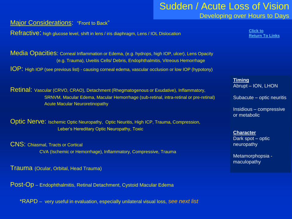
Sudden / Acute Loss of VisionDeveloping over Hours to Days
Major Considerations: “Front to Back”
Refractive: high glucose level, shift in lens / iris diaphragm, Lens / IOL Dislocation
Media Opacities: Corneal Inflammation or Edema, (e.g. hydrops, high IOP, ulcer), Lens Opacity
(e.g. Trauma), Uveitis Cells/ Debris, Endophthalmitis, Vitreous Hemorrhage
IOP: High IOP (see previous list) - causing corneal edema, vascular occlusion or low IOP (hypotony)
Retinal: Vascular (CRVO, CRAO), Detachment (Rhegmatogenous or Exudative), Inflammatory,
SRNVM, Macular Edema, Macular Hemorrhage (sub-retinal, intra-retinal or pre-retinal)
Acute Macular Neuroretinopathy
Optic Nerve: Ischemic Optic Neuropathy, Optic Neuritis, High ICP, Trauma, Compression,
Leber’s Hereditary Optic Neuropathy, Toxic
CNS: Chiasmal, Tracts or Cortical
CVA (Ischemic or Hemorrhage), Inflammatory, Compressive, Trauma
Trauma (Ocular, Orbital, Head Trauma)
Post-Op – Endophthalmitis, Retinal Detachment, Cystoid Macular Edema
*RAPD – very useful in evaluation, especially unilateral visual loss, see next list
Timing
Abrupt – ION, LHON
Subacute – optic neuritis
Insidious – compressive
or metabolic
Character
Dark spot – optic
neuropathy
Metamorphopsia -
maculopathy
Click to
Return To Links
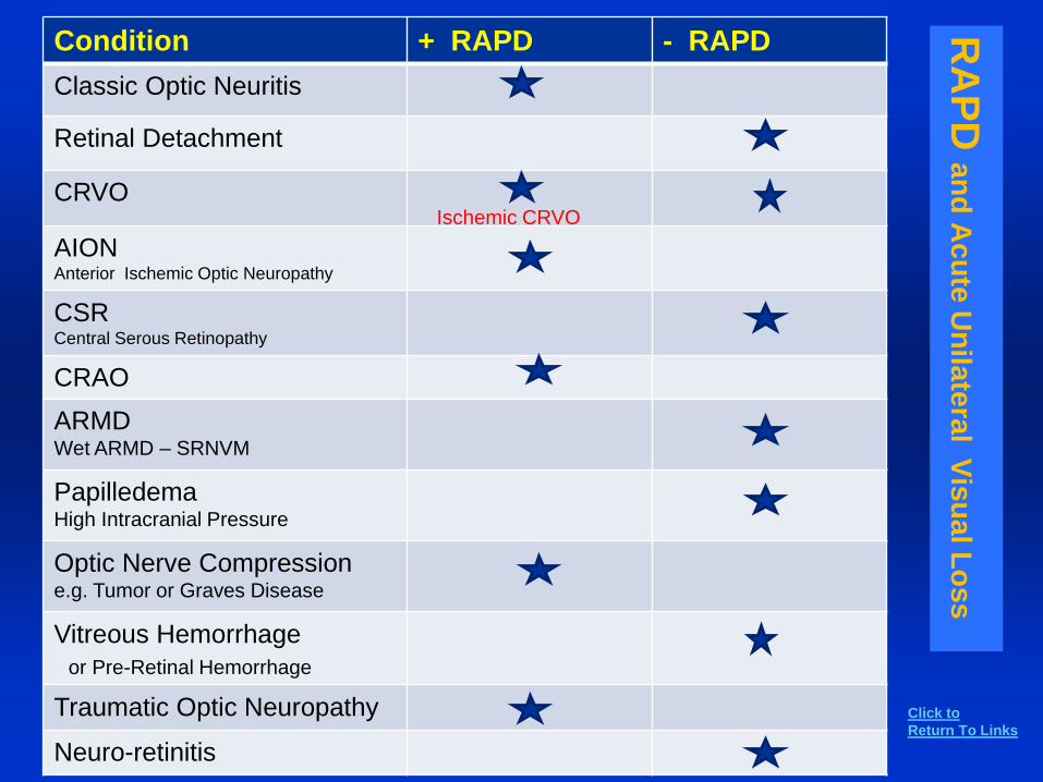
RA
PD
an
d A
cu
te U
nila
tera
l Vis
ua
l Lo
ss
Condition + RAPD - RAPD
Classic Optic Neuritis
Retinal Detachment
CRVO
AIONAnterior Ischemic Optic Neuropathy
CSRCentral Serous Retinopathy
CRAO
ARMDWet ARMD – SRNVM
PapilledemaHigh Intracranial Pressure
Optic Nerve Compression e.g. Tumor or Graves Disease
Vitreous Hemorrhage
or Pre-Retinal Hemorrhage
Traumatic Optic Neuropathy
Neuro-retinitis
Ischemic CRVO
Click to
Return To Links
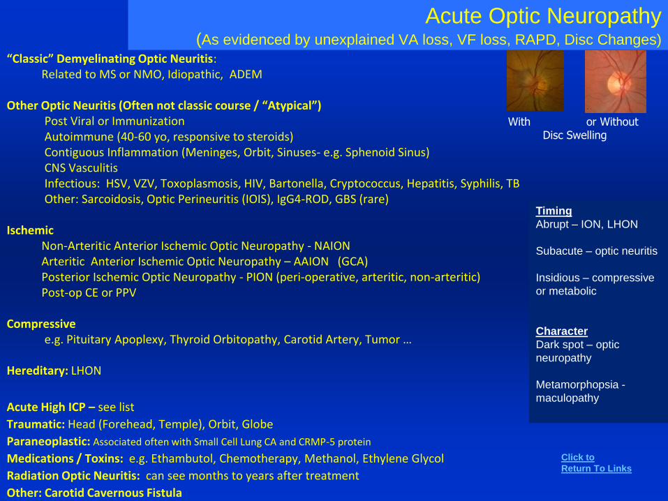
“Classic” Demyelinating Optic Neuritis: Related to MS or NMO, Idiopathic, ADEM
Other Optic Neuritis (Often not classic course / “Atypical”) Post Viral or ImmunizationAutoimmune (40-60 yo, responsive to steroids)Contiguous Inflammation (Meninges, Orbit, Sinuses- e.g. Sphenoid Sinus)CNS VasculitisInfectious: HSV, VZV, Toxoplasmosis, HIV, Bartonella, Cryptococcus, Hepatitis, Syphilis, TBOther: Sarcoidosis, Optic Perineuritis (IOIS), IgG4-ROD, GBS (rare)
IschemicNon-Arteritic Anterior Ischemic Optic Neuropathy - NAIONArteritic Anterior Ischemic Optic Neuropathy – AAION (GCA)Posterior Ischemic Optic Neuropathy - PION (peri-operative, arteritic, non-arteritic)Post-op CE or PPV
Compressivee.g. Pituitary Apoplexy, Thyroid Orbitopathy, Carotid Artery, Tumor …
Hereditary: LHON
Acute High ICP – see list
Traumatic: Head (Forehead, Temple), Orbit, Globe
Paraneoplastic: Associated often with Small Cell Lung CA and CRMP-5 protein
Medications / Toxins: e.g. Ethambutol, Chemotherapy, Methanol, Ethylene Glycol
Radiation Optic Neuritis: can see months to years after treatment
Other: Carotid Cavernous Fistula
With or Without Disc Swelling
Acute Optic Neuropathy(As evidenced by unexplained VA loss, VF loss, RAPD, Disc Changes)
Timing
Abrupt – ION, LHON
Subacute – optic neuritis
Insidious – compressive
or metabolic
Character
Dark spot – optic
neuropathy
Metamorphopsia -
maculopathy
Click to
Return To Links
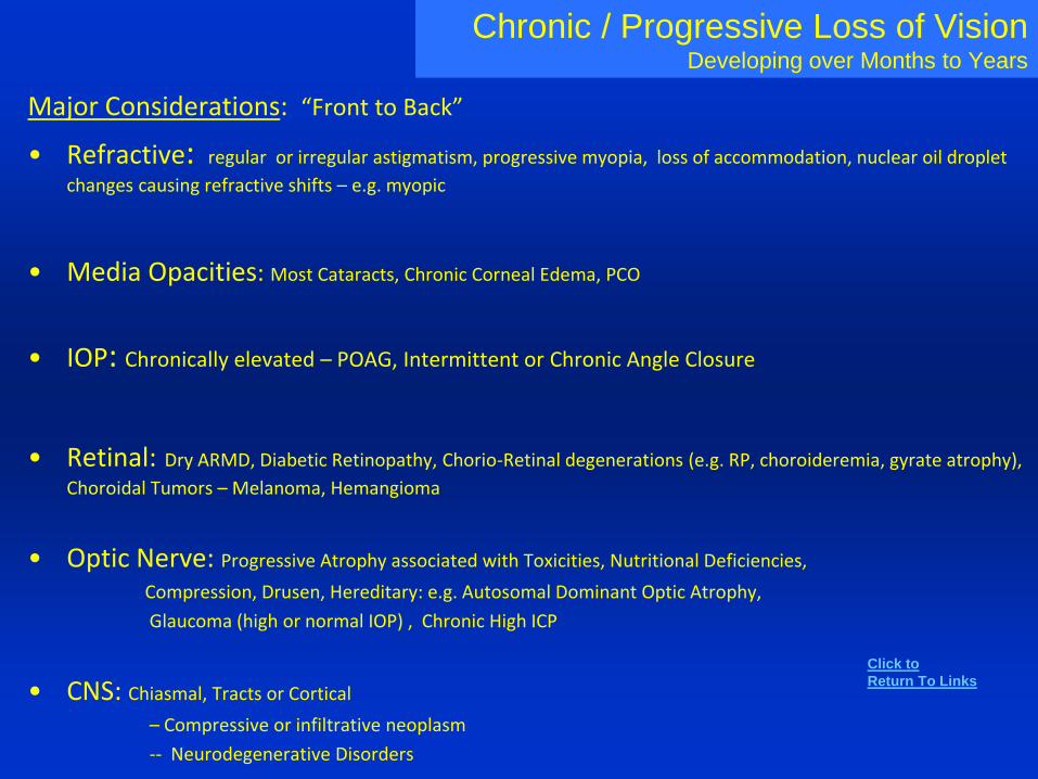
Chronic / Progressive Loss of VisionDeveloping over Months to Years
Major Considerations: “Front to Back”
• Refractive: regular or irregular astigmatism, progressive myopia, loss of accommodation, nuclear oil droplet
changes causing refractive shifts – e.g. myopic
• Media Opacities: Most Cataracts, Chronic Corneal Edema, PCO
• IOP: Chronically elevated – POAG, Intermittent or Chronic Angle Closure
• Retinal: Dry ARMD, Diabetic Retinopathy, Chorio-Retinal degenerations (e.g. RP, choroideremia, gyrate atrophy),
Choroidal Tumors – Melanoma, Hemangioma
• Optic Nerve: Progressive Atrophy associated with Toxicities, Nutritional Deficiencies,
Compression, Drusen, Hereditary: e.g. Autosomal Dominant Optic Atrophy,
Glaucoma (high or normal IOP) , Chronic High ICP
• CNS: Chiasmal, Tracts or Cortical
– Compressive or infiltrative neoplasm
-- Neurodegenerative Disorders
Click to
Return To Links
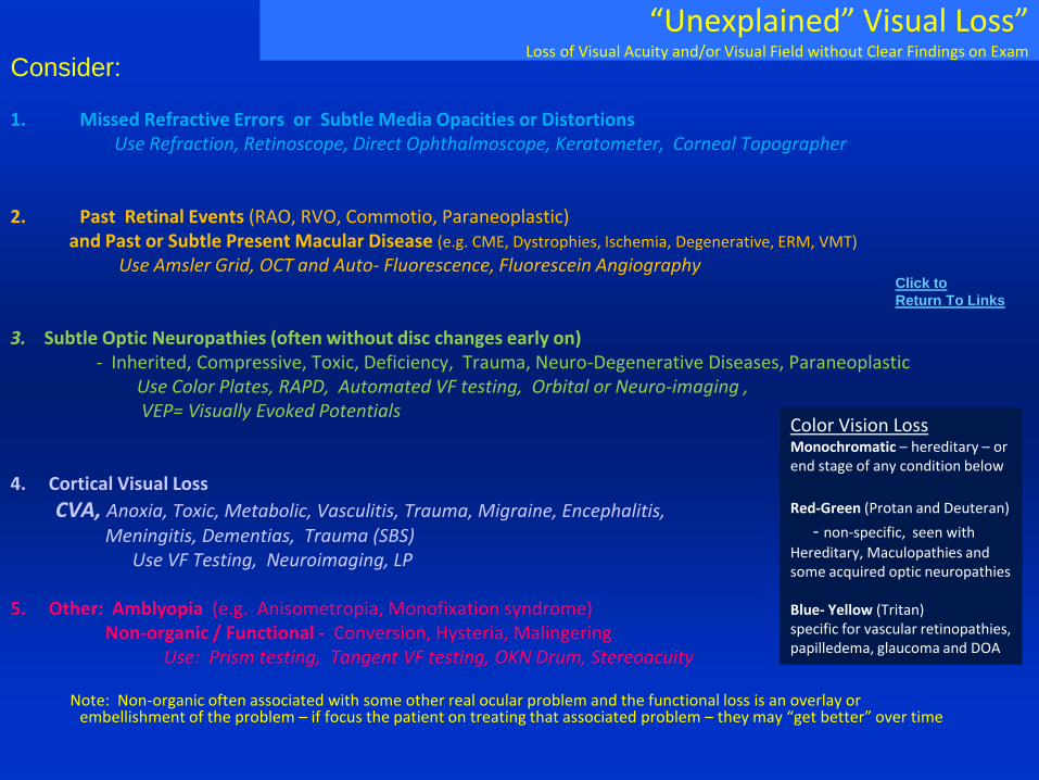
“Unexplained” Visual Loss”Loss of Visual Acuity and/or Visual Field without Clear Findings on Exam
Consider:
1. Missed Refractive Errors or Subtle Media Opacities or Distortions Use Refraction, Retinoscope, Direct Ophthalmoscope, Keratometer, Corneal Topographer
2. Past Retinal Events (RAO, RVO, Commotio, Paraneoplastic) and Past or Subtle Present Macular Disease (e.g. CME, Dystrophies, Ischemia, Degenerative, ERM, VMT)
Use Amsler Grid, OCT and Auto- Fluorescence, Fluorescein Angiography
3. Subtle Optic Neuropathies (often without disc changes early on)- Inherited, Compressive, Toxic, Deficiency, Trauma, Neuro-Degenerative Diseases, Paraneoplastic
Use Color Plates, RAPD, Automated VF testing, Orbital or Neuro-imaging , VEP= Visually Evoked Potentials
4. Cortical Visual Loss
CVA, Anoxia, Toxic, Metabolic, Vasculitis, Trauma, Migraine, Encephalitis, Meningitis, Dementias, Trauma (SBS)
Use VF Testing, Neuroimaging, LP
5. Other: Amblyopia (e.g. Anisometropia, Monofixation syndrome)Non-organic / Functional - Conversion, Hysteria, Malingering
Use: Prism testing, Tangent VF testing, OKN Drum, Stereoacuity
Note: Non-organic often associated with some other real ocular problem and the functional loss is an overlay or embellishment of the problem – if focus the patient on treating that associated problem – they may “get better” over time
Color Vision LossMonochromatic – hereditary – or end stage of any condition below
Red-Green (Protan and Deuteran)
- non-specific, seen with
Hereditary, Maculopathies and some acquired optic neuropathies
Blue- Yellow (Tritan) specific for vascular retinopathies, papilledema, glaucoma and DOA
Click to
Return To Links
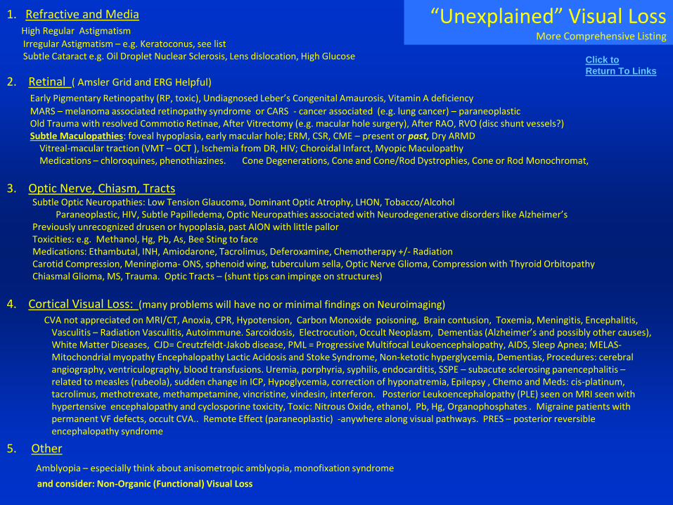
“Unexplained” Visual LossMore Comprehensive Listing
1. Refractive and MediaHigh Regular Astigmatism
Irregular Astigmatism – e.g. Keratoconus, see list Subtle Cataract e.g. Oil Droplet Nuclear Sclerosis, Lens dislocation, High Glucose
2. Retinal ( Amsler Grid and ERG Helpful)
Early Pigmentary Retinopathy (RP, toxic), Undiagnosed Leber’s Congenital Amaurosis, Vitamin A deficiency
MARS – melanoma associated retinopathy syndrome or CARS - cancer associated (e.g. lung cancer) – paraneoplasticOld Trauma with resolved Commotio Retinae, After Vitrectomy (e.g. macular hole surgery), After RAO, RVO (disc shunt vessels?)Subtle Maculopathies: foveal hypoplasia, early macular hole; ERM, CSR, CME – present or past, Dry ARMD
Vitreal-macular traction (VMT – OCT ), Ischemia from DR, HIV; Choroidal Infarct, Myopic MaculopathyMedications – chloroquines, phenothiazines. Cone Degenerations, Cone and Cone/Rod Dystrophies, Cone or Rod Monochromat,
3. Optic Nerve, Chiasm, TractsSubtle Optic Neuropathies: Low Tension Glaucoma, Dominant Optic Atrophy, LHON, Tobacco/Alcohol
Paraneoplastic, HIV, Subtle Papilledema, Optic Neuropathies associated with Neurodegenerative disorders like Alzheimer’sPreviously unrecognized drusen or hypoplasia, past AION with little pallorToxicities: e.g. Methanol, Hg, Pb, As, Bee Sting to faceMedications: Ethambutal, INH, Amiodarone, Tacrolimus, Deferoxamine, Chemotherapy +/- RadiationCarotid Compression, Meningioma- ONS, sphenoid wing, tuberculum sella, Optic Nerve Glioma, Compression with Thyroid OrbitopathyChiasmal Glioma, MS, Trauma. Optic Tracts – (shunt tips can impinge on structures)
4. Cortical Visual Loss: (many problems will have no or minimal findings on Neuroimaging)
CVA not appreciated on MRI/CT, Anoxia, CPR, Hypotension, Carbon Monoxide poisoning, Brain contusion, Toxemia, Meningitis, Encephalitis, Vasculitis – Radiation Vasculitis, Autoimmune. Sarcoidosis, Electrocution, Occult Neoplasm, Dementias (Alzheimer’s and possibly other causes), White Matter Diseases, CJD= Creutzfeldt-Jakob disease, PML = Progressive Multifocal Leukoencephalopathy, AIDS, Sleep Apnea; MELAS-Mitochondrial myopathy Encephalopathy Lactic Acidosis and Stoke Syndrome, Non-ketotic hyperglycemia, Dementias, Procedures: cerebral angiography, ventriculography, blood transfusions. Uremia, porphyria, syphilis, endocarditis, SSPE – subacute sclerosing panencephalitis –related to measles (rubeola), sudden change in ICP, Hypoglycemia, correction of hyponatremia, Epilepsy , Chemo and Meds: cis-platinum, tacrolimus, methotrexate, methampetamine, vincristine, vindesin, interferon. Posterior Leukoencephalopathy (PLE) seen on MRI seen with hypertensive encephalopathy and cyclosporine toxicity, Toxic: Nitrous Oxide, ethanol, Pb, Hg, Organophosphates . Migraine patients with permanent VF defects, occult CVA.. Remote Effect (paraneoplastic) -anywhere along visual pathways. PRES – posterior reversible encephalopathy syndrome
5. Other
Amblyopia – especially think about anisometropic amblyopia, monofixation syndrome
and consider: Non-Organic (Functional) Visual Loss
Click to
Return To Links

Unexplained Bilateral or Quickly Sequential Acute Visual LossRapid loss of vision in both eyes simultaneously or sequentially with minimal ocular findings
VascularHypotension – e.g. PION after trauma, surgery, code
Severe Systemic Hypertension
Vertebrobasilar Insufficiency
Temporal Arteritis – e.g. PION
RetinalParaneoplastic: MARS and CARS*
Optic NerveLHON
Bilateral / Sequential Retrobulbar Optic Neuritis (e.g. Neuromyelitis Optica (NMO) , MS not as likely)
Other Inflammatory – Post-infectious, Autoimmune, Infectious ON, Meningitis, Vasculitis, Sarcoidosis, GBS
Other Optic Neuropathy – Toxic (e.g. Methanol, Chemo), Nutritional, infiltrative
Paraneoplastic Optic Neuropathy* (e.g. small cell Lung CA)
PION – e.g. post-op, trauma, shock
CNS
Migraine
Compressive Lesion – e.g. rapidly expanding like pituitary apoplexy
Cortical Blindness – hypoxia, hypotension, PRES*,see more complete list under unexplained visual loss
Other
Sudden Refractive Changes: e.g. loss of accommodation, high Blood Glucose, etc.
Click to
Return To Links
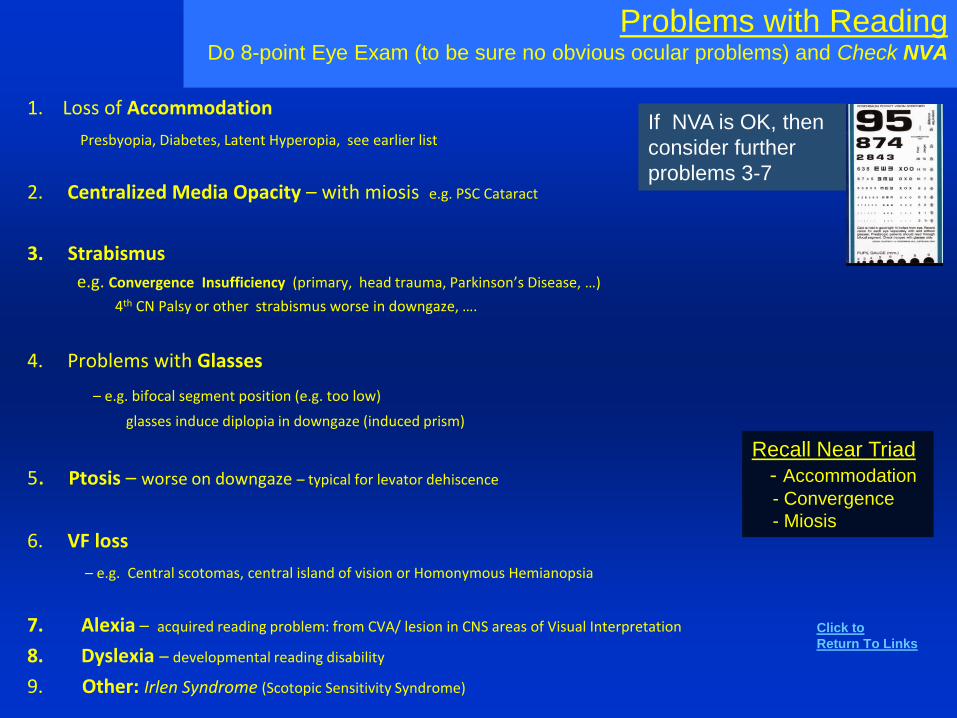
Problems with Reading Do 8-point Eye Exam (to be sure no obvious ocular problems) and Check NVA
1. Loss of Accommodation
Presbyopia, Diabetes, Latent Hyperopia, see earlier list
2. Centralized Media Opacity – with miosis e.g. PSC Cataract
3. Strabismus
e.g. Convergence Insufficiency (primary, head trauma, Parkinson’s Disease, …)
4th CN Palsy or other strabismus worse in downgaze, ….
4. Problems with Glasses
– e.g. bifocal segment position (e.g. too low)
glasses induce diplopia in downgaze (induced prism)
5. Ptosis – worse on downgaze – typical for levator dehiscence
6. VF loss
– e.g. Central scotomas, central island of vision or Homonymous Hemianopsia
7. Alexia – acquired reading problem: from CVA/ lesion in CNS areas of Visual Interpretation
8. Dyslexia – developmental reading disability
9. Other: Irlen Syndrome (Scotopic Sensitivity Syndrome)
Recall Near Triad
- Accommodation
- Convergence
- Miosis
If NVA is OK, then
consider further
problems 3-7
Click to
Return To Links

Suspected Loss of Vision in an InfantSigns of Poor Vision in Infant:
Absence of blink response to bright light, Overlooking, Oculodigital sign
Poor Fixation, Loss of Opto-Kinetc Nystagmus (OKN) (when spinning with child)
“Congenital Nystagmus” – sign of anterior pathway disease (retinal dystrophy, optic nerve, chiasm, optic tract). Onset usually not for 8-12 weeks. Usually in Cortical Visual loss there is no Nystagmus.
Roving Eye Movements – Usually sign of affixational patients with vision less than 20/400
First do a complete eye exam looking for more obvious causes
– e.g. refractive error, media opacities, strabismus / amblyopia , retinal changes, optic nerve Atrophy, Hypoplasia,
etc. If no obvious findings then consider….
Considerations:
Delayed Maturation of Visual System
Motor Dysfunction – e.g. Cerebral Palsy with poor eye movements
Leber’s Congenital Amaurosis
Retinal Dystrophies e.g. Achromatopsia, Congenital Stationary Night Blindness
Cone Dystrophy
Albinism / Foveal Aplasia
Subtle Optic Neuropathy – Hypoplasia? Atrophy?
Cortical Visual Loss: Peri-natal Hypoxia or Hemorrhage, Hydrocephalus, Head Trauma / Abuse, Metabolic (hypoglycemia, CO poisoning, uremia) Toxins (e.g. Pb), Meningitis, Encephalitis, Malformations – Encephaloceles, Chiari malformations, porencephalic cysts, neurodegenerative disorders
These would give appearance of poor
vision, yet could still could have good
vision in each eye
ERG appropriate in suspected
Retinal Dystrophy
Neuroimaging needed when
see an optic nerve
abnormality or cannot explain
cortical visual loss
Click to
Return To Links
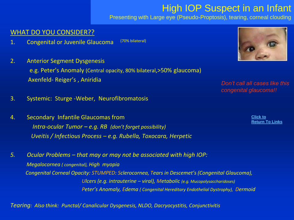
High IOP Suspect in an InfantPresenting with Large eye (Pseudo-Proptosis), tearing, corneal clouding
WHAT DO YOU CONSIDER??
1. Congenital or Juvenile Glaucoma (70% bilateral)
2. Anterior Segment Dysgenesis
e.g. Peter’s Anomaly (Central opacity, 80% bilateral,>50% glaucoma)
Axenfeld- Reiger’s , Aniridia
3. Systemic: Sturge -Weber, Neurofibromatosis
4. Secondary Infantile Glaucomas from
Intra-ocular Tumor – e.g. RB (don’t forget possibility)
Uveitis / Infectious Process – e.g. Rubella, Toxocara, Herpetic
5. Ocular Problems – that may or may not be associated with high IOP:
Megalocornea ( congenital), High myopia
Congenital Corneal Opacity: STUMPED: Sclerocornea, Tears in Descemet’s (Congenital Glaucoma),
Ulcers (e.g. intrauterine – viral), Metabolic (e.g. Mucopolysaccharidoses)
Peter’s Anomaly, Edema ( Congenital Hereditary Endothelial Dystrophy), Dermoid
Tearing: Also think: Punctal/ Canalicular Dysgenesis, NLDO, Dacryocystitis, Conjunctivitis
Don’t call all cases like this
congenital glaucoma!!
Click to
Return To Links
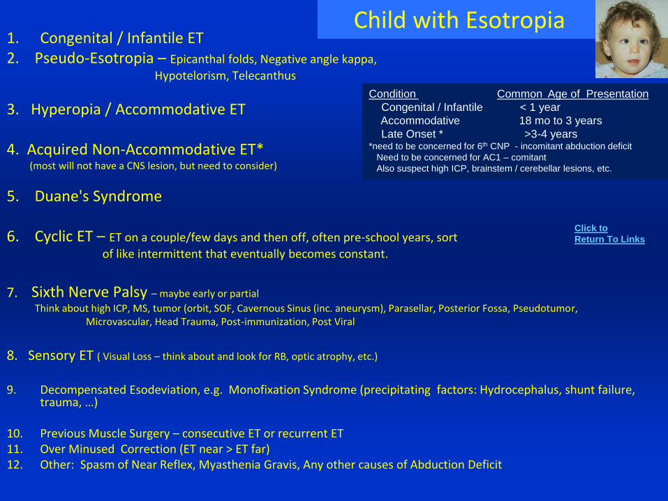
Child with Esotropia1. Congenital / Infantile ET2. Pseudo-Esotropia – Epicanthal folds, Negative angle kappa,
Hypotelorism, Telecanthus
3. Hyperopia / Accommodative ET
4. Acquired Non-Accommodative ET*(most will not have a CNS lesion, but need to consider)
5. Duane's Syndrome
6. Cyclic ET – ET on a couple/few days and then off, often pre-school years, sort
of like intermittent that eventually becomes constant.
7. Sixth Nerve Palsy – maybe early or partial
Think about high ICP, MS, tumor (orbit, SOF, Cavernous Sinus (inc. aneurysm), Parasellar, Posterior Fossa, Pseudotumor, Microvascular, Head Trauma, Post-immunization, Post Viral
8. Sensory ET ( Visual Loss – think about and look for RB, optic atrophy, etc.)
9. Decompensated Esodeviation, e.g. Monofixation Syndrome (precipitating factors: Hydrocephalus, shunt failure, trauma, …)
10. Previous Muscle Surgery – consecutive ET or recurrent ET11. Over Minused Correction (ET near > ET far)12. Other: Spasm of Near Reflex, Myasthenia Gravis, Any other causes of Abduction Deficit
Condition Common Age of Presentation
Congenital / Infantile < 1 year
Accommodative 18 mo to 3 years
Late Onset * >3-4 years *need to be concerned for 6th CNP - incomitant abduction deficit
Need to be concerned for AC1 – comitant
Also suspect high ICP, brainstem / cerebellar lesions, etc.
Click to
Return To Links
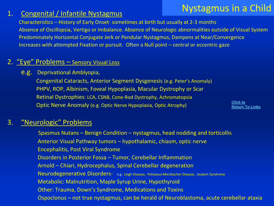
1. Congenital / Infantile NystagmusCharacteristics – History of Early Onset- sometimes at birth but usually at 2-3 months
Absence of Oscillopsia, Vertigo or Imbalance. Absence of Neurologic abnormalities outside of Visual System
Predominately Horizontal Conjugate Jerk or Pendular Nystagmus, Dampens at Near/Convergence
Increases with attempted Fixation or pursuit. Often a Null point – central or eccentric gaze
2. “Eye” Problems – Sensory Visual Loss
e.g. Deprivational Amblyopia,
Congenital Cataracts, Anterior Segment Dysgenesis (e.g. Peter’s Anomaly)
PHPV, ROP, Albinism, Foveal Hypoplasia, Macular Dystrophy or Scar
Retinal Dystrophies: LCA, CSNB, Cone-Rod Dystrophy, Achromatopsia
Optic Nerve Anomaly (e.g. Optic Nerve Hypoplasia, Optic Atrophy)
3. “Neurologic” Problems
Spasmus Nutans – Benign Condition – nystagmus, head nodding and torticollis
Anterior Visual Pathway tumors – hypothalamic, chiasm, optic nerve
Encephalitis, Post Viral Syndrome
Disorders in Posterior Fossa – Tumor, Cerebellar Inflammation
Arnold – Chiari, Hydrocephalus, Spinal Cerebellar degeneration
Neurodegenerative Disorders- e.g. Leigh Disease, Pelizaeus-Merzbacher Disease, Joubert Syndrome
Metabolic: Malnutrition, Maple Syrup Urine, Hypothyroid
Other: Trauma, Down’s Syndrome, Medications and Toxins
Ospoclonus – not true nystagmus, can be herald of Neuroblastoma, acute cerebellar ataxia
Nystagmus in a Child
Click to
Return To Links
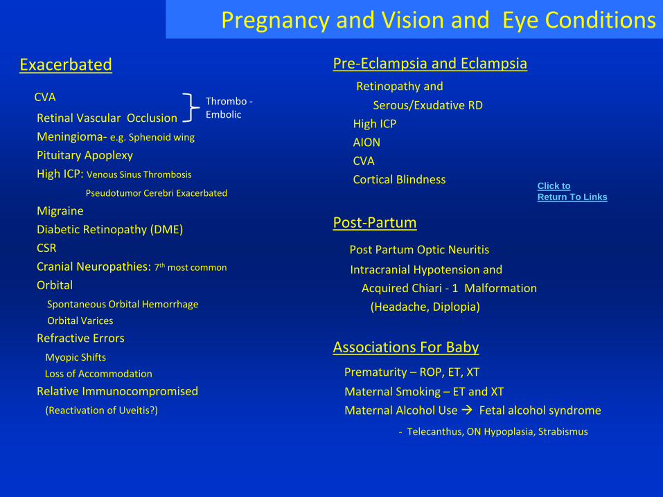
Pregnancy and Vision and Eye Conditions
Exacerbated
CVA
Retinal Vascular Occlusion
Meningioma- e.g. Sphenoid wing
Pituitary Apoplexy
High ICP: Venous Sinus Thrombosis
Pseudotumor Cerebri Exacerbated
Migraine
Diabetic Retinopathy (DME)
CSR
Cranial Neuropathies: 7th most common
Orbital
Spontaneous Orbital Hemorrhage
Orbital Varices
Refractive Errors
Myopic Shifts
Loss of Accommodation
Relative Immunocompromised
(Reactivation of Uveitis?)
Pre-Eclampsia and Eclampsia
Retinopathy and
Serous/Exudative RD
High ICP
AION
CVA
Cortical Blindness
Post-Partum
Post Partum Optic Neuritis
Intracranial Hypotension and
Acquired Chiari - 1 Malformation
(Headache, Diplopia)
Associations For Baby
Prematurity – ROP, ET, XT
Maternal Smoking – ET and XT
Maternal Alcohol Use Fetal alcohol syndrome
- Telecanthus, ON Hypoplasia, Strabismus
Click to
Return To Links
Thrombo -Embolic
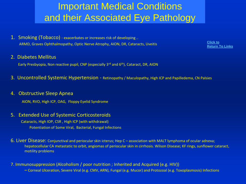
Important Medical Conditions
and their Associated Eye Pathology
1. Smoking (Tobacco) - exacerbates or increases risk of developing ..
ARMD, Graves Ophthalmopathy, Optic Nerve Atrophy, AION, DR, Cataracts, Uveitis
2. Diabetes Mellitus
Early Presbyopia, Non reactive pupil, CNP (especially 3rd and 6th), Cataract, DR, AION
3. Uncontrolled Systemic Hypertension - Retinopathy / Maculopathy, High ICP and Papilledema, CN Palsies
4. Obstructive Sleep Apnea
AION, RVO, High ICP, OAG, Floppy Eyelid Syndrome
5. Extended Use of Systemic CorticosteroidsCataracts, High IOP, CSR , High ICP (with withdrawal)
Potentiation of Some Viral, Bacterial, Fungal Infections
6. Liver Disease: Conjunctival and periocular skin icterus; Hep C – association with MALT lymphoma of ocular adnexa;
hepatocellular CA metastatic to orbit, angiomas of periocular skin in cirrhosis. Wilson Disease; KF rings, sunflower cataract, motility problems
7. Immunosuppression (Alcoholism / poor nutrition ; Inherited and Acquired (e.g. HIV)) – Corneal Ulceration, Severe Viral (e.g. CMV, ARN), Fungal (e.g. Mucor) and Protozoal (e.g. Toxoplasmosis) Infections
Click to
Return To Links
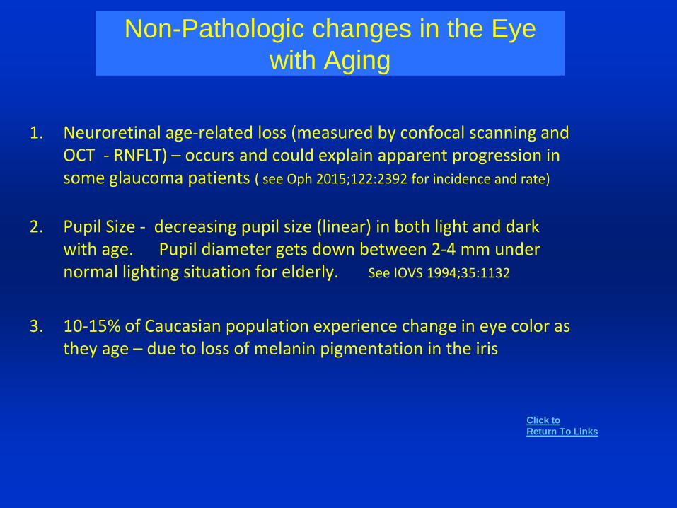
Non-Pathologic changes in the Eye
with Aging
1. Neuroretinal age-related loss (measured by confocal scanning and OCT - RNFLT) – occurs and could explain apparent progression in some glaucoma patients ( see Oph 2015;122:2392 for incidence and rate)
2. Pupil Size - decreasing pupil size (linear) in both light and dark with age. Pupil diameter gets down between 2-4 mm under normal lighting situation for elderly. See IOVS 1994;35:1132
3. 10-15% of Caucasian population experience change in eye color as they age – due to loss of melanin pigmentation in the iris
Click to
Return To Links
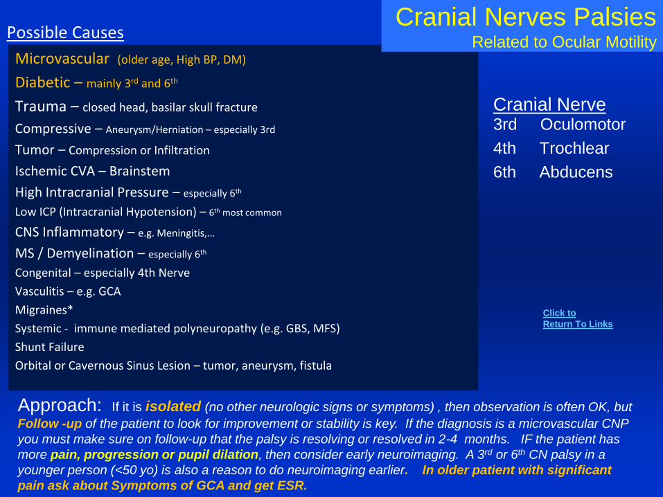
Microvascular (older age, High BP, DM)
Diabetic – mainly 3rd and 6th
Trauma – closed head, basilar skull fracture
Compressive – Aneurysm/Herniation – especially 3rd
Tumor – Compression or Infiltration
Ischemic CVA – Brainstem
High Intracranial Pressure – especially 6th
Low ICP (Intracranial Hypotension) – 6th most common
CNS Inflammatory – e.g. Meningitis,…
MS / Demyelination – especially 6th
Congenital – especially 4th Nerve
Vasculitis – e.g. GCA
Migraines*
Systemic - immune mediated polyneuropathy (e.g. GBS, MFS)
Shunt Failure
Orbital or Cavernous Sinus Lesion – tumor, aneurysm, fistula
Cranial Nerves PalsiesRelated to Ocular Motility
Cranial Nerve3rd Oculomotor
4th Trochlear
6th Abducens
Possible Causes
Approach: If it is isolated (no other neurologic signs or symptoms) , then observation is often OK, but
Follow -up of the patient to look for improvement or stability is key. If the diagnosis is a microvascular CNP
you must make sure on follow-up that the palsy is resolving or resolved in 2-4 months. IF the patient has
more pain, progression or pupil dilation, then consider early neuroimaging. A 3rd or 6th CN palsy in a
younger person (<50 yo) is also a reason to do neuroimaging earlier. In older patient with significant
pain ask about Symptoms of GCA and get ESR.
Click to
Return To Links
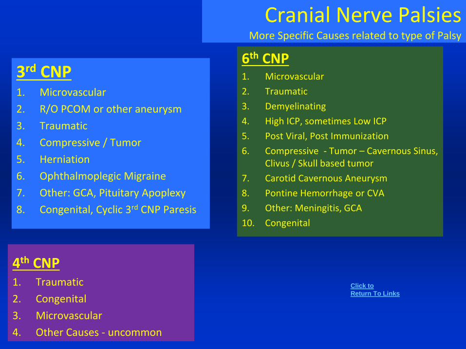
Cranial Nerve PalsiesMore Specific Causes related to type of Palsy
3rd CNP1. Microvascular
2. R/O PCOM or other aneurysm
3. Traumatic
4. Compressive / Tumor
5. Herniation
6. Ophthalmoplegic Migraine
7. Other: GCA, Pituitary Apoplexy
8. Congenital, Cyclic 3rd CNP Paresis
6th CNP1. Microvascular
2. Traumatic
3. Demyelinating
4. High ICP, sometimes Low ICP
5. Post Viral, Post Immunization
6. Compressive - Tumor – Cavernous Sinus, Clivus / Skull based tumor
7. Carotid Cavernous Aneurysm
8. Pontine Hemorrhage or CVA
9. Other: Meningitis, GCA
10. Congenital
4th CNP1. Traumatic
2. Congenital
3. Microvascular
4. Other Causes - uncommon
Click to
Return To Links
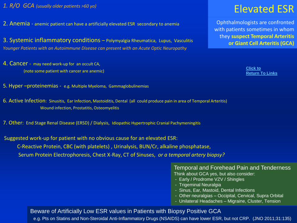
Elevated ESR1. R/O GCA (usually older patients >60 yo)
2. Anemia - anemic patient can have a artificially elevated ESR secondary to anemia
3. Systemic inflammatory conditions – Polymyalgia Rheumatica, Lupus, Vasculitis
Younger Patients with an Autoimmune Disease can present with an Acute Optic Neuropathy
4. Cancer - may need work-up for an occult CA,
(note some patient with cancer are anemic)
5. Hyper –proteinemias - e.g. Multiple Myeloma, Gammaglobulinemias
6. Active Infection: Sinusitis, Ear Infection, Mastoiditis, Dental (all could produce pain in area of Temporal Arteritis)
Wound infection, Prostatitis, Osteomyelitis
7. Other: End Stage Renal Disease (ERSD) / Dialysis, Idiopathic Hypertrophic Cranial Pachymeningitis
Suggested work-up for patient with no obvious cause for an elevated ESR:
C-Reactive Protein, CBC (with platelets) , Urinalysis, BUN/Cr, alkaline phosphatase,
Serum Protein Electrophoresis, Chest X-Ray, CT of Sinuses, or a temporal artery biopsy?
Ophthalmologists are confronted with patients sometimes in whom
they suspect Temporal Arteritisor Giant Cell Arteritis (GCA)
Beware of Artificially Low ESR values in Patients with Biopsy Positive GCA
e.g. Pts on Statins and Non-Steroidal Anti-Inflammatory Drugs (NSAIDS) can have lower ESR, but not CRP. (JNO 2011;31:135)
Temporal and Forehead Pain and Tenderness Think about GCA yes, but also consider:
- Early / Prodrome VZV / Shingles
- Trigeminal Neuralgia
- Sinus, Ear, Mastoid, Dental Infections
- Other neuralgias – Occipital, Cervical, Supra Orbital
- Unilateral Headaches – Migraine, Cluster, Tension
Click to
Return To Links
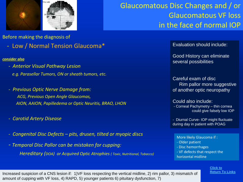
Glaucomatous Disc Changes and / orGlaucomatous VF loss
in the face of normal IOP
Before making the diagnosis of
- Low / Normal Tension Glaucoma*
consider also
- Anterior Visual Pathway Lesion
e.g. Parasellar Tumors, ON or sheath tumors, etc.
- Previous Optic Nerve Damage from:
ACG, Previous Open Angle Glaucomas,
AION, AAION, Papilledema or Optic Neuritis, BRAO, LHON
- Carotid Artery Disease
- Congenital Disc Defects – pits, drusen, tilted or myopic discs
- Temporal Disc Pallor can be mistaken for cupping:
Hereditary (DOA) or Acquired Optic Atrophies ( Toxic, Nutritional, Tobacco)
Evaluation should include:
Good History can eliminate
several possibilities
Careful exam of disc
Rim pallor more suggestive
of another optic neuropathy
Could also include:- Corneal Pachymetry – thin cornea
could give falsely low IOP
- Diurnal Curve- IOP might fluctuate
during day in patient with POAG
Increased suspicion of a CNS lesion if: 1)VF loss respecting the vertical midline, 2) rim pallor, 3) mismatch of
amount of cupping with VF loss, 4) RAPD, 5) younger patients 6) pituitary dysfunction, 7)
More likely Glaucoma if :- Older patient- Disc hemorrhages- VF defects that respect the horizontal midline
Click to
Return To Links
Ocular Effects of Medications• Illusions and Hallucinations:
Psychogenic Medications and Drugs -
Dopaminergics: levadopa, bromocryptine,
Anticholinergics: atropine, scopolamine, cyclopentolate
Tricyclic Antidepressants, Beta-Blockers
Adrenergic – albuterol, Phenylephrine, Theophyline, Amphetamine, Cocaine
Alcohol
Misc: Benzodiazepines, corticosteroids, NSAIDS, Ca Channel blockers, Narcotics, anticonvulsants,…
Trazodone, Nefazodone, Topiramate, Risperidone, Clomiphene
Illicit Drugs: LSD, Cannibus, Methamphetamine, Psylocibin (mushrooms)
• Transient Refractive Shifts
Myopic – Topiramate
Hyperopic - Phenothiazides, Antihistamines, Cholorquine, Anticholinergics, Cannabis
• Cataract Corticosteroids (PSC), Statins, Phenothiazines (Chlorpromazine)
• Angle Closure Glaucoma – increased risk
Ant-histamines, Anti-psychotics, Tri-cyclic Antidepressants, Ephedrine
• Retinal Pigmentary Changes Phenothiazines - Thioridazine (Mellaril), Chlorpromazine ( Thorazine), Chloroquine, Hydroxychloroquine Tamoxifen, Indomethacine, Deferoxamine
Click to
Return To Links
Ocular Effects of Medications
• Papilledema / Increased Intracranial Pressure
Vitamin A , Retinoids e.g. Isotretinoin (Accutane) , Tetracylines: Doxycycline, Minocycline, Lithium, Human Growth Hormone, Thyroid supplementation, Nalidixic Acid, Oral Contraceptives, Withdrawal of Corticosteroids,
• NystagmusPhenytoin, Barbituates, Lithium, Salicylates, Antihistamines
• Loss of VF and/ or NFL Vigabatrin
• Optic NeuropathyAmiodarone, Ethambutol, INH, Quinine, Sulfonamides, Chloramphenicol, SSRI’s
Anti TNF Ab (Remicade, Embrel), Tacrolimus
Interferon; Chemotherapy (Doxorubicin, Cisplatin, Methotrexate, Vincristine) especially with Radiation;
Lindane Shampoo (absorbed)
• Myasthenia Gravis (Induced, Aggravated or Mimic) - Statins (3HMG CoA reductase inhibitors)
Also consider: Ca Channel Blockers (e.g. Verapamil), Beta Blockers (e.g Propranolol or Timolol), Anti-Arrhythmics (e.g. procainamide), Quinine, chloroquine, Penicillamine, Corticosteroids, Antibiotics (e.g. Fluoroquinolones, Aminoglycosides, Macrolides), Anti-Seizure (e.g. phenytoin, carbamazepine and Gabapentine), Chemotherapy, Interferon, Some IV Anesthetics,
Click to
Return To Links
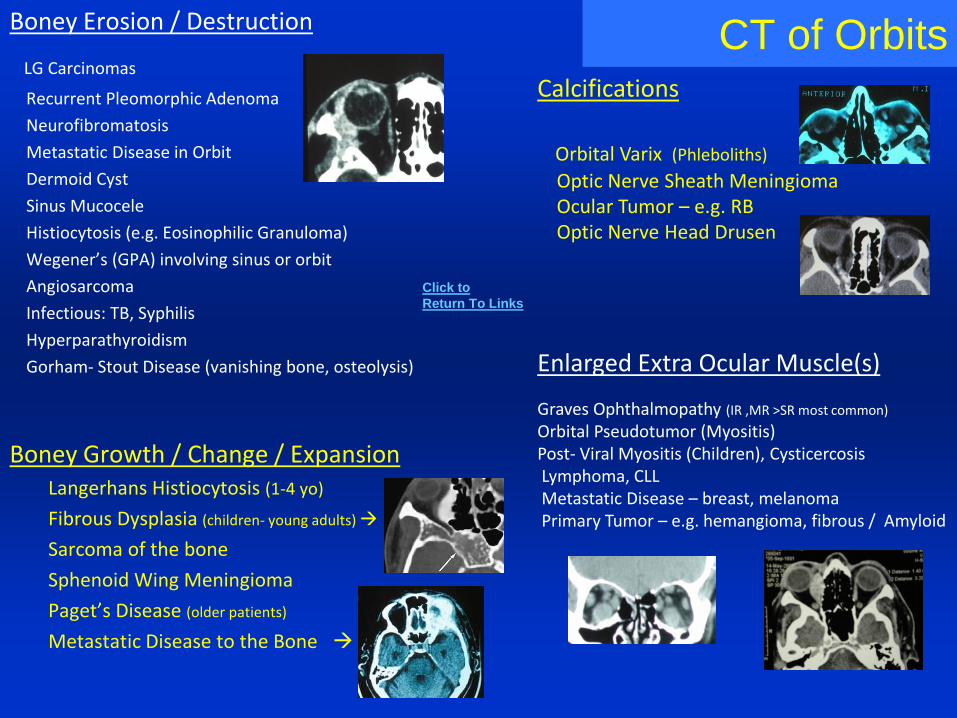
CT of OrbitsBoney Erosion / Destruction
LG Carcinomas
Recurrent Pleomorphic Adenoma
Neurofibromatosis
Metastatic Disease in Orbit
Dermoid Cyst
Sinus Mucocele
Histiocytosis (e.g. Eosinophilic Granuloma)
Wegener’s (GPA) involving sinus or orbit
Angiosarcoma
Infectious: TB, Syphilis
Hyperparathyroidism
Gorham- Stout Disease (vanishing bone, osteolysis)
Boney Growth / Change / ExpansionLangerhans Histiocytosis (1-4 yo)
Fibrous Dysplasia (children- young adults)
Sarcoma of the bone
Sphenoid Wing Meningioma
Paget’s Disease (older patients)
Metastatic Disease to the Bone
Calcifications
Orbital Varix (Phleboliths)
Optic Nerve Sheath MeningiomaOcular Tumor – e.g. RBOptic Nerve Head Drusen
Enlarged Extra Ocular Muscle(s)
Graves Ophthalmopathy (IR ,MR >SR most common)
Orbital Pseudotumor (Myositis)Post- Viral Myositis (Children), CysticercosisLymphoma, CLLMetastatic Disease – breast, melanomaPrimary Tumor – e.g. hemangioma, fibrous / Amyloid
Click to
Return To Links
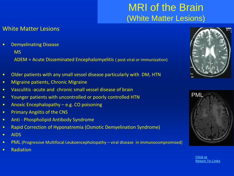
MRI of the Brain(White Matter Lesions)
White Matter Lesions
• Demyelinating Disease
MS
ADEM = Acute Disseminated Encephalomyelitis ( post viral or immunization)
• Older patients with any small vessel disease particularly with DM, HTN
• Migraine patients, Chronic Migraine
• Vasculitis -acute and chronic small vessel disease of brain
• Younger patients with uncontrolled or poorly controlled HTN
• Anoxic Encephalopathy – e.g. CO poisoning
• Primary Angiitis of the CNS
• Anti - Phospholipid Antibody Syndrome
• Rapid Correction of Hyponatremia (Osmotic Demyelination Syndrome)
• AIDS
• PML (Progressive Multifocal Leukoencepholopathy – viral disease in immunocompromised)
• Radiation
PML
Click to
Return To Links
DizzinessDizzy patients are often referred for eye exams
Vertigo - Peripheral or Central causes – associated with nystagmus
Peripheral: BPPV* (seconds < minute associated with head posturing, no tinnitus, hearing OK)
Acute Labyrinthitis/ Vestibular Neuritis (hours to 2 weeks, no tinnitus, hearing OK)
Meniere’s Disease (hours, +hearing loss, +tinnitus)
Drug Toxicity (Aminoglycosides, anti- seizure meds, alcohol, ASA, chemo)
Superior Canal (labyrinth semicircular) Dehiscence Syndrome - Loud noises cause patient to suffer sudden transient vertigo and blurred vision
Central: CPA tumor e.g. Acoustic Neuroma (hearing loss, tinnitus, other CN deficits)
Ischemia, Stroke involving Vertebral basilar circulationMSAcute Cerebellar DiseaseVestibular Migraine
Disequilibrium – elderly issues like fear of walking, musculoskeletal problems, peripheral neuropathy
Consider also eye Problems: visual loss or causes of oscillopsia and diplopia or problem with glasses
Pre-syncope – Orthostatic Hypotension from medications, arrhythmias, vaso-vagal
Psychiatric – anxiety, hyperventilation, depression, substance abuse
Multifactorial or unknown
- Acute Vestibular Syndrome – rapid onset of vertigo, N/V, nystagmus, unsteady gait, head motion intolerance, most commonly due to vestibular neuritis or posterior fossa stroke.
- *BPPV- Benign Paroxysmal Positional Vertigo
- Shopping Cart Syndrome (B. Farris – COS 2013) – symptoms of dizziness in patients (with prior BPPV or labyrinthitis) – when in an
environment which can induce OKN – such as pushing shopping cart down visually busy grocery aisle.
Click to
Return To Links
AC - Anterior Chamber
AC1 – Arnold Chiari Malformation Type 1
ACE – Angiotensin Converting Enzyme
ACG – Angle Closure Glaucoma
AD – Autosomal Dominant
ADOA – Autosomal Dominant Optic Atrophy
AIBSES – Acute Idiopathic Blind Spot Enlargement Syndrome
AIDS – Acquired Immune Deficiency Syndrome
AION – Anterior Ischemic Optic Neuropathy
AAION– Arteritic AION (GCA)
AK – Astigmatic Keratotomy
AMPPE – Acute Multifocal Posterior Placoid Epitheliopathy
ANA – Anti-Nuclear Antibody (Lupus Diagnosis)
AR – Autosomal Recessive
ARMD – Age Related Macular Degeneration
ARN – Acute Retinal Necrosis
AVM – Arterial Venous Malformation
BEB – Benign Essential Blepharospasm
BCCA – Basal Cell Carcinoma
BCP – Birth Control Pills, Contraceptives
BDUMP – Bilateral Diffuse Uveal Melanocytic Proliferation
BP – Blood Pressure
BPES - Blepharophimosis- Ptosis- Epicanthus Inversus Syndrome
BPV – Benign Positional Vertigo
BRAO- Branch Retinal artery Occlusion
BRVO- Branch RVO
CA – Carcinoma
CARS – Cancer Associated Retinopathy Syndrome
CC – Carotid Cavernous
CE – Cataract Extraction (Surgery)
CFEOM – Congenital Fibrosis of Extraocular Muscles
CHRPE – Congenital Hypertrophy of the RPE
CIN – Carcinoma in Situ
CLL – Chronic Lymphocytic Leukemia
CME – Cystoid Macular Edema
CMV – Cytomegalovirus
CN- Cranial Nerve
CNP – CN palsy
CNS – Central Nervous System
CPA – Cerebellar Pontine Angle
CPEO – Chronic Progressive External Ophthalmoplegia
CR – Chorioretinal
CRAO- central retinal artery occlusion, CRVO- central ret. vein occlusion
CSR – Central Serous Retinopathy
CSNB – Congenital Stationary Night Blindness
CT- Computerized Tomography
CTA – CT Angiogram
CTL – Contact Lens
CVA – Cerebral (CNS) Vascular Accident (Stroke)
CXR – Chest X-Ray
DDX – Differential Diagnosis
DOA – Dominant Optic Atrophy
DR – Diabetic Retinopathy
DME- Diabetic Macular Edema
DM – Diabetes Mellitus
DUSN – Diffuse Unilateral Subacute Neuroretinitis from Nematode
EOM – Extra Ocular Muscle
ERG – Electroretinogram
ERM – Epi - Retinal Membrane
ESR – Erythrocyte Sedimentation Rate
ET – Esotropia
FA- Fluorescein Angiogram
FB- Foreign Body
FBS – Foreign Body Sensation
FEVR - Familial exudative vitreoretinopathy
FHx – Family Medical History
GBS- Guillain-Barre Syndrome
GCA – Giant Cell Arteritis (aka Temporal Arteritis)
Lis
t of A
bb
revia
tion
sClick to
Return To Links
GPA- Granulomatosis with Polyangiitis (Wegener's)
HA - Headache
HIV – Human Immunodeficiency Virus (AIDS)
HLA – Human Leukocyte Antigen
HSV – Herpes Simplex Virus
HT – Hypertropia
HoT - Hypotropia
HTN – Hypertension (high blood pressure)
HZO- Herpes Zoster Ophthalmicus
IBD - Inflammatory Bowel Diseases
ICE – Iridocorneal Endothelial (Syndrome)
ICP - Intracranial Pressure
IgG4-ROD – Immunoglobulin G4 Related Ophthalmic Disease
IIH – Idiopathic Intracranial Hypertension (pseudotumor cerebri)
INO – Internuclear Ophthalmoplegia
IO – Inferior Oblique
IOFB – Intra-Ocular Foreign Body
IOIS – Idiopathic Orbital Inflammatory Syndrome
IOL – Intraocular lens
ION- Ischemic Optic Neuropathy
IOP – Intraocular Pressure
IR – Inferior Rectus
JIA – Juvenile Idiopathic Arthritis
JRA - Juvenile Rheumatoid Arthritis
KC – Keratoconjunctivitis
KP – Keratoprecipitates
LASIK – Laser Assisted In Situ Keratomileusis
LCA – Leber’s Congenital Amaurosis
LHON – Leber’s Hereditary Optic Neuropathy
LG – Lacrimal Gland
LL – Lower Lid (Lower Eyelid)
LLL – Left Lower Eyelid
LP – Lumbar Puncture (Spinal Tap)
LR – Lateral Rectus
LSCD- Limbal Stem Cell Deficiency
LUL – Left Upper Lid
MARS – Melanoma Associated Retinopathy Syndrome
MEWDS – Multiple Evanescent White Dot Syndrome
MFS – Miller Fisher Syndrome (polyneuropathy)
MG – Myasthenia Gravis
MM – Multiple Myeloma
MR – Medial Rectus
MRA – Magnetic Resonance Angiography
MRD – Margin Reflex Distance (Upper Eyelids)
MRI - Magnetic Resonance Imaging
MRV- MR Venography
MS – Multiple Sclerosis
NAION- Non-Arteritic AION
NF - Neurofibromatosis
NFL – Nerve Fiber Layer
NLO – Nasolacrimal Obstruction
NLDO – NL Duct Obstruction
NPDR – Non-Proliferative Diabetic Retinopathy
NVA- Near Visual Acuity
NVD – Neovascularization of the Disk
NVI – Neovascularization of the Iris
OAG – Open Angle Glaucoma
OCP – Ocular Cicatricial Pemphigoid
OCT – Ocular Coherence Tomography
OIS – Ocular Ischemic Syndrome
OKN – Opto-Kinetic Nystagmus
ONH – Optic Nerve Head
OMG – Ocular Myasthenia Gravis
ONM – Ocular Neuromyotonia
OSSN – Ocular Surface Squamous Neoplasia
PAN – Polyarteritis Nodosa
PC – Posterior Chamber
PCO - Posterior Capsular Opacity (Pseudophakic eye)
Lis
t of A
bb
revia
tion
s
Click to
Return To Links
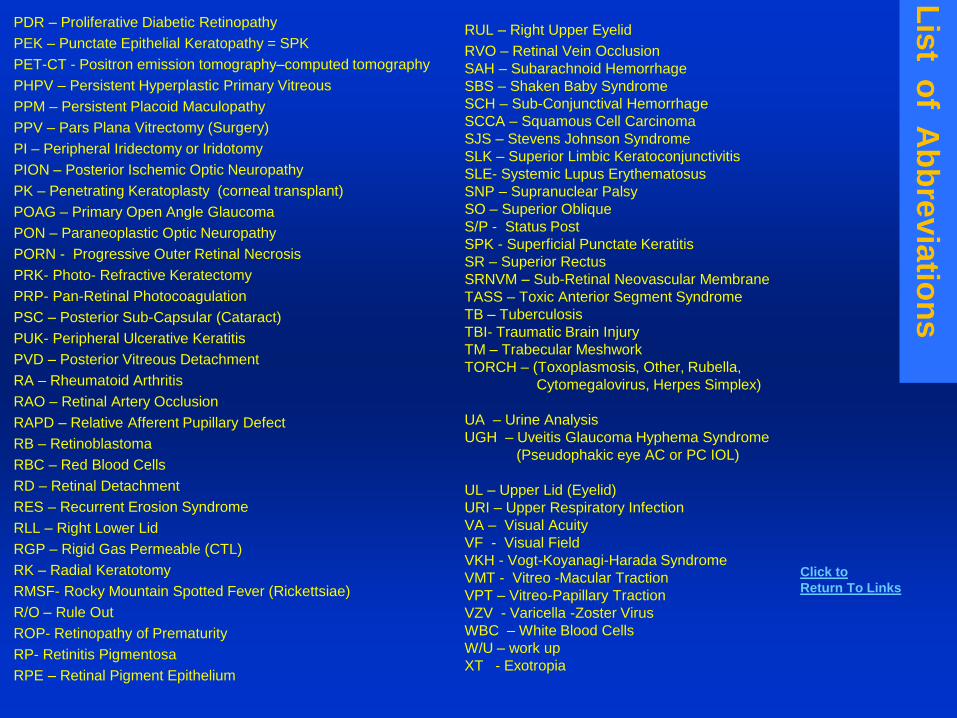
PDR – Proliferative Diabetic Retinopathy
PEK – Punctate Epithelial Keratopathy = SPK
PET-CT - Positron emission tomography–computed tomography
PHPV – Persistent Hyperplastic Primary Vitreous
PPM – Persistent Placoid Maculopathy
PPV – Pars Plana Vitrectomy (Surgery)
PI – Peripheral Iridectomy or Iridotomy
PION – Posterior Ischemic Optic Neuropathy
PK – Penetrating Keratoplasty (corneal transplant)
POAG – Primary Open Angle Glaucoma
PON – Paraneoplastic Optic Neuropathy
PORN - Progressive Outer Retinal Necrosis
PRK- Photo- Refractive Keratectomy
PRP- Pan-Retinal Photocoagulation
PSC – Posterior Sub-Capsular (Cataract)
PUK- Peripheral Ulcerative Keratitis
PVD – Posterior Vitreous Detachment
RA – Rheumatoid Arthritis
RAO – Retinal Artery Occlusion
RAPD – Relative Afferent Pupillary Defect
RB – Retinoblastoma
RBC – Red Blood Cells
RD – Retinal Detachment
RES – Recurrent Erosion Syndrome
RLL – Right Lower Lid
RGP – Rigid Gas Permeable (CTL)
RK – Radial Keratotomy
RMSF- Rocky Mountain Spotted Fever (Rickettsiae)
R/O – Rule Out
ROP- Retinopathy of Prematurity
RP- Retinitis Pigmentosa
RPE – Retinal Pigment Epithelium
Lis
t of A
bb
revia
tion
sRUL – Right Upper Eyelid
RVO – Retinal Vein Occlusion
SAH – Subarachnoid Hemorrhage
SBS – Shaken Baby Syndrome
SCH – Sub-Conjunctival Hemorrhage
SCCA – Squamous Cell Carcinoma
SJS – Stevens Johnson Syndrome
SLK – Superior Limbic Keratoconjunctivitis
SLE- Systemic Lupus Erythematosus
SNP – Supranuclear Palsy
SO – Superior Oblique
S/P - Status Post
SPK - Superficial Punctate Keratitis
SR – Superior Rectus
SRNVM – Sub-Retinal Neovascular Membrane
TASS – Toxic Anterior Segment Syndrome
TB – Tuberculosis
TBI- Traumatic Brain Injury
TM – Trabecular Meshwork
TORCH – (Toxoplasmosis, Other, Rubella,
Cytomegalovirus, Herpes Simplex)
UA – Urine Analysis
UGH – Uveitis Glaucoma Hyphema Syndrome
(Pseudophakic eye AC or PC IOL)
UL – Upper Lid (Eyelid)
URI – Upper Respiratory Infection
VA – Visual Acuity
VF - Visual Field
VKH - Vogt-Koyanagi-Harada Syndrome
VMT - Vitreo -Macular Traction
VPT – Vitreo-Papillary Traction
VZV - Varicella -Zoster Virus
WBC – White Blood Cells
W/U – work up
XT - Exotropia
Click to
Return To Links

Journal References
Ophthalmology (AAO Journal) OPH
Survey of Ophthalmology Survey
JAMA Ophthalmology (Archives) JAMA Oph
American Journal of Ophthalmology AJO
Journal of Neuro-ophthalmology JNO
Investigative Ophthalmology and Visual Science IOVS
Abbreviation Used

Major References
Hampton Roy, Ocular Differential Diagnosis Book
American Academy of Ophthalmology – Basic and Clinical Science Series Monograph
Emedicine Ophthalmology – Multiple articles from website Internet
EyeRounds.org
AAO- EyeWiki
Survey of Ophthalmology – Many of the Major Reviews Journal
Picture Credits:
1) Personal Collection
2) University of Iowa site – uiowa.edu
3) Redatlas.org
4) University of Michigan , Kellogg Eye Center
5) Some images seem to be in wide circulation and have no credit given. However, if any image is seen and recognized as copyrighted and needs to be removed or given proper credit – please contact me.
Medium

The heavens declare the glory of God;
the skies proclaim the work of his handsPsalm 19:1