
Paper · Web viewIn case of umbilical hernia, a discrete spherical swelling is obvious at the...
20
Researcher 2019;11(10) http://www.sciencepub.net/researcher RSJ Types Of Hernia And Surgical Management Approaches In Domestic Animals: Review Dese Kefyalew, Abebe Firomsa and Tolessa Ebissa Department of surgery, Bishoftu campus, Addis Ababa University, Ethiopia E-mails: [email protected] , [email protected] . Telephones: +251-917-068-648, +251-921-889-089 Abstract: Hernia is bulging of abdominal contents in abdominal wall caused by naturally through canal (congenital) or traumatic (acquired). Hernias have several deleterious effects, such as lowering the productivity and re- productivity of the affected animals where, abdominal, inguinal and perineal hernias could entrap a fetus in their hernial sacs leading to dystocia in domestic animals. Therefore, the aim of this paper is to review on the different types of hernia and its surgical management in domestic animals. Hernia is classified as internal and external hernia. External hernia is a displacement of an organ, part of an organ, or a tissue outside the abdominal cavity through an abnormal opening in the abdominal wall which can be noted from the outside of animal’s body and can be detected with external examination. It is the most common form of problems of domestic animals which can be umbilical, inguinal, scrotal, femoral, perineal, and ventral (or abdominal) hernias based on anatomic location. Umbilical hernia is usually occurs in calves due to failure to closed of the umbilical cord, while ventral hernia are usually present in cattle and horses because of trauma and heavy weight. It can causes decreases in reproductive performance, production loss and even leads to the death of animal depending on the types of hernias. External hernia was easy to diagnose by case history, inspection and palpation where it can be overcome by both surgical and non-surgical approaches to correct different types of hernias occurring in domestic animals. Moreover it can be prevented to a large extent by modifying the prevailing management practices. However, still information on the associated risk factors and its prevalence is not well studied. Therefore further studies are needed to identify the associated risk factors and farmers should be aware of the diseases and improve prevention approaches through implementing better management practices. [Dese K., Abebe F. and Tolessa E. Types Of Hernia And Surgical Management Approaches In Domestic Animals: Review. Researcher 2019;11(10):35-46]. ISSN 1553-9865 (print); ISSN 2163-8950 (online). http://www.sciencepub.net/researcher . 6. doi: 10.7537/marsrsj111019.0 6 . Key words: Domestic animals, Hernia, Hernioplasty, Hernioraphy, Mesh, Surgical managment 1. Introduction Hernia is defined as the protrusion of an organ or tissue 1
Transcript of Paper · Web viewIn case of umbilical hernia, a discrete spherical swelling is obvious at the...

Researcher 2019;11(10) http://www.sciencepub.net/researcher RSJ
Types Of Hernia And Surgical Management Approaches In Domestic Animals: Review
Dese Kefyalew, Abebe Firomsa and Tolessa Ebissa
Department of surgery, Bishoftu campus, Addis Ababa University, EthiopiaE-mails: [email protected], [email protected].
Telephones: +251-917-068-648, +251-921-889-089
Abstract: Hernia is bulging of abdominal contents in abdominal wall caused by naturally through canal (congenital) or traumatic (acquired). Hernias have several deleterious effects, such as lowering the productivity and re-productiv-ity of the affected animals where, abdominal, inguinal and perineal hernias could entrap a fetus in their hernial sacs leading to dystocia in domestic animals. Therefore, the aim of this paper is to review on the different types of hernia and its surgical management in domestic animals. Hernia is classified as internal and external hernia. External hernia is a displacement of an organ, part of an organ, or a tissue outside the abdominal cavity through an abnormal open-ing in the abdominal wall which can be noted from the outside of animal’s body and can be detected with external examination. It is the most common form of problems of domestic animals which can be umbilical, inguinal, scrotal, femoral, perineal, and ventral (or abdominal) hernias based on anatomic location. Umbilical hernia is usually occurs in calves due to failure to closed of the umbilical cord, while ventral hernia are usually present in cattle and horses because of trauma and heavy weight. It can causes decreases in reproductive performance, production loss and even leads to the death of animal depending on the types of hernias. External hernia was easy to diagnose by case history, inspection and palpation where it can be overcome by both surgical and non-surgical approaches to correct different types of hernias occurring in domestic animals. Moreover it can be prevented to a large extent by modifying the pre-vailing management practices. However, still information on the associated risk factors and its prevalence is not well studied. Therefore further studies are needed to identify the associated risk factors and farmers should be aware of the diseases and improve prevention approaches through implementing better management practices.[Dese K., Abebe F. and Tolessa E. Types Of Hernia And Surgical Management Approaches In Domestic Ani-mals: Review. Researcher 2019;11(10):35-46]. ISSN 1553-9865 (print); ISSN 2163-8950 (online). http://www.sci-encepub.net/researcher. 6. doi:10.7537/marsrsj111019.0 6 .
Key words: Domestic animals, Hernia, Hernioplasty, Hernioraphy, Mesh, Surgical managment
1. IntroductionHernia is defined as the protrusion of an organ or
tissue through artificial or natural opening (Mcardle, 1997). This may be from accidental or normal anatomical opening, which does not completely fulfill its physiological function and the majority of hernia involves the protrusion of the abdominal contents through the part of the abdominal wall, diaphragm, or perineum, although herniations can occur elsewhere in the body (Sutradhar et al., 2009).
A typical hernia always consist of the “hernia ring” or an opening in the muscle which may have been brought about as a result of an accident, or may have been present at birth; a swelling appearing below the skin “hernia sac”, and the “hernia content” (Polli-cino et al., 2007). Depending on the anatomical site of herniation parts hernia classified as ventral or lateral abdominal hernia, diaphragmatic, inguinal, scrotal, umbilical, perineal and femoral hernia (Rahman et al., 2001). Hernia causes may by congenital or acquired (Sabev and Kanakov, 2009). The type of the herniated tissue as enterocel containing portion of intestine, epi-plocel or omentocele of omentum, enteroepiplocele of intestine and omentum, Gastrocele of stomach, vesic-
ocele (of bladder), hepatocele of liver, hysterocele of uterus and it may be, external or internal hernia (Kilich, 2014).
External hernia is the most common form of dis-eases occur in domestic animals which is character-ized by a displacement of an organ, part of an organ, or a tissue outside the abdominal cavity through an abnormal opening in the abdominal wall which can be noted from the outside of animal’s body and can be detected with external examination (Das et al., 2012).
Risk factors significantly associated with preva-lence of hernia are: hereditary, environmental and/ or animal handling factors are the factors that enhance the occurrence of the problems (Bates and Straw, 2008).
Hernias have several deleterious effects, such as lowering the productivity and re-productivity of the affected animals. It can enlarge over time and if not repaired surgically, it may cause pain, anorexia, weight loss, or dystocia when a gravid horn is found in the hernial sac. For instance, abdominal, inguinal and perineal hernias could entrap a foetus or foetuses in their hernial sacs leading to dystocia (Muggli et al.,
1

Researcher 2019;11(10) http://www.sciencepub.net/researcher RSJ
2014; Ruhil et al., 2018). Therefore, the objectives of this review paper are:
To review different types of hernia in domes-tic animals
To review on non-surgical and surgical ap-proaches for the treatment herniated domestic animal
To provide information on prevention of her-nia in domestic animals
2. Literature ReviewHernia is a protrusion of abdominal content/ vis-
cera, omentum, or abdominal organs through a natural or artificial opening in the abdominal wall to form a swelling covered by skin, subcutaneous tissue and peritoneum (Bayl et al., 2017). This possibly will oc-cur by accident or due to normal anatomical opening, which does not completely fulfill its functional (Planellas et al., 2012). The common risk factor for developing hernia is sex, age difference and animal management system. Hernias of the abdominal wall are common in all domestic species and include um-bilical hernias and inguinal or scrotal hernias (Niebauer et al., 2005). Hernias may be direct (through a rent in the body wall) or indirect (through an already existing ring, such as the inguinal ring or umbilical ring) (Meylan, 2008).2.1. Anatomy of Hernia
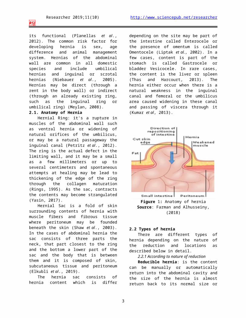
Hernial Ring: it's a rupture in muscles of the ab-dominal wall such as ventral hernia or widening of natural orifices of the umbilicus, or may be a natural passageway the inguinal canal (Petritz et al., 2012). The ring is the actual defect in the limiting wall, and it may be a small as a few millimeters or up to several centimeters and spontaneous attempts at healing may be lead to thickening of the edge of the ring through the collagen maturation (Rings, 1995). As the sac, contracts the contents may become strangulated (Yasin, 2017).
Hernial Sac is a fold of skin surrounding con-tents of hernia with muscle fibers and fibrous tissue where peritoneum may be founded beneath the skin (Shaw et al., 2003). In the cases of abdominal hernia the sac consists of three parts the neck, that part clos-est to the ring and the bottom a lower part of the sac and the body that is between them and it is composed of skin, subcutaneous tissue and peritoneum (Elkubli et al., 2019).
The hernia sac consists of hernia content which is differ depending on the site may be part of the in-testine called Enterocele or the presence of omentum is called Omentocele (Liptak et al., 2002). In a few cases, content is part of the stomach is called Gastro-cele or bladder Vesicocele. In rare cases, the content is the liver or spleen (Thas and Harcourt, 2013). The hernia either occur when there is a natural weakness in the inguinal canal and femoral or the umbilicus area
caused widening in these canal and passing of viscera through it (Kumar et al, 2013).
Figure 1: Anatomy of herniaSource: Farman and Alhusseiny, (2018)
2.2 Types of herniaThere are different types of hernia depending on
the nature of the reduction and locations as described below in detail.
2.2.1 According to nature of reductionReducible hernia: is the content can be manu-
ally or automatically return into the abdominal cavity and the size of the hernia is almost return back to its normal size or with no external protrusions (Wiedner et al., 2008).
Irreducible hernia: is the content that cannot be easily reduced; may be due to the adhesion between the content and peritoneum (Lisciandro, 2013). This may leads to another complication like Incarcerated hernia where the content of hernial sac is too volumi-nous to pass through small hernia ring (Moll et al., 1999). When incarcerated hernia form adhesion form between the contents and surrounding tissue, and the contents fixed in the abnormal location, the hernia is classified as incarcerated (Sagar et al., 2010).
Further irreducible hernia can cause Strangula-tion where incarcerated hernia may get strangulated. It is caused when the hernia ring evert pressure on the mesentery and obstructed the blood vessels to the con-tents. In this type pressure of the contents of the her-nia leads to cutting blood supplying and leads to Is-chemia in the part of viscera that entering through the hernial ring causing necrosis and Gangrene (Williams et al., 2014).
2.2.2. According to nature of locationUmbilical hernia: The contents usually consist
of omentum and small intestine the condition is com-
2
Researcher 2019;11(10) http://www.sciencepub.net/researcher RSJ
mon in foals, pigs, calves and pups but rare in lambs and kids (Venclauskas et al., 2014). The disease can be congenital or acquired. Congenital umbilical her-nias result from closure of the peritoneal ring but an incomplete closure of the body wall around the um-bilicus in utero leading to apposition of the peri-toneum to fascia and skin (Amle et al., 2004). The ac-quired umbilical hernia occurs due to resection of the umbilical cord too close to the abdominal wall, exces-sive straining due to diarrhea and constipation and in-fection of umbilical cord preventing the natural clo-sure of umbilical orifice (Greber et al., 2013). Umbili-cal hernias in calves commonly secondary to failure of the normal closure of the umbilical ring, and which re-sult in cutting of the umbilical cord near the body or when animals chewed the umbilical cord or contami-nated handling with the umbilical cord during cae-sarean section, leading to Omphalitis and weaknesses making them convertible to hernia (Anderson, 2004). Umbilical hernias vary in size and may contain only fat or omentum, to more severe cases that contains in-testinal loops (Weaver et al., 2005).
Figure 2: Umbilical hernia in calf Source: Kumar (2013).
Inguinal hernia: is protrusion of an abdominal organ through the inguinal canal (Al-sobayil and Ahmed, 2007). The incidence is common in bitches, horses, bulls and pigs (Merchav et al., 2005). The cause of inguinal hernia is acquired or congenital where congenital inguinal hernias are rare in dogs and often co-exist with the umbilical hernia (Zama et al., 2013). Congenital inguinal hernia develop more often in male dogs than in females, possibly due to delayed narrowing of the inguinal ring as a result of late testic-ular descent (Grunkemeyer et al., 2010).
Traumatic inguinal hernia may occur as a result of congenital weakness of the musculature or abnor-mality of the inguinal ring (Parizi, 2012). It is rela-tively common in dogs and most often involve the middle aged intact bitches and are mostly due to
trauma that weakens the abdominal musculature re-sulting in abnormality of the inguinal ring (Wilderjans et al., 2012). Obesity increases intra-abdominal pres-sure, forcing abdominal fat through the inguinal canals and furthermore, accumulation of fat around the round ligament may dilate the vaginal process and inguinal canal leads to the formation of hernia (Smeak, 2003).
Figure 3: Inguinal hernia in dogsSource: Kumar et al. (2017)
Scrotal hernia: The hernia marks as extension of the inguinal hernia when viscera reaching to scro-tum through the internal and external inguinal canal may be unilateral or bilateral (Du et al., 2009). In some cases, a common occurrence in male dogs as well as horses and bulls and rarely in sheep and goats (Gilbert et al., 2004). The causes of this type of hernia are genetic or acquired, in most cases scrotal hernia is acquired, usually caused by a trauma such as a horn injury (Tanko et al., 2015).
Figure 4: Goat kid with big scrotal herniaSource: Tanko et al. (2015)
Ventral hernia: Ventral or lateral abdominal hernia is the term used to describe a hernia through
3

Researcher 2019;11(10) http://www.sciencepub.net/researcher RSJ
any part of the abdominal wall other than a natural orifice (Williams, 2010). This condition is common in horse, goat and cattle and it is generally acquired in nature where it is commonly seen along the costal arch, high or low in the flank between the last few ribs or in the ventral abdominal wall near the midline (Sarker, 2012). In animals ventral hernia occurs mainly due to any trauma such as a kick, blow, horn thrust or falling on blunt objects and rupture of pre pu-bic tendon (Hanson and Todhunter, 1986; William, Rao and Kumar, 2010). It is also observed in multi-parous ruminants in advanced pregnancy with multi-ple fetus which leads to fragility of abdominal mus-cles or prepubic tendon (Vijayanand et al., 2009).
In horses, these are usually the result of trauma, associated with facility problems where the animal is maintained and trauma caused by kicks from other an-imals (Wilson et al., 1995). The prevalence of ventral hernia was higher in bovine and ovine (32.3%) (Parsad, 2019) and in horses, the rate of hernia is as few as 5–10% (Gibson and Brisson, 2005). Ventral hernias are common, classically taught to occur in do-mestic animals with a prevalence of 2.83% when compared to other types of hernias (Hassen et al., 2017).
Figure 5: Ventral hernia in doeSource: Preethi et al. (2018)
Incisional hernia: Are a major problem follow-ing abdominal surgery, which occur when previous abdominal surgery has weakened the abdominal wall or an infection at the surgical site causes a breakdown of the wound closure (Paudel et al., 2017). The skin is sometimes (that covers the hernia) very light and sep-tic wounds after the operation, which is the most dan-gerous predisposing factor and metabolic disorders such as weight gain and kidney deficit, diabetes, lack of protein or vitamin C and the use of some treat-ments, such as steroids and chemotherapy in addition to the increase in intra-abdominal pressure high per-
centage to causes incisional hernia (Niles and Williams, 1999; Klinge et al., 2005).
Diaphragmatic hernia: Diaphragmatic hernias are not seen very often and it must be assumed that such spontaneous defects are extremely rare (Schuh, 1987). Accidental rupture usually occurs from abdom-inal crushing due to jumping from height seen in dogs, following blunt trauma or penetrating injuries to the abdomen and chest (Sullivan and Reid, 1990). It is caused by congenital or acquired and occasionally the clinical findings at presentation include lethargy, res-piratory difficulties and exercise intolerance (Aref and Abdel-Hakiem, 2013). The condition is rare in cattle but is likely to occur with traumatic reticulo peritonitis and although clinical symptoms can be variable, signs of anterior stenosis predominate (Mohindroo et al., 2005).
The clinical signs of Diaphragmatic hernia were dullness, depression, tympany and scanty feces. Retic-ulum of cattle and buffalo with diaphragmatic hernia was detected at the level of 4th/5th intercostal space by ultrasonography (Kelmer et al., 2008). The reliability of ultrasonography in diagnosis of diaphragmatic her-nia was more reliable to detect the relation of reticu-lum to adjacent thoracic organs then, observe its motility inside the thoracic cavity (Abdelaal, 2005).
Femoral hernia: Drooping part of the intestine throw the passing region of the femoral vein and artery from the abdomen to the femora (Beittenmiller et al., 2009). Diagnosis of this hernia is by making the animal standing on hind limbs and feel the bulging ventrally to Inguinal ligament and laterally to Pelvic brim this is a very rare condition in veterinary practice it is recognized as swelling on the inner aspect of the thigh between the sartorious and gracils muscle due to the protrusion of abdominal viscera through the femoral canal. The content protrudes between the liga-ment and Sertorius muscle, lifting the facial covering the Sertorius and gracils muscle (Noakes, 2009).
The treatment is usually not attempted but radi-cal operation can be done in which skin is incised over the swelling and to facilitate reduction the incision may extend upward. Care is necessary to prevent in-jury to the femoral vessels (Slatter, 2002). After return of contents into the abdomen re-herniation is pre-vented by suturing the inguinal ligament to the Serto-rius muscle (Maxie, 2007).
Perineal hernia: Is characterized by protrusion of the abdominal or pelvic organs through the rup-tured pelvic diaphragm which supports the rectal wall (Spreull and Frankland, 1980). Due to weakened pelvic diaphragm, there is abnormal displacement of these pelvic organs into the perineal region. Although, exact cause of muscle weakness is unknown but some factors have been proposed, such as neurogenic, con-genital predisposition, prostatic disease, chronic con-
4

Researcher 2019;11(10) http://www.sciencepub.net/researcher RSJ
stipation, myopathies and hormonal alterations (Hen-rique et al., 2001). The incidence of perineal hernia is more common in aged intact dogs rarely in females, which may be due to weakness of pelvic diaphragm muscles that lead to the displacement of pelvic and/or abdominal contents such as small intestine, bladder, rectum, prostrate and fat caudally to the perineal re-gion (Morello et al., 2015). Herniation usually occurs between the external anal sphincter and the levator ani muscles and occasionally between the levator ani and coccygeus muscle. Approximately 59% of the per-ineal hernia is unilateral while 41% are bilateral and Perineal hernia rarely reported in buffaloes and cows (Sharma et al., 2018).
Figure 6: Perineal hernia in male dogSource: Henrique et al. (2001)
2.3 Clinical Signs of HerniaThere are physical symptom and functional
symptoms: Physical symptoms include presence of hernial swelling which is the classic sign of herniation and the swelling varies in size and shape (Burns et al., 2013). In uncomplicated hernia no pain is elicited on palpation, and has consistency of the doughy if the content is intestine (Ellison et al., 1987). Inflamma-tion due to trauma or infection can be super-imposed on these sign, making palpation difficult (Steenholdt, 2004). The site of a swelling may be some distance from the hernial ring, because of the migration of the contents in the subcutaneous space (Velguth et al., 2009). Functional symptoms are ordinary absent in re-ducible hernia and may be severe pain, rise of temper-ature and colic are pronounced in strangulated hernia (Karen, 2010).
In case of umbilical hernia, a discrete spherical swelling is obvious at the umbilicus (Jena and Ahmed, 2015) Inguinal hernia strangulation is not common but may occur rapidly and affected animal shows signs of intestinal obstruction including abdominal pain, and decreases fecal output (Rossignol et al., 2014). The
condition must be distinguished from intussusceptions and volvulus of the root of mesentery (Formaggini et al., 2008). The hernia may be contained entirely within inguinal canal without visible scrotal swelling. In ventral or lateral abdominal hernia, the hernial swelling is very prominent (Abdin-Bey and Ramadan, 2001). Systemic symptoms are usually absent, the contents usually omentum or intestines or both and may be reducible or irreducible and strangulation is rare (Venugopalan, 2000).2.4 Diagnosis of Hernia
A primary diagnosis was made from the history and by palpation of the hernial region (Burns et al., 2013). Diagnosis of the cases, however, was con-firmed by exploratory puncture of the swelling and demonstration of intestinal contents (Misk et al., 2016). Either, the reducibility of continent after placed animal in dorsal recumbency and the contents were pushed back into the abdomen (Atkinson et al., 2017). In case of reducible hernia, the contents went back to the abdominal cavity and the hernial ring became evi-dent (Salim, et al., 2015). In diaphragmatic hernia, a lateral and a ventrodorsal radiography of the thorax may help to confirm the diagnosis (Kumar and Saini, 2011). Condition like hydrothorax, aspiration pneu-monia, cardiac diseases, and foreign bodies in the esophagus should differentiate from diaphragmatic hernia (Pratschke et al., 1998). Exploratory laparo-tomy used to diagnosis the defect and X-ray is also used differentiates abdominal wall hernias from fib-rino cystic, abscess, and inflammatory swellings in bovine animals (Muggli, et al., 2014).2.5 Differential diagnosis of hernia
A hernia should be differentiated from abscess, tumor, hematoma and cyst (Ali and El-Hakiem, 2012). Abscess, tumor and cyst develop slowly whereas her-nia is of sudden occurrence and in developing abscess, there are symptoms of local inflammation and it does not fluctuate under the skin. In hematoma, the collec-tion of blood may feel like free fluid or may give a slight crepitating sound on palpation (Misk et al., 2010). A cyst fluctuates uniformly and has no ten-dency to point and pain or functional symptoms’ are absent (Hodgkis et al., 2015). Exploratory puncture or radiography may also be done for confirmation (Aboulnasr et al., 2016).2.6. Types of Treatment
2.4.1. Surgical treatment of herniaMost hernias enlarge over time and, if not re-
paired surgically, they may cause pain, anorexia, weight loss, or it may cause dystocia when a gravid horn is found in the hernial sac. The only effective treatment of hernia is surgery to restore integrity of the abdominal wall and prevent incarceration and strangulation of herniated contents (Jahromi et al., 2009).
5

Researcher 2019;11(10) http://www.sciencepub.net/researcher RSJ
A. HernioraphyA primary repair (Hernioraphy) a surgical repair
of simple hernia done with sutures placed in a straight line in the abdomen (Abou et al., 2004). Most hernias are best approached by elliptical incision over the sac or ring (Baird, 2008). Adequate surgical exposure and access to the hernia contents are essential, and the tis-sue may be friable requiring gentle handling. It may be necessary to enlarge hernia ring (keletomy) to achieve proper access. The hernial ring is closed by overlapping suture or horizontal mattress by approxi-mation of local tissue (Salim et al., 2015). B. Hernioplasty
A mesh repair (Hernioplasty) surgical repair of large and complex hernia by using networks and may be using a laparoscope (Rahman et al., 2001). Using mesh provides additional support to weakened or damaged tissue (Zinther et al., 2010). The majority of surgical mesh devices currently available for use are constructed from synthetic materials or animal tissue (Hjort et al., 2012). Mesh is used in some complex ab-dominal wall defects and hernias can be repaired by a primary closure while massive defects, including irre-ducible hernia, need special attention, since they can-not be treated by simple methods of reduction (Wilderjans et al., 2012).
This type of hernia requires surgical procedures to rectify the defect by the use of biomaterial for the repair of abdominal wall defects has gained an in-creasing recognition in achieving a tension-free repair, resulting in a significant reduction of postoperative pain, shortening the recovery period, and the fre-quency of recurrence (Bellows et al., 2006). The mesh is placed beneath the muscle, bigger than the hernial opening. The body will create tissue that will adhere to the mesh, combining the mesh with the abdominal wall (Scheidbach et al., 2004). This new growth tissue keeps the mesh in place (Klosterhalfen, 2012). Types of mesh
Synthetic Mesh: which is made of nylon may be used for the repair of large abdominal hernias with ad-equate strength in adult bovines as an economic alter-native to the costly prosthetic (Kiranjeet et al., 2012). The synthetic materials are divided into non-ab-sorbable and absorbable mesh. Non-absorbable syn-thetic mesh is one of the most widely used prosthetic material for reconstruction of abdominal wall hernias (Zinther et al., 2010). This material allows for a ten-sion free repair, which significantly reduces the hernia recurrence rate compared with primary suture repair. Synthetic mesh like sterilized nylon mosquito net is less traumatic uses as hernia repair and has started to gain popularity because they induce less tissue dam-age and less postoperative pain (Burger et al., 2004). The absorbable mesh includes fewer materials, namely, polyglycolic acid, polyglactin, and Bulgarian
antimicrobial polyamide. The less traumatic use of surgical adhesives rather than sutures for mesh fixa-tion in hernia repair has started to gain popularity be-cause they induce less tissue damage and less postop-erative pain (Pascual et al., 2017).
Biologic Mesh: Is derived from the hard skin of human cadavers and from porcine (pig) or bovine (cow) sources. The advantage of biologic meshes is that they are more resistant to infection and they pro-mote tissue growth for healing and closing the hernia defect (Scheidbach et al., 2004). Bovine fetal collagen was found to effectively support component repairs and undergo an assimilation process including rapid revascularization and repopulation with host cells fol-lowed by gradual extracellular matrix (Cornwell and Zhang, 2015).Implantation techniques of mesh
Most surgical meshes used currently are chemi-cally and physically inert, non-toxic, stable and non-immunogenic (Ławniczak et al., 2011). There are three techniques are known for implantation of bioma-terials to bridge an abdominal wall defect. These tech-niques include “onlay” (a superficial technique) in which the fascial suture is reinforced by placing a mesh over it, “inlay” sewing the mesh into the fascial defect and the “underlay” positioning of the mesh in the retromuscular space, posterior to the rectus abdo-minis muscle and indirect contact with viscera after omentalization (Schumpelick et al., 2004). The “in-lay” technique is preferred in the repair of large ven-tral incisional hernia, in which the mesh is sewn to the margins of the defect by simple continuous suture or interrupted only at the corners are sutured. Although, the inlay technique is the simplest form of repair, it has a disadvantage of lacking fixation of the implant by intra-abdominal pressure, due to minimal surface area of contact between the implant and the adjacent tissue, leading to higher frequency of relapse (Geldere et al., 2004).
The “onlay” technique of implantation has the advantages of easiness in implanting the mesh, the easiness in removal of infected stitches, and the de-creased strain on the suture line due to the spread of the tension across the mesh (Karrouf et al., 2016). However, it has minor ability to relieve tension and may cause local discomfort and erosions of mesh through the subcutaneous tissue and skin (Hjort et al., 2012). The “onlay” hernia repair has several disadvan-tages including tenderness of the abdominal wall, seroma formation, and highest rate of surgical site in-fection as well as mesh displacement from the intra-abdominal pressure (Sharma, 2013).
The “underlay” retromuscular position has the advantage of excellent incorporation into the abdomi-nal wall with sufficient protection of the viscera, al-though an extensive tissue dissection is required
6

Researcher 2019;11(10) http://www.sciencepub.net/researcher RSJ
(Sharma, 2013). The “underlay” technique is consid-ered the best method because of its relatively low her-nia recurrence rates. Intraperitoneal placement of polytetrafluoroethylene mesh has several advantages over other techniques, including minimal dissection, providing better fixation and possibly a decreased risk of infection. The disadvantage of intraperitoneal placement of mesh grafts is the contact of the prosthe-sis with viscera, which could lead to inflammatory re-sponse, resulting in intra-abdominal adhesion, for which omental inter positioning as a physical barrier is recommended (Ferzoco et al., 2015). This technique needs a covering of the mesh with a fascial flap de-rived from the hernial sac, to provide an additional strength to the wound and to reduce the serous effu-sion. In addition, it was stressed that a belly bandage should be applied to counteract seroma formation and to prevent soiling of the incision (Lantz, 2006).
3. Conclusion And RecommendationsHernia is the problem encountered in all domes-
tic animals including cattle, horse, goats, pig, dog and sheep under all age and sex categories related to dif-ferent attributable causes, site and severity. Majority of the hernias that accounts in domestic animals are abdominal hernia followed by scrotal hernia and um-bilical hernia. Cattle and sheep were known to be fre-quently affected by hernial problems, due to traumatic hernia which seem to happen in most of the cases like horn puncture and falling on blunt materials. Both sur-gical and non-surgical treatment approaches are ap-plied to correct the defects or problems which usually involve the use of both biological and synthetic mesh. Moreover hernia is also prevented by modifying the management practices. However, it is still thee prob-lems of domestic animals where the associated risk factor and further study on its prevalence and eco-nomic importance is not well studied. Therefore, based on the above conclusive ideas the following rec-ommendations are forwarded;
Awareness should be created on the manage-ment practices to minimize the occurrence of the dis-eases.
Cattle and small ruminants should be kept separately this is to reduces horn thrust/gore.
Aggressive cattle should be dehorned More research has to be carried out on the
identification of risk factors, prevalence and economic importance.
Modern techniques for hernia diagnoses and corrections should also be employed.
Acknowledgements We all Authors are grateful to the individuals
who shared their experience and provide their idea to prepare this review work.
Corresponding Author:Dr. Dese Kefyalew (Department of veterinary surgery)Dr. Abebe Firomsa (Department of veterinary surgery)Dr. Tolessa Ebissa (Department of veterinary microbi-ology) Department of surgeryBishoftu Campus, Addis Ababa University, EthiopiaTelephones:+251-917-068-648+251-921-889-089E-mails: [email protected] [email protected].
References1. Abdelaal, A. (2015): Reticular Diaphragmatic
Hernia in Egyptian Buffaloes: Clinical, Haemato- Biochemical and Ultrasonographic Findings. Pak Vet J., 34(4): 541-544.
2. Abdin-Bey, M. and Ramadan, R. (2001): Retro-spective study of hernias in goats. Sci. J., 2: 77–88.
3. Abouelnasr, K., Mosbah, E. and El-khodery, S. (2016): Utility of ultrasonography for diagnosis of superficial swellings in buffalo (Bubalus bubalis). J Vet Med Sci., 78(8)1303–1309.
4. Abou-Madi, N., Kollias, G., Hackett, R., Ducharme, N., Gleed, R. and Moakler, J. (2004): Umbilical herniorrhaphy in a juvenile Asian ele-phant (Elephas maximus). J. Zoo Wildl. Med., 35:221–226.
5. Ali, M. and El-Hakiem, M. (2012): Ultrasono-graphic differential diagnosis of superficial swellings in farm animals. J. Adv. Vet. Res., 2:292–298.
6. Al-Sobayil, F. and Ahmed A. (2007): Surgical treatment for different forms of hernias in sheep and goats. Qassim, Saudi Arabia. Vet Sci., 8(2)185-19.
7. Amle, M., Shelar, R., Thorat, M. and Zope, A. (2004): Congenital umbilical hernia and cryp-torchidism in a Pandharpuri buffalo calf. Vet Sci., 23: 82–83.
8. Anderson, D. (2004): Surgical diseases of the neonate. Med. Vet. Du Quebec., 34:12.
9. Aref, N. and Abdel-Hakiem, M. (2013): Clinical and diagnostic methods for evaluation of sharp foreign body syndrome in buffaloes. Veterinary World, 6(9)586–591.
10. Atkinson, M., Amezcua, R., DeLay, J., Wid-owski, T. and Friendship, R. (2017): Evaluation
7

Researcher 2019;11(10) http://www.sciencepub.net/researcher RSJ
of the effect of umbilical hernias on play behav-iors in growing pigs. Can. Vet. J., 58: 1065.
11. Baird, A. (2008): Umbilical surgery in calves. Vet. Clin. North Am. Food Anim. Pract., 24:467–477.
12. Bates, R. and Straw, B. (2008): Hernias in grow-ing pigs. Michigan state Univ. pork Q., 13:1–4.
13. Bayl, K., Rodr, P., El, A., Gilkerson, R. and Lozano, K. (2017): Past, Present and Future of Surgical Meshes : A Review. J Vet Med Sci., Pp. 1–23.
14. Beittenmiller, M., Mann, F., Constantinescu, G. and Luther, J. (2009): Clinical anatomy and sur-gical repair of prepubic hernia in dogs and cats. J. Am. Anim. Hosp. Assoc., 45:284–290.
15. Bellows, C., Alder, A. and Helton, W. (2006): Abdominal wall reconstruction using biological tissue grafts : present status and future opportuni-ties. Rev. Med. Devices., 3(5)657–675.
16. Burger, J., Luijendijk, R., Hop, W., Halm, J., Verdaasdonk, E. and Jeekel. J. (2004): Long term follow up of a randomized controlled trial of suture verses mesh repair of incisional hernia. Annals of Surgery, 240:578-583.
17. Burns, C., Bergh, M. and McLoughlin, M. (2013): Surgical and nonsurgical treatment of peritoneopericardial diaphragmatic hernia in dogs and cats: 58 cases (1999–2008). J. Am. Vet. Med. Assoc., 242:643–650.
18. Cornwell, K. and Zhang, F. (2015): Science Di-rect Bovine fetal collagen reinforcement in a small animal model of hernia with component repair. J. Surg. Res., 201:1–9.
19. Das, B., Mannan, A. and Biswas, D. (2012): Successful management of ventral abdominal hernia in goat : a case report, International Jour-nal of Natural Science, 2(2):60-62.
20. Du, Z. (2009). Association and haplotype analy-ses of positional candidate genes in five genomic regions linked to scrotal hernia in commercial pig lines, PLoS One., 4(3): 487.
21. Elkbuli, A., Kinslow, K., Ehrhardt, J., Hai, S., Mckenney, M. and Boneva, D. (2019): Case Re-port – Open Access International Journal of Surgery Case Reports Surgical management for an infected urachal cyst in an adult : Case report and literature review. Int. J. Surg. Case Rep., 57:130–133.
22. Ellison, G., Lewis, D., Phillips, L. and Tarvin, G. (1987): Esophageal hiatal hernia in small ani-mals: literature review and a modified surgical technique. J. Am. Anim. Hosp. Assoc., 23:391–399.
23. Farman, R. and Al-husseiny, S. (2018): Surgical treatment of hernia in cattle : A review Surgical
treatment of hernia in cattle : A review. Vet. J., 17.
24. Formaggini, L., Schmidt, K. and Delorenzi, D. (2008): Gastric dilatation–volvulus associated with diaphragmatic hernia in three cats: clinical presentation, surgical treatment and presumptive aetiology. J. Feline Med. Surg., 10:198–201.
25. Ferzoco, S. (2015). Mesh and prosthesis Hernia. Springer Science and Business Media, 19:147.
26. Fossum, T. (2007). Surgery of the abdominal cavity. In: Small animal surgery. 3rd Edition, Mosby Elsevier, Philadelphia. Pp. 317-338.
27. Gadre, K., Shingatgeri, R. and Panchabnai, V. (1989): Biometry of umbilical hernia in cross breed calfs (Bos-trus). India Vet J., 66:89.
28. Geldere, D., Van, K., Jong, D., Wilt, D. and Van Der, G. (2004): Repair of large midline inci-sional hernias with polypropylene mesh : Com-parison of three operative techniques. Vet. Med. Assoc., 8(1)56-59.
29. Gibson, T., Brisson, B. and Sears, W. (2005): Perioperative survival rates after surgery for di-aphragmatic hernia in dogs and cats: 92 cases (1990-2002). J. Am. Vet. Med. Assoc., 227:105–109.
30. Gilbert, R. and Fubini, F. (2004): Surgical man-agement of specific condition. In: Farm Animal Surgery. Fubini SL, Ducharme N (eds.). Saun-ders. St. Louis, Pp. 335-361.
31. Greber, D., Doherr, M., Drögemüller, C. and Steiner, A. (2013): Occurrence of congenital dis-orders in Swiss sheep. Acta. Vet. Scand., 55(1)27.
32. Grunkemeyer, V., Sura, P., Baron, M. and Souza, M. (2010): Surgical repair of an inguinal herniation of the urinary bladder in an intact fe-male domestic rabbit (Oryctolagus cuniculus). J. Exot. Pet Med., 19:249–254.
33. Hanson, R. and Todhunter, R. (1986): Herniation of the abdominal wall in pregnant mares. J Am Vet Med Assoc., 189:790–793.
34. Hassen, D., Kawo, H. and Gondore, M. (2017): A Preliminary Study on Hernia in Domestic Ani-mals in Gondar Town, North Gondar, North West Ethiopia. J. Vet. Sci. Technol., 8:420–427.
35. Henrique, C., Souza, D. and Mann, T. (2001): Small Animal Soft Tissue Surgery, Desai. Pp. 286–296.
36. Hjort, H., Mathisen, T., Alves, A., Clermont, G. and Boutrand, J. (2012): Three-year results from a preclinical implantation study of a long-term resorbable surgical mesh with time-dependent mechanical characteristics. Hernia. 16:191–197.
37. Hodgkiss, G., Palermo, V., Liuti, T., Philbey, A. and Marques, A. (2015): Pericardial cyst in a 2-year-old Maine Coon cat following perito-
8
Researcher 2019;11(10) http://www.sciencepub.net/researcher RSJ
neopericardial diaphragmatic hernia repair. J. Feline Med. Surg., 17:381–386.
38. Jahromi, A., Dehghani, S. and Javdani, M. (2009): Concurrent bilateral inguinal and umbili-cal hernias in a bitch - A case report Concurrent bilateral inguinal and umbilical hernias in a bitch-a case report. Vet. J., 6:281–284.
39. Jena, B. and Ahmed, A. (2015): successful man-agement of umbilical hernia in a buffalo calf. J. Vet. med., 5:28–32.
40. Karen, M. and Tobias, D. (2010): Manual of small animal soft tissue surgery. 1st edition, Black well Publishing, Iowa State, USA, Pp. 88-93.
41. Karrouf, G., Zaghloul, A., Abou-Alsaud, M., Barbour, E. and Abouelnasr, K. (2016): Prosthet-ics and Techniques in Repair of Animal’s Ab-dominal Wall. Scientifica (Cairo). Pp. 8.
42. Kelmer, G., Kramer, J. and Wilson, D. (2008): Diaphragmatic hernia: Etiology, clinical presen-tation, and diagnosis. Compendium on the Con-tinuing Education of Equine Practice, 3(1)28–36.
43. Kilic, N. (2014). Surgical correction of umbilical disease in calves : A retrospective study of 95 Cases” Surgical Correction of Umbilical Disease in Calves : A Retrospective Study of 95 Case. Vet. Fak. Derg., 16 (2)35-38.
44. Kiranjeet, S., Sangwan, V., Angad, G., Kumar, A., Arun, A. and Veterinary, D. (2012): Hernio-plasty using nylon mesh for massive ventral ab-dominal hernia in adult bovine. Indian Journal of Animal Sciences, 82(10)1153–1155.
45. Klinge, U., Conze, J., Krones, C. and Schumpelick, V. (2005): Incisional Hernia : Open Techniques, World J. Surg., 29 (8)1066-1072.
46. Klosterhalfen, U. (2012). Modified classification of surgical meshes for hernia repair based on the analyses of 1,000 explanted meshes. Hernia, 16(3)251-258.
47. Knecht, A. and Williams, J. (1987): Fundamental techniques in veterinary surgery. 3rd edition. Pp. 197-216.
48. Kumar, A. and Saini, N. (2011): Reliability of ultrasonography at the fifth intercostal space in the diagnosis of reticular diaphragmatic hernia. Vet. Res., 169:391.
49. Kumar, B., Phaneendra, M. and Lakshmi, N. (2017): Surgical management of perineal hernia associated with inguinal hernia in a Spitz. J Am Anim Hosp Assoc., 5(3)902–904.
50. Kumar, N., Mathew, D., Gangwar, A., Remya, V., Muthalavi, M., Maiti, S. and Sharma, A. (2014): Reconstruction of large ventro-lateral hernia in a calf with acellular dermal matrix : a
method for treating large hernias in animals - a case report. 8:691–699.
51. Kumar, S. (2013). Using acellular aortic matrix to repair umbilical hernias of calves. Aust. Vet. J., 91(6)251-253.
52. Lantz, G. (2006). Tensile Strength Comparison of Small Intestinal Submucosa. J. Surg. Res., 135(1)9-17.
53. Ławniczak, P., Grobelski, B. and Pasieka, Z. (2011): Properties Comparison of Intraperitoneal Hernia Meshes in Reconstruction of the Abdomi-nal Wall-Animal Model Study. Polish Journal of Surgery. Versita., 83(1)19–26.
54. Liptak, J., Bissett, S., Allan, G., Zaki, S. and Ma-lik, R. (2002): Hepatic cysts incarcerated in a peritoneopericardial diaphragmatic hernia. J. Fe-line Med. Surg., 4:123–125.
55. Lisciandro, G. (2013). The Use of Ultrasound for Dogs and Cats in the Emergency Room. Vet. Clin. North Am. Small Anim. Pract., 43(4)773-797.
56. Matthews, B., Pratt, B., Pollinger, H., Backus, C., Kercher, K., Sing R. and Heniford B. (2003): Assessment of adhesion formation to intra-ab-dominal polypropylene mesh and polytetrafluo-roethylene mesh. J. Surg. Res., 114 (2)126-132.
57. Maxie, G. (2007): Pathology of domestic ani-mals. 5th edition. Volume 1, Saunders, London UK. Pp. 92.
58. Mcardle, G. (1997): Is inguinal hernia a defect in human evolution and would this insight improve concepts for methods of surgical repair. Clin. Anat. Off. J. Am. Assoc., 10: 47–55.
59. Merchav, R., Feuermann, Y., Shamay, A., Ra-nen, E., Stein, U., Johnston, D. and Shahar, R. (2005): Expression of relaxin receptor, canine re-laxin, and relaxin‐like factor in the pelvic di-aphragm musculature of dogs with and without perineal hernia. Vet. Surg., 34:476–481.
60. Meylan, M. (2008). Surgery of the Bovine Large Intestine. Vet. Clin. NA Food Anim. Pract. 24:479–496.
61. Misk, N., Misk, T. and Semieka, M. (2016): Field diagnosis and differential diagnosis of body surface swellings in different domestic ani-mals, in: Proceedings of the 13th Congress of Egyptian Society for Cattle Diseases. Pp. 55–71.
62. Misk, N., Semieka, M. and Misk, T. (2010): Swellings on body surface in different domestic animals, in: 26th World Buiatrics. Congress Nov. Pp. 14–18.
63. Mohindroo, J., Kumar, M., Kumar, A. and Singh, S. (2005): Short Communications diagno-sis of reticular diaphragmatic hernias in buf-faloes. J. Vet. sci., 42:153-159.
9

Researcher 2019;11(10) http://www.sciencepub.net/researcher RSJ
64. Moll, H., Wallace, M., Sysel, A. and Cheramie, H. (1999): Large colon strangulation due to a di-aphragmatic hernia in a mare: a case report. J. Equine Vet. Sci., 19:58–59.
65. Morello, E. (2015): Modified semitendinosus muscle transposition to repair ventral perineal hernia in 14 dogs. Journal of Small Animal Prac-tice, 56(6)370–376.
66. Muggli, E., Lesser, M., Braun, U. and Nuss, K. (2014): Herniation of the gravid uterus through a mesoduodenal defect and concurrent omental hernia in a cow. Vet. Surg., 43:91–94.
67. Niebauer, G., Shibly, S., Seltenhammer, M., Pirker, A. and Brandt, S. (2005): Relaxin of pro-static origin might be linked to perineal hernia formation in dogs. Ann. N. Y. Acad. Sci., 1041:415–422.
68. Niles, J. and Williams, J. (1999): Perineal hernia with bladder retroflexion in a female cocker spaniel. J. Small Anim. Pract., 40:92–94.
69. Noakes, E., Parkinson, J. and England, W. (2009): Veterinary reproduction and obstetrics. 9th edition, Saunders, Philadelphia, USA. Pp. 382.
70. Ortega, F., Gracia, L., Ezquerra, J. and Pena, F. (2014): Use of colour and spectral Doppler ultra-sonography in stallion andrology. Reprod. Domest. Anim., 49:88–96.
71. Parizi, M. (2012): Anesthetic management of di-aphragmatic hernia repair in a dog : a case report and literature review of anesthetic techniques. 13(2)156-160.
72. Pascual, G., Rodríguez, M., Pérez-köhler, B., Mesa-ciller, C., Fernández, M., Román, J. and Bellón, J. (2017): Host tissue response by the ex-pression of collagen to cyanoacrylate adhesives used in implant fixation for abdominal hernia re-pair. J. Mater. Sci: Mater. Med., 28:58.
73. Paudel, B. Acharya, A. Chapagain, A. Shrestha, S. Gurung, A. and Shrestha, D. (2017): Analysis of Risk Factors for Incisional Hernias and its Management. J. Med. Nepal., 10(2):16.
74. Petritz, O., Guzman, D., Gandolfi, R. and Stef-fey, M. (2012): Inguinal-scrotal urinary bladder hernia in an intact male domestic rabbit (orycto-lagus cuniculus). J. Exot. pet Med., 21:248–254.
75. Pinggera, G., Mitterberger, M., Bartsch, G., Strasser, H., Gradl, J., Aigner, F., Pallwein, L. and Frauscher, F. (2008): Assessment of the in-tratesticular resistive index by colour Doppler ul-trasonography measurements as a predictor of spermatogenesis. J. Int., 101:722–726.
76. Planellas, M., Martin, N., Pons, C., Font, J. and Cairo, J. (2012): Mummified fetus in the thoracic cavity of a domestic short-haired cat. Top. Com-panion Anim. Med., 27:36–37.
77. Pollicino, P., Gandini, M., Perona, G., Mattoni, M. and Farca, A. (2007): Use of Elastrator rings to repair umbilical hernias in young swine. J. Swine Heal. Prod., 15:92–95.
78. Prasad, C. (2019): Comparative Evaluation of Open and Closed Method of Herniorrhaphy for Management of Umbilical Hernia in Bovine calves Comparative Evaluation of Open and Closed Method of Herniorrhaphy, Publishing, Iowa State, USA. Pp. 88-93.
79. Pratschke, K., Hughes, J., Shelly, C. and Bel-lenger, C. (1998): Hiatal herniation as a compli-cation of chronic diaphragmatic herniation. J. Small Anim. Pract., 39:33–38.
80. Preethi, K., Sravanti, M., Kumar, V. and Raghavender, K. (2018): Surgical management of ventral hernia ( Hysterocele ) in a doe : A case report. International Journal of Natural Sci-ences, 2(2)60-67.
81. Rahman, M., Biswas, D. and Hossain, M. (2001): Occurrence of umbilical Hernia and comparative efficacy of different suture materi-als and techniques for its correction in calves. Pak. J. Biol. Sci., 4:1026–1028.
82. Rings, D. (2000). Umbilical hernias, umbilical abscesses, and urachal fistulas: surgical consider-ations. Vet. Clin. Food Anim. Pract., 11:137–148.
83. Romero, A. and Rodgerson, D. (2010): Di-aphragmatic herniation in the horse: 31 cases from 2001-2006. Can. Vet. J., 51:1247-1250.
84. Rossignol, F., Mespoulhes, C., Vitte, A., Lechartier, A. and Boening, K. (2014): Standing laparoscopic inguinal hernioplasty using cyanoacrylate for preventing recurrence of ac-quired strangulated inguinal herniation in 10 stal-lions. Vet. Surg. 43:6–11.
85. Ruhil, S., Kumar, P. and Khichar, V. (2018): Dystocia in Sheep and its Correction by Feto-tomy–A Case Report. J. Adv. Vet. Anim. Res., 2(2)6–12.
86. Sabev, S. and Kanakov, D. (2009): Diaphrag-matic hernia in a horse-a case report. Vet Arh., 79:97–103.
87. Sagar, P., Harish, D. and Babu, P. (2010): Ven-tral hernia in an Ongole cow : A case Report. Vet. World., 3(2)90-9.
88. Salim, M., Hashim, M., Juyena, N., Arafat, Y., Dey, R., Bag, M. and Islam, M. (2015): Preva-lence of hernia and evaluation of herniorrhaphy in calves. Int. J. Nat. Soc. Sci., 2:35–43.
89. Sankar, P., William, B., Shafiuzama, M., Rao, G. and Suresh, R. (2010): Repair of rumino -entero -omentocele (ventral hernia) in a cow-a case re-port. Indian J. Anim. Res., 44(3)214-216.
10

Researcher 2019;11(10) http://www.sciencepub.net/researcher RSJ
90. Saperstein, G. (1993): Congenital abnormalities of internal organs and body cavities. Vet. Clin. North Am. Food Anim. Pract., 9:115–125.
91. Sarker, G. (2012): Clinical studies on recurrent ruminal tympany in cattle Department of Veteri-nary Surgery and Radiology Clinical studies on recurrent ruminal tympany in cattle. J. vet. Med., 4:1-105.
92. Scheidbach, H., Tannapfel, A., Schmidt, U., Lip-pert, H. and Köckerling, F. (2004): Influence of titanium coating on the biocompatibility of a heavyweight polypropylene mesh. Eur. Surg. Res., 36:313–317.
93. Schuh, J. (1987). Hepatic nodular myelolipo-matosis (myelolipomas) associated with a perito-neo-pericardial diaphragmatic hernia in a cat. J. Comp. Pathol., 97:231–235.
94. Schumpelick, V., Klinge, U., Junge, K. and Stumpf, M. (2004): Incisional abdominal hernia : the open mesh repair. Langenbecks Arch Surg., 389(1)1-5.
95. Sharma, M. and Sharma, D. (2013): Histopatho-logical Comparison of Mosquito Net with Polypropylene Mesh for Hernia Repair : An Ex-perimental Study in Rats. Indian. J Surg., 77(2)511–514.
96. Sharma, S., Chaudhary, R. and Niwas, R. (2018): Surgical Management of Perineal Hernia in a Crossbred Cow : A Case Report, Interna-tional Journal of Current Microbiology and Ap-plied Sciences, 7(12)385-388.
97. Shaw, S., Rozanski, E. and Rush, J. (2003): Traumatic body wall herniation in 36 dogs and cats, Journal of the American Animal Hospital Association. Am. Animal Hosp Assoc., 39(1)35–46.
98. Slatter, D. (2002). Textbook of small animal surgery. 3rd edition, Volume I, Saunders. Pp. 140-141.
99. Smeak, D. (2003). Abdominal Hernias. In: Text Book of Small Animal Surgery. 3rd edition, Phil-adelphia, U.S.A: W B. Saunders. Pp. 452-455.
100. Spreull, J. and Frankland, A. (1980): Transplant-ing the superficial gluteal muscle in the treat-ment of perineal hernia and flexure of the rectum in the dog’. Journal of Small Animal Practice, 21(5)265–278.
101. Steenholdt, C. and Hernandez, J. (2004): Risk factors for umbilical hernia in Holstein heifers during the first two months after birth. Am. Vet Med Assoc., 224:1487-1490.
102. Sullivan, M. and Reid, J. (1990): Management of 60 cases of diaphragmatic rupture. Journal of Small Animal Practice, 31(9)425–430.
103. Sutradhar, B., Hossain, M., Das, B. and Kim, G. (2009): Comparison between open and closed
methods of herniorrhaphy in calves affected with umbilical hernia. 10:343–347.
104. Tanko, F., Odinya, A., Augustine, A., Dupe, R., Bala, U., Garba, K. and Olu, O. (2015): An Eight Year Retrospective Study on the Prevalence of Hernias in Large Animals at the Veterinary Teaching Hospital Ahmadu Bello University, Zaria. 3:125–127.
105. Thas, I., Harcourt, D. and Brown, F. (2013): Six cases of inguinal urinary bladder herniation in entire male domestic rabbits. Journal of Small Animal Practice, 54(12)662–666.
106. Velguth, K., Rochat, M., Langan, J. and Back-ues, K. (2009): Acquired umbilical hernias in four captive polar bears (Ursus maritimus). J. Zoo Wildl. Med., 40:767–772.
107. Venclauskas, L., Jolita, Š. And Kiudelis, M. (2014): Umbilicalhernia : Factors indicative of recurrence Umbilical hernia. 44(11)855-859.
108. Venugopalan, A. (2000). Essential of veterinary surgery. 8th edition. Pp. 275-279.
109. Vijayanand, V., Gokulakrihnan, M. and Rajasun-daram, R. (2009): Ventral Hernia (Hysterocele-Gravid) In A Goat-A Case Report. Vet Res., 43:148–150.
110. Weaver, A., Jean, G. and Steiner, A. (2005): Umbilical hernia and abscess. In: Bovine surgery and lameness 2nd edition, UK: Blackwell pub-lisher. Pp. 125-126.
111. Wiedner, E. (2008): Nonsurgical repair of an umbilical hernia in two Asian elephant calves (Elephas maximus). Journal of Zoo and Wildlife Medicine, 39(2)248–252.
112. Wilderjans, H., Meulyzer, M. and Simon, O. (2012): Standing laparoscopic peritoneal flap hernioplasty technique for preventing recurrence of acquired strangulating inguinal herniation in stallions. Vet. Surg., 41:292–299.
113. William, P., Rao, S. and Kumar, R. (2010): Re-pair Of Rumino - Entero - Omentocele (Ventral Hernia) In a Cow-A Case Report. J. Vet. Med., 44:214–216.
114. Williams, H., Gillespie, A., Oultram, J., Cripps, P. and Holman, A. (2014): Outcome of surgical treatment for umbilical swellings in bovine youngstock, 2012–2015. Vet Res., 174(9)221.
115. Wilson, D., Baker, G. and Boero, M. (1995): Complications of Celiotomy Incisions in Horses. Vet. Surg., 24:506–514.
116. Yasin, M. (2017): External Hernias in Ruminants in Dohuk. 16:348–368.
117. Zama, H., Aithal, P., Pawde, A. (2013): Surgical Management of Inguinal Hernia in a Dog. In-dian. Vet. Res, Izatnagar., 5:4–5.
118. Zinther, N., Wara, P., Friis, A. (2010): Intraperi-toneal on lay mesh: an experimental study of ad-
11

Researcher 2019;11(10) http://www.sciencepub.net/researcher RSJ
hesion formation in a sheep model, Hernia. Vet. Surg., 14:283–289.
10/17/2019
12


