Volume LXXXI, pp. 477-490 January-March 2002
14
Volume LXXXI, pp. 477-490 January-March 2002 MYCOACIELLA, A SYNONYM OF PHLEBIA Karen K. Nakasone Center for Forest Mycology Research Forest Products Laboratory 1 USDA Forest Service Madison, Wisconsin Abstract. Mycoaciella (Steccherinaceae, Stereales, Basidiomycota) is a genus of resupinate, hydnaceous, corticioid fungi with a dimitic hyphal system. Presently, three taxa are accepted in the genus: M. bispora, the generic type, M. brunnea, and M. hinnulea. As a result of this microscopic study, Mycoaciella is placed in synonymy with Phlebia (Meruliaceae, Stereales), two new combinations, Phlebia bispora and P. hinnulea, are proposed, and Mycoaciella brunnea is retained in Ceraceohydnum. The new combination Phlebia badia is proposed for Odontia badia, a species with a dimitic hyphal system of simple septate generative and thick-walled skeletal hyphae. Phlebia bispora and P. hinnulea possess a trimitic hyphal system consisting of thin-walled, nodose septate generative, thick-walled, aseptate skeletal, and slender, highly branched micro-binding hyphae. Resinous-capped hymenial cystidia are present in P. bispora, P. hinnulea, and P. badia. These three taxa and C. brunneum are described and illustrated. Keywords. Ceraceohydnum brunneum, Resinicium bisporum, Odontia hinnulea, Odontia badia, Acia denticulata, micro-binding hyphae. INTRODUCTION Mycoaciella J. Erikss. & Ryvarden was described by Eriksson et al. (1978) based on the type species Resinicium bisporum Stalpers from Europe. They noted the similarity of R. bisporum to species of Mycoacia Donk except for the presence of skeletal hyphae. Mycoaciella is characterized by resupinate, ceraceous basidiomes with a hydnoid hymenophore, dimitic hyphal system of nodose septate generative and thick-walled skeletal hyphae, cylindrical cystidia with an apical cap of resinous-like material, clavate basidia with four sterigmata, and ellipsoid, thin-walled basidiospores. 1 The Forest Products Laboratory is maintained in cooperation with the University of Wisconsin. This article was written and prepared by US. Government employees on official time, and it is therefore in the public domain and not subject to copyright. The use of trade or firm names in this publication is for reader information and does not imply endorsement by the U.S. Department of Agriculture of any product or service.
Transcript of Volume LXXXI, pp. 477-490 January-March 2002
Volume LXXXI, pp. 477-490 January-March 2002
MYCOACIELLA, A SYNONYM OF PHLEBIA
Karen K. Nakasone
Center for Forest Mycology Research Forest Products Laboratory1
USDA Forest Service Madison, Wisconsin
Abstract. Mycoaciella (Steccherinaceae, Stereales, Basidiomycota) is a genus of resupinate, hydnaceous, corticioid fungi with a dimitic hyphal system. Presently, three taxa are accepted in the genus: M. bispora, the generic type, M. brunnea, and M. hinnulea. As a result of this microscopic study, Mycoaciella is placed in synonymy with Phlebia (Meruliaceae, Stereales), two new combinations, Phlebia bispora and P. hinnulea, are proposed, and Mycoaciella brunnea is retained in Ceraceohydnum. The new combination Phlebia badia is proposed for Odontia badia, a species with a dimitic hyphal system of simple septate generative and thick-walled skeletal hyphae. Phlebia bispora and P. hinnulea possess a trimitic hyphal system consisting of thin-walled, nodose septate generative, thick-walled, aseptate skeletal, and slender, highly branched micro-binding hyphae. Resinous-capped hymenial cystidia are present in P. bispora, P. hinnulea, and P. badia. These three taxa and C. brunneum are described and illustrated.
Keywords. Ceraceohydnum brunneum, Resinicium bisporum, Odontia hinnulea, Odontia badia, Acia denticulata, micro-binding hyphae.
INTRODUCTION
Mycoaciella J. Erikss. & Ryvarden was described by Eriksson et al. (1978) based on the type species Resinicium bisporum Stalpers from Europe. They noted the similarity of R. bisporum to species of Mycoacia Donk except for the presence of skeletal hyphae. Mycoaciella is characterized by resupinate, ceraceous basidiomes with a hydnoid hymenophore, dimitic hyphal system of nodose septate generative and thick-walled skeletal hyphae, cylindrical cystidia with an apical cap of resinous-like material, clavate basidia with four sterigmata, and ellipsoid, thin-walled basidiospores.
1 The Forest Products Laboratory is maintained in cooperation with the University of Wisconsin. This article was written and prepared by US. Government employees on official time, and it is therefore in the public domain and not subject to copyright. The use of trade or firm names in this publication is for reader information and does not imply endorsement by the U.S. Department of Agriculture of any product or service.
478
Mycoaciella remains a small genus that includes two other taxa: M. hinnulea (Bres.) Hjortstam & Ryvarden from Brazil (Hjortstam and Ryvarden, 1980) and M. brunnea (Jülich) Hjortstam & Spooner from New Guinea (Hjortstam et al., 1990). An investigation of the genus was undertaken to assess its relationship to Mycoacia, now a synonym of Phlebia Fr. (Nakasone, 1997). Type specimens of all Mycoaciella species and Odontia badia Pat. were examined. Descriptions and illustrations of the taxa are provided.
MATERIAL AND METHODS
Thin, freehand sections of specimens were mounted in a drop of aqueous potassium hydroxide (2% w/v, KOH) and aqueous phloxine (1% w/v) or Melzer’s reagent (Hawksworth et al., 1995, p. 437) and examined with an Olympus BH2 compound microscope. Drawings were made with a camera lucida attachment. An Olympus zoom stereo microscope, model SZH, with a DP10 digital camera system, was used for photographs of the hymenophore. Color names are from Kornerup and Wanscher (1978) and Ridgway (1912), and herbarium abbreviationsfollow Holmgren et al. (1990).
DESCRIPTION OF TAXA
Phlebia badia (Pat.) Nakasone, comb. nov. Figs. 1 ,2,6 º Odontia badia Pat., Journal de Bot. (Morot) 11: 342. 1897.
Basidiome effused to marginally effused-reflexed, patches offertile areas coalescing, up to 10 x 5 cm, thin, 70-350 µm thick between spines, crustaceous to subceraceous, hydnaceous with smooth, finely tomentose, loosely felty, or porose areas between spines, occasionally spines borne on tubercules, greyish yellow (3B6, 4B4) when fresh, then drying to greyish orange [5B(4–5)], yellowish brown [5D(6–8)] to brown [ 6 ( D – F)(5-8)]or Chamois, Honey Yellow, Cinnamon Buff, Buckthorn Brown, Tawny Olive, Sudan Brown, Cinnamon Brown or Saccardo’s Umber; not reacting to KOH; cracks absent or extensive; context consisting of a thin, subceraceous, yellowish brown upper layer and a thicker, membranous, yellow to white lower layer next to substrate; hymenial surface ofwell-developed spines, spines long, slender, up to 2.5 x 0 . 1 – 0 4 mm, 2–4(–5) spines per mm, single or fused at base or along entire length, sometimes branched or bifurcate, terete to spathulate, smooth, ceraceous, brittle, tapering to an acute apex, dark brown to nearly black at base, then gradually turning brownish yellow toward apex, finally yellow to white at apex, occasionally developing a white, calcareus swelling along length of spine; margin up to 1 mm wide, indistinct, fibrillose or abrupt, adherent, finely tomentose, brownish orange (5C4) to greyish orange (5B4) when dried, light yellow (3A4) or greyish yellow (3B5) when fresh, edges with short, radiating, light yellow (4A4) to white strands or fascicles of hyphae.
Hyphal system dimitic with simple septate generative and thick-walled skeletal hyphae. Spines composed of a dense, central column of agglutinated skeletal and tramal hyphae, often moderately to heavily encrusted with coarse, hyaline crystals and with embedded brownish yellow, resinous-like materials, then enclosed by subhymenial and hymenial layers, apex sterile, consisting of protruding, typically dextrinoid, coarsely encrusted skeletal hyphae; tramal hyphae 1.5–4.5 µm diam, inflating up to 6 µm diam, simple septate, occasionally branched, agglutinated, walls thin to slightly thick, hyaline, occasionally dextrinoid, smooth or encrusted with loosely adherent coarse, hyaline
479
crystals; skeletal hyphae 2-3.5 µm diam, unbranched, agglutinated, walls up to 2 µm thick but thinning toward base and apex, hyaline or dextrinoid, especially in spine apices, smooth or encrusted with loosely adherent coarse, hyaline crystals. Subiculum up to 350 µm thick, at first a moderately dense, homogeneous tissue of nonagglutinated hyphae, later developing a lower layer of dense, agglutinated hyphae arranged parallel next to substrate, 80-180µm thick, and an upper layer of open, loose tissue of vertical hyphae, 150-250µm thick; subicular hyphae 1–4 µm diam, simple septate, frequently branched, forming numerous H-connections, becoming agglutinated, walls thin to slightly thickened, hyaline, smooth. Subhymenium up to 100 µm thick, a moderately dense tissue of vertically arranged, short-celled hyphae; subhymenial hyphae 2–3.5 µm diam, simple septate, moderately to frequently branched, walls thin, hyaline, smooth. Hymenium consisting of a dense palisade of hyphidia, cystidia and basidia. Hyphidia rare to locally numerous, cylindrical to obclavate, tapering slightly to a rounded apex, 16–32 x 2–4 µm, tapering to 2-2.5µm diam at base, simple septate at base, occasionally knobby or branched at apex, walls thin, hyaline, smooth. Cystidia abundant, clavate, 17– 33 x 4-6µm, tapering to 1-2 µm diam at base, simple septate at base, apex capped with a globular, yellowish-brown, homogeneous, transparent, resinous-like material, up to 15 µm diam, walls thin, hyaline, smooth. Basidia narrowly clavate to cylindrical, 17-25 x 4–5 µm, tapering to 1.5-2.5 µm diam at base, simple septate at base, 4-sterigmate, walls thin, hyaline, smooth. Basidiospores ellipsoid, (3.5–)4.5–5(–6) x 2–2.5(–3) µm, walls thin, hyaline, smooth, negative in Melzer’s reagent.
Habitat. On wood and bark of angiospermouslogs and branches. Distribution. Costa Rica, Iran, United States (Florida), Vietnam. Specimens examined. COSTA RICA. San José province, Hondura, 5600 ft, on (decorticated) hardwood, 20 June 1963, J. L. Lowe 12836 (CFMR). IRAN. Gilan, Asalem, on fallen (corticate) branch, 16 July 1976, L. and N. Hallenberg and D. Ershad, NH 1896, GB 23608, ut Mycoaciella bispora (GB); Gorgan, Khanbebin, Jangle shir abad, on (bark and moss of) fallen trunk, 7 July 1976, L. and N. Hallenberg and D. Ershad, NH 1633, GB 23606 and NH 1625b, GB 23607, ut M. bispora (GB); Mazanderan, Jangale liresar, S of Khorramobad, on fallen (decorticated) branch, 13 July 1976, L. and N. Hallenberg and D. Ershad, NH 1801, GB 23605, ut M. bispora (GB). UNITED STATES. Florida. Alachua County, Buzzards Roost, on Quercus sp., 21 July 1970, H. H. Burdsall, Jr., 4470 (CFMR). Gainesville, Sugarfoot Hammock, on Quercus nigra L., 27 July 1970, H. H. Burdsall, Jr., 4763 (CFMR). Leon County, Tall Timbers Research Station, Cypress Pond, on (bark and wood of) Acer sp., 24 July 1977, H. H. Burdsall, Jr., 9491 (CFMR). VIETNAM. (HN, Tonkin), Thinh Chaû, vieux troncs de Psidium, 13 Oct 1891, Bon 4969 (FH, holotype of Odontia badia).
Remarks. The diagnostic characters of P. badia are the ceraceous, dark brown spines, simple septate generative hyphae, thick-walled, distally dextrinoid skeletal hyphae, capped cystidia, and small, ellipsoid basidiospores. Skeletal hyphae are found only in the spines, and the striking, but inconsistent, dextrinoid reaction of the walls at the apex can be observed in well-squashed sections mounted in Melzer’s reagent. The reflexed feature is minimally developed but distinct in some specimens. The resinous-capped cystidia of P. badia are similar to those found in P. bispora and P. hinnulea.
480
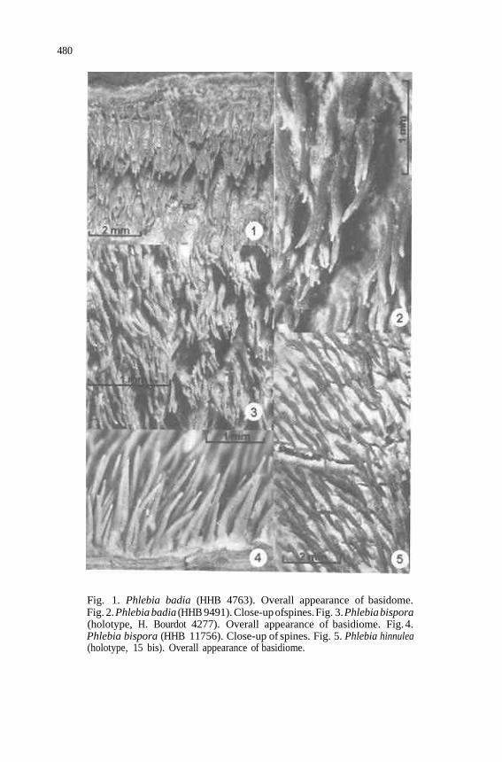
Fig. 1. Phlebia badia (HHB 4763). Overall appearance of basidome. Fig. 2. Phlebia badia (HHB 9491). Close-up ofspines. Fig. 3. Phlebia bispora (holotype, H. Bourdot 4277). Overall appearance of basidiome. Fig. 4. Phlebia bispora (HHB 11756). Close-up of spines. Fig. 5. Phlebia hinnulea (holotype, 15 bis). Overall appearance of basidiome.
481
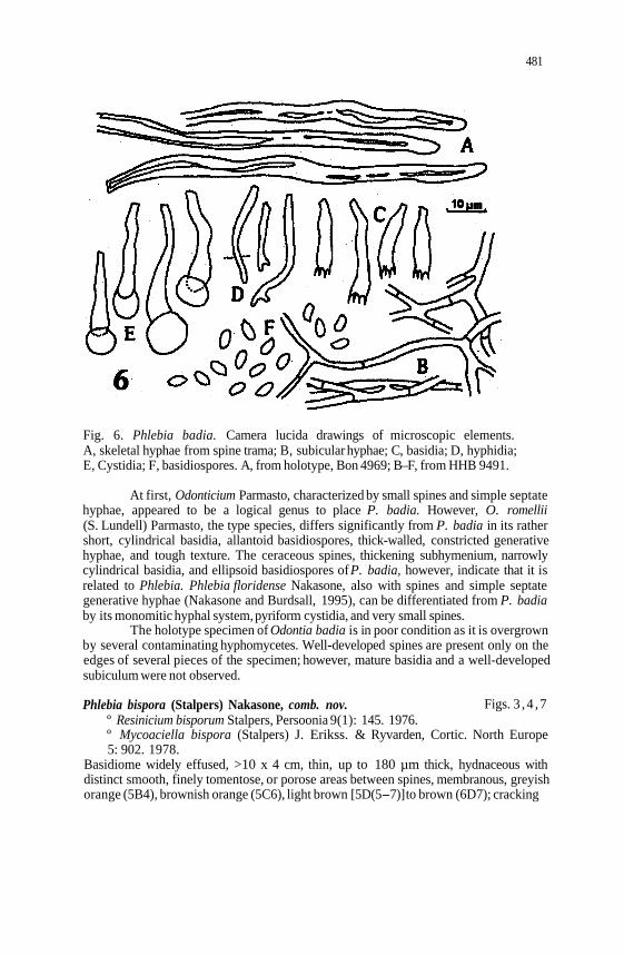
Fig. 6. Phlebia badia. Camera lucida drawings of microscopic elements. A, skeletal hyphae from spine trama; B, subicular hyphae; C, basidia; D, hyphidia; E, Cystidia; F, basidiospores. A, from holotype, Bon 4969; B–F, from HHB 9491.
At first, Odonticium Parmasto, characterized by small spines and simple septate hyphae, appeared to be a logical genus to place P. badia. However, O. romellii (S. Lundell) Parmasto, the type species, differs significantly from P. badia in its rather short, cylindrical basidia, allantoid basidiospores, thick-walled, constricted generative hyphae, and tough texture. The ceraceous spines, thickening subhymenium, narrowly cylindrical basidia, and ellipsoid basidiospores of P. badia, however, indicate that it is related to Phlebia. Phlebia floridense Nakasone, also with spines and simple septate generative hyphae (Nakasone and Burdsall, 1995), can be differentiated from P. badia by its monomitic hyphal system, pyriform cystidia, and very small spines.
The holotype specimen of Odontia badia is in poor condition as it is overgrown by several contaminating hyphomycetes. Well-developed spines are present only on the edges of several pieces of the specimen; however, mature basidia and a well-developed subiculum were not observed.
Phlebia bispora (Stalpers) Nakasone, comb. nov. Figs. 3 , 4 ,7 º Resinicium bisporum Stalpers, Persoonia 9(1): 145. 1976. º Mycoaciella bispora (Stalpers) J. Erikss. & Ryvarden, Cortic. North Europe 5: 902. 1978.
Basidiome widely effused, >10 x 4 cm, thin, up to 180 µm thick, hydnaceous with distinct smooth, finely tomentose, or porose areas between spines, membranous, greyish orange (5B4), brownish orange (5C6), light brown [5D(5-7)]to brown (6D7); cracking
482
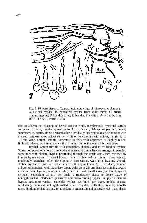
Fig. 7. Phlebia bispora. Camera lucida drawings of microscopic elements. A, skeletal hyphae; B, generative hyphae from spine trama; C, micro-binding hyphae; D, basidiospores; E, basidia; F, cystidia. A-D and F, from HHB 11756; E, from GB 758.
rare or absent; not reacting to KOH; context white, membranous; hymenial surface composed of long, slender spines up to 3 x 0.35 mm, 3–6 spines per mm, terete, subceraceous, brittle, single or fused at base, gradually tapering to an acute point or with a broad, setulose apex, apices sterile, white or concolorous with spines; margin up to 1.5 mm wide, abrupt, smooth, tomentose to felty with appressed to slightly raised, fimbriate edge or with small spines, then thinning out, with a white, fibrillose edge.
Hyphal system trimitic with generative, skeletal, and micro-binding hyphae. Spines composed of a core of skeletal and generative tramal hyphae arranged in parallel, sometimes with skeletal hyphae protruding through the sterile apex, then enclosed by thin subhymenial and hymenial layers; tramal hyphae 2–3 µm diam, nodose septate, moderately branched, often developing H-connections, walls thin, hyaline, smooth, skeletal hyphae arising from subiculum or within spine trama, 2.5–6 µm diam, clamped at base, unbranched, with secondary septa, walls up to 2.5 µm diam but thinning toward apex and base, hyaline, smooth or lightly encrusted with small, closely adherent, hyaline crystals. Subiculum 30–130 µm thick, a moderately dense to dense tissue of nonagglutinated, intertwined generative and micro-binding hyphae, in upper subiculum hyphae becoming vertical; subicular hyphae 1.5–4(–6) µm diam, nodose septate, moderately branched, not agglutinated, often irregular, walls thin, hyaline, smooth, micro-binding hyphae lacking to abundant in subiculum and substrate, 0.5–1 µm diam,
483
aseptate, moderately to frequently branched, nonstaining in phloxine, walls hyaline, smooth. Subhymenium up to 30 µm thick, a compact tissue of short-celled hyphae; subhymenial hyphae 3–6.5 µm diam, inflating up to 8 µm diam, moderately to frequently branched, often irregular, walls thin, hyaline, smooth. Hymenium composed of a dense palisade of hyphidia, cystidia, and basidia. Hyphidia rare, cylindrical, 30 x 2 µm, with basal clamp connection, simple to knobby at apex, walls thin, hyaline, smooth. Cystidia abundant, embedded in hymenium or protruding up to 20 µm, cylindrical to slightly clavate or fusiform, 18–40(–65) x 3.5–6 µm, tapering to 1.5–3(–4) µm diam at base, with a clamp connection at base, capped with a spherical, brownish yellow, resinous-like material that is dehiscent and sometimes dissolving in KOH, walls thin, hyaline, smooth. Basidia narrowly clavate to clavate, 2 4 – 3 2 x 4.5–6 µm, tapering to 1.5-3 µm diam at base, with basal clamp connection, 4-sterigmate, walls thin, hyaline, smooth. Basidiospores broadly cylindrical to ellipsoid, (4.5–)5–6.5 x (2–)2.5–3 µm, walls thin to slightly thickened, hyaline, smooth; negative in Melzer’s regent.
Habitat. On bark and wood of angiosperms. Distribution. Czech Republic (Cejp, 1930), Denmark, France, Germany (Grosse-Brauckmann, 1983; Heller, 1989), Italy (Losi, 1999), Slovakia (Pilát, 1926), Ukraine (Nikolajeva, 1961), United States (Illinois, Louisiana, New York). Specimens examined. DENMARK. Själland, Korsör skov., on (bark of) Fagus sp., 21 Nov 1959, K. Hauerslev 758 (GB). Fyen, Gamborg, Lindgravgaard i mergelgrav, på rodden pilestamme, 1 Nov 1956, M. P. Christiansen (GB). FRANCE. Allier, bordure Garnafag, entre le Mazeau et la Roche (Chappes), sur br. d’aune toucheé, 9 Août 1905, H. Bourdot 4277, ut Acia denticulata (Pers.) Bourd. & Galzin (PC, holotype of R. bisporum). GERMANY. Hessische Oberrheinebene, Kühkopf, Königsinsel, 17 Oct 1983, K.-H. Larsson and K. Hjortstam, KHL 4706 (GB); NSG, Kisselworth, on overside of trunk of Salk alba L., 3 Nov 1980, H. Grosse-Brauckmann 1437 (GB). UNITED STATES. Illinois, Johnson County, Lower Cache River, on (wood and bark of branch of) Nyssa sylvatica Marsh., 9 Sept 1983, H. H. Burdsall, Jr., 11756 (CFMR). Louisiana, Baton Rouge, on wood and bark, 25 Aug 1909, C. J. Humphrey, FP 5427 (CFMR); on decaying (decorticate) poplar, 29 Jan 1886, A. B. Langlois 239, US0259007 (BPI). New York, Jamesville, Clarke Reservation, on (decorticate) hardwood, 30 Aug 1959, J. L. Lowe and R. L. Gilbertson, JLL 10792 (CFMR).
Remarks. Phlebia bispora is an uncommon species characterized by slender spines, trimitic hyphal system, resinous-capped hymenia1 cystidia, and broadly cylindrical basidiospores. The presence of micro-binding hyphae is reported for the first time. These hyphae are similar to those found in P. chrysocreas (Berk. & M. A. Curtis) Burds. Micro-binding hyphae were observed in the subiculum and substrate of most specimens cited. The skeletal hyphae remain in the axis of the spines and never penetrate the hymenium.
Macroscopically, the slender spines of P. bispora are similar to those of P. aurea (Fr.) Nakasone, and the narrowly cylindrical basidia, cylindrical basidiospores, and embedded, resinous-like materials of P. bispora also suggest a close relationship to the genus Phlebia. In particular, P. subfasicularis (Wakef.) Nakasone and P. brunneofusca (Hjortstam & Ryvarden) Nakasone & Gilb. appear to be dimitic because of the thick-walled, heavily encrusted, terminal, sclerified hyphae (up to 65-150 µm long) in the spines (Nakasone and Gilbertson, 1998). Although Stalpers (1976)

484
believed that the capitate cystidia of P. bispora resembled those of Resinicium species, Eriksson et al. (1978) noted that this resemblance is superficial.
According to Stalpers (1976) and Eriksson et al. (1978), P. bispora was first labeled by Bourdot and Galzin (1914) as Acia denticulata (Pers.) Bourd. & Galzin. Thereafter, Cejp (1930), Pilát (1926), Nikolajeva (1961, as Sarcodontia denticulata (Pers.) Nikol.), and Parmasto (1967, as Mycoacia denticulata (Pers.) Parmasto) followed Bourdot and Galzin’s concept of A. denticulata. Maas Geesteranus (1974, p. 531) concluded that the type of Hydnum denticulatum Pers. is a specimen of H. ochraceum Pers. Later, Stalpers (1976) renamed the Bourdot and Galzin specimens Resinicium bisporum.
The holotype specimen of R. bisporum (H. Bourdot 4277) is in very good condition, with abundant basidiospores and capitate hymenial cystidia. Skeletal and micro-binding hyphae were observed, but not mature basidia.
Other descriptions and illustrations ofP. bispora can be found in Cejp (1930) as A. denticulata, Nikolajeva (1961) as S. denticulata, Stalpers (1976), Eriksson and Ryvarden (1978), Grosse–Brauckmann (1983), Duhem (1989), and Losi (1999). The specimens ofM. bispora reported from Iran by Hallenberg (1 981) are simple septate and are cited above as P. badia.
Phlebiahinnulea (Bres.) Nakasone, comb. nov. Figs. 5 , 8 º Odontia hinnulea Bres., Mycologia 18: 42. 1920. º Mycoaciella hinnulea (Bres.) Hjortstam & Ryvarden, Mycotaxon 10(2): 281.
1980. Basidiome effused, appressed but loosely attached, up to 5.5 x 1 cm and 250 µm thick, hydnaceous with smooth, fertile areas between spines, cracking between spines and exposing a thin, white context; hymenial surface of well-developed spines, spines terete, slender, smooth, single, up to 2.5 x 0.3 mm, 2-5spines per mm, tapering gradually to an acute, entire, sterile apex, brown (6E8) to pale yellow (4A3), fertile areas between spines pale yellow to cream colored; margin closely appressed, up to 1 mm wide, silky, blending into substrate or felty, white to tan, with a few, small developing spines.
Hyphal system trimitic with generative, skeletal, and micro-binding hyphae. Spines composed of a central core of skeletal and generative tramal hyphae in parallel and protruding through the sterile apex, then surrounded and enclosed by subhymenial and hymenial layers, hyphae at apex smooth or encrusted with adherent, finely granular crystals; tramal generative hyphae 2.5–3.5 µm diam, nodose septate, sparingly branched, walls thin, hyaline, smooth; skeletal hyphae cylindrical, slightly enlarged at apex and tapering gradually to the base, up to 250 x 5–10 µm, with basal clamp connection, arising in the subiculum and within the spine trama, walls hyaline to dark yellow, up to 3 µm thick but thinning toward the base and apex, smooth or encrusted at the apex with adherent, finely granular or rod-shaped, hyaline crystals. Subiculum of fertile areas between spines up to 180 µm thick, with hyphae arranged parallel to substrate in a dense, nonagglutinated tissue; subicular hyphae 2.5–4.5 µm diam, nodose septate, moderately branched, forming numerous H-connections, walls hyaline, thin, smooth, micro-binding hyphae scarce or abundant in substrate, up to 1 µm diam, aseptate, frequently branched, walls thick, hyaline, smooth. Subhymenium 35-50µm thick, a dense tissue of vertically arranged short-celled, nonagglutinated hyphae, sometimes with embedded, dark gold-colored, resinous-like substances; subhymenial hyphae 2 . 2 – 6 µm diam, nodose septate, frequently branched, walls hyaline, thin, smooth. Hymenium a dense palisade of basidia
485
and cystidia, sometimes with dark gold-colored, resinous-like substances embedded among the elements. Cystidia of two types: (1) numerous, arising from hymenium, clavate, 21–45 x 5–6.5 µm, tapering to 2–4 µm diam at base, with basal clamp connection, apex capped with a dark gold-colored resinous-like substance that is easily dislodged, 8–16 µm diam, walls hyaline, thin, smooth except at apex; (2) rare, found only in the type specimen, arising in the upper subiculum and lower subhymenium, cylindrical to narrowly clavate, 50–70 x 6–7 µm, tapering to 2 µm diam at base, with basal clamp connection, walls hyaline, thin, smooth except for apex that is encrusted with fine, closely appressed granules. Basidia narrowly clavate to cylindrical, 25-35 x 5–6 µm, tapering to 1.5–3 µm diam at base, with basal clamp connection, 4-sterigmate, walls hyaline, thin, smooth. Basidiospores cylindrical to ellipsoid, 5.5–7(–8) x 3–4 µm, walls hyaline, thin, smooth, negative in Melzer’s reagent.
Habitat. On bark of angiosperms. Distribution. Brazil. Specimens examined. BRAZIL. S. Leopoldo, ad ramos Mimosae (on bark of small twigs, 10 mm diam), leg. Rick (S, holotype of Odontia hinnulea ); S. Leopoldo, ad lignum frondosum, Rick 15 bis •20(S, BPI-US0265786).
Remarks. Phlebia hinnulea is characterized by a trimitic hyphal system, hymenia1 cystidia capped with dark, golden-colored materials, and relatively large basidiospores. Microbinding hyphae were observed only in the substrate. Mature basidia were difficult to find. Phlebia hinnulea is similar to P. bispora but is easily distinguished by its larger basidiospores. Phlebia hinnulea also shares with P. albida H. Post ex Fr. and P. albomellea (Bondartsev) Nakasone a similar texture of the subicultun and arrangement and branching of the subicular hyphae. In addition, these taxa have relatively large basidia and basidiospores compared to other Phlebia species.
The above description is based on the available specimens. Hjortstam and Ryvarden (1980, p. 28 1) also described and illustrated P. hinnulea.
Ceraceohydnum brunneum Jülich, Persoonia 10(1): 138.1978. Figs. 9, 10, 1 1 º Mycoaciella brunnea (Jülich) Hjortstam & Spooner, Kew Bull. 45(2): 309.
1990. Basidiome effused, appressed but easily separable, up to 10 x 5 cm, thin, up to 500 µm thick, ceraceous to corneous, hydnaceous to merulioid with smooth areas between spines and ridges, brown [6(E-F)8]; not reacting to KOH; cracks scarce; context usually bilayered with a thicker, dark brown, ceraceous upper layer and next to substrate a narrower, white lower layer (sometimes absent); hymenophore starting as low connecting ridges, then forming spines, spines long, slender, up to 3–4(–7) x 0.4 mm diam, 2-3spines per mm, terete, single or fused at base, ceraceous, tapering to a sterile, acute apex, flexuous when dried; margins up to 4 mm wide, smooth to odontoid, easily separable from substrate, ceraceous and brittle when dried, brown (6D7).
486
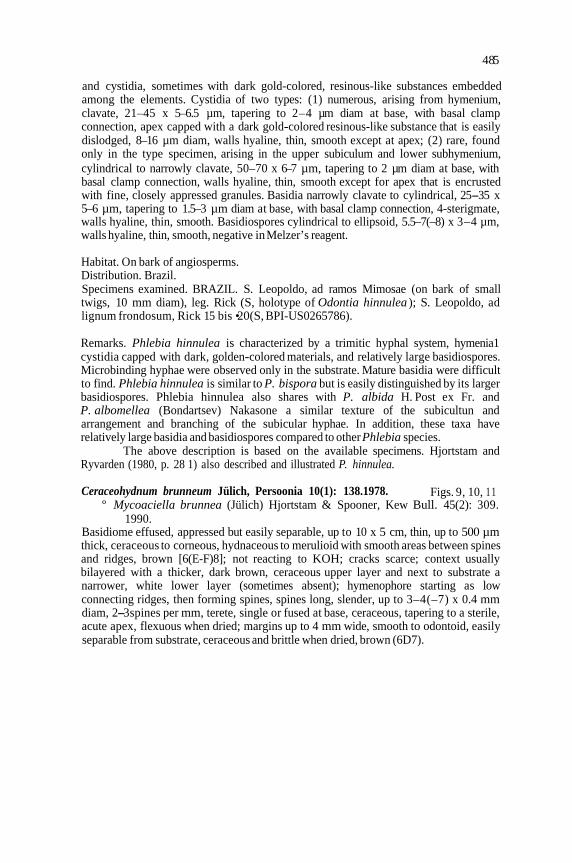
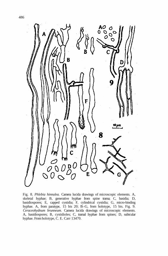
Fig. 8. Phlebia hinnulea. Camera lucida drawings of microscopic elements. A, skeletal hyphae; B, generative hyphae from spine trama; C, basidia; D, basidiospores; E, capped cystidia; F, cylindrical cystidia; G, micro-binding hyphae. A, from paratype, 15 bis 20; B–G, from holotype, 15 bis. Fig. 9. Ceraceohydnum brunneum. Camera lucida drawings of microscopic elements. A, basidiospores; B, cystidioles; C, tramal hyphae from spines; D, subicular hyphae. From holotype, C. E. Carr 13470.
487
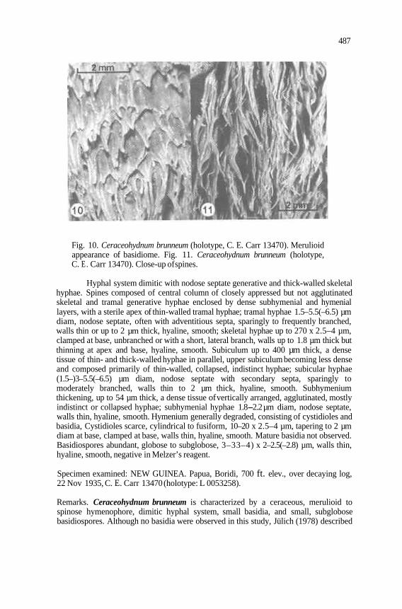
Fig. 10. Ceraceohydnum brunneum (holotype, C. E. Carr 13470). Merulioid appearance of basidiome. Fig. 11. Ceraceohydnum brunneum (holotype, C. E . Carr 13470). Close-up ofspines.
Hyphal system dimitic with nodose septate generative and thick-walled skeletal hyphae. Spines composed of central column of closely appressed but not agglutinated skeletal and tramal generative hyphae enclosed by dense subhymenial and hymenial layers, with a sterile apex of thin-walled tramal hyphae; tramal hyphae 1.5–5.5(–6.5) µm diam, nodose septate, often with adventitious septa, sparingly to frequently branched, walls thin or up to 2 µm thick, hyaline, smooth; skeletal hyphae up to 270 x 2.5–4 µm, clamped at base, unbranched or with a short, lateral branch, walls up to 1.8 µm thick but thinning at apex and base, hyaline, smooth. Subiculum up to 400 µm thick, a dense tissue of thin- and thick-walled hyphae in parallel, upper subiculum becoming less dense and composed primarily of thin-walled, collapsed, indistinct hyphae; subicular hyphae (1.5–)3–5.5(–6.5) µm diam, nodose septate with secondary septa, sparingly to moderately branched, walls thin to 2 µm thick, hyaline, smooth. Subhymenium thickening, up to 54 µm thick, a dense tissue of vertically arranged, agglutinated, mostly indistinct or collapsed hyphae; subhymenial hyphae 1.8-2.2 µm diam, nodose septate, walls thin, hyaline, smooth. Hymenium generally degraded, consisting of cystidioles and basidia, Cystidioles scarce, cylindrical to fusiform, 10–20 x 2.5–4 µm, tapering to 2 µm diam at base, clamped at base, walls thin, hyaline, smooth. Mature basidia not observed. Basidiospores abundant, globose to subglobose, 3–33–4) x 2–2.5(–2.8) µm, walls thin, hyaline, smooth, negative in Melzer’s reagent.
Specimen examined: NEW GUINEA. Papua, Boridi, 700 ft. elev., over decaying log, 22 Nov 1935, C. E. Carr 13470 (holotype: L 0053258).
Remarks. Ceraceohydnum brunneum is characterized by a ceraceous, merulioid to spinose hymenophore, dimitic hyphal system, small basidia, and small, subglobose basidiospores. Although no basidia were observed in this study, Jülich (1978) described
488
the basidia as narrowly clavate, hyaline, 4-sterigmate, 6–10 x 2.5–3 µm, with a basal clamp. Skeletal hyphae are limited to the spines, and although similar to the thick-walled subicular hyphae, they are morphologically distinct. Although C. brunneum was transferred into Mycoaciella (Hjortstam et al., 1990), it does not appear to be closely related to M. bispora or Phlebia because of its small basidia and basidiospores. The affinities of C. brunneum are not known at this time; therefore, it is accepted in the monotypic Ceraceohydnum Jülich.
DISCUSSION
Mycoaciella is placed in synonymy under Phlebia, necessitating the transfer of M. bispora and M. hinnulea into Phlebia. Phlebia bispora is reported from Europe and eastern United States, and P. hinnulea is limited to Brazil. Similarly, Odontia badia, with a circumglobal distribution, is transferred into Phlebia. Mycoaciella brunnea, however, is morphologically distinct from Phlebia and Phlebia bispora and is accepted under its original name, Ceraceohydnum brunneum.
A shared feature of P. bispora, P. hinnulea, and P. badia is the development of hymenia1 cystidia with a globular, resinous-like cap. Nevertheless, P. bispora and P. hinnulea, which share many morphological features and are probably closely related, do not appear to be closely related to P. badia. In particular, the overall membranous texture and open hyphal structure of the subiculum in P. bispora and P. hinnulea are more similar to that found in P. albida and P. albomellea than to the ceraceous texture and agglutinated subicular tissue ofP. badia. Furthermore, the basidia and basidiospores are slightly smaller in P. badia compared with those in P. bispora and P. hinnulea.
Trimitic hyphal systems in corticioid fungi are unusual. The first trimitic corticioid species, Schizopora trametoides Suhirman & Núñez, was described by Suhirman and Núñez (1990). Phlebia bispora and P. hinnulea are the only other examples known at present. In these species, the trimitic hyphal system consists of generative, skeletal, and micro-binding hyphae. Skeletal hyphae occur only in the spine trama whereas micro-binding hyphae are found in the substrate and subiculum.
The term micro-binding hyphae refers to narrow (< 1 µm diam), highly branched, often at right angles, aseptate, nonstaining hyphae arising from generative hyphae (Nakasone, 2001). These hyphae are not commonly described in corticioid fungi and were referred to previously as “narrow, much-branched hyphae” in Phlebia chrysocreas (Berk. & Curtis) Burds. (Lombard et al., 1975), “branched skeletal hyphae” in Amethicium Hjortstam (Hjortstam, 1983), “quasi-binding hyphae” (Wu, 1990, p. 16), and “arboriform” in Cericium Hjortstam (Hjortstam, 1995). Micro-binding hyphae in corticioid fungi are more delicate and highly branched than are the binding hyphae present in the polypores (Gilbertson and Ryvarden, 1986,p. 21).
Skeletal hyphae occur more frequently than micro-binding hyphae in corticioid fungi. Skeletal hyphae are undifferentiated, unbranched or sparingly branched, and thick-walled, and they lack septa or clamp connections throughout their length, although occasional secondary septa may be present. Skeletal hyphae can be confused with sclerified generative hyphae or tramal cystidia. Typically embedded in the subicular trama, skeletal hyphae are labeled tramal cystidia if they terminate in or beyond the hymenium. The apices of tramal cystidia are often enlarged or encrusted. Sclerified generative hyphae differ from skeletal hyphae in that clamp connections or primary simple septa are present.
489
With the inclusion of P. bispora, P. hinnulea, and P. badia, Phlebia now contains species with increasingly diverse morphologies. In addition to the varied hymenophore types, the 'circumscription of Phlebia now includes monomitic, dimitic, or trimitic hyphal systems consisting of simple or nodose septate generative hyphae, with or without skeletal or micro-binding hyphae. Future phylogenetic studies may resolve the taxonomic significance of the morphological heterogeneity in Phlebia.
ACKNOWLEDGEMENTS
The specimens used in this study were loaned by the curators of the following herbaria: BPI, FH, GB, L, PC, and S. Presubmission reviews of this manuscript by Drs. Karen Wikler, James H. Ginns, and Harold H. Burdsall, Jr. are gratefully acknowledged.
LITERATURE CITED

490