View - Journal of Neurology, Neurosurgery & Psychiatry
5
Journal of Neurology, Neurosurgery, and Psychiatry 1992;55:121-125 Crossed cerebellar diaschisis accompanied by hemiataxia: a PET study Makoto Tanaka, Susumu Kondo, Shunsaku Hirai, Koji Ishiguro, Tomio Ishihara, Mitsunori Morimatsu Abstract To study crossed cerebeilar diaschisis (CCD), cerebeilar blood flow and oxygen metabolism were measured with positron emission tomography (PET) in 12 patients who showed a minimal degree of hemiparesis due to single unilateral supratentorial lesion. Six patients presenting with mild to moderate cerebellar type hemiataxia showed CCD, that is, decreased blood flow and oxygen metabolism in the cerebellar hemisphere contralateral to the side of supratentorial lesion. Hemiataxia and reduced cerebellar blood flow and metabolism occurred in the ipsilateral side. Lesions were located in the thalamus in four patients and the parietal lobe and inter- nal capsule in one each. The other six patients did not exhibit ataxia, and oxygen metabolism was not reduced in the contralateral cerebellar hemisphere. In two of these cases, however, reduced cerebellar perfusion was observed in the contralateral cerebellar hemisphere. These findings indicate that CCD occurs with hemiataxia and suggest that it results not only from disruption of the corticopontocerebellar pathway but also of the dentatorubrothalamic pathway. CCD associated with hemiataxia, demon- strated in patients with thalamic lesions, was presumed to result from retrograde deactivation of the cerebellar hemisphere via the dentatorubrothalamic pathway. Gunma University School of Medicine, Maebashi, Japan Department of Neurology Makoto Tanaka Susumu Kondo Shunsaku Hirai Koji Ishiguro Department of Radiology Tomio Ishihara Department of Neurology, Ymaguchi University School of Medicine, Japan Mitsunori Morimatsu Correspondence to: Dr Tanaka, Department of Neurology, Gunma University School of Medicine, Showa-machi, Maebashi 371, Japan. Received 20 August 1990 and in final revised form 14 May 1991. Accepted 21 May 1991 Diaschisis is a concept developed by von Mon- akow' to explain temporary deactivation in an undamaged area of the brain after injury to a remote site. A good example is crossed cerebellar diaschisis (CCD), wherein damage in one cerebral hemisphere produces a func- tional depression in the contralateral cerebellar hemisphere.2 Loss of cerebellar excitatory afferent input is presumed to cause CCD primarily through disruption of the cor- ticopontocerebellar tract.2' The clinical importance of CCD, however, is not under- stood despite repeated demonstration with positron emission tomography (PET) and single photon emission tomography. Patients with cerebrovascular disorders often show CCD, but cerebellar manifestations are rarely detected on neurological examina- tion. Only a few case reports exist."6 This may imply that the depression of the cerebellar function seen in CCD has no or little relation to the clinical presentation of ataxia, a major manifestation of cerebellar dysfunction. An alternative interpretation is that the motor weakness and hypertonicity usually occurring with CCD mask additional motor deficits, including ataxia. Thalamic injury, especially to the ven- trolateral portion, is a well known cause of contralateral hemiataxia.7 Although this is explained as being due to destruction of major projections from the cerebellum via the den- tato(rubro)thalamic pathway8 at the terminal, cerebellar efferent pathways have not been investigated with reference to CCD. We therefore initiated the present study in patients without hemiparesis, which can mask ataxia, and with single lesions in various supraten- toral regions. Materials and methods Twelve patients with cerebral infarction or haemorrhage presenting minimal hemiparesis were studied by PET (table 1). They were selected from a consecutive series of patients studied since March 1986 at the Gunma University Hospital Cyclotron Center for PET Diagnosis. All were examined neurologically to ensure the absence of motor impairment due to muscle weakness or hypertonicity, which can mask ataxia. Clinical diagnosis and lesion site were also confirmed by neuroradiological evaluation including brain CT in all patients, angiography in five (cases 1, 8, 9, 11, 12) and magnetic resonance imaging (MRI) in six (cases 1, 2, 7, 8, 9, 12), and by clinical course and neurological findings. All but one patient (case 3) had a single supratentorial lesion without clinical or radiological evidence of cerebellar or brain stem involvement. Of these, six (cases 1 to 6) showed mild to moderate cerebellar ataxia with preserved deep sensation (vibration, proprioception, and kinesthesia), while the remaining patients (cases 7 to 12) had no ataxia. A clinical history of cases 1 and 5 has been reported in detail previously.9 Four hemiataxic patients had a history of old thalamic haemorrhage or infarction, and one patient (case 1) had infarction in the parietal lobe. Case 3 showed pure motor hemiplegia on examination at the time of onset, which had improved to mild hemiataxia 48 hours later. Although a small capsular or pontine lacune not revealed by x ray CT was suspected as the cause of these transient symptoms,'0 hypoper- fusin and oxygen hypometabolism on the left cerebral hemisphere later revealed by PET favoured a capsular lacune. Thirteen healthy 121 on 8 January 2019 by guest. Protected by copyright. http://jnnp.bmj.com/ J Neurol Neurosurg Psychiatry: first published as 10.1136/jnnp.55.2.121 on 1 February 1992. Downloaded from
Transcript of View - Journal of Neurology, Neurosurgery & Psychiatry
Journal ofNeurology, Neurosurgery, and Psychiatry 1992;55:121-125
Crossed cerebellar diaschisis accompanied byhemiataxia: a PET study
Makoto Tanaka, Susumu Kondo, Shunsaku Hirai, Koji Ishiguro, Tomio Ishihara,Mitsunori Morimatsu
AbstractTo study crossed cerebeilar diaschisis(CCD), cerebeilar blood flow and oxygenmetabolism were measured with positronemission tomography (PET) in 12patients who showed a minimal degree ofhemiparesis due to single unilateralsupratentorial lesion. Six patientspresenting with mild to moderatecerebellar type hemiataxia showed CCD,that is, decreased blood flow and oxygenmetabolism in the cerebellar hemispherecontralateral to the side ofsupratentoriallesion. Hemiataxia and reducedcerebellar blood flow and metabolismoccurred in the ipsilateral side. Lesionswere located in the thalamus in fourpatients and the parietal lobe and inter-nal capsule in one each. The other sixpatients did not exhibit ataxia, andoxygen metabolism was not reduced inthe contralateral cerebellar hemisphere.In two of these cases, however, reducedcerebellar perfusion was observed in thecontralateral cerebellar hemisphere.These findings indicate that CCD occurs
with hemiataxia and suggest that itresults not only from disruption of thecorticopontocerebellar pathway but alsoof the dentatorubrothalamic pathway.CCD associated with hemiataxia, demon-strated in patients with thalamic lesions,was presumed to result from retrogradedeactivation ofthe cerebellar hemispherevia the dentatorubrothalamic pathway.
Gunma UniversitySchool of Medicine,Maebashi, JapanDepartment ofNeurologyMakoto TanakaSusumu KondoShunsaku HiraiKoji IshiguroDepartment ofRadiologyTomio IshiharaDepartment ofNeurology, YmaguchiUniversity School ofMedicine, JapanMitsunori MorimatsuCorrespondence to: DrTanaka, Department ofNeurology, GunmaUniversity School ofMedicine, Showa-machi,Maebashi 371, Japan.Received 20 August 1990and in final revised form14 May 1991.Accepted 21 May 1991
Diaschisis is a concept developed by von Mon-akow' to explain temporary deactivation in an
undamaged area of the brain after injury to a
remote site. A good example is crossedcerebellar diaschisis (CCD), wherein damagein one cerebral hemisphere produces a func-tional depression in the contralateral cerebellarhemisphere.2 Loss of cerebellar excitatoryafferent input is presumed to cause CCDprimarily through disruption of the cor-
ticopontocerebellar tract.2' The clinicalimportance of CCD, however, is not under-stood despite repeated demonstration withpositron emission tomography (PET) andsingle photon emission tomography.
Patients with cerebrovascular disordersoften show CCD, but cerebellar manifestationsare rarely detected on neurological examina-tion. Only a few case reports exist."6 This mayimply that the depression of the cerebellarfunction seen in CCD has no or little relation to
the clinical presentation of ataxia, a majormanifestation of cerebellar dysfunction. Analternative interpretation is that the motorweakness and hypertonicity usually occurringwith CCD mask additional motor deficits,including ataxia.Thalamic injury, especially to the ven-
trolateral portion, is a well known cause ofcontralateral hemiataxia.7 Although this isexplained as being due to destruction of majorprojections from the cerebellum via the den-tato(rubro)thalamic pathway8 at the terminal,cerebellar efferent pathways have not beeninvestigated with reference to CCD. Wetherefore initiated the present study in patientswithout hemiparesis, which can mask ataxia,and with single lesions in various supraten-toral regions.
Materials and methodsTwelve patients with cerebral infarction orhaemorrhage presenting minimal hemiparesiswere studied by PET (table 1). They wereselected from a consecutive series of patientsstudied since March 1986 at the GunmaUniversity Hospital Cyclotron Center for PETDiagnosis. All were examined neurologically toensure the absence ofmotor impairment due tomuscle weakness or hypertonicity, which canmask ataxia. Clinical diagnosis and lesion sitewere also confirmed by neuroradiologicalevaluation including brain CT in all patients,angiography in five (cases 1, 8, 9, 11, 12) andmagnetic resonance imaging (MRI) in six(cases 1, 2, 7, 8, 9, 12), and by clinical courseand neurological findings. All but one patient(case 3) had a single supratentorial lesionwithout clinical or radiological evidence ofcerebellar or brain stem involvement. Of these,six (cases 1 to 6) showed mild to moderatecerebellar ataxia with preserved deep sensation(vibration, proprioception, and kinesthesia),while the remaining patients (cases 7 to 12) hadno ataxia. A clinical history of cases 1 and 5 hasbeen reported in detail previously.9 Fourhemiataxic patients had a history of oldthalamic haemorrhage or infarction, and onepatient (case 1) had infarction in the parietallobe. Case 3 showed pure motor hemiplegia onexamination at the time of onset, which hadimproved to mild hemiataxia 48 hours later.Although a small capsular or pontine lacunenot revealed by x ray CT was suspected as thecause of these transient symptoms,'0 hypoper-fusin and oxygen hypometabolism on the leftcerebral hemisphere later revealed by PETfavoured a capsular lacune. Thirteen healthy
121 on 8 January 2019 by guest. P
rotected by copyright.http://jnnp.bm
j.com/
J Neurol N
eurosurg Psychiatry: first published as 10.1136/jnnp.55.2.121 on 1 F
ebruary 1992. Dow
nloaded from
Tanaka, Kondo, Hirai, Ishiguro, Ishihara, Morimatsu
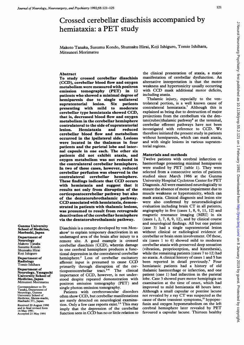
Table 1 Details ofpatients with cerebral infarction or haemorrhage presenting minimal hemiparesis
Case Age Timefrom onsetnumber (years) Sex Neuroimagingfindings Presenting signs and symptoms Neurologicalfindings on PET study to PET study
1 34 F Infarction in left parietal cortex and subcortical Dysesthesia and clumsiness of Right sided hemiataxia, mildly 13 Monthswhite matter right hand disturbed combined sensations
2 56 M Old haemorrhage localised in left thalamus Sensorimotor disturbance Right sided hemiataxia, dys- and 18 Months(ventrolateral nucleus) hyperesthesia
3 71 M No abnormal findings on repeated CT Pure motor hemiplegia in right Right sided hemiataxia without 24 Hoursside motor weakness
4 47 F Small localised haemorrhage in right lateral Sensorimotor disturbances Left sided hemiataxia, dys- and 2 Yearsthalamus at onset, no abnormalities on follow hyperesthesia, hyperalgesiaup CT
5 70 F Old haemorrhage localised in left thalamus Severe sensory and mild motor Right sided hemiataxia, dys- and 4 Yearsdisturbances hyperesthesia, hyperalgesia
6 77 F Small infarction in left thalamus (ventrolateral Sensorimotor disturbances Right side hemiataxia, hand tremor, 7 Yearsnucleus), periventricular lucency dys- and hypesthesia, hypalgesia
7 58 M Infarction in right centrum semiovale and mild Sensorimotor disturbances Left sided minimal hemisensory 8 Yearscortical involvement disturbance
8 37 M Right temporoparietooccipital infarction due to Subjective hemisensory Left-sided hemispatial neglect, 1 MonthMoyamoya disease disturbances disturbed combined sensations
9 49 M Infarction in right temporoparietal cortex and Sensorinotor disturbances Left sided disturbed combined 1 Monthsubcortex due to obstruction of middle sensationscerebral artery
10 82 F Infarction in left posterior inferior frontal lobe Speech disturbance Motor aphasia 1 Month11 72 M Infarction in subcortex of left temporoparietal Speech disturbance Conduction aphasia 3 Months
junction12 61 M Small infarction in anterior medial portion of Mental deterioration Amnestic syndrome 7 Months
left thalamus
volunteers were subjected to PET study as
control subjects.PET was performed with a PCT-H1 (Hita-
chi, Japan) with four rings with 128 bismuthgermanate oxide detectors providing seven
views per scan cycle. The best spatial resolu-tion was 7-4 mm full width at half maximum(FWHM) at the centre of the scan field, andaxial resolution at the centre was 16 mm (slicethickness). Before PET study, x ray CT (CT-HSF, Hitachi) images were obtained to iden-tify anatomical structures in the PET views.PET and x ray CT scanners were located sideby side and joined by rails on which the bedmoved with the patient's position fixed, thusproviding identical tomographic planes. Thepatient's head was fixed to the headrest toobtain a tomographic plane parallel to theorbitomeatal line. The initial positioning andthe absence of head tilt during the scan were
ensured by crossed beams projected on inkmarks drawn on the patient's face. A transmis-sion scan with Germanium-68 and gallium-68was performed for 10 minutes for attenuationcollection. Calibration factors between thePET scanner and well counter were obtained ineach study. The oxygen-15 steady state tech-nique"' was used to measure regional cerebralblood flow (rCBF) and regional cerebraloxygen metabolic rate (rCMRO2). Oxygen-15gas (750-1,100 MBq per minute) then oxygen-
15 labelled carbon dioxide (350-550 MBq per
minute) were inhaled continuously. Scan data
were collected for 5 to 8 minutes. The bloodwas sampled three times during each scan
process from a cannulated radial artery andsubjected to determinations of oxygen-15radioactivity in whole blood and plasma as wellas blood cell count and blood gas analysis. Nocorrection was performed for intravascularoxygen-15 labelled oxyhaemoglobin.Image data was processed with a Hitachi
image processing computer with systemsubroutines to reconstruct functional imagesconstituting 128 x 128 pixels. The region ofinterest was determined on both cerebellarhemispheres identified with x ray CT by trackball. The region was set as the x ray CT imageon which the full width of both cerebellarhemispheres appeared. The outlines were
traced by track ball, carefully avoiding thelateral sinuses and the fourth ventricles. Thesame region of interest was superimposed on
cerebellar rCBF and rCMRO2 tomographicplanes identical to the x ray CT plane. Thedegree of asymmetry in rCBF and rCMRO2was expressed by the asymmetry index (AI)defined as follows;
AI (%) = lt - rt x 200/(lt + rt)lt and rt are the left and right cerebellar rCBFor CMRO2, respectively.We analysed cerebellar rCBF and rCMRO2
with two way analysis of variance (ANOVA)with two fixed factors (a group factor withataxic and non-ataxic and a side factor withipsilateral and contralateral to the cerebral
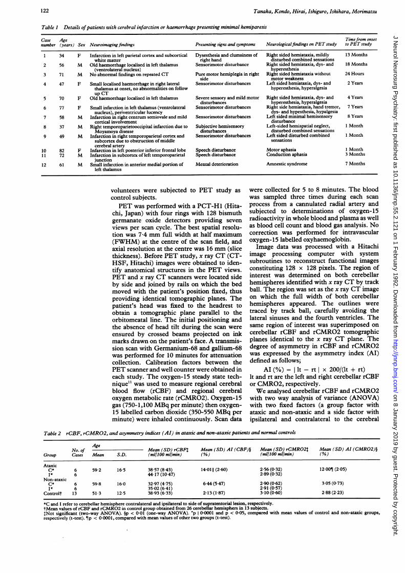
Table 2 rCBF, rCMR02, and asymmetry indices (AI) in ataxic and non-ataxic patients and normal controls
AgeNo. of Mean (SD) rCBF$ Mean (SD) AI (CBF)§ Mean (SD) rCMR02t Mean (SD) AI (CMR02)§
Group Cases Mean S.D. (m100 mil/min) (%) (ml/lO mil/min) (%)
AtaxicC* 6 59-2 16 5 38-57 (8-43) 14 0111 (260) 2-56 (0 32) 12 0011 (2 05)I* 6 44 17(10 47) 2-89 (0 32)
Non-ataxicC* 6 59-8 16-0 32-97 (4-75) 6-44 (5-47) 2-90 (0-62) 3 05 (0 73)I* 6 35-02 (6-41) 2-91(0 57)
Controlt 13 51 3 12 5 38-93 (6-33) 2 13 (1-87) 3-10(0-60) 2-88 (2-23)
*C and I refer to cerebellar hemisphere contralateral and ipsilateral to side of supratentorial lesion, respectively.tMean values of rCBF and rCMRO2 in control group obtained from 26 cerebellar hemisphers in 13 subjects.tNot significant (two-way ANOVA). §p < 0-01 (one-way ANOVA). "p ll 0-0001 and p < 0-05, compared with mean values of control and non-ataxic groups,respectively (t-test). 1IP < 0-0001, compared with mean values of other two groups (t-test).
122 on 8 January 2019 by guest. P
rotected by copyright.http://jnnp.bm
j.com/
J Neurol N
eurosurg Psychiatry: first published as 10.1136/jnnp.55.2.121 on 1 F
ebruary 1992. Dow
nloaded from
123Crossed cerebellar diaschisis accompanied by hemiataxia: a PET study
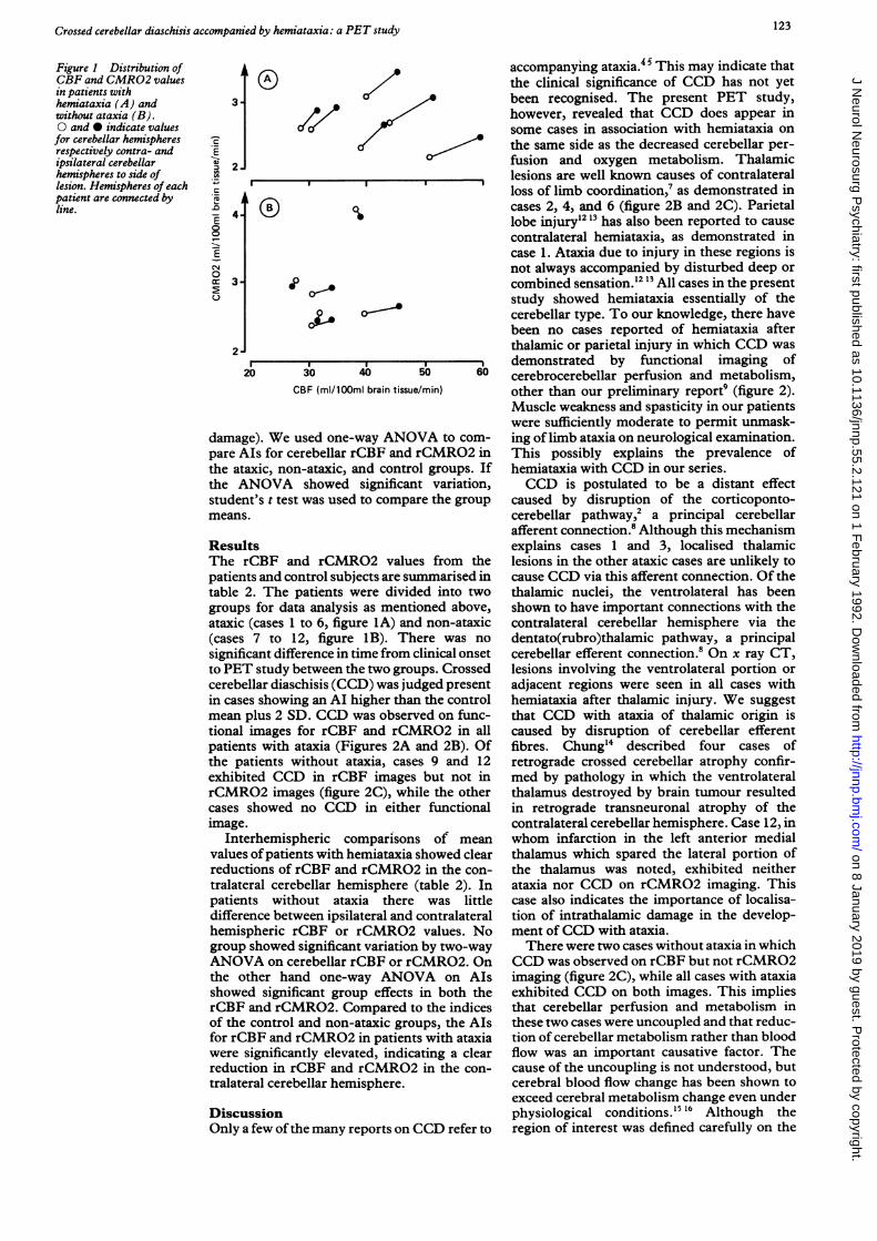
Figure 1 Distribution ofCBF and CMR02 valuesin patients withhemiataxia (A) andwithout ataxia (B).0 and 0 indicate valuesfor cerebellar hemispheresrespectively contra- andipsilateral cerebellarhemispheres to side oflesion. Hemispheres of eachpatient are connected byline.
m 2.
E 4'4-0
0
ECN
0cr- 3.
2
CBF (ml/lOOml brain tissue!
damage). We used one-way ANONpare AIs for cerebellar rCBF and r
the ataxic, non-ataxic, and controlthe ANOVA showed significantstudent's t test was used to comparmeans.
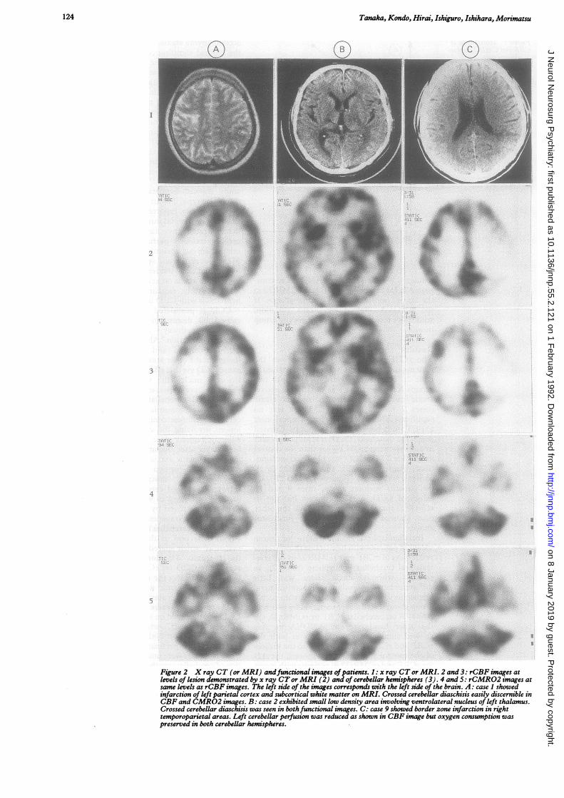
ResultsThe rCBF and rCMR02 valuespatients and control subjects are suntable 2. The patients were dividegroups for data analysis as mentioataxic (cases 1 to 6, figure 1A) and(cases 7 to 12, figure 1B). Thesignificant difference in time from clto PET study between the two groulcerebellar diaschisis (CCD) was judin cases showing an AI higher thanmean plus 2 SD. CCD was observetional images for rCBF and rCMpatients with ataxia (Figures 2A athe patients without ataxia, cases
exhibited CCD in rCBF imagesrCMR02 images (figure 2C), whilcases showed no CCD in eitherimage.
Interhemispheric comparisons
values ofpatients with hemiataxia si
reductions of rCBF and rCMRO2tralateral cerebellar hemisphere (tpatients without ataxia theredifference between ipsilateral and clhemispheric rCBF or rCMR02group showed significant variation IANOVA on cerebellar rCBF or rCthe other hand one-way ANOVshowed significant group effects irCBF and rCMR02. Compared toof the control and non-ataxic grou
for rCBF and rCMR02 in patientswere significantly elevated, indicaireduction in rCBF and rCMR02tralateral cerebellar hemisphere.
DiscussionOnly a few ofthe many reports on C
accompanying ataxia. 5 This may indicate thatthe clinical significance of CCD has not yet
A been recognised. The present PET study,however, revealed that CCD does appear insome cases in association with hemiataxia onthe same side as the decreased cerebellar per-fusion and oxygen metabolism. Thalamiclesions are well known causes of contralateralloss of limb coordination,7 as demonstrated incases 2, 4, and 6 (figure 2B and 2C). Parietallobe injury12 13 has also been reported to causecontralateral hemiataxia, as demonstrated incase 1. Ataxia due to injury in these regions isnot always accompanied by disturbed deep orcombined sensation.2 13 All cases in the presentstudy showed hemiataxia essentially of thecerebellar type. To our knowledge, there havebeen no cases reported of hemiataxia afterthalamic or parietal injury in which CCD was
'r- , demonstrated by functional imaging ofso 60 cerebrocerebellar perfusion and metabolism,,min) other than our preliminary report9 (figure 2).
Muscle weakness and spasticity in our patientswere sufficiently moderate to permit unmask-
TA to com- ing of limb ataxia on neurological examination.CMR02 in This possibly explains the prevalence ofgroups. If hemiataxia with CCD in our series.: variation, CCD is postulated to be a distant effecte the group caused by disruption of the corticoponto-
cerebellar pathway,2 a principal cerebellarafferent connection.8 Although this mechanismexplains cases 1 and 3, localised thalamic
from the lesions in the other ataxic cases are unlikely tonmarised in cause CCD via this afferent connection. Of thed into two thalamic nuclei, the ventrolateral has beenined above, shown to have important connections with thenon-ataxic contralateral cerebellar hemisphere via there was no dentato(rubro)thalamic pathway, a principallinical onset cerebellar efferent connection.8 On x ray CT,ps. Crossed lesions involving the ventrolateral portion orged present adjacent regions were seen in all cases withthe control hemiataxia after thalamic injury. We suggested on func- that CCD with ataxia of thalamic origin is.R02 in all caused by disruption of cerebellar efferentnd 2B). Of fibres. Chung'4 described four cases of9 and 12 retrograde crossed cerebellar atrophy confir-
but not in med by pathology in which the ventrolateralle the other thalamus destroyed by brain tumour resultedfunctional in retrograde transneuronal atrophy of the
contralateral cerebellar hemisphere. Case 12, inof mean whom infarction in the left anterior medial
howed clear thalamus which spared the lateral portion ofin the con- the thalamus was noted, exhibited neithertable 2). In ataxia nor CCD on rCMR02 imaging. Thiswas little case also indicates the importance of localisa-ontralateral tion of intrathalamic damage in the develop-values. No ment of CCD with ataxia.by two-way There were two cases without ataxia in whichIMRO2. On CCD was observed on rCBF but not rCMR02rA on AIs imaging (figure 2C), while all cases with ataxiain both the exhibited CCD on both images. This impliesithe indices that cerebellar perfusion and metabolism inips, the AIs these two cases were uncoupled and that reduc-with ataxia tion of cerebellar metabolism rather than bloodting a clear flow was an important causative factor. Thein the con- cause of the uncoupling is not understood, but
cerebral blood flow change has been shown toexceed cerebral metabolism change even underphysiological conditions.'5 16 Although the
:CD refer to region of interest was defined carefully on the
on 8 January 2019 by guest. Protected by copyright.
http://jnnp.bmj.com
/J N
eurol Neurosurg P
sychiatry: first published as 10.1136/jnnp.55.2.121 on 1 February 1992. D
ownloaded from
124 Tanaka, Kondo, Hirai, Ishiguro, Ishihara, Morimatsu
,...B, . /i
.CI.
... .....
Y.f... .
;.:.4:: n......
Figure 2 X ray CT (or MRI) andfunctional images ofpatients. 1: x ray CTor MRI. 2 and 3: rCBF images atlevels of lesion demonstrated by x ray CTor MRI (2) and of cerebellar hemispheres (3). 4 and 5: rCMR02 images atsame levels as rCBF images. The left side of the images corresponds with the left side of the brain. A: case I showedinfarction of left parietal cortex and subcortical white matter on MRI. Crossed cerebellar diaschisis easily discernible inCBF and CMR02 images. B: case 2 exhibited small low density area involving ventrolateral nucleus of left thalamus.Crossed cerebellar diaschisis was seen in bothfunctional images. C: case 9 showed border zone infarction in righttemporoparietal areas. Left cerebellar perfusion was reduced as shown in CBF image but oxygen consumption waspreserved in both cerebellar hemispheres.
I...
(A )i
It I
%I....
..........IF
x
iip
on 8 January 2019 by guest. Protected by copyright.
http://jnnp.bmj.com
/J N
eurol Neurosurg P
sychiatry: first published as 10.1136/jnnp.55.2.121 on 1 February 1992. D
ownloaded from
Crossed cerebellar diaschisis accompanied by hemiataxia: a PET study
cerebellar hemisphere delineated by x ray CT,the possible presence of some artifacts shouldbe taken into account. Radioactivity in theregion was more or less influenced by thatpresenting in the lateral sinuses, as intravas-cular blood volume correction was not carriedout in the present study.Although CCD accompanied by ataxia was
probably the result of damage to cerebellarafferent or efferent pathway, it is not neces-
sarily linked to ataxia."7 Pappata et al 7 reportedthat significant contralateral cerebellar hypo-metabolism was found in three of six patientswith pure internal capsule infarction, suggest-ing a pathogenetic role for the corticoponto-cerebellar system. They also observed thatcontralateral hypometabolism occurred in twoof six patients with thalamic lesions and madethe assumption that this phenomenon alsoresults either from damage to the ascendingcerebellothalamocortical system or indirectlyfrom hypometabolism of the cerebral cortex.Systemic association was not observed betweencrossed cerebellar hypometabolism and ipsi-lateral ataxia in their series.'7 Their findingssuggest that hypometabolism in the cerebralcortex, which is caused by transneuronal effector by underlying cerebral ischaemia, also playsa pathophysiological role in CCD accompaniedby ataxia. In our study, however, there seemedto be no assocition between ipsilateral hypo-metabolism in the cerebral cortex and CCDwith ataxia (data not shown). A specificallydesigned study is required to reveal the path-ogenesis producing CCD concurrent withataxia.
We thank assistant Professor S Aoki (Department of Public
Health, Gunma University School of Medicine) for his helpfulcomment on statistical procedure.
1 Von Monakow C. Die Lokalisation im Grosshirn und derAbbau der Funktion durch Kortikale Herde. Wiesbaden: J FBergmann, 1914:26-34.
2 Baron JC, Bousser MG, Comar D, Castaigne P. Crossedcerebellar diaschisis in human supratentorial brain infarc-tion. Ann Neurol 1980;8:128.
3 Pantano P, Baron JC, Samson Y, Bousser MG, DerouesneC, Comar D. Crossed cerebellar diaschisis: furtherstudies. Brain 1986;109:677-94.
4 Fukuyama H, Kameyama M, Harada K, et al. Thalamictumours invading the brain stem produce crossedcerebellar diaschisis demonstrated by PET. J NeurolNeurosurg Psychiatry 1986;49:524-8.
5 Bogousslavsky J, Regli F, Delaloye B, Delaloye-Bischoff A,Uske A, Despland PA. Hemiataxie et deficit -sensitifipsilateral: infarctus du territoire de l'artere choroidienneanterieure: diaschisis cerebelleux croise. Rev Neurol 1986;142:671-6.
6 Sakai F, Aoki S, Kan S, Igarashi H, Kanada T, Tazaki Y.Ataxic hemiparesis with reductions of ipsilateralcerebellar blood flow. Stroke 1986;17:1016-8.
7 Bogousslavsky J, Regli F, Uske A. Thalamic infarcts: clinicalsyndromes, etiology, and prognosis. Neurology 1988;38:837-45.
8 Carpenter MB, Sutin J. Human neuroanatomy, 8th ed.Baltimore: Williams and Wilkins, 1983:454-92.
9 Tanaka M, Hirai S, Morimatsu M, Kanai H, Mizuno Y.Clinical and etiological study ofcrossed cerebellar diasch-isis: report of two cases. Clin Neurol 1989;19:469-74.
10 Soisson T, Cabanis EA, Iban-Zizen MT, Bousser MG,Laplane D, Castaigne P. Hemiplegie motrice pure ettomodensitometrie. J Neuroradiol 1982;9:304-22.
11 Frackowiak RSJ, Pozzilli C, Legg NJ, et al. Regionalcerebral oxygen supply and utilization in dementia, aclinical and physiological study with oxygen-15 andpositron tomography. Brain 1981;104:753-78.
12 Appenzeller 0, Hanson JC. Parietal ataxia. Arch Neurol1966;15:264-9.
13 Yagnik PM, Dhaduk V, Huen L. Parietal ataxic hemiparesis.Eur Neurol 1988;28:164-6.
14 Chung HD. Retrograde crossed cerebellar atrophy. Brain1985;1O8:881-95.
15 Fox PT, Raichle ME. Focal physiological uncoupling ofcerebral -blood flow and oxidative metabolism during-somatosensory stimulation in human subjects. Proc NatlSci USA 1985;83:1140-4.
16 Lou HC, Edvinsson L, MacKenzie ET. The concept ofcoupling blood flow to brain function: revision required?Ann Neurol 1987;22:289-97.
17 Pappata S, Mazoyer B, Tran Dinh S, Cambon H, LevasseurM, Baron JC. Effects of capsular or thalamic stroke onmetabolism in the cortex and cerebellum: a positrontomography study. Stroke 1990;21:519-24.
125 on 8 January 2019 by guest. P
rotected by copyright.http://jnnp.bm
j.com/
J Neurol N
eurosurg Psychiatry: first published as 10.1136/jnnp.55.2.121 on 1 F
ebruary 1992. Dow
nloaded from