intracerebral arterioles - Journal of Neurology, Neurosurgery
6
Journal of Neurology, Neurosurgery, and Psychiatry 1982 ;45 :120-124 Ultrastructural characteristics of spasm in intracerebral arterioles WOLFGANG ROGGENDORF, JORGE CERVOS-NAVARRO From the Institut fur Neuropathologie, Klinikum Steglitz der Freien Universitdt Berlin (West) SUMMARY Following craniotomy, three groups of cats were subjected to three different stimuli: group A hyperventilation, group B electroshock, and group C direct electric current. During electric stimuli, pial vessels were observed through a cranial window. Immediately after electric current application, some arterial vessels showed segmental spastic constriction. Tissue samples for electron- microscopy were taken from the parietal lobe and nucleus caudatus. In all three groups of animals, different types of constriction of blood vessels were observed. The respiratory alkalosis achieved by hyperventilation led to physiological constriction of the arterioles. The electric stimuli led to spastic constriction of the meningeal and intracerebral arteries and arterioles in group B and C; the entire vessel wall was greatly deformed and the vessel lumen was almost obstructed. Electroshock resulted in only moderate structural changes of the smooth muscle cells. Direct current, however, caused an extreme and bizarre smooth muscle deformation. The results show that spastic constrictions of arterioles can be clearly distinguished from physiological, that is non-spastic constriction, by mor- phological parameters. Electric stimulation of cerebral vessels could be an experimental condition for further investigation of intracerebral vasospasm. Spastic constriction occurs in larger intracranial arteries and may influence cerebral circulation, especially after aneurysmal rupture followed by sub- arachnoid haemorrhage, as shown by clinical and experimental investigations by Echlin, Wilkens et al, and Peterson et al.1-3 Ultrastructural studies by Matakas et a14 and Cerv6s-Navarro et al5 have revealed spastic constrictions also in intracerebral arterioles under various experimental conditions. The purposes of this investigation were (1) definition of morphological criteria for identification of con- traction of smooth muscle cells in blood vessel walls under physiological conditions, and (2) to determine whether spastic contraction produced by various electrical stimuli was accompanied by specific morphological changes. Material and methods For this study we used 21 adult cats (2-3 kg), which were anaesthetised with intramuscular sodium pentobarbital (Nembutal 25 mg/kg bw). A catheter was inserted into the Address for reprint requests: Professor J Cerv6s-Navarro, Universitats Klinicum Steglitz, Hindenburgdamm 30, D 1000 Berlin 45, W. Germany. Received 27 May 1981 and in revised form 7 September 1981 Accepted 26 September 1981 aorta and into the inferior vena cava through the femoral artery and vein, respectively. Arterial blood pressure was monitored and remained at normal levels throughout the experiments. After relaxation with succinyl-bis-choline (2 mg) and intubation, the animals were artificially ventilated. Blood gases were kept normal (arterial pres- sure of CO2 35-45 mm Hg, pH 7 35, arterial pressure of 02 90-1 10 mm Hg). The cats were divided into 3 groups: Group A Respiratory alkalosis in nine cats was achieved by hyperventilation for 20 min (82 ml, frequency 28-33/min) (Cerv6s-Navarro et a16). Group B Electroconvulsive treatment was applied in six cats after craniotomy. Two metal electrodes, each with a diameter of 10 mm, were attached to the temporal muscle on each side. An alter- nating current of 220 V 50 Hz was applied for 500 ms at a time, two to five times within 5 minutes. After 20 minutes, the animals were killed by injecting 50 ml of India ink suspen- sion within 10 s into the inferior vena cava. Group C For electric stimulation by direct current (DC) in six cats after craniotomy, one electrode (10 mm diameter) was attached to the right temporal muscle. The left electrode was an Agar-gel silver electrode with a plastic tube of inner tip diameter of about 0-20 mm. DC with 20-100 V for 2 to 30 s (20-150 ma) was applied. In two animals, two assessments were made. In four animals, three were made. 120 Protected by copyright. on 20 December 2018 by guest. http://jnnp.bmj.com/ J Neurol Neurosurg Psychiatry: first published as 10.1136/jnnp.45.2.120 on 1 February 1982. Downloaded from
Transcript of intracerebral arterioles - Journal of Neurology, Neurosurgery

Journal of Neurology, Neurosurgery, and Psychiatry 1982 ;45 :120-124
Ultrastructural characteristics of spasm inintracerebral arteriolesWOLFGANG ROGGENDORF, JORGE CERVOS-NAVARRO
From the Institut fur Neuropathologie, Klinikum Steglitz der Freien Universitdt Berlin (West)
SUMMARY Following craniotomy, three groups of cats were subjected to three different stimuli:group A hyperventilation, group B electroshock, and group C direct electric current. During electricstimuli, pial vessels were observed through a cranial window. Immediately after electric currentapplication, some arterial vessels showed segmental spastic constriction. Tissue samples for electron-microscopy were taken from the parietal lobe and nucleus caudatus. In all three groups of animals,different types of constriction of blood vessels were observed. The respiratory alkalosis achieved byhyperventilation led to physiological constriction of the arterioles. The electric stimuli led to spasticconstriction of the meningeal and intracerebral arteries and arterioles in group B and C; the entirevessel wall was greatly deformed and the vessel lumen was almost obstructed. Electroshock resultedin only moderate structural changes of the smooth muscle cells. Direct current, however, caused anextreme and bizarre smooth muscle deformation. The results show that spastic constrictions ofarterioles can be clearly distinguished from physiological, that is non-spastic constriction, by mor-phological parameters. Electric stimulation of cerebral vessels could be an experimental conditionfor further investigation of intracerebral vasospasm.
Spastic constriction occurs in larger intracranialarteries and may influence cerebral circulation,especially after aneurysmal rupture followed by sub-arachnoid haemorrhage, as shown by clinical andexperimental investigations by Echlin, Wilkens et al,and Peterson et al.1-3 Ultrastructural studies byMatakas et a14 and Cerv6s-Navarro et al5 haverevealed spastic constrictions also in intracerebralarterioles under various experimental conditions.The purposes of this investigation were (1) definitionof morphological criteria for identification of con-traction of smooth muscle cells in blood vessel wallsunder physiological conditions, and (2) to determinewhether spastic contraction produced by variouselectrical stimuli was accompanied by specificmorphological changes.
Material and methods
For this study we used 21 adult cats (2-3 kg), which wereanaesthetised with intramuscular sodium pentobarbital(Nembutal 25 mg/kg bw). A catheter was inserted into the
Address for reprint requests: Professor J Cerv6s-Navarro,Universitats Klinicum Steglitz, Hindenburgdamm 30, D 1000Berlin 45, W. Germany.
Received 27 May 1981 and in revised form 7 September 1981Accepted 26 September 1981
aorta and into the inferior vena cava through the femoralartery and vein, respectively. Arterial blood pressure wasmonitored and remained at normal levels throughout theexperiments. After relaxation with succinyl-bis-choline(2 mg) and intubation, the animals were artificiallyventilated. Blood gases were kept normal (arterial pres-sure of CO2 35-45 mm Hg, pH 7 35, arterial pressure of02 90-1 10 mm Hg).The cats were divided into 3 groups:
Group A Respiratory alkalosis in nine cats was achievedby hyperventilation for 20 min (82 ml,frequency 28-33/min) (Cerv6s-Navarro et a16).
Group B Electroconvulsive treatment was applied in sixcats after craniotomy. Two metal electrodes,each with a diameter of 10 mm, were attachedto the temporal muscle on each side. An alter-nating current of 220 V 50 Hz was applied for500 ms at a time, two to five times within 5minutes. After 20 minutes, the animals werekilled by injecting 50 ml of India ink suspen-sion within 10 s into the inferior vena cava.
Group C For electric stimulation by direct current (DC)in six cats after craniotomy, one electrode(10 mm diameter) was attached to the righttemporal muscle. The left electrode was anAgar-gel silver electrode with a plastic tube ofinner tip diameter of about 0-20 mm. DC with20-100 V for 2 to 30 s (20-150 ma) was applied.In two animals, two assessments were made. Infour animals, three were made.
120
Protected by copyright.
on 20 Decem
ber 2018 by guest.http://jnnp.bm
j.com/
J Neurol N
eurosurg Psychiatry: first published as 10.1136/jnnp.45.2.120 on 1 F
ebruary 1982. Dow
nloaded from

Ultrastructural characteristics of spasm in intracerebral arterioles
In group B and C, the animals were killed 20 min afterelectrical stimulation by injecting 50 ml of India inksuspension rapidly into the inferior vena cava.A control group of 12 cats was used. Six of these cats
were only normally ventilated. The remaining six catswere treated as group B and C, but the cerebral vesselswere not subjected to electric stimulation. After treat-ment of 20 min (group A) or immediately after injectionof India ink (group B and C), 5 mm3 blocks of parietalcortex and subcortical white matter and of the caudatenucleus were excised quickly for electron-microscopy.The brain was then removed and fixed in formalin. Thetissue for electronmicroscopy was fixed for 2 hours inchilled 50% buffered glutaraldehyde, washed in bufferedsaccharose solution, postfixed in 1-0% osmium tetroxide,dehydrated and embedded in epon. The brain specimensin this study were fixed by immersion in order to preventreduction of the spasm which might occur with fixation byperfusion under pressure. Immersion fixation was alsonecessary for the groups of experimental animals whichwere perfused with India ink in order to observe the noreflow phenomenon and the spastic constriction ofarterioles along large segments.The control brains of regularly ventilated cats were also
fixed by immersion. Arterioles in these brains showed onlya slight contraction evidenced by undulation of the basallamina. Marked constriction or even spasm of arterioleswas not observed in any of the control brains. There wereno differences between normal cat brain arterioles fixed byimmersion or by perfusion with regard to the appearanceof the cellular organelles. In particular pinocytoticactivity and filamentous components in endothelial cellswere identical.The gross and microscopic aspects of groups B and C
after India ink injection have been described in detail byMatakas et a14 (we will give only a short summary). Insome cortical areas, patches without blackening of Indiaink were observed, indicating that cerebral circulationwas disturbed. In all cases, light-microscopically largearterioles and arteries varied in diameter and showedsome segments completely spastic. The spastic segmentswere exclusively observed in the grey substance of thebrain and in either the cortex or basal ganglia.
In the following description, we refer to spasticcontraction when marked deformation of the entirevessel wall leads to a completely obstructed vessel incontrast to physiological contraction produced byalkalosis, in which the basic configuration of the vessel isretained.
Results
I ENDOTHELIUM AND SUBENDOTHELIALSPACE
In groups A, B, and C, oval to round endothelial cellnuclei covered by a thin rim of cytoplasm protrudedfar into the vessel lumen (fig 1). In the endothelialcell body away from the nuclear zone, slender,digital cytoplasmic extensions projected towards thevessel lumen (figs 1, 2).
30 4
Fig 1 Arteriole from the cortex in physiologicalcontraction (group A) with finger-like narrowprotrusions of endothelium and strongly scallopedsubendothelial space. Fixation by immersion. x 2880.* , ,;. o. t.<.; + : ..... = . : u
i.
A .e,.
v B -^0a .... ............. ^ ......... b
Fig 2 Contracted arteriole from the nucleus caudatus(group A) with the characteristic deformation of thesmooth muscle cells: between attachment devices (A), thecell membrane bulges out (-). The endothelial tightjunctions are set upright (). Fixation by immersion.x 7160.
Slightly increased endothelial pinocytosis wasevident only in the first experimental group (physio-logical constriction after respiratory alkalosis). Inboth of the experimental groups of animals exposedto electric stimulation, no changes in endothelialpinocytotic activity were observed, possibly becauseof the short duration on the experiments. The endo-thelial tight junctions in dilated vessels ran diagonallyand overlapped. In contracted arterioles (group A),the junctions usually ran perpendicular to the basalmembrane (fig 2). Spicular cytoplasmic extensionscontaining clusters of 7 nm filaments were also seen,arising abluminally from the endothelial cells of
121
Is
...... %.
1. '. %
-AP' Protected by copyright.
on 20 Decem
ber 2018 by guest.http://jnnp.bm
j.com/
J Neurol N
eurosurg Psychiatry: first published as 10.1136/jnnp.45.2.120 on 1 F
ebruary 1982. Dow
nloaded from
Roggendorf, Cervos-Navarro
....
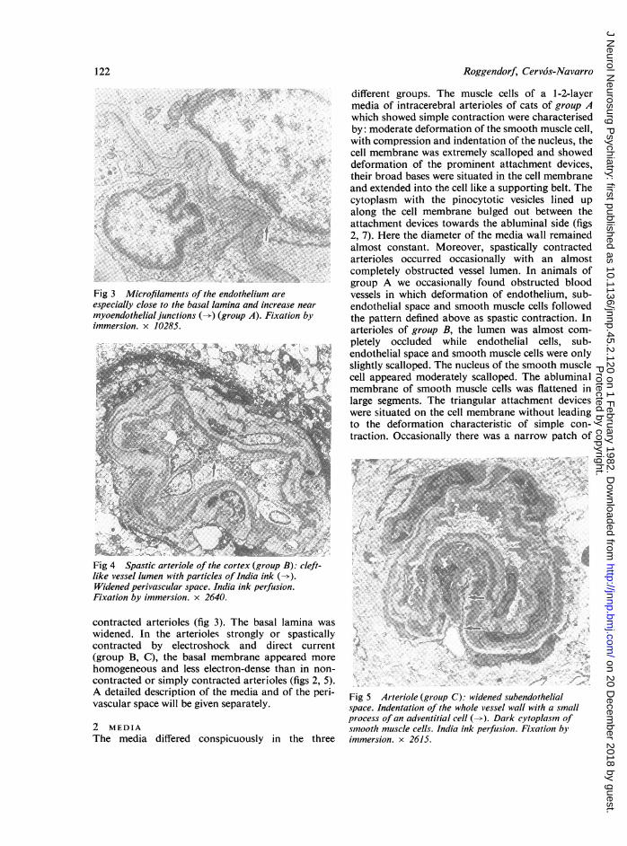
Fig 3 Microfilament's of the endothelium areespecially close to the basal lamina and increase nearmyoendothelial junctions (->) (group A). Fixation byimmersion. x 10285;
different groups. The muscle cells of a 1-2-layermedia of intracerebral arterioles of cats of group Awhich showed simple contraction were characterisedby: moderate deformation of the smooth muscle cell,with compression and indentation of the nucleus, thecell membrane was extremely scalloped and showeddeformation of the prominent attachment devices,their broad bases were situated in the cell membraneand extended into the cell like a supporting belt. Thecytoplasm with the pinocytotic vesicles lined upalong the cell membrane bulged out between theattachment devices towards the abluminal side (figs2, 7). Here the diameter of the media wall remainedalmost constant. Moreover, spastically contractedarterioles occurred occasionally with an almostcompletely obstructed vessel lumen. In animals ofgroup A we occasionally found obstructed bloodvessels in which deformation of endothelium, sub-endothelial space and smooth muscle cells followedthe pattern defined above as spastic contraction. Inarterioles of group B, the lumen was almost com-pletely occluded while endothelial cells, sub-endothelial space and smooth muscle cells were onlyslightly scalloped. The nucleus of the smooth musclecell appeared moderately scalloped. The abluminalmembrane of smooth muscle cells was flattened inlarge segments. The triangular attachment deviceswere situated on the cell membrane without leadingto the deformation characteristic of simple con-traction. Occasionally there was a narrow patch of
tX-.'
Fig 4 Spastic arteriole of the cortex (group B): cleft-like vessel lumen with particles of India ink (-*).Widened perivascular space. India ink perfusion.Fixation by immersion. x 2640.
contracted arterioles (fig 3). The basal lamina waswidened. In the arterioles strongly or spasticallycontracted by electroshock and direct current(group B, C), the basal membrane appeared morehomogeneous and less electron-dense than in non-contracted or simply contracted arterioles (figs 2, 5).A detailed description of the media and of the peri-vascular space will be given separately.
2 MEDIAThe media differed conspicuously in the three
Fig 5 Arteriole (group C): widened subendothelialspace. Indentation of the whole vessel wall with a smallprocess of an adventitial cell (). Dark cytoplasm ofsmooth muscle cells. India ink perfusion. Fixation byimmersion. x 2615.
122
Protected by copyright.
on 20 Decem
ber 2018 by guest.http://jnnp.bm
j.com/
J Neurol N
eurosurg Psychiatry: first published as 10.1136/jnnp.45.2.120 on 1 F
ebruary 1982. Dow
nloaded from
Ultrastructural characteristics of spasm in intracerebral arterioles
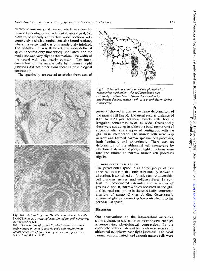
electron-dense marginal border, which was possiblyformed by contiguous attachment devices (figs 4, 6a).Next to spastically contracted vessel sections withcompletely occluded lumina, one also found sections,where the vessel wall was only moderately infolded.The endothelium was flattened, the subendothelialspace appeared only moderately undulated, and themedia showed very slight deformation. The width ofthe vessel wall was nearly constant. The inter-connection of the muscle cells by myomyal tightjunctions did not differ from those in physiologicalcontraction.The spastically contracted arterioles from cats of
4..... ......
SMc
/ , ~~~~~~J
:% f
,- > > k we.~~~~~~~~~~~~~~~~~~~~~~.4-f.,..>i" Ai|
.. ~~~~~~.. ..Y.
Fig 6(a) Arteriole (gi*oup B). The smooth mtiuscle cells(SMC) show no sti-ong deformtation of the cell memnbraneas opposed to (b).
(b) The arteriole of group C, which shows a bizarre-.
deformaAtationof smgrooth moousclecells and endothelium.
Snmall processes ofglia in the penivascular space ().
(a) x 6360 (b) x 5830.
Fig 7 Schematic presentation of the physiologicalconstriction mechanism: the cell membrane wasextremely scalloped and showed deformation byattachment devices, which work as a cytoskeleton duringconstriction.
grouip C showed a bizarre, extreme deformation ofthe muscle cell (fig 5). The usual regular distance of015 to 0-20 pm between muscle cells becameirregular, sometimes twice as wide. Occasionallythere were gap zones in which the basal membrane ofsubendothelial space appeared contiguous with theglial basal membrane. The muscle cells were verynarrow and formed narrow spicular cell processes,both luminally and abluminally. There was nodeformation of the abluminal cell membrane byattachment devices. Myomyal tight junctions wererare and limited to narrow muscle cell processes
(fig 6b).
3 PERIVASCULAR SPACEThe perivascular space in all three groups of catsappeared as a gap that only occasionally showed adilatation. It contained uniformly narrow adventitialcell branches, nerves, and collagen fibres. In con-trast to uncontracted arterioles and arterioles ofgroups A and B, narrow folds occurred in the glialand its basal membrane in the spastically contractedarteriole of group C (figs 5, 6b). Occasionallyattenuated glial processes (fig 6b) protruded into theperivascular space.
Discussion
Our observations on the intracerebral arteriolesshow a characteristic group of morphologic changesaccompanying physiological contraction. In theendothelial cells, clusters of filaments were seen in theabluminal cytoplasm near tight junctions. The basallamina was undulated, and smooth muscle cells were
4,
>4'
- ¾.'
K
123
Protected by copyright.
on 20 Decem
ber 2018 by guest.http://jnnp.bm
j.com/
J Neurol N
eurosurg Psychiatry: first published as 10.1136/jnnp.45.2.120 on 1 F
ebruary 1982. Dow
nloaded from
124
deformed by a scalloped cell membrane and promi-nent attachment devices. The vessel diameter was
markedly decreased. However, the vessel lumen was
not obstructed. These findings are in agreement withthe observations by light microscopy on smallarterioles undergoing physiological contraction7 andwith the ultrastructural findings in mesentericarterioles by Phelps and Luft.8 In particular thepresence of contractile filaments within the endothe-lial cells observed by us and others9-1" indicates thatthe endothelium plays an active role in the contractileprocess. Further, these contractile filaments may de-termine the typical arrangement in endothelial cells.The spastic constrictions of arterioles can be
clearly distinguished from the physiological, that isnon-spastic constriction by morphological param-
eters. Both DC and electroshock lead to a spasticconstriction with occlusion of the vessel lumen.There is no characteristic deformation of smoothmuscle cell membrane by attachment devices and no
typical undulation of the basal lamina. The peri-vascular glia participates in the spasm by folding ofthe astrocytic plasma membrane and the glial basallamina probably because of a reduction of the totalsurface of the glial ensheathment.
These morphological changes indicate that it ispossible to produce a complete obstruction of thevessel lumen with both electric stimulation methods,DC and electroshock. The effect of these electricalstimuli on the microcirculation is described in moredetail by Matakas et al4 and Cerv6s-Navarro et al.5
Morphologic alterations occurring with spasticprocesses are not homogeneous. There are morpho-logic differences between the spastic vessels in groupB and group C. The principal difference involves thereaction of the smooth muscle cell. In cases wherethe spasm has been induced by direct current (groupC), the spastic constriction is characterised by a
bizarre deformation of smooth muscle cells. Incases where spasm was produced by electroshock(group B) there is only a moderately scallopedsmooth muscle cell layer and no deformation of thesmooth muscle cell membrane by attachmentdevices. We believe that a spastic contraction is notan extreme deformation of the single muscle cell butrather a folding and indentation of the whole vesselwall which leads to occlusion of the lumen.The question arises as to whether spastic con-
traction takes place without typical signs of thephysiological constriction. We consider that thepattern of contraction depends on the arrangementof the smooth muscle cells of the media. The spiralpattern of the smooth muscle cells found in smallarteries and arterioles by Benninghoff12 by lightmicroscopy has been confirmed by ultrastructuralexaminations. Roggendorf and Cervos-Navarro'3
Roggendorf, Cervos-Navarro
showed non-contracted intracerebral arterioles witha nearly circular course with interruptions in thesmooth muscle cell layer by segments in which themyofilaments of neighbouring smooth muscle cellsare arranged vertically on top of one another in thecourse of a two layered media. In tangential sections,Rhodin14 demonstrated that the muscle cells of theoutermost layer are spirally arranged in a pitch of180. This spiral course of the smooth muscle cellrenders possible the extreme deformation and deepindentation of the vessel in the presence of spasticcontraction with occlusion of the vessel lumen.Lesions of single cells, for example in the endotheliumor in the smooth muscle cells were not found moreoften in experimental animals than in the controlanimals. There were no significant changes oforganelles, generalised vacuolisation or oedema.These observations are in agreement with the studiesof Fein et all5 and Tanabe et al,16 which revealed thatthere are no changes due to spasm in the first hourafter stimulation. The intriguing phenomenon of thesegmental arrangement of spasm, which is welldocumented in meningeal vessels after subarachnoi-dal haemorrhagel 2 17 18 cannot be explained by theresults of our electron microscopical study. A sys-tematic investigation of the precise mechanism ofintracerebral spasm, however, could be based, atleast in part, on experimental conditions derivedfrom this investigation with electric stimulation ofintracerebral arteries and arterioles.
References
Echlin FA. Spasm of the basilar and vertebral arteriescaused by experimental subarachnoid hemorrhage.J Neurosurg 1965 ;23 :1-11.
2 Wilkins RH, Alexander JA, Odom GL. Intracranialarterial spasm: a clinical analysis. J Neurosurg 1968;29 :121-34.
3Peterson EW, Searle R, Mandy FF et al. The reversalof experimental vasospasm by dinutyryl-3'-adenosinemonophosphate. J Neurosurg 1973 ;39 :730-4.
4Matakas F, Cerv6s-Navarro J, Roggendorf W et al.Spastic constriction of cerebral vessels after electricconvulsive treatment. Fortschr Psychiatr Nervenhlk1977;224:1-9.
5Cerv6s-Navarro J, Matakas F, Roggendorf W et al.The morphology of spastic intracerebral arterioles.Neuropath and Neurobiol 1978;4:369-79.
6 Cerv6s-Navarro J, Leipert M, Valencak E et al. Lamorphologie ultrastructurale du S.N. dans "laperfusion de luxe" de l'acidose respiratoire. Neuro-chirurgie 1971 ;17:595-600.
7Citters RL van, Wagern BM, Rushmer RF. Archi-tecture of small arteries during vasoconstriction.Circ Res 1962;10:668-75.
8 Phelps PC, Luft JH. Electron microscopical study of
Protected by copyright.
on 20 Decem
ber 2018 by guest.http://jnnp.bm
j.com/
J Neurol N
eurosurg Psychiatry: first published as 10.1136/jnnp.45.2.120 on 1 F
ebruary 1982. Dow
nloaded from
Ultrastructural characteristics of spasm in intracerebral arterioles
relaxation and constriction in frog arterioles. Am JAnat 1969 ;125 :399-429.
9 Becker CG, Murphy GE. Demonstration of contractileprotein in endothelium and cells of the heart valves,endocardium, intimal arteriosclerotic plaques, andAschoff bodies of rheumatic heart disease. Am JPath1969;55:1-37.
10 Becker CG, Nachman RL. Contractile proteins ofendothelial cells, platelets, and smooth muscle. Am JPath 1973 ;71 :1-22.
Owman Ch, Edvinsson L, Hardebo JH et al. Immuno-chemical demonstration of actin and myosin in braincapillaries. In: Cerv6s-Navarro J, ed. Advances inNeurology, Vol. 20. New York: Raven Press, 1978:35-7.
12 Benninghoff A. Blutgefal3e und Herz. In: Handbuchder mikroskopischen Anatomie des Menschen.Mollendorf W, ed. Berlin: Springer Verlag, 1930:134.
13 Roggendorf W, Cerv6s-Navarro J. Ultrastructure ofarterioles in the cat brain. Cell Tiss Res 1977;178:495-515.
14 Rhodin JAG. Fine structure of vascular walls inmammals with special reference to smooth musclecomponent. Physiol Rev 1962;42 :48-81.
15 Fein JM, Flor WJ, Cohan SL et al. Sequential changesof vascular ultrastructure in experimental vaso-
spasm. Myonecrosis of subarachnoid arteries. JNeurosurg 1974;41 :49-58.
16 Tanabe Y, Sakata K, Yamada H et al. Cerebral vaso-
spasm and ultrastructural changes in cerebralarterial wall. J Neurosurg 1978 ;49 :229-38.
17 Arutiunow Al, Baron MA, Majorova NA. Experi-mental and clinical study of the development ofspasm of cerebral arteries related to subarachnoidhemorrhage. J Neurosurg 1970;32 :617-25.
18 Lende RA. Local spasm in cerebral arteries. J Neuro-surg 1960;17:90-103.
125
Protected by copyright.
on 20 Decem
ber 2018 by guest.http://jnnp.bm
j.com/
J Neurol N
eurosurg Psychiatry: first published as 10.1136/jnnp.45.2.120 on 1 F
ebruary 1982. Dow
nloaded from