UvA-DARE (Digital Academic Repository) Novel insights in ... · Chapter 2 16 Applications of IVIg...
148
UvA-DARE is a service provided by the library of the University of Amsterdam (http://dare.uva.nl) UvA-DARE (Digital Academic Repository) Novel insights in human Fcy-receptors an IgS : on genes, transcripts and functional interactions van Mirre, E. Link to publication Citation for published version (APA): van Mirre, E. (2005). Novel insights in human Fcy-receptors an IgS : on genes, transcripts and functional interactions General rights It is not permitted to download or to forward/distribute the text or part of it without the consent of the author(s) and/or copyright holder(s), other than for strictly personal, individual use, unless the work is under an open content license (like Creative Commons). Disclaimer/Complaints regulations If you believe that digital publication of certain material infringes any of your rights or (privacy) interests, please let the Library know, stating your reasons. In case of a legitimate complaint, the Library will make the material inaccessible and/or remove it from the website. Please Ask the Library: http://uba.uva.nl/en/contact, or a letter to: Library of the University of Amsterdam, Secretariat, Singel 425, 1012 WP Amsterdam, The Netherlands. You will be contacted as soon as possible. Download date: 10 Jul 2018
Transcript of UvA-DARE (Digital Academic Repository) Novel insights in ... · Chapter 2 16 Applications of IVIg...

UvA-DARE is a service provided by the library of the University of Amsterdam (http://dare.uva.nl)
UvA-DARE (Digital Academic Repository)
Novel insights in human Fcy-receptors an IgS : on genes, transcripts and functionalinteractionsvan Mirre, E.
Link to publication
Citation for published version (APA):van Mirre, E. (2005). Novel insights in human Fcy-receptors an IgS : on genes, transcripts and functionalinteractions
General rightsIt is not permitted to download or to forward/distribute the text or part of it without the consent of the author(s) and/or copyright holder(s),other than for strictly personal, individual use, unless the work is under an open content license (like Creative Commons).
Disclaimer/Complaints regulationsIf you believe that digital publication of certain material infringes any of your rights or (privacy) interests, please let the Library know, statingyour reasons. In case of a legitimate complaint, the Library will make the material inaccessible and/or remove it from the website. Please Askthe Library: http://uba.uva.nl/en/contact, or a letter to: Library of the University of Amsterdam, Secretariat, Singel 425, 1012 WP Amsterdam,The Netherlands. You will be contacted as soon as possible.
Download date: 10 Jul 2018

Novel insights in human Fcγ-receptors and IgG: on genes, transcripts and functional interactions.

The work described in this thesis was performed at the department of Immunopathology and Experimental Immunohematology at Sanquin Research, Amsterdam, the Netherlands. The publication of this thesis was financially supported by: J.E. Jurriaanse Stichting Stichting voor Afweerstoornissen Genmab B.V., Utrecht Sanquin Reagents, Amsterdam Sanquin Plasmaproducts, Amsterdam Sanquin Research, Amsterdam University of Amsterdam ISBN 90-9019942-X ©2005, Edwin van Mirre Cover design: confocal microscopy by Erik Mul. Neutrophils stained for CD11b (green), CD16 (red) and immune complexes (blue). Co-localization of CD16 and immune complexes (purple). Co-localization of CD11b, CD16 and immune complexes (white). All rights reserved. No part of this publication may be reproduced, stored in a retrieval system, or transmitted in any form or by any means, without the written permission of the author. Printed by: Teewes/Textpresse grafische bedrijven

Novel insights in human Fcγ-receptors and IgG: on genes, transcripts and functional interactions.
Academisch proefschrift
Ter verkrijging van de graad van doctor aan de Universiteit van Amsterdam
op gezag van de Rector Magnificus prof. mr. P.F. van der Heijden ten overstaan van een door het college van promoties ingestelde commissie,
in het openbaar te verdedigen in de Aula der Universiteit.
op donderdag 20 oktober 2005, te 12.00 uur
door Edwin van Mirre
geboren te Vlaardingen

Promotores: Prof. dr. C.E. Hack
Prof. dr. T.W. Kuijpers
Commissie: Prof. dr. D. Roos Prof. dr. M. L. Kapsenberg Prof. dr. R.J.M. ten Berge Prof. dr J.W. M. van der Meer Prof. dr. J.G.J. van de Winkel Faculteit der Geneeskunde

Up, up, up some stairs we go… And then,
Through a tunnel…
Aan Muizekind, Uil & Kleine Rooie


Contents Chapter 1 Introduction. 9 Chapter 2 Immunoglobulins and FcγR: interactions and effects. 13 Chapter 3 Monomeric IgG in IVIg is a functional antagonist of FcγRII and FcγRIIIb. 39 Chapter 4 Amelioration of immune complex-mediated anaphylaxis by intravenous 59
immunoglobulins (IVIg) in rat. Chapter 5 Variation in the mRNA ratios of activating and inhibitory FcγRII determines 83
neutrophil responsiveness. Chapter 6 Identification of a novel gain- of- function splice variant of FcγRIIa and 103
its relation to hyperactivation of neutrophils. Chapter 7 Summary and discussion 125 Nederlandse samenvatting voor niet-immunologen 133 Curriculum vitae 142 Nawoord 144 List of publications 146


Chapter 1
9
Introduction

Chapter 1
10

Chapter 1
11
During evolution, man has built up a defense mechanism against pathogenic organisms such as bacteria, viruses, fungi and parasites. This defense mechanism is called the immune system. This system is comprised of the innate immunity and the adaptive immunity, also known as the aspecific and the specific immunity, respectively. Both innate and adaptive immunity can be subdivided in a cellular component (cell-mediated immunity) and a humoral component (humoral immunity). The innate and the adaptive immunity are strongly intertwined via their subdivisions. One of the strategies against pathogens is the generation of specific antibodies (immunoglobulins) by the adaptive immunity. Immunoglobulins contain two regions: one region (the Fab-domain) recognizes the pathogen, whereas the other region (Fc-domain) activates the immune system. This humoral component is linked to the cellular component via receptors for the immunoglobulins, so-called Fc-receptors.
There are several classes of immunoglobulins (Ig); i.e. IgD, IgM, IgG, IgA and IgE. IgG is the most important, most abundant, has a long circulation time and is present in the highest concentration within the blood. In addition, IgG is involved in the protection of the newborn against pathogens, since the mother transfers IgG to the child via the placenta. There are four classes of receptors for IgG: FcγRI, FcγRII, FcγRIII and FcRn. The latter has a different structure and is responsible for the transport of IgG from mother to child. In addition, this receptor is also responsible for the long circulation time of IgG, because it protects IgG from degradation in the cell. The FcγR are effector molecules that can, upon IgG binding, activate the cell. FcγRI is the high affinity receptor and capable of binding monomeric IgG. FcγRII and FcγRIII are low affinity receptors and bind preferentially IgG in complexed form (immune complexes). With the exception of FcγRII, FcγR are dependent on another protein for stable expression and cell activation. FcγRII contains its own signaling motif. Dependent on the isoform of this receptor, it is either an activating (FcγRIIa) or an inhibitory receptor (FcγRIIb). Intravenous gammaglobulin (IVIg) was originally designed for patients lacking IgG (a- or hypogammaglobulinemia). IVIg consists of IgG obtained from the blood of at least 1000 donors. The underlying thought is that in this way IVIg contains all IgG antibodies against common environmental pathogens. Since the discovery that IVIg is an effective treatment for idiopathic thrombocytopenia purpura (ITP), an autoimmune disease characterized by low platelet counts, IVIg is used in the treatment of various autoimmune diseases as well. However, the mechanism of action is not always clear. Several hypotheses have been formulated that might explain the mechanisms of action of IVIg. One of these depends on the functional interaction of the Fc-domain of IgG with FcγRs. In this thesis we investigated the mechanism of action of IVIg by studying the interaction with FcγRs. We performed our studies mostly in vitro with whole-blood cultures or neutrophilic granulocytes to increase our understanding of the biological effect of IVIg. In chapter 2 and 3, we provide evidence that monomeric IgG, in IVIg as well as in plasma IgG (chapter 2) might act as a functional antagonist for FcγR. In chapter 4 and 5, we studied the expression of isoforms of FcγRII in neutrophils. In chapter 4, we show that in healthy volunteers two distinct populations can be observed and that this correlate to the responsiveness to immune complexes. In chapter 5, we describe a novel gain-of-function splice variant of FcγRIIa observed within a family.

12

Chapter 2
13
Immunoglobulin and FcγR; interactions and effects. E. van Mirre, C. E. Hack and T. W. Kuijpers.

Chapter 2
14

Chapter 2
15
Manufacturing of IVIg
Polyspecific immunoglobulins for intravenous use (IVIg) are obtained from a plasma pool of at least 10.000 donors. Its current formulation is based on the procedure of Cohn and colleagues (1) for fractionation of plasma proteins by means of cold ethanol, nowadays known as Cohn fractionation (figure 1). One of the fractions derived by this method, Cohn II or paste II, contains mainly immunoglobulins, the main part of these being IgG (figure 1). However, the immunoglobulins obtained in this way are not suitable to administer intravenously because they cause side effects. Therefore, such immunoglobulins can only be injected intramuscular in small amounts (2). Barandun et al. found that aggregates present in the formulation caused these side effects 2. To eliminate aggregates a small amount of pepsin is added to the immunoglobulins and the mixture is incubated at pH 4.
In addition to aggregates, also IgG dimers are present in IVIg preparations. These dimers seem to be inherent to the large plasma pool required to obtain the antibodies against all common environmental pathogens (3), and result from idiotype-anti-idiotype interactions (4). Interestingly, idiotype-anti-idiotype interactions are not stable; dimerization is reversed by increasing the temperature, increasing the ionic strength through addition of salt or decreasing the pH (3). Nowadays, intravenous immunoglobulins (IVIg) is approved when it consists of >90% monomeric IgG and less than 3% aggregates and polymeric IgG (5).
Figure 1: Overview of Cohn fractionation. In order to obtain the IgG from the plasma pool and to eliminate potential pathogens, the plasma first undergoes cryo precipitation. Subsequently, several fractionation steps yield different fractions and pastes, each containing different plasma proteins. Finally, out of filtrate III the immunoglobulin is obtained.

Chapter 2
16
Applications of IVIg
Intravenous immunoglobulins are commonly indicated for substitution of Immunoglobulin G (IgG) in primary immune deficiencies (PID), such as X-linked agammaglobulinemia, common variable immune deficiency (CVID), severe combined immune deficiency (SCID) and X-linked agammaglobulinemia with hyper IgM (6). Treatment of secondary immune deficiencies, caused by chronic lymphatic leukemia, with IVIg is also commonly accepted. In addition, IVIg is also utilized in auto-immune diseases, immune-hematological disorders, inflammatory diseases, sepsis, dermatological diseases and several neurological diseases. IVIg is primarily indicated for diseases listed below.
1. Primary immunodeficiency-syndromes (PIDs) and disorders of specific antibody formation:
o congenital agammaglobulinemia and hypogammaglobulinemia o common variable immune deficiency o severe combined immune deficiency o Wiskott-Aldrich syndrome o DiGeorge syndrome o ataxia-telangiectasia o IgG-subclass deficiencies or disorders of specific antibody formation
2. Secondary immunodeficiency (SID):
o chronic lymphatic leukemia o children with AIDS o allogeneic bone marrow transplantations o premature infants with a birthweight below 1500 gram
3. Idiopatic Thrombocytopenia Purpura (ITP), especially in the acute form in children 4. Kawasaki disease 5. Guillain-Barré Syndrome (GBS).
In addition to those mentioned in the table, there is an enormous off-label use of IVIg in many diseases in which the effectiveness is controversial.

Chapter 2
17
Mechanisms of action of IVIg
Apart from suppletion of antibodies missing in PID or conditions of SID, one of the compelling features of IVIg is that it has a broad range of action, influencing various components of the immune network. Among the mechanisms supposed to explain efficacy of IVIg are:
1. functional blockade of FcγR 2. opsonization and neutralization of pathogens or pathogen-derived toxins not provided by
endogenous Ig’s 3. neutralization of autantibodies through idiotype ant-idiotype interactions 4. attenuation of complement cascade activation 5. modulation of the cytokine network 6. modulation of the differentiation and maturation of dendritic cells Whether used to supplete the lack of endogenous IgG or administered as a form of immune
therapy, one of the major features is the modulation of the immune response via FcγR. 1. FcγR and IgG interactions.
Fcγ-receptors Fc gamma receptors (FcγR) are IgG-binding molecules belonging to the immunoglobulin
superfamily encoded on chromosome 1q21.1 and 1q23-24, linking both innate and adaptive immunity. Depending on their expression on effector cells, FcγR exert different effects. For example, on phagocytes they mediate phagocytosis, endocytosis, antibody-dependent cellular cytotoxicity (ADCC) and induction of the respiratory burst (7).
Three types of FcγR, type I, II and III, are discriminated, based on their affinity for monomeric IgG. Type I (Ka = 108-109 M-1) is considered a high-affinity receptor, whereas type II (Ka = 106 M-1) and III (Ka = 5.5 x 10⁵ M-1)(8,9) are considered to be low-affinity receptors (figure 2). Therefore, it is postulated that FcγRI binds monomeric IgG in vivo, whereas FcγRII and FcγRIII preferentially interact with immune complexes. Thus, regarding IVIg, FcγRII and FcγRIII will interact predominantly with di- or polymeric IgG, whereas FcγRI likely reacts with monomeric IgG as well.
Chapter 2
18
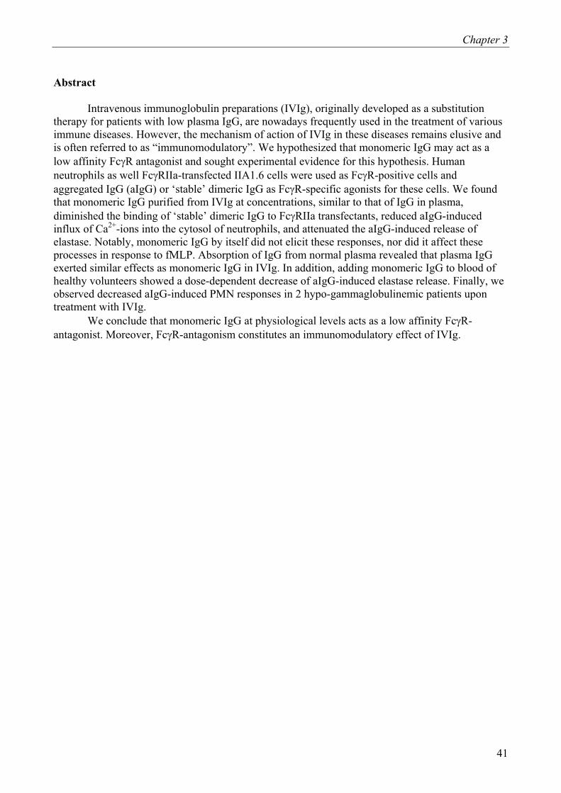
Figure 2: Overview of the human FcγR. Representation of the FcγR expressed in man. FcγRI is associated with the common γ-chain homo-dimer, which contains ITAM motifs, indicated in green. FcγRII contains its own signaling motif, either an ITAM motif (FcγRIIa) or an ITIM motif, indicated in red (FcγRIIb). FcγRIIa contains a polymorphism, indicated in yellow, at position 131. FcγRIIb contains a polymorphism, indicated in green, at position 187. FcγRIIIa is associated with the ζ-γ hetero-dimer. FcγRIIIa contains a polymorphism indicated in purple at position 158. FcγRIIIb has a GPI-anchor and contains the NA1/NA2 polymorphism indicated in light blue.
1. FcγRI
Human FcγRI is represented by three highly homologous genes, FCGR1A, FCGR1B and FCGR1C (10,11) and are mapped on chromosome 1 band q21.1 (12). FCGR1A encodes a glycoprotein of 72 kD containing three extracellular Ig-like domains involved in IgG binding. FCGR1B has two transcripts, one with a stopcodon in extracellular domain 3 and one lacking this domain. Although it is expressed in myeloid cells, this transcript resides in the endoplasmatic reticulum and therefore does not contribute to cell activation (13). For FCGR1C it has not yet been clarified whether it encodes for a functional protein (14). Association and interaction with the FcR-γ-chain signaling subunit has been shown for FcγRI (15). Although the FcR γ chain is not needed for folding and targeting of FcγRI to the cell membrane, association is critical for both FcγRI signal transduction and stable expression in vitro and in vivo (16). However, recent evidence suggests that residual FcγRI expression and functionality is maintained through the interaction with periplakin (17,18).
2. FcγRII
Of the Fcγ receptors FcγRII is the most widely distributed class and is expressed on most types of blood cells (19-21). FcγRII is encoded by FCGR2A, FCGR2B and FCGR2C located on chromosome 1q23-24 (19-21). All three FCGR2 genes encode 40 kD glycoproteins, containing two extracellular Ig-like domains. FCGR2C results very likely from an unequal crossover event between the first part of FCGR2B and the terminal part of FCGR2A (22) and has been postulated to be a pseudogene. However, it has been shown that 88 % of the Caucasian population have a SNP in exon 3 that results in a stopcodon (23). Thus, it is questionable whether the contribution of this isoform is relevant in most individuals.
FcγRII is the only family member known to contain its own signaling motif, as FcγRI and FcγRIII are dependent on the association with another molecule for signal transduction. Several isoforms of FcγRII exist, which are highly homologous in their extracellular and transmembrane

Chapter 2
19
regions but differ in their intracellular domains (24,25). FcγRII contains, depending on the isoform, either an immunoreceptor tyrosine-based activation motif (ITAM) or an immunoreceptor tyrosine-based inhibitory motif (ITIM). FcγRIIa and FcγRIIc contain an ITAM motif and are therefore activating receptors, whereas FcγRIIb contains an ITIM motif and is thus an inhibitory receptor (26,27). In case of FcγRIIc four splice variant have been described of which only the FcγRIIc1 and FcγRIIc3 variants contain an ITAM (28,29). The isoform FcγRIIb has two functional splice variants, FcγRIIb1 and FcγRIIb2. Next to differential expression patterns, these splice variants differ in the presence or absence of exon 6, respectively (23).
In contrast, FcγRIIa has only one known functional variant. However, a functional polymorphism has been identified in FcγRIIa (24). Sequencing revealed that FcγRIIa either expresses an arginine (R) or a histidine (H) at amino acid position 131. These polymorphic variants have been shown to interact differently with various ligands; FcγRIIa-H131 binds human IgG2, whereas FcγRIIa-R131 does not (30). A polymorphism in the transmembrane domain of FcγRIIb has been identified that affects the activity of the ITIM (31). Not until recently, transgenic animals were available to study the role of FcγRIIa in vivo (32). In comparison to their wild-type littermates, induction of thrombocytopenia by antibodies was more severe (32), suggesting an important role of FcγRIIa in this disease. Other studies in animal models and in vitro suggest that, the ratio between activating and inhibitory FcγR may determine the responsiveness of immune cells to immune-complexes (33,34). 3. FcγRIII
FcγRIII is encoded by two genes FCGR3A and FCGR3B, both located on chromosome 1q23-24 (19-21). Both gene products are heterogeneous in size as a result of variable N-linked glycosylation. Molecular masses range between 50-80 kD. FCGR3A and FCGR3B encode for proteins with two extracellular Ig-like domains. In contrast to FcγRIIIa, FcγRIIIb does not contain a transmembrane domain or a cytoplasmatic tail but is anchored to the cell membrane via a glycosyl-phosphatidylinositol (GPI)-anchor. Unlike FcγRI, FcγRIIIa is critically dependent on the FcR γ chain for trafficking towards the cell membrane, due to an endoplasmatic reticulum retention signal (35).
Genetic polymorphisms affecting IgG-subclass binding exist in both FCGR3A (36,37) and FCGR3B (38). In FcγRIIIa a polymorphism at amino acid 158, a valine or phenylalanine, has been identified. As a result, FcγRIIIa-V158 has a higher affinity for IgG1 and IgG3 than FcγRIIIa-F158 (36,37). Allelic variation in FcγRIIIb is comprised of differences in four amino acids, referred to as neutrophils antigen 1 (NA1) and NA2 39. FcγRIIIb-NA1 internalizes IgG1- or IgG3-opsonized particles more efficiently than FcγRIIIb-NA2 (38).
4. FcRn
The α-chain of the non-classical Fc-receptor FcRn is encoded on chromosome 19q13.3 and is dependent for its expression on β2-microglobulin, which is located on chromosome 15q21-22.2. FcRn is a 45 kD glycoprotein that is structurally related to MHC class I (40,41). This molecule was originally identified in the intestinal epithelium of suckling rats as the receptor responsible for the well-known transport of maternal IgG across the intestinal epithelium into the bloodstream (42 43). Next to that, FcRn is also implicated as a mediator of protection for IgG against catabolism (44) and transport across the placenta.
FcRn-IgG interaction is highly pH-dependent, with binding occurring at pH 6.0 but progressively less as neutral pH is approached (45,46). Hence, IgG is bound in the endosomal compartment of the cell, but is released at the cell surface.

Chapter 2
20
Cellular distribution and signal transduction
Engagement of Fc receptors by crosslinking agonists initiates a cascade of signaling effects.
Phosphorylation of tyrosine residues that reside in the signaling motif (ITAM) within the FcγR molecule or the molecule to which the activating Fc receptor is associated, is subsequently followed by recruitment of phosphor-inositol-3-kinase (PI3K) and formation of inositolphosphates that lead to increases in [Ca2+]i. However, when an inhibitory FcγR is co-crosslinked with an activating FcγR, both signaling motifs (ITAM and ITIM) are phosphorylated by protein tyrosine kinases (PTKs). Although the phosphorylated ITAM of the activating FcγRII still recruits PI3K, the phosphorylated ITIM of the inhibitory FcγR recruits phosphatases [Src homology 2 domain-containing inositol polyphosphate phosphatase-1 (SHIP-1) and Src homology 2 domain-containing polyphosphate phosphatase-1 (SHP-1)], which revert the product formed by PI3K back to its original substrate, thereby ablating signal transduction (figure 3). It should be noted that in order to phosphorylate the ITIM of the inhibitory FcγR, a phosphorylated ITAM is required in the direct vicinity of the inhibitory FcγR.
Figure 3: Schematic representation of FcγRIIa coligated with FcγRIIb and its signaling pathways. Phosphorylation of tyrosine residues that reside within the signaling motif (ITAM or ITIM) occur when an inhibitory FcγR is co-crosslinked with an activating FcγR by protein tyrosine kinases (PTKs), such as Lyn kinase. Although the phosphorylated ITAM of the activating FcγRII recruits phosphoinositol-3 kinase (PI3K), the phosphorylated ITIM of the inhibitory FcγR recruits phosphatases [Src homology 2 domain-containing inositol polyphosphate phosphatase-1 (SHIP-1), Src homology 2 domain-containing polyphosphate phosphatase-1 (SHP-1)], which revert the product formed by PI3K back to its original substrate, thereby ablating signal transduction.

Chapter 2
21
1. FcγRI Constitutional high expression of FcγRI is observed on monocytes and macrophages. Under resting conditions neutrophils do not express FcγRI (38). However, expression can be enhanced by G-CSF in vivo (47) and by IFN-γ in vitro and in vivo (48,49). Monocyte FcγRI expression can also be enhanced by IFN-γ 48 or IL-10 (50) and by infections in vivo (51). FcγRI mediates multiple biological functions upon ligation and subsequent crosslinking. FcγRI mediates phagocytosis of opsonized erythrocytes (52), internalization of small immune complexes (53) and subsequent loading onto MHC classes resulting in antigen presentation (54,55). Killing of opsonised cellular targets via ADCC (56,57), cytokine release (58,59) and superoxide production (60-62) are also well characterized effects mediated by FcγRI. 2. FcγRII CD32 is the most widely expressed FcγR and therefore most blood cells express one or more isoforms, although which isoform has not always been elucidated. FcγRII is the sole FcγR expressed by basophils, platelets, langerhans cells, placental endothelial cells and B cells (19-21).
Under resting conditions neutrophils express FcγRIIa (1-4 x 10⁴ molecules per cell) (38). In addition, neutrophils also express the splice variant FcγRIIb2 (34) although quantification has not been substantiated. Monocytes have also been shown to express both FcγRIIa and FcγRIIb2 (34). Interestingly, IFN-γ and IL-4 influence the balance of FcγRIIa and FcγRIIb2 on monocytes; IL-4 induces upregulation of FcγRIIb2 and a simultaneous downregulation of FcγRIIa, where IFN-γ has the opposite effect (34).
B cells express only FcγRIIb1 and when co-ligated to the B cell receptor (BCR) functions to downregulate the BCR activation signal (27). Interestingly, IL-4 has been reported to reduce FcγRIIb1 expression on B cells (63).
NK cells have been reported to express FcγRIIc (29,64) which enhances the cytotoxicity of these cells. FcγRIIa has been shown to have very similar biological functions as FcγRI (52,56,59). Although FcγRIIb contains an ITIM, this receptor has been shown to have phagocytic capacity in the absence of inflammation in mice (65). Strikingly, one of the mechanisms of action of IVIg in inflammatory disorders has been shown to be mediated via FcγRIIb in mice (33). In this study, a murine model for immune thrombocytopenia purpura (ITP), upregulation of FcγRIIb induced by IVIg was observed. Furthermore, FcγRIIb¯/¯ mice were completely unresponsive to IVIg treatment, indicating a critical role for FcγRIIb in amelioration of the disease. However, this finding remains to be confirmed in man, because humans express the activating FcγRIIa/c isoforms, whereas mice are devoid of these FcγRs. 3. FcγRIII Monocytes and NK cells are the major cell populations expressing FcγRIIIa (19-21), whereas FcγRIIIb is solely expressed by neutrophils and eosinophils (66). FcγRIIIa can be induced by TGF-β on monocytes (67). In contrast, IL-4 strongly reduces FcγRIIIa expression on these cells (50,68). Clearance of immune complexes and mediation of ADCC have been reported as functions of FcγRIII (69). In NK cells, next to mediating ADCC, triggering of FcγRIIIa also leads to enhanced transcription of cytokine genes (70,71) and increased adhesion (72). The role of FcγRIIIb in activation of these cells has been debated. Some investigators have shown that this receptor is indeed capable of inducing signal transduction (73), possibly with the

Chapter 2
22
help of FcγRIIa (74), whereas others have suggested that FcγRIIIb does not contribute to effector functions (75). In fact, it has been demonstrated that elastase release from neutrophils induced by dimeric IgG is fully dependent on FcγRII, and not on FcγRIIIb (76). In addition, FcγRIIIb─/─ individuals have been reported to be clinically healthy (77).
Functional blockade of FcγR by IVIg leading to attenuation of signaling effects resulting in e.g. reduced phagocytosis, ADCC, cytokine production and release of cytotoxic mediators, has been hypothesized as a mechanism of action. As described before, IVIg treatment of a murine model for ITP has been shown to be critically dependent on FcγRIIb (33). However, in the same year observations were published showing that disease induction of murine ITP was critically dependend on FcγRIII (78). Furthermore, this study showed that immune protection depended on IgG dimers present in the IVIg preparation. Therefore, functional blockade of FcγRIII by dimeric IgG present in IVIg was postulated. Alternatively, it can be hypothesized that dimeric IgG co-crosslinks both the inhibitory FcγR as well as the activating FcγR, which results in attenuation of the activation signal. 4. FcRn Although expression of FcRn was initially described in the gut of neonatal rats (43) and subsequently in placental cells (42) functioning as transport vehicles for IgG, FcRn is also expressed by monocytes, macrophages and dendritic cells (79). Interestingly, all these cell types express functional FcγR and FcRn has therefore been hypothesized to function in the protection of IgG in complex to antigen from degradation in the lysosomal compartment (79). Increased catabolism of IgG through saturation of FcRn has been hypothesized (80).
An interesting study has shown that IVIg therapy induces a relatively long-lasting but modest reduction of autoantibody levels by accelerating IgG clearance (81). This mechanism has clinical relevance in the sense that it can explain, as the sole mechanism, the gradual 20% to 40% decrease in autoantibody levels observed in several patient studies. However, larger or more rapid effects observed in some other clinical studies cannot be explained by accelerated clearance, implying that IVIg may also reduce autoantibody levels through other mechanisms (81). 2. Effects of IVIg independent of FcγR.
Superantigens and microbial toxins Due to the primary use of IVIg, i.e. substitution therapy in immune-compromised
individuals, the European Pharmacopoeia prescribes that certain levels of neutralizing antibodies against several common environmental pathogens or microbial toxins should at least be present (5). A subgroup a microbial antigens are the so-called superantigens. Superantigens stimulate expansion of large fractions of Vβ-chain-positive unsensitized T cells and induce cytokine secretion in an antigen-aspecific manner. IVIg has the ability to interfere with superantigen-induced T cell activation (82), either through direct neutralization of the superantigen (83) or blockade of T cell receptors by antibodies against Vβ3, Vβ8 and Vβ17 gene families (84).
1. Idiotype anti-idiotype interactions
As mentioned before and inherent to the nature of IVIg, dimerization of IgG molecules can occur due to idiotype anti-idiotype interactions (3,4). In fact, the presence of several anti-idiotypes in the preparation has been reported (85,86). It has been proposed that anti-idiotypes present in the IVIg preparation may ameliorate auto-antibody-mediated disease. Attenuation of disease through anti-idiotypes can occur in several ways, i.e. either by direct neutralization of auto-antibodies (87), thereby inhibiting binding to their target, or by feedback regulation of auto-aggressive B cells (88).

Chapter 2
23
Feedback regulation by anti-idiotype interaction with membrane-bound Ig, i.e. the BCR, is thought to be mediated through co-ligation of FcγRIIb on the cell surface by the Fc-tail of the IgG bound to the BCR which leads to attenuation of the signal delivered by the BCR and subsequent apoptosis of the auto-antibody-producing B cells (89). 2. IVIg and complement activation
Another mechanism of action of IVIg is via the complement system. The complement system is strongly intertwined with the IgG-FcγR system at several levels. First of all IgG-containing immune complexes have been shown to activate the classical pathway of complement sytem (90). Secondly, it has been shown that signaling of complement receptors occurs via common γ-chain or FcγRIIa (91-93). Next to that, one study has implicated C5a as a modulator of the ratio of activating and inhibitory FcγRs (94). Here, it was shown that C5a stimulation resulted in an upregulation of FcγRIII and simultaneous downregulation of FcγRIIb on alveolar macrophages in mice.
IVIg has been implicated to have various effects on the complement system. This has been shown for the first time in vivo in the Forssman shock model in guinea pigs where IVIg protected the animals from acute complement-mediated tissue damage (95). Inhibition of complement-mediated tissue damage was achieved by scavenging activated complement C3 and C4 due to offering a high concentration of acceptor sites for the thioesters exposed in activated C3 and C4 thereby ameliorating the damage mediated by these components (95,96). Alternatively, competition of IVIg with the immune complex for binding of C1q has been suggested (96,97). However, it has also been proposed that influencing the C3-convertase is the mechanism of attenuation of complement activation (98). Finally, neutralization of anaphylatoxins C3a and C5a by IVIg has been reported (99). Interestingly, this would imply that IVIg can potentially modulate the activating-/inhibitory FcγR ratio and tilt the balance to inhibition or an anti-inflammatory response through neutralization of anaphylatoxins. 3. IVIg and cytokines
Modulation of the cytokine network by IVIg has been observed. In vitro studies at the single
cell level indicate that especially T cell lymphokines, e.g. IL-2, IL-3, IL-4, IL-5, IL-10, TNF-β and GM-CSF, were reduced upon stimulation by IVIg (100,101). In contrast, the monokines were rather unaffected (100,102). Only IL-6 was downregulated, whereas IL-1-receptor antagonist (IL1ra) and IL-8 were upregulated (103). In vivo, modulation of cytokine synthesis has been reported (104). Natural antibodies present in IVIg against cytokines with neutralizing effects have been reported (102). In addition, downregulation of expression levels of cytokine receptors as an effect of IVIg have also been observed (105). Thus, in general, pro-inflammatory cytokines are downregulated by IVIg, whereas anti-inflammatory cytokines are enhanced.
Finally, evidence indicates that by influencing the cytokine network the delicate balance between activating and inhibitory FcγR on monocytes is affected (34). This demonstrates the broad range of action of IVIg on the immune system.
4. Differentiation and maturation of dendritic cells
Although IVIg inhibits T-cell proliferation and T-cell cytokine production, it is unclear
whether these effects are directly dependent on the effects of IVIg on T cells or whether they are dependent on the inhibition of antigen-presenting cell activity. Recent studies implicate that IVIg

Chapter 2
24
has a strong influence on the differentiation and maturation of professional antigen-presenting cells, i.e. dendritic cells (DCs) (106).
The differentiation and maturation of dendritic cells (DCs) is governed by various signals in the microenvironment. Monocytes and DCs circulate in peripheral blood, which contains high levels of natural antibodies (NAbs). NAbs are germ-line-encoded (107,108) and are generated independently of antigens, i.e. they are formed in the absence of deliberate immunization or microbial infection. NAbs are known to react with CD40 molecules, which importantly contributes to the development of DCs. Therefore, it has been suggested that B cells promote bystander DC development through NAbs and the interaction between NAbs and DCs may play a role in the steady-state migration of DCs (109). 3. Side effects 1. Adverse effects associated with IgG aggregates
IVIg products often cause mild adverse effects, such as fever, chills, flushing, headache, low back pain and nausea. Aggregates present in the IVIg formulation evoke serious side effects, which was thought to result from activation of the complement system (2). Side effects are usually dependent on the infusion rate and subside rapidly after slowing down the infusion (110). In rare cases of moderately severe reactions such as in bronchoconstriction, the infusion has to be interrupted and the patient should be given intravenous hydrocortisone and antihistamine when needed. Anaphylactic reactions are very rare and can sometimes be associated with preformed IgA antibodies in a patient with complete IgA deficiency. Traditionally, an assay of complement binding, the so-called spontaneous anticomplementary activity (ACA) has been used as the most important biologic measurement of of complement activation by aggregated IgG (111). 2. Adverse effects associated with IgA and other protein contaminants Plasma protein impurities in the product may also cause adverse effects. Agammaglobulinemia patients receiving immunoglobulin treatment may have a complete IgA deficiency. Interestingly, these patients may develop IgG or IgE class anti-IgA antibodies (these patients are then not completely agammaglobulinemic). In these cases, intravenous infusion of a product containing IgA can lead to a severe anaphylactic reaction (112). Anaphylactic reactions are treated according to the usual principles such as by giving epinephrine. Anaphylactic reactions, however, are very rare and not all patients with preformed anti-IgA antibodies react to IgA (113,114). Because the appearance and severity of reactions are unpredictable, patients with a high anti-IgA titer should use an IVIg product with a minimum amount of IgA (110). The upper limit of an IgA level in this sense is unknown and varies between patients. Laschinger et al. (115) showed that six patients with class-specific preformed anti-IgA antibodies tolerated 14 µg to 2.65 mg IgA without reactions. On the other hand, Nadorp et al (116) reported an anaphylactic reaction in one patient receiving less than 1 mg. As a rule, patients with preformed IgA antibodies should receive an IgG product containing less than 1 mg IgA per treatment. Infusion of IVIg can also cause hypotension mediated by prekallikrein activator (PKA) (117). 3. Other adverse effects associated with IVIg products A high level of IgG class anti-A, anti-B or anti-RhD blood group antibodies can sometimes cause a hemolytic reaction with Coombs positivity, especially in immunomodulatory treatment with

Chapter 2
25
high doses (110,118,119). The level of these isoagglutinins is considered safe when it is less than 1:64 at a protein concentration of 3 g/L (5). Mild and moderate reactions with IVIg are encountered in hypogammaglobulinemia replacement therapy. Reactions mostly occur during ongoing active infection. This suggests that the underlying mechanism is the formation of immune complexes between antibodies in IVIg and microbial antigens in the patient. Therefore, adequate treatment with antibiotics is recommended before infusion of IVIg. In the 1990's, reversible acute aseptic meningitis has been reported in connection with high-dose IVIg therapy in ITP or neuromuscular disease (110,120,121). In these patients, headache and nuchal rigidity appeared 10 hours to 7 days after a high-dose IVIg infusion. Pleocytosis and mildly elevated protein concentrations have been found in the spinal fluid; however, no pathogen was detected in microbiological studies. Complete resolution of symptoms was seen in all patients after a few days. Patients with a history of migraine have an increased risk for developing aseptic meningitis. So far, the precise mechanism of the reaction is unclear.

Chapter 2
26
Reference List
1. Cohn, E., LuetscherJJ, J. Oncley, S. J. Armstrong, and B. Davis. 1940. Preparation and properties of serum and plasma proteins. Journal of the American Chemical.Society. 62:3396-3400:3396-3400.
2. Barandun, S., P. Kistler, F. Jeunet, and H. Isliker. 1962. Intravenous administration of human gammaglobulin. Vox Sang. 7:157-174.:157-174.
3. Tankersley, D. L., M. S. Preston, and J. S. Finlayson. 1988. Immunoglobulin G dimer: an idiotype-anti-idiotype complex. Mol.Immunol. 25:41-48.
4. Roux, K. H. and D. L. Tankersley. 1990. A view of the human idiotypic repertoire. Electron microscopic and immunologic analyses of spontaneous idiotype-anti-idiotype dimers in pooled human IgG. J.Immunol. 144:1387-1395.
5. 2004. European Pharmacopoeia. European Pharmacopoeia., Supplement.4.6.Strasbourg., France.: Directorate.for the quality of medicines of the Council.of Europe (EDQM.)4030-4032.
6. Buckley, R. H. and R. I. Schiff. 1991. The use of intravenous immune globulin in immunodeficiency diseases. N Engl J Med 325:110-117.
7. Ravetch, J. V. 1994. Fc receptors: rubor redux. Cell 78:553-560.
8. Galon, J., M. W. Robertson, A. Galinha, N. Mazieres, R. Spagnoli, W. H. Fridman, and C. Sautes. 1997. Affinity of the interaction between Fc gamma receptor type III (Fc gammaRIII) and monomeric human IgG subclasses. Role of Fc gammaRIII glycosylation. Eur.J.Immunol. 27:1928-1932.
9. Maenaka, K., P. A. van der Merwe, D. I. Stuart, E. Y. Jones, and P. Sondermann. 2001. The human low affinity Fcgamma receptors IIa, IIb, and III bind IgG with fast kinetics and distinct thermodynamic properties. J.Biol.Chem. 276:44898-44904.
10. Van de Winkel, J. G., L. K. Ernst, C. L. Anderson, and I. M. Chiu. 1991. Gene organization of the human high affinity receptor for IgG, Fc gamma RI (CD64). Characterization and evidence for a second gene. J.Biol.Chem. 266:13449-13455.
11. Ernst, L. K., J. G. Van de Winkel, I. M. Chiu, and C. L. Anderson. 1992. Three genes for the human high affinity Fc receptor for IgG (Fc gamma RI) encode four distinct transcription products. J.Biol.Chem. 267:15692-15700.
12. de Wit, T. P., R. F. Suijkerbuijk, P. J. Capel, A. Geurts van Kessel, and J. G. Van de Winkel. 1993. Assignment of three human high-affinity Fc gamma receptor I genes to chromosome 1, band q21.1. Immunogenetics 38:57-59.
13. van Vugt, M. J., E. Reefman, I. Zeelenberg, G. Boonen, J. H. Leusen, and J. G. Van de Winkel. 1999. The alternatively spliced CD64 transcript FcgammaRIb2 does not specify a surface-expressed isoform. Eur J Immunol 29:143-149.

Chapter 2
27
14. Porges, A. J., P. B. Redecha, R. Doebele, L. C. Pan, J. E. Salmon, and R. P. Kimberly. 1992. Novel Fc gamma receptor I family gene products in human mononuclear cells. J.Clin.Invest 90:2102-2109.
15. Ernst, L. K., A. M. Duchemin, and C. L. Anderson. 1993. Association of the high-affinity receptor for IgG (Fc gamma RI) with the gamma subunit of the IgE receptor. Proc Natl Acad Sci U S A 90:6023-6027.
16. van Vugt, M. J., A. F. Heijnen, P. J. Capel, S. Y. Park, C. Ra, T. Saito, J. S. Verbeek, and J. G. Van de Winkel. 1996. FcR gamma-chain is essential for both surface expression and function of human Fc gamma RI (CD64) in vivo. Blood 87:3593-3599.
17. Beekman, J. M., J. E. Bakema, J. G. Van de Winkel, and J. H. Leusen. 2004. Direct interaction between FcgammaRI (CD64) and periplakin controls receptor endocytosis and ligand binding capacity. Proc Natl Acad Sci U S A 101:10392-10397.
18. Beekman, J. M., J. E. Bakema, L. J. van der, B. Tops, M. Hinten, M. van Vugt, J. G. Van de Winkel, and J. H. Leusen. 2004. Modulation of FcgammaRI (CD64) ligand binding by blocking peptides of periplakin. J Biol Chem 279:33875-33881.
19. Hulett, M. D. and P. M. Hogarth. 1994. Molecular basis of Fc receptor function. Adv.Immunol. 57:1-127.
20. Van de Winkel, J. G. and C. L. Anderson. 1991. Biology of human immunoglobulin G Fc receptors. J.Leukoc.Biol. 49:511-524.
21. Ravetch, J. V. and J. P. Kinet. 1991. Fc receptors. Annu.Rev.Immunol. 9:457-492.
22. Warmerdam, P. A., N. M. Nabben, S. A. van de Graaf, J. G. Van de Winkel, and P. J. Capel. 1993. The human low affinity immunoglobulin G Fc receptor IIC gene is a result of an unequal crossover event. J.Biol.Chem. 268:7346-7349.
23. Su, K., J. Wu, J. C. Edberg, S. E. McKenzie, and R. P. Kimberly. 2002. Genomic organization of classical human low-affinity Fcgamma receptor genes. Genes Immun. 3 Suppl 1:S51-S56.
24. Warmerdam, P. A., J. G. Van de Winkel, E. J. Gosselin, and P. J. Capel. 1990. Molecular basis for a polymorphism of human Fc gamma receptor II (CD32). J.Exp.Med. 172:19-25.
25. Brooks, D. G., W. Q. Qiu, A. D. Luster, and J. V. Ravetch. 1989. Structure and expression of human IgG FcRII(CD32). Functional heterogeneity is encoded by the alternatively spliced products of multiple genes. J.Exp.Med. 170:1369-1385.
26. Van den Herik-Oudijk, I. E., P. J. Capel, T. van der Bruggen, and J. G. Van de Winkel. 1995. Identification of signaling motifs within human Fc gamma RIIa and Fc gamma RIIb isoforms. Blood 85:2202-2211.
27. Van Den Herik-Oudijk IE, N. A. Westerdaal, N. V. Henriquez, P. J. Capel, and J. G. Van de Winkel. 1994. Functional analysis of human Fc gamma RII (CD32) isoforms expressed in B lymphocytes. J.Immunol. 152:574-585.

Chapter 2
28
28. Metes, D., M. Manciulea, D. Pretrusca, H. Rabinowich, L. K. Ernst, I. Popescu, A. Calugaru, A. Sulica, W. H. Chambers, R. B. Herberman, and P. A. Morel. 1999. Ligand binding specificities and signal transduction pathways of Fc gamma receptor IIc isoforms: the CD32 isoforms expressed by human NK cells. Eur.J.Immunol. 29:2842-2852.
29. Metes, D., L. K. Ernst, W. H. Chambers, A. Sulica, R. B. Herberman, and P. A. Morel. 1998. Expression of functional CD32 molecules on human NK cells is determined by an allelic polymorphism of the FcgammaRIIC gene. Blood 91:2369-2380.
30. Parren, P. W., P. A. Warmerdam, L. C. Boeije, J. Arts, N. A. Westerdaal, A. Vlug, P. J. Capel, L. A. Aarden, and J. G. Van de Winkel. 1992. On the interaction of IgG subclasses with the low affinity Fc gamma RIIa (CD32) on human monocytes, neutrophils, and platelets. Analysis of a functional polymorphism to human IgG2. J.Clin.Invest 90:1537-1546.
31. Li, X., J. Wu, R. H. Carter, J. C. Edberg, K. Su, G. S. Cooper, and R. P. Kimberly. 2003. A novel polymorphism in the Fcgamma receptor IIB (CD32B) transmembrane region alters receptor signaling. Arthritis Rheum. 48:3242-3252.
32. McKenzie, S. E., S. M. Taylor, P. Malladi, H. Yuhan, D. L. Cassel, P. Chien, E. Schwartz, A. D. Schreiber, S. Surrey, and M. P. Reilly. 1999. The role of the human Fc receptor Fc gamma RIIA in the immune clearance of platelets: a transgenic mouse model. J Immunol 162:4311-4318.
33. Samuelsson, A., T. L. Towers, and J. V. Ravetch. 2001. Anti-inflammatory activity of IVIG mediated through the inhibitory Fc receptor. Science 19;291:484-486.
34. Pricop, L., P. Redecha, J. L. Teillaud, J. Frey, W. H. Fridman, C. Sautes-Fridman, and J. E. Salmon. 2001. Differential modulation of stimulatory and inhibitory Fc gamma receptors on human monocytes by Th1 and Th2 cytokines. J.Immunol. 166:531-537.
35. Kurosaki, T., I. Gander, and J. V. Ravetch. 1991. A subunit common to an IgG Fc receptor and the T-cell receptor mediates assembly through different interactions. Proc Natl Acad Sci U S A 88:3837-3841.
36. Koene, H. R., M. Kleijer, J. Algra, D. Roos, A. E. dem Borne, and M. de Haas. 1997. Fc gammaRIIIa-158V/F polymorphism influences the binding of IgG by natural killer cell Fc gammaRIIIa, independently of the Fc gammaRIIIa-48L/R/H phenotype. Blood 90:1109-1114.
37. de Haas, M., H. R. Koene, M. Kleijer, E. de Vries, S. Simsek, M. J. van Tol, D. Roos, and A. E. dem Borne. 1996. A triallelic Fc gamma receptor type IIIA polymorphism influences the binding of human IgG by NK cell Fc gamma RIIIa. J Immunol 156:3948-3955.
38. Huizinga, T. W., M. Kerst, J. H. Nuyens, A. Vlug, B. von dem, D. Roos, and P. A. Tetteroo. 1989. Binding characteristics of dimeric IgG subclass complexes to human neutrophils. J.Immunol. 142:2359-2364.

Chapter 2
29
39. Huizinga, T. W., M. Kleijer, P. A. Tetteroo, D. Roos, and A. E. dem Borne. 1990. Biallelic neutrophil Na-antigen system is associated with a polymorphism on the phospho-inositol-linked Fc gamma receptor III (CD16). Blood 75:213-217.
40. Gastinel, L. N., N. E. Simister, and P. J. Bjorkman. 1992. Expression and crystallization of a soluble and functional form of an Fc receptor related to class I histocompatibility molecules. Proc Natl Acad Sci U S A 89:638-642.
41. Simister, N. E. and K. E. Mostov. 1989. An Fc receptor structurally related to MHC class I antigens. Nature 337:184-187.
42. Abrahamson, D. R. and R. Rodewald. 1981. Evidence for the sorting of endocytic vesicle contents during the receptor-mediated transport of IgG across the newborn rat intestine. J Cell Biol 91:270-280.
43. Brambell, F. W. 1966. The transmission of immunity from mother to young and the catabolism of immunoglobulins. Lancet 2:1087-1093.
44. Junghans, R. P. and C. L. Anderson. 1996. The protection receptor for IgG catabolism is the beta2-microglobulin-containing neonatal intestinal transport receptor. Proc Natl Acad Sci U S A 93:5512-5516.
45. Raghavan, M., L. N. Gastinel, and P. J. Bjorkman. 1993. The class I major histocompatibility complex related Fc receptor shows pH-dependent stability differences correlating with immunoglobulin binding and release. Biochemistry 32:8654-8660.
46. Raghavan, M., V. R. Bonagura, S. L. Morrison, and P. J. Bjorkman. 1995. Analysis of the pH dependence of the neonatal Fc receptor/immunoglobulin G interaction using antibody and receptor variants. Biochemistry 34:14649-14657.
47. Repp, R., T. Valerius, A. Sendler, M. Gramatzki, H. Iro, J. R. Kalden, and E. Platzer. 1991. Neutrophils express the high affinity receptor for IgG (Fc gamma RI, CD64) after in vivo application of recombinant human granulocyte colony-stimulating factor. Blood 78:885-889.
48. Guyre, P. M., P. M. Morganelli, and R. Miller. 1983. Recombinant immune interferon increases immunoglobulin G Fc receptors on cultured human mononuclear phagocytes. J Clin Invest 72:393-397.
49. Perussia, B., M. Kobayashi, M. E. Rossi, I. Anegon, and G. Trinchieri. 1987. Immune interferon enhances functional properties of human granulocytes: role of Fc receptors and effect of lymphotoxin, tumor necrosis factor, and granulocyte-macrophage colony-stimulating factor. J Immunol 138:765-774.
50. Te Velde, A. A., M. R. de Waal, R. J. Huijbens, J. E. de Vries, and C. G. Figdor. 1992. IL-10 stimulates monocyte Fc gamma R surface expression and cytotoxic activity. Distinct regulation of antibody-dependent cellular cytotoxicity by IFN-gamma, IL-4, and IL-10. J Immunol 149:4048-4052.
51. Simms, H. H., M. M. Frank, T. C. Quinn, S. Holland, and T. A. Gaither. 1989. Studies on phagocytosis in patients with acute bacterial infections. J Clin Invest 83:252-260.

Chapter 2
30
52. Anderson, C. L., L. Shen, D. M. Eicher, M. D. Wewers, and J. K. Gill. 1990. Phagocytosis mediated by three distinct Fc gamma receptor classes on human leukocytes. J Exp Med 171:1333-1345.
53. Harrison, P. T., W. Davis, J. C. Norman, A. R. Hockaday, and J. M. Allen. 1994. Binding of monomeric immunoglobulin G triggers Fc gamma RI-mediated endocytosis. J Biol Chem 269:24396-24402.
54. Gosselin, E. J., K. Wardwell, D. R. Gosselin, N. Alter, J. L. Fisher, and P. M. Guyre. 1992. Enhanced antigen presentation using human Fc gamma receptor (monocyte/macrophage)-specific immunogens. J.Immunol 149:3477-3481.
55. van Vugt, M. J., M. J. Kleijmeer, T. Keler, I. Zeelenberg, M. A. van Dijk, J. H. Leusen, H. J. Geuze, and J. G. Van de Winkel. 1999. The FcgammaRIa (CD64) ligand binding chain triggers major histocompatibility complex class II antigen presentation independently of its associated FcR gamma-chain. Blood 94:808-817.
56. Graziano, R. F. and M. W. Fanger. 1987. Fc gamma RI and Fc gamma RII on monocytes and granulocytes are cytotoxic trigger molecules for tumor cells. J Immunol 139:3536-3541.
57. Fanger, M. W., L. Shen, R. F. Graziano, and P. M. Guyre. 1989. Cytotoxicity mediated by human Fc receptors for IgG. Immunol Today 10:92-99.
58. Krutmann, J., R. Kirnbauer, A. Kock, T. Schwarz, E. Schopf, L. T. May, P. B. Sehgal, and T. A. Luger. 1990. Cross-linking Fc receptors on monocytes triggers IL-6 production. Role in anti-CD3-induced T cell activation. J Immunol 145:1337-1342.
59. Debets, J. M., J. G. Van de Winkel, J. L. Ceuppens, I. E. Dieteren, and W. A. Buurman. 1990. Cross-linking of both Fc gamma RI and Fc gamma RII induces secretion of tumor necrosis factor by human monocytes, requiring high affinity Fc-Fc gamma R interactions. Functional activation of Fc gamma RII by treatment with proteases or neuraminidase. J Immunol 144:1304-1310.
60. Anderson, C. L., P. M. Guyre, J. C. Whitin, D. H. Ryan, R. J. Looney, and M. W. Fanger. 1986. Monoclonal antibodies to Fc receptors for IgG on human mononuclear phagocytes. Antibody characterization and induction of superoxide production in a monocyte cell line. J Biol Chem 261:12856-12864.
61. Pfefferkorn, L. C. and M. W. Fanger. 1989. Cross-linking of the high affinity Fc receptor for human immunoglobulin G1 triggers transient activation of NADPH oxidase activity. Continuous oxidase activation requires continuous de novo receptor cross-linking. J Biol Chem 264:14112-14120.
62. Pfefferkorn, L. C. and M. W. Fanger. 1989. Transient activation of the NADPH oxidase through Fc gamma RI. Oxidase deactivation precedes internalization of cross-linked receptors. J Immunol 143:2640-2649.
63. Rudge, E. U., A. J. Cutler, N. R. Pritchard, and K. G. Smith. 2002. Interleukin 4 reduces expression of inhibitory receptors on B cells and abolishes CD22 and Fc gamma RII-mediated B cell suppression. J Exp Med 195:1079-1085.

Chapter 2
31
64. Sulica, A., P. Morel, D. Metes, and R. B. Herberman. 2001. Ig-binding receptors on human NK cells as effector and regulatory surface molecules. Int.Rev.Immunol. 20:371-414.
65. van Lent, P., K. C. Nabbe, P. Boross, A. B. Blom, J. Roth, A. Holthuysen, A. Sloetjes, S. Verbeek, and B. W. van den. 2003. The inhibitory receptor FcgammaRII reduces joint inflammation and destruction in experimental immune complex-mediated arthritides not only by inhibition of FcgammaRI/III but also by efficient clearance and endocytosis of immune complexes. Am.J.Pathol. 163:1839-1848.
66. Huizinga, T. W., C. E. van der Schoot, C. Jost, R. Klaassen, M. Kleijer, A. E. dem Borne, D. Roos, and P. A. Tetteroo. 1988. The PI-linked receptor FcRIII is released on stimulation of neutrophils. Nature 333:667-669.
67. Wahl, S. M., J. B. Allen, G. R. Welch, and H. L. Wong. 1992. Transforming growth factor-beta in synovial fluids modulates Fc gamma RII (CD16) expression on mononuclear phagocytes. J Immunol 148:485-490.
68. Wong, H. L., G. R. Welch, M. E. Brandes, and S. M. Wahl. 1991. IL-4 antagonizes induction of Fc gamma RIII (CD16) expression by transforming growth factor-beta on human monocytes. J Immunol 147:1843-1848.
69. Clarkson, S. B., J. B. Bussel, R. P. Kimberly, J. E. Valinsky, R. L. Nachman, and J. C. Unkeless. 1986. Treatment of refractory immune thrombocytopenic purpura with an anti-Fc gamma-receptor antibody. N.Engl.J.Med 314:1236-1239.
70. Anegon, I., M. C. Cuturi, G. Trinchieri, and B. Perussia. 1988. Interaction of Fc receptor (CD16) ligands induces transcription of interleukin 2 receptor (CD25) and lymphokine genes and expression of their products in human natural killer cells. J Exp Med 167:452-472.
71. Cassatella, M. A., I. Anegon, M. C. Cuturi, P. Griskey, G. Trinchieri, and B. Perussia. 1989. Fc gamma R(CD16) interaction with ligand induces Ca2+ mobilization and phosphoinositide turnover in human natural killer cells. Role of Ca2+ in Fc gamma R(CD16)-induced transcription and expression of lymphokine genes. J Exp Med 169:549-567.
72. Gismondi, A., F. Mainiero, S. Morrone, G. Palmieri, M. Piccoli, L. Frati, and A. Santoni. 1992. Triggering through CD16 or phorbol esters enhances adhesion of NK cells to laminin via very late antigen 6. J Exp Med 176:1251-1257.
73. Vossebeld, P. J., C. H. Homburg, R. C. Schweizer, I. Ibarrola, J. Kessler, L. Koenderman, D. Roos, and A. J. Verhoeven. 1997. Tyrosine phosphorylation-dependent activation of phosphatidylinositide 3-kinase occurs upstream of Ca2+-signalling induced by Fcgamma receptor cross-linking in human neutrophils. Biochem.J. 323:87-94.
74. Chuang, F. Y., M. Sassaroli, and J. C. Unkeless. Convergence of Fc gamma receptor IIA and Fc gamma receptor IIIB signaling pathways in human neutrophils. J Immunol 2000.Jan 1;164.(1):350.-60. 164:350-360.

Chapter 2
32
75. Scott-Zaki, P., D. Purkall, and S. Ruddy. Neutrophil chemotaxis and superoxide production are induced by cross-linking FcgammaRII receptors. Cell Immunol 2000.May.1;201.(2):89.-93. 201:89-93.
76. Teeling, J. L., E. R. De Groot, A. J. Eerenberg, W. K. Bleeker, G. Van Mierlo, L. A. Aarden, and C. E. Hack. 1998. Human intravenous immunoglobulin (IVIG) preparations degranulate human neutrophils in vitro. Clin.Exp Immunol 114:264-270.
77. de Haas, M., M. Kleijer, R. van Zwieten, D. Roos, and A. E. dem Borne. 1995. Neutrophil Fc gamma RIIIb deficiency, nature, and clinical consequences: a study of 21 individuals from 14 families. Blood 86:2403-2413.
78. Teeling, J. L., T. Jansen-Hendriks, T. W. Kuijpers, M. de Haas, J. G. Van de Winkel, C. E. Hack, and W. K. Bleeker. Therapeutic efficacy of intravenous immunoglobulin preparations depends on the immunoglobulin G dimers: studies in experimental immune thrombocytopenia. Blood 2001.Aug.15.;98.(4.):1095.-9. 98:1095-1099.
79. Zhu, X., G. Meng, B. L. Dickinson, X. Li, E. Mizoguchi, L. Miao, Y. Wang, C. Robert, B. Wu, P. D. Smith, W. I. Lencer, and R. S. Blumberg. MHC class I-related neonatal Fc receptor for IgG is functionally expressed in monocytes, intestinal macrophages, and dendritic cells. J Immunol 2001.Mar 1;166.(5.):3266.-76. 166:3266-3276.
80. Yu, Z. and V. A. Lennon. 1999. Mechanism of intravenous immune globulin therapy in antibody-mediated autoimmune diseases. N.Engl.J.Med. 340:227-228.
81. Bleeker, W. K., J. L. Teeling, and C. E. Hack. Accelerated autoantibody clearance by intravenous immunoglobulin therapy: studies in experimental models to determine the magnitude and time course of the effect. Blood 2001.Nov.15.;98.(10.):3136.-42. 98:3136-3142.
82. Takei, S., Y. K. Arora, and S. M. Walker. 1993. Intravenous immunoglobulin contains specific antibodies inhibitory to activation of T cells by staphylococcal toxin superantigens [see comment]. J Clin Invest 91:602-607.
83. Nishi, J. I., S. Kanekura, S. Takei, I. Kitajima, T. Nakajima, M. R. Wahid, K. Masuda, M. Yoshinaga, I. Maruyama, and K. Miyata. 1997. B cell epitope mapping of the bacterial superantigen staphylococcal enterotoxin B: the dominant epitope region recognized by intravenous IgG. J.Immunol. 158:247-254.
84. Marchalonis, J. J., H. Kaymaz, F. Dedeoglu, S. F. Schluter, D. E. Yocum, and A. B. Edmundson. 1992. Human autoantibodies reactive with synthetic autoantigens from T-cell receptor beta chain. Proc Natl Acad Sci U S A 89:3325-3329.
85. Rossi, F., D. R. Jayne, C. M. Lockwood, and M. D. Kazatchkine. 1991. Anti-idiotypes against anti-neutrophil cytoplasmic antigen autoantibodies in normal human polyspecific IgG for therapeutic use and in the remission sera of patients with systemic vasculitis. Clin Exp Immunol 83:298-303.

Chapter 2
33
86. Sultan, Y., M. D. Kazatchkine, P. Maisonneuve, and U. E. Nydegger. 1984. Anti-idiotypic suppression of autoantibodies to factor VIII (antihaemophilic factor) by high-dose intravenous gammaglobulin. Lancet 2:765-768.
87. Kazatchkine, M. D., G. Dietrich, V. Hurez, N. Ronda, B. Bellon, F. Rossi, and S. V. Kaveri. 1994. V region-mediated selection of autoreactive repertoires by intravenous immunoglobulin (i.v.Ig). Immunol Rev 139:79-107.
88. Tankersley, D. L. 1994. Dimer formation in immunoglobulin preparations and speculations on the mechanism of action of intravenous immune globulin in autoimmune diseases. Immunol.Rev. 139:159-72.:159-172.
89. Heyman, B. 2000. Regulation of antibody responses via antibodies, complement, and Fc receptors. Annu.Rev.Immunol. 18:709-37.:709-737.
90. Cowdery, J. S., Jr., P. E. Treadwell, and R. B. Fritz. 1975. A radioimmunoassay for human antigen-antibody complexes in clinical material. J.Immunol. 114:5-9.
91. Jones, S. L., U. G. Knaus, G. M. Bokoch, and E. J. Brown. 1998. Two signaling mechanisms for activation of alphaM beta2 avidity in polymorphonuclear neutrophils. J Biol Chem 273:10556-10566.
92. Downey, G. P., T. Fukushima, L. Fialkow, and T. K. Waddell. 1995. Intracellular signaling in neutrophil priming and activation. Semin Cell Biol 6:345-356.
93. Fukushima, T., T. K. Waddell, S. Grinstein, G. G. Goss, J. Orlowski, and G. P. Downey. 1996. Na+/H+ exchange activity during phagocytosis in human neutrophils: role of Fcgamma receptors and tyrosine kinases. J Cell Biol 132:1037-1052.
94. Shushakova, N., J. Skokowa, J. Schulman, U. Baumann, J. Zwirner, R. E. Schmidt, and J. E. Gessner. 2002. C5a anaphylatoxin is a major regulator of activating versus inhibitory FcgammaRs in immune complex-induced lung disease. J.Clin.Invest 110:1823-1830.
95. Basta, M., P. Kirshbom, M. M. Frank, and L. F. Fries. 1989. Mechanism of therapeutic effect of high-dose intravenous immunoglobulin. Attenuation of acute, complement-dependent immune damage in a guinea pig model. J.Clin.Invest 84:1974-1981.
96. Mollnes, T. E., I. H. Andreassen, K. Hogasen, C. E. Hack, and M. Harboe. 1997. Effect of whole and fractionated intravenous immunoglobulin on complement in vitro. Mol.Immunol. 34:719-729.
97. Miletic, V. D. and M. M. Frank. 1995. Complement-immunoglobulin interactions. Curr.Opin.Immunol. 7:41-47.
98. Lutz, H. U., P. Stammler, V. Bianchi, R. M. Trueb, T. Hunziker, R. Burger, E. Jelezarova, and P. J. Spath. 2004. Intravenously applied IgG stimulates complement attenuation in a complement-dependent autoimmune disease at the amplifying C3 convertase level. Blood 103:465-472.

Chapter 2
34
99. Basta, M., F. Van Goor, S. Luccioli, E. M. Billings, A. O. Vortmeyer, L. Baranyi, J. Szebeni, C. R. Alving, M. C. Carroll, I. Berkower, S. S. Stojilkovic, and D. D. Metcalfe. 2003. F(ab)(2)-mediated neutralization of C3a and C5a anaphylatoxins: a novel effector function of immunoglobulins. Nat.Med. 9:431-438.
100. Andersson, J., U. Skansen-Saphir, E. Sparrelid, and U. Andersson. 1996. Intravenous immune globulin affects cytokine production in T lymphocytes and monocytes/macrophages. Clin Exp Immunol 104 Suppl 1:10-20.
101. Skansen-Saphir, U., J. Andersson, L. Bjork, C. Ekberg, T. E. Fehniger, J. I. Henter, and U. Andersson. 1998. Down-regulation of lymphokine synthesis by intravenous gammaglobulin is dependent upon accessory cells. Scand J Immunol 47:229-235.
102. Andersson, U., L. Bjork, U. Skansen-Saphir, and J. Andersson. 1994. Pooled human IgG modulates cytokine production in lymphocytes and monocytes. Immunol Rev 139:21-42.
103. Ruiz, d. S., V, M. P. Carreno, S. V. Kaveri, A. Ledur, H. Sadeghi, J. M. Cavaillon, M. D. Kazatchkine, and N. Haeffner-Cavaillon. 1995. Selective induction of interleukin-1 receptor antagonist and interleukin-8 in human monocytes by normal polyspecific IgG (intravenous immunoglobulin). Eur J Immunol 25:1267-1273.
104. Sewell, W. A., M. E. North, R. Cambronero, A. D. Webster, and J. Farrant. 1999. In vivo modulation of cytokine synthesis by intravenous immunoglobulin. Clin Exp Immunol 116:509-515.
105. Andersson, U. G., L. Bjork, U. Skansen-Saphir, and J. P. Andersson. 1993. Down-regulation of cytokine production and interleukin-2 receptor expression by pooled human IgG. Immunology 79:211-216.
106. Bayry, J., S. Lacroix-Desmazes, C. Carbonneil, N. Misra, V. Donkova, A. Pashov, A. Chevailler, L. Mouthon, B. Weill, P. Bruneval, M. D. Kazatchkine, and S. V. Kaveri. 2003. Inhibition of maturation and function of dendritic cells by intravenous immunoglobulin. Blood 101:758-765.
107. Marchalonis, J. J., M. K. Adelman, B. J. Zeitler, P. M. Sarazin, P. M. Jaqua, and S. F. Schluter. 2001. Evolutionary factors in the emergence of the combinatorial germline antibody repertoire. Adv Exp Med Biol 484:13-30.
108. Hayakawa, K., M. Asano, S. A. Shinton, M. Gui, D. Allman, C. L. Stewart, J. Silver, and R. R. Hardy. 1999. Positive selection of natural autoreactive B cells. Science 285:113-116.
109. Bayry, J., S. Lacroix-Desmazes, V. Donkova-Petrini, C. Carbonneil, N. Misra, Y. Lepelletier, S. Delignat, S. Varambally, E. Oksenhendler, Y. Levy, M. Debre, M. D. Kazatchkine, O. Hermine, and S. V. Kaveri. 2004. Natural antibodies sustain differentiation and maturation of human dendritic cells. Proc Natl Acad Sci U S A 101:14210-14215.
110. Misbah, S. A. and H. M. Chapel. 1993. Adverse effects of intravenous immunoglobulin. Drug Saf 9:254-262.

Chapter 2
35
111. Romer, J., A. Gardi, and P. Kistler. 1979. Assay of anticomplementary activity in solutions of immunoglobulins. Dev Biol Stand 44:147-151.
112. Wells, J. V., R. H. Buckley, M. S. Schanfield, and H. H. Fudenberg. 1977. Anaphylactic reactions to plasma infusions in patients with hypogammaglobulinemia and anti-IgA antibodies. Clin Immunol Immunopathol 8:265-271.
113. Koskinen, S., H. Tolo, M. Hirvonen, and J. Koistinen. 1995. Long-term follow-up of anti-IgA antibodies in healthy IgA-deficient adults. J Clin Immunol 15:194-198.
114. Eijkhout, H. W., P. J. van den Broek, and J. W. Van Der Meer. 2003. Substitution therapy in immunodeficient patients with anti-IgA antibodies or severe adverse reactions to previous immunoglobulin therapy. Neth.J.Med. 61:213-217.
115. Laschinger, C., D. Gauthier, J. P. Valet, and D. H. Naylor. 1984. Fluctuating levels of serum IgA in individuals with selective IgA deficiency. Vox Sang 47:60-67.
116. Nadorp, J. H., M. Voss, W. C. Buys, P. J. van Munster, J. H. van Tongeren, R. C. Aalberse, and E. van Loghem. 1973. The significance of the presence of anti-IgA antibodies in individuals with an IgA deficiency. Eur J Clin Invest 3:317-323.
117. Alving, B. M., D. L. Tankersley, B. L. Mason, F. Rossi, D. L. Aronson, and J. S. Finlayson. 1980. Contact-activated factors: contaminants of immunoglobulins preparations with coagulant and vasoactive properties. J Lab Clin Med 96:334-346.
118. Nakamura, S., T. Yoshida, S. Ohtake, and T. Matsuda. 1986. Hemolysis due to high-dose intravenous gammaglobulin treatment for patients with idiopathic thrombocytopenic purpura. Acta Haematol 76:115-118.
119. Vincenzi, D., G. Lama, E. Mignani, and V. Pompili. 1989. Anti-A, anti-B agglutinins in some commercial intravenous gamma-globulins. Vox Sang 57:219.
120. Stangel, M., H. P. Hartung, P. Marx, and R. Gold. 1997. Side effects of high-dose intravenous immunoglobulins. Clin Neuropharmacol. 20:385-393.
121. Sekul, E. A., E. J. Cupler, and M. C. Dalakas. 1994. Aseptic meningitis associated with high-dose intravenous immunoglobulin therapy: frequency and risk factors. Ann Intern Med 121:259-262.

Chapter 2
36

37

38

Chapter 3
39
Monomeric IgG in IVIg is a functional antagonist of FcγRII and FcγRIIIb. Edwin van Mirre1, Jessica L. Teeling1, Jos W. M. van der Meer2, Wim K. Bleeker1, C. Erik Hack1,3 1 Department of Immunopathology, Sanquin Research, and Landsteiner Laboratory, Academic Medical Center, University of Amsterdam; 2 Department of Internal Medicine, UMC St. Radboud, Nijmegen; 3 Department of Clinical Chemistry, VU Medical Center, Amsterdam, the Netherlands Journal of Immunology, 2004, 173: 332-339.

Chapter 3
40
Chapter 3
41
Abstract
Intravenous immunoglobulin preparations (IVIg), originally developed as a substitution therapy for patients with low plasma IgG, are nowadays frequently used in the treatment of various immune diseases. However, the mechanism of action of IVIg in these diseases remains elusive and is often referred to as “immunomodulatory”. We hypothesized that monomeric IgG may act as a low affinity FcγR antagonist and sought experimental evidence for this hypothesis. Human neutrophils as well FcγRIIa-transfected IIA1.6 cells were used as FcγR-positive cells and aggregated IgG (aIgG) or ‘stable’ dimeric IgG as FcγR-specific agonists for these cells. We found that monomeric IgG purified from IVIg at concentrations, similar to that of IgG in plasma, diminished the binding of ‘stable’ dimeric IgG to FcγRIIa transfectants, reduced aIgG-induced influx of Ca2+-ions into the cytosol of neutrophils, and attenuated the aIgG-induced release of elastase. Notably, monomeric IgG by itself did not elicit these responses, nor did it affect these processes in response to fMLP. Absorption of IgG from normal plasma revealed that plasma IgG exerted similar effects as monomeric IgG in IVIg. In addition, adding monomeric IgG to blood of healthy volunteers showed a dose-dependent decrease of aIgG-induced elastase release. Finally, we observed decreased aIgG-induced PMN responses in 2 hypo-gammaglobulinemic patients upon treatment with IVIg.
We conclude that monomeric IgG at physiological levels acts as a low affinity FcγR-antagonist. Moreover, FcγR-antagonism constitutes an immunomodulatory effect of IVIg.

Chapter 3
42
Introduction
Polyspecific IgG for intravenous use (IVIg) was originally developed as a substitution therapy for hypo- and agammaglobulinemic patients. However, Imbach et al. observed that IVIg supplementation was in fact effective as a treatment in idiopathic thrombocytopenia purpura (1). This has led to the widely spread use of IVIg in various immune diseases. Yet, the biological effects of IVIg, explaining its efficacy in these diseases, are still poorly understood, although they are frequently referred to as “immunomodulatory” effects. One could postulate various mechanisms for these immunomodulatory effects of IVIg, some of which are dependent on a productive interaction of the Fcγ-region of infused immunoglobulin with the Fcγ-receptors on effector cells (1-4) or with proteins of the complement system (5,6).
Blockade of the Fcγ-receptors (FcγR) on phagocytic cells, preventing the removal of sensitised platelets by the macrophages in the spleen and liver, is believed to be the mechanism of action by which IVIg is effective as a treatment of ITP (1). Indeed, blockade of FcγR by IVIg on macrophages has been shown to inhibit macrophage-mediated phagocytosis of antigen-bearing target cells (2,7). Depending on their expression on effector cells, FcγR exert different effects. For example, on phagocytes they mediate phagocytosis, endocytosis, antibody-dependent cellular cytotoxicity (ADCC) and induction of the respiratory burst. Three types of FcγR, type I, II and III, are discriminated, based on their affinity for monomeric IgG. Type I (Ka = 108-109 M-1) is considered a high-affinity receptor, whereas type II (Ka = 106 M-1) and III (Ka = 5.5 x 10⁵ M-1) (8,9) are considered to be low-affinity receptors. Therefore, it is postulated that FcγRI binds monomeric IgG in vivo, whereas FcγRII and FcγRIII preferentially interact with immune complexes. Thus, regarding IVIg, FcγRII and FcγRIII will interact predominantly with di- or polymeric IgG, whereas FcγRI likely react with monomeric IgG as well.
IVIg preparations contain monomeric IgG as well as a variable amount of dimeric and polymeric IgG. In a mouse model for ITP, we have shown that dimeric IgG in IVIg potently inhibits removal of antibody-sensitized platelets, whereas preparations with low dimeric content were hardly active (2). Notably, the removal of sensitized platelets in this model is dependent on the low-affinity FcγRIII. Hence, one could postulate that dimeric IgG is the active principle in IVIg, explaining the efficacy of this drug in ITP. To what extent monomeric IgG in IVIg may contribute to the blockade of FcγRII and FcγRIII is not known. Although dimeric IgG is more potent in reducing immune complex-mediated anaphylaxis in rats; we also observed that dimeric IgG induces anaphylaxis itself. In contrast, a high dose monomeric IgG did not induce anaphylaxis, but still had a protective effect (Teeling et al. submitted). In the present study we investigated whether monomeric IgG at high concentrations could act as a low-affinity FcγR antagonist. We sought experimental evidence for this hypothesis, using FcγRIIa-transfected cells as well as neutrophils as a model. Neutrophils express both FcγRIIa and FcγRIIIb (1-4 x 10⁴ and 1-3 x 10⁵ molecules per cell, respectively), and, under resting conditions, no FcγRI (10). Possible antagonistic effects by IVIg as well as by plasma IgG on neutrophils were investigated. Our results indicate that monomeric IgG, in plasma as well as in IVIg, has sufficient affinity to displace the binding of immune-complexes to FcγRII and FcγRIII, resulting in an attenuation of signal transduction via these receptors.

Chapter 3
43
Materials & Methods
Patients and blood sampling Two patients with low plasma IgG due to late-onset common variable immune deficiency
(CVID), who had not been treated with IVIg before, received IVIg at 0.4 g per kg body weight. Before, halfway and at the end of IVIg infusion blood was collected in heparin-coated tubes. Responsiveness of neutrophils for aggregated IgG (aIgG; as a model for immune complexes) was tested ex vivo by incubating whole blood with aIgG as described below. The patients as well as healthy volunteers contributed to this study after informed consent.
Immunoglobulin preparations Immunoglobulin I.V. (IVIg, Lot. no. 01H03H443A, Sanquin, CLB, Amsterdam, the
Netherlands) contains 60 g protein per liter, of which at least 95% is IgG. To obtain monomeric IgG, IVIg was reconstituted and set at low pH by dialysis against 10
mM acetate buffer, containing 0.24 M glucose and 0.037 M NaCl, pH 4.15. Monomeric IgG content was verified by HPLC gel filtration (Superdex 200 HR 16/30; Amersham Biosciences, Uppsala, Sweden); no dimeric or polymeric content was detected. One part of monomeric IgG was biotinylated according to standard procedures (Pierce, Rockford, USA) and used for direct binding studies (see below).
IgG dimers were prepared as described by Huizinga et al (10). In short, mAb K37, directed against the λ-light chain of human IgG, was digested by pepsin (Cooper Biomedial) for 20 h at 37°C to generate F(ab’)2 fragments. The F(ab’)2 fragments were incubated with IVIG for 72 h at 4°C in a 1:2 ratio. Subsequently, complexes were purified on a Ultropac TSK-G4000SWG column (LKB-Producter AB, Bromma, Sweden) connected to an FPLC system with PBS-0.02% sodium azide as running buffer. Fractions were collected and concentrated by dialysis against PEG. Purity of the ‘stable’ dimeric fraction was checked by gelfiltration over a Superose 12 HR 10.30 column (Pharmacia). The chromatogram was analyzed by a computer-program (EZChrom, version 6.5, Pharmacia). The ‘stable’ dimeric IgG fraction was pooled and biotinylated according to standard procedures (Pierce, Rockford, USA).
Aggregated IgG (aIgG) was obtained by incubating IVIg at 10 mg per ml in phosphate-buffered saline, pH 7.4 (PBS), for 30 minutes at 63 °C (11). Gel filtration chromatography on a Superdex 200 HR 16/30 column revealed that the preparation contained 43 % aIgG, no dimeric and 57 % monomeric IgG, as analyzed by a computer program (Unicorn version 4.5, Amersham Biosciences).
Monoclonal antibodies and reagents The following monoclonal antibodies (mAb) against human Fcγ receptors (FcγR) were used:
CD16 (anti-FcγRIII, clone 3G8, prepared as F(ab’)2 fragments; a generous gift from Dr. Masja de Haas, Sanquin Research, Amsterdam, the Netherlands); CD16-PE (anti-FcγRIII, IgG2a isotype, clone CLB-FcR-gran/1, 5D2, Sanquin, Amsterdam, the Netherlands), CD32-biotin (anti-FcγRII, Fab fragments of clone IV.3; also a generous gift from Dr. Masja de Haas) and anti-CD64-FITC (anti-FcγRI, IgG1 isotype, clone 10.1, InstruChemie, Delfzijl, the Netherlands).
FITC-conjugated rabbit-anti-human-IgG polyclonal F(ab’)2 fragments were obtained from Sanquin and were reduced with 1 mM DTT. Subsequently, 2.5 mM iodoacetamide was added to obtain Fab fragments. The Fab fragments were then dialyzed against PBS o/n to remove DTT and

Chapter 3
44
iodoacetamide. Relevant isotype controls were obtained from Sanquin: isotype control IgG2a-PE (clone 713) and IgG1-FITC (clone 203). Streptavidin-allophycocyanin (strep-APC; BD PharMingen, San Diego, CA, USA) was used to visualize CD32-biotin on the cell surface.
Fab fragments of clone anti-C1q-85, a generous gift from Fabian McGrath M.Sc. and Diana Wouters M.Sc., Sanquin Research, were used to block the classical pathway of complement activation. Cell lines
IIA1.6 cells transfected with human FcγRIIa R131 were a generous gift of Dr. J.G.J. van de Winkel (UMCU, Utrecht, The Netherlands) (12). This IIA1.6 transfectant was cultured in Iscove’s Modified Dulbecco’s Medium (IMDM; Biowhittaker Europe, Verviers, Belgium) supplemented with 10%, v/v, fetal calf serum (FCS; Bodinco B.V., Alkmaar, The Netherlands), 100 U/ml penicillin (Gibco, Paisley, UK) and 100 µg/ml streptomycin (Gibco) under selection of 0.8 mg/ml geneticin (G-418-sulphate, Life Technologies, Gibco).
Isolation of neutrophils and preparation of plasma and IgG-depleted plasma Blood was obtained from healthy volunteers by venous puncture in heparin- or EDTA-
containing tubes (Vacuette, Greiner Bio-one, Alphen a/d Rijn, the Netherlands). Heparinised blood was diluted 1:1 in PBS containing 10% sodium citrate, layered on Percoll
(δ = 1.078 g/ml) and centrifuged at 2500 rpm for 15 minutes without brake. The pellet containing erythrocytes and neutrophils, was collected, and erythrocytes were lysed in ice-cold NH4Cl-buffer (155 mM NH4Cl, 10 mM KHCO3, 0.1 mM EDTA at pH 7.4). Subsequently, neutrophils were washed in HEPES buffer (123 mM NaCl, 5 mM KCl, 1 mM CaCl2, 1 mM MgCl2, 25 mM HEPES, 10 mM glucose, pH 7.4) and counted. Samples were taken and assessed by flow cytometry for the expression of FcγR. Purity and viability was > 95%, as determined by flow cytometry and trypan blue exclusion, respectively. All cells used in the experiments were negative for FcγRI and cell-surface-bound IgG. EDTA plasma was obtained by centrifugation of blood at 3000 rpm for 10 minutes (no brake). The plasma was supplemented with 100 µg/ml anti-C1q Fab fragments and 15 U/ml hirudin (a generous gift from Dr. Henk Te Velthuis, Sanquin) to block classical pathway activation and clotting. The plasma was then recalcified with 20 mM CaCl2. IgG was depleted from plasma by batch-wise incubation with protein G coupled to Sepharose (Amersham Biosciences, Uppsala, Sweden). As a control, plasma was absorbed onto human serum albumin (Sanquin) coupled to CNBr-sepharose (Amersham Biosciences).
Detection of IgG on the cell surface of neutrophils Blood was obtained as described above and diluted 1:10 in FACS lysis-buffer (Becton Dickinson, San Jose, USA). Cells were then washed minimally, i.e. 1 time, or more extensively, and stained with FITC-conjugated rabbit-anti-human-IgG Fab fragments. IgG binding to neutrophils was then analysed by flow cytometry. Neutrophils were discriminated from other cell populations in the blood by their typical forward-scatter/sideward-scatter pattern, and checked for their absence of FcγRI expression. Binding of ‘stable’ dimers to FcγR
Wells of a 96-well round-bottom plate were pre-incubated with PBS containing 2%, w/v, human serum albumin (Sanquin, Amsterdam, the Netherlands). Then, 50 µl of dilutions of biotinylated ‘stable’ dimeric IgG were added into the wells, together with 50 µl of FACS buffer

Chapter 3
45
(PBS; 0.5%, w/v, bovine serum albumin; 0.1%, w/v, NaN3) with or without monomeric IgG (0.1 mg/ml). Next to that, 75 µg/ml biotinylated monomeric IgG with or without 4.9 µg/ml FcγRII specific F(ab’)2 fragments (AT10, a generous gift by Dr J.G.J van de Winkel), FACS buffer only or solely AT10 F(ab’)2 fragments were put in pre-coated wells. Subsequently, IIA1.6 cells transfected with FcγRIIa R131 were added and incubated for 15 minutes on ice. After washing, the cells were incubated with streptavidin-allophycocyanin (strep-APC: Pharmingen, CA, USA) for 15 minutes at 4°C in the dark, washed, and analyzed by flow cytometry on FACScalibur (Becton Dickinson, San Jose, USA).
Measurement of intracellular calcium in neutrophils Neutrophils were suspended at a concentration of 107 cells/ml in HEPES buffer and loaded
with 1 µM Fura-2/AM (Molecular Probes Europe, Leiden, the Netherlands) by incubation at 37 °C for 45 minutes. The cells were then washed twice with HEPES buffer and suspended at 2x10⁶/ml. Loaded neutrophils were transferred to a cuvette and stimulated either with monomeric IgG (1 or 10 mg/ml) or aIgG (1 mg/ml). Also, cells were pretreated with either 1 mg/ml or 10 mg/ml monomeric IgG for 5 minutes at 37 °C, and then stimulated with 1 mg/ml aIgG. The influx of Ca2+-ions was measured with a fluorometer (Luminescence Spectrometer LS55, Perkin Elmer, Fremont, CA, USA), and the data were analyzed with FLWinlab software (Perkin Elmer).
Ex vivo model to assess sensitivity of neutrophils for aIgG A titration of aIgG with either 1, 10 or 20 mg/ml monomeric IgG or an equivalent amount of
water to correct for dilution with monomeric IgG (in 90 µl IMDM) was prepared in wells of a 96 round-bottom well plate. The wells had been pre-coated with 2%, v/v, human serum albumin in PBS for 1 hour at 37 °C. When whole blood was used, 15 U/ml hirudin and 100 µg/ml anti-C1q Fabs were added. Whole blood or an equivalent amount of blood depleted for plasma, was then diluted 1:10 in the wells. The mixtures were then incubated for 2 hours at 37 °C in humidified air containing 5% CO2. Elastase release was then measured with sandwich ELISA as described (3). As a positive control for degranulation, 1 µM N-Formyl-L-methionyl-L-leucyl-L-phenylalanine (fMLP; Sigma-Aldrich, St. Louis, MO, USA) and 5 µg/ml cytochalasin B (Sigma-Aldrich) were added. In the experiments where plasma and IgG-depleted plasma were used, 10% plasma was added to the titrated aIgG. Thereafter, the cells were diluted 1:10 as described above.
Analysis of data Results are depicted as mean ± SD; where applicable Student’s t test was used. A two-sided p-value <0.05 was considered to indicate a significant difference.

Chapter 3
46
Results
IgG is present on neutrophils in the circulation We postulated that monomeric IgG at plasma concentration has sufficient affinity to bind to low-affinity FcγR. Hence, it is expected that circulating cells having low-affinity FcγR carry surface-bound IgG. To assess this, we collected fresh blood samples from 4 healthy donors and tested the neutrophils for surface-bound IgG using limited and more extensive washing procedures. Neutrophils had expression levels of 40.5 ± 21.8 MFI and 255.8 ± 66.7 MFI for FcγRII and FcγRIII, respectively. Results obtained with one donor are shown in Figure 1. As can be seen, IgG was present on the cell surface on neutrophils that were washed 1 time, to become rapidly dissociated from the cells after more extensive washing procedures. For example, after 10 times washing in FACS lysis buffer, IgG was undetectable on the cell surface of neutrophils by flow cytometry. Notably, the neutrophils used in this experiment were negative for the high-affinity FcγRI (figure 1B). Results obtained with 3 other healthy donors yielded similar results.
A B Figure 1. IgG is detectable on the surface of circulating neutrophils. Blood was obtained from a healthy volunteer in heparin-coated tubes by venous puncture and diluted 1:10 in FACS lysisbuffer. Cells were then washed as indicated, and stained with FITC-conjugated rabbit-anti-human IgG Fab fragments. A IgG binding on neutrophils was analysed by flow cytometry. Results obtained with 3 other volunteers were comparable. B Neutrophils, in gate R1 in whole blood were FcγRI negative, whereas monocytes, in gate R2, are FcγRI positive. The dotted line indicates the isotype control, whereas the thick line indicates CD64-FITC mAb.
Fcγ receptor binding studies To evaluate whether the binding of immune complexes to surface Fcγ receptors was altered
by the presence of monomeric IgG, we performed binding studies in vitro using an FcγRIIa-transfected cell line. The cells had a high expression of FcγRIIa (1441.8 ± 170.8 MFI) as assessed
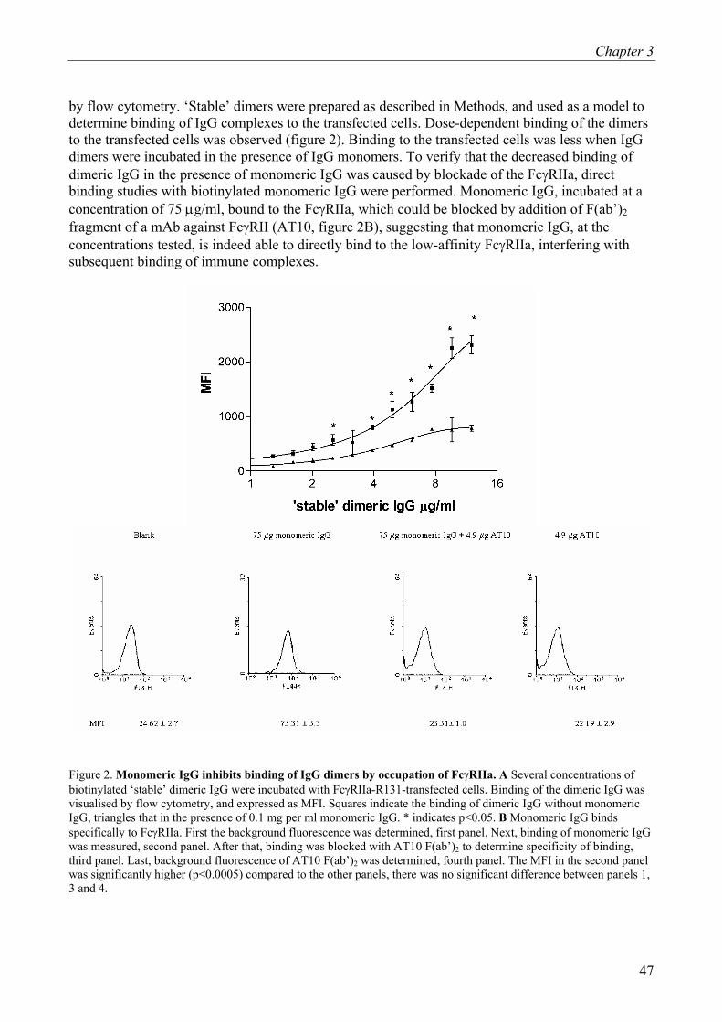
Chapter 3
47
by flow cytometry. ‘Stable’ dimers were prepared as described in Methods, and used as a model to determine binding of IgG complexes to the transfected cells. Dose-dependent binding of the dimers to the transfected cells was observed (figure 2). Binding to the transfected cells was less when IgG dimers were incubated in the presence of IgG monomers. To verify that the decreased binding of dimeric IgG in the presence of monomeric IgG was caused by blockade of the FcγRIIa, direct binding studies with biotinylated monomeric IgG were performed. Monomeric IgG, incubated at a concentration of 75 µg/ml, bound to the FcγRIIa, which could be blocked by addition of F(ab’)2 fragment of a mAb against FcγRII (AT10, figure 2B), suggesting that monomeric IgG, at the concentrations tested, is indeed able to directly bind to the low-affinity FcγRIIa, interfering with subsequent binding of immune complexes.
Figure 2. Monomeric IgG inhibits binding of IgG dimers by occupation of FcγRIIa. A Several concentrations of biotinylated ‘stable’ dimeric IgG were incubated with FcγRIIa-R131-transfected cells. Binding of the dimeric IgG was visualised by flow cytometry, and expressed as MFI. Squares indicate the binding of dimeric IgG without monomeric IgG, triangles that in the presence of 0.1 mg per ml monomeric IgG. * indicates p<0.05. B Monomeric IgG binds specifically to FcγRIIa. First the background fluorescence was determined, first panel. Next, binding of monomeric IgG was measured, second panel. After that, binding was blocked with AT10 F(ab’)2 to determine specificity of binding, third panel. Last, background fluorescence of AT10 F(ab’)2 was determined, fourth panel. The MFI in the second panel was significantly higher (p<0.0005) compared to the other panels, there was no significant difference between panels 1, 3 and 4.

Chapter 3
48
AIgG-mediated rise of intracellular Ca2+ is reduced by monomeric IgG Upon crosslinking by specific monoclonal antibodies or immune complexes both FcγRII and
FcγRIIIb on neutrophils will induce a rise in cytosolic free Ca2+ (13). To assess its possible antagonistic effects on FcγR, we investigated the effect of monomeric IgG on the increase in cytosolic Ca2+ upon triggering of FcγR by aIgG, as a model for immune complexes. AIgG indeed induced an increase in intracellular Ca2+ in neutrophils, whereas treatment with monomeric IgG did not (figure 3a). Pretreatment of the cells with monomeric IgG reduced the influx of Ca2+ induced by aIgG (figure 3b). To assess whether this effect was due to desensitisation by monomeric IgG, an FcγR-independent stimulus, fMLP, was used to stimulate the cells. Monomeric IgG did not have a significant effect on the fMLP-mediated signal (figure 3c).
Figure 3. Monomeric IgG attenuates on aIgG-induced Ca2+ influx in neutrophils. 107 neutrophils per ml were loaded with 1 µM Fura-2/AM. A. Under continuous stirring background fluorescence was measured in a fluorometer for 100 seconds. The cells were then stimulated with either 1 mg/ml aIgG (….) or 10 mg/ml monomeric IgG (—). B. Loaded neutrophils were either pretreated with 0 (—), 1 (---) or 10 (….) mg/ml monomeric IgG for 5 minutes. After measurement of background fluorescence for 100 s, cells were stimulated with 1 mg/ml aIgG. C. Loaded neutrophils were pretreated with 0 (….) or 10 (—) mg/ml monomeric IgG for 5 minutes. Cells were then stimulated with 1 µM fMLP. The experiments shown were repeated 6 times with identical results. AIgG-triggered neutrophil degranulation is attenuated by monomeric IgG
Downstream of the signalling cascade of FcγR is the release of azurophilic granule content (13,14). To study the effect of monomeric IgG on FcγR-induced degranulation, blood was depleted from plasma and stimulated for 2 hours with aIgG with or without monomeric IgG. Degranulation was assessed by measuring the release of elastase. AIgG-mediated elastase release was significantly reduced by the addition of monomeric IgG (figure 4A). To study whether this effect was due to desensitisation by monomeric IgG, the effect on fMLP/cytochalasin B-induced degranulation was

Chapter 3
49
also studied. As can be seen in figure 4B, monomeric IgG had no effect on FMLP/cytochalasin B-mediated elastase release. Also, monomeric IgG itself did not induce degranulation of neutrophils in the blood (figure 4B). Furthermore, addition of blocking Fab or F(ab’)2 fragments of mAbs against FcγRII and FcγRIII (IV.3 Fab and 3G8 F(ab’)2, respectively) attenuated the release of elastase (data not shown and ref 3).
Two patients diagnosed with late-onset common variable immune deficiency resulting in hypo- or agammaglobulinemia were treated for the first time with IVIg. Blood was sampled prior, during and after infusion and stimulated ex vivo with aIgG. Neutrophils of both patients responded to stimulation with aIgG before infusion with IVIg (figure 4C). However, at the end of infusion, the responsiveness to stimulation with aIgG had decreased in both patients.
Figure 4. AIgG-mediated elastase release by neutrophils is diminished by monomeric IVIg. A. Blood cells were washed three times with an excess of IMDM to remove plasma, and added 1:10 to wells pre-coated with human serum albumin and IMDM containing aIgG at the indicated concentrations with (■) or without (♦) 1 mg/ml monomeric IgG. * indicates p<0.05. B. The light grey bar indicates where 1 mg/ml monomeric IgG was added. The dark grey bars represent the wells where no monomeric IgG was added. Either medium or 1 µM fMLP with 5 µg/ml cytochalasin B was added to the wells. The wells were then incubated for 2 hours at 37 °C. Finally, elastase concentrations in the wells were measured with ELISA. The experiment was performed 3 times. Data represent mean ± SD. C. Blood was obtained from 2 patients with low IgG during a first treatment with IVIg, before, halfway and at the end of infusion. Whole blood was diluted 1 to 10 in IMDM containing 1 mg/ml aIgG, final concentration, and incubated for 2 hours at 37 °C. Thereafter, elastase content of the mixtures was measured. Data represent mean ± SD.

Chapter 3
50
Plasma IgG downmodulates aIgG-induced neutrophil degranulation One may postulate that the tertiary structure of monomeric IgG in IVIg differs from that of
plasma IgG due to manufacturing artefacts, and that the effects observed with monomeric IgG purified from IVIg are not representative for plasma IgG. In addition, one may postulate the presence of anti-neutrophil antibodies in the IVIg preparation, which via a Kurlander phenomenon (15) can cause a blockade of FcγR. Hence, we studied whether autologous plasma IgG could exert a similar effect as monomeric IgG in IVIg. Therefore, we depleted plasma for IgG, as described in Methods. IgG concentration, as determined with nephelometry, in this plasma was <0.04 g/L, versus 9.58 g/L ± 3.57 IgG before depletion. Plasma, either or not depleted for IgG, was mixed with IMDM and with peripheral blood cells to yield 10% plasma, final concentration. Before incubation with neutrophils, a monoclonal antibody that inhibits C1q, was added to block the classical pathway.
Blood reconstituted with 10% plasma or 10% IgG-depleted plasma, was stimulated for 2 hours with aIgG or fMLP/cytochalasin B. Neutrophils stimulated with aIgG in the absence of plasma IgG released more elastase than did neutrophils stimulated in the presence of plasma IgG (figure 5A), whereas cells stimulated with fMLP/cytochalasin B responded equally, irrespective of the presence or absence of plasma IgG (figure 5B). Furthermore, plasma that had been adsorbed over HSA-coated CNBr sepharose did not affect aIgG-induced degranulation of neutrophils (figure 5B).
Although physiological levels of IgG in plasma already occupy most FcγR on the blood cells, immune complexes (ICs) can still bind to leukocytes and exert, albeit attenuated, a biological response. However, ITP is nowadays treated with high-dose IVIg while these patients more or less have normal IgG levels. Therefore, we used blood from healthy volunteers and stimulated this with aIgG in the presence or absence of various doses of monomeric IgG and we evaluated the responses by elastase release. As seen in figure 5C, addition of monomeric IgG resulted in a dose-dependent decrease in aIgG-induced elastase release. Furthermore, when a dose of 20 mg/ml, a dosage easily reached during infusion therapy, was added to the blood, elastase release was decreased to background levels (as shown by single dots).

Chapter 3
51
Figure 5. Plasma IgG reduces aIgG-induced elastase release by neutrophils. A. EDTA blood was centrifuged, and plasma was obtained and depleted for IgG as described in methods. Blood cells were washed with IMDM, and added 1:10 to wells pre-coated with albumin and containing a titration of aIgG in IMDM, supplemented with either 10% plasma (▲) or 10% IgG-depleted plasma (■). * indicates p<0.05. B. Buffer (light gray bars) or 1 µM fMLP with 5 µg/ml cytochalasin B (dark gray bars) were used as blank and positive control, respectively. The wells were incubated for 2 hours at 37 °C. C. Blood was added 1:10 to wells pre-coated with human serum albumin and IMDM containing 15 U/ml hirudin, anti-C1q Fab, 1 mg/ml aIgG and various concentrations of monomeric IgG, as represented by the curves. The wells were then incubated for 2 hours at 37 °C. Single dots represent blanks without aIgG. Each curve represents an independent individual experiment. Elastase content of the wells was then measured with ELISA. Data represent mean ± SD (n=3).
Acknowledgements
The authors wish to thank Dr. Anton J. Tool and Dr. Arthur Verhoeven for their excellent assistance with the measurement of calcium-transients and providing us with fMLP and cytochalasin B.
Discussion
The low affinity FcγRII and FcγRIII are stimulated by polyvalent interactions with
multimerized IgG, for example IgG in immune complexes (16,17). Conversely, monomeric IgG should be unable to induce signal transduction via these receptors. Indeed also in the present study we observed that monomeric IgG did not stimulate Ca2+-influx or release of elastase by neutrophils. However, we also show that in spite of its inability to induce signal transduction via these receptors, monomeric IgG has sufficient affinity for FcγRII and FcγRIIIb to bind to these receptors thereby attenuating signal transduction by aIgG. This antagonism of FcγRII and FcγRIIIb by monomeric IgG was observed with IgG in IVIg as well as with plasma IgG, and at concentrations that occur in vivo. Furthermore, infusion of IVIg in patients with low plasma IgG who had never received this drug before, resulted in diminished responsiveness of neutrophils to aIgG, supporting the notion

Chapter 3
52
that the observed effects are relevant in vivo. In addition, at therapeutic concentrations, monomeric IgG, the major constituent of IVIg, attenuates aIgG-induced effects in blood. Therefore, functional blockade of low affinity FcγR might be an important early mechanism of action of IVIg.
Immune complexes (IC) rather than aIgG constitute major agonists for low-affinity FcγR in vivo. However, well established that aIgG and IC are very much alike; since both activate the classical pathway of the complement system (18) and both fully interact with FcγR on phagocytes(19,20). As it is more stable and more easily to prepare with human antibodies than IC, we decided to use aIgG in our studies. AIgG or IC can activate complement in plasma and thus become opsonized with complement factors such as C3b (18). Hence, in the experiments with plasma, aIgG potentially could interact with complement receptors on cells as well, which may blur the effects mediated by FcγR. To prevent this, we added a monoclonal antibody that blocked the interaction of C1q with aIgG, and thereby prevented activation of complement by aIgG.
Monomeric IgG derived from IVIg contains a variable amount of contaminating dimeric IgG. To remove this contaminating dimeric IgG, IVIg was dialysed to low pH resulting in the disruption of any dimers formed (21). Furthermore, control experiments in which we tested the effect of small amounts of dimeric IgG revealed that small amounts of contaminating dimers (< 0.5%) in the monomeric IgG fraction cannot explain the inhibiting effect of monomeric IgG on the binding of dimeric IgG to FcγRIIa-transfected cells (data not shown). Such an amount of dimeric IgG in the monomeric preparation would have been detected upon gel filtration, but yet was not found in the monomeric IgG preparation we used for the experiments. Hence, contaminating dimers do not explain the effects of monomeric IgG on the binding to FcγRIIa. Moreover, as these dimers have the capability to cross-link low-affinity FcγRII or FcγRIII and induce signal transduction (3,22), the observed inhibiting effect of monomeric IgG on aIgG-induced activating effects cannot be explained by the presence of dimeric or polymeric IgG.
Although obtained from plasma pools of more than 1000 donors, IgG in IVIg may be slightly distinct from plasma IgG. For example, the manufacturing processes may alter the tertiary structure of IgG somewhat as compared to native IgG in plasma. However, in the experiments in which plasma was either or not depleted for IgG, we also found an inhibiting effect of autologous non-purified plasma IgG on aIgG-induced neutrophil stimulation. This suggests that, although there may be structural differences between monomeric IgG in plasma and that in IVIg, the effect of monomeric IgG on low-affinity FcγR is not unique for IVIg-derived IgG, but apparently is a general property of monomeric IgG.
The lack of fucose groups on human IgG improves binding of this IgG to human FcγRIII and enhances antibody-dependent cellular cytotoxicity (23). This effect is, however, as far as we know, unique for the interaction with FcγRIIIa and not shared by other low-affinity FcγR. FcγRIIIa is not expressed by neutrophils (24). Hence, it is unlikely that the effects of monomeric IgG, as described in the present article, are related to the presence or absence of this fucose-group in the IgG molecules. In addition, it is commonly accepted that FcγR crosslinking is required for signal transduction and activation of the cells. There is some debate whether monomeric IgG can interact with just one or possibly two FcγR (25,26). Our results show that monomeric IgG does not activate neutrophils, although it binds to at least FcγRIIa (figure 2). Thus, monomeric IgG apparently is incapable of crosslinking the low-affinity FcγR. This strongly suggests that monomeric IgG binds low-affinity FcγR in a 1:1 ratio without inducing activation.
Activation of human granulocytes by IVIg has been reported to occur via stimulation of
FcγRII and FcγRIIIb (3,22) . This activation is mainly mediated by di- and polymeric IgG present in these preparations. Monomeric IgG cannot activate neutrophils unless these cells express FcγRI, for example in inflammatory disease (27-29). We found that some donors constitutively express this

Chapter 3
53
high-affinity receptor (data not shown). The presence of the high-affinity FcγRI would be an obvious explanation for the observed binding of monomeric IgG. However, we screened potential donors for FcγRI expression on neutrophils, and only used neutrophils that had undetectable expression of FcγRI. Hence, interaction of monomeric IgG with FcγRI is excluded to explain our results. In case of monomeric IgG purified from IVIg, one could postulate that the inhibiting effect was mediated via anti-neutrophil antibodies in IVIg, that after binding via their antigen-binding sites to antigens on the neutrophils, interacted with low-affinity FcγR on the cells via their Fc-fragments, the so-called Kurlander phenomenon (15). However, the inhibiting effect was also observed with autologous IgG in plasma. As antibodies against autologous neutrophils in general do not occur in normal plasma, a Kurlander phenomenon cannot explain the inhibiting effect by monomeric IgG.
We showed that physiological levels of monomeric IgG (10 mg/ml) are unable to trigger an FcγR-mediated intracellular Ca2+-rise. It could be argued that monomeric IgG cannot bind to low-affinity FcγR at all. However, we observed binding of monomeric IgG to cells transfected with FcγRIIa, which was completely blocked by the addition of an Fab fragment specific for FcγRII. In addition, the results of Galon et al. (8) and Maenaka et al. (9) show that monomeric IgG as well as Fc-fragments bind both FcγRIIa and FcγRIII with fast kinetics. Importantly, at physiological concentrations, given the affinities mentioned above, IgG would occupy 98.5% and 97.4% of FcγRIIa and FcγRIIIb, respectively. Furthermore, our current data show that pretreatment with high dose of monomeric IVIg, down-modulates aIgG-induced changes in intracellular Ca2+. This suggests, that monomeric IgG indeed binds in a dose-depend manner to low affinity FcγR. It could also be stated that monomeric IVIg induces a functional refractory state of the neutrophils. Nevertheless, our experiments show that, although pretreated with high-dose of monomeric IgG, neutrophils still respond to fMLP as well as untreated neutrophils. Thus, monomeric IgG does not render neutrophils refractory to further stimulation. Another explanation for the effects of monomeric IgG could be downmodulation of the FcγR. However, binding experiments were performed in the cold in the presence of NaN3 and mAb against FcγRII revealed expression levels compared to untreated cells (data not shown) rendering downmodulation highly unlikely. Furthermore, it is unlikely that monomeric IgG exerts its effect through induction of FcγRIIb expression on the neutrophils, since the preincubation time (5 minutes) presumably was too short to establish this (30).
In conclusion, we show that monomeric IgG is able to bind and saturate low-affinity FcγR to attenuate signal transduction by aIgG via these receptors. Hence, monomeric IgG at physiological as well as therapeutical levels acts as a functional receptor antagonist for low-affinity FcγR. This also holds in vivo, as shown by the results of two patients who received a first dose of IVIg. Importantly, these results together implicate that under normal conditions, low-affinity FcγR on neutrophils in the circulation are partially saturated, making it difficult for ICs to bind. Patients with hypo- or agammaglobulinemia lack this inhibiting effect of monomeric IgG, and consequently may be more prone than persons with normal IgG levels to develop side effects after administration of IVIg containing some dimeric or polymeric IgG. Observations in patients indeed support this notion (16).
Chapter 3
54
Reference List
1. Imbach, P., S. Barandun, V. d'Apuzzo, C. Baumgartner, A. Hirt, A. Morell, E. Rossi, M. Schoni, M. Vest, and H. P. Wagner. 1981. High-dose intravenous gammaglobulin for idiopathic thrombocytopenic purpura in childhood. Lancet 1:1228-1231.
2. Teeling, J. L., T. Jansen-Hendriks, T. W. Kuijpers, M. de Haas, J. G. Van de Winkel, C. E. Hack, and W. K. Bleeker. Therapeutic efficacy of intravenous immunoglobulin preparations depends on the immunoglobulin G dimers: studies in experimental immune thrombocytopenia. Blood 2001.Aug.15;98.(4.):1095.-9. 98:1095-1099.
3. Teeling, J. L., E. R. De Groot, A. J. Eerenberg, W. K. Bleeker, G. Van Mierlo, L. A. Aarden, and C. E. Hack. 1998. Human intravenous immunoglobulin (IVIG) preparations degranulate human neutrophils in vitro. Clin.Exp Immunol 114:264-270.
4. Lazarus, A. H., J. Freedman, and J. W. Semple. 1998. Intravenous immunoglobulin and anti-D in idiopathic thrombocytopenic purpura (ITP): mechanisms of action. Transfus.Sci. 19:289-294.
5. Mollnes, T. E., I. H. Andreassen, K. Hogasen, C. E. Hack, and M. Harboe. 1997. Effect of whole and fractionated intravenous immunoglobulin on complement in vitro. Mol.Immunol. 34:719-729.
6. Basta, M., P. Kirshbom, M. M. Frank, and L. F. Fries. 1989. Mechanism of therapeutic effect of high-dose intravenous immunoglobulin. Attenuation of acute, complement-dependent immune damage in a guinea pig model. J.Clin.Invest 84:1974-1981.
7. Dalakas, M. C. 1998. Mechanism of action of intravenous immunoglobulin and therapeutic considerations in the treatment of autoimmune neurologic diseases. Neurology 51:S2-S8.
8. Galon, J., M. W. Robertson, A. Galinha, N. Mazieres, R. Spagnoli, W. H. Fridman, and C. Sautes. 1997. Affinity of the interaction between Fc gamma receptor type III (Fc gammaRIII) and monomeric human IgG subclasses. Role of Fc gammaRIII glycosylation. Eur.J.Immunol. 27:1928-1932.
9. Maenaka, K., P. A. van der Merwe, D. I. Stuart, E. Y. Jones, and P. Sondermann. 2001. The human low affinity Fcgamma receptors IIa, IIb, and III bind IgG with fast kinetics and distinct thermodynamic properties. J.Biol.Chem. 276:44898-44904.
10. Huizinga, T. W., M. Kerst, J. H. Nuyens, A. Vlug, B. von dem, D. Roos, and P. A. Tetteroo. 1989. Binding characteristics of dimeric IgG subclass complexes to human neutrophils. J.Immunol. 142:2359-2364.
11. Hack, C. E. and A. J. Belmer. 1986. The IgG detected in the C1q solid-phase immune-complex assay is not always of immune-complex nature. Clin.Immunol Immunopathol. 38:120-128.
12. Van Den Herik-Oudijk IE, N. A. Westerdaal, N. V. Henriquez, P. J. Capel, and J. G. Van de Winkel. 1994. Functional analysis of human Fc gamma RII (CD32) isoforms expressed in B lymphocytes. J.Immunol. 152:574-585.

Chapter 3
55
13. Vossebeld, P. J., C. H. Homburg, R. C. Schweizer, I. Ibarrola, J. Kessler, L. Koenderman, D. Roos, and A. J. Verhoeven. 1997. Tyrosine phosphorylation-dependent activation of phosphatidylinositide 3-kinase occurs upstream of Ca2+-signalling induced by Fcgamma receptor cross-linking in human neutrophils. Biochem.J. 323:87-94.
14. Naucler, C., S. Grinstein, R. Sundler, and H. Tapper. 2002. Signaling to localized degranulation in neutrophils adherent to immune complexes. J.Leukoc.Biol. 71:701-710.
15. Kurlander, R. J. 1983. Blockade of Fc receptor-mediated binding to U-937 cells by murine monoclonal antibodies directed against a variety of surface antigens. J.Immunol. 131:140-147.
16. Barandun, S., P. Kistler, F. Jeunet, and H. Isliker. 1962. Intravenous administration of human gammaglobulin. Vox Sang. 7:157-174.:157-174.
17. De Grandmont, M. J., C. Racine, A. Roy, R. Lemieux, and S. Neron. 2003. Intravenous immunoglobulins induce the in vitro differentiation of human B lymphocytes and the secretion of IgG. Blood 101:3065-3073.
18. Cowdery, J. S., Jr., P. E. Treadwell, and R. B. Fritz. 1975. A radioimmunoassay for human antigen-antibody complexes in clinical material. J.Immunol. 114:5-9.
19. Daha, M. R. and L. A. van Es. 1984. Fc- and complement receptor-dependent degradation of soluble immune complexes and stable immunoglobulin aggregates by guinea pig monocytes, peritoneal macrophages, and Kupffer cells. J.Leukoc.Biol. 36:569-579.
20. Gormus, B. J., R. L. Vessella, L. N. Martin, and M. E. Kaplan. 1982. Heterogeneity of human lymphocyte Fc receptors: studies using heat-aggregated and antigen-complexed IgG from human, rabbit, guinea pig, horse and goat. Comp Immunol.Microbiol.Infect.Dis. 5:483-499.
21. Tankersley, D. L., M. S. Preston, and J. S. Finlayson. 1988. Immunoglobulin G dimer: an idiotype-anti-idiotype complex. Mol.Immunol. 25:41-48.
22. Nemes, E., F. Teichman, D. Roos, and L. Marodi. Activation of human granulocytes by intravenous immunoglobulin preparations is mediated by FcgammaRII and FcgammaRIII receptors. Pediatr.Res.2000.Mar.;47.(3):357.-61. 47:357-361.
23. Shields, R. L., J. Lai, R. Keck, L. Y. O'Connell, K. Hong, Y. G. Meng, S. H. Weikert, and L. G. Presta. 2002. Lack of fucose on human IgG1 N-linked oligosaccharide improves binding to human Fcgamma RIII and antibody-dependent cellular toxicity. J.Biol.Chem. 277:26733-26740.
24. Huizinga, T. W., C. E. van der Schoot, C. Jost, R. Klaassen, M. Kleijer, A. E. dem Borne, D. Roos, and P. A. Tetteroo. 1988. The PI-linked receptor FcRIII is released on stimulation of neutrophils. Nature 333:667-669.
25. Kato, K., W. H. Fridman, Y. Arata, and C. Sautes-Fridman. 2000. A conformational change in the Fc precludes the binding of two Fcgamma receptor molecules to one IgG. Immunol.Today 21:310-312.
Chapter 3
56
26. Kato, K., Sautes-Fridman, W. Yamada, K. Kobayashi, S. Uchiyama, H. Kim, J. Enokizono, A. Galinha, Y. Kobayashi, W. H. Fridman, Y. Arata, and I. Shimada. 2000. Structural basis of the interaction between IgG and Fcgamma receptors. J.Mol.Biol. 295:213-224.
27. Reilly, M. P. and S. E. McKenzie. 2002. Mechanisms of action of IVIg: physiology of Fc receptors. Vox Sang. 83 Suppl 1:57-63.:57-63.
28. Basta, M., F. Van Goor, S. Luccioli, E. M. Billings, A. O. Vortmeyer, L. Baranyi, J. Szebeni, C. R. Alving, M. C. Carroll, I. Berkower, S. S. Stojilkovic, and D. D. Metcalfe. 2003. F(ab)(2)-mediated neutralization of C3a and C5a anaphylatoxins: a novel effector function of immunoglobulins. Nat.Med. 9:431-438.
29. Lamoureux, J., E. Aubin, and R. Lemieux. 2003. Autoimmune complexes in human serum in presence of therapeutic amounts of intravenous immunoglobulins. Blood 101:1660-1662.
30. Samuelsson, A., T. L. Towers, and J. V. Ravetch. 2001. Anti-inflammatory activity of IVIG mediated through the inhibitory Fc receptor. Science 19;291:484-486.

Chapter 3
57

58

Chapter 4
59
Amelioration of immune complex-mediated anaphylaxis by intravenous immunoglobulins (IVIg) in a rat model. Jessica L. Teeling1*, Edwin van Mirre1*, Wim K. Bleeker1, Gemma M.M. Rigter1, Taco W. Kuijpers2,3 and C. Erik Hack1,4. 1 Department of Immunopathology and 2 of Experimental Immunohematology, Sanquin Research and Landsteiner Laboratory, Academic Medical Center, University of Amsterdam, 3 Department of Pediatrics, Academical Medical Center, University of Amsterdam, 4 Department of Clinical Chemistry, VU Medical Center, Amsterdam, The Netherlands. Submitted * Jessica Teeling and Edwin van Mirre contributed equally to this manuscript Keywords: IVIg, neutrophils, macrophages, Fc receptors, immune complexes

Chapter 4
60

Chapter 4
61
Abstract
We have previously shown that monomeric IgG in intravenous immunoglobulin preparations (IVIg) as well as in plasma can react as an antagonist for low-affinity Fcγ receptors (FcγR). In the present article, we used an in vivo model, i.e. a rat model of immune-complex-mediated anaphylaxis, to explore this hypothesis further. We observed that dimeric IgG, though more potent in preventing anaphylaxis, also induces anaphylaxis itself to some extent. Monomeric IgG, on the other hand, does not induce anaphylaxis itself and, at high doses, is capable of preventing anaphylaxis as well. Interestingly, whereas dimeric IgG induces a dose-dependent anaphylaxis, simultaneous injection of both dimeric and monomeric IgG resulted in reduced anaphylaxis compared to the same dose of dimeric IgG. The effect of monomeric IgG was verified in ex-vivo binding studies, which showed that dimeric, as well as monomeric IgG inhibited binding of immune complexes to blood cells.
Our results indicate that IVIg can modulate FcγR-dependent responses elicited in vivo by immune complexes. The effects are mediated by dimeric IgG, which themselves trigger cells via FcγR, and, at higher concentrations, by monomeric IgG, which do not stimulate cells via FcγR. We propose that similar effects may explain some of the beneficial effects of IVIg observed in immune-complex disease.

Chapter 4
62
Introduction
Intravenous immunoglobulin preparations (IVIg) have proved to be effective and safe in the long-term treatment of antibody deficiencies. After the first report on its efficacy in the treatment of idiopathic thrombocytopenic purpura (ITP) (1, 2) IVIg has increasingly been used in autoimmune and inflammatory conditions. Yet, in spite of the frequent use of IVIg in the treatment of these conditions, the biological mechanisms for its efficacy are not clear. Some of these mechanisms are dependent on a productive interaction of the Fcγ-region of infused immunoglobulin with Fcγ receptors (FcγR) on target cells or with complement proteins (3,4) whereas other mechanisms are primarily dependent on the variable regions of antibodies (5). The ability of IVIg to interfere with complement activation in vivo was demonstrated in the model of the Forssman shock in guinea pigs, where IVIg protected the animals from acute complement-mediated tissue damage (6).Blockade of the FcγR on phagocytic cells is believed to be the mechanism of action by which IVIg is effective in the treatment of patients with ITP, thus preventing the removal of sensitized platelets by the reticuloendothelial system in the spleen and liver (1,7). Indeed, blockade of FcγR by IVIg on macrophages has been shown to inhibit macrophage-mediated phagocytosis of antigen bearing target cells (8,9).
Depending on their expression on effector cells, FcγRs exert different effects. For example, on phagocytes they mediate phagocytosis, endocytosis, antibody-dependent cellular cytotoxicity (ADCC) and induction of the respiratory burst. In humans and mice three types of FcγR, type I, II and III, are discriminated based on their affinity for IgG. Type I (Ka = 108-109 M-1) is considered a high-affinity receptor whereas type II (Ka < 107 M-1) and III (Ka < 107 M-1) are considered to be low-affinity receptors. Rat FcγR have not been as extensively characterized as their human and mouse counterparts. Published literature suggests that rat FcγR are similar to those in mice (10,11).
We have previously shown that monomeric IgG in IVIg and in plasma can react as an antagonist for low affinity FcγR (12). In the present paper, we used an in vivo model to further explore this hypothesis. Inflammatory responses to immune complexes, as exemplified by the Arthus reaction, have been attributed to the presence of antibodies, neutrophils, and complement components. Depletion of either antibodies or neutrophils indeed results in the ablation of the Arthus reaction, whereas depletion of complement components results in a more variable effect (13,14). Studies in FcγR-deficient mice demonstrated the requirement of FcγR for immune-complex-mediated inflammation in these mice (15-19). In contrast, complement depletion with cobra venom factor had little effect (17). Thus, these studies established a key role of FcγR in initiating the inflammatory response to immune complexes and suggest a secondary role for complement.
We have previously used rat models to evaluate the effects of IVIg in vivo (20,21), and hence we decided to set up a model for immune-complex-induced anaphylaxis in rats. In this model, antibodies directed against ovalbumin (OVA) are injected intraperitoneally one day prior to an intravenous challenge with OVA, leading to various systemic responses. We demonstrate that both dimeric IgG and, albeit at higher concentrations, also monomeric IgG in IVIg preparations can completely inhibit the systemic responses elicited by OVA in this model.

Chapter 4
63
Material and Methods Animal model
The animal experiments were approved by the institutional ethics committee and governed by the pertinent national regulations. Any discomfort was avoided by the use of anesthesia throughout the whole experiment.
Female Wistar rats (Harlan, Zeist, The Netherlands), weighing 200-250 g, were injected intraperitoneally with an IgG fraction of rabbit anti-OVA serum (Cappel, ICN Biomedicals, Germany; 0.5 mg/kg). Twenty-four hours later the animals were anesthetized by intraperitoneal injection of 10 mg/kg of pentobarbital, and subcutaneous injection of 0.5 ml/kg of a solution containing fentanyl citrate (0.3 mg/ml) and fluanisone (10 mg/ml) (Hypnorm; Janssen Pharmaceutica, Belgium) and 0.05 mg/kg of atropine. Cannulas (silastic) were introduced into in the left carotid artery for blood pressure monitoring, infusion of test solution and blood sampling. Saline was infused at a rate of 2 ml/h in the cannula to ensure patency. The cannula was connected to a pressure transducer for continuous recording of the mean arterial blood pressure (MABP), averaged over 10-second intervals. Human IgG preparations were infused into the cannula within 10 seconds, at different dosages (ranging from 15-500 mg/kg). One hour after the infusion of IVIg or saline, rats were challenged with OVA (Sigma Chemical Co, Steinheim, Germany) administered via the cannula at a dose of 2 mg/kg unless stated otherwise, and monitored for another 60 minutes. The rats remained under anesthesia throughout this part of the experiment and each rat was challenged only once. At various time points, leukocyte and erythrocyte counts were determined with an electronic cell counter (Coulter Electronics, model 2F, Dunstable, UK) in blood samples anti-coagulated with EDTA. Leukocyte differentiation was performed by microscopy of stained blood smears and by FACS analysis. Plasma was prepared from EDTA blood by centrifugation and stored in aliquots at -80°C until analysis. Immunoglobulin preparations
Human γ-globulin for intravenous use was obtained from our institute (Immunoglobulin I.V., 6% IgG, w/v). This is a freeze-dried product prepared from pooled plasma from at least 1000 donors by Cohn fractionation followed by pepsin incubation at pH 4. Fresh IVIg was immediately frozen after reconstitution and kept at -80°C until further use. Two percent of the preparation represented dimeric IgG. An aged IVIg preparation was prepared by storing the reconstituted IVIg preparation at 4°C for several months. This yielded an increase in IgG dimer content to about 12%. To obtain monomeric IgG, fresh IVIg was dialyzed in 10 mM acetate buffer at pH 4.15 containing 0.24 M glucose and 0.037 M NaCl. An IgG preparation for intramuscular use (IMIG, 16% IgG, w/v), containing >15% dimeric IgG, was also obtained from our institute. A dimer fraction was isolated from IMIG by gelfiltration on an Ultropac TSK-G4000SWG column (LKB-Producter AB, Bromma, Sweden) connected to a FPLC system (Pharmacia, Upsala, Sweden). This yielded an enriched dimer preparation with a stable content of 75% dimeric IgG, 25% monomeric IgG and no detectable aggregated IgG.
All IgG preparations and sub-fractions used in the experiments were analyzed for actual monomer, dimer and polymer contents throughout the period of the experiments on a calibrated Superose 12 gelfiltration column connected to an FPLC system. A computer program (Ezchrom Chromatography Data System version 6.5) was applied to determine the peak areas of the chromatograms. The contents of IgG preparations and sub-fractions were found to be stable throughout the period of the experiments.

Chapter 4
64
Immune-complex formation in vitro OVA was trace radio-labeled for in vitro studies with carrier-free 125I (Amersham
International, Aylesbury, U.K.) and Iodo-Beads (Pierce, Rockford, IL) as oxidizing reagent, followed by extensive dialysis to remove free 125I. To analyze immune-complex formation, different concentrations of OVA were incubated with a trace amount of 125I-labelled OVA (0.05 mg/ml), 0.8 mg/ml anti-OVA serum and normal rat plasma, in the presence or absence of IVIg (12 mg/ml). Immune-complexes were allowed to form during an overnight incubation at 4°C. Immune complexes were precipitated by addition of polyethylene glycol (PEG, MW 15.000-20.000; Sigma, St Louis, MO, USA) at different concentrations (1%, 2% and 3%, w/v, respectively). After 1 hour, the samples were centrifuged at 2500 g for 30 minutes at 4°C. The amount for 125I-OVA in the precipitate was measured with a gamma counter (Wallac model 500, Turku, Finland)
Immune-complex formation was also analyzed by gel filtration. Trace amounts of radiolabeled OVA were added to different amounts of non-labeled OVA. The mixtures were subsequently incubated for 30 minutes at room temperature with plasma of the rats that had received rabbit anti-OVA. Thereafter, samples were separated by gelfiltration over a Superose-6 column (Pharmacia, Upsala, Sweden). Fractions were analyzed for the amount of 125I-OVA, as well as for protein content (A280).
CH50 assay Total classical pathway hemolytic activity (CH50) of rat serum was measured by mixing
rabbit-antibody-sensitized sheep red blood cells (2 x 10⁷) with appropriate dilutions of rat plasma in veronal-buffered saline, pH 7.4, containing 4 mM MgCl2 and 20 mM CaCl2 in a final volume of 200 µl. The mixtures were incubated for 1 hour at 37oC under constant shaking. After centrifugation, the hemoglobin release was determined by measuring the absorbance of the supernatants at 405 nm in a microplate reader. The CH50 was calculated as the reciprocal dilution giving 50% hemolysis.
Binding immune complexes and human IgG to human leukocytes in vitro
Human whole blood was diluted 1:10 in FACS buffer (phosphate-buffered saline, pH 7.4 [PBS], 0.5%, w/v, bovine serum albumin [BSA]; 0.02%, w/v, sodium azide) and then incubated for 30 minutes at 37°C with either dimeric IgG or monomeric IgG. Biotinylated OVA/anti-OVA complexes were isolated on a calibrated Superose-12 column connected to an FLPC system. Fractions containing immune complexes were pooled. Then, different concentrations of biotinylated OVA/anti-OVA complexes were added, and the cells were further incubated for 1h at 37°C . Thereafter, the cells were washed and binding of the complexes was visualised by streptavidin-FITC by use of flow cytometry. Neutrophils and monocytes were distinguished based on differences in forward and sideward scatter characteristics. ELISA for anti-OVA For the detection of OVA-specific antibodies, plasma samples were added to a 96-well flatbottom microtiter plate (Maxisorb; Nunc, Roskilde, Denmark) coated with OVA (2 µg.ml; Sigma), and bound antibodies were detected by alkaline-phosphatase-labelled antibodies to rabbit IgG (Sigma). After addition of p-nitrophenyl phosphate as substrate, the optical density was measured at 405 nm with Titertek Multiscan (Flowlabs. Maclean, VA, USA). Serial dilutions of polyclonal rabbit anti-OVA were used as a reference. The amount of anti-OVA was expressed as percentage of this standard.

Chapter 4
65
Measurement of neutrophil activation in blood samples by flow cytometry. Blood samples were obtained at several time points. Cells were washed and incubated with FACS buffer with FITC-conjugated mouse-anti-rat-CD11b mAb (ED8, IgG1, Instruchemie B.V., Hilversum, the Netherlands) or isotype control (IgG1-FITC, Sanquin, Amsterdam, the Netherlands). Subsequently, erythrocytes were lysed in FACS lysing solution according to the manufacturer’s instructions (Becton Dickinson, San Jose, CA, USA). Leukocytes were resuspended in FACS buffer and analyzed by flow cytometry.
Statistical analysis Student’s t-tests or ANOVA were performed to show significant differences. A p-value <
0.05 was considered to indicate significant differences.

Chapter 4
66
Results Immune-complex model
To determine the effect of IVIg in immune-complex-mediated inflammation we set up a rat model for systemic anaphylactic shock. After a standard intraperitoneal dose of polyclonal anti-OVA, the rats were challenged intravenously with different doses of OVA. At doses of 2 mg/kg or higher, OVA induced a sustained decrease in blood pressure of about 40% (Fig 1). To establish a potential role of complement activation in the model, CH50 levels were determined in plasma of anti-OVA sensitized rats, challenged with 2 mg/kg OVA (n=5, data not shown). CH50 levels decreased less than 20% of baseline values during the observation period.
Figure 1 Average change in blood pressure from t=5 min to t=60 min after OVA. Rats were challenged at t = 0 with different doses of OVA after intraperitoneal administration of polyclonal anti-OVA (2 mg/ml) at t= -24 h. The average blood pressure during the interval from 5 to 60 minutes after OVA challenge was calculated for each rat, and expressed as change (%) relative to pre-infusion level. Data are mean ± SD of n=6 experiments for the 2 mg/kg OVA challenge and n=3 for the other groups. Influence of IVIg on OVA/anti-OVA immune complex formation
Plasma levels of anti-OVA, at the time of OVA administration, as measured in OVA coated plates with labeled goat-anti-rabbit IgG, were about 0.2% of that in the preparation given the previous day. This corresponded to an apparent distribution volume of about 200 ml/kg. No change in the level of circulating anti-OVA antibodies was found in rats after pretreatment with IVIg, indicating that IVIg does not contain antibodies capable of neutralizing anti-OVA. These findings were confirmed in vitro, where we did not detect any neutralizing effects of IVIg to anti-OVA. Five, thirty or sixty minutes after challenge with OVA at 2 mg/ml, no circulating anti-OVA antibodies were detected in the rats, indicating that all circulating anti-OVA antibodies were in complex (data not shown).
To determine the effect of IVIg on the formation and size of the immune complexes, OVA-anti-OVA complexes were formed in vitro by incubating different concentrations of OVA with rat plasma containing anti-OVA at levels similar to those occurring in vivo. Immune-complex formation was monitored with 125I-OVA as a tracer, using PEG precipitation and gel-filtration as described in Materials and Methods. The amount of immune complexes as well as their size

Chapter 4
67
increased with increasing concentrations of OVA. Two IVIg preparations, i.e. aged IVIg and fresh IVIg containing 11.2% and 2% dimeric IgG, respectively (22), added to yield a final concentration of 12 mg/ml, did not have any effect on the amount or size of the OVA/anti-OVA immune complexes formed. An example of these experiments is shown in Figure 2.
Figure 2 Influence of IVIg on OVA/anti-OVA immune complex formation in vitro Three different concentrations of OVA were incubated with a trace amount of 125I-labelled OVA, anti-OVA IgG and normal rat plasma, with (open symbols) or without (closed symbols) addition of fresh IVIg (12 mg/ml). Immune complexes were allowed to form and samples were analyzed by gelfiltration as described in Materials and Methods. The amount of 125I OVA in different fractions was measured with a gamma counter. Effect of IVIg on immune-complex-mediated anaphylaxis
Rats were pretreated with aged or fresh IVIg, 60 minutes prior to the OVA challenge. Pretreatment with fresh IVIg at a dose of 250 mg/kg did not change the OVA-induced systemic responses. However, pretreatment of rats with 250 mg/kg aged IVIg, resulted in an attenuated response to OVA. Figure 3 shows typical examples of the change in blood pressure upon infusion of IVIg and OVA in more detail. Pooled data are depicted in Figure 4. Administration of aged IVIg at t= -60 min induced a change in blood pressure, that normalized after 45 minutes. Fresh IVIg at a dose of 250 mg/kg or saline did not induce changes in blood pressure. Intravenous administration of OVA caused immediate systemic anaphylaxis in saline-pretreated rats, manifested as a rapid change in blood pressure in the first minute followed by a more gradual decrease over 10 minutes. No hypotensive responses were observed in 6 out of 6 rats pretreated with aged IVIg upon administration of OVA. Pretreatment of rats with fresh IVIg at the same dose, had only a partial effect on the OVA-induced anaphylaxis
Prior to the administration of OVA, infusion of aged IVIg resulted in a decrease in circulating neutrophils, i.e., -54 ± 6.7% of baseline up to -263 ± 106% of baseline at the time of the OVA challenge. Administration of saline or fresh IVIg did not have such an effect on the number of circulating neutrophils. Upon infusion of OVA, rats pretreated with saline or fresh IVIg showed a decrease in circulating neutrophils by -54 ± 22.7% and -59 ± 4.0%, respectively. In contrast, rats

Chapter 4
68
pretreated with aged IVIg showed a smaller decrease in circulating neutrophils, i.e., -9.7 ± 5.5%, indicating that neutrophil activation after OVA challenge is inhibited by pretreatment with an IVIg preparation containing relatively high amounts of dimeric IgG. Next to that, OVA/anti-OVA induced CD11b upregulation was also hampered in these samples (p< 0.01). Administration of OVA also affected the number of erythrocytes in peripheral blood (table 1). Both saline- and fresh- IVIg treated rats showed an increase in the number of erythrocytes after challenge, indicative for leakage of plasma proteins, whereas no such effect was observed in rats pretreated with aged IVIg (table 1).
We previously showed that administration of aged IVIg results in activation of macrophages and release of platelet activating factor (PAF) from these cells (20). A possible explanation for the inhibitory effect of aged IVIg on the OVA-induced systemic responses might be that the endothelial cells were desensitized for PAF by pretreatment with aged IVIg. To rule out this possibility, short PAF infusions were given intravenously before and one hour after administration of aged IVIg, as well as directly after the OVA challenge (data not shown). The short-lasting hypotensive effects of PAF were unchanged by IVIg pre-treatment, ruling out that endothelial cells had become refractory to secondary stimulation
Figure 3 Course of mean arterial blood pressure (MABP) in anaphylactic rats pretreated with IVIg. Typical examples of the course of mean arterial blood pressure (MABP; represented as percentage of baseline) of OVA-sensitized rats receiving saline (solid line), aged IVIg (dotted line, A) or fresh IVIg (dotted line B) prior to OVA challenge (2 mg/kg). Arrows indicate the moment of IVIg and OVA injection.
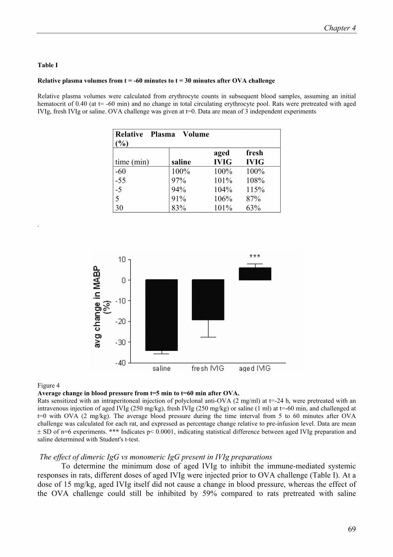
Chapter 4
69
Table I
Relative plasma volumes from t = -60 minutes to t = 30 minutes after OVA challenge
Relative plasma volumes were calculated from erythrocyte counts in subsequent blood samples, assuming an initial hematocrit of 0.40 (at t= -60 min) and no change in total circulating erythrocyte pool. Rats were pretreated with aged IVIg, fresh IVIg or saline. OVA challenge was given at t=0. Data are mean of 3 independent experiments
Relative Plasma Volume (%)
time (min) saline aged IVIG
fresh IVIG
-60 100% 100% 100% -55 97% 101% 108% -5 94% 104% 115% 5 91% 106% 87% 30 83% 101% 63%
.
Figure 4 Average change in blood pressure from t=5 min to t=60 min after OVA. Rats sensitized with an intraperitoneal injection of polyclonal anti-OVA (2 mg/ml) at t=-24 h, were pretreated with an intravenous injection of aged IVIg (250 mg/kg), fresh IVIg (250 mg/kg) or saline (1 ml) at t=-60 min, and challenged at t=0 with OVA (2 mg/kg). The average blood pressure during the time interval from 5 to 60 minutes after OVA challenge was calculated for each rat, and expressed as percentage change relative to pre-infusion level. Data are mean ± SD of n=6 experiments. *** Indicates p< 0.0001, indicating statistical difference between aged IVIg preparation and saline determined with Student's t-test. The effect of dimeric IgG vs monomeric IgG present in IVIg preparations
To determine the minimum dose of aged IVIg to inhibit the immune-mediated systemic responses in rats, different doses of aged IVIg were injected prior to OVA challenge (Table I). At a dose of 15 mg/kg, aged IVIg itself did not cause a change in blood pressure, whereas the effect of the OVA challenge could still be inhibited by 59% compared to rats pretreated with saline

Chapter 4
70
(p<0.0001). Administration of 30 mg/kg caused a short lasting vasoactive response and responses to OVA were inhibited by 43% (p<0.01). Pretreatment with 60 mg/kg gave similar results as 250 mg/kg and resulted in a slight increase in blood pressure after OVA challenge. However, the latter doses themselves induced significant hypotensive effects. The responses to OVA were not significantly changed in rats pretreated with fresh IVIg at a dose of 250 mg/kg. However, this seemed to be close to a threshold for effect, since 3 out of 6 rats analyzed indeed demonstrated a diminished effect upon OVA challenge. Fresh IVIg administered in a dose of 500 mg/kg showed no significant changes in blood pressure by the IVIg preparation itself, whereas the response to OVA was inhibited by 87.3% (p<0.0001). These results suggest that monomeric IgG at high doses is also able to modulate systemic responses to OVA in vivo, without inducing hypotension itself.
To further investigate the modulatory effect of monomeric IgG a series of experiments was performed in which we established dose-response curves for the hypotensive effect of dimeric IgG in rats, with or without simultaneous administration of monomeric IgG (Figure 5). Dimeric IgG was administered in doses ranging from 1 to 40 mg/kg mixed with either saline or monomeric IgG (pH 4-treated IVIg containing no detecable dimeric IgG or polymeric IgG upon analysis with HPLC), giving a final dose of 250 mg/kg . Addition of monomeric IgG to dimeric IgG preparation resulted in a marked shift of the dose-response curve, clearly demonstrating that monomeric IgG inhibits the hypotensive effect of dimeric IgG. In two control experiments a 3% human serum albumin (HSA) solution, was added to the dimeric IgG, instead of saline. This experiment made clear that the shift was not due to an osmotic effect of the monomeric IgG, although it affected the maximum decrease in blood pressure. Thus, the effect on the maximum decrease in blood pressure seems to be related to filling of the capillaries unrelated to monomeric IgG but related to addition of protein as indicated by addition of human serum albumin.
Binding studies To evaluate whether the binding of immune complexes to the cell surface was altered in the
presence of monomeric IgG, we performed binding studies. Blood cells were pre-incubated with buffer (no IgG), aged IVIg, dimeric IgG or monomeric IgG. Subsequently, varying concentrations of labeled OVA/anti-OVA complexes were added and binding of these complexes to the cells was assessed. The complexes bound to the cells in a dose-dependent fashion (figure 6). Binding to the cells was diminished upon pre-treatment with aged IVIg, dimeric IgG or monomeric IgG. Interestingly, the effect observed with in vivo pretreatment is similar to that with ex-vivo pretreatment. At low concentrations (1 mg/ml) the attenuation of immune-complex-mediated effects was mainly mediated by dimeric IgG, whereas at higher doses (2.5 mg/ml) monomeric IgG exhibited similar effects as IVIg with high dimer content.

Chapter 4
71
Table II Average change in blood pressure from t= -60 min to t= 0 before OVA, and t=5 min to t=60 min after OVA (%) Rats were challenged with OVA (2 mg/kg) after intraperitoneal administration of polyclonal anti-OVA (0.5 mg/ml; at t= -24) and aged IVIg, fresh IVIg or saline pretreatment at t=-60 min. For the individual rats, the average blood pressure was measured in the time interval from -60 to 0 min before OVA and 5 to 60 minutes after OVA challenge and was expressed as percentage change with respect to the pre-infusion level. Data are mean ± SEM. Student's t-tests were used to compare IVIg-treated animals with saline pretreatment. ns stands for not significant.
IVIg pretreatment Effect on bloodpressure (%) (mg/kg) pretreatment
OVA challenge
n t=-60-0 min
SEM p-value t=5-60 min SEM p-value
saline 5 -1.0 2.3 -34.2 1.5 aged (15) 3 4.0 9.7 Ns -14.0 2.5 0.0001 aged (30) 3 -11.0 5.0 0.04 -19.7 5.9 0.008 aged (60) 3 -23.7 1.5 0.0001 8.0 3.8 0.0001 aged (250) 6 -13.2 0.8 0.0003 5.7 2.2 0.0001 fresh (250) 6 2.3 1.9 ns -19.0 8.4 ns fresh (500) 3 5.2 3.9 ns -4.0 5.4 0.0001

Chapter 4
72
Figure 5 Effect of monomeric IgG on the hypotensive effect of dimeric IgG in rats. Purified dimeric IgG was administered intravenously in a freshly prepared mixture with either monomeric IgG (pH 4-treated IVIg, <0.5% dimers), giving a dose of 250 mg/kg, or an equal volume of saline. In two control experiments a 3% solution of human serum albumin (HSA), was added to the dimers, instead of saline. Each data point concerns an independent experiment in a different rat, giving the mean arterial blood pressure (MABP) 15 minutes after administration of dimers, expressed as percentage of baseline. Each rat received only one dose of dimers. The lines give sigmoid curves fitted to the data by non-linear regression.

Chapter 4
73
Figure 6 Binding of OVA-anti-OVA immune complexes to neutrophils and monocytes in vitro. Binding of biotinylated OVA-anti-OVA immune complexes to A. neutrophils and B monocytes in human whole blood cultures simultaneously incubated with 2.5 mg/ml monomeric IgG, aged IVIg or buffer alone. Binding was measured by flow cytometry after staining with streptavidin-FITC. Data are expressed as MFI and represent the mean ± SEM of 3 individual experiments.

Chapter 4
74
Discussion
Clinical experience suggests an immune-modulating effect of IVIg in a number of autoimmune and immune-complex-mediated diseases (2). Several underlying mechanisms have been suggested to account for these effects. For example, the rapid increase in the circulating platelet counts in patients with autoimmune idiopathic thrombocytopenia after IVIg infusion has been postulated to be due to blockade of FcγR on phagocytes (1). We have previously shown that monomeric IgG in IVIg and plasma IgG can act as an antagonist for low-affinity FcγR (12). In the present paper, we used an in vivo model to explore this hypothesis further. We show that IVIg can modulate FcγR-dependent inflammatory reactions in vivo, presumably by blockade of FcγR. This effect is mediated by relatively low concentrations of dimeric IgG, but can also be brought about by monomeric IgG, though at higher concentrations. The possible effects of IVIg therapy on immune-complex-induced inflammatory responses were studied in a rat model, in which anti-OVA-sensitized rats were challenged with OVA. Intravenous injection of OVA resulted in a short-lasting biphasic change in blood pressure, followed by a long-lasting decrease in blood pressure. Evaluation of peripheral blood cells showed a transient decrease in circulating neutrophil numbers and upregulation of CD11b on these cells, indicating activation of these cells. In addition an increase in the number of erythrocytes was observed, indicative for vascular leakage and hemo-concentration. Changes in erythrocyte counts directly reflect changes in plasma volume (if there are no changes in the red blood cell mass) and, therefore, we choose to use hemoconcentration as an indicator of plasma extravasation. Previously, we have used erythrocyte counts to monitor anaphylactoid reactions on IVIg infusions, and we prefer this over the use of dyes which seems to be more appropriate for monitoring local inflammatory reactions. The interpretation of changes in erythrocyte count can be complicated by hemodilution due to the infusion of IVIg. Infusion of IVIg at a dose of 250 mg/kg is expected to give an expansion of the blood volume of about 4 ml/kg, which corresponds to about 6% of the blood volume. This increase in blood volume corresponds to the observed decrease in erythrocyte count after fresh IVIg in the present study. After aged IVIg no decrease was observed, indicating that some plasma extravasation accompanied the hypotensive response. As expected, saline infusion at the same volume induced little changes since it has no osmotic activity. Since it seems clear that the changes after infusion are relatively small compared to those observed after OVA administration, we consider the erythrocyte count to be a good indicator of anaphylaxis in these experiments.
A similar model to study immune-complex-induced inflammation has been developed in mice (23). In these animals the anaphylactic reaction was shown to be dependent on the low-affinity FcγRIII, rather than on complement. In the present study, however, measurement of CH50 titer in plasma of the OVA-challenged rats revealed that some complement activation occurred in our rat model for systemic anaphylaxis. In previous studies with the same model, we observed no correlation between hypotensive responses and reductions in CH50 titer following infusion of IgG preparations containing IgG aggregates (24). In addition, complement depletion with cobra venom factor did not affect the hypotensive response, whereas macrophage depletion completely abrogated the systemic effects of infusing IgG preparations (21). Hence, we assume that complement activation did not play a major role in the anaphylactic reaction in the rats challenged with OVA, but rather depended on the triggering of low-affinity FcγR. Indeed, administration of dimeric IgG, which is a specific agonist of low-affinity FcγR, induced a hypotensive reaction closely resembling that seen after OVA challenge.
In previous studies, we demonstrated that polymeric and dimeric IgG in IVIg interacts with FcγR, resulting in the activation of macrophages and neutrophils (22,25). Activation of neutrophils

Chapter 4
75
in vitro was mediated by direct triggering of low-affinity FcγR by dimeric IgG present in the IVIg preparation (21,25). Activation of neutrophils in vivo was mediated by macrophage activation, due to triggering of FcγR by dimeric IgG (21,22). In the present study, we demonstrate that pretreatment of rats with IVIg preparations containing a relatively high amount of dimeric IgG, i.e. aged IVIg, was more effective in inducing tolerance to OVA challenge than an IVIg preparation with low IgG dimer content, i.e. fresh IVIg. Immune-complex formation was not affected by IVIg and there was no evidence for the presence of anti-OVA Ab present in IVIg preparations, indicating that IVIg was effective at the level of effectors (phagocytes) and not by modulation of immune-complex formation.
Rat FcγR have not been as extensively characterized as their human or murine counterparts. Available literature suggests that FcγR expression in rats is similar as compared to mice (10,11) FcγRIII is the activating low-affinity FcγR in mice. Several studies have described the importance of this FcγR in inflammatory reactions in mice models. Hence, it is likely that also in rats FcγRIII is the main low-affinity FcγR mediating inflammation. In contrast to mice and rats, humans express two types of activating low affinity FcγR, FcγRIIa and FcγRIII. In vitro studies (25) have shown that IVIg binds and activates human leukocytes in a similar fashion as compared to rat cells. Hence, we believe that our data are relevant for the effect of IVIg in humans as well.
Various mechanisms can explain modulation of FcγR number and function. First, the blockade of FcγR in the presence of an excess of ligand. Second, ligand-oriented diffusion of FcγR in the planar cell membrane (induced by surface-bound ligands), and third, down-modulation of FcγR due to shedding or ingestion and degradation of ligand-receptor complexes. IVIg indeed may influence numbers of FcγR (21,26,27). For example, polymeric IgG induced down-modulation of FcγR, which correlated with decreased expression of FcγRIII on monocytes (21,27). Kimberly et al. demonstrated that treatment of IVIg in patients suffering from ITP resulted in a marked increase in intracellular IgG and decreased clearance of autologous IgG-sensitized erythrocytes due to inhibition of FcγR-ligand interactions by monomeric IgG (21,26). In experiments not shown here, we observed an increase in intracellular human IgG in rat leukocytes, suggesting internalization of FcγR. However, ex vivo stimulation of whole blood from a rat treated with IVIg in vivo, showed that neutrophils could still be activated by aged IVIg in vitro, suggesting that functional FcγR were still available on the surface of this cell type. It should be noted that the discrepancy between these results and those of Kimberly et al. may reflect that we studied neutrophil rather than macrophage function.
Although higher doses of aged IVIg were more potent in inhibition of systemic responses to OVA challenge, these doses induced a drop in blood pressure themselves. However, immune-complex-mediated diseases are, preferentially not treated with an agent that itself induces a similar clinical outcome, and IVIg containing high amounts of dimeric IgG seems, therefore, not suitable for clinical application. This also puts forward the possibility that the inhibiting effect of aged IgG was due to desensitization of the effector pathways. For several reasons we think that desensitization does not explain our results. First, we found no sign of desensitization of endothelial cells for PAF, the most likely mediator of the hypotensive response. Second, we observed efficacy of aged IVIg to significantly attenuate OVA-induced hypotension, at a dose of 15 mg/kg, which in itself did not induce vasoactive reactions.Yet, IVIg preparations containing significant amounts of dimeric IgG are not attractive therapeutic agents since they have the inherent risk of side effects in patients. Finally, the drop in blood pressure induced by dimeric IgG could be blocked by the simultaneous administration of monomeric IgG, indicating that IgG modulates the anaphylactic reaction in rats at a different level than desensitization of endothelial cells.
We noted that 50% of the rats pretreated with fresh IVIg (250 mg/kg) hardly developed hypotension upon OVA challenge. Increasing the dose of fresh IVIg (500 mg/kg) could inhibited

Chapter 4
76
responses to OVA without apparent effect of the preparation itself on the blood pressure. Although this suggests that monomeric IgG, at a high dose, can inhibit systemic responses triggered by immune complexes without activation, one may postulate that sub-threshold activation by low amounts of dimeric IgG is responsible for the modulation. Because it is virtually impossible to assure complete absence of di- and polymers in monomeric IgG fractions, we chose a different approach to rule out this possibility. We focused on the activating effects of dimeric IgG and established a dose-response curve for IgG dimer-induced hypotension (see figure 5). Mixing the dimeric preparation with monomeric IgG caused the dose-response curve to shift to the right. In case low amounts of contaminating dimeric IgG in the preparation would be the active principle in the monomeric preparation, the effect of mixing monomeric and dimeric IgG would have been similar to simply increasing the dose of dimeric IgG. As can be clearly seen in figure 5, increasing the dose of the dimeric IgG preparation did not result in a diminished drop of blood pressure. Taken together, these experiments strongly suggest that monomeric IgG can interfere with Fcγ receptor function without activation.
The initiation of the inflammatory cascade by immune complexes mainly depends on the engagement and activation of the ITAM-containing γ-chain-associated FcγR (28). We have previously shown that activation of human neutrophils via FcγRII and FcγRIIIb by immune complexes can be inhibited by monomeric IgG. This inhibition was mediated via a functional blockade of the low-affinity FcγR in the absence of cell activation (12). The in vivo data described in this paper can be best explained by a non-activating blockade of FcγR. In line herewith, we found that ex vivo human blood cells also showed diminished binding of OVA/immune complexes in the presence of IVIg (figure 6).
In the present study we demonstrate in an in vivo model in rats that IVIg modulates immune complex-mediated inflammation. The effect was most pronounced with IVIg with a high content of dimeric IgG, which themselves can trigger FcγR-dependent inflammatory reactions, and hence are not suitable for clinical application. However, high doses of IVIg consisting of almost exclusively monomeric IgG, which do not trigger low-affinity FcγR, similarly attenuated immune-complex induced hypotension. Hence, we suggest that IVIg, preferably those with low dimeric IgG contents, can be used in the treatment of immune-complex-mediated human diseases.

Chapter 4
77
Reference List 1. Imbach, P., S. Barandun, V. d'Apuzzo, C. Baumgartner, A. Hirt, A. Morell, E. Rossi, M.
Schoni, M. Vest, and H. P. Wagner. 1981. High-dose intravenous gammaglobulin for idiopathic thrombocytopenic purpura in childhood. Lancet 1:1228-1231.
2. Dwyer, J. M. 1992. Manipulating the immune system with immune globulin. N.Engl.J.Med. 326:107-116.
3. Basta, M., F. Van Goor, S. Luccioli, E. M. Billings, A. O. Vortmeyer, L. Baranyi, J. Szebeni, C. R. Alving, M. C. Carroll, I. Berkower, S. S. Stojilkovic, and D. D. Metcalfe. 2003. F(ab)(2)-mediated neutralization of C3a and C5a anaphylatoxins: a novel effector function of immunoglobulins. Nat.Med. 9:431-438.
4. Lutz, H. U., P. Stammler, V. Bianchi, R. M. Trueb, T. Hunziker, R. Burger, E. Jelezarova, and P. J. Spath. 2004. Intravenously applied IgG stimulates complement attenuation in a complement-dependent autoimmune disease at the amplifying C3 convertase level. Blood 103:465-472.
5. Baudet, V., V. Hurez, C. Lapeyre, S. V. Kaveri, and M. D. Kazatchkine. 1996. Intravenous immunoglobulin (IVIg) modulates the expansion of V beta 3+ and V beta 17+ T cells induced by staphylococcal enterotoxin B superantigen in vitro. Scand.J.Immunol. 43:277-282.
6. Basta, M., P. Kirshbom, M. M. Frank, and L. F. Fries. 1989. Mechanism of therapeutic effect of high-dose intravenous immunoglobulin. Attenuation of acute, complement-dependent immune damage in a guinea pig model. J.Clin.Invest 84:1974-1981.
7. Lazarus, A. H., J. Freedman, and J. W. Semple. 1998. Intravenous immunoglobulin and anti-D in idiopathic thrombocytopenic purpura (ITP): mechanisms of action. Transfus.Sci. 19:289-294.
8. Dalakas, M. C. 1998. Mechanism of action of intravenous immunoglobulin and therapeutic considerations in the treatment of autoimmune neurologic diseases. Neurology 51:S2-S8.
9. Teeling, J. L., T. Jansen-Hendriks, T. W. Kuijpers, M. de Haas, J. G. Van de Winkel, C. E. Hack, and W. K. Bleeker. Therapeutic efficacy of intravenous immunoglobulin preparations depends on the immunoglobulin G dimers: studies in experimental immune thrombocytopenia. Blood 2001.Aug.15.;98.(4):1095-9.
10. Bocek, P., Jr., L. Draberova, P. Draber, and I. Pecht. 1995. Characterization of Fc gamma receptors on rat mucosal mast cells using a mutant Fc epsilon RI-deficient rat basophilic leukemia line. Eur.J.Immunol. 25:2948-2955.
11. Farber, D. L. and D. W. Sears. 1991. Rat CD16 is defined by a family of class III Fc gamma receptors requiring co-expression of heteroprotein subunits. J.Immunol. 146:4352-4361.
12. Van Mirre, E., J. L. Teeling, J. W. Van Der Meer, W. K. Bleeker, and C. E. Hack. 2004. Monomeric IgG in intravenous Ig preparations is a functional antagonist of FcgammaRII and FcgammaRIIIb. J Immunol. 173:332-339.

Chapter 4
78
13. Cochrane, C. G. and H. J. Muller-Eberhard. 1968. The derivation of two distinct anaphylatoxin activities from the third and fifth components of human complement. J.Exp.Med. 127:371-386.
14. Cochrane, C. G., H. J. Muller-Eberhard, and B. S. Aikin. 1970. Depletion of plasma complement in vivo by a protein of cobra venom: its effect on various immunologic reactions. J.Immunol. 105:55-69.
15. Clynes, R. and J. V. Ravetch. 1995. Cytotoxic antibodies trigger inflammation through Fc receptors. Immunity. 3:21-26.
16. Clynes, R., J. S. Maizes, R. Guinamard, M. Ono, T. Takai, and J. V. Ravetch. 1999. Modulation of immune complex-induced inflammation in vivo by the coordinate expression of activation and inhibitory Fc receptors. J.Exp.Med. 189:179-185.
17. Ravetch, J. V. 1994. Fc receptors: rubor redux. Cell 78:553-560.
18. Ravetch, J. V. and R. A. Clynes. 1998. Divergent roles for Fc receptors and complement in vivo. Annu.Rev.Immunol. 16:421-32.:421-432.
19. Sylvestre, D., R. Clynes, M. Ma, H. Warren, M. C. Carroll, and J. V. Ravetch. 1996. Immunoglobulin G-mediated inflammatory responses develop normally in complement-deficient mice. J.Exp.Med 184:2385-2392.
20. Bleeker, W. K., J. L. Teeling, A. J. Verhoeven, G. M. Rigter, J. Agterberg, A. T. Tool, A. H. Koenderman, T. W. Kuijpers, and C. E. Hack. Vasoactive side effects of intravenous immunoglobulin preparations in a rat model and their treatment with recombinant platelet-activating factor acetylhydrolase. Blood 2000.Mar 1;95.(5):1856-61.
21. Bleeker, W. K., J. Agterberg, G. Rigter, N. van Rooijen, and J. C. Bakker. 1989. Key role of macrophages in hypotensive side effects of immunoglobulin preparations. Studies in an animal model. Clin.Exp.Immunol. 77:338-344.
22. Teeling, J. L., W. K. Bleeker, G. M. Rigter, N. van Rooijen, T. W. Kuijpers, and C. E. Hack. Intravenous immunoglobulin preparations induce mild activation of neutrophils in vivo via triggering of macrophages--studies in a rat model. Br.J Haematol.2001.Mar;112.(4):1031.-40.
23. Hazenbos, W. L., J. E. Gessner, F. M. Hofhuis, H. Kuipers, D. Meyer, I. A. Heijnen, R. E. Schmidt, M. Sandor, P. J. Capel, M. Daeron, J. G. Van de Winkel, and J. S. Verbeek. 1996. Impaired IgG-dependent anaphylaxis and Arthus reaction in Fc gamma RIII (CD16) deficient mice. Immunity. 5:181-188.
24. Bleeker, W. K., J. Agterberg, G. Rigter, A. Vries-van Rossen, and J. C. Bakker. 1987. An animal model for the detection of hypotensive side effects of immunoglobulin preparations. Vox Sang. 52:281-290.
25. Teeling, J. L., E. R. De Groot, A. J. Eerenberg, W. K. Bleeker, G. Van Mierlo, L. A. Aarden, and C. E. Hack. 1998. Human intravenous immunoglobulin (IVIG) preparations degranulate human neutrophils in vitro. Clin.Exp Immunol 114:264-270.

Chapter 4
79
26. Kimberly, R. P., J. E. Salmon, J. B. Bussel, M. K. Crow, and M. W. Hilgartner. 1984. Modulation of mononuclear phagocyte function by intravenous gamma-globulin. J.Immunol. 132:745-750.
27. Mannhalter, J. W. and M. M. Eibl. 1989. Down regulation of Fc receptors by IVIgG. Int.Rev.Immunol. 5:173-179.
28. Van de Winkel, J. G. and P. J. Capel. 1993. Human IgG Fc receptor heterogeneity: molecular aspects and clinical implications. Immunol.Today 14:215-221.

Chapter 4
80

Chapter 4
81

82

Chapter 5
83
Variation in the mRNA ratios of activating and inhibitory FcγRII determines neutrophil responsiveness. Edwin van Mirre 1, Judy Geissler 2, Martin de Boer 2, C. Erik Hack 1, 3, and Taco W. Kuijpers 2, 4 1 Department of Immunopathology and 2 Experimental Immunohematology, Sanquin Research at CLB, and Landsteiner Laboratory, Academic Medical Center, University of Amsterdam; 3 Department of Clinical Chemistry, VU Medical Center, Amsterdam, and 4 Emma Children’s Hospital, Academical Medical Center, Amsterdam, the Netherlands. Submitted

Chapter 5
84

Chapter 5
85
Abstract
In this study we tested the hypothesis that the ratio between activating and inhibitory Fcγ receptor type 2 (FcγRII) determines the responsiveness of neutrophils to immune complexes. We measured mRNA levels of FcγRII isoforms and observed differences in the ratio of FcγRIIa/FcγRIIb2 in granulocytes of 20 healthy volunteers, being either 2:1 or 1:1. The response to either dimeric IgG or aggregated IgG (aIgG) was subsequently assessed for neutrophils with high or low FcγRIIa:FcγRIIb2 ratio. The upregulation of CD11b on the surface as well as the elastase release from azurophil granules was significantly more pronounced in neutrophils with a high FcγRIIa/FcγRIIb2 ratio of 2:1 as compared to a 1:1 ratio (p=0.0006; and p=0.0059, respectively). The ratio as well as the functional responsiveness of neutrophils was constant over a 2-6 month period. In support of the in vivo stability over time, neutrophil stimulation with various agents in vitro (endotoxin, interleukin-4, interferon-γ or GM-CSF) did not alter the mRNA levels of FcγRIIa or FcγRIIb2 in the neutrophils of these donors, in contrast to the findings in their mononuclear cells. These data show that responsiveness of neutrophils to IgG agonists is dependent on an apparently fixed ratio of activating and inhibitory FcγRII.

Chapter 5
86
Introduction
Fc gamma receptors (FcγR) are IgG binding molecules belonging to the immunoglobulin superfamily that links innate and adaptive immunity. Depending on their expression on effector cells FcγR exert different effects. For example, on phagocytes they mediate phagocytosis, endocytosis, antibody-dependent cellular cytotoxicity (ADCC) and release of reactive oxygen species (1).
Three types of FcγR, type I, II and III, are discriminated based on their affinity for monomeric IgG. Type I (Ka = 108-109 M-1) is a high-affinity receptor whereas type II (Ka = 106 M-
1) and III (Ka = 5.5 x 10⁵ M-1) (2,3) are to be regarded as low-affinity receptors. Of these receptors FcγRI and FcγRIII are dependent on the association with another molecule for signal transduction. In contrast, FcγRII contains, depending on the isoform, either an ITAM or an ITIM motif in its intracellular tail. FcγRIIa contains an immune-receptor tyrosine-based activation motif (ITAM motif) and is therefore an activating receptor, whereas FcγRIIb contains an immune-receptor inhibition motif (ITIM motif) and is described to be an inhibitory receptor (4,5).
Under resting conditions, neutrophils express both FcγRIIa and FcγRIIIb (1-4 x 10⁴ and 1-3 x 10⁵ molecules per cell, respectively), but no FcγRI (6). In addition, neutrophils also express the splice variant FcγRIIb2 7 although quantification has not been substantiated. It has been suggested that the ratio between activating and inhibitory FcγR may determine the responsiveness of immune cells to immune-complexes (7,8).
In the present paper, we tested the hypothesis that the ratio between activating and inhibitory FcγRII on neutrophils may differ among individuals, resulting in a different sensitivity of these phagocytes to IgG agonists. Therefore, the mRNA coding for FcγRIIa and FcγRIIb2 was quantitated in highly purified neutrophils from various donors by specific PCRs, and related to the induction of CD11b expression 9 from specific granules and to elastase release from the azurophil granules (10,11) upon stimulation of the neutrophils with polymeric IgG.

Chapter 5
87
Materials & methods
Immunoglobulin preparations Immunoglobulin I.V. (IVIg, Lot. no. 01H03H443A, Sanquin, CLB, Amsterdam, the
Netherlands) contains 60 g/l protein, of which at least 95% is IgG. Aggregated IgG (aIgG) was obtained by incubating IVIg at 10 mg per ml in phosphate-buffered saline, pH 7.4 (PBS), for 30 minutes at 63 °C (12). Gelfiltration chromatography on a Superdex 200 HR 16/30 column revealed that the preparation contained 43 % aIgG, no dimeric and 57 % monomeric IgG as analyzed by a computer program (Unicorn version 4.5, Amersham Biosciences).
Immunoglobulin I.M. (IMIg, Lot. No. 01B26H403A, Sanquin, CLB, Amsterdam, the Netherlands) contains 160 g/l protein of which at least 90% is IgG. Dimeric IgG was isolated from IMIg by gelfiltration on Hiload 16/60 Superdex 200 and subsequently pooling of the peaks containing mainly dimeric IgG.
Monoclonal antibodies and reagents The following monoclonal antibodies (mAb) against human Fcγ receptors (FcγR) were used:
CD16 (anti-FcγRIII, clone 3G8, prepared as F(ab’)2 fragments; a generous gift from Dr. Masja de Haas, Sanquin Research, Amsterdam, the Netherlands); CD16-PE (anti-FcγRIII, IgG2a isotype, clone CLB-FcR-gran/1, 5D2, Sanquin, Amsterdam, the Netherlands), CD32 (anti-FcγRII, Fab fragments of clone IV.3; also a generous gift from Dr. Masja de Haas) and anti-CD64-FITC (anti-FcγRI, IgG1 isotype, clone 10.1, InstruChemie, Delfzijl, the Netherlands). CD11b-FITC (anti-LFA-1, IgM isotype, clone CLB-mon-gran/1B2) Relevant isotype controls were obtained from Sanquin: isotype control IgG2a-PE (clone 713) and IgG1-FITC (clone 203). Goat-anti-mouse-Ig-PE F(ab’)2 (DakoCytomation, Glostrup, Denmark) was used to visualize CD32 on the cell surface.
Isolation of neutrophils and PBMCs. Blood was obtained from healthy volunteers by venous puncture in heparin- or EDTA
containing tubes (Vacuette, Greiner Bio-one, Alphen a/d Rijn, the Netherlands). EDTA blood was diluted 1:1 in PBS containing 10%, v/v, sodium citrate, layered on Lymphoprep (δ = 1.078 g/ml, Nycomed, Oslo, Norway) and centrifuged at 1000 x g for 25 minutes without brake. The interface containing the peripheral blood mononuclear cells (PBMCs) and the pellet containing erythrocytes and neutrophils were collected, and erythrocytes were lysed in ice-cold NH4Cl-buffer (155 mM NH4Cl, 10 mM KHCO3, 0.1 mM EDTA at pH 7.4). Neutrophils and PBMCs were washed in PBS and counted. Purity and viability was > 95%, as determined by flow cytometry and tryptan blue exclusion, respectively. In the experiments, only neutrophil fractions were used that were negative for FcγRI (CD64). RNA isolation and reverse transcription mRNA was isolated from 107 purified neutrophils by use of QiaAmp RNA blood mini kit (Qiagen, Hilden, Germany) according to manufacturer’s instructions. Subsequently, first-strand complementary DNA (cDNA) was synthesized with the Superscript III first-strand synthesis system for RT-PCR (Invitrogen, Breda, the Netherlands). In short, RNA was primed with 2.5 µM oligo-dT for 5 minutes at 65 ˚C. Reverse transcription was performed with 10 U/µl Superscript III in the presence of 5 mM MgCl2, 20 mM Tris-HCl and 50 mM KCl, pH 8.4 (RT-buffer), 0.5 mM dNTP, 2 U/µl RNaseOUT™, lacking DTT for reasons described by Lekanne Deprez et al.(13) for 50 minutes at 50 ˚C. After that, Superscript III was inactivated by incubation for 5 minutes at 85 ˚C,

Chapter 5
88
followed by chilling on ice. Immediately thereafter, 2 U RNase H was added and incubated at 37 ˚C for 20 minutes. Subsequently cDNA was stored at -20 ˚C until further use. Primers Intron-spanning primers were designed to specifically amplify cDNA and exclude amplification of genomic DNA, yielding products of 100 bp for GUS, 244 bp for FcγRIIa and 243 bp for FcγRIIb2. β-glucuronidase (GUS): forward primer: 5’-GAAAATATGTGGTTGGAGAGCTCATT-3’, reverse primer: 5’-CCGAGTGAAGATCCCCTTTTTA-3’. FcγRII primer sequences can be obtained upon request. Polymerase chain reaction Amplification by PCR was performed on a LightCycler instrument (Roche, Almere, the Netherlands), with software version 3.5. The reaction was performed with Lightcycler FastStart DNA MasterPLUS SYBR Green I (Roche), which has been optimized by the manufacturer so that MgCl2 optimalisation is no longer needed. The annealing temperature used for all primers was 60 °C. The reaction mixture consisted of 2 µl of cDNA, 1 µM of each primer combination and 4 µl of LightCycler FastStart DNA MasterPLUS SYBR Green I mix (Roche) in a total volume of 20 µl. All cDNA amplified was compared to the standard within the same run and in every run the same standard was used, although there was very little variation in the standard between runs. For amplification the following LightCycler protocol was used. The cDNA was denatured by preincubation for 10 minutes at 95 °C; the template was amplified for 40 cycles of denaturation of 5 seconds at 95 °C, annealing of the primers at 60 °C for 30 seconds, followed by extension at 72 °C for 15 seconds. At the end of 40 cycles, a melting curve was generated to determine the unique features of the DNA amplified. To identify the product obtained, it was submitted to a 1 %, w/v, agarose gel to determine the size. Subsequently, the band obtained was excised and isolated by means of GFX PCR DNA and Gel Band purification kit (Amersham Biosciences) according to manufacturer’s instructions. The product was sequenced by Big-dye Terminator Sequencing and ABI Prism software (Applied Biosystems, Foster City, CA, USA). The sequence obtained was verified with BLAST (http://www.ncbi.nlm.nih.gov/BLAST/) to determine specificity. All products obtained were unique and had no overlap with other isoforms. Standard curves and relative quantitation As a source of cDNA for standard curves to which all samples were normalised, neutrophils were isolated from an apheresis buffy coat obtained from the bloodbank (Sanquin). Serial 10-fold dilutions from the cDNA obtained were made to which each sample was quantified with the method described in Technical Note No. LC 13/2001 (Roche Applied Science). In short, the threshold cycle (CT) values, determined by the LightCycler software, were used to calculate and plot a linear regression curve, as performed by the software. From this regression, the quality of the standard curve can be evaluated by the slope and the correlation coefficient (r). The slope of the line was used to determine the efficiency of the reaction (E). From the CT’s and the efficiencies obtained, the normalized ratio can be calculated with the following formula: ET
CpT(C) – CpT(S) x ERCpR(S) – CpR(C). In
which ET is the efficiency of the PCR of the target gene; ER the efficiency of the PCR of the reference gene; CpT(C) is the measured CT of the target gene determined for standard or calibrator; CpT(S) is the measured CT of the target gene determined for the sample; CpR(C) is the measured CT of the reference gene of the calibrator or standard; and CpR(S) is the measured CT of the reference gene of the sample.

Chapter 5
89
In vitro model to assess sensitivity of neutrophils for dimeric IgG and aIgG A titration of dimeric IgG or aIgG (in 90 µl of IMDM) was prepared in the wells of a 96-
round-bottom wells plate. Heparinized whole blood was then diluted 1:10 in the wells. The mixtures were then incubated for 2 hours at 37 °C in humidified air containing 5% CO2. Elastase release was estimated by assessing the amount of elastase released into the supernatant in relation to the total elastase content of the cells, which was determined in 1%, w/v, Triton X-100 lysates of the cells. Elastase concentrations were measured with sandwich ELISA as described (14). After stimulation, the cells were also analysed by flow cytometry on a FACScalibur for the expression of FcγR and CD11b. Sensitivity to stimulation with either dimeric IgG or aIgG was evaluated as fold increase in CD11b expression, which was calculated with the following formula; (MFI for CD11b of the stimulated cells – MFI for the isotype control of the stimulated cells)/ (MFI for CD11b of the control cells – MFI for the isotype control of the control cells). In vitro activation of neutrophils and PBMCs. Neutrophils and PBMCs were isolated as described above and cultured in 24-well plates at a density of 106 cells/ml. Wells contained either medium alone (control cells), 50 U/ml rhGM-CSF, 200 ng/ml rhIL-4 (generous gifts from Dr. Lucien Aarden, Sanquin Research, Amsterdam, the Netherlands), 200 U/ml rhIFNγ (Boehringer Ingelheim, Germany), or 20 ng/ml LPS (Sigma-Aldrich, St. Louis, MO, USA) supplemented with rhLBP (LPS-binding protein, Boehringer Ingelheim ). Each condition was applied in triplicate. After 4 hours samples were taken for analysis by flow cytometry. The remainder of cells was used for determination of the FcγRIIa/FcγRIIb2 mRNA ratio. Statistical analysis
Results are depicted as mean ± SEM. Where applicable Student’s t test or one-way ANOVA was used. A p-value <0.05 was considered to indicate a significant difference. For comparison of the various time-points a two-way ANOVA was performed, a p-value <0.05 was considered a significant difference.

Chapter 5
90
Results Validation of quantitative RT-PCR for FcγRII isoforms on the LightCycler
To study the expression levels of FcγRII isoforms on neutrophils, we set up a relative quantitative RT-PCR by means of the LightCycler instrument. This technique yielded a highly sensitive and specific method to determine FcγRIIa and FcγRIIb2 mRNA expression levels in neutrophils. As shown in figure 1a, the slopes of the standards for each PCR reaction were around -3.3, yielding an efficiency of about 2, indicating that during each cycle the specific product was doubled. Furthermore, each set of primers used resulted in a specific melting curve (as shown in figure 1b) with its own melting temperature (Tm), whereas the non-template controls displayed a different melting curve or no product at all. In addition, when the PCR products were separated on a 1% agarose gel, each product showed one distinct band of the predicted size. Finally, the sequence of each band was determined and verified through BLAST. All products were fully specific for their target mRNA. FcγRIIa/FcγRIIb2 mRNA ratio relates to increase in CD11b expression on neutrophils in response to IgG complexes. We explored the expression of FcγRII isoforms mRNA in neutrophils of 20 healthy volunteers, and found expression of FcγRIIa and FcγRIIb2, but not FcγRIIb1 (data not shown). Upon examination of the FcγRIIa/FcγRIIb2 mRNA ratio on neutrophils of healthy volunteers, we distinguished two categories of donors, either with a ratio of 1:1 or 2:1 (figure 2). We investigated whether this ratio was linked to differences in sensitivity to activation by immune complexes. There was a highly significant difference in the upregulation of CD11b expression on the cell surface of neutrophils in response to dimeric IgG (50 µg/ml) in the individuals with a ratio of 2:1 compared to those with ratio of 1:1 (p=0.0006) (Fig 3A). This was true for various concentrations of IgG dimers and aIgG tested (Fig 3B). Blockade of FcγRIII had no effect whatsoever on the expression of CD11b in response to 50 µg/ml dimeric IgG, whereas anti-FcγRII blocked this phenomenon completely. Moreover, the fMLP-induced CD11b up-regulation was identical in the individuals tested, excluding any inherent difference in neutrophil reactivity per se (data not shown). FcγRIIa/FcγRIIb2 mRNA ratio relates to elastase release by neutrophils in response to IgG-complexes
The release of elastase from the azurophilic granules of neutrophils was subsequently tested. We found a highly significant difference in elastase release in response to dimeric IgG (50 µg/ml) when neutrophils with a 1:1 ratio of FcγRIIa mRNA to FcγRIIb2 mRNA were compared with cells with a 2:1 ratio (figure 4, p=0.0059). Again, blockade of FcγRIII had no effect, whereas anti-FcγRII fully attenuated the elastase release in response to 50 µg/ml dimeric IgG, irrespective of the ratio. Again, there was no significant difference in the responsiveness toward fMLP relative to the 100% elastase content in these individuals (data not shown).

Chapter 5
91
Figure1: Validation of quantitative RT-PCR for FcγRII isoforms on the LightCycler. A. Standard curves, obtained by 10-fold serial dilution of the cDNA, for GUS, FcγRIIa and FcγRIIb2, respectively. B. Melting curves for GUS, FcγRIIa and FcγRIIb2, respectively, indicating that each product has its own specific melting temperature (Tm) as indicated by the single peaks, proving the specificity of the products. C. The products obtained in PCR were separated on 1% agarose gel. GUS, FcγRIIb2 and FcγRIIa, respectively, were loaded. The first three slots are the serial dilutions of the standard curve, followed by the non-template control, followed by duplicates of two samples.

Chapter 5
92
Fluctuation of FcγRIIa/FcγRIIb2 mRNA ratio over time. Subsequently, the FcγRIIa/FcγRIIb2 mRNA ratio in neutrophils was measured 3 times at various time intervals (range 2-6 months) in six individuals, three having neutrophils with a 1:1 ratio whereas the others had neutrophils with a 2:1 ratio. At all time points we found no deviations from the ratios observed at the first measurement (Table 1). Furthermore, the CD11b and elastase responses of the neutrophils upon stimulation with dimeric IgG did also not vary significantly over time (Table 1).
Figure 2: Variation in FcγRIIa/FcγRIIb2 mRNA ratio within healthy volunteers. Neutrophils from twenty healthy volunteers were isolated and FcγRIIa/FcγRIIb2 mRNA ratio’s were determined. Two individual, non-overlapping groups were observed. Activation of neutrophils and PBMCs
The stable ratio in neutrophils was unexpected because of the earlier observations in mononuclear phagocytes that Th1 and Th2 cytokines could affect the FcγRIIa/FcγRIIb2 mRNA ratio (7). To gain insight in the nature of the stability of the FcγRIIa/FcγRIIb2 mRNA ratio, isolated neutrophils were cultured for 4 hours in the presence or absence of known neutrophil activators, i.e. GM-CSF (15), LPS and LBP (16) or cytokines reported to influence the FcγRIIa/FcγRIIb2 mRNA ratio on monocytes (7), i.e. IFNγ and IL-4. As Table 2 shows, the FcγRIIa/FcγRIIb2 mRNA ratio in neutrophils did not change (p=0.3). On the other hand, these cells were activated under the conditions applied as indicated by the increased surface expression of CD11b, CD66, FcγRI (CD64) and the loss of FcγRIII (CD16).
At the same time, we confirmed that the monocytes of the same individuals showed a significant up-regulation of the mRNA encoding FcγRIIb2 in response to IL-4 as well as GM-CSF compared to control cells (p=0.0005 and p=0.001, respectively), thereby strongly decreasing the FcγRIIa/FcγRIIb2 mRNA ratio (Table 2). Interestingly, the difference in FcγRIIa/FcγRIIb2 mRNA ratio between the donors was also observed within the PBMCs (Table 2). However, the observed ratios in neutrophils did not seem to be linked to the FcγRIIa/FcγRIIb2 mRNA ratios observed in the PBMC fraction.

Chapter 5
93
Figure 3: Increase in CD11b exposure on neutrophils with different FcγRIIa/FcγRIIb2 mRNA ratio upon stimulation with polymeric IgG. A. Induction of CD11b accumulation upon stimulation with 50 µg/ml dimeric IgG. The difference in exposure between the two groups of neutrophils was significant (p=0.0006). B. Histograms showing the upregulation of CD11b on neutrophils stimulated with various concentrations of polymeric IgG. Representative examples of each group are shown.

Chapter 5
94
Figure 4: Elastase release from neutrophils with different FcγRIIa/FcγRIIb2 mRNA ratio’s upon stimulation with dimeric IgG. Elastase release from neutrophils with different FcγRIIa/FcγRIIb2 ratio’s upon stimulation with 50 µg/ml dimeric IgG. The difference between the two groups was significant (p=0.0059).

Chapter 5
95
Discussion
It has been hypothesized that the ratio of activating versus inhibitory FcγR determines the responsiveness of phagocytic cells to IgG agonists (7,8). In the present paper we provide evidence for this hypothesis by showing that healthy individuals, with interindividual variation in the FcγRIIa/FcγRIIb2 mRNA ratio, display significant differences in their responsiveness towards polymeric IgG. We show that two independent activation markers are affected by this variation in the FcγRIIa/FcγRIIb2 mRNA ratio. Neutrophils express both FcγRIIa and FcγRIIb (7). However, these cells are also known to express FcγRIIIb, a GPI-anchored molecule (6). The role of FcγRIIIb in activation of these cells has been debated. Some have shown that this receptor is indeed capable of inducing signal transduction (11), possibly with the help of FcγRIIa (17), whereas others havesuggested that FcγRIIIb does not contribute to effector functions (18). In fact, our group has demonstrated previously that elastase release from neutrophils induced by dimeric IgG is fully dependent on FcγRII, and not on FcγRIIIb (14). In addition, FcγRIIIb─/─ individuals have been reported to be clinically healthy (19). Using blocking anti-FcγRIIIb antibodies, we found that elastase release and CD11b upregulation on the cell surface of neutrophils was indeed independent of FcγRIIIb. Thus, with respect to the agonists applied in our study, FcγRIIIb seems redundant on neutrophils. We only used neutrophils that did not express FcγRI. The observed variability in responsiveness to polymeric IgG was determined by the ratio of FcγRIIa and FcγRIIb, independent of either FcγRIIIb or FcγRI.
What determines the differences in the ratios of FcγRIIa/FcγRIIb2? A recent study has shown that a polymorphism in the promoter region of the FCGR2B gene exist, linked to transcriptional activity (20). This polymorphism determines the affinity of binding sites in the promoter region for the transcription factors Yin-Yang (YY1) and GATA-4. Hence, the differences in the ratios of FcγRIIa/FcγRIIb2 may be related to this polymorphism.
Indeed, FcγRIIb has been implicated as an important regulator in antibody-mediated disease (8,21,22). Furthermore, FcγRIIb has been demonstrated to clear immune-complexes in the absence of inflammation (23,24). Therefore, a high FcγRIIa/FcγRIIb mRNA ratio might predispose to a more rapid progression to inflammation during immune-complex diseases. Interestingly, we observed that the FcγRIIa/FcγRIIb2 mRNA ratio was stable throughout time. This is remarkable since cytokines have been shown to influence this balance, at least in monocytes (7) as we confirmed in the present study. Strikingly however, we found that none of these cytokines known to activate neutrophils, influenced the FcγRIIa/ FcγRIIb2 mRNA ratio in these cells. This lack of any alteration in the ratio upon cytokine treatment of neutrophils in vitro further supports the finding of a stable individual FcγRIIa/FcγRIIb2 mRNA ratio in these cells over time in vivo. Neutrophils are cells that are short-lived and need to respond rapidly in response to pathogens. In contrast, monocytes are long-lived cells that have the capacity to differentiate into more mature cells, i.e. macrophages and dendritic cells. During their life-span monocytes may need to be regulated more stringently as opposed to neutrophils, and therefore do respond to exogenous stimuli such as IL-4 and IFNγ (7).
In conclusion, we have shown that the ratio between activating FcγRIIa and inhibitory FcγRIIb2 mRNA in neutrophils varies among individuals and is accompanied by differences in responsiveness of these cells to IgG complexes. In each individual this ratio in neutrophils is constant over time, which suggests a fixed level of transcription for these receptors. When indeed independent of in vivo conditions of cytokine environment, inflammation or excerbations of clinical

Chapter 5
96
symptoms, these stable ratio’s between FcγRIIa and FcγRIIb2 mRNA levels in neutrophils allow for studies on the balance of activating and inhibitory FcγRII receptors in various diseases. Table 1: Fluctuation of FcγRIIa/FcγRIIb2 mRNA ratio over time.
Table 2: Influence of the cytokine environment on the FcγRIIa/FcγRIIb2 mRNA ratio in neutrophils and PBMCs.
8 .1 ±0 .2 9
8 .1 ±0 .1 5
7 .8 ±0 .1 65 .96 .16 .0
2 .1±0 .0 8
2 .1±0 .1 1
2 .3±0 .0 5
D o n o r 6
1 1 .5 ±1 .7 4
1 1 .2 ±1 .3 2
1 0 .4 ±1 .5 16 .66 .76 .8
2 .1±0 .0 7
2 .1±0 .1 1
2 .2±0 .0 1
D o n o r 5
9 .8 ±0 .2 7
1 0 .5 ±0 .3 4
9 .0 ±0 .5 65 .85 .95 .6
2 .2±0 .1 1
1 .9±0 .0 4
2 .2±0 .0 3
D o n o r 4
4 .2 ±0 .1 6
4 .0 ±0 .0 7
4 .3 ±0 .0 93 .13 .23 .0
1 .0±0 .0 2
1 .0±0 .0 4
1 .0±0 .0 1
D o n o r 3
2 .9 ±0 .1 1
3 .3 ±0 .2 9
2 .9 ±0 .1 92 .42 .72 .8
0 .9±0 .0 3
0 .9±0 .0 2
0 .9±0 .0 2
D o n o r 2
3 .4 ±0 .0 3
3 .1 ±0 .1 4
2 .8 ±0 .1 63 .83 .73 .5
0 .9±0 .0 1
1 .0±0 .0 1
1 .0±0 .0 1
D o n o r 1
321321321tim ep o in ts
E la sta se r e lea se (% )C D 1 1 b u p reg u la tio n (% )F cγR IIa /F cγR IIb 2 ra tio
0.2 ± 0.001.5 ± 0.071.1 ± 0.130.4 ± 0.011.6 ± 0.07PBMCs
1.9 ± 0.001.7 ± 0.011.9 ± 0.011.7 ± 0.021.9 ± 0.03NeutrophilsDonor 4
0.1 ± 0.000.4 ± 0.010.5 ± 0.010.1 ± 0.000.6 ± 0.02PBMCs
1.9 ± 0.071.9 ± 0.062.2 ± 0.072.0 ± 0.061.9 ± 0.02NeutrophilsDonor 3
0.1 ± 0.000.6 ± 0.020.5 ± 0.030.2 ± 0.020.7 ± 0.04PBMCs
0.9 ± 0.020.9 ± 0.050.9 ± 0.020.8 ± 0.000.9 ± 0.01NeutrophilsDonor 2
0.1 ± 0.000.7 ± 0.040.9 ± 0.030.2 ± 0.000.7 ± 0.12PBMCs
0.9 ± 0.000.9 ± 0.000.8 ± 0.080.9 ± 0.040.9 ± 0.04NeutrophilsDonor 1
IL-4 (200 ng/ml)IFNγ (200 U/ml)LPS (20 ng/ml)GM-CSF(50 U/ml)no stimulus
FcγRIIa/FcγRIIb2 mRNA ratio

Chapter 5
97
Reference List
1. Ravetch, J. V. 1994. Fc receptors: rubor redux. Cell 78:553-560.
2. Galon, J., M. W. Robertson, A. Galinha, N. Mazieres, R. Spagnoli, W. H. Fridman, and C. Sautes. 1997. Affinity of the interaction between Fc gamma receptor type III (Fc gammaRIII) and monomeric human IgG subclasses. Role of Fc gammaRIII glycosylation. Eur.J.Immunol. 27:1928-1932.
3. Maenaka, K., P. A. van der Merwe, D. I. Stuart, E. Y. Jones, and P. Sondermann. 2001. The human low affinity Fcgamma receptors IIa, IIb, and III bind IgG with fast kinetics and distinct thermodynamic properties. J.Biol.Chem. 276:44898-44904.
4. Van den Herik-Oudijk, I. E., P. J. Capel, T. van der Bruggen, and J. G. Van de Winkel. 1995. Identification of signaling motifs within human Fc gamma RIIa and Fc gamma RIIb isoforms. Blood 85:2202-2211.
5. Van Den Herik-Oudijk IE, N. A. Westerdaal, N. V. Henriquez, P. J. Capel, and J. G. Van de Winkel. 1994. Functional analysis of human Fc gamma RII (CD32) isoforms expressed in B lymphocytes. J.Immunol. 152:574-585.
6. Huizinga, T. W., M. Kerst, J. H. Nuyens, A. Vlug, B. von dem, D. Roos, and P. A. Tetteroo. 1989. Binding characteristics of dimeric IgG subclass complexes to human neutrophils. J.Immunol. 142:2359-2364.
7. Pricop, L., P. Redecha, J. L. Teillaud, J. Frey, W. H. Fridman, C. Sautes-Fridman, and J. E. Salmon. 2001. Differential modulation of stimulatory and inhibitory Fc gamma receptors on human monocytes by Th1 and Th2 cytokines. J.Immunol. 166:531-537.
8. Samuelsson, A., T. L. Towers, and J. V. Ravetch. 2001. Anti-inflammatory activity of IVIG mediated through the inhibitory Fc receptor. Science 19;291:484-486.
9. Jongstra-Bilen, J., R. Harrison, and S. Grinstein. 2003. Fcgamma-receptors induce Mac-1 (CD11b/CD18) mobilization and accumulation in the phagocytic cup for optimal phagocytosis. J.Biol.Chem. 278:45720-45729.
10. Naucler, C., S. Grinstein, R. Sundler, and H. Tapper. 2002. Signaling to localized degranulation in neutrophils adherent to immune complexes. J.Leukoc.Biol. 71:701-710.
11. Vossebeld, P. J., C. H. Homburg, R. C. Schweizer, I. Ibarrola, J. Kessler, L. Koenderman, D. Roos, and A. J. Verhoeven. 1997. Tyrosine phosphorylation-dependent activation of phosphatidylinositide 3-kinase occurs upstream of Ca2+-signalling induced by Fcgamma receptor cross-linking in human neutrophils. Biochem.J. 323:87-94.
12. Hack, C. E. and A. J. Belmer. 1986. The IgG detected in the C1q solid-phase immune-complex assay is not always of immune-complex nature. Clin.Immunol Immunopathol. 38:120-128.

Chapter 5
98
13. Lekanne Deprez, R. H., A. C. Fijnvandraat, J. M. Ruijter, and A. F. Moorman. 2002. Sensitivity and accuracy of quantitative real-time polymerase chain reaction using SYBR green I depends on cDNA synthesis conditions. Anal.Biochem. 307:63-69.
14. Teeling, J. L., E. R. De Groot, A. J. Eerenberg, W. K. Bleeker, G. Van Mierlo, L. A. Aarden, and C. E. Hack. 1998. Human intravenous immunoglobulin (IVIG) preparations degranulate human neutrophils in vitro. Clin.Exp Immunol 114:264-270.
15. van Pelt, L. J., M. V. Huisman, R. S. Weening, A. E. dem Borne, D. Roos, and R. H. van Oers. 1996. A single dose of granulocyte-macrophage colony-stimulating factor induces systemic interleukin-8 release and neutrophil activation in healthy volunteers. Blood 87:5305-5313.
16. Tool, A. T., M. J. Pabst, D. Roos, and A. J. Verhoeven. 1994. Phosphatidyl-inositol-linked CD14 is involved in priming of human neutrophils by lipopolysaccharide (LPS), but not in the inactivation of LPS. Prog.Clin.Biol.Res. 388:137-45.:137-145.
17. Chuang, F. Y., M. Sassaroli, and J. C. Unkeless. Convergence of Fc gamma receptor IIA and Fc gamma receptor IIIB signaling pathways in human neutrophils. J Immunol 2000.Jan 1;164.(1):350-60.
18. Scott-Zaki, P., D. Purkall, and S. Ruddy. Neutrophil chemotaxis and superoxide production are induced by cross-linking FcgammaRII receptors. Cell Immunol 2000.May.1;201(2):89-93.
19. de Haas, M., M. Kleijer, R. van Zwieten, D. Roos, and A. E. dem Borne. 1995. Neutrophil Fc gamma RIIIb deficiency, nature, and clinical consequences: a study of 21 individuals from 14 families. Blood 86:2403-2413.
20. Su, K., X. Li, J. C. Edberg, J. Wu, P. Ferguson, and R. P. Kimberly. 2004. A promoter haplotype of the immunoreceptor tyrosine-based inhibitory motif-bearing FcgammaRIIb alters receptor expression and associates with autoimmunity. II. Differential binding of GATA4 and Yin-Yang1 transcription factors and correlated receptor expression and function. J.Immunol. 172:7192-7199.
21. Bruhns, P., A. Samuelsson, J. W. Pollard, and J. V. Ravetch. 2003. Colony-Stimulating Factor-1-Dependent Macrophages Are Responsible for IVIG Protection in Antibody-Induced Autoimmune Disease. Immunity. 18:573-581.
22. Clynes, R., J. S. Maizes, R. Guinamard, M. Ono, T. Takai, and J. V. Ravetch. 1999. Modulation of immune complex-induced inflammation in vivo by the coordinate expression of activation and inhibitory Fc receptors. J.Exp.Med. 189:179-185.
23. Yada, A., S. Ebihara, K. Matsumura, S. Endo, T. Maeda, A. Nakamura, K. Akiyama, S. Aiba, and T. Takai. 2003. Accelerated antigen presentation and elicitation of humoral response in vivo by FcgammaRIIB- and FcgammaRI/III-mediated immune complex uptake. Cell Immunol. 225:21-32.
24. van Lent, P., K. C. Nabbe, P. Boross, A. B. Blom, J. Roth, A. Holthuysen, A. Sloetjes, S. Verbeek, and B. W. van den. 2003. The inhibitory receptor FcgammaRII reduces joint

Chapter 5
99
inflammation and destruction in experimental immune complex-mediated arthritides not only by inhibition of FcgammaRI/III but also by efficient clearance and endocytosis of immune complexes. Am.J.Pathol. 163:1839-1848.

Chapter 5
100

Chapter 5
101

102

Chapter 6
103
Identification of a novel gain-of-function splice variant of FcγRIIa and its relation to hyperactivation of neutrophils. Edwin van Mirre 1, Judy Geissler 2,5, Diana Wouters 1, Marcel van Deuren 3, Jos W.M. van der Meer 3, C. Erik Hack 1,4 and Taco W. Kuijpers 2,5 1 Department of Immunopathology and 2 Experimental Immunohematology, Sanquin Research at CLB and Landsteiner Laboratory, Academical Medical Center, Amsterdam; 3 Department of Internal Medicine, Radboud University Nijmegen Medical Centre , Department of Clinical Chemistry, VU Medical Center, Amsterdam, and 5 Emma Children’s Hospital, Academical Medical Center, Amsterdam, the Netherlands. Submitted

Chapter 6
104

Chapter 6
105
Abstract Here we report a novel splice variant of FcγRIIa containing exon 6, detected in three members of a Caucasian family. The heterozygous expression of this abnormal splice variant correlated with an A→G point mutation in nucleotide 5 of intron 6 at the DNA level. Expression levels of FcγRII (CD32) on blood cells of affected persons were normal. The splice variant resulted in enhanced activation compared to its native form, as assessed by elastase release from neutrophils in response to agonist.
In this family, two members with the splice variant were asymptomatic. One member suffered from common variable immunodeficiency (CVID) and showed severe side effects to a first infusion of IVIg. During shock and 1 hour later we observed strongly elevated levels of elastase in the circulation in the absence of complement activation products. Aggregates were not detected in the IVIg preparation, but the patient had anti-IgA antibodies.
In the absence of immune complexes, this novel splice variant does not contribute to clinically relevant hyperinflammation. In contrast, we postulate that the FcγRIIa containing exon 6 contributed in the patient to the severe reaction to IVIg infusion upon binding IgG-anti-IgA complexes.

Chapter 6
106
Introduction Human Fcγ receptors are glycoproteins that bind the Fc region of IgG. Fcγ receptors are encoded on chromosome 1q23-24. Depending on their expression on effector cells FcγR exert different effects. For example, on phagocytes they mediate endocytosis, antibody-dependent cellular cytotoxicity (ADCC) and induction of the respiratory burst (1). Three types of FcγRs, type I, II and III, are discriminated, based on their affinity for monomeric IgG. Type I (Ka = 108-109 M-
1) is a high-affinity receptor whereas type II (Ka = 106 M-1) and type III (Ka = 5.5 x 10⁵ M-1) (2,3) are low-affinity receptors. Of these receptors, FcγRII is the most widely distributed as it is expressed on most types of blood cells (4-6). Deficiencies or abnormalities of FcγRII have never been observed, and it has been assumed that the lack of FcγRII would be lethal. FcγRII is the only Fcγ receptor that contains its own signaling motif, whereas FcγRI and FcγRIII are dependent on the association with another molecule for signal transduction.
Several isoforms of FcγRII exist, which are highly homologous in their extracellular and transmembrane regions, but differ in their intracellular domains (7,8). FcγRII contains, depending on the isoform, either an immunoreceptor tyrosine-based activation motif (ITAM) or an immunoreceptor tyrosine-based inhibitory motif (ITIM). FcγRIIa contains an ITAM motif and is therefore an activation receptor, whereas FcγRIIb contains an ITIM motif and is considered to an inhibitory receptor (9,10). The isoform FcγRIIb has two functional splice variants, FcγRIIb1 and FcγRIIb2. Next to differential expression patterns, these splice variants differ in the presence or absence of exon 6, respectively (11). Similar to FcγRIIb2, FcγRIIa lacks exon 6, whereas FcγRIIb1 does contain exon 6. Splice variants of FcγRIIa have never been reported to date. Since rodents lack an FcγRIIa homologue, transgenic animals have been generated to study (human) FcγRIIa in vivo (12). In comparison to their wild-type littermates induction of thrombocytopenia by antibodies was more severe (12), suggesting an important pro-inflammatory role of FcγRIIa in disease. Other experimental animal models and in vitro studies suggest that the ratio between activating and inhibitory FcγR’s determines the responsiveness of immune cells to immune complexes (13,14). We have therefore developed a highly sensitive RT-PCR for the quantitation of mRNA of FcγRII isoforms (van Mirre et al., submitted) to test whether differential mRNA ratios for FcγRIIa and FcγRIIb2 in neutrophils of healthy volunteers would correlate with the cellular responsiveness to immune complexes. For this reason we also investigated the FcγRIIa/ FcγRIIb2 mRNA ratio in a patient with an acquired form of hypogammaglobulinemia -common variable immunodeficiency (CVID)- who needed antibody substitution therapy because of recurrent infections, but suffered from a severe adverse reaction to her first dose of intravenous immunoglobulins (IVIg).
Here we report for the first time a novel gain-of-function FcγRIIa splice variant containing exon 6, which was expressed in the granulocytes as well as in the mononuclear cell fraction, in a family with three affected individuals.

Chapter 6
107
Materials & Methods Patient description:
The propositus is a 28-year old Caucasian female patient, diagnosed recently as suffering from common variable immune Deficiency (CVID), when - after a period of recurrent upper airway infections and chronic Giardia lamblia related diarrhoea – examination revealed widespread gastric and intestinal nodular lymphoid hyperplasia and low serum immunoglobulins (table 1). Both parents as well as the brother of the patient were appearently healthy. There was no history of infectious or auto-immune diseases among other family members.
Under the assumption that anti-IgA antibodies were absent (the reported laboratory data appeared to be incorrect later) it was decided to start supplementation with immunoglobulins intravenously. Within 40 min after the start of the first infusion (IVIg 6%, Lot. No. 03K21H443A, Sanquin, Amsterdam, the Netherlands), at a rate of 0.5 ml/min, the patient complained of abdominal pain and nausea while developing a generalized rash, a respiration rate of 36 /min, tachycardia (136 /min) with a fall in blood pressure from 137/95 to 106/74 mm Hg. Half an hour later, there were chills followed by fever (39.2 oC).
Directly after the onset of the symptoms the IVIg infusion was stopped and treatment was started with 2 mg clemastin-fumarate i.v., 25 mg di-adreson-F i.v. and 2 x 500 mL 0.9% NaCl solution. The condition of the patient improved within the next hours and she could be discharged 24 hours later. Blood cultures and cultures from the immunoglobulin batch remained sterile. The concentrations of complement activation products (see table 2) and serum tryptase during the insult were not increased.
Re-analysis of pre-infusion serum samples showed the presence of anti-IgA IgG1 antibodies. Two weeks after the incident and under appropriate monitoring the patient received 16% immunoglobulin (Immunoglobulin I.M., Sanquin, Amsterdam, the Netherlands) subcutaneously in gradually increasing dosages during 3 subsequent days (15). Subcutaneous administration was repeated at day 7 and day 14. At day 15, when anti-IgA was undetectable, 9 g of IVIg could be infused intravenously without noticeable side effects. Two months later, when the patient received 3-weekly 12 g of IVIg without problems, IgA was present in trace amounts (5.4 mg/L), still without the presence of anti-IgA.
Monoclonal antibodies and reagents The following monoclonal antibodies (mAb) against human Fcγ receptors (FcγR) were used:
CD16 (anti-FcγRIII, clone 3G8, prepared as F(ab’)2 fragments) and CD32 (anti-FcγRII, Fab fragments of clone IV.3) were generous gifts from Dr. Masja de Haas, Sanquin Research, Amsterdam, the Netherlands. CD16-PE (anti-FcγRIII, IgG2a isotype, clone CLB-FcR-gran/1, 5D2) was from Sanquin, Amsterdam, the Netherlands, CD32-F (anti-FcγRII, IgG1 isotype, clone KB61) from DakoCytomation, Glostrup, Denmark, and anti-CD64-FITC (anti-FcγRI, IgG1 isotype, clone 10.1) from InstruChemie, Delfzijl, the Netherlands. Relevant isotype controls were obtained from Sanquin: isotype control IgG2a-PE (clone 713) and IgG1-FITC (clone 203). anti-hIgG1 (clone MH161, IgG2b,κ, Sanquin), anti-hIgG2 (clone MH162, IgG1,κ), anti-hIgG3 (clone MH163, IgG1,κ), anti-hIgG4 (clone MH164, IgG1,κ) and biotinylated rat anti-mouse κ-light chain (clone 226, Sanquin). Streptavidin-polyHRP (Sanquin) was used for the detection of hIgGs.

Chapter 6
108
Isolation of neutrophils and PBMCs. Blood was obtained from healthy volunteers by venipuncture in heparin- or EDTA
containing tubes (Vacuette, Greiner Bio-one, Alphen a/d Rijn, the Netherlands). EDTA blood was diluted 1:1 in phosphate-buffered saline (PBS) containing 10%, v/v, sodium citrate, layered on Lymphoprep (δ = 1.078 g/ml, Nycomed, Oslo, Norway) and centrifuged at 1000 x g for 25 minutes without brake. The interface containing the peripheral blood mononuclear cells (PBMCs) and the pellet containing erythrocytes and neutrophils were collected, and the erythrocytes were lysed in ice-cold NH4Cl-buffer (155 mM NH4Cl, 10 mM KHCO3, 0.1 mM EDTA at pH 7.4). Neutrophils and PBMCs were washed in PBS, counted and subsequently cultured in Iscove’s Modified Dulbecco’s Medium (IMDM; Biowhittaker Europe, Verviers, Belgium) supplemented with 10%, v/v, fetal calf serum (FCS; Bodinco B.V., Alkmaar, The Netherlands), 100 U/ml penicillin (Gibco, Paisley, UK) and 100 µg/ml streptomycin (Gibco). Purity and viability was >95%, as determined by flow cytometry and trypan blue exclusion, respectively. In the experiments, only neutrophil suspensions were used that were negative for FcγRI (CD64). Detection and relative quantification of FcγRII isoform-specific mRNA. The detection and relative quantification of FcγRIIa and FcγRIIb2 isoform mRNA has been described in detail elsewhere. In short, FcγRII isoform specific primers were developed and applied to cDNA generated from mRNA from cells obtained as described above. mRNA was isolated from 107 purified neutrophils by use of QiaAmp RNA blood mini kit (Qiagen, Hilden, Germany) according to the manufacturer’s instructions. Subsequently, cDNA was synthesized with the Superscript III first-strand synthesis system for RT-PCR (Invitrogen, Breda, the Netherlands). In short, RNA was primed with 2.5 µM oligo-dT for 5 minutes at 65 ˚C. Reverse transcription was performed with 10 U/µl Superscript III in the presence of 5 mM MgCl2, 20 mM Tris-HCl and 50 mM KCl, pH 8.4 (RT-buffer), 0.5 mM dNTP, 2 U/µl RNaseOUT™, lacking DTT for reasons described by Lekanne Deprez et al.(16) for 50 minutes at 50 ˚C. Thereafter, Superscript III was inactivated by incubation for 5 minutes at 85 ˚C, followed by chilling on ice. Immediately thereafter, 2 U of RNase H was added and incubated at 37 ˚C for 20 minutes. Subsequently cDNA was stored at -20 ˚C until further use.
Quantitation of cDNA was performed with the Lightcycler Instrument (Roche Applied Science, Almere, the Netherlands). Serial 10-fold dilutions from the cDNA obtained were made to which each sample was quantified with the method described in Technical Note No. LC 13/2001 (Roche Applied Science). Sequence analysis of the introns surrounding exon 6 on genomic DNA.
Specific primers were designed for the introns surrounding exon 6 of FcγRIIa to investigate the splice sites in these introns. Primer sequences can be obtained upon request.
Immunoglobulin preparations Immunoglobulin I.V. (IVIg, Lot. no. 03K21H443A, Sanquin, CLB, Amsterdam, the
Netherlands) contains 60 g/l protein, of which at least 95% is IgG and up to 1.5 g/l is IgA. Monomeric content was verified by HPLC gel filtration (Superdex 200 HR 16/30; Amersham Biosciences, Uppsala, Sweden) and the result was analyzed with a computer program (Unicorn 4.01, Amersham Biosciences).
Immunoglobulin I.M. (IMIg, Lot. No. 01B26H403A, Sanquin, CLB, Amsterdam, the Netherlands) contains 160 g/l protein of which at least 90% is IgG. Dimeric IgG was isolated from IMIg by gelfiltration on Hiload 16/60 Superdex 200 and subsequent pooling of the peaks containing mainly dimeric IgG.

Chapter 6
109
In vitro model to assess sensitivity of neutrophils for dimeric IgG. A titration of dimeric IgG (in 90 µl IMDM) was prepared in wells of a 96-round-bottom-
well plate. Neutrophils (2 x 10⁴) were then added to the wells. The mixtures were then incubated for 2 hours at 37 °C in humidified air containing 5% CO2. Elastase release was estimated by assessing the amount of elastase released into the supernatant. Elastase concentrations were measured with sandwich ELISA as described (17). Determination of isotype and subclass of anti-IgA antibodies A plasma pool from 33 healthy volunteers and plasma of the CVID patient sampled before IVIg administration were absorbed over IgA-coated sepharose by o/n incubation. The absorbed Ig’s were then eluted with glycin-HCl pH 2.5/ 0.5 M NaCl and sequently dialysed against 5000-fold excess PBS. The obtained Ig’s and the original plasma pool were then spotted onto nitrocellulose followed by o/n incubation with PBS/ 4% (v/w) milk powder. The nitrocellulose was then cut into four pieces each containing spots of the obtained Ig’s described above and incubated with mAbs to IgG subclasses. After extensive washing with PBS/0.01% Tween the nitrocellulose was incubated with biotinylated rat anti-mouse κ-light chain. Subsequently, the blots were incubated with streptavidin-poly HRP and washed. The spots were then visualized with ECL (Amersham Biosciences, Uppsala, Sweden). Measurement of complement activation products. Activation of C4 and C3 was assessed by ELISA as described previously (18,19). Statistical analysis
Results are depicted as mean ± SD. Where applicable Student’s t test and ANOVA was used. A two-sided p-value <0.05 was considered to indicate a significant difference.

Chapter 6
110
Results Detection and sequencing of a novel splice variant of FcγRIIa. We have previously developed and validated primers that are specific for the mRNA of FcγRIIa or FcγRIIb2. The forward primer specific for FcγRIIa binds in exon 5, whereas the reverse primer binds in exon 8. Using these primers, we have generated a highly sensitive RT-PCR test for the quantitation of FcγRII isoforms at the mRNA level (van Mirre et al. submitted). Via the melting curve generated at the end of a run, product specificity is routinely evaluated.
Testing the FcγRIIa/FcγRIIb2 mRNA ratio in the CVID patient with an anaphylactic shock upon administration of the first dose of IVIg, we observed a melting curve different from the others (Fig. 1A). The samples were applied to a 1.5% agarose gel and we observed two bands in the sample with the aberrant melting curve (Fig. 1B). The sequence of all products was determined. Alignment of the cDNA product sequence to the genomic sequence of FcγRIIa revealed that the extra band observed was indeed FcγRIIa, but now containing exon 6, which normally is only present in the FcγRIIb1 sequence. However, exon 6 was not derived from FCGR2B, but from FCGR2A, as assessed by sequence analysis revealing a single nucleotide polymorphism (SNP) in exon 6 that distinguishes FCGR2A from FCGR2B. The expression of this novel splice variant of FcγRIIa (FcγRIIaexon-6) was observed in the granulocyte fraction as well as in the mononuclear fraction (Fig. 1B).
In contrast, in FcγRIIb2 cDNA no aberrant forms were detected (Fig. 1B). Upon sequencing the cDNA from all four family members, we found the heterozygous expression of this splice variant of FcγRIIa in three family members: i.e. apart from the patient suffering from CVID, her brother and her father were also affected but not the mother (Fig. 1C).

Chapter 6
111
Figure 1: Melting curves and gel electropheresis of FcγRII isoforms. A. PCR products obtained during RT-PCR were subsequently heated to determine the melting temperature. The obtained melting curve is indicative for the specificity of the PCR product. A shift to the right in the melting curves of two individuals is observed for FcγRIIa, but not for FcγRIIb2. B. Analysis of the PCR products of the cDNA in the RT-PCR on agarose gel. From left to right, markers, healthy non-related control, non-template control, patient, mother, sibling. C. Pedrigree showing inheritance of FcγRIIa containing exon 6 and the initial discovery in the female of the second generation.

Chapter 6
112
Alternative splicing of FcγRIIa is linked to a point mutation in the conserved splice site in intron 6. Specific primers around exon 6 of FcγRIIa were developed, based on of the fact that FcγRIIa
and FcγRIIb can be distinguished by several nucleotide differences between the two isoforms (Fig. 2A). Evaluation of the genomic DNA sequence showed that all three individuals expressing exon 6 in FcγRIIa had the same heterozygous A→G point mutation at + 5 position of intron 6 (figure 2B). In contrast, 20 other individuals, including a close relative of the family members who expressed exon 6, did not express exon 6 in FcγRIIa and did not have this mutation. We also examined three individuals, two suffering from CVID, who had shown in the past severe side effects to IVIg. All three did not express FcγRIIaexon 6 or other FcγRIIa splice variants, nor did they have the A→G mutation in intron 6.
Figure 2: Site of point mutation in the genome. In order to determine whether genomic aberrations were responsible for the abnormal splicing of FcγRIIa, primers were developed for the introns surrounding exon 6 of FCGR2A. A. Forward primer and reverse primer alignment with FCGR2A, FCGR2B and FCGR2C reveals specificity of the primers for FCGR2A. Numbers correspond to the positions in the sequence. B. Site of A→G point mutation is located at base 5 at the 5’ site of intron 6.

Chapter 6
113
Neutrophils expressing FcγRIIa exon 6 have increased sensitivity to small immune complexes. As stated above, we have previously investigated the balance of FcγRIIa and FcγRIIb by
determination of their relative mRNA levels. Within the healthy population we found two distinct non-overlapping groups: i.e. those with a 1:1 and those with a 2:1 ratio of FcγRIIa/FcγRIIb2 mRNA levels in neutrophils. In addition, we have shown that the neutrophils from these two groups significantly differ in their responsiveness towards immune complexes; the 2:1 group responding more strongly than the 1:1 group (van Mirre et al., submitted). Interestingly, the FcγRIIa/FcγRIIb2 mRNA ratio was 1:1 in all individuals of the affected family. As described previously, we found that the FcγRIIa/FcγRIIb2 mRNA ratio in healthy volunteers varied. In half of the healthy volunteers investigated in the present study this ratio was 2:1.
Neutrophils from all four family members, three of whom were expressing FcγRIIaexon 6, as well as neutrophils from 10 healthy volunteers were isolated and stimulated with 50 µg/ml dimeric IgG - a dose previously defined as optimal. The individuals expressing FcγRIIaexon 6 showed a dramatically increased elastase release in response to stimulation compared to the healthy volunteers, irrespective of having a 2:1 or 1:1 ratio (Fig. 3). Specific involvement of the FcγRII in this neutrophil response was demonstrated by inhibitory antibodies against FcγRII or FcγRIII. The elastase release in response to dimeric IgG was almost completely ablated in all individuals when FcγRII was blocked, whereas blockade of FcγRIII led to a decrease of <10%. Blockade of both FcγR had no additive effect compared to blockade of FcγRII alone. Responsiveness to stimulation with fMLP with cytochalasin B, however, was equal in all individuals tested (Fig. 3).
Figure 3: Neutrophils expressing FcγRIIa containing exon 6 respond more vigorous to dimeric IgG. Neutrophils from three relatives expressing FcγRIIa containing exon 6 as well as neutrophils from ten unrelated healthy volunteers were isolated. Cells were stimulated for two hours with 50 µg/ml dimeric IgG in the presence or absence of blocking Fab (IV.3, anti-FcγRII) or F(ab’)2 (3G8, anti-FcγRIII) fragments. Bars represent mean values and lines indicate SEM.

Chapter 6
114
Clinical relevance of FcγRIIa exon 6. The propositus has recently been diagnosed with common variable immune deficiency
(CVID), based on hypogammaglobulinemia with a complete lack of IgA and concomitant presence of anti-IgA antibodies of the IgG1 isotype (Table 1). CVID is a heterogeneous group of diseases of the immune system leading to hypogammaglobulinemia (20). These patients frequently lack antibodies of other subclasses, mostly IgA. Remarkably, the few antibodies that reside within such patients are often auto-aggressive. Next to that, anti-IgA antibodies are frequently detected in IgA-deficient patients (15,21).
When this patient was infused for the first time with intravenous immunoglobulin (IVIg), severe side effects occurred. These side effects to IVIg can be caused by multiple mechanisms, of which activation of the complement system by aggregates in the IVIg preparation is a major one (22). However, no aggregates were observed when the IVIg preparation was submitted to gel filtration (figure 4). The IVIg preparation was titrated into plasma of the patient obtained before IVIg administration and to plasmas obtained from 5 healthy volunteers. No significant differences were observed between the patient and healthy controls with regard to complement activation (Table 2). In addition, no rise in complement activation products was measured in any of the plasmas obtained prior to IVIg infusion, during anaphylaxis and 1 hour afterwards (Table 3). In contrast, elastase levels increased dramatically during anaphylaxis up to at least 1 hour later.
The patient was resubmitted to the hospital for subcutaneous suppletion with IVIg. Interestingly, after two rounds of subcutaneous suppletion anti-IgA antibodies were no longer detected until the present day. Subsequently, IVIg was infused intravenously and this was well tolerated.
Table 1: Immunoglobulin levels in the sera of the family with abnormally spliced FcγRIIa. Immunoglobulin levels were determined by nephelometry. Nt indicates not tested.
IgM (g/L)
IgG (g/L) IgG1 (g/L)
IgG2 (g/L)
IgG3 (g/L)
IgG4 (g/L) IgA (g/L) IgE
(IU/mL)
Father 1.46 12.10 nt nt nt nt 2.14 nt
Mother 1.10 11.00 5.89 3.65 0.562 0.475 2.96 nt
Sibling (male)
1.50 10.60 6.05 3.25 0.660 0.753 2.27 nt
Patient
(female) 0.53 2.45 1.04 0.85 0.309 0.005 <0.002 12

Chapter 6
115
Table 2: IVIg-induced complement activation in plasma. IVIg that was infused to the patient was titrated to plasma of the patient obtained before infusion and to plasma of healthy controls. Subsequently, complement activation products were measured as described in M&M.
C3 b/c normal value < 57 nM
IVIg 0.4
mg/ml IVIg 2 mg/ml IVIg 10 mg/ml aIgG VB++ ice
Patient 170 115 512 2223 52 55 Control 1 121 252 344 792 36 19 Control 2 207 288 412 4131 44 35 Control 3 143 200 543 1250 133 32 Control 4 240 589 709 1596 76 58 Control 5 454 n/a 647 918 196 18
C4b/c normal value < 8 nM
Patient 19 32 33 943 24 35 Control 1 34 68 166 788 17 5 Control 2 41 57 77 491 92 25 Control 3 22 25 72 817 54 45 Control 4 31 46 45 746 16 6 Control 5 86 n/a 40 1306 24 10
C1q-C4 normal value < 0.9 nM
Patient 0.2 0.3 0.3 51 0.04 0.07 Control 1 0.1 0.3 0.4 99 0.3 0.1 Control 2 0.4 0.3 0.2 60 0.3 0.3 Control 3 0.4 0.4 0.3 55 0.4 0.3 Control 4 0.2 0.2 0.2 47 0.1 0.1 Control 5 0.4 n/a 0.3 68 0.1 0.1

Chapter 6
116
Discussion
We identified three closely related individuals who heterozygously expressed an aberrantly spliced mRNA of FcγRIIa. Comparison with the native form of FcγRIIa, showed that this novel splice variant contained exon 6. At the genomic level, a heterozygous point mutation, located at the 5th nucleotide 5’ in intron 6, was observed that was neither found in an unaffected relative nor in 20 unrelated healthy controls. At the mRNA level, these individuals did also not express any other splice variant of FcγRIIa. Thus, the A→G mutation in intron 6 is strongly correlated with the expression of exon 6 in FcγRIIa mRNA. We also examined three individuals, two suffering from CVID, who had shown in the past severe side effects to IVIg. All three did not express FcγRIIaexon 6 or other FcγRIIa splice variants, nor did they have the A→G mutation in intron 6. Thus, the FcγRIIaexon 6 splice variant seems restricted to the members of the same family. Although the sample tested is very small, severe anaphylaxis toward IVIg does not seem to be linked with aberrant splice products of FcγRIIa per se.
The individuals expressing FcγRIIaexon 6 had an FcγRIIa/FcγRIIb2 mRNA ratio of 1:1. Five out of ten unrelated individuals tested also had an FcγRIIa/FcγRIIb2 mRNA ratio of 1:1 but the responsiveness of their neutrophils to stimulation with dimeric IgG was markedly lower. In fact, neutrophils from the other five unrelated individuals had an FcγRIIa/FcγRIIb2 mRNA ratio of 2:1, and were also found to be less responsive to stimulation with dimeric IgG than the individuals expressing FcγRIIaexon 6.
Neutrophils express both FcγRIIa and FcγRIIb2 (14). However, these cells are also known to express FcγRIIIb, a GPI-anchored molecule (23). The role of FcγRIIIb in activation of these cells has been debated. Blockade of FcγRII completely blocked elastase release upon stimulation with IgG dimers or aggregates (leaving the response to other stimuli intact), whereas blockade of FcγRIII only had a minor effect. However, responsiveness to fMLP with cytochalasin B, FcγR-independent stimuli, was equal in all individuals tested. Thus, hyperresponsiveness seems to be related to FcγRII.
We have now shown that FcγRIIaexon 6-expressing neutrophils are hyperresponsive to stimulation with dimeric IgG. This might explain the extremely severe side-effects upon IVIg infusion. On the other hand, those family members expressing FcγRIIaexon 6 who did not suffer from CVID appeared healthy otherwise but had never received IVIg (Table 1). Although variability in clinical penetrance of certain heterozygous mutations cannot be excluded, it seems as if the FcγRIIaexon 6 as such is clinically silent. This, however, can be explained by earlier observations by our group (24). Endogenous IgG at physiological concentrations occupies 95% of FcγRIIa on neutrophils. In this way, endogenous IgG mediates a protective effect against unwanted cell activation by immune complexes by increasing the threshold for activation (24). Hypogammaglobulinemic patients have decreased endogenous IgG levels. Therefore, the threshold for cell activation by immune complexes is substantially lowered 24. The patient had high levels of anti-IgA antibodies of the IgG1 subclass and the IVIg preparation used contains up to 1.5 mg/ml IgA. Therefore, small immune complexes may have formed in vivo that bound to FcγRIIaexon-6 on neutrophils and hence caused the severe side effects observed in this patient upon infusion with IVIg. Next to that, the absence of these anti-IgA IgG1 molecules went hand in hand with successful, well-tolerated intravenous infusions of IVIg after subcutaneous desensibilisation. The tolerance to further IVIg infusion is probably mediated by two mechanisms: 1) the disappearance of anti-IgA, and 2) the occupation of the FcRIIa by previously infused IgG.
Why this FcγRIIaexon 6 variant results in a hyperresponsive neutrophil may be explained by the extension of its cytoplasmic tail. It has been shown that a single nucleotide polymorphism in exon 4 of FcγRIIb, which changes isoleucine to threonine at residue 187 in the transmembrane

Chapter 6
117
domain alters receptor signaling (25). FCGR2B-187T is more prone to dephosphorylate co-ligated CD19 than FCGR2B-187I. Since threonine has a polar side chain and isoleucine an apolar side chain it is likely that the FCGR2B-187T extends further into the cytosol than FCGR2B-187I. In addition, studies on CD40 have shown that the potential of the signaling motif is strongly influenced by the distance to the membrane that determines the capability to interact with signaling molecules (26). In contrast, the known polymorphisms in FcγRIIa (H131 or R131) only affect the binding of IgG subclasses at the extracellular EC2 domain, not the function of FcγRIIa signaling as such (27). In parallel with the polymorphism in FCGR2B, insertion of exon 6 into FcγRIIa explains the enhanced signaling capabilities, as we indeed have shown, by elongation of the cytoplasmic tail. In summary, we describe a novel splice variant of FcγRIIa containing exon 6, which has a normal mRNA expression pattern and a gain-of-function through elongation of the cytoplasmic tail. The expression of this splice variant seems restricted to members of one family, including a patient suffering from CVID. We postulate that the combination of IgG-anti-IgA complexes and a more activation-prone FcγRIIaexon 6, has resulted in the severe reaction to the first IVIg infusion observed in this CVID patient.

Chapter 6
118
Table 3: Dynamics of complement activation products and elastase levels. Complement activation products and elastase levels were determined as described in M&M in plasma of the patient before IVIg infusion, during shock and 1 hour after shock. Normal levels for C3b/c, C4b/c and C1q-C4 are indicated.
Before infusion During shock 1 hr. After shock
C3b/c (normal <57nM) 18 21.2 28.7
C4b/c (normal < 8nM) 2.4 7.7 2.7
C1q-C4 (normal < 0.9 AU) 0 0 0
Elastase (normal < 100 ng/ml)
102
158
358

Chapter 6
119
Figure 4: Chromatogram of infused IVIg preparation. The IVIg preparation used to infuse the patient was analyzed by gel filtration. The blue line depicts the absorbance light at 280 nm measuring aromatic groups in the protein, the orange line the absorbance light at 214 nm measuring the peptide-bonds of the protein.

Chapter 6
120
Reference List
1. Ravetch, J. V. 1994. Fc receptors: rubor redux. Cell 78:553-560.
2. Galon, J., M. W. Robertson, A. Galinha, N. Mazieres, R. Spagnoli, W. H. Fridman, and C. Sautes. 1997. Affinity of the interaction between Fc gamma receptor type III (Fc gammaRIII) and monomeric human IgG subclasses. Role of Fc gammaRIII glycosylation. Eur.J.Immunol. 27:1928-1932.
3. Maenaka, K., P. A. van der Merwe, D. I. Stuart, E. Y. Jones, and P. Sondermann. 2001. The human low affinity Fcgamma receptors IIa, IIb, and III bind IgG with fast kinetics and distinct thermodynamic properties. J.Biol.Chem. 276:44898-44904.
4. Hulett, M. D. and P. M. Hogarth. 1994. Molecular basis of Fc receptor function. Adv.Immunol. 57:1-127.
5. Van de Winkel, J. G. and C. L. Anderson. 1991. Biology of human immunoglobulin G Fc receptors. J.Leukoc.Biol. 49:511-524.
6. Ravetch, J. V. and J. P. Kinet. 1991. Fc receptors. Annu.Rev.Immunol. 9:457-492.
7. Warmerdam, P. A., J. G. Van de Winkel, E. J. Gosselin, and P. J. Capel. 1990. Molecular basis for a polymorphism of human Fc gamma receptor II (CD32). J.Exp.Med. 172:19-25.
8. Brooks, D. G., W. Q. Qiu, A. D. Luster, and J. V. Ravetch. 1989. Structure and expression of human IgG FcRII(CD32). Functional heterogeneity is encoded by the alternatively spliced products of multiple genes. J.Exp.Med. 170:1369-1385.
9. Van den Herik-Oudijk, I. E., P. J. Capel, T. van der Bruggen, and J. G. Van de Winkel. 1995. Identification of signaling motifs within human Fc gamma RIIa and Fc gamma RIIb isoforms. Blood 85:2202-2211.
10. Van Den Herik-Oudijk IE, N. A. Westerdaal, N. V. Henriquez, P. J. Capel, and J. G. Van de Winkel. 1994. Functional analysis of human Fc gamma RII (CD32) isoforms expressed in B lymphocytes. J.Immunol. 152:574-585.
11. Su, K., J. Wu, J. C. Edberg, S. E. McKenzie, and R. P. Kimberly. 2002. Genomic organization of classical human low-affinity Fcgamma receptor genes. Genes Immun. 3 Suppl 1:S51-S56.
12. McKenzie, S. E., S. M. Taylor, P. Malladi, H. Yuhan, D. L. Cassel, P. Chien, E. Schwartz, A. D. Schreiber, S. Surrey, and M. P. Reilly. 1999. The role of the human Fc receptor Fc gamma RIIA in the immune clearance of platelets: a transgenic mouse model. J Immunol 162:4311-4318.
13. Samuelsson, A., T. L. Towers, and J. V. Ravetch. 2001. Anti-inflammatory activity of IVIG mediated through the inhibitory Fc receptor. Science 19;291:484-486.

Chapter 6
121
14. Pricop, L., P. Redecha, J. L. Teillaud, J. Frey, W. H. Fridman, C. Sautes-Fridman, and J. E. Salmon. 2001. Differential modulation of stimulatory and inhibitory Fc gamma receptors on human monocytes by Th1 and Th2 cytokines. J.Immunol. 166:531-537.
15. Eijkhout, H. W., P. J. van den Broek, and J. W. Van Der Meer. 2003. Substitution therapy in immunodeficient patients with anti-IgA antibodies or severe adverse reactions to previous immunoglobulin therapy. Neth.J.Med. 61:213-217.
16. Lekanne Deprez, R. H., A. C. Fijnvandraat, J. M. Ruijter, and A. F. Moorman. 2002. Sensitivity and accuracy of quantitative real-time polymerase chain reaction using SYBR green I depends on cDNA synthesis conditions. Anal.Biochem. 307:63-69.
17. Teeling, J. L., E. R. De Groot, A. J. Eerenberg, W. K. Bleeker, G. Van Mierlo, L. A. Aarden, and C. E. Hack. 1998. Human intravenous immunoglobulin (IVIG) preparations degranulate human neutrophils in vitro. Clin.Exp Immunol 114:264-270.
18. Hack, C. E., J. Paardekooper, R. J. Smeenk, J. Abbink, A. J. Eerenberg, and J. H. Nuijens. 1988. Disruption of the internal thioester bond in the third component of complement (C3) results in the exposure of neodeterminants also present on activation products of C3. An analysis with monoclonal antibodies. J Immunol 141:1602-1609.
19. Wolbink, G. J., J. Bollen, J. W. Baars, R. J. ten Berge, A. J. Swaak, J. Paardekooper, and C. E. Hack. 1993. Application of a monoclonal antibody against a neoepitope on activated C4 in an ELISA for the quantification of complement activation via the classical pathway. J Immunol Methods 163:67-76.
20. Hammarstrom, L., I. Vorechovsky, and D. Webster. 2000. Selective IgA deficiency (SIgAD) and common variable immunodeficiency (CVID). Clin.Exp.Immunol. 120:225-231.
21. de Albuquerque, C. R., M. N. Sato, and A. J. Silva Duarte. 2000. IgG anti-IgA subclasses in common variable immunodeficiency and association with severe adverse reactions to intravenous immunoglobulin therapy. J.Clin.Immunol. 20:77-82.
22. Barandun, S., P. Kistler, F. Jeunet, and H. Isliker. 1962. Intravenous administration of human gammaglobulin. Vox Sang. 7:157-174.:157-174.
23. Huizinga, T. W., C. E. van der Schoot, C. Jost, R. Klaassen, M. Kleijer, A. E. dem Borne, D. Roos, and P. A. Tetteroo. 1988. The PI-linked receptor FcRIII is released on stimulation of neutrophils. Nature 333:667-669.
24. Van Mirre, E., J. L. Teeling, J. W. Van Der Meer, W. K. Bleeker, and C. E. Hack. 2004. Monomeric IgG in Intravenous Ig Preparations Is a Functional Antagonist of FcgammaRII and FcgammaRIIIb. J Immunol. 173:332-339.
25. Li, X., J. Wu, R. H. Carter, J. C. Edberg, K. Su, G. S. Cooper, and R. P. Kimberly. 2003. A novel polymorphism in the Fcgamma receptor IIB (CD32B) transmembrane region alters receptor signaling. Arthritis Rheum. 48:3242-3252.

Chapter 6
122
26. Hornung, M., D. Lindemann, C. Kraus, A. Peters, and I. Berberich. 1998. The CD40 TRAF family member interacting motif carries the information to rescue WEHI 231 cells from anti-IGM-induced growth arrest. Eur J Immunol 28:3812-3823.
27. Parren, P. W., P. A. Warmerdam, L. C. Boeije, J. Arts, N. A. Westerdaal, A. Vlug, P. J. Capel, L. A. Aarden, and J. G. Van de Winkel. 1992. On the interaction of IgG subclasses with the low affinity Fc gamma RIIa (CD32) on human monocytes, neutrophils, and platelets. Analysis of a functional polymorphism to human IgG2. J.Clin.Invest 90:1537-1546.

Chapter 6
123

124

Chapter 7
125
Summary and discussion

Chapter 7
126

Chapter 7
127
Summary and discussion Polyspecific immunoglobulin for intravenous use (IVIg) is nowadays broadly applied to treat various immune disorders, such as immune-deficiencies, hematological diseases, autoimmune diseases, neuro-inflammatory disorders and dermatological diseases. However, the effectiveness of IVIg in these conditions has not been established yet. Despite frequent and broad use of IVIg, the mechanisms of action remain for the greater part elusive, although in substitution therapy it seems obvious. In addition to a broad range of action, IVIg has been indicated to have less and milder side effects than other treatments commonly used in (auto)immune- or inflammatory disorders, such corticosteroids. The potential range of action of IVIg is broad, due to multiple interactions with various parts of the immune system, rendering IVIg multi-applicable and intriguing to study. Since the greater part of IVIg consists of IgG, at least 95%, we chose to study interactions between IgG and its receptors, Fcγ-receptors (FcγR) to get more insight into possible mechanisms of action of IVIg. FcγR are broadly expressed by immune cells, but also non-immune cells have been shown to express classes of FcγR. Humans express, next to the high-affinity FcγR (FcγRI), also two classes of low-affinity FcγR (FcγRII and FcγRIII). Of the latter, FcγRII is unique in that it contains its own signaling motif. FcγRIIa is an activating receptor and unique for higher primates, mice only express FcγRIIb which is an inhibitory receptor. Functional antagonism of FcγR by IgG. One of the possible mechanisms of action of IVIg has been hypothesized to be functional blockade of FcγR. In chapter 3 and 4, we provide evidence that monomeric IgG, in IVIg as well as plasma IgG (chapter 3) might act as a functional antagonist for FcγR. We show that monomeric IgG, obtained from IVIg (its major constituent), is capable of displacing dimeric IgG from FcγRIIa-transfected cells by competition for the binding site on this receptor. Furthermore, we were also able to show that plasma IgG occupies FcγR on neutrophils ex vivo, indicating that in vivo circulating neutrophils also bind monomeric IgG. Strikingly, these cells did not express FcγRI. Thus, the possibility of interaction of plasma IgG with the high-affinity FcγR was ruled out. Interestingly, neutrophils do express two other classes of FcγR, i.e. FcγRII and FcγRIIIb. Therefore, in line with the binding of monomeric IgG on FcγRIIa-transfected cells, plasma IgG is likely to bind the low-affinity FcγR on neutrophils. In addition, we show that monomeric IgG, from IVIg as well as plasma, occupies FcγRII and FcγRIII in absence of cell activation. Intriguingly, we show that monomeric IgG is capable of downmodulating aggregated-IgG-induced cell activation. This downmodulation is probably due to functional blockade, in absence of signaling events, of the FcγR. Interestingly, plasma IgG exerts similar effects as monomeric IgG obtained from IVIg. This implies that endogenous IgG might increase the threshold of cell activation by occupation of the FcγR in vivo.
We explored the protective effect of endogenous IgG (i.e. plasma IgG) in two hypogammaglobulinemia patients who received IVIg for the first time. Whole blood was stimulated ex vivo with aggregated IgG (aIgG) at various time points, before, halfway and at the end of infusion. We observed that before infusion with IVIg both patients secreted more elastase from the neutrophils in response to stimulation with aIgG than at the end of infusion. In one patient there already was an effect halfway infusion. Thus, IVIg substitution in vivo elevates the threshold for stimulation with immune-complexes. This observation coincides with our results in vitro and we therefore assume that saturation of the FcγR by monomeric IgG explains this phenomenon. Furthermore, these results also provide an explanation for the enhanced sensitivity to adverse

Chapter 7
128
reactions to IVIg reported in this patient group; since low endogenous IgG results in less saturated FcγR. Therefore, more free binding sites are available for contaminating dimeric – or polymeric IgG in the IVIg preparation which could then activate circulating neutrophils, resulting in adverse reactions due to degranulation. Next to in vitro studies, we also explored this phenomenon in vivo; we developed a rat model for immune-complex mediated anaphylactic shock and studied whether intervention with IVIg, monomer fraction or the dimer fraction, would lead to amelioration of the symptoms. We found that although the dimer fraction (250 mg/kg) was more potent in attenuation of anaphylactic responses, it also induced hypotensive effects themselves. On the other hand, higher doses of monomeric IgG (500 mg/kg) were capable of downmodulating the anaphylactic response towards immune complexes without inducing anaphylactic events itself. When dimeric IgG was infused in rats, this resulted in a dose-dependent decrease in blood pressure. However, when dimeric IgG was mixed with monomeric IgG and subsequently infused into rats, the dose response curve shifted to the right, indicating an antagonistic effect of monomeric IgG on the hypotensive effects mediated by dimeric IgG.
Taken together, these results indicate that raising the serum concentrations of monomeric IgG leads to an elevated threshold for cellular activation by immune complexes. This process is mediated by functional blockade of the low-affinity FcγR on these cells. However, this is a dynamic phenomenon, because each IgG molecule bound to low-affinity FcγR is constantly replaced by another. When more IgG molecules are present, the chance of finding a free binding site on the FcγR is decreased. Therefore, immune complexes also have a decreased chance to bind to free FcγR. Thus, the functional blockade of low affinity FcγR by monomeric IgG seems to be based on competition based saturation of these receptors. Ratios of activating FcγR and inhibitory FcγR; role for responsiveness to agonists. It has been hypothesized that the ratio of activating and inhibitory FcγR may determine the responsiveness of the cell to agonists (i.e. complexed IgG or with regard to IVIg; dimeric IgG). Moreover, studies in mice showed that this ratio might be important with regard to IVIg. Therefore, we developed a semi-quantitative real-time PCR to determine the ratios of FcγRIIa and FcγRIIb in neutrophils (chapter 5). Upon examination within the healthy population, we observed differential expression levels: individuals had an FcγRIIa/ FcγRIIb mRNA ratio of 1:1 or 2:1. The differences in activating and inhibitory FcγR coincided with responsiveness to agonists (i.e., dimeric and aggregated IgG). Furthermore, we found that within neutrophils these ratios were stable for up to 12 months and apparently not influenced by the cytokine environment. In contrast, monocytes were clearly affected by the cytokine environment. Ratios observed in neutrophils were not always correlated to ratios observed in the mononuclear population, indicating differential gene transcriptional regulation. Interestingly, the differences in the mRNA ratios observed in neutrophils might predict whether or not an individual is more likely to develop adverse effects to IVIg treatment due to activation of neutrophils. In order to investigate this hypothesis, we decided to study patient commonly substituted with IVIg, such as hypogammaglobulinemic patients (i.e. CVID patients). Within this group of patients, adverse effects to IVIg are most frequently reported. However, the cause of these side effects is often elusive. In chapter 6, we report a CVID patient, who had severe adverse effects to a first infusion of IVIg. The FcγRIIa/FcγRIIb mRNA ratio in the neutrophils of this patient was 1:1, a ratio associated with a lower responsiveness towards immune complexes than individuals with an FcγRIIa/FcγRIIb mRNA ratio of 2:1. However, we observed that this patient also expressed a previously unidentified variant of FcγRIIa; i.e. FcγRIIa containing exon 6

Chapter 7
129
(FcγRIIaexon 6). Her brother and father, but not her mother, were heterozygous for this variant. In addition, all three individuals expressing FcγRIIaexon 6, had an FcγRIIa/FcγRIIb mRNA ratio of 1:1. Interestingly, neutrophils from these individuals were more responsive to stimulation with immune-complexes than neutrophils from individuals lacking this variant. However, these cells all responded equally to stimulation with fMLP and cytochalasin B. The responsiveness of the neutrophils expressing FcγRIIaexon 6 was more pronounced than those from individuals with an FcγRIIa/FcγRIIb mRNA ratio of 2:1. Furthermore, the expression of FcγRIIaexon 6 was associated with an A→G point mutation at + 5 position of intron 6. Intriguingly, we did not find this point mutation in individuals homozygously expressing native FcγRIIa. In addition, when we examined FcγRIIa in two non-related individuals who experienced severe adverse reactions upon IVIg before, we did not find FcγRIIaexon 6, nor the A→G point mutation at + 5 position of intron 6. In fact, the FcγRIIa/FcγRIIb mRNA ratio was in one individual 2:1 and in the other 1:1, suggesting that the adverse reactions to IVIg in these tow patients were unrelated to the FcγRIIa/FcγRIIb mRNA ratio.
The severe adverse reaction to IVIg in the patient in which we initially discovered FcγRIIaexon 6 is probably caused by several non-exclusive mechanisms. As mentioned before, low endogenous IgG increases the sensitivity to neutrophil activation by immune complexes in this patient group, this would also explain why her brother and her father are clinically silent. In addition, triggering with dimeric IgG of FcγRIIaexon 6 resulted in more extensive neutrophil activation than neutrophils homozygously expressing native FcγRIIa. However, upon examination by gelfiltration the IVIg preparation infused to the patient did not contain IgG complexes or increased levels of dimeric IgG. On the other hand, the patient was diagnosed with common variable immune deficiency (CVID), based on hypogammaglobulinemia with a complete lack of IgA and the concomitant presence of anti-IgA antibodies of the IgG1 isotype. The IVIg preparation used contains up to 1.5 mg/ml IgA. Therefore, it seems likely that small immune complexes may have formed in vivo that bound to FcγRIIaexon 6 on neutrophils and hence caused the severe side effects observed in this patient upon infusion with IVIg. Interestingly, the absence of these anti-IgA IgG1 molecules after subcutaneous desensibilisation went hand in hand with successful, well-tolerated intravenous infusions of IVIg.
In summary, we describe a novel splice variant of FcγRIIa containing exon 6, with a normal mRNA expression pattern and a gain-of-function through elongation of the cytoplasmic tail. The expression of this splice variant seems restricted to members of one family, including a patient suffering from CVID. We postulate that the combination of low endogenous IgG, IgG-anti-IgA complexes and a more activation-prone FcγRIIaexon 6, has resulted in the severe reaction to IVIg infusion observed in this CVID patient. Concluding remarks.
In this thesis, we decribe a mechanism that might explain one of the immediate effects of IVIg infusion, i.e. functional blockade of low-affinity FcγR by monomeric IgG. This mechanism of action seems relevant with regard to the effects of IVIg in the treatment of immune-complex-mediated disease. Furthermore, the observation that plasma IgG exerts similar effects as monomeric IgG obtained from IVIg, suggests that endogenous IgG may be protective against cell activation by low amounts of immune complexes. In addition, this also bears relevance to IVIg treatment of hypo- and agammaglobulinemia patients. In this patient group adverse effects to IVIg infusion are most frequently observed. We believe that this can be explained by a decreased protection to cell activation by contaminating dimeric IgG due to lowered plasma IgG levels.
Next to the effects of endogenous IgG, the balance of activating and inhibitory FcγR might also determine the responsiveness of cells to immune complexes. Indeed, we show that already
Chapter 7
130
within the healthy population there are striking differences in the FcγRIIa/FcγRIIb mRNA ratios on neutrophils and that this coincides with the responsiveness to immune complexes. Therefore, with regard to IVIg treatment, IgG levels combined with the FcγRIIa/FcγRIIb mRNA ratio might predict the clinical outcome of such treatment. In addition, when assessing these parameters one should keep in mind of the possibility of aberrantly spliced variants of FcγR. Especially the splice variant of FcγRIIa we describe in this thesis can have a major contribution to adverse reactions, even when the FcγRIIa/FcγRIIb mRNA ratio is 1:1. In addition, the work is this thesis also points towards the necessity of an IVIg preparation that contains only monomeric IgG and is devoid of IgA. Nowadays, IVIg preparations are made that are stored at pH 4.5, and therefore contain mainly monomeric IgG (approx. 99%), and have been depleted for contaminating IgA. These preparations are therefore preferred for the treatment of hypo- and agammaglobulinemia patients.

Chapter 7
131

132

133
Nederlandse samenvatting voor niet-immunologen

134

135
Nederlandse samenvatting voor niet- immunologen. Gedurende de evolutie heeft de mens een afweermechanisme opgebouwd tegen ziekteverwekkende organismen zoals bacteriën, virussen, schimmels en parasieten: het immuunsysteem. Dit bestaat uit twee verschillende componenten; een aangeboren immuniteit (innate immunity) en een verworven immuniteit (adaptive immunity). Deze worden ook wel aangeduid met respectievelijk de a-specifieke afweer en de specifieke afweer. De aangeboren en verworven immuniteit zijn beide weer onder te verdelen in een cellulaire component (cel-gemedieerde immuniteit) en een humorale component (eiwit-gemedieerde immuniteit). De aangeboren en de verworven immuniteit werken vaak samen en hun componenten zijn onderling zeer nauw met elkaar verweven. Zo draagt de cellulaire component vaak herkenningseiwitten (receptoren) voor onderdelen van de humorale component. Eén van de strategieën tegen ziekteverwekkers is het maken van specifieke antistoffen (immunoglobulines) door de verworven immuniteit. Immunoglobulines bestaan uit twee delen; het ene deel (Fab-domein) herkent de ziekteverwekker, het andere deel (Fc-domein) alarmeert het immuunsysteem. Deze humorale immunoglobuline component is verbonden met de cellulaire component via receptoren voor de antistoffen, de zogenaamde Fc-receptoren. Op deze manier is ook het verworven immuunsysteem met het aangeboren immuunsysteem verbonden; de cellen van het aangeboren immuunsysteem dragen Fc-receptoren. Er zijn verschillende klassen immunoglobulines: IgD, IgM, IgG, IgA en IgE. IgG is hiervan de belangrijkste, het meest voorkomend, langst circulerend en in de hoogste concentratie aanwezig in het bloed. Bovendien is IgG van belang bij de bescherming van pasgeborenen tegen ziekteverwekkers, aangezien IgG van de moeder via de placenta aan het kind wordt doorgegeven. Er zjin vier klassen receptoren voor IgG, te weten FcγRI, FcγRII, FcγRIII en FcRn. De laatste is afwijkend qua structuur en is verantwoordelijk voor het transport van IgG van moeder naar kind via de placenta. Tevens is deze receptor verantwoordelijk voor de lange circulatietijd van IgG in het bloed, doordat het IgG beschermt tegen afbraak in een cel. De FcγR zijn zogenaamde effector receptoren die, wanneer ze IgG binden, de cel kunnen activeren. FcγRI, een hoge-affiniteitreceptor, is in staat monomeer IgG te binden. FcγRII en FcγRIII zijn lage-affiniteitreceptoren en binden bij voorkeur IgG in gecomplexeerde vorm (immuun complexen). Met uitzondering van FcγRII, zijn FcγR afhankelijk van een ander eiwit voor een stabiele expressie op de cel en voor de activatie van de cel. FcγRII heeft een eigen signaleringsmotief. Afhankelijk van de isoform van deze receptor is het een activerende (FcγRIIa) of een remmende receptor (FcγRIIb). Intraveneus gammaglobuline (IVIg) is oorspronkelijk gemaakt voor patiënten die geen of minder IgG aanmaken (a- of hypogammaglobulinemie, respectievelijk). IVIg bestaat uit IgG dat uit het bloed van tenminste 1000 donoren is verkregen. De gedachte hierachter is dat op die manier alle IgG antistoffen tegen veel voorkomende ziekteverwekkers in het preparaat aanwezig zijn. Sinds de ontdekking dat IVIg effectief is bij idiopathische trombocytopenie purpura (ITP), een autoimmuunziekte waarbij antistoffen tegen eigen bloedplaatjes aanwezig zijn, wordt IVIg ook toegepast in verschillende aandoeningen waarbij een autoimmuunstoornis een rol speelt. Echter, het werkingsmechanisme van IVIg hierbij is veelal onduidelijk. Er zijn een aantal hypotheses geformuleerd die de werking van IVIg zouden kunnen verklaren. Eén daarvan berust op de functionele interactie tussen het Fc-domein van IgG met FcγRs. In dit proefschrift is het werkingsmechanisme van IVIg onderzocht door de interactie met FcγRs te bestuderen. Als modelsyteem hebben wij met name in vitro studies gedaan met volbloed of neutrofiele granulocyten om zodoende het biologische mechanisme van IVIg beter te begrijpen.

136
Functioneel antagonisme van FcγR door IgG. Eén van de mogelijke werkingsmechanismes van IVIg zou de functionele blokkade van FcγR zijn. In hoofdstuk 3 en 4, laten we zien dat monomeer IgG, in IVIg maar ook in plasma (hoofdstuk 3), een functionele antagonist voor FcγR kan zijn. We laten zien dat het voornaamste bestandsdeel van IVIg, monomeer IgG, in staat is om de binding van dimeer IgG te verminderen aan cellen die FcγRIIa tot expressie brengen. Deze vermindering komt tot stand door competitie tussen monomeer IgG en dimeer IgG voor dezelfde bindingsplaats op de receptor. Daarnaast waren we ook in staat om plasma IgG binding aan neutrofielen ex vivo aan te tonen. Dit toont aan dat neutrofielen in het lichaam bedekt zijn met IgG. Opvallend was dat de cellen desondanks geen FcγRI tot expressie brachten. Neutrofielen brengen echter wel twee andere klassen FcγR tot expressie; FcγRIIa en FcγRIIIb. Het lijkt er daarom op dat in het lichaam monomeer IgG in staat is om aan lage-affiniteit FcγR te binden. Dit komt ook overeen met de observatie dat monomeer IgG uit IVIg in staat is om aan cellen met FcγRIIa te binden. Daarnaast lieten we ook zien dat monomeer IgG, uit IVIg en uit plasma, FcγRII en FcγRIIIb bezetten zonder de cel te activeren. Het is daarbij opmerkelijk dat monomeer IgG in staat is om celactivatie door immuuncomplexen te verminderen. Deze vermindering van celactivatie komt waarschijnlijk tot stand door fuctionele blockade van de FcγR. Dit gebeurt in afwezigheid van signalering in de cel. Interessant hierbij is dat plasma IgG dezelfde effecten heeft als monomeer IgG uit IVIg. Dit is opmerkelijk, omdat deze waarneming impliceert dat het lichaamseigen IgG de drempel voot celactivatie kan verhogen door de FcγR in het lichaam te bezetten.
We hebben het beschermende effect van het lichaams eigen IgG (plasma IgG) onderzocht in twee hypogammaglobulinemie patiënten die voor de eerste keer IVIg kregen toegediend. Bloed van deze patiënten afgenomen op verschillende tijdspunten, voor, tijdens en na het infuus, werd gestimuleerd met geaggregeerd IgG (aIgG) en de elastasesecretie vanuit de neutrofielen werd gemeten. Het viel op dat beide patiënten vóór behandeling met IVIg meer elastase secreteerden als gevolg van stimulatie met aIgG dan aan het einde van de behandeling. In één patiënt was dit effect al zichtbaar halverwege de behandeling met IVIg. IVIg substitutie in hypogammaglobulinemie patiënten leidt dus tot een verhoging van de drempel van celactivatie door immuuncomplexen. Deze observatie komt overeen met onze in vitro resultaten, en we nemen daarom aan dat de verzadiging van FcγR door monomeer IgG dit fenomeen verklaart. Deze resultaten verklaren vervolgens ook de gevoeligheid voor bijwerkingen op IVIg in deze patiëntengroep. Lage spiegels van het lichaamseigen IgG resulteert in een verminderde verzadiging van de FcγR Er zijn daarom meer vrije bindingsplaatsen beschikbaar voor verontreinigd dimeer of polymeer IgG. Deze immuun complexen activeren vervolgens de neutrofielen, hetgeen in bijwerkingen resulteert. Naast in vitro studies hebben we dit fenomeen ook in diermodellen onderzocht. Hiervoor is gebruik gemaakt van een rattenmodel waarin toediening van immuuncomplexen tot shock leidt. We hebben onderzocht of IVIg, de monomeerfractie of de dimeerfractie, in dit model de symptomen van shock kan beïnvloeden. We vonden dat de dimeerfractie (250 mg/kg) meer potentie had om de shocksymptomen te verminderen. Desondanks leidde het toedienen van dimeer IgG zelf tot shock symptomen. Echter, de monomeerfractie was bij hogere doses (500 mg/kg) in staat de symptomen te verlichten zonder deze zelf te induceren. Toediening van dimeer IgG leidde tot een dosis-afhankelijke daling van de bloeddruk (een symptoom van shock). Wanneer dimeer IgG echter van te voren werd gemengd met monomeer IgG, leidde toediening tot een verschuiving van de dosis-response curve naar rechts. Deze verschuiving van de curve wijst op een antagonistisch effect van monomeer IgG op de symptomen die zijn geïnduceerd door dimeer IgG.
Samengevat, wijzen de resultaten erop dat verhoging van de IgG concentraties in het bloed de drempel voor celactivatie door immuuncomplexen verhoogt. Dit proces wordt gemedieerd door

137
een functionele blokkade van FcγR op deze cellen. Echter, deze blokkade is een dynamisch fenomeen: elk FcγR-gebonden IgG molecuul wordt constant vervangen door ander IgG molecuul. De kans om een vrije bindingsplaats op de FcγR neemt af wanneer er meer IgG moleculen aanwezig zijn. Hierdoor hebben immuuncomplexen ook minder kans om vrije FcγR te binden. De functionele blokkade van lage-affiniteit FcγR door monomeer IgG lijkt dus te zijn gebaseerd op een competitie-gemedieerde verzadiging van deze receptoren. Ratio’s van activerende FcγR en remmende FcγR: een rol voor gevoeligheid voor activatie door agonisten. Verondersteld wordt dat de ratio van activerende en remmende FcγR de gevoeligheid van de cel voor activatie door agonisten (immuuncomplexen, of in geval van IVIg; dimeer IgG) bepaalt. Bovendien heeft onderzoek in verschillende muizenmodellen laten zien dat deze ratio ook belangrijk kan zijn bij het werkingsmechanisme van IVIg. Op neutrofielen wordt de ratio van activerende en remmende FcγR bepaald door respectievelijk FcγRIIa, FcγRIIIb en FcγRIIb. FcγRIIIb draagt echter niet bij aan de celactivatie, maar maakt gebruik van het signaleringsmotief van FcγRII. Om onderscheid tussen FcγRIIa en FcγRIIb te kunnen maken en het relatieve aantal te bepalen, hebben we een semi-quantitative real-time PCR ontwikkeld (hoofdstuk 5). We vonden verschillen in expressie niveau’s in de neutrofielen van gezonde personen. We vonden FcγRIIa/FcγRIIb mRNA ratio’s van 2:1 danwel 1:1. Daarop besloten we te onderzoeken of de verschillen in de ratio’s correleerde aan de gevoeligheid voor activatie door agonisten. De verschillende FcγRIIa/FcγRIIb mRNA ratios correleerde inderdaad met de gevoeligheid voor activatie door agonisten (dimeer IgG en aIgG). De neutrofielen van personen met een FcγRIIa/FcγRIIb mRNA ratio van 2:1 waren gevoeliger voor activatie door dimeer IgG en aIgG dan de cellen van personen met een ratio van 1:1. Daarbij vonden wij dat deze ratio’s bijzonder stabiel zijn in neutrofielen (in een periode van 2-12 maanden) en blijkbaar niet beïnvloed worden door signaalstoffen (cytokines). Monocyten echter, worden duidelijk wel beïnvloed door cytokines; de FcγRIIa/FcγRIIb mRNA ratio’s veranderen enorm onder inloed van sommige cytokines. Opmerkelijk was daarbij dat de FcγRIIa/FcγRIIb mRNA ratio’s in de neutrofielen van een persoon niet altijd correleerde met de ratio’s in de monocyten van dezelfde persoon. Dit impliceert differentiële gen-transcriptie regulatie. De verschillen in de mRNA ratio’s zoals we die zien in neutrofielen zouden het onderscheid kunnen maken tussen personen die het risico lopen bijwerkingen te krijgen op IVIg behandeling of niet. Om deze hypothese te onderzoeken hebben binnen een patiëntengroep gekeken die frequent IVIg krijgt toegediend en waarbij bijwerkingen veelvuldig worden geconstateerd: hypogammaglobulinemie patiënten (CVID patiënten). De oorzaak van de bijwerkingen op IVIg substitutie is niet altijd duidelijk. In hoofdstuk 6 bespreken we een CVID patiënt die ernstige bijwerkingen kreeg op een eerste gift IVIg. Opmerkelijk is dat de FcγRIIa/FcγRIIb mRNA ratio in de neutrofielen van deze patiënt 1:1 was, een ratio die is geassocieerd met een lagere gevoeligheid voor cel activatie door immuuncomplexen dan personen met een FcγRIIa/FcγRIIb mRNA ratio van 2:1. Echter, we zagen in deze patiënt expressie van een voorheen onbekende variant van FcγRIIa, nl. FcγRIIa met daarin exon 6 (FcγRIIaexon 6). Daarnaast brachten haar broer en haar vader, maar niet haar moeder, heterozygoot deze variant tot expressie. Bovendien hadden de broer en vader ook een FcγRIIa/FcγRIIb mRNA ratio van 1:1. Het was daarom ook opmerkelijk dat de neutrofielen van deze personen gevoeliger waren voor activatie door dimeer IgG dan neutrofielen van personen die de variant van FcγRIIa niet hadden. De cellen van alle personen reageerden echter gelijkwaardig op een FcγR-onhankelijke agonist. De expressie van FcγRIIaexon 6 was sterk geassocieerd met een verandering in het erfelijk materiaal van deze personen, een A→G puntmutatie op de + 5 positie

138
van intron 6. Deze verandering zagen we niet terug in personen die de FcγRIIaexon 6 variant niet tot expressie brachten. We hebben FcγRIIa ook onderzocht in twee personen die geen familie van elkaar of de bovengenoemde patiënt zijn, maar die in het verleden wel ernstige bijwerkingen op IVIg hebben gehad. Opmerkelijk was dat deze personen geen FcγRIIaexon 6 tot expressie brachten of de A→G punt mutatie in het erfelijk materiaal hadden. Daarnaast was de FcγRIIa/FcγRIIb mRNA ratio in één persoon 2:1 en in de andere 1:1, dit suggereert dat de bijwerkingen op IVIg niet gerelateerd zijn aan de FcγRIIa/FcγRIIb mRNA ratio.
De ernstige bijwerkingen op IVIg in de patiënt waar we FcγRIIaexon 6 als eerste ontdekten zijn waarschijnlijk veroorzaakt door verschillende elkaar versterkende mechanismen. Zoals eerder besproken, leidt een lage spiegel van lichaamseigen IgG tot een verhoogde gevoeligheid van neutrofielen voor activatie door immuuncomplexen binnen deze patiëntengroep. Dit zou verklaren waarom de broer en vader van deze patiënt geen klinische hinder van de afwijkende FcγRIIa ondervinden. Hoewel activatie van FcγRIIaexon 6 door dimeer IgG een sterkere neutrofielactivatie gaf dan cellen die alleen “normaal” FcγRIIa tot expressie brachten, leverde een analyse van het gebruikte IVIg preparaat geen aanwijsbare hoeveelheid dimeer of polymeer IgG op. Echter, de patiënt had de diagnose CVID resulterend in hypogammaglobulinemie met complete IgA-deficiëntie, samengaand met de aanwezigheid van anti-IgA antistoffen van de IgG klasse. Het gebruikte IVIg preparaat bevat tot 1,5 mg/ml IgA. Het is daarom aannemelijk dat in het bloed van de patiënt kleine immuuncomplexen zijn gevormd die vervolgens aan FcγRIIaexon 6 op neutrofielen bonden en toen de bijwerkingen veroorzaakten. Opmerkelijk is dat met het verdwijnen van de anti-IgA IgG moleculen na subcutane desensibilisatie (via onderhuidse toediening ongevoelig maken) ook de bijwerkingen op intraveneuze toediening van IVIg verdwenen. Slotopmerkingen.
In dit proefschrift beschrijven we een mechanisme dat één van de vroege effecten van IVIg kan verklaren, nl. functionele blockade van lage-affiniteits FcγR door monomeer IgG. Dit werkingsmechanisme lijkt relevant te zijn met betrekking tot de werking van IVIg in de behandeling van immuuncomplex-gemedieerde ziekten. Daarbij suggereert de observatie dat plasma IgG dezelfde effecten als monomeer IgG uit IVIg heeft, dat lichaamseigen IgG beschermend kan zijn voor celactivatie door lage hoeveelheden immuuncomplexen. Dit is vooral van belang bij IVIg behandeling van hypo- en agammaglobulinemie patiënten. In deze patiëntengroep komen het meest frequent bijwerkingen op de IVIg toediening voor. We denken dat deze gevoeligheid voor bijwerkingen kan worden verklaard door een verminderde bescherming tegen celactivatie door dimeer IgG als gevolg van verlaagd plasma IgG niveaus.
Naast de effecten van lichaamseigen IgG kan ook de balans van activerende en remmende FcγR de gevoeligheid voor celactivatie door immuuncomplexen beïnvloeden. We hebben laten zien dat er verschillenen zijn in de FcγRIIa/FcγRIIb mRNA ratio’s op neutrofielen van gezonde personen en dat deze correleren aan de gevoeligheid voor celactivatie door immuuncomplexen. Daarom, met het oog op IVIg behandeling, zouden plasma IgG niveau’s in combinatie met de FcγRIIa/FcγRIIb mRNA ratio de klinische uitkomst van zo’n behandeling kunnen voorspellen. Nota bene, men moet er op bedacht zijn dat abnormale vormen van FcγR de interpretatie kunnen verstoren. Voornamelijk de variant van FcγRIIa die we in dit proefschrift beschrijven kan een grote bijdrage leveren aan de bijwerkingen zelfs wanneer de FcγRIIa/FcγRIIb mRNA ratio 1:1 is. Daarnaast wijst het werk in dit proefschrift ook op het belang van een IVIg preparaat dat voornamelijk uit monomeer IgG bestaat en geen IgA bevat. Tegenwoordig worden IVIg preparaten gemaakt die opgeslagen worden op pH 4.5, en daardoor voornamelijk uit monomeer IgG (ongeveer

139
99%) bestaan, en waaruit IgA is verwijderd. Deze preparaten verdienen daarom de voorkeur bij de behandeling van hypo- en agammaglobulinemie.

140

141

142
Curriculum Vitae Edwin van Mirre werd op 4 mei 1978 geboren te Vlaardingen. Na het behalen van het Havo en VWO diploma aan scholengemeenschap Echnaton te Almere, begon hij in 2001 aan de studie Medische Biologie aan de Vrije Universiteit te Amsterdam. Tijdens deze opleiding liep hij stage op de afdeling Moleculaire Celbiologie van deze Universiteit met als onderwerp ‘Karakterisatie van glucocorticoid geinduceerde oppervlaktemoleculen op macrofagen’, onder begeleiding van dr. R. Beelen en dr. T. van de Berg. De vervolgstage werd afgelegd bij de afdeling Celbiologie en Histologie op het Academisch Medisch Centrum te Amsterdam. Onder begeleiding van prof. dr. M. Kapsenberg en drs. P. Vieira, met onderwerp ‘Karakterisatie van perifere bloed CD2+CD14+ monocyten op de functie van dendritische cellen’. Hierna schreef hij zijn scriptie op dezelfde afdeling onder begeleiding van dr. E. Wierenga getiteld: ‘Regulatoire T-cellen: oorsprong, specificiteit, functie en mogelijke therapeutische toepassingen’. Van mei 2001 tot mei 2005 was hij aangesteld als onderzoeker in opleiding bij de afdeling Immunopathologie van het toenmalig CLB, nu Sanquin, te Amsterdam, onder begeleiding van prof. dr. C. E. Hack en prof. dr. T.W. Kuijpers, waar het in dit proefschrift beschreven onderzoek werd verricht. Vanaf mei 2005 is hij werkzaam als postdoctoraal onderzoeker bij de afdeling Bloedcel Research (Sanquin) onder begeleiding van prof. dr. D. Roos en prof. dr. T. W. Kuijpers. Binnen dit project zal het FCGR2 gencluster nader onderzocht worden.

143

144
Nawoord
Welkom bij, wat waarschijnlijk het meest populaire stukje van een proefschrift is, het nawoord… Gezien de aard van de meeste hoofdstukken van dit proefschrift, lijkt het mij passend om allereerst alle patiënten die door hun medewerking hebben bijgedragen aan dit onderzoek te bedanken. Daarnaast wil ik ook alle gezonde vrijwilligers die, soms week na week, toch weer welwillend waren om een donatie te doen “voor het goede doel”. Ik hoop dat dit proefschrift een afspiegeling mag zijn van het feit dat jullie donaties ten goede gebruikt zijn.
Het feit dat ik in de afgelopen vier jaar drie keer van kantoor ben gewisseld, heeft mij in staat gesteld de verschillende collega’s beter te leren kennen. Allereerst, deelde ik een kamer met Wim. Wim, ik ben nog steeds erg blij dat ik de eerste periode “dicht bij het vuur” heb mogen zitten. Ik heb in een korte tijd veel van je mogen leren. Voornamelijk, de trucendoos die je nodig had om de oude FPLC aan de gang te krijgen en te houden was erg nuttig. Hierna heb ik een kamer gedeeld met Atoosa, Ineke en Niubel. Ik heb een leuke tijd in deze kamer doorgemaakt mede door het bonte gezelschap van deze kamer. Direct grenzend aan deze kamer was altijd de drukte van het lab. Waar Anke en Gerard altijd direct in buurt waren om de sfeer te maken. Anke door haar verhalen en haar “karakteristieke” lach en Gerard door zijn zingen van min of meer bekende liedjes. Gerard, ik wil je nog even uitlichten door je te bedanken voor de bijzondere discussies die we hebben gehad. Op een of andere manier wist je altijd zulke vragen te stellen dat ik daarna toch weer de literatuur in moest duiken om een antwoord te kunnen geven. Dorina en Angela, jullie wil bedanken voor de zeer regelmatige “activiteiten” als “partners in crime” die jullie voor mij hebben willen ondernemen. Zonder jullie had dit proefschrift niet tot stand kunnen komen. (Che)Mieke, jou wil ik bedanken voor het aandragen van “de handige oplossingen” voor de dagelijkse problemen. Jij had altijd wel een protocol klaarliggen om het een en ander te biotinyleren of een snelle methode om een polyclonaal te zuiveren. Aan jou heb ik ook mijn bijnaam te danken, ik weet niet of je hier voor moet bedanken of niet… Jij bent toch wel altijd een van de meest “gewillige slachtoffers” geweest, mijn dank hiervoor.
En dan mijn laatste verblijfplaats, U158, de plek waar heel IP samenkwam, Joep, Liesbeth, Marijn, Merel, Rob, Tineke enYuri, dank jullie voor de gezellige tijd! Natuurlijk wil ik ook alle andere AIO’s noemen met wie ik vier jaar of een gedeelte daarvan heb doorgebracht; Caroline, Diana, Fabian en Gwen. Patty en Fatima, hartstikke bedankt voor jullie fantastische secretariële ondersteuning. Annet, jij bedankt voor het (in het begin) samen gruwen bij dierproeven en (later) eelt op de ziel krijgen en samen goede proeven doen in al die muizen. Ik hoop dat we in de toekomst nog vaker samen mogen werken.
Erik en Taco, bedankt dat jullie mijn promoteres wilden zijn. Erik, jij in het bijzonder bedankt voor de begeleiding de eerste jaren. Ik heb veel van je mogen leren. Taco, bedankt voor de laatste paar jaar van de begeleiding. Je wist me altijd weer enthousiast te krijgen voor de kliniek achter de proeven…
Judy, allereerst bedankt dat je mijn paranimf wilde zijn. Na een paar jaar “illegaal” en later als pseudo-analist op mijn project, mag ik dan eindelijk officieel proeven samen met je gaan doen. Dankzij jouw enorme inzet, altijd maar weer Lightcycler runs inzetten en alle sequencing is een groot gedeelte van het proefschrift geworden tot wat het nu is… We hebben de afgelopen tijd veel van elkaar geleerd en ik hoop dat we dat de komende drie jaar blijven doen. Last but absolutely not least, Marisa, Hans en Marthe. Wat heb ik ontzettend veel steun van jullie gehad de afgelopen vier jaar! Zonder de bonte beestenverzameling had ik niet volgehouden… Marisa, dank je voor je engelengeduld…je hebt een hoop voor me betekend alle jaren en ik ben blij dat jij in 2002 definitief verder met me wilde…

145

146
List of publications 1. Van Mirre E., Van Royen A., & Hack C.E. (2004) IVIg-mediated amelioration of murine ITP via Fc{gamma}RIIb is not necessarily independent of SHIP-1 and SHP-1 activity. Blood 103, 1973-1974.
2. Van Mirre E., Teeling J.L., Van Der Meer J.W., Bleeker W.K., & Hack C.E. (2004) Monomeric IgG in Intravenous Ig Preparations Is a Functional Antagonist of FcgammaRII and FcgammaRIIIb. J Immunol. 173, 332-339.
3. Van Mirre E., and Hack C.E. 2004. Immune modulation by intravenous immunoglobulin: some remarks on the role of Fc receptors. In Intravenous Immunoglobulins in the Third Millenium, 1st Ed. M.C. Dalakas and P.J. Späth, eds. The Parthenon Publishing Group, London, U.K., p. 133-136 4. Van Mirre E., and Hack C.E. 2004. Immune-modulation by Intravenous gammaglobulin (IVIg); a role for FcγR. In Immunology 2004: Autoimmunity, genetic and degenerative disorders. Medimond S.r.l., Bologna, Italy, p. 297-302 5. Teeling J.L., Van Mirre E., Bleeker W.K., Rigter G.M.M., Kuijpers T.W., and Hack CE. Amelioration of immune complex-mediated anaphylaxis by intravenous immunoglobulins (IVIg) in a rat model. Submitted. 6. Van Mirre E., Geissler J., De Boer M., Hack C.E., and Kuijpers T.W. Variation in the mRNA ratios of activating and inhibitory FcγRII determines neutrophil responsiveness. Submitted. 7. Van Mirre E., Geissler J., Wouters D., Van Deuren M., Van der Meer J.W.M., Hack C.E., and Kuijpers T.W. Identification of a novel gain-of-function splice variant of FcγRIIa and its relation to hyperactivation of neutrophils. Submitted.

147