UvA-DARE (Digital Academic Repository) Molecular ... filePruritus is a seriously disabling symptom...
29
UvA-DARE is a service provided by the library of the University of Amsterdam (http://dare.uva.nl) UvA-DARE (Digital Academic Repository) Molecular mechanisms of pruritus in cholestasis Kremer, A.E. Link to publication Citation for published version (APA): Kremer, A. E. (2015). Molecular mechanisms of pruritus in cholestasis. General rights It is not permitted to download or to forward/distribute the text or part of it without the consent of the author(s) and/or copyright holder(s), other than for strictly personal, individual use, unless the work is under an open content license (like Creative Commons). Disclaimer/Complaints regulations If you believe that digital publication of certain material infringes any of your rights or (privacy) interests, please let the Library know, stating your reasons. In case of a legitimate complaint, the Library will make the material inaccessible and/or remove it from the website. Please Ask the Library: https://uba.uva.nl/en/contact, or a letter to: Library of the University of Amsterdam, Secretariat, Singel 425, 1012 WP Amsterdam, The Netherlands. You will be contacted as soon as possible. Download date: 18 Aug 2019
Transcript of UvA-DARE (Digital Academic Repository) Molecular ... filePruritus is a seriously disabling symptom...

UvA-DARE is a service provided by the library of the University of Amsterdam (http://dare.uva.nl)
UvA-DARE (Digital Academic Repository)
Molecular mechanisms of pruritus in cholestasis
Kremer, A.E.
Link to publication
Citation for published version (APA):Kremer, A. E. (2015). Molecular mechanisms of pruritus in cholestasis.
General rightsIt is not permitted to download or to forward/distribute the text or part of it without the consent of the author(s) and/or copyright holder(s),other than for strictly personal, individual use, unless the work is under an open content license (like Creative Commons).
Disclaimer/Complaints regulationsIf you believe that digital publication of certain material infringes any of your rights or (privacy) interests, please let the Library know, statingyour reasons. In case of a legitimate complaint, the Library will make the material inaccessible and/or remove it from the website. Please Askthe Library: https://uba.uva.nl/en/contact, or a letter to: Library of the University of Amsterdam, Secretariat, Singel 425, 1012 WP Amsterdam,The Netherlands. You will be contacted as soon as possible.
Download date: 18 Aug 2019

Chapter 4
127
4
Serum autotaxin is increased in pruritus of cholestasis, but not of
other origin and responds to therapeutic interventions
Kremer AE, van Dijk R, Leckie P, Schaap FG, Kuiper EM, Mettang T, Reiners KS, Raap U,
van Buuren HR, van Erpecum KJ, Davies NA, Rust C, Engert A, Jalan R, Oude Elferink RP,
Beuers U
Hepatology. 2012; 56:1391-1400.

Autotaxin in cholestatic pruritus
128

Chapter 4
129
ABSTRACT
Pruritus is a seriously disabling symptom accompanying many cholestatic liver disorders.
Recent experimental evidence implicated the lysophospholipase autotaxin (ATX) and its
product, lysophosphatidic acid (LPA), as potential mediators of cholestatic pruritus. In this
study we highlight that increased serum ATX levels are specific for pruritus of cholestasis
but not pruritus of uremia, Hodgkin’s disease or atopic dermatitis. Treatment of cholestatic
patients with the bile salt sequestrant colesevelam, but not placebo effectively reduced total
serum bile salts and fibroblast growth factor 19 levels, but only marginally altered pruritus
intensity and ATX activity. Rifampicin significantly reduced itch intensity and ATX
activity in pruritic patients not responding to bile salt sequestrants. In vitro, rifampicin
inhibited ATX expression in human HepG2 hepatoma cells and hepatoma cells over-
expressing the pregnane X receptor (PXR), but not in hepatoma cells in which PXR was
knocked down. Treatment of severe, refractory pruritus by Molecular Adsorbents
Recirculation System or nasobiliary drainage improved itch intensity which again
correlated with the reduction of ATX levels. Upon reoccurrence of pruritus ATX activity
returned to pretreatment values.
Conclusion: Serum ATX activity is specifically increased in patients with cholestatic, but
not other forms of systemic pruritus and closely correlates with effectiveness of therapeutic
interventions. The beneficial antipruritic action of rifampicin may be explained, at least
partly, by PXR-dependent transcriptional inhibition of ATX expression. Thus, ATX likely
represents a novel therapeutic target for pruritus of cholestasis.

Autotaxin in cholestatic pruritus
130
INTRODUCTION
Chronic pruritus can be a seriously debilitating symptom accompanying various cutaneous
and systemic disorders.1 It represents one of the most prominent clinical features in
numerous liver disorders such as primary biliary cirrhosis, primary sclerosing cholangitis,
cholangiocarcinoma, inherited forms of cholestasis and intrahepatic cholestasis of
pregnancy.2 This form of itching is designated cholestatic pruritus as impaired bile flow is a
common denominator in these disorders.3 The molecular mechanisms involved in the
pathogenesis of cholestatic pruritus remain enigmatic and treatment of these patients often
represents a clinical challenge due to limited therapeutic options. The current guidelines for
the treatment of cholestatic pruritus recommend the use of bile salt sequestrants such as
cholestyramine or colesevelam as first line therapy and rifampicin as second line
treatment.2,4 If patients do not respond to these drugs, experimental approaches may be
applied. The Molecular Absorbance Recirculating System (MARS) is an extracorporeal
liver dialysis system that is capable of removing mainly albumin-bound molecules such as
bile salts, bilirubin, ammonia and other amphiphilic toxins. MARS therapy has been shown
to effectively alleviate intractable pruritus of cholestasis in patients who do not respond to
any medicinal therapy.2,4,5 Nasobiliary drainage transiently relieves severe pruritus in
BRIC6 and PBC patients7 who did not respond to standard anti-pruritic treatment.2,4
However, pruritus may even become refractory to all medical treatments and can be an
indication for liver transplantation, even in the absence of liver failure.4
By functional screening of sera of cholestatic patients suffering from pruritus on
neuronal cells we recently identified lysophosphatidic acid (LPA) as a potent neuronal
activator.8 Serum levels of this phospholipid were increased in cholestatic patients that
suffered from pruritus. Circulating LPA is formed by a lysophospholipase D called
autotaxin (ATX) which hydrolyses the choline group from lysophosphatidylcholine.9 In
mice, the amount of circulating LPA depends on serum ATX activity.10 In line with the
observed increase in LPA, ATX activity was higher in sera of pruritic patients with
cholestatic disorders compared to those without pruritus. Furthermore, itch intensity highly
correlated with ATX activity. Intradermal injection of LPA caused a dose-dependent
scratch response in mice.8

Chapter 4
131
ATX was initially identified as a cell motility factor, that is over-expressed in
various tumors and involved in proliferation and generation of metastases.11 The effects of
autotaxin are largely mediated by the enzymatic formation of LPA which activates at least
six different G-protein-coupled receptors (GPCRs).9,11 ATX is also essential for
angiogenesis, neuronal development and lymphocyte homing10 and LPA mediates initiation
of neuropathic pain, hair growth and embryo implantation.9
Here, we studied whether increased serum ATX activity is specific for pruritus of
cholestasis. We also aimed to investigate the effect of various therapeutic interventions
such as treatment with colesevelam, rifampicin, MARS and nasobiliary drainage on ATX
activity. Finally, the effects of rifampicin on ATX expression were studied in vitro.
MATERIALS AND METHODS
Human subjects. Peripheral venous blood was obtained from healthy donors and patients
with cholestatic disorders, uremia, Hodgkin’s disease, and atopic dermatitis after informed
consent according to the Declaration of Helsinki. The study was approved by the local
Medical Ethical Committees. Treatment interventions such as colesevelam,12 rifampicin,
MARS therapy, and nasobiliary drainage7 were conducted, recorded, and reported in
compliance with the International Conference on Harmonisation Good Clinical Practice and
national regulations. Blood samples were allowed to clot for an hour before they were
centrifuged at 4°C and serum was cryo-preserved in aliquots at -80°C. Itch intensity was
quantified in all patients at the time point of blood drawing using a VAS ranging from 0 (no
pruritus) to 100 (unbearable pruritus). In the colesevelam study12 35 patients were
evaluable of whom 17 patients received colesevelam 1875 mg twice daily and 18 patients
were treated with an identical placebo for three weeks. The study population consisted of
22 female and 13 male patients being mainly diagnosed for primary biliary cirrhosis
(N=14) or primary sclerosing cholangitis (N=14). MARS treatment was performed in
10 patients (8 female / 2 male) with intractable pruritus due to PBC (n=6), PSC (n=2) or
other liver disorders (n=2; Supplementary Table 4).

Autotaxin in cholestatic pruritus
132
Materials. Choline oxidase, horseradish peroxidase, homovanillinic acid, dimethyl
sulfoxide (DMSO), bovine serum albumine (BSA) and rifampicin were purchased from
Sigma-Aldrich (Steinheim, Germany); Stearoyl-lysophosphatidic acid (LPA 18:1) and
Myristoyl-lysophosphatdiylcholine (LPC 14:0) were from Avanti Lipids (Alabaster, AL).
Cell culture. Human HepG2 hepatoma cells were grown in Dulbecco's modified Eagle's
medium (DMEM; Lonza BioWhittaker, Cologne, Germany) supplemented with 10% fetal
calf serum, 4 mM L-glutamine and a mixture of antibiotics (5 mg/mL penicillin, 5 mg/mL
streptomycin). Cells were incubated at 37 ° C in a humidified atmosphere containing 5%
CO2. For studying the effect of rifampicin, cells were seeded in 6-well plates at a density of
8 × 10⁵ cells/well until reaching 80% confluence. The subconfluent cells were cultured
overnight in serum-free medium containing 0.2% BSA. Following brief washing, cells
were incubated for 24 hours in DMEM/0.2% BSA containing 10 μM rifampicin. As a
solvent control 0.1% DMSO was added to control cells. HepG2 cells over-expressing PXR
and PXR knock-down HepG2 cells (see below) were identically analyzed.
Lentiviral Transduction. Short hairpin RNAs for PXR (TRCN0000021623) and plasmids
encoding non-target control (SHC002) as control were obtained from Sigma-Aldrich in a
lentiviral vector system. Lenti-viral supernatant was generated from HEK 293T cells as
packaging cells calcium phosphate based transfection (ClonTech, Mountain View, CA).
HepG2 cells were grown to 50–60% confluence and incubated with virus-containing
supernatants/DMEM (1:1) supplemented with 10 μg/mL diethylaminoethyl-dextrane for 24
hours. Selection of transduced cells was achieved by addition of puromycin 30 μg/mL and
knock-down of PXR was verified by quantitative PCR (see below). HepG2 cells over-
expressing PXR were generously provided by Dr. R. Hoekstra (Academic Medical Center,
University of Amsterdam).13

Chapter 4
133
RNA isolation and quantification of transcript levels. Total RNA was extracted from
cultured cells using Trizol reagent (Invitrogen). cDNA was synthesized from total RNA
with an oligo-dT primer and Superscript III reverse transcriptase (Invitrogen). Realtime
PCR measurements were performed at 60°C in a Lightcycler apparatus (Roche) with
Lightcycler Faststart DNA Master Plus CYBR Green I (Roche). Transcript levels were
normalized to the housekeeping gene 36b4 (acidic ribosomal phosphoprotein P0). For
quantitative PCR experiments the following primer sequences were used: ATX Forward:
TGCAATAGCTCAGAGGACGA; ATX Reverse: AGAAGTCCAGGCTGGTGAGA;
CYP3A4 Forward: TGTTTTCAGCCCATCTCCTT; CYP3A4 Reverse:
CATTGCATCGAGACAGTTGG; 36B4 Forward: TCATCAACGGTACAAACGA; 36B4
Reverse: GCCTTGACCTTTTCAGCAAG.
Autotaxin activity assay. ATX activity was quantified in diluted sera as recently
described.8 Briefly, serum samples were incubated with a buffer containing 500 mmol/L
NaCl, 5 mmol/L MgCl2, 100 mmol/L Tris (pH = 9.0) and 0.05% Triton X-100 for 60 min at
37°C. Parallel incubations were performed in the presence and absence of 1 mmol/L of
LPC 14:0. The lysophospholipase activity of ATX was determined by the amount of
liberated choline, as detected by enzymatic fluorimetry using choline oxidase (2 U/mL),
horseradish peroxidase (1.6 U/mL), and homovanillinic acid as substrate for peroxidase.
After addition of both enzymes in a buffer (consisting of 20 mmol/L CaCl2, 2 mmol/L
homovanillinic acid, 50 mmol/L 3-(N-morpholino)propanesulfonic acid (pH = 8.0) and
0.1% Triton X-100) the increase in fluorescence was monitored at 37°C on a Novostar
analyzer (excitation 320 nm, emission 405 nm). The (endogenous) amount of choline
present in the sample without addition of LPC was subtracted from the amount measured in
the presence of LPC. The inter-assay variance of the assay was less than 15%, the intra-
assay variance of the assay was below 10%. For studying the effect of rifampicin on in
vitro ATX activity, healthy control serum was incubated using above mentioned buffers
containing different concentrations of rifampicin. Stock solutions of rifampicin were
dissolved in PBS and diluted 100-fold in above mentioned buffer. PBS was used as a
solvent control.

Autotaxin in cholestatic pruritus
134
Determination of FGF19. Serum FGF19 levels were determined using a sandwich
enzyme-linked immunosorbent assay specific for FGF19 as described recently.14
Determination of bile salts. Total serum bile salt levels were quantified using Diazyme
total bile salts kit (Diazyme Laboratories, Poway, CA) according to the manufacturer’s
instructions.
SDS-PAGE and Western Blotting. Serum samples or albumin dialysates were diluted and
incubated for 10 min at 37°C with SDS-PAGE loading buffer containing β-mercapto-
ethanol. Amounts corrected for protein content were separated by SDS-PAGE, blotted on
PVDF membranes, blocked with 5% skim milk in phosphate-buffered saline, and incubated
with a rat anti-human ATX antibody (mAb 4F1, 1:10000; kindly provided by J. Aoki)15 and
appropriate secondary detection reagents. Immunoreactive bands were visualized by
enhanced chemoluminescence (Roche, Amersham, Buckinghamshire, UK).
Statistical analysis. Statistical differences were evaluated for two groups by Student’s t-
test and for three or more groups by one-way ANOVA with Bonferroni correction using
SPSS (version 18.0). A paired t-test was used if values before and after therapy were
compared. Pearson’s correlation coefficient and corresponding p-values were calculated to
assess the relationship between tested parameters. A multivariable test score was
constructed from a logistic regression model with disease status as the dependent and ATX
as the independent variable. Test performance was then assessed by calculating c-statistic
(area under the receiver operating characteristic ROC). All data are expressed as means ±
standard deviations (SD).

Chapter 4
135
RESULTS
Increased ATX activity is specific for pruritus of cholestasis
Compared to healthy controls, ATX activity was slightly, but significantly increased in
patients with atopic dermatitis and Hodgkin lymphoma and strongly in patients with
cholestatic liver diseases (Figure 1A). However, the strong elevation in ATX activity seen
in cholestatic patients with pruritus compared to non-pruritic cholestatic controls was not
observed in age- and gender-matched cohorts of Hodgkin’s lymphoma and uremia with vs.
without pruritus (Figure 1A, Supplementary Tables 1–3). Since all patients with atopic
dermatitis suffer from itch, this comparison could not be made for this disease group.
Strongly increased ATX activity appears therefore specific for pruritus of cholestasis.
Our cohort of patients with chronic liver diseases suffering from pruritus consisted of
primary biliary cirrhosis (PBC), primary sclerosing cholangitis (PSC), benign recurrent
intrahepatic cholestasis (BRIC), progressive familial intrahepatic cholestasis (PFIC),
chronic viral hepatitis C infection (HCV), cholangiocarcinoma (CCC), hepatic sarcoidosis,
liver cirrhosis, and drug- or toxin-induced intrahepatic cholestasis (Figure 1B). Irrespective
of the underlying cause of cholestasis, ATX activity was increased in all patients suffering
from cholestatic pruritus. Enzymatic activity and itch intensity correlated linearly in this
large group of patients (Supplementary Figure 1). In contrast, neither total bile salts nor
FGF19 levels did show any correlation with itch intensity in our patient cohort (data not
shown).
Using a cut-off level of 8.5 nmol·mL-1·min-1 ATX activity had a sensitivity of 71%,
a specificity of 91% and a positive predictive value of 70% to diagnose pruritus due to liver
disorders in comparison to atopic dermatitis, uremia or Hodgkin lymphoma (Figure 1C).
Thus, in patients with pruritus of unknown origin (PUO) or in case of coexistence of two or
more potentially pruritus-inducing disorders ATX activity might be a useful diagnostic tool
to identify patients suffering from a yet undiagnosed liver disorder.

Autotaxin in cholestatic pruritus
136
Figure 1: Elevated serum ATX activity is specific for pruritus of cholestasis. (A): Patients with atopic dermatitis, Hodgkin‘s Disease, or cholestasis had a significantly increased ATX activity compared to healthy controls. In patients with Hodgkin‘s disease and uremia there was no significant difference in serum ATX activity between pruritic and non-pruritic patients. In contrast, all cholestatic patients with pruritus had a significantly increased serum ATX activity compared to cholestatic patients without pruritus. *p<0.05, ***p<0.001 (ANOVA). (B): Increased ATX activity in pruritic patients with cholestatic liver disorders was observed irrespective of the underlying disease. (C): Receiver operator characteristic curve distinguishing patients with cholestatic pruritus from patients with pruritus due to atopic dermatitis, Hodgkin lymphoma or uremia. Sensitivity, specificity and positive predictive value were calculated using a cut-off value of 8.5 nmol·mL-1·min-1.
Colesevelam marginally lowers ATX activity and itch intensity
The current guidelines for the treatment of cholestatic pruritus recommend the use of bile
salt sequestrants as first line therapy.2,4 In a recent double-blind, randomized, placebo-
controlled multicenter study12, however, colesevelam had only a mild effect in alleviating
pruritus of cholestasis and was not more effective than placebo (Figure 2A&B). As
expected, bile salt levels were lowered in patients taking colesevelam (-49%; p<0.01;
Figure 2A). This alteration was physiologically relevant as shown by a similar reduction in

Chapter 4
137
circulating levels of FGF19, the product of the bile salt receptor FXR-stimulated FGF19
gene (-47%; p<0.01; Figure 2A). ATX activity was slightly reduced (-13%) in the verum
group (13.3±5.6 nmol·mL-1·min-1 at baseline vs. 11.6±4.4 nmol·mL-1·min-1 after treatment,
p<0.05; Figure 2B), whereas in the placebo group ATX, TBS and FGF19 levels all
remained unchanged (Figures 2B).
Figure 2: Marginal effects of colesevelam on ATX activity and itch intensity. Patients receiving either colesevelam (A) or placebo (B) were analyzed for itch intensity, ATX activity, TBS, and FGF19 levels before and after three weeks of treatment. Colesevelam effectively reduced TBS and FGF19 levels by approximately 50%, but pruritus was similarly reduced compared to the placebo group.12 ATX activity slightly dropped in the colesevelam group.
Rifampicin attenuates itch intensity and ATX activity
When bile salt sequestrants are ineffective, rifampicin is recommended as second line
therapy of cholestatic pruritus.2,4 Six patients who did not experience improvement of
pruritus using bile salt sequestrants were treated with 150 mg rifampicin twice daily. Itch
intensity improved within two weeks of rifampicin treatment (-65%, p<0.01; Figure 3)
which was accompanied by a concomitant significant decrease of ATX activity (-32%,

Autotaxin in cholestatic pruritus
138
p<0.05; Figure 3). TBS and FGF19 levels remained unaltered during this treatment
(Figure 3).
Figure 3: Rifampicin therapy attenuated pruritus and reduced ATX activity. Patients with cholestatic pruritus not responding to colesevelam were treated with rifampicin for two weeks. Serum samples taken before and after treatment were analyzed for itch intensity, ATX activity, TBS, and FGF19 levels. Rifampicin attenuated itch severity and reduced ATX activity whereas TBS and FGF19 levels remained unaffected.
Rifampicin does not affect ATX activity, but reduces ATX expression in human
hepatoma cells in vitro
To elucidate the molecular mechanism of the antipruritic properties of rifampicin we
analyzed the effects of rifampicin on ATX activity and expression in vitro. Rifampicin at
concentrations up to 100 μmol/L did not modify ATX activity in serum (data not shown).
Using HepG2 cells, however, rifampicin attenuated ATX gene expression in HepG2 cells
(p<0.01; Figure 4A). As rifampicin exerts its transcriptional effects via the Pregnane X
Receptor (PXR), we further analyzed its effect in HepG2 cells overexpressing PXR or after
knock-down of PXR. In PXR-overexpressing cells rifampicin caused a stronger inhibition
of ATX transcription (p<0.02; Figure 4B), whereas this effect was lost in HepG2 cells after
knock-down of PXR using shRNA (Figure 4C). For all experiments CYP3A4 gene
expression served as a positive control to verify the action of rifampicin (Supplementary
Figures 4A–C). These data show that expression of autotaxin is reduced at the
transcriptional level by rifampicin and that this mechanism is mediated via PXR.

Chapter 4
139
Figure 4: Rifampicin diminished ATX mRNA expression in vitro via a PXR-dependent mechanism. ATX mRNA expression was reduced (-25%, p<0.01; n=6) in HepG2 cells after incubation with 10 μmol/L rifampicin for 24 hours (A). This inhibitory effect of rifampicin was increased (-40%, p<0.02; n=3) in HepG2 cells overexpressing PXR (B), whereas it was lost in PXR knock-down cells, but not cells being transduced with scrambled shRNA (C).
MARS responders show diminished ATX activity in parallel with reduced itch
severity
Severity of pruritus was evaluated in patients undergoing MARS therapy using VAS and a
recently published itch severity score (ISS).16 The ISS showed a strong linear correlation
with VAS (r=0.92, p<0.001; Supplementary Figure 2A). Eight patients had a marked
improvement in itch intensity on VAS (-63.6%; p<0.01) and ISS (-60.9%; p<0.01,
Supplementary Figure 2B) after MARS therapy and were designated ‘responders’, whereas
two ‘non-responders’ showed no change in severity of pruritus on VAS (-4.2%) or ISS (-
2.2%) (Figure 5 A&B). A mean reduction of ATX activity of -29% (p<0.01) was seen in
responders, whereas non-responders remained unchanged (Figure 5A&B, Supplementary
Figure 2C). The change in ATX activity directly correlated with the reduction in ISS
(r=0.71; p<0.01, Supplementary Figure 2D) and VAS (r=0.61; p<0.03, Supplementary
Figure 2E). TBS concentrations and FGF19 levels (Figures 5A) dropped in responders
without reaching significance, whereas an apparent increase was observed in the two Non-
Responders (Figure 5B). Neither ATX activity nor ATX protein was detectable in the
albumin dialysate (Figures 5 C&D), in line with the MARS membrane pores having a
Molecular Weight cut-off of 50 kD which is approximately half the size of autotaxin.
Intriguingly, ATX levels returned to pre-treatment values with relapse of itching which
occurred in responders between 6 weeks and 4 months. Two patients underwent a second

Autotaxin in cholestatic pruritus
140
MARS treatment upon relapse of pruritus. During the 2nd intervention pruritus improved
again accompanied by a concomitant reduction of ATX activity (Figures 5E).
Figure 5: MARS therapy diminished itch intensity in most patients with refractory pruritus and caused a concomitant reduction in ATX activity. Patients undergoing MARS were divided into responders (A) and non-responders (B) on the basis of VAS and itch severity score (Supplementary Figure 2). Pruritus and ATX dropped significantly in responders and remained unchanged in non-responders. Neither ATX activity (C) nor ATX protein (D) could be detected in the albumin dialysate of MARS patients. Panel E represent individual courses of itch intensity and ATX activity in two patients undergoing sequential MARS therapy to treat otherwise intractable pruritus. Improvement of pruritus correlated with the reduction of ATX levels. Upon reoccurrence of pruritus ATX levels had returned to pre-treatment values.
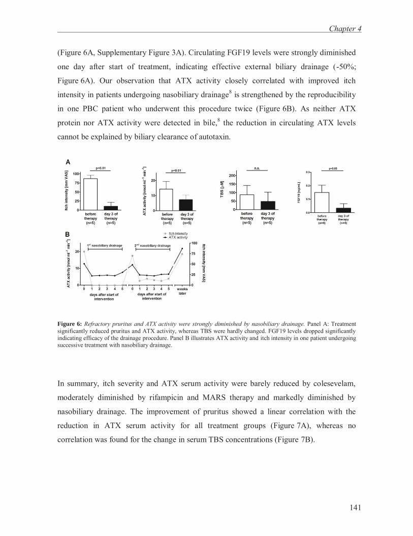
Nasobiliary drainage strongly reduced pruritus and ATX levels
Nasobiliary drainage effectively alleviated intractable pruritus in PBC patients not
responding to standard treatment.7 Simultaneously with the improvement of itch severity (-
85%; Figure 6A), ATX serum activity dropped in these patients to approximately half of
the baseline values (-50%; Figure 6A), whereas TBS initially dropped but rose back to
baseline values already during nasobiliary drainage, as in part reported previously7,8
Chapter 4
141
(Figure 6A, Supplementary Figure 3A). Circulating FGF19 levels were strongly diminished
one day after start of treatment, indicating effective external biliary drainage (-50%;
Figure 6A). Our observation that ATX activity closely correlated with improved itch
intensity in patients undergoing nasobiliary drainage8 is strengthened by the reproducibility
in one PBC patient who underwent this procedure twice (Figure 6B). As neither ATX
protein nor ATX activity were detected in bile,8 the reduction in circulating ATX levels
cannot be explained by biliary clearance of autotaxin.
Figure 6: Refractory pruritus and ATX activity were strongly diminished by nasobiliary drainage. Panel A: Treatment significantly reduced pruritus and ATX activity, whereas TBS were hardly changed. FGF19 levels dropped significantly indicating efficacy of the drainage procedure. Panel B illustrates ATX activity and itch intensity in one patient undergoing successive treatment with nasobiliary drainage.
In summary, itch severity and ATX serum activity were barely reduced by colesevelam,
moderately diminished by rifampicin and MARS therapy and markedly diminished by
nasobiliary drainage. The improvement of pruritus showed a linear correlation with the
reduction in ATX serum activity for all treatment groups (Figure 7A), whereas no
correlation was found for the change in serum TBS concentrations (Figure 7B).

Autotaxin in cholestatic pruritus
142
Figure 7: Overview of therapeutic interventions in chronic cholestatic patients suffering from pruritus. The effect on itch intensity and ATX activity are shown for colesevelam, rifampicin, MARS, and nasobiliary drainage. The individual changes of itch intensity and ATX activity (A) but not TBS (B) after therapy (related to the pre-treatment value) showed a linear correlation (Pearson’s correlation coefficient: r=0.62, p<0.0001). Dashed line represents no change in itch intensity and ATX activity, respectively.
DISCUSSION
In the present study we demonstrate that elevated serum ATX activity has a high specificity
for pruritus of cholestasis and might therefore serve as a diagnostic marker in cases of
pruritus of unknown origin (PUO) or multiple underlying diseases. A strong correlation
between ATX activity and efficacy of pruritus treatment further strengthens the role of
ATX in the pathogenesis of cholestatic pruritus. The beneficial effect of rifampicin on
cholestatic pruritus may be explained at least in part by PXR-dependent inhibition of ATX
expression as observed in vitro.
LPA is generated by ATX and serum levels of both correlate with the occurrence of
cholestatic itch.8 Quantification of LPA can be artificially altered after blood sampling
through release by platelets and levels may vary dependent on processing and storage.17 To
circumvent these potential artifacts, we analyzed ATX activity as a reliable parameter for
LPA formation. The source of the increased circulating ATX levels remains elusive but
might either be due to reduced clearance, increased expression, or a combination of both. A
reduced clearance may result from decreased uptake by liver sinusoidal endothelial cells.18
Despite their completely different mechanisms of action rifampicin, MARS treatment, and

Chapter 4
143
nasobiliary drainage all markedly reduced ATX serum levels, whereas ATX protein was
neither directly drained into bile8 nor removed in the albumin dialysate. We hypothesize
that a factor which is capable of increasing ATX expression (or reducing its clearance) is
removed by these treatments. This yet to be identified factor might accumulate in the
circulation during cholestasis, and might be metabolized in the liver and/or the gut,
followed by biliary secretion and reabsorption via enterohepatic circulation. The different
therapeutic approaches might intervene at different stages in this cycle.
Colesevelam binds various amphiphilic substances in the gut lumen and was
believed to effectively improve pruritus in cholestatic patients. The binding capacity of
colesevelam for the ATX-inducing factor might be minimal as opposed to that for bile salts,
which is underlined by only a small, though significant, decrease in ATX activity. As
cholestyramine has been reported in uncontrolled trials to attenuate pruritus, it might be that
cholestyramine could bind the ATX-inducing factor better than colesevelam which was not
superior to placebo in diminishing pruritus.12
Rifampicin alleviates pruritus in cholestasis by so far unknown molecular
mechanisms. Our in vitro data suggest that the antipruritic action of rifampicin in
cholestasis can be explained at least in part by transcriptional inhibition of ATX expression
in a PXR-dependent fashion. This may explain why rifampicin is effective in pruritus of
cholestasis, but not in pruritus of other origin such as uremia, Hodgkin’s disease or atopic
dermatitis where systemic ATX does not play a major pathogenetic role. One could
speculate that, in addition, rifampicin may reduce an ATX-inducing factor by modulation
of PXR-regulated genes involved in hepatic and intestinal detoxification and secretion
and/or by alteration of the gut flora.4 Determination of plasma ATX activity and LPA levels
in animal models of cholestasis in the presence and absence of an effective PXR agonist
may teach us more about ATX and LPA turnover under these pathological conditions in the
future. Alternatively, one has to consider that rifampicin might exert antipruritic effects, at
least in part, by PXR-independent mechanisms. An experimental approach to test this
option could be to compare the scratch response of mice towards injection of LPA with or
without prior administration of rifampicin - a PXR agonist in men, but not in mice.

Autotaxin in cholestatic pruritus
144
MARS therapy removes countless undefined substances from the circulation5
possibly including the ATX-inducing factor. Nasobiliary drainage removes secreted bile
from the body and thereby possibly also removes the ATX-inducing factor from the
enterohepatic circulation. Further in vitro analyses in cell culture systems of bile or albumin
dialysates of pruritic patients undergoing nasobiliary drainage or MARS treatment,
respectively, could possibly help to identify the ATX inducing factor in cholestatic pruritus.
It is of note that in as much as 10–35% of patients presenting with chronic
generalized pruritus an internal disease can be determined as underlying cause.19 Despite
extensive diagnostic examination, the cause of itching could not be identified in 8–20% of
patients with generalized pruritus.20-22 Eisendle et al. reported in a study with 117 patients
with pruritus of unknown origin that almost 30% of these patients had elevated TBS
concentrations without any evidence for liver disease.22 Identifying the underlying disease
causing pruritus apparently is a clinical challenge and diagnostic parameters are warranted
to make a differential diagnosis. ATX may represent such a novel marker for pruritus of
cholestasis. In this study an increased enzymatic activity above 8.5 nmol·mL-1·min-1 had a
positive predictive value of 70% in differentiating cholestatic pruritus from pruritus
associated with atopic dermatitis, uremia and Hodgkin lymphoma. Determination of ATX
serum activity in PUO or, more importantly, in cases of coexistence of two or more
potentially pruritus-inducing disorders might help clinicians in choosing a targeted
therapeutic regimen.
Slightly increased serum ATX activities were observed in patients with atopic
dermatitis and Hodgkin lymphoma compared to healthy controls in our cohort. A local
overproduction of ATX with only marginal increases in the systemic circulation could be a
conceivable mechanism causing itch perception in these patients. In line with our results
slightly enhanced ATX levels have been reported in a small cohort of 11 Hodgkin
lymphoma patients compared to healthy controls.23 In relation to uremia it has been
reported that patients with renal failure have three-fold elevated circulating LPA levels
compared to control subjects.24 More recently, this was confirmed in a rat model of
unilateral urethral obstruction.25 Strikingly, in this study the elevated plasma LPA was
accompanied by increased autotaxin activity in renal effluent rather than in plasma. It

Chapter 4
145
could be hypothesized that in renal failure autotaxin is primarily secreted into primary urine
but its product LPA may also end up in the plasma. Hence, this leaves open the possibility
that LPA plays a role in itch perception also in atopic dermatitis, Hodgkin’s disease and
uremia.
Several experimental and clinical observations favored increased levels of bile salts
as causative pruritogens in hepatobiliary disorders in the past.26 However, no correlation
between the level of any naturally occurring bile salt in the circulation or skin and severity
of pruritus could be proven.26 In addition, several observations in the present study argue
further against a direct causal role of bile salts in pruritus: (i) colesevelam halved TBS
levels without being more effective than placebo regarding improvement of itch intensity;
(ii) rifampicin or MARS therapy did not significantly reduce bile salt levels, yet strongly
diminished itch severity; (iii) in patients undergoing nasobiliary drainage TBS levels
dropped initially but returned to baseline values during the treatment long before pruritus
re-occurred, and (iv) the lack of correlation between TBS concentrations and itch
perception.
In our patient cohorts, markedly elevated ATX activity was specific for pruritus of
cholestasis. Thus, ATX might represent a useful diagnostic tool for those cases in whom
chronic pruritus remains unclassified. In addition, our study provides further clinical and
experimental evidence that ATX inhibitors and LPA receptor blockers may have potential
as future therapeutic agents to effectively treat pruritus in cholestatic liver disorders.
ACKNOWLEDGEMENTS
This work was supported by a ZonMW TOP grant to R.O.E. (#TOP 40-00812-98-10054),
research grants “Primary sclerosing cholangitis” from the Deutsche Crohn-Colitis-
Vereinigung and the Norwegian Primary Sclerosing Cholangitis Foundation to UB, and a
grant from the Deutsche Forschungsgemeinschaft to A.E.K. (KR3618/1-1). The authors
thank Dr. J. Aoki, University of Tokyo, for kindly providing 4F1-ATX antibody, Dr. R.

Autotaxin in cholestatic pruritus
146
Hoekstra, University of Amsterdam for generously providing HepG2 cells overexpressing
PXR, and Dagmar Tolenaars for expert technical assistance.

Chapter 4
147
REFERENCES
1. Ikoma A, Steinhoff M, Stander S, Yosipovitch G, Schmelz M. The neurobiology of itch. Nat Rev Neurosci. 2006;7:535-547.
2. Kremer AE, Beuers U, Oude-Elferink RP, Pusl T. Pathogenesis and treatment of pruritus in cholestasis. Drugs 2008;68:2163-2182.
3. Beuers U. Drug insight: Mechanisms and sites of action of ursodeoxycholic acid in cholestasis. Nat Clin Pract Gastroenterol Hepatol 2006;3:318-328.
4. EASL Clinical Practice Guidelines: Management of cholestatic liver diseases. J Hepatol 2009;51:237-267.
5. Pares A, Herrera M, Aviles J, Sanz M, Mas A. Treatment of resistant pruritus from cholestasis with albumin dialysis: combined analysis of patients from three centers. J Hepatol 2010;53:307-312.
6. Stapelbroek JM, van Erpecum KJ, Klomp LW, Venneman NG, Schwartz TP, van Berge Henegouwen GP, et al. Nasobiliary drainage induces long-lasting remission in benign recurrent intrahepatic cholestasis. Hepatology. 2006;43:51-53.
7. Beuers U, Gerken G, Pusl T. Biliary drainage transiently relieves intractable pruritus in primary biliary cirrhosis. Hepatology 2006;44:280-281.
8. Kremer AE, Martens JJ, Kulik W, Rueff F, Kuiper EM, van Buuren HR, et al. Lysophosphatidic acid is a potential mediator of cholestatic pruritus. Gastroenterology 2010;139:1008-1018.
9. Nakanaga K, Hama K, Aoki J. Autotaxin--an LPA producing enzyme with diverse functions. J Biochem 2010;148:13-24.
10. van Meeteren LA, Ruurs P, Stortelers C, Bouwman P, van Rooijen MA, Pradere JP, et al. Autotaxin, a secreted lysophospholipase D, is essential for blood vessel formation during development. Mol Cell Biol 2006;26:5015-5022.
11. Mills GB, Moolenaar WH. The emerging role of lysophosphatidic acid in cancer. Nat Rev Cancer 2003;3:582-591.
12. Kuiper EM, van Erpecum KJ, Beuers U, Hansen BE, Thio HB, de Man RA, et al. The potent bile acid sequestrant colesevelam is not effective in cholestatic pruritus: Results of a double-blind, randomized, placebo-controlled trial. Hepatology 2010;52:1334-1340.
13. Nibourg GA, Huisman MT, van der Hoeven TV, van Gulik TM, Chamuleau RA, Hoekstra R. Stable overexpression of pregnane X receptor in HepG2 cells increases its potential for bioartificial liver application. Liver transplantation : official publication of the American Association for the Study of Liver Diseases and the International Liver Transplantation Society 2010;16:1075-1085.
14. Schaap FG, van der Gaag NA, Gouma DJ, Jansen PL. High expression of the bile salt-homeostatic hormone fibroblast growth factor 19 in the liver of patients with extrahepatic cholestasis. Hepatology 2009;49:1228-1235.
15. Tanaka M, Okudaira S, Kishi Y, Ohkawa R, Iseki S, Ota M, et al. Autotaxin stabilizes blood vessels and is required for embryonic vasculature by producing lysophosphatidic acid. J Biol Chem 2006;281:25822-25830.
16. Leckie P, Tritto G, Proven A, Cheshire L, Mookerjee RP, Jones DE, et al. ”Outpatient” albumin dialysis for intractable itch in chronic cholestatic liver disease patients is a feasible, safe and efficacious alternative to liver transplantation Hepatology 2009;50:376A.
17. Nakamura K, Ohkawa R, Okubo S, Tozuka M, Okada M, Aoki S, et al. Measurement of lysophospholipase D/autotaxin activity in human serum samples. Clin Biochem 2007;40:274-277.
18. Jansen S, Andries M, Vekemans K, Vanbilloen H, Verbruggen A, Bollen M. Rapid clearance of the circulating metastatic factor autotaxin by the scavenger receptors of liver sinusoidal endothelial cells. Cancer Lett 2009;284:216-221.
19. Weisshaar E, Dalgard F. Epidemiology of itch: adding to the burden of skin morbidity. Acta Derm Venereol 2009;89:339-350.
20. Zirwas MJ, Seraly MP. Pruritus of unknown origin: a retrospective study. J Am Acad Dermatol 2001;45:892-896.
21. Sommer F, Hensen P, Bockenholt B, Metze D, Luger TA, Stander S. Underlying diseases and co-factors in patients with severe chronic pruritus: a 3-year retrospective study. Acta Derm Venereol 2007;87:510-516.

Autotaxin in cholestatic pruritus
148
22. Eisendle K, Muller H, Ortner E, Talasz H, Graziadei I, Vogel W, et al. Pruritus of unknown origin and elevated total serum bile acid levels in patients without clinically apparent liver disease. J Gastroenterol Hepatol 2011;26:716-721.
23. Masuda A, Nakamura K, Izutsu K, Igarashi K, Ohkawa R, Jona M, et al. Serum autotaxin measurement in haematological malignancies: a promising marker for follicular lymphoma. Br J Haematol 2008;143:60-70.
24. Sasagawa T, Suzuki K, Shiota T, Kondo T, Okita M. The significance of plasma lysophospholipids in patients with renal failure on hemodialysis. Journal of nutritional science and vitaminology 1998;44:809-818.
25. Tsutsumi T, Adachi M, Nikawadori M, Morishige J, Tokumura A. Presence of bioactive lysophosphatidic acid in renal effluent of rats with unilateral ureteral obstruction. Life sciences 2011;89:195-203.
26. Kremer AE, Oude Elferink RP, Beuers U. Pathophysiology and current management of pruritus in liver disease. Clin Res Hepatol Gastroenterol 2011;35:89-97.

Chapter 4
149
SUPPLEMENTARY INFORMATION
Supplementary Figure 1: ATX activity showed a linear correlation with the itch intensity represented on a VAS ranging from 0 (no pruritus) to 100 (unbearable pruritus). Pearson’s correlation coefficient: r=0.45, p<0.0001.
Supplementary Figure 2: (A): Itch severity score (ISS) showed a linear correlation with the VAS in patients undergoing MARS therapy (Pearson’s correlation coefficient: r=0.92, p<0.0001). (B): Improvement in ISS in patients responding (Responders) to MARS therapy. (C): No change in ISS was observed in Non-Responders. (D/E): Changes of itch intensity (ISS and VAS) and ATX activity after therapy (related to the pre-treatment value) showed a linear correlation (Pearson’s correlation coefficient: ISS: r=0.71, p<0.01, VAS: r=0.61, p<0.03)

Autotaxin in cholestatic pruritus
150
Supplementary Figure 3: Change in total serum bile salt (TBS) levels and itch intensity during the course of nasobiliary drainage. After an initial drop on the first and second day of the intervention, TBS rose back to baseline values long before pruritus re-occurred in these patients.
Supplementary Figure 4: Rifampicin induced CYP3A4 in a PXR-dependent manner. CPY3A4 gene expression tended to be increased (n=6; not significant) after incubation with 10 μM rifampicin for 24 hours (A). This induction was increased (p<0.01; n=3) in HepG2 cells overexpressing PXR (B), whereas it vanished in PXR knock-down cells, but not cells being transduced with scrambled shRNA (C).

Chapter 4
151
Supplementary Table 1: Characteristics of cholestatic patients without and with
pruritus.
Patients without
pruritus (n=40)
Patients with
pruritus (n=91) P-value
Male/Female 10/30 35/56 0.13
Age (yrs) 54.8 ± 18.0 51.5 ± 12.8 0.36
Disease (PBC/PSC/Other) 27/2/11 41/17/33 0.07
AP (IU/L) 289.7 ± 203.8 321.4 ± 239.2 0.54
γGT (IU/L) 160.6 ± 191.4 187.2 ± 233.0 0.61
Bilirubin (mg/dL) 3.1 ± 5.5 3.7 ± 6.3 0.65
TBS (μmol/L) 46.9 ± 60.1 135.9 ± 122.9 0.01
Albumin (g/dL) 4.4 ± 1.1 4.6 ± 1.2 0.48
ALT (IU/L) 71.9 ± 95.0 72.6 ± 55.3 0.96
AST (IU/L) 73.7 ± 75.5 118.6 ± 150.9 0.15
CRP (mg/dL) 0.5 ± 0.3 0.4 ± 0.5 0.87
ATX (nmol mL-1 min-1) 6.6 ± 2.9 13.6 ± 7.2 < 0.001
Supplementary Table 1: Characteristics and serum chemistry of cholestatic patients with and without pruritus. All values are expressed as means ± SD. P-values are for comparison between the subgroups of cholestatic patients with and without pruritus. Abbreviations: AP = alkaline phosphatase, γGT = γ-glutamyltransferase, TBS = total serum bile salts, ALT = alanine aminotransferase, AST = aspartate aminotransferase, CRP = C-reactive protein.

Autotaxin in cholestatic pruritus
152
Supplementary Table 2: Characteristics of Hodgkin lymphoma patients without and
with pruritus.
Patients without
pruritus (n=29)
Patients with
pruritus (n=30) P-value
Male/Female 18/11 13/17 0.16
Age (yrs) 39.0 ± 13.0 32.5 ± 10.3 0.13
Stage of disease (early /
intermittent / advanced) 16/13/0 4/19/7 0.01
ESR (mm/h) 26.7 ± 26.9 39.2 ± 24.2 0.07
Erythrocytes (109/L) 4.9 ± 0.5 4.7 ± 0.4 0.23
Haemoglobin (g/dL) 14.1 ± 1.5 12.7 ± 1.4 0.02
Leucocytes (109/L) 8.2 ± 2.5 11.7 ± 4.1 0.01
Albumin (g/dL) 4.3 ± 0.6 4.1 ± 0.9 0.22
TBS (μmol/L) 5.2 ± 5.9 5.6 ± 4.1 0.78
ATX (nmol mL-1 min-1) 5.5 ± 1.5 6.7 ± 4.9 0.23
Supplementary Table 2: Characteristics and serum chemistry of Hodgkin lymphoma patients with and without pruritus. All values are expressed as means ± SD. P-values are for comparison between the subgroups of patients with and without pruritus. Abbreviations: ESR = erythrocyte sedimentation rate, TBS = total serum bile salts.

Chapter 4
153
Supplementary Table 3: Characteristics of uremic patients without and with pruritus.
Patients without
pruritus (n=20)
Patients with
pruritus (n=21) P-value
Male/Female 9/11 14/7 0.22
Age (yrs) 67.9 ± 11.8 63.5 ± 17.1 0.34
Time of dialysis (months) 53.8 ± 34.2 36.3 ± 41.6 0.14
Calcium 2.2 ± 0.2 2.1 ± 0.5 0.57
Phosphat 1.6 ± 0.4 1.7 ± 0.7 0.49
PTH 383.2 ± 400.6 260.6 ± 276.6 0.26
1α-OH-Vit D3 0.6 ± 0.5 0.7 ± 0.5 0.84
25-OH-Vit D3 14.5 ± 8.3 15.5 ± 8.5 0.70
Albumin (g/dL) 3.7 ± 0.5 3.6 ± 0.9 0.59
CRP (mg/dL) 5.6 ± 7.9 8.7 ± 8.4 0.24
TBS (μmol/L) 4.9 ± 3.8 4.2 ± 3.2 0.83
ATX (nmol mL-1 min-1) 4.6 ± 1.3 5.1 ± 1.7 0.33
Supplementary Table 3: Characteristics and serum chemistry of uremic patients with and without pruritus. All values are expressed as means ± SD. P-values are for comparison between the subgroups of patients with and without pruritus. Abbreviations: PTH = parathyroid hormone, CRP = C-reactive protein, TBS = serum bile salts.

Autotaxin in cholestatic pruritus
154
Supplementary Table 4: Characteristics of Responders and Non-responders of MARS
therapy
Responder (N=8) Non-Responder (N=2)
Pre-
treatment
Post-
treatment
Pre-
treatment
Post-
treatment
Male/Female 7/1 1/1
Disease
(PBC/PSC/Other) 5/1/2 1/1/0
MELD score 10.5 ± 6.6 6.0
AP (IU/L) 505.1 ± 168.4 427.7 ± 111.2 319.5 251.5
ALT (IU/L) 103.9 ± 78.1 85.0 ± 52.2 76.5 64.5
Bilirubin (μmol/L) 28.6 ± 26.7 21.7 ± 16.9 22.5 28.0
TBS (μmol/L) 133.1 ± 140.4 43.6 ± 13.3 43.6 176.7
Albumin (g/dL) 3.9 ± 0.5 3.9 ± 0.6 4.4 4.3
Creatinine (mg/dL) 75.0 ± 34.4 61.3 ± 14.0 54.0 42.0
INR (IU/L) 0.96 ± 0.05 0.94 ± 0.03 1.00 1.06
Platelets (109/L) 223.3 ± 87.4 148.4 ± 52.7 306.0 229.0
Haemoglobin (g/L) 11.5 ± 1.6 10.0 ± 1.1 13.3 12.1
ATX (nmol mL-1 min-1) 10.8 ± 6.5 7.8 ± 4.0 12.3 12.2
Supplementary Table 4: Characteristics and serum chemistry of cholestatic patients undergoing MARS therapy. All values are expressed as means ± SD. P-values are for comparison between the subgroups of patients with and without pruritus. Abbreviations: AP = alkaline phosphatase, ALT = alanine aminotransferase, TBS = serum bile salts, INR = prothrombin time.