UvA-DARE (Digital Academic Repository) Genomic ... · Savci-Heijink CD, Halfwerk H, Hooijer GK,...
185
UvA-DARE is a service provided by the library of the University of Amsterdam (http://dare.uva.nl) UvA-DARE (Digital Academic Repository) Genomic characteristics of metastatic breast cancer Savcı Heijink, C.D. Link to publication Citation for published version (APA): Savcı Heijink, C. D. (2019). Genomic characteristics of metastatic breast cancer. General rights It is not permitted to download or to forward/distribute the text or part of it without the consent of the author(s) and/or copyright holder(s), other than for strictly personal, individual use, unless the work is under an open content license (like Creative Commons). Disclaimer/Complaints regulations If you believe that digital publication of certain material infringes any of your rights or (privacy) interests, please let the Library know, stating your reasons. In case of a legitimate complaint, the Library will make the material inaccessible and/or remove it from the website. Please Ask the Library: https://uba.uva.nl/en/contact, or a letter to: Library of the University of Amsterdam, Secretariat, Singel 425, 1012 WP Amsterdam, The Netherlands. You will be contacted as soon as possible. Download date: 02 Jan 2020
Transcript of UvA-DARE (Digital Academic Repository) Genomic ... · Savci-Heijink CD, Halfwerk H, Hooijer GK,...
UvA-DARE is a service provided by the library of the University of Amsterdam (http://dare.uva.nl)
UvA-DARE (Digital Academic Repository)
Genomic characteristics of metastatic breast cancer
Savcı Heijink, C.D.
Link to publication
Citation for published version (APA):Savcı Heijink, C. D. (2019). Genomic characteristics of metastatic breast cancer.
General rightsIt is not permitted to download or to forward/distribute the text or part of it without the consent of the author(s) and/or copyright holder(s),other than for strictly personal, individual use, unless the work is under an open content license (like Creative Commons).
Disclaimer/Complaints regulationsIf you believe that digital publication of certain material infringes any of your rights or (privacy) interests, please let the Library know, statingyour reasons. In case of a legitimate complaint, the Library will make the material inaccessible and/or remove it from the website. Please Askthe Library: https://uba.uva.nl/en/contact, or a letter to: Library of the University of Amsterdam, Secretariat, Singel 425, 1012 WP Amsterdam,The Netherlands. You will be contacted as soon as possible.
Download date: 02 Jan 2020


Genomic characteristics of metastatic breast cancer
Cemile Dilara Savcı Heijink

Genomic characteristics of metastatic breast cancer
PhD Thesis, University of Amsterdam, The Netherlands
The publication of this thesis was kindly supported by Center for Translational Molec-ular Medicine (BreastCARE), Amsterdam UMC (AMC), ChipSoft and Pfizer BV.
Cover design by Wim J.V. van Est , modified from Blue Nude II, Gouache-painted pa-per cut-out, by Henri Matisse completed in 1952. Layout and design by David de Groot, persoonlijkproefschrift.nl. Printed by Ipskamp Printing BV
Copyright © 2018 Cemile Dilara Savcı Heijink
All rights reserved. No part of this thesis may be reproduced, stored or transmitted in any way or by any means without the prior permission of the author, or when applica-ble, of the publishers of the scientific papers.
ISBN/EAN: 978-94-028-1297-8

GENOMIC CHARACTERISTICS OF METASTATIC BREAST CANCER
ACADEMISCH PROEFSCHRIFT
ter verkrijging van de graad van doctor
aan de Universiteit van Amsterdam
op gezag van de Rector Magnificus
prof. dr. ir. K.I.J. Maex
ten overstaan van een door het College voor Promoties ingestelde commissie,
in het openbaar te verdedigen in de Agnietenkapel
op vrijdag 18 januari 2019, 14.00 uur
door Cemile Dilara Savcı Heijink
geboren te Adana, Turkije

Promotiecommissie
Promotor: Prof. dr. M.J. van de Vijver AMC-UvA
Copromotor: Dr. H.M. Horlings NKI-AvL
Overige leden: Prof. dr. V.T.H.B.M Smit Universiteit Leiden
Prof. dr. P. J. van Diest Universiteit Utrecht
Prof. dr. L.F.A. Wessels TU Delft
Prof. dr. E.J.T. Rutgers AMC-UvA
Dr. A.D. Bins AMC-UvA
Dr. N. Bijker AMC-UvA
Faculteit der Geneeskunde

For Şuşu,
The most beautiful sea,
hasn’t been crossed yet
The most beautiful child,
hasn’t grown up yet
Our most beautiful days,
we haven’t seen yet
And the most beautiful words I wanted to tell you,
I haven’t said yet…
Nâzım Hikmet Ran September 24th 1945

Table of contents
Chapter 1 9
General introduction and outline of the thesis
Chapter 2 25
Translating the genomic architecture of breast cancer into clinical
application
Horlings HM, Savci-Heijink CD, van de Vijver MJ
Sci Transl Med. 2010 Jun 30;2(38):38ps32.
Chapter 3 41
Retrospective analysis of metastatic behavior of breast cancer
subtypes
Savci-Heijink CD, Halfwerk H, Hooijer GK, Horlings HM, Wesseling J, van de
Vijver MJ
Breast Cancer Res Treat. 2015 Apr;150(3):547-57.
Chapter 4 67
A novel gene expression signature for bone metastasis in breast
carcinomas
Savci-Heijink CD, Halfwerk H, Koster J, van de Vijver MJ
Breast Cancer Res Treat. 2016 Apr;156(2):249-59.
Chapter 5 95
A specific gene expression signature for visceral organ metastasis in
breast cancer
Savci-Heijink CD, Halfwerk H, Koster J, Horlings HM, van de Vijver MJ
Submitted

Chapter 6 115
Association between gene expression profile of the primary tumor
and chemotherapy response of metastatic breast cancer
Savci-Heijink CD, Halfwerk H, Koster J, van de Vijver MJ
BMC Cancer. 2017 Nov 13;17(1):755.
Chapter 7 135
Epithelial-mesenchymal transition status of primary breast
carcinomas and its correlation with metastatic behavior
Savci-Heijink CD, Halfwerk H, Hooijer GK, Koster J, Horlings HM, Meijer SL, van
de Vijver MJ
Accepted for publication in Breast Cancer Res Treat.
Chapter 8 155
General discussion and concluding remarks
Chapter 9 171
Summary in English and Dutch
Appendix 181
Nawoord / Acknowledgement
About the author


General introduction and outline of the thesis
Chapter 1

10
Chapter 1
Metastatic breast cancer
Breast cancer is the most common cancer in women in the western world and the
most prevalent type of cancer in the Netherlands with a ten-year prevalence of
128.000 cases [1]. Notwithstanding the advances in early detection and improved cure
rates, breast cancer remains a major cause of cancer related deaths for women [2-
4]. Mortality rates are closely related to the development of distant metastases and
associated complications [5, 6]. Approximately 5-10% of the patients initially present
with metastatic breast cancer, while 20-30% of patients eventually develop distant
metastases during the course of the disease [7, 8].
Metastatic progression in cancer is a heterogeneous process and encompasses
stepwise sequential events initiating with invasion of tumor cells through the basal
membrane followed by penetration into the bloodstream (intravasation). When cancer
cells survive in the circulation, subsequent extravasation and eventual colonization
leads to form a detectable macroscopic tumor. Considering that only less than 0.01%
of tumor cells that reach the circulation give rise to an overt metastasis, metastasis has
been regarded as a highly inefficient process [9, 10]. Therefore, accomplishment of this
complex process requires multistep interactions between the circulating tumor cells and
the microenvironment of the organ in which metastatic disease develops.
Organ-specific metastasisOrganotropism (Gk, organon, tool of the body + tropism, turning movement of a
biological organism), i.e. organ-specific metastasis, depicts the concept of non-
random involvement of the particular organ within a specific cancer type. Mechanisms
and determinants of organotropism are intriguing and therefore are often subject of
investigation for many researchers [5, 11-14]. The “Anatomical/mechanical” hypothesis,
a rather conventional theory, proposes that the blood circulation pattern, the anatomy
of the primary tumor and the surrounding vessels principally shape the metastatic
spread pattern. This theory fails to fully relate the clinically observed metastasis pattern
for many types of cancer. More than a century ago, Stephen Paget introduced a theory
implicating that the essence of close interactions between the circulating tumor cells
would constitute the “seed” and the microenvironment within the targeted area,

11
General introduction and outline of the thesis
the “soil”. Since its introduction, Paget’s “Seed and Soil” hypothesis, along with the
“Anatomical/mechanical” hypothesis, has been acknowledged by experimental and
clinical research [10, 15-17].
Several experimental studies using animal models to decipher the underlying framework
of these non-random distinct organ metastases, have shown that as well as extrinsic
factors, tumor intrinsic factors play a substantial role in the development of metastatic
disease [18-22]. These studies have investigated the underlying biology of organ-
specific metastasis by using animal models of metastatic breast cancer, which were
developed by injecting human breast cancer cell lines in immune compromised mice.
The organ-tropic metastatic variants selected from these animal models were further
analyzed by genomic profiling, which was subsequently combined with clinical genomic
studies [23-30]. Some of these studies, carried out by Massague and colleagues,
resulted in distinct gene expression profiling signatures associated with metastasis to
bone, lung and brain [24-26, 30, 31]. Based on these site-specific signatures, several
individual genes were further explored by means of validating these discovered genes
in cohorts of primary breast tumors, to comprehend the biology of metastatic disease.
Clinical Genomics and its application in management
of breast cancer
In addition to the histomorphologic features, such as histologic type and the grade of the
primary tumor, tissue-based biomarkers and clinical characteristics play an important
role in decision making in the management of breast cancer. Tissue-based biomarkers
include hormone receptor status (Estrogen receptor, ER; Progesteron receptor, PR;
Human epidermal growth factor, HER2 and proliferation index, Ki67) of the tumor and
are most widely assessed in combination of immunohistochemistry and in situ DNA
hybridization techniques. Despite compelling improvement in disease control, the
heterogeneity of the breast tumors is still not fully reflected by this basic stratification
approach.
1

12
Chapter 1
In the past decades, extensive research applying genomics on breast cancers was
carried out to identify the molecular characteristics of distinctive biological behavior of
these tumors. Genomics is defined as the field of science concerned with applying high-
throughput techniques to the genetic mapping and DNA sequencing of sets of genes
or the complete genetic material and RNA expression profiling of selected organisms/
samples. Clinical genomics, which employs genomic studies to improve patient care,
has resulted in several molecular classifiers, particularly in the breast cancer field.
Several widely accepted molecular classifiers for breast cancer were developed based on
gene expression profiling analyses of breast cancer [32-37]. Gene expression profiling
experiments are based on measuring relative amounts of mRNA simultaneously for many
genes and reflect the pattern of the transcription which is encoded in DNA sequences
[38]. The data generated by these experiments, following a normalization step, can
be analyzed in an unsupervised and a supervised manner. Unsupervised classification
aims to group the genes or samples together with similar traits. Hierarchical clustering is
one of the most common approaches used in unsupervised classification and operates
by repetitively joining the two closest clusters from individual clusters or repetitively
separating clusters starting with the initial dataset [39, 40]. Another frequently used
unsupervised classification method is the K-means clustering algorithm which works
by classifying the given data set into a certain number of clusters chosen a priori. This
algorithm repeatedly calculates the center points for each cluster, following initial
separation of the linear space into given K components. Using unsupervised hierarchical
clustering analysis, several investigators have observed distinct gene expression traits of
ER-positive and ER-negative tumors [34, 35]. These analyses have led to the discovery
of several subgroups within breast carcinomas with distinct clinical behavior. Based on
these studies different “intrinsic” subtypes of breast carcinomas are defined: luminal A
and luminal B tumors (ER-positive tumors characterized by gene expression pattern
similar to breast luminal cells), HER2-like tumors (ER-negative tumors with HER2 gene
overexpression), basal-like tumors (ER-negative tumors with gene expression pattern
overlapping with myoepithelial cells) and normal-like tumors. Even though a very similar
classification of breast carcinomas into subgroups can be done by immunohistochemical
analysis of ER, PR and HER2, the results are not consistently in concordance with the
‘intrinsic’ subtypes assigned by molecular classification of these tumors [41].

13
General introduction and outline of the thesis
Supervised classification is described as a knowledge-driven classification of a data
set to design a classifier with prognostic and predictive value. Usually a subset of
tumors with known characteristics are used to train a model to classify samples and
subsequently this classifier is tested in another dataset to create similar groups. Using
the supervised classification approach, several gene sets have been described with the
purpose of identifying breast tumors with distinctive clinical behavior [34, 35, 37, 42],
eventual prediction of the metastatic potential and response to therapy of the tumors
[24-26]. Some of these gene expression profiling studies have led to commercially
available gene-expression-based molecular tests [32, 33, 36, 37, 43, 44]. The 21-gene
recurrence classifier (Oncotype DX®) is one of these molecular tests that estimates the
recurrence score (RS), which can be defined as a risk of developing distant metastasis
at 10 years for the patients with ER-positive and lymph-node negative breast cancer.
This estimation is based on the expression level of 21 genes (16 cancer related genes
and 5 reference genes) and can be easily applied as a clinical assay on formalin-fixed,
paraffin-embedded tumor tissue of breast cancer patients. This test is most commonly
used (most widely in the USA) as a prognostic indicator in addition to tissue-based
markers and clinicopathological characteristics of the primary tumor in decision-making
regarding administration of chemotherapy [32, 45, 46]. Another well-known prognostic
classifier and a companion diagnostic tool is the 70-gene signature (MammaPrint®).
The 70-gene prognosis signature, classifies the breast tumors into 2 groups as a “good”
or “poor” prognosis, and gives an estimation of developing distant metastasis, mainly in
lymph-node negative breast cancer patients with tumor size <5 cm [37]. The prognostic
value of this signature has been validated in large-scale prospective studies and has
shown that this signature is also valid to predict the outcome in breast cancer patients
with 1 to 3 lymph node metastases [47].
Clinical management of metastatic breast cancer
Metastatic breast cancer is considered to be an incurable disease. The main aim of
treating metastatic breast cancer is to prolong survival of the patients with acceptable
toxicity and to palliate the disease-related symptoms. The decisions of treating patients
with metastatic disease and the choice of therapy (hormonal, chemotherapeutic and/or
1

14
Chapter 1
targeted) depends on the patient’s age/performance, site and the number of the distant
metastases, hormone receptor status, HER2 status, menopausal status, type and extent
of prior adjuvant therapy, and time between the last administered therapy [48-50].
Hormonal therapyFor patients with hormone receptor positive metastatic breast cancer, hormone therapy
plays an important role. Even though a well-established consensus on the sequence is
lacking, in the absence of visceral crisis (severe organ dysfunction as assessed by signs
and symptoms, laboratory studies, and rapid progression of disease) two to four lines
of hormonal therapy may be administered before considering to start chemotherapeutic
therapy. For the patients who are premenopausal at the initial diagnosis, surgical
(oophorectomy) or medical (gonotropin-releasing hormone analogs) introduction of
menopause is indicated as an effective therapeutic action. First-line hormonal therapy
options for the postmenopausal patients include aromatase inhibitors, selective
estrogen receptor modulators (tamoxifen) or a selective estrogen receptor degrader
(fulvestrant). Combination therapies with inclusion of cyclin-dependent kinase 4/6
inhibitors or everolimus can be considered in the treatment of metastatic breast cancer
as second or further-line therapy [50-52].
ChemotherapyIn the metastatic setting, administration of chemotherapy is considered for the patients
with hormone receptor negative tumors and for patients with hormone receptor
positive disease resistant to hormonal therapy and rapidly progressive disease. Choice
of chemotherapeutic agent depends prior (adjuvant) chemotherapeutic treatments and
the status of the patient. For the first-line chemotherapy, anthracyline- and/or taxane-
based regimens are the most commonly used therapy agents [49-51, 53]. In case of
resistance to anthracyclines or taxanes, an antimetabolite cytotoxic agent (capecitabine)
is usually used as a second-line chemotherapy agent [54].
Targeted therapy HER2 is amplified and overexpressed in approximately 15% of breast carcinomas,
and the tumors with HER2 overexpression are associated with shorter overall survival
times [55-57]. For the patients with HER2-positive tumors and metastastic disease,
targeted therapy including combination of HER2- receptor antagonists (trastuzumab,

15
General introduction and outline of the thesis
pertuzumab) and taxanes is recommended [49, 50, 58]. As second-line targeted therapy
option or in case of development of metastases within 6 months after completion of
an anti-HER2 adjuvant therapy, administration of ado-trastuzumab emtansine (T-DMI)
is indicated [59]. Dual inhibitors of epidermal growth factor 1 (EGFR1) and HER2
(lapatinib) can also be used as a single therapy agent or in combination with other
chemotherapeutics in treatment of metastatic breast carcinoma [60].
ImmunotherapyThe immune microenvironment of the tumor has been shown to play an important role in
cancer progression. Cancer immunotherapy aims to boost antitumor immune response
and has been historically implemented in passive forms (Interferons, Interleukin-2,
Calmette-Guerin Bacillus and antibody dependent cell-mediated cytotoxicity) in
various cancer types [61-65]. Immunotherapy has evolved to a therapeutic approach
using an immune checkpoint blockade (ICB) with promising results in a variety of tumor
types [66-70]. Several recent studies have pointed out the association between the
immune-related genes and the tumor-infiltrating lymphocytes with prognosis in triple-
negative and HER2-positive breast cancer subtypes [71-74]. Subsequent clinical trials
investigating the usage of novel immunomodulatory agents in treatment of advanced
breast cancer, have revealed that the blockading of programmed cell death protein
1(PD1)/programmed death ligand 1 (PDL1) by means of monotherapy or in combination
with chemotherapy/anti-cytotoxic T lymphocyte antigen-4 (CTLA4) was paired with
prolonged survival times in patients with triple negative tumors [75-77]. More recently,
it has been showed that the PD-1/PD-L1 blockade in combination with chemotherapy
resulted in increased pathologic response rates in the neoadjuvant setting for triple
negative and HER2-negative breast tumors [78-80]. Despite the emerging role of
immunotherapy in treatment of advanced breast cancer, refinement of patient selection
and evaluation of response to these agents remain unsolved [81].
Evaluation of response to systemic therapyAssessment of response to given therapeutic agents is determined by estimating the
changes in tumor burden. To evaluate these changes, a widely accepted guideline,
Response Evaluation of Solid Tumors (RECIST) criteria, which was introduced by the
European Organization and Treatment of Cancer (EORTC), the National Cancer Institute
of the United States and the National Cancer Institute of Canada Clinical Trials Group,
1

16
Chapter 1
is commonly used [82]. According to the RECIST criteria, evaluation of an objective
response is classified as; complete response (CR), partial response (PR), stable disease
(SD) and progressive disease (PD). This assessment of therapy response in metastatic
breast carcinoma is performed by means of imaging techniques and tumor marker
determination [50].
Within the metastatic setting, response to combined chemotherapy agents varies
between 50-70% [48, 83, 84]. To this day, no consensus exists on the optimal
chemotherapy options or the sequence and duration of the given chemotherapeutics.
As the current therapy approaches for metastatic breast cancer are mainly of a trial-
and-error approach, predictors of response are needed for optimization the benefit and
minimization of the toxic side-effects of the therapy.
Radiotherapy may be implemented to palliate the pain and discomfort for the patients
with metastatic disease.
Rationale of this thesis
The aim of this thesis is to identify the characteristics of primary breast tumors that
are predictive of metastatic behavior in terms of organ-specific metastasis, response to
systemic therapy and associated patient outcomes.
Outline of this thesis
Chapter 1 provides a brief introduction to metastatic breast cancer and genomic
research of breast cancer.
In chapter 2, a viewpoint on genomic alterations of breast cancer and their translation
into clinical application is presented (in conjunction with article by Russness et al: “

17
General introduction and outline of the thesis
Genomic architecture characterizes tumor progression paths and fate in breast cancer
patients” [85]).
Chapter 3 describes a retrospective study, analyzing the clinicopathologic features of
primary breast carcinomas focusing on the association between breast cancer subtypes
and their metastatic behavior, including site-specific metastasis and metastasis specific
survival outcomes.
In chapters 4 and 5, we describe our results of gene expression profiling experiments
of 157 primary breast carcinomas of patients who all developed distant metastases.
The correlation of the gene expression profiling data to metastatic behavior is reported.
Furthermore, specific gene expression profiling signatures, which were found to be
associated with development of bone metastasis (in chapter 4) and visceral organ
metastasis (in chapter 5), are presented.
In chapter 6, we investigated the link between primary breast carcinoma features and
the chemotherapy response in the frame of metastatic disease. We sought to develop
genomic identifiers of chemotherapy responsiveness by comparing the gene expression
profiling of primary tumors of the responders and non-responders.
In chapter 7, the concept of epithelial-to mesenchymal transition (EMT) in metastatic
breast cancer is elaborated. Using a generic EMT-core signature, the EMT-status of
each primary breast tumor in our data set was assessed and compared with tumor
characteristics and their metastasis pattern. The concept to reconcile the EMT-status
of the tumor and to identify the tumor cells with EMT-phenotype, using conventional
immunohistochemistry, is also explored.
Chapter 8 provides a general discussion followed by concluding remarks, based on the
findings generated in the abovementioned studies which form this thesis.
In chapter 9, a brief summary of this thesis, in the English and Dutch language, is
presented.
1

18
Chapter 1
References1. Rijksinstituut voor gezondheid en milieu. Public health info, breast cancer in the Netherlands.
2018; Available from: https://www.rivm.nl/en/Topics/B/Breast_cancer_screening_programme/Breast_cancer_in_the_Netherlands.
2. Malvezzi, M., et al., European cancer mortality predictions for the year 2012. AnnOncol, 2012. 23(4): p. 1044-1052.
3. Sant, M., et al., Breast carcinoma survival in Europe and the United States. Cancer, 2004. 100(4): p. 715-22.
4. Thomssen, C., et al., Follow-up after breast cancer diagnosis. Breast Care (Basel), 2013. 8(6): p. 457-60.
5. Chambers, A.F., A.C. Groom, and I.C. MacDonald, Dissemination and growth of cancer cells in metastatic sites. NatRevCancer, 2002. 2(8): p. 563-572.
6. Chiang, A.C. and J. Massague, Molecular basis of metastasis. NEnglJMed, 2008. 359(26): p. 2814-2823.
7. Early Breast Cancer Trialists’ Collaborative, G., Effects of chemotherapy and hormonal therapy for early breast cancer on recurrence and 15-year survival: an overview of the randomised trials. Lancet, 2005. 365(9472): p. 1687-717.
8. O’Shaughnessy, J., Extending survival with chemotherapy in metastatic breast cancer. Oncologist, 2005. 10 Suppl 3: p. 20-9.
9. Fidler, I.J., Metastasis: quantitative analysis of distribution and fate of tumor emboli labeled with 125 I-5-iodo-2’-deoxyuridine. J Natl Cancer Inst, 1970. 45(4): p. 773-82.
10. Langley, R.R. and I.J. Fidler, The seed and soil hypothesis revisited--the role of tumor-stroma interactions in metastasis to different organs. IntJCancer, 2011. 128(11): p. 2527-2535.
11. Aslakson, C.J. and F.R. Miller, Selective events in the metastatic process defined by analysis of the sequential dissemination of subpopulations of a mouse mammary tumor. Cancer Res, 1992. 52(6): p. 1399-1405.
12. Ding, L., et al., Genome remodelling in a basal-like breast cancer metastasis and xenograft. Nature, 2010. 464(7291): p. 999-1005.
13. Hu, G., Y. Kang, and X.F. Wang, From breast to the brain: unraveling the puzzle of metastasis organotropism. JMolCell Biol, 2009. 1(1): p. 3-5.
14. Lorusso, G. and C. Ruegg, New insights into the mechanisms of organ-specific breast cancer metastasis. SeminCancer Biol, 2012. 22(3): p. 226-233.
15. Fidler, I.J., The pathogenesis of cancer metastasis: the ‘seed and soil’ hypothesis revisited. Nat Rev Cancer, 2003. 3(6): p. 453-8.
16. Lu, X. and Y. Kang, Organotropism of breast cancer metastasis. JMammaryGlandBiolNeoplasia, 2007. 12(2-3): p. 153-162.

19
General introduction and outline of the thesis
17. Paget, S., The distribution of secondary growths in cancer of the breast. 1889. Cancer Metastasis Rev, 1989. 8(2): p. 98-101.
18. Chi, J.T., et al., Gene expression programs in response to hypoxia: cell type specificity and prognostic significance in human cancers. PLoS Med, 2006. 3(3): p. e47.
19. Feinberg, A.P., R. Ohlsson, and S. Henikoff, The epigenetic progenitor origin of human cancer. Nat Rev Genet, 2006. 7(1): p. 21-33.
20. Fidler, I.J. and G. Poste, The “seed and soil” hypothesis revisited. Lancet Oncol, 2008. 9(8): p. 808.
21. Nuyten, D.S., et al., Combining biological gene expression signatures in predicting outcome in breast cancer: An alternative to supervised classification. Eur J Cancer, 2008. 44(15): p. 2319-29.
22. Semenza, G.L., Targeting HIF-1 for cancer therapy. Nat Rev Cancer, 2003. 3(10): p. 721-32.
23. Bos, P.D., D.X. Nguyen, and J. Massague, Modeling metastasis in the mouse. Curr Opin Pharmacol, 2010. 10(5): p. 571-7.
24. Bos, P.D., et al., Genes that mediate breast cancer metastasis to the brain. Nature, 2009. 459(7249): p. 1005-1009.
25. Kang, Y., et al., A multigenic program mediating breast cancer metastasis to bone. Cancer Cell, 2003. 3(6): p. 537-49.
26. Landemaine, T., et al., A six-gene signature predicting breast cancer lung metastasis. Cancer Res, 2008. 68(15): p. 6092-6099.
27. Minn, A.J., et al., Lung metastasis genes couple breast tumor size and metastatic spread. Proc Natl Acad Sci U S A, 2007. 104(16): p. 6740-5.
28. Minn, A.J., et al., Distinct organ-specific metastatic potential of individual breast cancer cells and primary tumors. J Clin Invest, 2005. 115(1): p. 44-55.
29. Zhang, X.H., et al., Selection of bone metastasis seeds by mesenchymal signals in the primary tumor stroma. Cell, 2013. 154(5): p. 1060-73.
30. Zhang, X.H., et al., Latent bone metastasis in breast cancer tied to Src-dependent survival signals. Cancer Cell, 2009. 16(1): p. 67-78.
31. Harrell, J.C., et al., Genomic analysis identifies unique signatures predictive of brain, lung, and liver relapse. Breast Cancer ResTreat, 2012. 132(2): p. 523-535.
32. Paik, S., et al., A multigene assay to predict recurrence of tamoxifen-treated, node-negative breast cancer. N Engl J Med, 2004. 351(27): p. 2817-26.
33. Parker, J.S., et al., Supervised risk predictor of breast cancer based on intrinsic subtypes. J Clin Oncol, 2009. 27(8): p. 1160-7.
34. Perou, C.M., et al., Molecular portraits of human breast tumours. Nature, 2000. 406(6797): p. 747-752.
1
20
Chapter 1
35. Sorlie, T., et al., Gene expression patterns of breast carcinomas distinguish tumor subclasses with clinical implications. Proc Natl Acad Sci U S A, 2001. 98(19): p. 10869-74.
36. Sotiriou, C., et al., Gene expression profiling in breast cancer: understanding the molecular basis of histologic grade to improve prognosis. J Natl Cancer Inst, 2006. 98(4): p. 262-72.
37. van de Vijver, M.J., et al., A gene-expression signature as a predictor of survival in breast cancer. NEnglJMed, 2002. 347(25): p. 1999-2009.
38. Brazma, A. and J. Vilo, Gene expression data analysis. FEBS Lett, 2000. 480(1): p. 17-24.
39. Alon, U., et al., Broad patterns of gene expression revealed by clustering analysis of tumor and normal colon tissues probed by oligonucleotide arrays. Proc Natl Acad Sci U S A, 1999. 96(12): p. 6745-50.
40. Eisen, M.B., et al., Cluster analysis and display of genome-wide expression patterns. Proc Natl Acad Sci U S A, 1998. 95(25): p. 14863-8.
41. van de Vijver, M.J., Molecular tests as prognostic factors in breast cancer. Virchows Arch, 2014. 464(3): p. 283-91.
42. van ‘t Veer, L.J., et al., Gene expression profiling predicts clinical outcome of breast cancer. Nature, 2002. 415(6871): p. 530-536.
43. Filipits, M., et al., A new molecular predictor of distant recurrence in ER-positive, HER2-negative breast cancer adds independent information to conventional clinical risk factors. Clin Cancer Res, 2011. 17(18): p. 6012-20.
44. Ma, X.J., et al., A two-gene expression ratio predicts clinical outcome in breast cancer patients treated with tamoxifen. Cancer Cell, 2004. 5(6): p. 607-16.
45. Albain, K.S., et al., Prognostic and predictive value of the 21-gene recurrence score assay in postmenopausal women with node-positive, oestrogen-receptor-positive breast cancer on chemotherapy: a retrospective analysis of a randomised trial. Lancet Oncol, 2010. 11(1): p. 55-65.
46. Goldhirsch, A., et al., Strategies for subtypes--dealing with the diversity of breast cancer: highlights of the St. Gallen International Expert Consensus on the Primary Therapy of Early Breast Cancer 2011. AnnOncol, 2011. 22(8): p. 1736-1747.
47. Mook, S., et al., The 70-gene prognosis-signature predicts disease outcome in breast cancer patients with 1-3 positive lymph nodes in an independent validation study. Breast Cancer Res Treat, 2009. 116(2): p. 295-302.
48. Johnston, S.R., The role of chemotherapy and targeted agents in patients with metastatic breast cancer. Eur J Cancer, 2011. 47 Suppl 3: p. S38-47.
49. Jones, S.E., Metastatic breast cancer: the treatment challenge. Clin Breast Cancer, 2008. 8(3): p. 224-33.
50. Oncoline Richtlijnen Oncologische zorg. Gemetastaseerde borstkanker. 2018; Available from: https://www.oncoline.nl/borstkanker.

21
General introduction and outline of the thesis
51. Cardoso, F., et al., 3rd ESO-ESMO International Consensus Guidelines for Advanced Breast Cancer (ABC 3). Ann Oncol, 2017. 28(12): p. 3111.
52. Rugo, H.S., et al., Endocrine Therapy for Hormone Receptor-Positive Metastatic Breast Cancer: American Society of Clinical Oncology Guideline. J Clin Oncol, 2016. 34(25): p. 3069-103.
53. Beslija, S., et al., Second consensus on medical treatment of metastatic breast cancer. Ann Oncol, 2007. 18(2): p. 215-25.
54. Karachaliou, N., et al., A multicenter phase II trial of docetaxel and capecitabine as salvage treatment in anthracycline- and taxane-pretreated patients with metastatic breast cancer. Cancer Chemother Pharmacol, 2012. 70(1): p. 169-76.
55. Heitz, F., et al., Cerebral metastases in metastatic breast cancer: disease-specific risk factors and survival. AnnOncol, 2011. 22(7): p. 1571-1581.
56. Kennecke, H., et al., Metastatic behavior of breast cancer subtypes. JClinOncol, 2010. 28(20): p. 3271-3277.
57. Wolff, A.C., et al., American Society of Clinical Oncology/College of American Pathologists guideline recommendations for human epidermal growth factor receptor 2 testing in breast cancer. ArchPatholLab Med, 2007. 131(1): p. 18-43.
58. Lin, N.U., et al., International guidelines for management of metastatic breast cancer (MBC) from the European School of Oncology (ESO)-MBC Task Force: Surveillance, staging, and evaluation of patients with early-stage and metastatic breast cancer. Breast, 2013. 22(3): p. 203-10.
59. Dieras, V., et al., Trastuzumab emtansine versus capecitabine plus lapatinib in patients with previously treated HER2-positive advanced breast cancer (EMILIA): a descriptive analysis of final overall survival results from a randomised, open-label, phase 3 trial. Lancet Oncol, 2017. 18(6): p. 732-742.
60. de Azambuja, E., et al., Lapatinib with trastuzumab for HER2-positive early breast cancer (NeoALTTO): survival outcomes of a randomised, open-label, multicentre, phase 3 trial and their association with pathological complete response. Lancet Oncol, 2014. 15(10): p. 1137-46.
61. Achkar, T. and A.A. Tarhini, The use of immunotherapy in the treatment of melanoma. J Hematol Oncol, 2017. 10(1): p. 88.
62. Eggermont, A.M., et al., Adjuvant therapy with pegylated interferon alfa-2b versus observation alone in resected stage III melanoma: final results of EORTC 18991, a randomised phase III trial. Lancet, 2008. 372(9633): p. 117-26.
63. Ferris, R.L., E.M. Jaffee, and S. Ferrone, Tumor antigen-targeted, monoclonal antibody-based immunotherapy: clinical response, cellular immunity, and immunoescape. J Clin Oncol, 2010. 28(28): p. 4390-9.
64. Morales, A., Treatment of superficial bladder cancer. Can Med Assoc J, 1980. 122(10): p. 1133-8.
1

22
Chapter 1
65. Ratta, R., et al., Immunotherapy advances in uro-genital malignancies. Crit Rev Oncol Hematol, 2016. 105: p. 52-64.
66. Ferris, R.L., et al., Nivolumab for Recurrent Squamous-Cell Carcinoma of the Head and Neck. N Engl J Med, 2016. 375(19): p. 1856-1867.
67. Motzer, R.J., et al., Nivolumab versus Everolimus in Advanced Renal-Cell Carcinoma. N Engl J Med, 2015. 373(19): p. 1803-13.
68. Reck, M., et al., Pembrolizumab versus Chemotherapy for PD-L1-Positive Non-Small-Cell Lung Cancer. N Engl J Med, 2016. 375(19): p. 1823-1833.
69. Robert, C., et al., Pembrolizumab versus Ipilimumab in Advanced Melanoma. N Engl J Med, 2015. 372(26): p. 2521-32.
70. Rosenberg, J.E., et al., Atezolizumab in patients with locally advanced and metastatic urothelial carcinoma who have progressed following treatment with platinum-based chemotherapy: a single-arm, multicentre, phase 2 trial. Lancet, 2016. 387(10031): p. 1909-20.
71. Bohling, S.D. and K.H. Allison, Immunosuppressive regulatory T cells are associated with aggressive breast cancer phenotypes: a potential therapeutic target. Mod Pathol, 2008. 21(12): p. 1527-32.
72. Ingold Heppner, B., et al., Tumor-Infiltrating Lymphocytes: A Predictive and Prognostic Biomarker in Neoadjuvant-Treated HER2-Positive Breast Cancer. Clin Cancer Res, 2016. 22(23): p. 5747-5754.
73. Salgado, R., et al., The evaluation of tumor-infiltrating lymphocytes (TILs) in breast cancer: recommendations by an International TILs Working Group 2014. Ann Oncol, 2015. 26(2): p. 259-71.
74. Savas, P., et al., Clinical relevance of host immunity in breast cancer: from TILs to the clinic. Nat Rev Clin Oncol, 2016. 13(4): p. 228-41.
75. Adams, S., et al., Phase 2 study of pembrolizumab as first-line therapy for PD-L1-positive metastatic triple-negative breast cancer (mTNBC): Preliminary data from KEYNOTE-086 cohort B. Journal of Clinical Oncology, 2017. 35.
76. Adams, S., et al., Phase 2 study of pembrolizumab (pembro) monotherapy for previously treated metastatic triple-negative breast cancer (mTNBC): KEYNOTE-086 cohort A. Journal of Clinical Oncology, 2017. 35.
77. Nanda, R., et al., Pembrolizumab in Patients With Advanced Triple-Negative Breast Cancer: Phase Ib KEYNOTE-012 Study. J Clin Oncol, 2016. 34(21): p. 2460-7.
78. Nanda, R., et al., Pembrolizumab plus standard neoadjuvant therapy for high-risk breast cancer (BC): Results from I-SPY 2. Journal of Clinical Oncology, 2017. 35.
79. Pusztai, L., et al., Safety of MED14736 (anti-PD-Li antibody) administered concomitant with weekly nab-paclitaxel and dose dense doxorubicin/cyclophosphamic (ddAC) as neoadjuvant chemotherapy for stage I-III triple negative breast cancer (TNBC): A Phase I/II trial. Journal of Clinical Oncology, 2017. 35.

23
General introduction and outline of the thesis
80. Schmid, P., et al., Pembrolizumab (pembro) plus chemotherapy (chemo) as neoadjuvant treatment for triple negative breast cancer (TNBC): Preliminary results from KEYNOTE-173. Journal of Clinical Oncology, 2017. 35.
81. Solinas, C., et al., Targeting immune checkpoints in breast cancer: an update of early results. ESMO Open, 2017. 2(5): p. e000255.
82. Eisenhauer, E.A., et al., New response evaluation criteria in solid tumours: revised RECIST guideline (version 1.1). EurJCancer, 2009. 45(2): p. 228-247.
83. El Saghir, N.S., et al., Treatment of metastatic breast cancer: state-of-the-art, subtypes and perspectives. Crit Rev Oncol Hematol, 2011. 80(3): p. 433-49.
84. Hortobagyi, G.N., Treatment of breast cancer. N Engl J Med, 1998. 339(14): p. 974-84.
85. Russnes, H.G., et al., Genomic architecture characterizes tumor progression paths and fate in breast cancer patients. Sci Transl Med, 2010. 2(38): p. 38ra47.
1


Sci Transl Med. 2010 Jun 30;2(38):38ps32
Hugo M. Horlings, C. Dilara Savci Heijink, Marc J. van de Vijver
Translating the genomic architecture of breast cancer into clinical application
Chapter 2

26
Chapter 2
Abstract
The genetic in breast cancer have in recent years been studied through a variety of
techniques: analysis of alterations in individual oncogenes and tumor suppressor genes;
gene expression profiling of both messenger RNA and microRNA; global analysis of
DNA copy number changes; and most recently, whole-genome sequence analysis.
Analysis of the association between genetic alterations and gene expression profiles
with prognosis and response to specific treatments will lead to improved possibilities
for patient-tailored treatment. Russnes et al. now add an additional view on the
complex genetic makeup of breast carcinomas by developing algorithms that can be
used to subclassify tumors based on their patterns of genome-wide DNA copy number
gains and losses, which vary from very simple (only a few gains and losses) to complex.
The algorithms provide indices that can be used in conjunction with results from other
genetic analyses to subclassify breast cancer, with the aim of defining subgroups of
patients that differ with respect to prognosis and response to therapy.

27
Translating the genomic architecture of breast cancer into clinical application
Introduction
Breast cancer is markedly heterogeneous with respect to distinctive biological
characteristics and clinical behavior; this attribute is also reflected by the heterogeneity
in genetic alterations that have been identified by analyzing large series of tumors.
Breast cancer, like all malignancies, arises from a multistep process of genetic alterations
in oncogenes and tumor suppressor genes that affect the function of individual genes
and cellular processes [1]. DNA copy number alterations are a reflection of the genetic
aberrations of tumors: An increase from the two copies of a gene present in a normal
diploid genome to several copies (usually 10 or more) represents gene amplification,
producing a “gain of function” in the affected tumor cell, and is one way to activate a
“normal” proto-oncogene to become an oncogene. Inactivation of a tumor suppressor
gene by the mutation of one tumor suppressor gene allele and complete loss of the
second allele, producing a “loss of function” in the tumor cell, is another step contributing
to a malignant phenotype [2].
Because genetic alterations are the cause of cancer development, it is expected that
the combination of specific genetic alterations in tumors will be predictive of clinical
behavior [3-5]. The need to recognize genetically defined subtypes of breast cancer is
enhanced by the increasing availability of specific and more effective therapy regimens.
One important example of a genetically defined tumor type that guides treatment is
human epidermal growth factor receptor 2 (HER2)–positive breast cancer; a positive
HER2 status predicts the response to HER2 targeted therapy such as trastuzumab
(Herceptin) [6]. Meticulous selection of patients for specific therapies could lead to
improved treatment outcomes. Determining the molecular mechanism of primary
or acquired drug resistance can be critical for identifying patients that will fail to
respond to therapy and might help to design more efficient treatment protocols [7].
Genome-wide approaches for the identification of molecular genetic changes therefore
provide powerful instruments to study cancer. Relevant molecular techniques include
cytogenetic banding, spectral karyotyping, analysis of loss of heterozygosity, fluorescent
and chromogenic in situ hybridization, and comparative genomic hybridization (CGH). In
this issue of Science Translational Medicine, Russnes et al. [8] describe two algorithms
2

28
Chapter 2
to measure changes in genomic architecture using data from CGH experiments; one
measurement independently predicts breast cancer outcome.
CGH
Array-based CGH (aCGH) offers a good approach to screen whole genomes for a
detailed analysis of DNA copy alterations [9]. In a CGH experiment, total genomic
DNA is isolated from test cell populations (tumor tissue) and reference cell populations
(normal tissue), differentially labeled with green and red fluorescent dyes, mixed in a
1:1 ratio, and hybridized to a microarray containing DNA fragments representing the
whole genome, which allows the binding of sequences at different genomic locations
to be distinguished [9]. Unlabeled human Cot-1 DNA (placental DNA that is enriched
in repetitive DNA sequences) is contained in the mix to block nonspecific hybridization.
Data processing of the scanned microarray slide includes signal intensity measurements
with specialized image software and a fluorescent microscope. Deviation from 1:1
log-scaled intensity ratios (green/red) is counted as a change in DNA copy number.
Normally, a threshold is set (gain, ratio 1.2; loss, 0.75), and statistical verification is
applied (95 to 99% confidence intervals) [10].
The sensitivity of aCGH methods depends on the proportion of tumor cells in the tissue
(a desirable proportion is > 70%) and the extent of the aberration. Smaller alterations
in size [for example, a few hundred base pairs (bp)] and copy number are more difficult
to detect than larger changes. The sensitivity of the technique has improved with
the advent of high-resolution aCGH platforms. The resolution for the identification
of genomic gains and losses is determined by the distance between two contiguous
probes and varies depending on the type of probe: for example, bacterial artificial
chromosome (BAC) clones (with a length of 100 to 200 kb), cDNA clones (~100 to 1000
bp), or oligonucleotides (30 to 100 bp). A drawback of aCGH is that it only recognizes
physical changes in DNA copy number and is unable to identify smaller variations in
DNA sequence or balanced chromosomal translocations (rearrangements that do not
involve the loss or gain of any genetic material). aCGH has multiple applications; it can
be used (i) to identify genomic regions that harbor oncogenes and tumor suppressor

29
Translating the genomic architecture of breast cancer into clinical application
genes that have been amplified or deleted, and (ii) to build class discovery tools for
categorizing independent breast cancers [11].
Identifying aberrant chromosomal regionsAn important challenge during the analysis of aCGH data is the detection of regions
of concentrated high or low fluorescence ratios-that is, aberrant chromosomal regions
specific to the problem under study. Broadly, there are two obstacles: (i) determining the
statistical significance of the alteration and (ii) defining the boundaries of the alteration.
To reach these goals, different approaches and algorithms have been used [12, 13].
The first approach uses only aCGH data. Amplifications and deletions in each sample
are individually identified, and common aberrations between the samples are sought.
The identification of amplifications and deletions can be simply done by setting a
threshold and determining which DNA probes (which can be BAC clones, cDNA clones,
or oligonucleotides) result in hybridization signals that exceed the threshold. These
regions are considered to be amplified or deleted [14]. More complex algorithms employ
the fact that copy number changes involve chromosome segments; ratios at contiguous
sets of probes should be identical, except for occasional abrupt steps to another level
(indicating a chromosomal breakpoint) [13]. Identification of these breakpoints is
referred to as “segmentation” and produces “segmented data.”
A second approach to detect aberrations across samples is to use gene expression
data together with the chromosomal location of the genes [15]. This approach assumes
that amplification directly affects gene expression. Therefore, the genes in an amplified
region should have a detectable common overexpression. Similarly, the genes located
in a deleted region would have a detectable underexpression. Because the alteration
in expression may be caused by mechanisms other than a change in copy number, the
potentially underlying chromosomal aberrations would need to be verified either by
polymerase chain reaction or fluorescent in situ hybridization, if the number of loci to
be tested is tractable; otherwise, the alterations would need to be confirmed by aCGH
data.
A third approach combines aCGH and expression data to detect regions of chromosomal
aberrations. The stepwise linkage analysis of microarray signatures (or SLAM) algorithm
2

30
Chapter 2
[16] is an excellent example of this approach. First, significance analysis of microarrays
(or SAM) analysis [17] is applied to the aCGH data to identify the DNA probes that
distinguish tumor versus normal DNA. Then, the focus is on the DNA probes that
display hybridization patterns that are correlated with the gene expression pattern. An
algorithm to study this correlation is supervised identification of regions of aberration (or
SIRAC) of aCGH data sets [12], which has been used to identify chromosomal regions
associated with the classes of breast tumors defined by prognostic gene expression
signatures or clinical and pathological characteristics.
Genomic alterations in breast cancer
Using these different approaches and algorithms, frequently observed genomic
alterations in breast carcinomas include the gain of chromosomal regions 1q, 8q, 16p,
17q, and 20q; the loss of 16q and 17p; and DNA amplification in 8q12-24, 11q11-
13, 17q12- 21, 17q22-24, and 20q13-ter (reviewed by Reis-Filho et al. [3]) . The
histological grade of the tumor is strongly associated with the amount and complexity
of the genomic aberrations; tumors with higher histological grades harbor more
chromosomal alterations. Well-differentiated tumors (grade 1) often show only gain
at 1q and loss of 16q, whereas poorly differentiated tumors (grade 3) exhibit more
amplifications and less frequent loss of 16q [3].
aCGH data have also been correlated with the prognosis of patients with breast cancer.
Tumors from patients with a poor prognosis exhibited significantly more changes
than those from patients with a good prognosis. Additionally, DNA gains and losses
have been shown to vary between tumors with different prognostic gene expression
signatures and clinical and pathological features. For example, the previously identified
70-gene signature indicating poor prognosis is associated with the gain of 3q26-27,
8q22-24, and 17q24-25; the 70-gene good prognosis profile is associated with the
loss of 16q11-24 [18].
The wealth of data derived from aCGH experiments has the potential to improve
biological understanding of breast cancer development and progression, and in

31
Translating the genomic architecture of breast cancer into clinical application
combination with well-known clinical and pathological prognostic markers may also
result in improved prediction of prognosis and response to breast cancer therapy [19].
Despite these ample data, translation into clinical practice remains a challenge. Until
a resultant model can be of practical use, some limiting factors may hamper progress:
(i) The size of sample sets available for microarray-based studies has so far been
limited, (ii) studies often include a heterogeneous mix of patients with respect to clinical
stage and treatment received, (iii)combining data sets to increase their size has been
challenging because various types of array platforms have been used, (iv) validation of
molecular classification in independent data sets has so far been limited, (v) analyses
of high dimensional data are complex, and (vi) new genetic factors should demonstrate
improved prediction accuracy over the combination of standard prognostic factors [20,
21].
Patterns of genomic aberrations
In the article published in this issue of Science Translational Medicine, a team of
Norwegian, U.S., Swedish, and British scientists collaborated to overcome some of
these obstacles. Russnes et al. (8) developed an objective estimate of genome-wide
architectural distortion and investigated the ability of this marker of genomic complexity
to provide prognostic information. They used aCGH data from 4 clinical cohorts,
including 595 breast carcinomas. Their aim to develop such an estimate was inspired
by three different patterns of segmented genome profiles “Simplex,” “Firestorm,” and
“Sawtooth” (Fig. 1) that were previously visually recognized by Hicks et al. [22]. These
patterns were presented as tools for distinguishing distinct processes of genomic
rearrangement. Simplex has broad segments of duplications and deletions, usually
comprising entire chromosomes or chromosome arms, with occasional isolated narrow
peaks of amplification (Fig. 1A). Sawtooth is characterized by many narrow segments
of duplications and deletions, more or less affecting all the chromosomes. Typically,
the events in these tumors do not involve high copy number amplification, although
little of the genome remains at the normal copy number (Fig. 1B). Firestorm resembles
the Simplex type, except that the genomes contain at least one localized region of
clustered, relatively narrow peaks of amplification, with each cluster confined to a single
2

32
Chapter 2
chromosome arm. In these tumors, the amplifications often occur at high copy number
(Fig. 1C) [22].
Russnes et al. [8] developed two new algorithms to estimate genomic complexity
objectively using segmented aCGH data: (i) whole-arm aberration index (WAAI) and
(ii) complex armwise aberration index (CAAI). This approach to classifying tumors
is new, because the criteria include not only specific genomic regions but also the
architectural type of rearrangement, such as the gain or loss of whole chromosome
arms. Segmentation was performed on data from three different studies that each used
slightly different methods to obtain DNA copy number data. Because the authors aimed
to pool all the segmented aCGH profiles, they scaled different parameters to obtain
roughly equal segmentation resolutions for the three studies.
The WAAI score was designed to capture events that involve whole chromosome
arms rather than more localized gains and losses of DNA; this was done to reflect
underlying defects in DNA maintenance, such as processes that lead to the formation
of isochromosomes (which lack one arm and contain a duplication of the remaining arm)
and translocations with a breakpoint close to the centromere. Russnes et al. [8] defined
chromosomal arms with a WAAI score ≥ 0.8 as whole arms, and arms with a score of
≤–0.8 as whole-arm losses. In contrast, CAAI measures local distortion to recognize
regions with structural complexity. For each breakpoint found by the segmentation
algorithm, three scores were calculated: (i) the proximity to neighboring breakpoints,
(ii) the magnitude of change, and (iii) a weight of importance. Areas of complex
rearrangements were found by selecting chromosome arms with a CAAI score ≥ 0.5.
To define subgroups based on genomic architecture, the authors first distinguished
four groups of tumors, based on previously identified genomic alterations that tend to
occur in different breast cancer subtypes: (i) those with whole-arm gain of 1q and/or
loss of 16q (group A), (ii) those with regional loss on 5q and/or gain on 10p (group B),
(iii) those with both group A and group B alterations (group AB), and (iv) those with
neither (group C). The subgroups displayed pronounced differences with respect to the
number of whole–chromosome arm loss or gain events. To characterize these groups
further, each was split into two CAAI sub- groups, depending on the level of complex
rearrangement: those with CAAI < 0.5 for all arms (low-level CAAI; A1, B1, AB1, and

33
Translating the genomic architecture of breast cancer into clinical application
Figure 1. Segmented genome profiles. Examples of patterns representing simplex (A), sawtooth (B), and Firestorm (C). the y axis displays the geometric mean value of two experiments on a log scale. chromosomes 1 to 22, plus X and Y, are displayed in order from left to right, according to the probe position.
2

34
Chapter 2
C1) and those with CAAI ≥ 0.5 for at least one arm (high-level CAAI; A2, B2, AB2, and
C2). The results show that all tumors with complex rearrangements had more whole
arms affected than those without complex rearrangements.
These subgroups tend to have other characteristics in common. Breast tumors can be
classified into several subtypes based on their hormone receptor [estrogen receptor
(ER) and progesterone receptor (PR)] and HER2 status; based on gene expression
profiling, luminal A, luminal B, HER2-enriched, basal-like, and normal-like tumors can
be distinguished. The type A tumor class was dominated by ER-positive, luminal A
tumors. These tumor genomes had high-magnitude WAAI scores, as well as 1q gain
and 16q loss. As compared to A1 tumors, genomes from A2 tumors had chromosomes
with more arms with high-magnitude WAAI scores and were more frequently
aneuploid. A2 tumors tended to be of high grade and were associated with worse
outcomes than A1 tumors. The B1 class was dominated by the basal-like subtype. Type
B tumors had different and more heterogeneous genomic patterns; B1 tumor genomes
were dominated by losses. A majority of HER2-enriched and normal-like tumors were
classified as C tumors, and almost 30% of all basal-like tumors were classified as C
tumors.
Next, the CAAI score was shown to have independent prognostic power in 451
breast carcinomas with clinical follow-up data. Patients with a B type tumor had a
twofold increased risk of dying of breast cancer as compared with those with the A
type, independent of lymph node status, tumor size, histological grade, and treatment.
Individuals with tumors with a high-magnitude CAAI score had a twofold increased
risk of dying of breast cancer as compared to those with a low-magnitude CAAI score,
independent of lymph node status, tumor size, histological grade, and WAAI class.
Conclusion
The analyses presented by Russnes et al. [8] illustrate the complexity of the types of
analysis that are needed to integrate large-scale genomic analysis with clinical and
pathological parameters. In recent years, it has become possible to give tailored therapy

35
Translating the genomic architecture of breast cancer into clinical application
to many breast cancer patients as a result of the multitude of choices for surgical,
radiotherapy, and adjuvant or neoadjuvant systemic treatment. (Adjuvant therapy is given
after all detectable disease has been removed to reduce the risk of relapse; neoadjuvant
therapy is given before the primary treatment.) The prognostic and predictive factors on
which these choices are presently based include clinical and pathological factors (Fig.
2). Prognostic factors (defined as factors that predict the course of the disease) and
predictive factors (defined as factors that predict the response to specific therapies)
are used to guide treatment in individual patients [23]. With increasing knowledge of
specific genetic alterations and gene expression profiles of tumors, and the prognostic
and predictive value of these genetic tumor characteristics, more refined patient
therapy is starting to be possible. Axillary lymph node status, tumor size, histological
grade, histological subtype, HER2 status, and hormone receptor status are still the
most important factors for determining treatment [23]. The St. Gallen [24], National
Institutes of Health [25], and Nottingham Prognostic Index guidelines [26], as well as
the Adjuvant Online! decision-making tool [27], use a combination of these prognostic
Figure 2. Better decisions. Integration of clinical, pathological, and possible genetic factors to improve treatment decisions in breast cancer. 2

36
Chapter 2
factors to guide decision-making about adjuvant systemic treatment of patients with
early breast cancer. However, using these guidelines, a substantial proportion of breast
cancer patients who would also survive without adjuvant systemic therapy undergo
systemic therapy and suffer from its side effects without gaining any benefit [28, 29].
In addition, more (genetic) tests are urgently needed to predict the responsiveness of
tumors to chemotherapy and targeted therapies.
To a progressively increasing extent, genetic factors are being added to clinical and
pathological characteristics to derive individualized predictions of disease outcome and
response to therapy. The information derived from gene expression profiling, aCGH, and
more recently, massive parallel sequencing has been used for these reasons. Translation
into the clinical area has so far been limited by (i) the heterogeneity of the studies, (ii)
the complexity of breast cancer biology, (iii) the complex analyses, (iv) small sample
sizes and lack of independent validation, and (v) the relatively rare occurrence of most
genetic alterations in breast cancer (most alterations occur in only 10 to 20% of breast
carcinomas); if subgroups of tumors are defined on the basis of combinations of genetic
alterations, these subgroups become very small.
The study by Russnes et al. [28, 29] demonstrates a correlation between structural
genomic alterations, molecular subtypes, and clinical outcomes and reveals that an
objective score of genomic complexity can give independent prognostic information in
breast cancer. In this way, an additional tool has been added to the arsenal of analysis
algorithms for genetic data that is currently being used to decipher the association
between the genetic makeup of breast carcinomas and their clinical behavior.
37
Translating the genomic architecture of breast cancer into clinical application
References1. Hanahan D, Weinberg RA: The hallmarks of cancer. Cell 2000, 100(1):57-70.
2. Sherr CJ: Principles of tumor suppression. Cell 2004, 116(2):235-246.
3. Reis-Filho JS, Simpson PT, Gale T, Lakhani SR: The molecular genetics of breast cancer: the contribution of comparative genomic hybridization. Pathol Res Pract 2005, 201(11):713-725.
4. Sotiriou C, Piccart MJ: Taking gene-expression profiling to the clinic: when will molecular signatures become relevant to patient care? Nat Rev Cancer 2007, 7(7):545-553.
5. Straehle C, Cardoso F, Azambuja E, Dolci S, Meirsman L, Vantongelen K, Ignatiadis M, Sotiriou C, Piccart-Gebhart MJ: Better translation from bench to bedside: breakthroughs in the individualized treatment of cancer. Crit Care Med 2009, 37(1 Suppl):S22-29.
6. Slamon DJ, Leyland-Jones B, Shak S, Fuchs H, Paton V, Bajamonde A, Fleming T, Eiermann W, Wolter J, Pegram M et al: Use of chemotherapy plus a monoclonal antibody against HER2 for metastatic breast cancer that overexpresses HER2. N Engl J Med 2001, 344(11):783-792.
7. Berns K, Horlings HM, Hennessy BT, Madiredjo M, Hijmans EM, Beelen K, Linn SC, Gonzalez-Angulo AM, Stemke-Hale K, Hauptmann M et al: A functional genetic approach identifies the PI3K pathway as a major determinant of trastuzumab resistance in breast cancer. Cancer Cell 2007, 12(4):395-402.
8. Russnes HG, Vollan HKM, Lingjaerde OC, Krasnitz A, Lundin P, Naume B, Sorlie T, Borgen E, Rye IH, Langerod A et al: Genomic architecture characterizes tumor progression paths and fate in breast cancer patients. Sci Transl Med 2010, 2(38):38ra47.
9. Pinkel D, Segraves R, Sudar D, Clark S, Poole I, Kowbel D, Collins C, Kuo WL, Chen C, Zhai Y et al: High resolution analysis of DNA copy number variation using comparative genomic hybridization to microarrays. Nat Genet 1998, 20(2):207-211.
10. Weiss MM, Hermsen MA, Meijer GA, van Grieken NC, Baak JP, Kuipers EJ, van Diest PJ: Comparative genomic hybridisation. Mol Pathol 1999, 52(5):243-251.
11. van Beers EH, Nederlof PM: Array-CGH and breast cancer. Breast Cancer Res 2006, 8(3):210.
12. Lai C, Horlings HM, van de Vijver MJ, van Beers EH, Nederlof PM, Wessels LF, Reinders MJ: SIRAC: Supervised Identification of Regions of Aberration in aCGH datasets. BMC Bioinformatics 2007, 8:422.
13. Lai WR, Johnson MD, Kucherlapati R, Park PJ: Comparative analysis of algorithms for identifying amplifications and deletions in array CGH data. Bioinformatics 2005, 21(19):3763-3770.
2

38
Chapter 2
14. Naylor TL, Greshock J, Wang Y, Colligon T, Yu QC, Clemmer V, Zaks TZ, Weber BL: High resolution genomic analysis of sporadic breast cancer using array-based comparative genomic hybridization. Breast Cancer Res 2005, 7(6):R1186-1198.
15. Dressman MA, Baras A, Malinowski R, Alvis LB, Kwon I, Walz TM, Polymeropoulos MH: Gene expression profiling detects gene amplification and differentiates tumor types in breast cancer. Cancer Res 2003, 63(9):2194-2199.
16. Adler AS, Lin M, Horlings H, Nuyten DS, van de Vijver MJ, Chang HY: Genetic regulators of large-scale transcriptional signatures in cancer. Nat Genet 2006, 38(4):421-430.
17. Tusher VG, Tibshirani R, Chu G: Significance analysis of microarrays applied to the ionizing radiation response. Proceedings of the National Academy of Sciences of the United States of America 2001, 98(9):5116-5121.
18. Horlings HM, Lai C, Nuyten DS, Halfwerk H, Kristel P, van Beers E, Joosse SA, Klijn C, Nederlof PM, Reinders MJ et al: Integration of DNA copy number alterations and prognostic gene expression signatures in breast cancer patients. Clinical cancer research : an official journal of the American Association for Cancer Research 2010, 16(2):651-663.
19. Faratian D, Clyde RG, Crawford JW, Harrison DJ: Systems pathology--taking molecular pathology into a new dimension. Nat Rev Clin Oncol 2009, 6(8):455-464.
20. Dunkler D, Michiels S, Schemper M: Gene expression profiling: does it add predictive accuracy to clinical characteristics in cancer prognosis? Eur J Cancer 2007, 43(4):745-751.
21. Subramanian J, Simon R: What should physicians look for in evaluating prognostic gene-expression signatures? Nat Rev Clin Oncol 2010, 7(6):327-334.
22. Hicks J, Krasnitz A, Lakshmi B, Navin NE, Riggs M, Leibu E, Esposito D, Alexander J, Troge J, Grubor V et al: Novel patterns of genome rearrangement and their association with survival in breast cancer. Genome Res 2006, 16(12):1465-1479.
23. Chang JC: Diseases of the Breast. In., edn. Edited by Harris J. LME. Philadelphia: Lippincott Williams & Wilkins; 2004: 675-696.
24. Goldhirsch A, Ingle JN, Gelber RD, Coates AS, Thurlimann B, Senn HJ, Panel m: Thresholds for therapies: highlights of the St Gallen International Expert Consensus on the primary therapy of early breast cancer 2009. Ann Oncol 2009, 20(8):1319-1329.
25. Eifel P. AJA, Costa J., Crowley J, Curran W.J., Deshler A., Fulton S., Hendricks C.B., et al.: Adjuvant therapy for breast cancer. In: National Institutes of Helath consensus development conference statement. USA; 2000.
26. Blamey RW, Ellis IO, Pinder SE, Lee AH, Macmillan RD, Morgan DA, Robertson JF, Mitchell MJ, Ball GR, Haybittle JL et al: Survival of invasive breast cancer according to the Nottingham Prognostic Index in cases diagnosed in 1990-1999. Eur J Cancer 2007, 43(10):1548-1555.
27. Ravdin PM, Siminoff LA, Davis GJ, Mercer MB, Hewlett J, Gerson N, Parker HL: Computer program to assist in making decisions about adjuvant therapy for women with early breast cancer. J Clin Oncol 2001, 19(4):980-991.

39
Translating the genomic architecture of breast cancer into clinical application
28. Early Breast Cancer Trialists’ Collaborative G: Effects of chemotherapy and hormonal therapy for early breast cancer on recurrence and 15-year survival: an overview of the randomised trials. Lancet 2005, 365(9472):1687-1717.
29. Smith I, Procter M, Gelber RD, Guillaume S, Feyereislova A, Dowsett M, Goldhirsch A, Untch M, Mariani G, Baselga J et al: 2-year follow-up of trastuzumab after adjuvant chemotherapy in HER2-positive breast cancer: a randomised controlled trial. Lancet 2007, 369(9555):29-36.
2


Breast Cancer Res Treat. 2015 Apr;150(3):547-57
C. Dilara Savci Heijink, Hans Halfwerk, Gerrit K. Hooijer, Hugo M. Horlings, Jelle Wesseling, Marc J. van de Vijver
Retrospective analysis of metastatic behavior of breast cancer subtypes
Chapter 3

42
Chapter 3
Abstract
Among breast cancer patients who develop distant metastases there is marked
variability in the clinical course, including metastasis pattern. Here, we present a
retrospective study of breast cancer patients who all developed distant metastases
focusing on the association between breast cancer subtype and clinical course, including
organ-specific metastasis. Tissue microarrays (TMAs) were assembled and stained for
ER, PR, HER2, EGFR, CK5/6, CK14, E-Cadherin, TP53 and Ki67 for 263 breast cancer
patients with metastatic disease. Tumors were classified into ER+/HER2-/Ki67high,
ER+/HER2-/Ki67low, ER+/HER2+, ER-/HER2+ and ER-/HER2- groups. Relevant data
related to metastasis pattern, metastasis timeline, systemic treatment and survival were
retrieved. Associations between site-specific relapse and patient/tumor characteristics
were assessed with multivariate models using logistic regression. Median time for
development of distant metastasis was 30 months (range 0-15.3 years); 75.8% of the
distance metastases developed in the first 5 years after treatment of the primary tumor.
Patients with ER-/HER2- tumors had a median overall survival of 27 months; those
with HER2+ tumors of 52 months; those with ER+/HER2-/Ki67high of 76 months and
those with ER+/HER2-/Ki67low of 79 months. Bone was the most common site for
distant metastasis (70.6%) followed by liver (54.5%) and lung (31.4%) respectively.
Visceral metastasis was found in 76.8% of the patients. Patients with ER-/HER2-
tumors developed visceral metastases in 81% and bone metastases in 55.2%; those
with HER2+ tumors developed visceral metastases in 77.4% and bone metastases
in 69. 8%; those with ER+/HER2-/Ki67high developed visceral metastases in 75.7%
and bone metastases in 87.8% and those with ER+/HER2-/Ki67low developed
visceral metastases in 76.9% and bone metastases in 73.1%. In metastatic breast
cancer patients, tumor subtypes are associated with survival and pattern of distant
metastases. These associations are of help in choices for surveillance and therapy in
individual patients.

43
Retrospective analysis of metastatic behavior of breast cancer subtypes
Introduction
Although the cure rate of breast cancer is increasing in the western world, breast cancer
remains the leading cause of female cancer deaths [1]. Most breast cancer deaths are
related to distant organ metastasis, which is considered to be essentially incurable. The
development of metastatic breast cancer is a complex multi-step process manifesting
with distinct patterns of distal organ involvement [2-6]. Using gene expression profiling
studies, several molecular mechanisms associated with organ-specific metastasis
patterns have been reported [4, 7-15]. Even though these gene expression signatures
have already provided useful information in the characterization of novel molecular
mediators of organ-specific metastasis, translation of these recently published data to
clinical practice has not been accomplished. Moreover, the number of studies focusing
on association of more conventional clinicopathologic findings to metastasis pattern is
limited [3, 7, 16, 17].
The metastasis pattern of breast cancer varies by hormone receptor status. It has been
shown that triple-negative tumors show increased incidence of visceral and cerebral
distant metastasis, while hormone receptor-positive tumors have been shown to have
a greater tendency to develop bone metastasis. HER2-positive tumors have been
reported to metastasize to the brain more frequently than HER2-negative tumors [12,
16, 18-27].
Population-based studies suggest that the survival for metastatic breast cancer patients
has been prolonged in recent years as a result of more effective systemic treatment
[28-30]. However patients with triple negative breast cancer continue to have dismal
outcome after the development of distant metastases [19, 22, 31-33] with a shorter
median survival compared to hormone receptor and/or HER2 positive breast cancer
[28].
To improve our understanding of the time course and pattern of distant metastases, a
retrospective study was carried out using tissue microarrays of primary invasive breast
carcinomas of patients who developed distant metastatic disease. Our objectives were
3

44
Chapter 3
to compare the clinicopathologic findings with metastatic behavior of the breast tumors
in terms of organ-specific metastasis and associated patient outcomes.
Material and Methods
Patients and tumor samplesPatients with metastatic breast cancer diagnosed between 1983 and 2009 were
identified from the archives of the Academic Medical Center and the Netherlands
Cancer Institute (total n=263) and relevant clinical information was abstracted from
their clinical charts. This study material was strictly handled after coding of the data
according to national ethical guidelines of ‘Code for Proper Secondary Use of Human
Tissue’ developed by Federation of Medical Societies (FMWV) in the Netherlands [34].
Therefore the need for obtaining informed consent was waived by the Medical Ethical
Committee of the Academic Medical Center.
Metastatic disease was defined as recurrence of breast cancer occurring beyond the
confines of the ipsilateral breast, chest wall and regional lymph nodes. Metastatic site
was classified as bone, lung, liver, pleura/peritoneum, brain, distant lymph nodes and
other (including skin, spleen, ovary, eye and other organs). These individual metastasis
sites were further used to separate patients in subgroups; for each metastatic site it was
assessed whether patients developed metastases during follow-up (ever versus never
for each organ site); when patients developed metastases to any organ site, it was
recorded whether this was the first metastasis or a metastasis arising after metastases
to other organ site arose (first/not first); and it was recorded when a patient developed
metastases to one organ site only (only/not only). The presence of multiple metastases
was also carefully recorded at the time of diagnosis of the first metastases as well
as after the complete follow up. In instances where patients developed another distal
organ involvement within less than two months after initial diagnosis of a metastasis,
this was also considered as multiple organ metastases at first presentation.
Time from surgery to development of first metastasis, time from first metastasis to last
event (metastasis specific survival, MSS) and overall survival (OS) time for each patient

45
Retrospective analysis of metastatic behavior of breast cancer subtypes
was calculated. Last event date was recorded as most recent follow-up date for the
patients who were alive and time of death for the others. Nineteen of the patients were
lost to follow up.
Furthermore, data on systemic treatment (chemotherapy, hormonal therapy, HER2-
targeted therapy) used to treat primary and metastatic disease was collected for a
subset of the patients (n= 149 and n=124, respectively).
Morphological features and immunophenotypic analysisFrom all tumors, hematoxylin-eosin stained-slides from paraffin embedded tissues
were evaluated and tumor type, histologic grade according to Elston and Ellis [35]
and the presence of lymfangioinvasion were assessed. Tissue microarrays (TMAs)
were constructed by a manual tissue arrayer (Beecher Instruments, Silver Spring,
MD, USA) from the selected representative blocks (n=263). Immunohistochemical
staining for Estrogen Receptor (ER) [clone SP1, Ventana], Progesterone Receptor
(PR) [clone 1E2, Ventana], Human Epidermal growth Factor receptor 2 (HER2) [clone
SP3, Thermo Scientific], Epidermal Growth Factor Receptor (EGFR) [clone H11, Dako],
Cytokeratin-5/6 (CK5/6) [clone D5/16 B4, Dako], Cytokeratin-14 (CK14) [clone LL002,
Leica] E-Cadherin [clone HECD-1, Invitrogen], TP53 [clone DO-7 +BP53-12, Thermo
Scientific], and Ki67 [clone SP6, Thermo Scientific] was performed using an automated
slide preparation system (Benchmark XT, Ventana Medical Systems, Tucson Arizona,
USA). On the same platform a Silver In Situ Hybridisation (SISH) was performed
with INFORM HER2 DNA probe obtained from Ventana Medical Systems. The signal
detection for IHC was performed with a biotine-free ultraview universal DAB detection
Kit (Ventana Medical Systems) and for SISH with an ultraview SISH detection kit
(Ventana Medical Systems).
The immunohistochemistry results were scored independently by two pathologists
(C.D.S-H and MJvdV). ER and PR positivity were defined as nuclear staining in 10% or
more of tumor cells. Scoring for HER2 immunohistochemistry and in situ hybridization
was performed according to ASCO guidelines [36]. Briefly, HER2 staining was scored
as 0, 1+, 2+ or 3+; a score of 3+ was considered to be HER2 positive and 0 or 1+ HER2
negative and 2+ scores were evaluated by silver enhanced in situ hybridization (SISH)
to determine final HER2 status. For mono color SISH, the number of nuclear spots was
3
46
Chapter 3
counted in 30 adjacent tumor cells and tumors with an average number of HER2 signals
≥ 6 were considered as HER2 amplified; all other tumors were considered as HER2
non-amplified. Tumors were further grouped by ER/HER2 expression pattern as: ER-
positive/HER2-positive, ER-positive/HER2-negative, ER-negative/HER2-positive and
ER-negative/HER2-negative tumors. ER-positive/HER2-negative tumors were further
divided into 2 subgroups according to their Ki67 immunopositivity. For Ki-67 staining,
the percentage of positively staining tumor cells was counted and a cut-off for low
versus high of 13% was used according to the St Gallen consensus guidelines [37,
38]. Hormone receptor-negative group was also divided into 2 subgroups according to
their so-called basal cell marker status. The hormone receptor-negative tumors which
were positive for CK5/6 and/or CK14 and/or EGFR and/or C-kit was considered to be
basal-like tumors whereas the others considered to be non-basal-like group of tumors
[39, 40].
Samples were considered to be positive for TP53 if more than 50% of tumor cells
showed positive staining in the nuclei. E-cadherin was scored as positive when there
was any membranous staining. CK5/6, CK14, C-kit, E-cadherin, and EGFR were scored
as positive if ≥10% of the tumor cells showed staining.
Statistical analysisAssociation between immunophenotypic findings and metastatic behavior (including
metastasis site and metastasis pattern) was assessed using either the Fisher Exact
test (variable with two classes) or Chi-square test. To further explore this association,
multivariate logistic regression analyses were applied to model the relationship between
site-specific relapse and patient/tumor characteristics. All statistical tests were two
sided and p < 0.05 was considered to be statistically significant. Survival analyses were
estimated by the Kaplan-Meier method and were compared using the log-rank test.
Analyses were performed using SPSS Statistics for Windows (Release version 21.0;
IBM Corp. 2012, Armond, NY).

47
Retrospective analysis of metastatic behavior of breast cancer subtypes
Results
Clinicopathologic featuresFor 263 patients treated for breast cancer who all developed distant metastases during
follow-up, we have collected paraffin-embedded tumor tissue of the primary tumor;
assessed the histopathological features; and performed immunohistochemical staining
on tissue micro arrays (TMA’s). Clinicopathologic data are shown in Table 1. The mean
age at diagnosis was 50 years old (range 27-86). Median follow-up was 57 months
for all patients (range 0.5-22.4 years) and 11.6 years (range 6.2 to 17.3 years) for
patients (n=14) who were alive at last follow-up. The majority of the tumors (88.2%)
was classified as invasive ductal carcinoma and 90.9% of the tumors were grade 2 or 3
[35]. Tumor size varied from 0,5 cm to 9 cm with a mean size of 3,2 cm.
Out of 149 patients with available adjuvant therapy data, 85 (57%) patients received
chemotherapy, whereas 61 (40.9%) patients received hormonal therapy. More
specifically 46.5% of patients with ER+ tumors was noted to receive hormonal therapy.
Among 122 patients with available chemotherapy data for the metastatic disease,
50 patients received chemotherapy as first line treatment after the development of
metastatic disease, whereas 66 patients received hormonal therapy (40.7% and 50.7%,
respectively). Selective oestrogen receptor modulators were the most common (45.8%)
administered first-line hormonal therapy regimen, followed by aromatase inhibitors
(41.7%) and LH blockers (46.8%). Only 14 patients received Herceptin therapy for
treating metastatic disease.
Results of immunohistochemical staining can be seen in Table 2. When grouped into
subtypes according to ER/HER2 expression, 27.6% were ER-/HER2-, 24.8% were HER2
positive and 47.6% were ER+/HER2-. Of ER+/HER2- tumors, 93.7% were Ki67 high and
6.3% were Ki67 low, 31.8% were TP53 positive; 4.1% were EGFR positive; 9.4% were
CK14 positive and 15.4% were CK5/6 positive. 61.1% of the ER-/HER2- tumors were
positive for one of the so-called basal cell markers (CK5/6, CK14, EGFR or C-kit). Of
note, within the hormone receptor negative group no significant difference was found
between the tumors with and without basal-like markers regarding clinicopathological
3

48
Chapter 3
Table 1. Clinical and pathological characteristics of metastatic breast cancer pa-tients
N %
Age at diagnosis, years <50 146 55.5
>50 117 44.5
Lmphy node status negative 54 34.4
1-3 positive 52 33.1
>3 positive 51 32.5
Histology ductal 231 88.2
lobular 20 7.6
other 11 4.2
Tumor grade 1 23 9.1
2 134 53.0
3 96 37.9
Tumor size 0-2 cm 63 28.5
2-5 cm 131 59.3
>5 cm 27 12.2
Tumor subtype ER(-) HER2(-) 59 27.6
ER (+) HER2 (-) Ki67high 75 35.0
ER (+) HER2 (-) Ki67low 27 12.6
ER (+) HER2 (+) 28 13.1
ER (-) HER2 (+) 25 11.7
Time to distant metastasisa early 194 75.8
late 62 24.2
Multiple metastasis sites at first presentation
no 160 62.7
yes 95 37.3
Multiple metastasis sites during follow up
no 50 19.6
yes 205 80.4
CT chemotherapy, HT hormonal therapy, ER, estrogen receptor, PR progesterone receptor; HER2 Human epidermal growth factor receptor.a Cut-off point 5 years.

49
Retrospective analysis of metastatic behavior of breast cancer subtypes
Table 2. Results of immunohistochemical staining in the primary tumors
N %*
ER negative 84 34.1
positive 162 65.9
PR negative 93 37.1
positive 158 62.9
HER2 negative 199 78.3
positive 55 21.7
E-cadherin negative 24 10.4
positive 206 89.6
CK5/6 negative 193 84.6
positive 35 15.4
CK14 negative 213 90.6
positive 22 9.4
EGFR negative 232 95.9
positive 10 4.1
TP53 negative 163 69.1
positive 73 30.9
c-kit negative 233 96.7
positive 8 3.3
Ki67 low 37 19.3
high 155 80.7
ER estrogen receptor, PR progesterone receptor, HER2 Human epidermal growth factor receptor, CK cytokeratine, EGFR epidermal growth factor receptor. * Valid percentages
3

50
Chapter 3
characteristics, metastatic behavior and survival outcomes. Therefore, we have chosen
to proceed with hormone receptor-negative group as one group.
Findings of immunohistochemical staining for TP53, CK5/6, CK14, EGFR, c-kit and Ki67
in hormone receptor-negative tumors are displayed in Table 3. 10.4% of tumors were
E-cadherin negative; 54. 2% of these were classified as invasive lobular carcinomas.
Time to distant metastasisMedian time to develop metastasis was 30 months (range 0-15.3 years) and median
time from metastasis to death was 19 months and to last follow-up for patients alive
was 64 months. Using the cut-off point of 5 years, 75.8% of the tumors were recorded
as early metastasizing tumors. In table 4, the association between histologic and
immunohistochemical variables and early versus late metastasis is shown.
Table 3. Results of immunohistochemical staining in ER-/HER2- tumors
N %*
TP53 negative 24 42.1
positive 33 5.,9
CK5/6 negative 28 52,8
positive 25 47.2
CK14 negative 39 70.9
positive 16 29.1
EGFR negative 47 82.5
positive 10 17.5
c-kit negative 52 91.2
positive 5 8.8
Ki67 low 2 4.5
high 42 95.5
CK Cytokeratine, EGFR Epidermal growth factor receptor.*Valid percentages
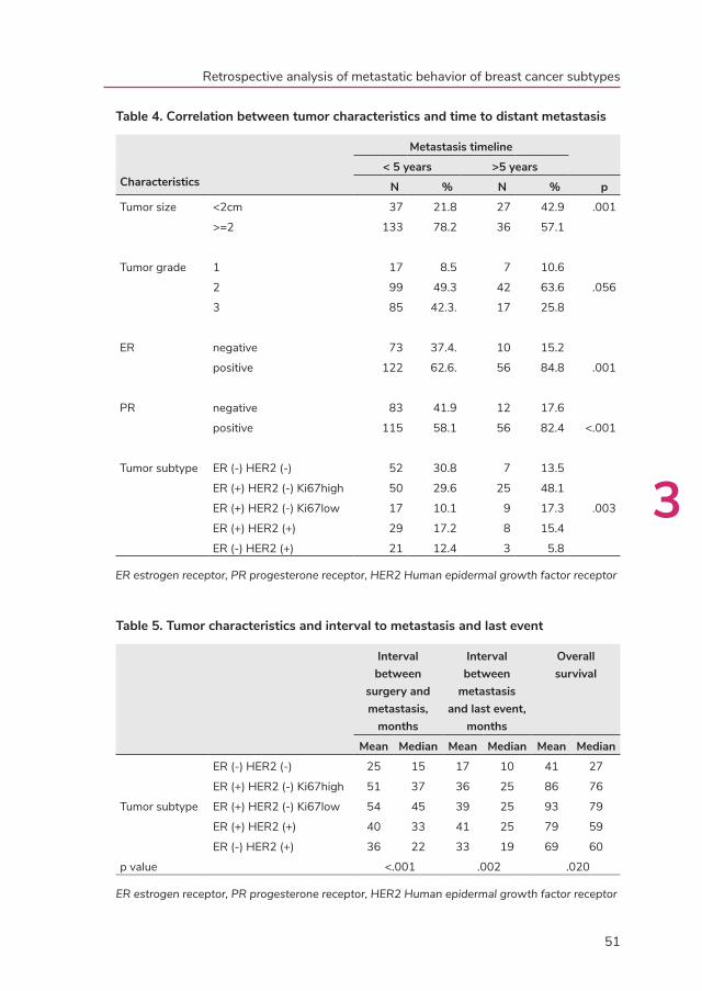
51
Retrospective analysis of metastatic behavior of breast cancer subtypes
Table 4. Correlation between tumor characteristics and time to distant metastasis
Characteristics
Metastasis timeline
< 5 years >5 years
N % N % p
Tumor size <2cm 37 21.8 27 42.9 .001
>=2 133 78.2 36 57.1
Tumor grade 1 17 8.5 7 10.6
2 99 49.3 42 63.6 .056
3 85 42.3. 17 25.8
ER negative 73 37.4. 10 15.2
positive 122 62.6. 56 84.8 .001
PR negative 83 41.9 12 17.6
positive 115 58.1 56 82.4 <.001
Tumor subtype ER (-) HER2 (-) 52 30.8 7 13.5
ER (+) HER2 (-) Ki67high 50 29.6 25 48.1
ER (+) HER2 (-) Ki67low 17 10.1 9 17.3 .003
ER (+) HER2 (+) 29 17.2 8 15.4
ER (-) HER2 (+) 21 12.4 3 5.8
ER estrogen receptor, PR progesterone receptor, HER2 Human epidermal growth factor receptor
Table 5. Tumor characteristics and interval to metastasis and last event
Interval between
surgery and metastasis,
months
Interval between
metastasis and last event,
months
Overall survival
Mean Median Mean Median Mean Median
Tumor subtype
ER (-) HER2 (-) 25 15 17 10 41 27
ER (+) HER2 (-) Ki67high 51 37 36 25 86 76
ER (+) HER2 (-) Ki67low 54 45 39 25 93 79
ER (+) HER2 (+) 40 33 41 25 79 59
ER (-) HER2 (+) 36 22 33 19 69 60
p value <.001 .002 .020
ER estrogen receptor, PR progesterone receptor, HER2 Human epidermal growth factor receptor
3

52
Chapter 3
Figure 1. Metastasis specific (A) and overall (B) survival curves of breast cancer patients with and without visceral metastasis.Kaplan-Meier plots of patients show that tumors with visceral metastasis had worse survival outcomes than the tumors without visceral metastasis.Patients who had visceral metastasis had shorter survival time from detection of metastasis to last event and from initial diagnosis of the disease to last event (p 0.009 and 0.073, respectively)
Figure 2. Metastasis specific (A) and overall (B) survival curves of breast cancer patients according to tumor subtypes. ER Estrogen receptor; PR progesterone receptor; HER2 Human epidermal growth factor receptor type 2.Kaplan-Meier plots of patients show that ER-/HER2- had worse survival outcomes compared to other tumor subtypes. Patients with hormone receptor-negative (ER-/HER2-) tumors had shorter survival time from detection of metastatic disease to last event and from the initial diagnosis of the disease to last event (p < 0.001).

53
Retrospective analysis of metastatic behavior of breast cancer subtypes
As can be seen, ER-/HER2- and ER-/HER2+ tumors metastasized earlier than other
subgroups of tumors (p 0.003). Almost 90% of hormone receptor-negative breast
cancer patients developed distant metastases early; versus 66% of ER+/HER2-; within
this group there was no significant difference between Ki67 high (66.7%) and Ki67 low
(65.4%) tumors (p 0.54).
Survival after development of distant metastasisFigure 1 shows that overall survival and survival after the detection of distant metastasis
for patients who developed visceral metastases (n=198) is worse than for those who
did not develop visceral metastases (p 0.073 and p 0.009, respectively). Figure 2 shows
overall survival and survival after the detection of distant metastasis for the subgroups
of patients defined by ER and HER2 status of the primary tumor.
Table 5 demonstrates the differences of time to develop metastasis and survival time
after development of metastatic disease in various subgroups. As can be seen, patients
with ER-/HER2- tumors had a median survival of 10 months after the detection of
distant metastasis whereas ER-/HER2+ tumors had 19 months median survival (p
0.020). ER+/HER2- (Ki67 high as well as Ki67 low groups) and ER+/HER2+ tumors had
a median survival time of 25 and 24 months, respectively (p 0.75).
Site of distant metastasisDetailed information about metastatic behavior was available for 256 patients;
11(4.3%) patients presented with multiple metastasis simultaneously, while 205
(80.4%) developed multiple metastases during the course of follow-up. Bone was the
most common site for metastasis (70.6%) followed by liver (54.5%) and lung (31.4%)
respectively. Visceral (liver, lung and brain) metastases were found in 77.6% of the
patients.
Twenty-five (9.8%) of the patients developed only bone metastasis and 29 (11.4%)
of the patients developed only visceral metastasis during the course of the disease.
Among these patients median time to develop bone metastasis and visceral metastasis
differed and was 40 months and 23 months, respectively.
3

54
Chapter 3
Multivariate analyses further revealed that patients who developed visceral metastasis
had a higher prevalence of multiple metastases during follow-up (p < 0.001).
The metastasis pattern was similar for patients who received adjuvant systemic therapy
compared to patients who did not undergo adjuvant systemic treatment.
Along with 81.3% of ER+ tumors, 88% of ER+/HER2- Ki67 high tumors noted to have
bone metastasis. Contrarily, hormone receptor-negative (ER-/HER2-) tumors were
associated with visceral organ metastasis, yet composing 55% of the tumors with only
visceral metastasis.
ER status of the tumor was significantly positively correlated to bone metastasis in
the univariate as well as in the multivariate analyses. Several immunohistochemical
markers, such as E-Cadherin and Cytokeratine 14, were found to be correlated to
visceral metastasis (p 0.013 and p 0.018). E-cadherin was found to be positive in
primary tumors of patients who developed visceral organ metastasis and the ones with
visceral metastasis as initial site of metastasis (p 0.028 and p 0.040). TP53 positive
tumors developed brain metastasis with a rate of 38%, as opposed to 21.2% in TP53
negative ones ( p 0.007).
Discussion
Breast cancer is a heterogeneous disease and this is also reflected in the clinical patterns
of the development of distant metastases. There is marked variability in the time interval
between treatment of the primary tumor and the occurrence of distant metastases; in
the organs involved with distant metastases; and in the response to systemic treatment
in patients with metastatic breast cancer. The concept of organotropism compasses
the non-random distinct organ involvement of different cancer types as well as within
a given type of cancer, which usually implies a more subtle intrinsic heterogeneity
among organotropic cancer cells [4]. Along with the conventional metastatic model of
“anatomical/mechanical” hypothesis, Paget’s “seed and soil” hypothesis [41] are widely
accepted models for site-specific metastasis. Stephen Paget’ s century old theory
that proposed the organ-preference patterns of tumor metastasis are the product of
favourable interactions between metastatic tumor cells (the “seed”) and their organ

55
Retrospective analysis of metastatic behavior of breast cancer subtypes
microenvironment (the “soil”) was confirmed by clinical and experimental research [4,
41-43]. Better understanding of this complex interaction between two compartments
and consequently the mechanisms that lie beneath the site-specific metastasis may
improve the clinical management, including developing novel therapeutic options, for
metastatic disease.
Despite the increasing tendency to classify breast tumors into molecular subtypes based
on gene expression profiles first described by Perou et al. [33], immunophenotypic
characteristics of the tumor also remain an important cornerstone of defining subgroups
of the disease. In the current study, we investigated the presence of site-specific
metastasis and concomitant characteristics of the metastatic disease in a retrospective
series of 263 breast cancer patients, focusing on the immunophenotypic features of the
primary tumor.
Together with clinical observations, recent comprehensive molecular studies unveiled
the considerable differences between ER-positive and ER-negative tumors. It has also
been shown that ER status has a time-varying prognostic effect mainly pronounced in
the early follow-up period [44-48]. ER-positive tumors are known for their tendency to
relapse later with higher rate of bone recurrences than their ER-negative counterparts.
In agreement with published literature, our data clearly indicates the close relation
between ER-positive tumors and metastasis specific survival and bone metastasis
[18, 45, 46, 49]. In addition to confirming the well-established prognostic markers in
breast cancer, this study was also able to verify that ER-status is also an important
factor for bone-only and bone-first metastasis. Likewise, in agreement with previously
published data, ER-negative tumors showed a higher proportion of patients with
visceral metastases [18, 20, 44, 45, 50]. HER2-positive tumors have been found as a
risk factor for cerebral metastasis development (5,12,13,19,22,24,27,29,32,33,52,55)
. However, in this study HER2 positivity was not identified as a strong predictive factor
for site-specific metastasis and early metastatic disease; of note, in our study we did not
find an association between HER2 positive status and brain metastasis. The fact that
almost none of the patients included in this study received HER2-targeted therapy may
play a role in the absence of a correlation between HER2 status and brain metastases
(although we could not retrieve data on adjuvant systemic therapy for all patients, we
3

56
Chapter 3
know that HER2-targeted therapy was not yet available as adjuvant therapy during
large part of the period in which patients were treated).
A recent cohort study demonstrated that hormone receptor HR+/HER2+subtype was
associated with the best prognosis after diagnosis of metastatic disease, with a median
survival of 34.4 months even better than HR+/HER2- subgroup [51]. This subgroup
was followed by HR+/HER2-, HR-/HER2+ and hormone receptor negative tumors.
In our study, regardless of their HER2 status, hormone receptor-positive tumors had
better survival after the diagnosis of distant metastasis. Similarly to Lobbezoo et al., ER-/
HER2+ tumors had better survival than hormone receptor-negative tumors. Improved
survival rates of HER2+ tumors have already been reported [28, 29, 52]. Even though
our study includes patients before the implementation of HER2-targeted therapy for
metastatic disease, our results are comparable to this recent cohort study. Additionally,
further subgrouping of ER+/HER2- tumors according to their Ki67 status revealed that
only minority (6.3%) of these tumors had low Ki67 status. Within this group of tumors
there was also no significance regarding metastasis pattern. This result may suggest
that if once metastatic event occurs, prognostic relevance of Ki67 might be limited.
Our analyses indicate the noticeable distinction between breast tumors with visceral
metastasis and the ones without visceral metastasis. Additional to the remarkably
shorter overall survival and metastasis-specific survival compared to the tumors without
visceral metastasis, higher frequency of developing multiple metastasis during the
course of disease, make this subgroup of tumors challenging and therefore worth to be
recognized [49, 53]. Several immunohistochemical markers are known to be associated
with hormone receptor-negative breast tumors especially the ones with basal-like
features. It is also claimed that triple-negative status cannot be used as a surrogate for
the basal cell phenotype [54-56]. In our study, we showed that a group of frequently
registered immunohistochemistry markers such as CK 5/6, CK 14 and EGFR were related
to the hormone receptor negative subgroup (p < 0.001), while c-kit was not found to
be related to this subgroup of tumors (p 0.098). Further analyses within this group
revealed no significant difference between basal-like group and non-basal-like group
in relation to metastatic behavior and survival outcomes. Based on gene expression
profiling studies, Lehmann et al showed that this aggressive type of breast cancer
can be divided into seven subtypes as; basal-like 1, basal-like 2, immunomodulatory,

57
Retrospective analysis of metastatic behavior of breast cancer subtypes
mesenchymal-like, mesenchymal stem-like, luminal androgen receptor and unstable.
They also showed that independent analysis of five data sets based on triple-negative
tumors identified by immunohistochemical staining had similar clustering [57]. In
conjunction with this information, it is indicated that hormone receptor-negative group
contains heterogeneous group of tumors with distinct phenotypes. We believe that
further studies are indicated to explore the role of immunohistochemistry to portray
these heterogeneous subgroups.
The role of E-cadherin in metastatic potential of the tumors has already been a topic of
interest. The absence of E-cadherin expression as a result of genetic alterations in the
E-cadherin gene is observed in the majority of lobular carcinomas. Reduced expression
of E-cadherin has been reported in breast carcinoma cases with a frequency ranging
from 4 5% to 63% of cases [58-60]. Several studies showed a higher metastatic
potential for tumors with reduced E-cadherin expression [58, 61-65], whereas others
were not able to prove such a relation [66, 67]. Interestingly, in the current study, we
showed that immunostaining of E-cadherin was positively correlated with developing
visceral metastases, also with developing visceral metastasis as first site of metastasis.
It has been shown that tumors with TP53 gene mutations are associated with brain
metastases. [27, 68, 69]. Recently, Lo et al. have demonstrated that mutation of TP53
is the most common genetic change identified in brain metastases from breast cancer.
They identified that 87% of CNS metastatic lesions in their study contained TP53
mutations compared to 25-34% mutations in all breast cancers. [70]. Consistent with
the previous reports, we showed that TP53 immunopositivity is significantly associated
with subsequent brain metastasis. The cohort of patients in our study was treated
between 1983 and 2009; the median size of the primary tumors was 3.2 cm. It may
well be that metastatic pattern will differ for patients who were treated more recntly
and for patients who presented with smaller tumors. It will be therefore be of interest
to perform a similar study to the one presented here in the future for a cohort of more
recently treated patients.
3
58
Chapter 3
Conclusions
This study demonstrates that subtypes of breast cancer mainly defined by ER, PR
and HER2 and are strongly related to the metastasis pattern, in terms of site-specific
relapse, early/late metastasis and survival outcomes. Hormone receptor-positive tumors
have tendency to develop bone metastasis and they have better survival outcomes
compared to hormone receptor-negative tumors with a tendency of developing visceral
metastasis. HER2 status was not associated with pattern of distant metastases; in
agreement with previous reports, P53-positive tumors were more likely to metastasize
to the brain than P53-negative tumors. In addition, we show that tumors that develop
visceral metastasis have worse prognosis than the ones without visceral metastasis
and immunostaining for E-cadherin and Cytokeratine 14 can be of help to identify
such tumors. These associations are of help in choices for surveillance and therapy in
individual patients.

59
Retrospective analysis of metastatic behavior of breast cancer subtypes
References 1. Malvezzi M, Bertuccio P, Levi F, La VC, Negri E: European cancer mortality predictions for
the year 2012. AnnOncol 2012, 23(4):1044-1052.
2. Lorusso G, Ruegg C: New insights into the mechanisms of organ-specific breast cancer metastasis. SeminCancer Biol 2012, 22(3):226-233.
3. St RP, Madan R, Tawfik OW, Damjanov I, Fan F: Organotropism and prognostic marker discordance in distant metastases of breast carcinoma: fact or fiction? A clinicopathologic analysis. HumPathol 2012, 43(3):398-404.
4. Lu X, Kang Y: Organotropism of breast cancer metastasis. JMammaryGlandBiolNeoplasia 2007, 12(2-3):153-162.
5. Chiang AC, Massague J: Molecular basis of metastasis. NEnglJMed 2008, 359(26):2814-2823.
6. Chambers AF, Groom AC, MacDonald IC: Dissemination and growth of cancer cells in metastatic sites. NatRevCancer 2002, 2(8):563-572.
7. Altundag K, Bondy ML, Mirza NQ, Kau SW, Broglio K, Hortobagyi GN, Rivera E: Clinicopathologic characteristics and prognostic factors in 420 metastatic breast cancer patients with central nervous system metastasis. Cancer 2007, 110(12):2640-2647.
8. Bos PD, Zhang XH, Nadal C, Shu W, Gomis RR, Nguyen DX, Minn AJ, van de Vijver MJ, Gerald WL, Foekens JA et al: Genes that mediate breast cancer metastasis to the brain. Nature 2009, 459(7249):1005-1009.
9. Harrell JC, Prat A, Parker JS, Fan C, He X, Carey L, Anders C, Ewend M, Perou CM: Genomic analysis identifies unique signatures predictive of brain, lung, and liver relapse. Breast Cancer ResTreat 2012, 132(2):523-535.
10. Landemaine T, Jackson A, Bellahcene A, Rucci N, Sin S, Abad BM, Sierra A, Boudinet A, Guinebretiere JM, Ricevuto E et al: A six-gene signature predicting breast cancer lung metastasis. Cancer Res 2008, 68(15):6092-6099.
11. Paik S, Shak S, Tang G, Kim C, Baker J, Cronin M, Baehner FL, Walker MG, Watson D, Park T et al: A multigene assay to predict recurrence of tamoxifen-treated, node-negative breast cancer. NEnglJMed 2004, 351(27):2817-2826.
12. Slimane K, Andre F, Delaloge S, Dunant A, Perez A, Grenier J, Massard C, Spielmann M: Risk factors for brain relapse in patients with metastatic breast cancer. AnnOncol 2004, 15(11):1640-1644.
13. van de Vijver MJ, He YD, van’t Veer LJ, Dai H, Hart AA, Voskuil DW, Schreiber GJ, Peterse JL, Roberts C, Marton MJ et al: A gene-expression signature as a predictor of survival in breast cancer. NEnglJMed 2002, 347(25):1999-2009.
3

60
Chapter 3
14. Wang Y, Klijn JG, Zhang Y, Sieuwerts AM, Look MP, Yang F, Talantov D, Timmermans M, Meijer-van Gelder ME, Yu J et al: Gene-expression profiles to predict distant metastasis of lymph-node-negative primary breast cancer. Lancet 2005, 365(9460):671-679.
15. Zhang XH, Wang Q, Gerald W, Hudis CA, Norton L, Smid M, Foekens JA, Massague J: Latent bone metastasis in breast cancer tied to Src-dependent survival signals. Cancer Cell 2009, 16(1):67-78.
16. Koo JS, Jung W, Jeong J: Metastatic breast cancer shows different immunohistochemical phenotype according to metastatic site. Tumori 2010, 96(3):424-432.
17. Lin Y, Yin W, Yan T, Zhou L, Di G, Wu J, Shen Z, Shao Z, Lu J: Site-specific relapse pattern of the triple negative tumors in Chinese breast cancer patients. BMCCancer 2009, 9:342.
18. Campbell FC, Blamey RW, Elston CW, Nicholson RI, Griffiths K, Haybittle JL: Oestrogen-receptor status and sites of metastasis in breast cancer. BrJCancer 1981, 44(3):456-459.
19. Dent R, Trudeau M, Pritchard KI, Hanna WM, Kahn HK, Sawka CA, Lickley LA, Rawlinson E, Sun P, Narod SA: Triple-negative breast cancer: clinical features and patterns of recurrence. ClinCancer Res 2007, 13(15 Pt 1):4429-4434.
20. Dent R, Hanna WM, Trudeau M, Rawlinson E, Sun P, Narod SA: Pattern of metastatic spread in triple-negative breast cancer. Breast Cancer ResTreat 2009, 115(2):423-428.
21. Fuqua SA: The role of estrogen receptors in breast cancer metastasis. JMammaryGlandBiolNeoplasia 2001, 6(4):407-417.
22. Haffty BG, Yang Q, Reiss M, Kearney T, Higgins SA, Weidhaas J, Harris L, Hait W, Toppmeyer D: Locoregional relapse and distant metastasis in conservatively managed triple negative early-stage breast cancer. JClinOncol 2006, 24(36):5652-5657.
23. Heitz F, Harter P, Lueck HJ, Fissler-Eckhoff A, Lorenz-Salehi F, Scheil-Bertram S, Traut A, du BA: Triple-negative and HER2-overexpressing breast cancers exhibit an elevated risk and an earlier occurrence of cerebral metastases. EurJCancer 2009, 45(16):2792-2798.
24. Kennecke H, Yerushalmi R, Woods R, Cheang MC, Voduc D, Speers CH, Nielsen TO, Gelmon K: Metastatic behavior of breast cancer subtypes. JClinOncol 2010, 28(20):3271-3277.
25. Largillier R, Ferrero JM, Doyen J, Barriere J, Namer M, Mari V, Courdi A, Hannoun-Levi JM, Ettore F, Birtwisle-Peyrottes I et al: Prognostic factors in 1,038 women with metastatic breast cancer. AnnOncol 2008, 19(12):2012-2019.
26. Lin NU, Claus E, Sohl J, Razzak AR, Arnaout A, Winer EP: Sites of distant recurrence and clinical outcomes in patients with metastatic triple-negative breast cancer: high incidence of central nervous system metastases. Cancer 2008, 113(10):2638-2645.
27. Tham YL, Sexton K, Kramer R, Hilsenbeck S, Elledge R: Primary breast cancer phenotypes associated with propensity for central nervous system metastases. Cancer 2006, 107(4):696-704.
28. Chia SK, Speers CH, D’yachkova Y, Kang A, Malfair-Taylor S, Barnett J, Coldman A, Gelmon KA, O’reilly SE, Olivotto IA: The impact of new chemotherapeutic and hormone agents on

61
Retrospective analysis of metastatic behavior of breast cancer subtypes
survival in a population-based cohort of women with metastatic breast cancer. Cancer 2007, 110(5):973-979.
29. Dawood S, Broglio K, Gonzalez-Angulo AM, Buzdar AU, Hortobagyi GN, Giordano SH: Trends in survival over the past two decades among white and black patients with newly diagnosed stage IV breast cancer. JClinOncol 2008, 26(30):4891-4898.
30. Giordano SH, Buzdar AU, Smith TL, Kau SW, Yang Y, Hortobagyi GN: Is breast cancer survival improving? Cancer 2004, 100(1):44-52.
31. Bauer KR, Brown M, Cress RD, Parise CA, Caggiano V: Descriptive analysis of estrogen receptor (ER)-negative, progesterone receptor (PR)-negative, and HER2-negative invasive breast cancer, the so-called triple-negative phenotype: a population-based study from the California cancer Registry. Cancer 2007, 109(9):1721-1728.
32. Foulkes WD, Brunet JS, Stefansson IM, Straume O, Chappuis PO, Begin LR, Hamel N, Goffin JR, Wong N, Trudel M et al: The prognostic implication of the basal-like (cyclin E high/p27 low/p53+/glomeruloid-microvascular-proliferation+) phenotype of BRCA1-related breast cancer. Cancer Res 2004, 64(3):830-835.
33. Perou CM, Sorlie T, Eisen MB, van de Rijn M, Jeffrey SS, Rees CA, Pollack JR, Ross DT, Johnsen H, Akslen LA et al: Molecular portraits of human breast tumours. Nature 2000, 406(6797):747-752.
34. Vermeulen E, Geesink I, Schmidt MK, Steegers C, Verhue D, Brom FW, Aaronson NK, van Leeuwen FE: [Secondary use of human tissue: consent and better information required]. NedTijdschrGeneeskd 2009, 153:A948.
35. Elston CW, Ellis IO: Pathological prognostic factors in breast cancer. I. The value of histological grade in breast cancer: experience from a large study with long-term follow-up. C. W. Elston & I. O. Ellis. Histopathology 1991; 19; 403-410. Histopathology 2002, 41(3A):151-152, discussion.
36. Wolff AC, Hammond ME, Schwartz JN, Hagerty KL, Allred DC, Cote RJ, Dowsett M, Fitzgibbons PL, Hanna WM, Langer A et al: American Society of Clinical Oncology/College of American Pathologists guideline recommendations for human epidermal growth factor receptor 2 testing in breast cancer. ArchPatholLab Med 2007, 131(1):18-43.
37. Cheang MC, Chia SK, Voduc D, Gao D, Leung S, Snider J, Watson M, Davies S, Bernard PS, Parker JS et al: Ki67 index, HER2 status, and prognosis of patients with luminal B breast cancer. JNatlCancer Inst 2009, 101(10):736-750.
38. Goldhirsch A, Wood WC, Coates AS, Gelber RD, Thurlimann B, Senn HJ: Strategies for subtypes--dealing with the diversity of breast cancer: highlights of the St. Gallen International Expert Consensus on the Primary Therapy of Early Breast Cancer 2011. AnnOncol 2011, 22(8):1736-1747.
39. Kim MJ, Ro JY, Ahn SH, Kim HH, Kim SB, Gong G: Clinicopathologic significance of the basal-like subtype of breast cancer: a comparison with hormone receptor and Her2/neu-overexpressing phenotypes. Human pathology 2006, 37(9):1217-1226.
3

62
Chapter 3
40. Kreike B, van Kouwenhove M, Horlings H, Weigelt B, Peterse H, Bartelink H, van de Vijver MJ: Gene expression profiling and histopathological characterization of triple-negative/basal-like breast carcinomas. Breast Cancer Res 2007, 9(5):R65.
41. Paget S: The distribution of secondary growths in cancer of the breast. 1889. Cancer Metastasis Rev 1989, 8(2):98-101.
42. Fidler IJ: Selection of successive tumour lines for metastasis. NatNew Biol 1973, 242(118):148-149.
43. Langley RR, Fidler IJ: The seed and soil hypothesis revisited--the role of tumor-stroma interactions in metastasis to different organs. IntJCancer 2011, 128(11):2527-2535.
44. Andry G, Suciu S, Pratola D, Sylvester R, Leclercq G, da Costa PM, Legros N, Andry-t’Hooft M, Verhest A, Mattheiem W et al: Relation between estrogen receptor concentration and clinical and histological factors: their relative prognostic importance after radical mastectomy for primary breast cancer. EurJCancer ClinOncol 1989, 25(2):319-329.
45. Hess KR, Pusztai L, Buzdar AU, Hortobagyi GN: Estrogen receptors and distinct patterns of breast cancer relapse. Breast Cancer ResTreat 2003, 78(1):105-118.
46. Koenders PG, Beex LV, Langens R, Kloppenborg PW, Smals AG, Benraad TJ: Steroid hormone receptor activity of primary human breast cancer and pattern of first metastasis. The Breast Cancer Study Group. Breast Cancer ResTreat 1991, 18(1):27-32.
47. Raemaekers JM, Beex LV, Koenders AJ, Pieters GF, Smals AG, Benraad TJ, Kloppenborg PW: Disease-free interval and estrogen receptor activity in tumor tissue of patients with primary breast cancer: analysis after long-term follow-up. Breast Cancer ResTreat 1985, 6(2):123-130.
48. Winstanley J, Cooke T, Murray GD, Platt-Higgins A, George WD, Holt S, Myskov M, Spedding A, Barraclough BR, Rudland PS: The long term prognostic significance of c-erbB-2 in primary breast cancer. BrJCancer 1991, 63(3):447-450.
49. Solomayer EF, Diel IJ, Meyberg GC, Gollan C, Bastert G: Metastatic breast cancer: clinical course, prognosis and therapy related to the first site of metastasis. Breast Cancer ResTreat 2000, 59(3):271-278.
50. Hicks DG, Short SM, Prescott NL, Tarr SM, Coleman KA, Yoder BJ, Crowe JP, Choueiri TK, Dawson AE, Budd GT et al: Breast cancers with brain metastases are more likely to be estrogen receptor negative, express the basal cytokeratin CK5/6, and overexpress HER2 or EGFR. AmJSurgPathol 2006, 30(9):1097-1104.
51. Lobbezoo DJ, van Kampen RJ, Voogd AC, Dercksen MW, van den Berkmortel F, Smilde TJ, van de Wouw AJ, Peters FP, van Riel JM, Peters NA et al: Prognosis of metastatic breast cancer subtypes: the hormone receptor/HER2-positive subtype is associated with the most favorable outcome. Breast cancer research and treatment 2013, 141(3):507-514.
52. Planchat E, Durando X, Abrial C, Thivat E, Mouret-Reynier MA, Ferriere JP, Pomel C, Kwiatkowski F, Chollet P, Nabholtz JM: Prognostic value of initial tumor parameters after metastatic relapse. Cancer Invest 2011, 29(9):635-643.

63
Retrospective analysis of metastatic behavior of breast cancer subtypes
53. Rosa Mendoza ES, Moreno E, Caguioa PB: Predictors of early distant metastasis in women with breast cancer. JCancer ResClinOncol 2013.
54. Badve S, Dabbs DJ, Schnitt SJ, Baehner FL, Decker T, Eusebi V, Fox SB, Ichihara S, Jacquemier J, Lakhani SR et al: Basal-like and triple-negative breast cancers: a critical review with an emphasis on the implications for pathologists and oncologists. Modern pathology : an official journal of the United States and Canadian Academy of Pathology, Inc 2011, 24(2):157-167.
55. Keam B, Im SA, Lee KH, Han SW, Oh DY, Kim JH, Lee SH, Han W, Kim DW, Kim TY et al: Ki-67 can be used for further classification of triple negative breast cancer into two subtypes with different response and prognosis. Breast Cancer Res 2011, 13(2):R22.
56. Nielsen TO, Hsu FD, Jensen K, Cheang M, Karaca G, Hu Z, Hernandez-Boussard T, Livasy C, Cowan D, Dressler L et al: Immunohistochemical and clinical characterization of the basal-like subtype of invasive breast carcinoma. Clinical cancer research : an official journal of the American Association for Cancer Research 2004, 10(16):5367-5374.
57. Lehmann BD, Bauer JA, Chen X, Sanders ME, Chakravarthy AB, Shyr Y, Pietenpol JA: Identification of human triple-negative breast cancer subtypes and preclinical models for selection of targeted therapies. The Journal of clinical investigation 2011, 121(7):2750-2767.
58. Oka H, Shiozaki H, Kobayashi K, Inoue M, Tahara H, Kobayashi T, Takatsuka Y, Matsuyoshi N, Hirano S, Takeichi M et al: Expression of E-cadherin cell adhesion molecules in human breast cancer tissues and its relationship to metastasis. Cancer Res 1993, 53(7):1696-1701.
59. Rimm DL, Sinard JH, Morrow JS: Reduced alpha-catenin and E-cadherin expression in breast cancer. Lab Invest 1995, 72(5):506-512.
60. Siitonen SM, Kononen JT, Helin HJ, Rantala IS, Holli KA, Isola JJ: Reduced E-cadherin expression is associated with invasiveness and unfavorable prognosis in breast cancer. AmJClinPathol 1996, 105(4):394-402.
61. Chen WC, Obrink B: Cell-cell contacts mediated by E-cadherin (uvomorulin) restrict invasive behavior of L-cells. JCell Biol 1991, 114(2):319-327.
62. Frixen UH, Behrens J, Sachs M, Eberle G, Voss B, Warda A, Lochner D, Birchmeier W: E-cadherin-mediated cell-cell adhesion prevents invasiveness of human carcinoma cells. JCell Biol 1991, 113(1):173-185.
63. Mareel MM, Behrens J, Birchmeier W, De Bruyne GK, Vleminckx K, Hoogewijs A, Fiers WC, Van Roy FM: Down-regulation of E-cadherin expression in Madin Darby canine kidney (MDCK) cells inside tumors of nude mice. IntJCancer 1991, 47(6):922-928.
64. Pierceall WE, Woodard AS, Morrow JS, Rimm D, Fearon ER: Frequent alterations in E-cadherin and alpha- and beta-catenin expression in human breast cancer cell lines. Oncogene 1995, 11(7):1319-1326.
65. Vleminckx K, Vakaet L, Jr., Mareel M, Fiers W, van RF: Genetic manipulation of E-cadherin expression by epithelial tumor cells reveals an invasion suppressor role. Cell 1991, 66(1):107-119.
3

64
Chapter 3
66. Lipponen P, Saarelainen E, Ji H, Aaltomaa S, Syrjanen K: Expression of E-cadherin (E-CD) as related to other prognostic factors and survival in breast cancer. JPathol 1994, 174(2):101-109.
67. Parker C, Rampaul RS, Pinder SE, Bell JA, Wencyk PM, Blamey RW, Nicholson RI, Robertson JF: E-cadherin as a prognostic indicator in primary breast cancer. BrJCancer 2001, 85(12):1958-1963.
68. Ding L, Ellis MJ, Li S, Larson DE, Chen K, Wallis JW, Harris CC, McLellan MD, Fulton RS, Fulton LL et al: Genome remodelling in a basal-like breast cancer metastasis and xenograft. Nature 2010, 464(7291):999-1005.
69. Piccirilli C, Saxena A, Robertson J, Clark W, Ikejiri B, Oldfield E, Ali I: Allelic deletions on chromosome-17 and mutations in the p53 gene in tumors metastatic to brain. IntJOncol 1994, 4(1):37-42.
70. Lo NC, Vivenza D, Monteverde M, Lattanzio L, Gojis O, Garrone O, Comino A, Merlano M, Quinlan PR, Syed N et al: High frequency of complex TP53 mutations in CNS metastases from breast cancer. BrJCancer 2012, 106(2):397-404.

65
Retrospective analysis of metastatic behavior of breast cancer subtypes
3


Breast Cancer Res Treat. 2016 Apr;156(2):249-59
C. Dilara Savci Heijink, Hans Halfwerk, Jan Koster, Marc J. van de Vijver
A novel gene expression signature for bone metastasis in breast carcinomas
Chapter 4

68
Chapter 4
Abstract
Metastatic cancer remains the leading cause of death for patients with breast cancer.
To understand the mechanisms underlying the development of distant metastases to
specific sites is therefore important and of potential clinical value. From 157 primary
breast tumors of the patients with known metastatic disease, gene expression profiling
data were generated and correlated to metastatic behavior including site-specific
metastasis, metastasis pattern and survival outcomes. We analyzed gene expression
signatures specifically associated with the development of bone metastases. As a
validation cohort, we used a published dataset of 376 breast carcinomas for which
gene expression data and site specific metastasis information were available. 80.5%
of luminal-type tumors developed bone metastasis as opposed to 41.7% of basal and
55.6% of HER2-like tumors. A novel 15-gene signature identified 82.4% of the tumors
with bone metastasis, 85.2% of the tumors which had bone metastasis as first site
of metastasis and 100% of the ones with bone metastasis only (p 9.99e-09), in the
training set. In the independent data set, 81.2% of the positive tested tumors had known
metastatic disease to the bone (p 4.28e-10). This 15-gene signature showed much
better correlation with the development of bone metastases than previously identified
signatures and was predictive in both ER- positive as well as in ER-negative tumors.
Multivariate analyses revealed that together with the molecular subtype, our 15-gene
expression signature was significantly correlated to bone metastasis status (p <0.001,
95% CI 3.86-48.02 in the training set; p 0.001, 95% CI 1.54-5.00 in the independent
set). The 15 genes, APOPEC3B, ATL2, BBS1, C6orf61, C6orf167, MMS22L, KCNS1,
MFAP3L, NIP7, NUP155, PALM2, PH-4, PGD5, SFT2D2 and STEAP3, encoded
mainly membrane-bound molecules with molecular function of protein binding. The
expression levels of the upregulated genes (NAT1, BBS1 and PH-4) were also found
to be correlated to epithelial to mesenchymal transition (EMT) status of the tumor. We
have identified a novel 15-gene expression signature associated with the development
of bone metastases in breast cancer patients. This bone metastasis signature is the first
to be identified using a supervised classification approach in a large series of patients
and will help forward research in this area towards clinical applications.

69
A novel gene expression signature for bone metastasis in breast carcinomas
Introduction
After the initial treatment of primary breast cancer, 20-30% of patients develop
distant metastases [1, 2]. The survival outcomes and sites at which distant metastases
develop differ greatly among patients [3-6]. Several studies have already reported gene
expression profiles correlated with risk of distant metastasis, which are in the process
of being validated with prospective studies [7-9]. Moreover, breast cancer’s propensity
to spread to certain organs, so called “non-random organ-specific metastasis” has also
been investigated [10-14]. There have been several important studies using animal
models to unravel the mechanism of site specific distant metastases in breast cancer
[15-22]. These studies focusing on organotropism of metastatic breast cancer have
used human breast cancer cell lines which were injected in immune compromised mice.
By combining genomic profiling of organ-tropic metastatic variants selected in vivo from
the animal models of metastatic disease with clinical genomic studies, Massague and
his colleagues, were able to identify gene expression signatures that were associated
with metastasis to bone, lung and brain [16, 17, 19]. They have further explored the
association between specific patterns of gene expression and metastatic pattern. The
discovered candidate genes were then further investigated and their metastatic role
was confirmed by means of overexpressing or inactivating their expression. Hereafter
they have validated these gene expression signatures in several cohorts of primary
breast tumors with known metastatic disease.
We have recently described the metastatic behavior (organ-specific metastasis) related
immunophenotypic findings of the primary tumors in a retrospective study including
263 primary breast tumors with known metastatic disease [23]. We have shown that
the time to distant metastasis was less than 5 year in 90% of the hormone receptor
negative breast cancer patients as compared to 66% of hormone receptor positive
patients. The role of estrogen receptor (ER) positivity was found to be closely associated
to the development of bone metastasis including bone-only and bone-first metastasis
in the course of the disease; whereas ER negativity was found to be related to visceral
(liver, lung or brain) metastasis. Along with the hormone status, tumor size and tumor
grade, we found that patients who developed visceral metastasis had worse survival
outcome, in terms of metastasis specific survival and overall survival and additionally
4

70
Chapter 4
they frequently developed multiple metastasis during the course of the disease. We
have concluded that tumor types were associated with survival and pattern of distant
metastasis during the course of the disease. Gene expression profiling patterns
predicting site-specific metastasis may aid in better understanding the mechanisms for
the development of distant metastases.
In this study, we analysed the gene expression profile of 157 primary tumors that all
metastasized. In order to identify and validate tumor factors of metastatic breast cancer
that are predictive of metastatic behavior; gene expression profiling of primary tumors
are correlated to metastasis pattern and subsequently gene expression signatures are
investigated for prediction of the site specific distinct metastasis.
Materials and Methods
Patients and Tumor samplesThe study was carried out according to the national ethical guidelines of ‘Code for
Proper Secondary Use of Human Tissue’ developed by Federation of Medical Societies
(FMWV) in the Netherlands [24]. Selection of patients and tumor samples and definition
of metastatic disease has been previously described [23].
Briefly, metastatic breast cancer patients were identified and a subset of 157 of tumors
with available frozen material was selected for further gene expression profiling
experiments. Subsequently relevant clinical data were abstracted from the clinical
charts. Detailed information on metastasis site, metastatic behavior and survival
(metastasis specific and overall) data were collected for 151 patients.
The individual metastatic sites were recorded and used to separate tumors in subgroups.
For each metastatic site it was assessed whether patients developed metastases during
follow-up (ever versus never for each organ site); when patients developed metastases
to any organ site it was recorded whether this was the first metastasis or a metastasis
arising after metastases to other organ site arose (first/not first); and it was recorded
when a patient developed metastases to one organ site only (only/not only). Presence

71
A novel gene expression signature for bone metastasis in breast carcinomas
of multiple metastases was also carefully recorded at the time of diagnosis of the first
metastases as well as after complete follow up. In instances where patients developed
another distal organ involvement within less than two months after initial diagnosis of a
metastasis, this was also considered as multiple organ metastases at first presentation.
Time from surgery to development of first metastasis, time from first metastasis to last
event (metastasis specific survival, MSS) and overall survival (OS) time for each patient
was calculated. Last event date was recorded as most recent follow-up date for the
patients who were alive and time of death for the others.
Additionally, data on systemic treatment (chemotherapy, hormonal therapy and targeted
therapy) used to treat primary and metastatic disease was collected (n=142 and n=122,
respectively).
Hemotoxylin and eosin stained slides from all tumors were evaluated and histologic
subtype, tumor grade and hormone receptor status were immunohistochemically
assessed as previously described [23].
Gene expression profilingFrom primary tumors of 157 patients 30 sections of 20-μm were used for isolation of
RNA. The first and last sections (5 μm) were stained with hematoxylin and eosin; only
samples containing an average of at least 50% tumor cells were used in this analysis.
Total RNA was isolated with RNA-Bee (Tel-Test) and dissolved in RNase-free water.
Then, total RNA was treated with DNase with use of the Qiagen RNase-Free DNase
Kit and RNeasy spin columns and dissolved in RNase-free water. The gene expression
microarrays used in this study were the HumanHT-12 v4 Expression BeadChip arrays
(Illumina, Inc.) containing more than 47,000 probes. Details of RNA amplification,
labeling, and hybridization are available on the Illumina website (http://www.illumina.
com). The arrays were processed in the Central Microarray Facility of the Netherlands
Cancer Institute. The data was normalized using robust spline normalization (rsn) and
log2 transformed, followed by ComBat (http://www.bu.edu/jlab/wp-assets/ComBat/
Abstract.html) to adjust for batch effects.
4

72
Chapter 4
Human breast tumor microarray datasetsAdditional to the gene expression profiling data set of 157 primary breast tumors
generated herein with this study, a second data set which was a combined data set
of four studies captured from the public domain was analysed. This large data set
was employed as a combined data set as previously described by Harrell et al [25].
Aforementioned combined data set included total 855 tumors of which 376 with known
metastatic disease. Analyses in order to validate the gene signatures were performed
using this subset of tumors with known metastatic disease (n= 376) and whole data
set (n= 855). Four microarray sets are listed as: GSE2034, GSE12276, GSE2603 and
the NKI295 (microarray-pubs.stanford.edu/wound_NKI/Clinical_Data_Supplement.xls).
Microarray data analysis/ bioinformaticsAll data were analyzed using the R2 (Microarray Analysis and Visualization Platform)
web application, which is publicly available at http://r2.amc.nl.
Samples were classified into 5 intrinsic breast cancer subtypes by using the PAM50
classifier [26]. To assess the prognostic status of the tumors according to the 70-
gene prognostic signature [9], these 70 genes were mapped via Gene Symbol ID to
the Illumina platform. Out of the 70 genes 62 genes were found to be present on the
Illumina platform corresponding to 65 Illumina probes. In case of presence of multiple
probes for one gene, the probe with the highest variance across the samples was
selected. We calculated the Pearson correlation coefficient between the centroids of the
original good prognosis template and the gene expression levels of each sample with
regard to these mapped 62 gene to assign tumors in good or poor prognostic group.
To validate already published gene expression signatures for site specific metastasis for
bone metastasis [17] and epithelial to mesenchymal transition (EMT) gene signature
[27], the specified genes from bone metastasis signature and from EMT signature were
first mapped to the Illumina platform via Gene Symbol ID. As previously mentioned, in
the instance of presence of multiple probes for one gene, the probe with the highest
variance across the samples were selected. Respectively, a K-means method was used
to cluster the data in 2 groups and a t-test revealed the performance of these signatures
(bone metastasis and EMT signature) in our dataset.

73
A novel gene expression signature for bone metastasis in breast carcinomas
Identification and validation of site-specific metastasis signatureIn order to identify a gene expression signature associated an organ specific metastasis,
we used the one-way ANOVA function in R2. Samples were split into 2 groups; one
group in which the patient developed a metastasis and another group in which a patient
never developed a certain organ metastasis. The genes with an expression level above
the background expression level were selected (total 16036 genes. The calculated
p-values (<0.01) were corrected for multiple testing using Benjamini Hochberg False
Discovery Rate (FDR) calculation. The metastatic signature was validated in multiple
datasets using the K-means and t-test function in R2. To further investigate the
association between this gene signature and clinical variables multivariate logistic
regression tests were applied using SPSS Statistics for Windows (Release version
21.0; IBM Corp.2012, Armond, NY). All statistical tests were two sided and p < 0.05
was considered to be statistically significant.
Results
For 157 primary invasive breast carcinomas from patients who all developed metastatic
disease, mRNA expression signatures were assessed using microarray analysis. The
patient characteristics and metastasis patterns are described in Table 1. Tumors were
subdivided into 5 molecular subtypes using the PAM50 classifier [26]. Out of 157
cases, 67 (42.7%) were identified as Luminal A, 46 (29.3%) as Luminal B, 18 (11.5%)
as HER2-like and 25 (15.9%) as basal type. One (0.6%) of these tumors was identified
as normal-like. For statistical purposes, the normal like breast tumor was excluded from
the multivariate analysis. Median follow-up time for patients who were alive was 11.5
years (range 6.2-17.3 years). 79.4% of the patients with Luminal A, 72.5% of Luminal
B, 78.6% of HER2-like and 87.5% of basal-type tumors received adjuvant therapy.
None of the patients received trastuzumab as adjuvant therapy; a subgroup of patients
(n=10) received trastuzumab for treatment of metastatic disease.
Bone was the most frequent site of distant metastasis (71.5%) followed by liver (51.7%)
and lung (34.4%). 74.2% of the patients developed visceral organ metastasis (lung,
liver or brain).
4

74
Chapter 4
Table 1. Clinical and pathological characteristics of metastatic breast cancer pa-tients
N %
Age at diagnosis, years <50 83 52.9
>50 74 47.1
Surgical procedure none 4 2.8
mastectomy 73 51.8
breast conserving 64 45.4
Adjuvant therapy none 30 21.1
only CT 50 35.2
only HT 17 12.0
CT+HT 45 31.7
Lymph node status none 43 29.3
1-3 positive 48 32.7
>3 positive 56 38.1
Histology Ductal 134 86.5
Lobular 14 9.0
Other 7 4.5
Tumor grade 1 13 8.6
2 84 55.3
3 55 36.2
Time to distant metastasisa early 117 77.0
late 35 23.0
Metastasis at first presentation
no 141 92.8
yes 11 7.2
Multiple metastasis sites at first presentation
no 97 64.2
yes 54 35.8
Multiple metastasis sites during follow up
no 37 24.5
yes 114 75.5
CT chemotherapy, HT hormonal therapy.a Cut-off point 5 years.

75
A novel gene expression signature for bone metastasis in breast carcinomas
Survival analysis revealed that luminal-type tumors had better outcomes in terms of
metastasis specific and overall survival compared to basal-type tumors and HER2-like
tumors (p < 0.001). Median time to develop metastasis was 37, 27, 19 and 15 months
for Luminal A, Luminal B, HER2-like tumors and basal-type tumors, respectively. 88.3%
of basal-type and HER2-like tumors developed metastases within 5 years, versus
72.7% of luminal A and 76.7% of Luminal B tumors.
Among luminal subtype 80.5% of the tumors developed bone metastasis as opposed to,
respectively, 41.7 and 55.6% of basal-type and HER2-like tumors (p 0.001). This group
of tumors also composed the 81.8% of the tumors which metastasized to bone as initial
site of metastasis (p 0.001). The rate of development of visceral metastasis were 70.4%
in luminal-type tumors, 87.5% in basal-type tumors and 77.8% in HER2-like tumors.
Of basal-type tumors 66.7% developed visceral metastasis as first metastasis site and
29.2% of these tumors had only visceral site metastasis during the course of disease (p
0.061 and p 0.034).
The tumor samples from all patients were assigned to the poor prognostic group
according to the 70-gene signature [9]. Based on recently published epithelial
mesenchymal transition (EMT) gene classifiers [27], 100 of the tumors allocated as
EMT-activated and the rest, n=51, as EMT-non activated.
Validation of a previously identified gene signature for bone-specific metastasisFirst we have studied the predictive value of the previously published bone metastasis
signature of Kang et al [17]. This signature was assessed as positive in 110 of the
tumors in the current study set. All (100%) Luminal A tumors and 90,7% of the Luminal
B tumors were found be positive for the signature, whereas 33% of the HER2-like
tumors were positive. None of the basal type tumors were found be positive for this
site specific metastasis signature. Within this site-specific signature positive subgroup
of tumors, 80% had clinically identified bone metastasis (n=88, p 4.26e-04). Kang et
al’ s 102-gene expression signature for bone metastasis was able to identify 81.5%
of the tumors with bone metastasis, 84.1% of the tumors which had bone as initial
site of metastasis and 100% (n=18) of the tumors which had bone only metastasis in
the training set (p values <0.001, <0.001 and 0.002, respectively. Sensitivity: 81.5%
4

76
Chapter 4
and specificity: 48.8%). When tested in ER-positive (n=108) and ER-negative (n=43)
groups separately, 61.1% (n=66) of the ER-positive tumors and 60.4% (n=26) of the
ER-negative tumors were tested to be positive with this 102-gene expression signature.
Out of positively tested ER-positive tumors (n=66) 83.3% had clinically evident bone
metastasis (p 0.456). Of the 26 bone signature positive tested ER-negative tumors,
50% had bone metastatic disease (p 1.000).
Supervised classification of bone (specific) metastasis-related genesTo identify site-specific metastasis genes, differentially expressed genes between
tumors with bone metastasis and the ones without bone metastasis were explored.
A t test was conducted with a p value of <0.01. After application of filtering criteria,
differentially expressed genes were identified between two subgroups of tumors
with and without bone metastasis. The group of differentially expressed genes were
subsequently validated in the training data set as well as in the independent data set
with the help of K-means and t testing.
We identified 15 differentially expressed genes between tumors with bone metastasis
and the ones without bone metastasis (Table 2). The heat map with gene expression
pattern of these 15 genes is displayed in Figure 1. None of the genes in this set overlapped
with the bone signature of Kang et al. Three genes, namely NAT1, PH-4 and BBS1, were
up-regulated and the other genes were found to be down-regulated. Mapping into the
Gene Ontology and Kyoto Encyclopaedia of Genes and Genomes databases showed an
overrepresentation of membrane-bound molecules with molecular function of protein
binding (APOPEC3B, ATL2, BBS1, MMS22L, KCNS1, MFAP3L, NIP7, NUP155, PALM2,
PH-4 and STEAP3).
In order to validate this gene expression signature, conjointly with our training set, an
independent large combined microarray data set of four studies was analysed. This
combined previously published by Harrell et al [25]. With the help of K-means clustering
method, we have grouped our training data set and independent data set into two
groups based on their expression levels for our newly developed bone metastasis gene
expression signature and subsequently these two groups were compared using a t test.

77
A novel gene expression signature for bone metastasis in breast carcinomas
Figu
re 1
. The
gen
e ex
pres
sion
pat
tern
of 1
5 ge
nes
of b
one
met
asta
sis
gene
sig
natu
re.
Hea
t map
sho
ws
the
gene
exp
ress
ion
patt
ern
of 1
5-ge
nes
amon
g 15
1 pa
tient
s. P
rimar
y tu
mor
s w
ith c
linic
ally
evi
dent
bon
e m
etas
tasi
s ar
e ill
ustr
ated
in
blue
and
the
ones
with
out b
one
met
asta
sis
are
in y
ello
w .
For e
ach
prim
ary
tum
or, t
he e
xpre
ssio
n le
vel o
f the
spe
cific
gen
e is
exh
ibite
d as
red,
if u
p-re
gu-
late
d an
d gr
een,
if d
own-
regu
late
d. M
olec
ular
sub
type
s of
prim
ary
tum
ors
are
also
dem
onst
rate
d as
; dar
k bl
ue fo
r lum
inal
A, l
ight
blu
e fo
r lum
inal
B, y
ello
w
for H
ER2-
like,
pin
k fo
r bas
al ty
pe a
nd o
rang
e fo
r nor
mal
-like
tum
ors.
4

78
Chapter 4Ta
ble
2. T
he li
st o
f diff
eren
tially
exp
ress
ed g
enes
in b
one
met
asta
tic d
isea
se
Acc
essi
on
Num
ber
HU
GO
Des
crip
tion
R-v
alue
p-va
lue
Leve
l of
expr
essi
on*
1N
M_0
0490
0A
POB
EC3B
Apo
lipop
rote
in B
mR
NA
edi
ting
enzy
me,
cat
alyt
ic p
olyp
eptid
e-lik
e 3B
(A
POB
EC3B
), m
RN
A.
-0,4
073,
55e-
03<
2N
M_1
5348
5N
UP1
55N
ucle
opor
in 1
55kD
a (N
UP1
55),
tran
scrip
t var
iant
1, m
RN
A.
-0,3
858,
43e-
03<
3N
M_0
2164
7M
FAP3
LM
icro
fibril
lar-
asso
ciat
ed p
rote
in 3
-like
(MFA
P3L)
, tra
nscr
ipt v
aria
nt 1
, mR
NA
.-0
,382
6,77
e-03
<
4N
M_0
1610
1N
IP7
Nuc
lear
impo
rt 7
hom
olog
(S. c
erev
isia
e) (N
IP7)
, mR
NA
.-0
,375
8,67
e-03
<
5N
M_1
9846
8C
6orf
167
Chr
omos
ome
6 op
en re
adin
g fr
ame
167
(C6o
rf16
7), m
RN
A.
-0,3
717,
22e-
03<
6N
M_0
0225
1KC
NS1
Pota
ssiu
m v
olta
ge-g
ated
cha
nnel
, del
ayed
-rec
tifier
, sub
fam
ily S
, mem
ber 1
(K
CN
S1),
mR
NA
.-0
,368
7,41
e-03
<
7N
M_0
0125
8311
PGB
D5
Pigg
yBac
tran
spos
able
ele
men
t der
ived
5 (P
GB
D5)
, mR
NA
.-0
,364
7,88
e-03
<
8N
M_1
8291
5ST
EAP3
STEA
P fa
mily
mem
ber 3
(STE
AP3
), tr
ansc
ript v
aria
nt 1
, mR
NA
.-0
,364
8,77
e-03
<
9N
M_0
2018
8C
16or
f61
Chr
omos
ome
16 o
pen
read
ing
fram
e 61
(C16
orf6
1), m
RN
A.
-0,3
579,
84e-
03<
10N
M_0
5301
6PA
LM2
Para
lem
min
2 (P
ALM
2), t
rans
crip
t var
iant
2, m
RN
A.
-0,3
569,
02e-
03<
11N
M_0
2237
4AT
L2A
tlast
in G
TPas
e 2
(ATL
2), m
RN
A.
-0,3
549,
68e-
03<
12N
M_1
9934
4SF
T2D
2SF
T2 d
omai
n co
ntai
ning
2 (S
FT2D
2), m
RN
A.
-0,3
539,
66e-
03<
13N
M_0
0116
0170
NAT
1N
-ace
tyltr
ansf
eras
e 1
(ary
lam
ine
N-a
cety
ltran
sfer
ase)
(NAT
1), m
RN
A.
0,35
29,
24e-
03>
14N
M_1
7793
8PH
-4H
ypox
ia-in
duci
ble
fact
or p
roly
l 4-h
ydro
xyla
se (P
H-4
), tr
ansc
ript v
aria
nt 2
, m
RN
A.
0,35
79,
20e-
03>
15N
M_0
2464
9B
BS1
Bar
det-
Bie
dl s
yndr
ome
1 (B
BS1
), m
RN
A.
0,37
28,
29e-
03>
* >
up-
regu
late
d, <
dow
n-re
gula
ted
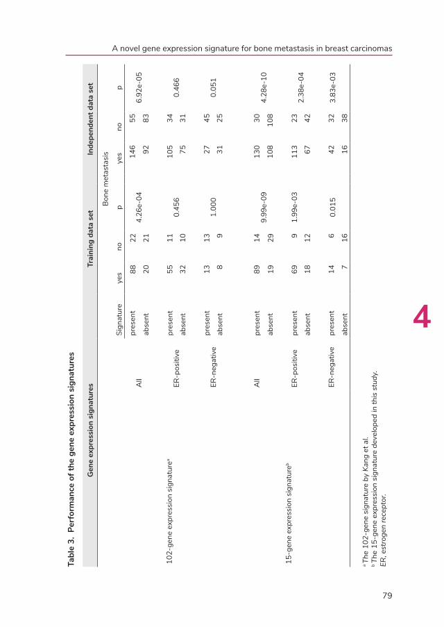
79
A novel gene expression signature for bone metastasis in breast carcinomas
Tabl
e 3.
Per
form
ance
of t
he g
ene
expr
essi
on s
igna
ture
s
Gen
e ex
pres
sion
sig
natu
res
Trai
ning
dat
a se
tIn
depe
nden
t dat
a se
t
Sign
atur
e
Bon
e m
etas
tasi
s
yes
nop
ye
sno
p
102-
gene
exp
ress
ion
sign
atur
ea
All
pres
ent
8822
4.26
e-04
146
556.
92e-
05ab
sent
2021
9283
ER-p
ositi
vepr
esen
t55
110.
456
105
340.
466
abse
nt32
1075
31
ER-n
egat
ive
pres
ent
1313
1.00
027
450.
051
abse
nt8
931
25
15-g
ene
expr
essi
on s
igna
ture
b
All
pres
ent
8914
9.99
e-09
130
304.
28e-
10ab
sent
1929
108
108
ER-p
ositi
vepr
esen
t69
91.
99e-
0311
323
2.38
e-04
abse
nt18
1267
42
ER-n
egat
ive
pres
ent
146
0.01
542
323.
83e-
03
ab
sent
716
16
38
a Th
e 10
2-ge
ne s
igna
ture
by
Kang
et a
l.b Th
e 15
-gen
e ex
pres
sion
sig
natu
re d
evel
oped
in th
is s
tudy
.ER
, est
roge
n re
cept
or.
4

80
Chapter 4
The 15-gene bone metastasis gene signature was found to be present in 103 tumors in
the training data set. With the help of this signature, 82.4% of the tumors with known
metastatic disease, 85.2% of the tumors which had bone metastasis as first metastasis
site and 100% of the ones with bone metastasis only were identified (p 9.99e-09,
sensitivity: 82.4% and specificity: 67.4%). When analysed in the independent data set,
the 15-gene expression signature was found to be present in 160 tumors (total n=376)
and 81.2% of these positive tested tumors had also clinically evident bone metastatic
disease (p 4.28e-10, sensitivity: 54.6% and specificity: 78.2%). The independent data
base of Harrell et al was also utilized to test the bone-specific metastasis of Kang et
al. The 102-gene expression signature was assessed as present in 201 tumors (total
n = 376) and 72.6% of these tumors reported to have bone metastasis (p 6.92e-05,
sensitivity: 61.3% and specificity: 60.1%).
In addition, the independent data set was analysed separately in ER-positive and
ER-negative tumors. Among ER-positive tumors (n = 245) the 15-gene expression
signature was found to be present in 136 tumors and 83.1% of these tumors had
known bone metastasis; 38.5% of the negatively tested tumors had no bone metastasis
(p 2.38e-04, sensitivity: 79.3% and specificity: 57.1%). Out of 139 ER-positive tumors
which were tested to be positive for the 102-gene expression signature, 75.5% had
bone metastatic disease and 29.2% of the negatively tested tumors had no bone
metastasis (p 0.466, sensitivity: 63.2% and specificity: 47.6%). Within the ER-negative
subgroup (n=128) 74 tumors were tested positive for the 15-gene expression signature
and 56.8% these tumors had bone metastasis; 70.4% of negatively tested tumors had
no evidence of bone metastasis (p 3.83e-03, sensitivity: 72.4% and specificity: 56.8%).
Out of 56 ER-negative tumors which were tested positive for 102-gene expression
signature, 55.4% had clinically bone metastasis; 62.5% of negatively tested tumors
had no bone metastasis (p 0.05, sensitivity: 53.5% and specificity: 64.3%). Table 3
summarizes the validation of gene signatures in training and independent data sets.
In addition, in a subsequent study a subset of 50-genes (out of initially identified 102
genes) was selected by Massague’s group [20]; this subset of 50 genes was also
analysed in our training and in the independent data sets for its predictive value for
bone-specific metastasis The 50-gene signature was able to identify the patients with
bone metastasis in the training set (p 1.14e-03) and the independent data set (p 0.014).

81
A novel gene expression signature for bone metastasis in breast carcinomas
When tested in the ER-positive and the ER-negative tumors separately, this 50-gene
signature was not predictive for bone metastatic disease.
When tested among all patients with metastatic and not-metastatic disease in
the independent data set (n=855), the 15-gene signature was able to identify the
patients with bone metastasis (p 5.48e04, sensitivity: 54.6% and specificity: 58.7%).
This gene expression signature remained statistically significant for identification of
bone metastasis when separately analysed in ER-positive and ER-negative tumors
(p 3.45e-04, sensitivity: 63.9% and specificity: 52.2%; p 3.82e-03, sensitivity: 75.9%
and specificity: 45.5%, respectively).
The up-regulated genes and their correlation with molecular subtypes and known
prognostic gene signatures were further explored. NAT1 was identified to be expressed
at the highest levels in Luminal A followed by Luminal B, HER2-like group and being
least expressed in the basal-type group. NAT1 expression was also correlated with the
EMT-activated group, being overexpressed in this group of tumors compared to the
not-EMT-activated group (p 5.7e-05) (Figure 2). Similarly the other up-regulated genes,
Figure 2. The expression levels ( log2) of NAT1 among molecular subtypes (A) and in EMT-activated and not-EMT-activated group (B).The box plots show that NAT1 expression was higher in Luminal-type tumors compared to the other molecular subtypes (p 7.2e-20). NAT1 expression was also found to be higher in the EMT (epithelial to mesenchymal transition)-activated group (p 5.7e-05)
4

82
Chapter 4
BBS1 and PH-4, were also found to be significantly correlated with the EMT-activated
group of tumors (p 5.8e-04 and p 0.01, respectively).
The 15-gene bone metastasis signature was positive in 96.9% of the Luminal A tumors,
in 79.7% of Luminal B tumors and in 38.9% of HER2-like tumors. Similar to Kang’s
bone metastasis signature, none of the basal like tumors were found to be positive for
this signature.
Univariate analyses showed that our bone metastasis signature was significantly
correlated to the development of bone metastasis especially in the group of patients who
developed only bone metastasis in the course of their disease (p < 0.001). As expected,
ER status and molecular subtypes were the parameters that were closely related to
bone metastasis status (p < 0.001). Subsequently, multivariate analyses were applied
in order to further explore the link between our gene signature and these parameters.
Table 4 displays the multivariate analyses results for ER status, molecular subtypes and
two separate gene data sets (training and independent) for bone specific metastasis. As
shown, the 15-gene signature was the only parameter that was significantly correlated
to bone metastasis status in the training data set (p <0.001, 95% CI 3.86-48.02). In the
independent data set, together with the molecular subtype, the 15-gene signature was
significantly correlated to bone metastasis status (p 0.001, 95% CI 1.54-5.00).
Table 4. Multivariate analyses results of predictive factors among the gene datasetsTraining data set
B Wald x2 p Odds ratio 95% CI
ER status -0.48 0.53 .468 0.620 0.17 – 2.25
Molecular subtype 0.53 0.07 .793 1.05 0.71 - 1.57
15-gene signaturea 2.61 16.49 < .000 13.61 3.86 – 48.03
Independent data set
B Wald x2 p Odds ratio 95% C.I.
ER status 0.25 0.06 .939 1.02 0.54 - 1.96
Molecular subtype 0.30 10.70 .001 1.36 1.13 - 1.64
15-gene signaturea 2.62 11.54 .001 2.78 1.54 – 5.00a Novel gene expression signatureER estrogen receptor, CI confidence interval

83
A novel gene expression signature for bone metastasis in breast carcinomas
Discussion
The metastatic potential of the primary tumor revolves around multistep biological
processes within host tissue and microenvironment of the distant organ site [28]. In
addition to the early origin of genetic instability [28-30] and hence the metastatic
potential of the tumor cells, several intrinsic and extrinsic factors are recognised as
potential promoters of metastatic relapse [31-33]. Upon sustaining the elementary
steps of dissemination, the circulating tumor cells can colonize a new organ, forming a
detectable metastasis [11, 28].
Experimental models of metastasis yielded distinct sets of genes that mediated site-
specific metastasis in breast cancer [16-19]. Kang et al. identified a bone metastasis
signature composed of 102 genes mostly encoding cell surface and secretory
proteins, with functions including bone marrow homing and extravasation, pericellular
proteolysis and invasion, angiogenesis, osteoclastogenesis, growth factor regulation
and extracellular matrix alteration [17]. The authors concluded that this gene set was
superimposed on a poor-prognosis gene signature to provide additional functions in
order to achieve an overt bone-specific metastasis.
Despite these interesting findings from mouse model system and validation of the
results from the mouse models in human breast cancer, no clinical application or follow-
up research has emerged since these first findings. Here we present results of the
largest study to date on the association between gene expression profiling of primary
breast cancer and the development of bone metastases, and the first study in which
supervised classification has been used to identify a bone metastasis associated gene
expression signature. This gene expression signature was composed of 15 genes, with
3 (NAT1, PH-4 and BBS1) of them being up-regulated in the primary tumor samples.
The overexpressed genes in this bone-specific metastasis signature were associated
with metabolic (NAT1) and oxidation-reduction (PH-4) processes, and protein transport
(BBS-1), in agreement with previous works hypothesizing their potential role in altering
the host tissue environment in order to achieve a bone metastasis [31, 33-35].
4

84
Chapter 4
N-acetyltransferase 1 (NAT1) was first reported to be associated with enhanced
growth and survival of breast epithelial cells by Adam et al. [36], and later reported to
be a potential biomarker for breast cancer [37-41]. In several studies, inhibiting NAT1
resulted in cell morphology change, a loss of surface filopodia and subsequent reduction
of invasive potential both in vitro and in vivo [40]. Likewise, knockdown of this gene
led to inhibition of invasion and metastasis, by means of modification/rearrangement
of filopodial (intracellular) actin [41, 42]. In agreement with other gene expression
profiling studies in human cancer samples, here we showed that NAT1 clusters close
to the estrogen receptor with higher expression levels in luminal-type tumors [36, 43,
44]. Tiang et al. also showed that the loss of NAT1 resulted in alteration of cell-to-
cell contact and up-regulation of E-cadherin. Based on aforementioned cell-line studies
a possible association between this gene and EMT/MET has been speculated [42].
Interestingly, in our data set overexpression of this gene was significantly correlated to
the so-called EMT-activated group (p 5.7e-05). To our knowledge, this is the first study
pointing to the association between NAT1 and EMT in human female breast cancer
samples. Along with the considerations of the potentiality of this gene as a drug target
[42, 45], we believe that further studies in human breast cancer samples are indicated
to explore this link.
The extracellular matrix (ECM) plays important role in diverse pathological and
physiological processes, including cancer invasion and metastasis [46, 47]. Collagens
compose the major component of ECM. Increased expression of collagens, thereupon
increase in deposition and stiffening in ECM, is associated with tumor progression [48,
49]. Collagen prolyl 4-hydroxylase (PH-4), a member of post-transcription modification
enzyme family, is required in collagen biosynthesis and angiogenesis. Hypoxia-induced
collagen prolyl 4-hydroxylase expression is reported to be associated with increased
progression and mortality in breast cancer [49-51]. Indeed, animal studies showed that
knockdown of PH-4 resulted in inhibition of tumor growth and lung metastasis [52, 53].
With gene expression profiling of breast cancer samples we have found that PH-4 was
positively correlated with site-specific metastasis to bone. This finding confirms the
observations by others [46-49] and advocates for the importance of extracellular matrix
alterations in disease progression.

85
A novel gene expression signature for bone metastasis in breast carcinomas
Twelve out of 15 genes were found to be down-regulated in the primary tumors of breast
cancer patients who developed bone metastasis. One of these genes, apolipoprotein B
mRNA editing enzyme, catalytic polypeptide-like-3B (APOBEC3B), is reported to be
up-regulated in a large proportion of breast tumors and high levels of APOBEC3B were
found to be associated with worse disease-free and overall survival [54-57]. Recently,
several independent genome-wide association studies have shown a deletion resulting
in complete elimination of the APOBEC3B gene-encoding region [58-60]. This
deletion has been indicated to be associated with decreased expression of APOBEC3B
in breast cancer cells [58]. In this study, we have also shown that APOBEC3B was
significantly down-regulated in the group of tumors with bone metastatic disease
(p 3.55e-03). We believe that further copy number variations studies are required to
explore such an association between APOBEC3B deletion and site-specific metastasis.
Six-transmembrane epithelial antigen of prostate 3 (STEAP3), which is thought be
involved in apoptosis and cell-cycle progression [61-63], is also found to be down-
regulated in the bone metastatic group of primary breast tumors in our study. STEAP3
expression is shown to be diminished in hepatocellular carcinoma nodules compared
to cirrhotic peritumoral tissue and healthy liver [64]. Another family member of these
proteins, STEAP1, has already shown to be overexpressed in breast cancer cells [65-
67]. However, we could not retrieve any similar data pointing STEAP3 expression levels
in breast cancer tissues.
In order to determine the validity of the experimentally derived 102 gene bone
metastasis signature, Kang et al. have utilized a cohort of 63 primary breast carcinomas
to test this signature. The authors have selected a subset of 50 genes to carry on their
validation studies and they have shown that this gene set was not able to identify the
group of tumors with bone metastasis. When the authors restricted their analyses to
25 breast tumors with known metastatic disease, they were able distinguish the tumors
preferentialy metastasized to bone rather than other distant organs [20]. In this current
study along with new identified 15-gene expression signature, we have shown that
the 102-gene expression signature and the subset of 50 genes as reported by Kang
et al. were informative in identifying likelihood of developing bone metastasis in the
training and the independent data sets. However, when datasets subdivided into 2
groups according to their ER-status, the 102-gene expression signature as well as the
50-gene signature were not effective in predicting bone metastasis, whereas herein
4

86
Chapter 4
identified 15-gene expression signature remained associated with the likelihood of
bone metastasis development in ER-positive and ER-negative tumor groups.
Notably, the bone-specific metastasis signature presented in this study did not include
any of the genes from already published Kang’ s bone signature [17]. The absence
of overlap between these gene sets could be justified with the fact that in the former
study tumor cells from the metastasis site were utilized to generate gene signatures in
contrast to primary tumors in the current study. Considering that tumor progression and
development of metastasis requires compiled steps of modification, we may assume
that these two different gene signature sets play a complementary role in separate
levels of this multi-complex process.
Notwithstanding several well received studies focusing on the biology of metastasic
breast cancer, little progress has been made over the past years to identify a robust
gene expression signature for site-specific metastasis. Moreover, the experimentally
derived gene expression signatures when tested in human breast carcinomas were not
as strongly associated with site-specific metastasis as in the experimental conditions.
A reproducible gene expression signature associated with the development of bone
metastases in breast cancer will have clinical utility in two ways: first, the knowledge of
the specific gene expressed at higher or lower levels in the metastatic disease will lead to
the investigation of targeted therapy options directed to the altered mechanism related
to this gene and second, reliable identification of the patients at high risk of developing
bone metastases may lead to therapeutic interventions specifically aimed at preventing
the development of bone metastases, for example treatment with bisphosphonates.
In summary, we present the largest study to date revealing the association between the
gene expression profiling patterns and bone specific metastasis in breast carcinomas.
The identification of novel 15-gene expression signature will forward this area of
research, including subsequent exploration of the underlying mechanisms of metastatic
behavior and ultimately help improve outcome for breast cancer patients.
87
A novel gene expression signature for bone metastasis in breast carcinomas
References 1. Early Breast Cancer Trialists’ Collaborative G: Effects of chemotherapy and hormonal therapy
for early breast cancer on recurrence and 15-year survival: an overview of the randomised trials. Lancet 2005, 365(9472):1687-1717.
2. O’Shaughnessy J: Extending survival with chemotherapy in metastatic breast cancer. Oncologist 2005, 10 Suppl 3:20-29.
3. Giordano SH, Buzdar AU, Smith TL, Kau SW, Yang Y, Hortobagyi GN: Is breast cancer survival improving? Cancer 2004, 100(1):44-52.
4. Greenberg PA, Hortobagyi GN, Smith TL, Ziegler LD, Frye DK, Buzdar AU: Long-term follow-up of patients with complete remission following combination chemotherapy for metastatic breast cancer. J Clin Oncol 1996, 14(8):2197-2205.
5. Hayes DF, Van Zyl JA, Hacking A, Goedhals L, Bezwoda WR, Mailliard JA, Jones SE, Vogel CL, Berris RF, Shemano I et al: Randomized comparison of tamoxifen and two separate doses of toremifene in postmenopausal patients with metastatic breast cancer. J Clin Oncol 1995, 13(10):2556-2566.
6. Sledge GW, Jr., Hu P, Falkson G, Tormey D, Abeloff M: Comparison of chemotherapy with chemohormonal therapy as first-line therapy for metastatic, hormone-sensitive breast cancer: An Eastern Cooperative Oncology Group study. J Clin Oncol 2000, 18(2):262-266.
7. Drukker CA, van Tinteren H, Schmidt MK, Rutgers EJ, Bernards R, van de Vijver MJ, Van’t Veer LJ: Long-term impact of the 70-gene signature on breast cancer outcome. Breast cancer research and treatment 2014, 143(3):587-592.
8. Mook S, Knauer M, Bueno-de-Mesquita JM, Retel VP, Wesseling J, Linn SC, Van’t Veer LJ, Rutgers EJ: Metastatic potential of T1 breast cancer can be predicted by the 70-gene MammaPrint signature. Annals of surgical oncology 2010, 17(5):1406-1413.
9. van de Vijver MJ, He YD, van’t Veer LJ, Dai H, Hart AA, Voskuil DW, Schreiber GJ, Peterse JL, Roberts C, Marton MJ et al: A gene-expression signature as a predictor of survival in breast cancer. NEnglJMed 2002, 347(25):1999-2009.
10. Aslakson CJ, Miller FR: Selective events in the metastatic process defined by analysis of the sequential dissemination of subpopulations of a mouse mammary tumor. Cancer Res 1992, 52(6):1399-1405.
11. Chambers AF, Groom AC, MacDonald IC: Dissemination and growth of cancer cells in metastatic sites. NatRevCancer 2002, 2(8):563-572.
12. Ding L, Ellis MJ, Li S, Larson DE, Chen K, Wallis JW, Harris CC, McLellan MD, Fulton RS, Fulton LL et al: Genome remodelling in a basal-like breast cancer metastasis and xenograft. Nature 2010, 464(7291):999-1005.
13. Hu G, Kang Y, Wang XF: From breast to the brain: unraveling the puzzle of metastasis organotropism. JMolCell Biol 2009, 1(1):3-5.
4

88
Chapter 4
14. Lorusso G, Ruegg C: New insights into the mechanisms of organ-specific breast cancer metastasis. SeminCancer Biol 2012, 22(3):226-233.
15. Bos PD, Nguyen DX, Massague J: Modeling metastasis in the mouse. Curr Opin Pharmacol 2010, 10(5):571-577.
16. Bos PD, Zhang XH, Nadal C, Shu W, Gomis RR, Nguyen DX, Minn AJ, van de Vijver MJ, Gerald WL, Foekens JA et al: Genes that mediate breast cancer metastasis to the brain. Nature 2009, 459(7249):1005-1009.
17. Kang Y, Siegel PM, Shu W, Drobnjak M, Kakonen SM, Cordon-Cardo C, Guise TA, Massague J: A multigenic program mediating breast cancer metastasis to bone. Cancer Cell 2003, 3(6):537-549.
18. Landemaine T, Jackson A, Bellahcene A, Rucci N, Sin S, Abad BM, Sierra A, Boudinet A, Guinebretiere JM, Ricevuto E et al: A six-gene signature predicting breast cancer lung metastasis. Cancer Res 2008, 68(15):6092-6099.
19. Minn AJ, Gupta GP, Padua D, Bos P, Nguyen DX, Nuyten D, Kreike B, Zhang Y, Wang Y, Ishwaran H et al: Lung metastasis genes couple breast tumor size and metastatic spread. Proceedings of the National Academy of Sciences of the United States of America 2007, 104(16):6740-6745.
20. Minn AJ, Kang Y, Serganova I, Gupta GP, Giri DD, Doubrovin M, Ponomarev V, Gerald WL, Blasberg R, Massague J: Distinct organ-specific metastatic potential of individual breast cancer cells and primary tumors. The Journal of clinical investigation 2005, 115(1):44-55.
21. Zhang XH, Jin X, Malladi S, Zou Y, Wen YH, Brogi E, Smid M, Foekens JA, Massague J: Selection of bone metastasis seeds by mesenchymal signals in the primary tumor stroma. Cell 2013, 154(5):1060-1073.
22. Zhang XH, Wang Q, Gerald W, Hudis CA, Norton L, Smid M, Foekens JA, Massague J: Latent bone metastasis in breast cancer tied to Src-dependent survival signals. Cancer Cell 2009, 16(1):67-78.
23. Savci-Heijink CD, Halfwerk H, Hooijer GK, Horlings HM, Wesseling J, van de Vijver MJ: Retrospective analysis of metastatic behaviour of breast cancer subtypes. Breast cancer research and treatment 2015, 150(3):547-557.
24. Vermeulen E, Geesink I, Schmidt MK, Steegers C, Verhue D, Brom FW, Aaronson NK, van Leeuwen FE: [Secondary use of human tissue: consent and better information required]. NedTijdschrGeneeskd 2009, 153:A948.
25. Harrell JC, Prat A, Parker JS, Fan C, He X, Carey L, Anders C, Ewend M, Perou CM: Genomic analysis identifies unique signatures predictive of brain, lung, and liver relapse. Breast Cancer ResTreat 2012, 132(2):523-535.
26. Parker JS, Mullins M, Cheang MC, Leung S, Voduc D, Vickery T, Davies S, Fauron C, He X, Hu Z et al: Supervised risk predictor of breast cancer based on intrinsic subtypes. J Clin Oncol 2009, 27(8):1160-1167.

89
A novel gene expression signature for bone metastasis in breast carcinomas
27. Groger CJ, Grubinger M, Waldhor T, Vierlinger K, Mikulits W: Meta-analysis of gene expression signatures defining the epithelial to mesenchymal transition during cancer progression. PLoS One 2012, 7(12):e51136.
28. Fidler IJ: The pathogenesis of cancer metastasis: the ‘seed and soil’ hypothesis revisited. Nat Rev Cancer 2003, 3(6):453-458.
29. Bartkova J, Horejsi Z, Koed K, Kramer A, Tort F, Zieger K, Guldberg P, Sehested M, Nesland JM, Lukas C et al: DNA damage response as a candidate anti-cancer barrier in early human tumorigenesis. Nature 2005, 434(7035):864-870.
30. Feinberg AP, Ohlsson R, Henikoff S: The epigenetic progenitor origin of human cancer. Nat Rev Genet 2006, 7(1):21-33.
31. Chi JT, Wang Z, Nuyten DS, Rodriguez EH, Schaner ME, Salim A, Wang Y, Kristensen GB, Helland A, Borresen-Dale AL et al: Gene expression programs in response to hypoxia: cell type specificity and prognostic significance in human cancers. PLoS Med 2006, 3(3):e47.
32. Nuyten DS, Hastie T, Chi JT, Chang HY, van de Vijver MJ: Combining biological gene expression signatures in predicting outcome in breast cancer: An alternative to supervised classification. Eur J Cancer 2008, 44(15):2319-2329.
33. Semenza GL: Targeting HIF-1 for cancer therapy. Nat Rev Cancer 2003, 3(10):721-732.
34. Harris AL: Hypoxia--a key regulatory factor in tumor growth. Nat Rev Cancer 2002, 2(1):38-47.
35. Pennacchietti S, Michieli P, Galluzzo M, Mazzone M, Giordano S, Comoglio PM: Hypoxia promotes invasive growth by transcriptional activation of the met protooncogene. Cancer Cell 2003, 3(4):347-361.
36. Adam PJ, Berry J, Loader JA, Tyson KL, Craggs G, Smith P, De Belin J, Steers G, Pezzella F, Sachsenmeir KF et al: Arylamine N-acetyltransferase-1 is highly expressed in breast cancers and conveys enhanced growth and resistance to etoposide in vitro. Mol Cancer Res 2003, 1(11):826-835.
37. Dolled-Filhart M, Ryden L, Cregger M, Jirstrom K, Harigopal M, Camp RL, Rimm DL: Classification of breast cancer using genetic algorithms and tissue microarrays. Clinical cancer research : an official journal of the American Association for Cancer Research 2006, 12(21):6459-6468.
38. Endo Y, Yamashita H, Takahashi S, Sato S, Yoshimoto N, Asano T, Hato Y, Dong Y, Fujii Y, Toyama T: Immunohistochemical determination of the miR-1290 target arylamine N-acetyltransferase 1 (NAT1) as a prognostic biomarker in breast cancer. BMC Cancer 2014, 14:990.
39. Laurieri N, Crawford MH, Kawamura A, Westwood IM, Robinson J, Fletcher AM, Davies SG, Sim E, Russell AJ: Small molecule colorimetric probes for specific detection of human arylamine N-acetyltransferase 1, a potential breast cancer biomarker. J Am Chem Soc 2010, 132(10):3238-3239.
4

90
Chapter 4
40. Tiang JM, Butcher NJ, Minchin RF: Small molecule inhibition of arylamine N-acetyltransferase Type I inhibits proliferation and invasiveness of MDA-MB-231 breast cancer cells. Biochem Biophys Res Commun 2010, 393(1):95-100.
41. Tiang JM, Butcher NJ, Minchin RF: Effects of human arylamine N-acetyltransferase I knockdown in triple-negative breast cancer cell lines. Cancer Med 2015, 4(4):565-574.
42. Tiang JM, Butcher NJ, Cullinane C, Humbert PO, Minchin RF: RNAi-mediated knock-down of arylamine N-acetyltransferase-1 expression induces E-cadherin up-regulation and cell-cell contact growth inhibition. PLoS One 2011, 6(2):e17031.
43. Bieche I, Girault I, Urbain E, Tozlu S, Lidereau R: Relationship between intratumoral expression of genes coding for xenobiotic-metabolizing enzymes and benefit from adjuvant tamoxifen in estrogen receptor alpha-positive postmenopausal breast carcinoma. Breast Cancer Res 2004, 6(3):R252-263.
44. Sorlie T, Perou CM, Tibshirani R, Aas T, Geisler S, Johnsen H, Hastie T, Eisen MB, van de Rijn M, Jeffrey SS et al: Gene expression patterns of breast carcinomas distinguish tumor subclasses with clinical implications. Proceedings of the National Academy of Sciences of the United States of America 2001, 98(19):10869-10874.
45. Sugamori KS, Wong S, Gaedigk A, Yu V, Abramovici H, Rozmahel R, Grant DM: Generation and functional characterization of arylamine N-acetyltransferase Nat1/Nat2 double-knockout mice. Mol Pharmacol 2003, 64(1):170-179.
46. Frantz C, Stewart KM, Weaver VM: The extracellular matrix at a glance. J Cell Sci 2010, 123(Pt 24):4195-4200.
47. Hynes RO: The extracellular matrix: not just pretty fibrils. Science 2009, 326(5957):1216-1219.
48. Levental KR, Yu H, Kass L, Lakins JN, Egeblad M, Erler JT, Fong SF, Csiszar K, Giaccia A, Weninger W et al: Matrix crosslinking forces tumor progression by enhancing integrin signaling. Cell 2009, 139(5):891-906.
49. Provenzano PP, Inman DR, Eliceiri KW, Knittel JG, Yan L, Rueden CT, White JG, Keely PJ: Collagen density promotes mammary tumor initiation and progression. BMC Med 2008, 6:11.
50. Conklin MW, Eickhoff JC, Riching KM, Pehlke CA, Eliceiri KW, Provenzano PP, Friedl A, Keely PJ: Aligned collagen is a prognostic signature for survival in human breast carcinoma. Am J Pathol 2011, 178(3):1221-1232.
51. Fox SB, Generali D, Berruti A, Brizzi MP, Campo L, Bonardi S, Bersiga A, Allevi G, Milani M, Aguggini S et al: The prolyl hydroxylase enzymes are positively associated with hypoxia-inducible factor-1alpha and vascular endothelial growth factor in human breast cancer and alter in response to primary systemic treatment with epirubicin and tamoxifen. Breast Cancer Res 2011, 13(1):R16.
52. Wottawa M, Leisering P, Ahlen M, Schnelle M, Vogel S, Malz C, Bordoli MR, Camenisch G, Hesse A, Napp J et al: Knockdown of prolyl-4-hydroxylase domain 2 inhibits tumor growth

91
A novel gene expression signature for bone metastasis in breast carcinomas
of human breast cancer MDA-MB-231 cells by affecting TGF-beta1 processing. Int J Cancer 2013, 132(12):2787-2798.
53. Gilkes DM, Bajpai S, Chaturvedi P, Wirtz D, Semenza GL: Hypoxia-inducible factor 1 (HIF-1) promotes extracellular matrix remodeling under hypoxic conditions by inducing P4HA1, P4HA2, and PLOD2 expression in fibroblasts. J Biol Chem 2013, 288(15):10819-10829.
54. Burns MB, Lackey L, Carpenter MA, Rathore A, Land AM, Leonard B, Refsland EW, Kotandeniya D, Tretyakova N, Nikas JB et al: APOBEC3B is an enzymatic source of mutation in breast cancer. Nature 2013, 494(7437):366-370.
55. Burns MB, Temiz NA, Harris RS: Evidence for APOBEC3B mutagenesis in multiple human cancers. Nat Genet 2013, 45(9):977-983.
56. Roberts SA, Lawrence MS, Klimczak LJ, Grimm SA, Fargo D, Stojanov P, Kiezun A, Kryukov GV, Carter SL, Saksena G et al: An APOBEC cytidine deaminase mutagenesis pattern is widespread in human cancers. Nat Genet 2013, 45(9):970-976.
57. Sieuwerts AM, Willis S, Burns MB, Look MP, Meijer-Van Gelder ME, Schlicker A, Heideman MR, Jacobs H, Wessels L, Leyland-Jones B et al: Elevated APOBEC3B correlates with poor outcomes for estrogen-receptor-positive breast cancers. Horm Cancer 2014, 5(6):405-413.
58. Komatsu A, Nagasaki K, Fujimori M, Amano J, Miki Y: Identification of novel deletion polymorphisms in breast cancer. Int J Oncol 2008, 33(2):261-270.
59. Long J, Delahanty RJ, Li G, Gao YT, Lu W, Cai Q, Xiang YB, Li C, Ji BT, Zheng Y et al: A common deletion in the APOBEC3 genes and breast cancer risk. J Natl Cancer Inst 2013, 105(8):573-579.
60. Xuan D, Li G, Cai Q, Deming-Halverson S, Shrubsole MJ, Shu XO, Kelley MC, Zheng W, Long J: APOBEC3 deletion polymorphism is associated with breast cancer risk among women of European ancestry. Carcinogenesis 2013, 34(10):2240-2243.
61. Aerbajinai W, Giattina M, Lee YT, Raffeld M, Miller JL: The proapoptotic factor Nix is coexpressed with Bcl-xL during terminal erythroid differentiation. Blood 2003, 102(2):712-717.
62. Liu F, Stanton JJ, Wu Z, Piwnica-Worms H: The human Myt1 kinase preferentially phosphorylates Cdc2 on threonine 14 and localizes to the endoplasmic reticulum and Golgi complex. Mol Cell Biol 1997, 17(2):571-583.
63. Yussman MG, Toyokawa T, Odley A, Lynch RA, Wu G, Colbert MC, Aronow BJ, Lorenz JN, Dorn GW, 2nd: Mitochondrial death protein Nix is induced in cardiac hypertrophy and triggers apoptotic cardiomyopathy. Nat Med 2002, 8(7):725-730.
64. Coulouarn C, Derambure C, Lefebvre G, Daveau R, Hiron M, Scotte M, Francois A, Daveau M, Salier JP: Global gene repression in hepatocellular carcinoma and fetal liver, and suppression of dudulin-2 mRNA as a possible marker for the cirrhosis-to-tumor transition. J Hepatol 2005, 42(6):860-869.
65. Hubert RS, Vivanco I, Chen E, Rastegar S, Leong K, Mitchell SC, Madraswala R, Zhou Y, Kuo J, Raitano AB et al: STEAP: a prostate-specific cell-surface antigen highly expressed
4

92
Chapter 4
in human prostate tumors. Proceedings of the National Academy of Sciences of the United States of America 1999, 96(25):14523-14528.
66. Korkmaz CG, Korkmaz KS, Kurys P, Elbi C, Wang L, Klokk TI, Hammarstrom C, Troen G, Svindland A, Hager GL et al: Molecular cloning and characterization of STAMP2, an androgen-regulated six transmembrane protein that is overexpressed in prostate cancer. Oncogene 2005, 24(31):4934-4945.
67. Maia CJ, Socorro S, Schmitt F, Santos CR: STEAP1 is over-expressed in breast cancer and down-regulated by 17beta-estradiol in MCF-7 cells and in the rat mammary gland. Endocrine 2008, 34(1-3):108-116.

93
A novel gene expression signature for bone metastasis in breast carcinomas
4


Submitted
C. Dilara Savci Heijink, Hans Halfwerk, Jan Koster, Hugo M. Horlings, Marc J. van de Vijver
A specific gene expression signature for visceral organ metastasis in breast cancer
Chapter 5

96
Chapter 5
Abstract
Visceral organ metastasis is associated with poor survival outcomes in terms of
metastasis free survival and overall survival in breast carcinomas. Identification of a
gene expression profile in tumors that selects a subpopulation of patients that is more
likely to develop visceral organ metastases will help elucidate mechanisms for the
development of distant metastases and could be of clinical value. Gene expression
profiling data of 157 primary tumors from breast cancer patients, who during follow-
up developed distant metastases were analyzed and differentially expressed genes
between the group of tumors with visceral metastasis (liver, lung and brain) and the
those without visceral metastases were identified. Published data were used to validate
our findings. Multivariate logistic regression tests were applied to further investigate
the association between the gene expression signature and clinical variables. Survival
analyses were performed by the Kaplan-Meier method. 14 differentially expressed
genes (WDR6, CDYL, ATP6V0A4, CHAD, IDUA, MYL5, PREP, RTN4IP1, BTG2,
TPRG1, ABHD14A, KIF18A, S100PBP and BEND3) were identified between the group
of tumors with and without visceral metastatic disease. Five of these genes (CDYL,
ATP6V0A4, PREP, RTN4IP1 and KIF18A) were up-regulated and the other genes were
down-regulated. This gene expression signature was validated in the training and
in the independent data set (p 2.13e-08 and p 9.68e-06, respectively). Multivariate
analyses revealed that the 14-gene expression signature was associated with visceral
metastatic disease (p 0.001, 95% CI 1.43-4.27), independent of other clinicopathologic
features. This signature has been also found to be associated with survival status of the
patients (p < 0.001). We have identified an unique gene expression signature which is
specific to visceral metastasis. This 14-gene expression signature may play a role in
identifying the subgroup of patients with potential to develop visceral metastasis.

97
A specific gene expression signature for visceral organ metastasis in breast cancer
Introduction
The implementation of breast cancer screening programmes and improved options for
the treatment of patients with early breast cancer have contributed to the improved
outcome in breast cancer [1]. However, once metastatic disease develops, breast
cancer is still a deadly disease [2, 3]. Predicting the likelihood of metastatic behavior and
organ-specific metastasis of the primary tumors could help to improve the modalities
for the treatment of the primary tumor and of metastatic disease.
The relationship between primary tumor and their metastases has been an important
area in cancer research since the “seed and soil’ theory proposed by Stephen Paget
[4]. Several studies have investigated the predictors for metastatic potential [5-7] of
the primary tumors and site- specific distant organ metastasis [8-11] in breast cancer.
Some of these studies using animal models and genomic profiling have identified gene
expression signatures that were associated with organ specific metastasis. In particular,
using an experimental system based on the in vivo selection of MDA-MB231- derived
breast cancer cell lines with specific organotropism, Massague and co-workers have
identified genes related to bone, lung and brain metastasis. They have suggested
that whereas some genes may determine a breast cancer’s overall classification and
prognostic signature, some other superimposed tissue-specific metastatic gene
expression profile(s) in a subset of tumor cells may affect the cancer’s organ specific
metastatic behavior [12].
It has been demonstrated that metastases and primary breast cancers show similar
gene expression profiles [13-15]. We previously reported gene expression profiling
experiments performed on primary breast cancers to identify gene expression profiles
for organ-specific metastasis. We have recently presented a novel 15-gene expression
signature for bone specific metastasis in breast cancer [16]. This new gene expression
signature was found to be associated with the likelihood of bone metastasis development
in ER-positive and ER-negative tumors, in the training set as well as in an independent
data set including 376 tumors with known clinical metastatic disease. We have shown
that 80.5% of the patients with luminal subtype tumors developed bone metastasis as
opposed to, respectively, 41.7% and 55.6% of the basal-type and HER2-like tumors
5

98
Chapter 5
(p 0.001). We have also identified that 70.4% of luminal type tumors, 87.5% of basal
type tumors and 77.8% of HER2-like tumors developed visceral organ metastasis (liver,
lung and brain). Among basal type tumors 66.7% developed visceral metastasis as
first metastasis site and 29.2% of these tumors had only visceral metastasis during
the course of disease. Survival analyses revealed that patients who developed visceral
metastasis had worse survival outcome, in terms of metastasis specific survival and
overall survival and they frequently developed multiple metastasis during the course of
the disease [17]. In this study we sought a gene expression profiling identifier to select
the subgroup of tumors that are most likely to develop visceral organ metastasis.
Here we present a gene expression signature which is found to be associated with
development of visceral organ metastasis in breast carcinomas.
Materials and Methods
Patients and Tumor samples157 primary breast carcinomas from patients who all developed distant metastases
were included in this study. This series of tumors has been described previously [17].
The national ethical guidelines of ‘Code for Proper Secondary Use of Human Tissue’
developed by Federation of Medical Societies (FMWV) in the Netherlands were followed
for this study [18].
Clinical data with detailed information on metastatic behavior, metastasis site and
survival outcomes were abstracted from the clinical charts for 151 patients as
previously published . Briefly, metastasis site was carefully recorded and classified into
ever versus never, first versus not first and only versus not only for each organ site. In
addition, data on systemic treatment (chemotherapy, hormonal therapy and targeted
therapy) used to treat primary and metastatic disease was also available for a subset
of patients (n=142 and n=122, respectively). Tumors were evaluated and histological
and immunohistochemical characteristics were assessed as previously published [17].

99
A specific gene expression signature for visceral organ metastasis in breast cancer
Gene expression profiling and human breast tumor microarray data setsRNA extraction, amplification, labeling and hybridization have been described and
details are available on Illumina website (http://www.illumina.com) [16]. As described,
the arrays were processed in the Central Microarray Facility of the Netherlands Cancer
Institute. The data was normalized using robust spline normalization (rsn) and log2
transformed, followed by ComBat (http://www.bu.edu/jlab/wp-assets/ComBat/
Abstract.html) to adjust for batch effects. Next to this already published gene expression
profiling data set of 157 primary breast tumors a combined data set (GSE2034,
GSE12276, GSE2603 and the NKI295 (microarray-pubs.stanford.edu/wound_NKI/
Clinical_Data_Supplement.xls) captured from public domain was used [19]. A subset of
tumors (n=376) with clinically proven metastatic disease was utilized for the analyses
[19].
Microarray data analysisAll data were analyzed using the R2 (Microarray Analysis and Visualization Platform)
web application, which is publicly available at http://r2.amc.nl. The tumors were also
designated to have a “good prognosis” or a “poor prognosis” profile based on the 70-
gene prognostic signature as described previously [16].
To validate already published gene expression signatures for lung metastasis [10, 11]
and brain metastasis [8], the indicated genes were mapped to Illumina platform via
Gene Symbol ID. As previously described [16], respectively a K-means method was
used to cluster the patients in 2 groups and a t-test revealed the performance of these
signatures in our dataset.
Identification and validation of site-specific metastasis signatureTo identify a gene expression signature associated with organ specific metastasis, we
used the one-way ANOVA function in R2. Only the genes with an expression level
above background level were included in the analysis (total 16051 genes). Samples
were split into 2 groups; one group in which the patient developed a metastasis and
another group in which a patient never developed a certain organ metastasis. The genes
that showed a significant differential expression between these groups (p value, 0.001)
were included in the signature. The metastatic signature was subsequently validated
5

100
Chapter 5
in multiple datasets using the K-means and t test function in R2. To further investigate
the association between this gene signature and clinical variables multivariate logistic
regression tests were applied using SPSS Statistics for Windows (Release version
21.0;IBM Corp.2012, Armond, NY). Overall survival and metastasis free survival were
analyzed by the Kaplan-Meier method in the training data set. Due to missing survival
data in the publicly available files, additional survival analyses were not conducted in the
independent dataset. All statistical tests were two sided and p < 0.05 was considered
to be statistically significant.
Results
From 157 primary invasive breast cancer from patients who developed distant
metastases during follow-up, mRNA expression signatures were assessed using micro
array analysis. The patient and tumor characteristics have been described previously
[16].
Tumors were subdivided into molecular subtypes with the help of the PAM50 classifier
[20]. The distribution of metastatic behavior including site of metastasis, metastasis
timeline and survival outcomes among the molecular subtypes have been published
previously and are summarized in Table 1. 79.4% Of the patients with Luminal A, 72.5%
of Luminal B, 78.6% HER2-like and 87.5% of basal-type tumors received adjuvant
systemic therapy. None of the patients received trastuzumab as adjuvant therapy; a
subgroup of patients (n = 10) received trastuzumab for treatment of metastatic disease.
The tumors were also subdivided in “good prognosis signature” and “poor prognosis
signature” based on their 70-gene expression profile.
Validation of previously identified gene signature(s) for lung and brain specific metastasisWe have investigated three previously published gene signatures for lung [10, 11] and
brain metastasis [8].

101
A specific gene expression signature for visceral organ metastasis in breast cancer
The gene expression signature (consisting of 54 genes) predicting lung metastases
identified by Minn et al. [11] identified 55 primary tumors as having a “lung metastasis”
gene expression signature. Out of the 55 tumors which positively tested for this
signature, 17 (30.9%) were found to have developed lung metastases. Of negatively
tested primary tumors 61 (63.5%) had no lung metastasis (p 0.594, sensitivity: 32.7%
and specificity: 61.6%). When separated according to ER status, of 29 positively tested
ER-positive tumors 6 (35.3%) and of 26 positively tested ER-negative tumors 11
(42.3%) developed metastatic disease to the lung (p 0.165 and p 0.368, respectively).
These results show that the 54 gene lung metastasis signature did not predict the
development of lung metastases in our patient series. 70.8% of the basal type tumors,
44.4% of the HER2-like tumors and 27.7% of the luminal type tumors was positive for
the lung metastasis associated gene expression signature.
The six-gene expression lung metastasis associated signature of Landemaine et al [10]
was present in 23 tumors. Nine (39.1%) of these patients with positively tested tumors
had lung metastasis, whereas of 128 patients with negatively tested tumors 85 (66.5%)
had no metastatic disease to lung (p 0.638, sensitivity: 17.3% and specificity: 85.9%).
87.5% of the basal-type tumors and 0.9% (n = 1) of the luminal-type tumors was
positive for the signature. None of the HER2-like tumors were positive. The 17-gene
expression signature of Bos et al. for brain specific metastasis was tested as positive
in 56 tumors. Sixteen (28.6%) of the patients with positively tested tumors developed
brain metastases and 79 (83.2%) of the patients with negatively tested tumors did
not develop brain metastases (p 0.102, sensitivity: 50% and specificity: 66.4%). When
the tumors which were positively tested for 17-gene signature [8] grouped according
to ER-status, 11 (44%, n = 25) of ER-positive tumors and 12 (38.7%, n = 31) of ER-
negative tumors had brain metastasis (p 0.795 and p 0.74, respectively). All basal-type
tumors, 33.3% of the HER2-like tumors and 24.1% of the luminal type tumors were
positive for the brain metastasis associated gene expression signature.
Supervised classification of visceral organ metastasis related genesSubsequently, we have performed supervised classification comparing 112 tumors
from patients who developed visceral metastases and 39 tumors from patients who did
not develop visceral metastases. Using this approach,14 differentially expressed genes
5

102
Chapter 5
Figu
re 1
. The
gen
e ex
pres
sion
pat
tern
of 1
4 ge
nes
of v
isce
ral m
etas
tasi
s ge
ne s
igna
ture
. H
eat m
ap d
ispl
ays
the
gene
exp
ress
ion
profi
ling
patt
ern
of th
e 14
diff
eren
tially
exp
ress
ed g
enes
am
ong
151
tum
ors.
Prim
ary
tum
ors
of th
e pa
tient
s w
ho
deve
lope
d vi
scer
al m
etas
tasi
s ar
e ill
ustr
ated
in b
lue
and
the
ones
with
out v
isce
ral m
etas
tatic
dis
ease
are
in y
ello
w. F
or e
ach
prim
ary
tum
or th
e ex
pres
sion
le
vel o
f the
spe
cific
gen
e is
exh
ibite
d as
red,
if u
p-re
gula
ted
and
gree
n, if
dow
n-re
gula
ted.
Mol
ecul
ar s
ubty
pes
of p
rimar
y tu
mor
s ar
e al
so d
emon
stra
ted
as;
dark
blu
e fo
r lum
inal
A, l
ight
blu
e fo
r lum
inal
B, y
ello
w fo
r HER
2-lik
e, p
ink
for b
asal
type
and
ora
nge
for n
orm
al-li
ke tu
mor
s.
Tabl
e 1.
Dis
trib
utio
n of
met
asta
tic b
ehav
ior a
mon
g m
olec
ular
sub
type
s
bone
met
asta
sis
visc
eral
met
asta
sis
met
asta
sis
timel
ine*
time
to d
evel
op
met
asta
sis*
*tim
e to
last
ev
ent*
**
yes
noye
sno
early
la
te
Mol
ecul
ar s
ubty
pe
Lum
inal
A54
1141
2448
1845
37
Lum
inal
B33
1035
833
1041
40
HER
2 lik
e10
814
415
335
21
Bas
al ty
pe10
1421
320
426
21
Nor
mal
like
10
10
10
101
p .0
01p
.090
p .7
49p
.052
p .0
01
* cu
t-of
f poi
nt, 5
yea
rs.
**tim
e fro
m s
urge
ry d
ate
to th
e de
velo
pmen
t of fi
rst m
etas
tasi
s in
mon
ths.
***t
ime
from
dev
elop
men
t of fi
rst m
etas
tasi
s to
the
last
eve
nt (d
eath
/last
follo
w-u
p) in
mon
ths.

103
A specific gene expression signature for visceral organ metastasis in breast cancer
Tabl
e 2.
The
list
of d
iffer
entia
lly e
xpre
ssed
gen
es in
vis
cera
l met
asta
tic d
isea
se
Acc
essi
on n
umbe
rH
UG
OD
escr
iptio
nR
-val
uep-
valu
eLe
vel o
fex
pres
sion
*
1IL
MN
_166
9484
WD
R6
WD
repe
at d
omai
n 6
-0,3
196,
50E-
05<
2IL
MN
_167
8075
CD
YLC
hrom
odom
ain
prot
ein,
Y-li
ke0,
316
7,84
E-05
>
3IL
MN
_167
8186
ATP6
V0A
4AT
Pase
, H+
tran
spor
ting,
lyso
som
al V
0 su
buni
t a4
0,31
86,
75E-
05>
4IL
MN
_170
0652
CH
AD
Cho
ndro
adhe
rin-0
,319
6,42
E-05
<
5IL
MN
_170
3041
IDU
AId
uron
idas
e, a
lpha
-L-
-0,3
254,
76E-
05<
6IL
MN
_174
6948
MYL
5M
yosi
n, li
ght c
hain
5, r
egul
ator
y-0
,313
8,97
E-05
<
7IL
MN
_175
1887
PREP
Prol
yl e
ndop
eptid
ase
0,35
76,
99E-
06>
8IL
MN
_175
8827
RTN
4IP1
Ret
icul
on 4
inte
ract
ing
prot
ein
10,
314
8,84
E-05
>
9IL
MN
_177
0085
BTG
2BT
G fa
mily
, mem
ber 2
-0,3
897,
83E-
07<
10IL
MN
_179
0350
TPR
G1
Tum
or p
rote
in p
63 re
gula
ted
1-0
,336
2,41
E-05
<
11IL
MN
_179
4213
AB
HD
14A
Abh
ydro
lase
dom
ain
cont
aini
ng 1
4A-0
,321
5,86
E-05
<
12IL
MN
_213
2161
KIF1
8AKi
nesi
n fa
mily
mem
ber 1
8A0,
341
1,81
E-05
>
13IL
MN
_229
4274
S100
PBP
S100
P bi
ndin
g pr
otei
n-0
,424
5,87
E-08
<
14IL
MN
_237
5032
BEN
D3
BEN
dom
ain
cont
aini
ng 3
0,31
67,
91E-
05>
* >
, up-
regu
late
d; <
, dow
n-re
gula
ted
5

104
Chapter 5
were identified. The 14 genes included in the visceral organ specific gene expression
signature were WDR6, CDYL, ATP6V0A4, CHAD, IDUA, MYL5, PREP, RTN4IP1, BTG2,
TPRG1, ABHD14A, KIF18A, S100PBP and BEND3 (Table 2). Figure 1 illustrates the
heat map with gene expression pattern of these 14 genes in all tumors. Six of these
genes, CDYL, ATP6V0A4, PREP, RTN4IP1, BEND3 and KIF18A were up-regulated
and the other genes were down-regulated. None of these genes overlapped with the
genes included in the already published gene expression signatures for lung and brain
metastasis [8, 10, 11]. Mapping to the Gene Ontology and Kyoto Encyclopaedia of
Genes and Genomes databases revealed that five of these genes (ABDHD14A, IDUA,
ATP6V0A4, PREP and KIF18A) were involved in hydrolase activity.
This 14-gene expression signature for visceral metastasis was subsequently validated
in the training and the independent datasets. This novel signature was found to be
positive in 72 primary tumors of the patients with metastatic breast carcinoma. Out
of the 14-gene expression signature positive 72 patients 68 (94%) had visceral
organ metastasis. Of 79 patients which were tested as negative for this signature, 35
(44.3%) did not develop visceral metastatic disease (p 2.13e-08, sensitivity: 60.7% and
specificity: 89.7% ). Among the group of patients which had only visceral metastasis
during the disease course (n = 18) 88.9% tested positive for the 14-gene expression
signature (p 2.0e-04). Among the ones which had visceral organ metastasis as first site
of metastasis (n = 68) 70.6% tested positive for the signature (p 3.4e-07).
When tested separately in ER-positive and ER-negative tumor groups, 50% of the
ER-positive tumors and 60.5% the ER-negative tumors were assessed as visceral
metastasis signature positive. Out of 54 ER+/signature+ tumors 94.4% developed
metastatic disease in a visceral organ; of 54 ER+ tumors/signature – tumors 53.7% did
not develop visceral metastases (p 3.2e-08, sensitivity: 67.1% and specificity: 90.6%).
Of 24 ER-/signature+ tumors 91.7% had visceral organ metastasis and of 19 ER-/
signature – tumors, 26.3% did not have a visceral organ metastasis (p 0.211, sensitivity:
61.1% and specificity: 71.4%).
Subsequently, the predictive value of 14-gene expression signature was investigated
in an independent data set including 376 primary tumors of patients with metastatic
breast carcinoma. Of 271 tumors assessed as visceral metastasis signature positive,

105
A specific gene expression signature for visceral organ metastasis in breast cancer
170 (62.7%) developed visceral organ metastases. Out of 105 tumors which were
tested as negative, 66 (62.9%) had no evidence of metastatic disease to the visceral
organs (p 9.68e-06, sensitivity: 81.3% and specificity: 39.5%). This 14-gene expression
signature was also assessed separately in ER-positive and ER- negative tumor groups
(n = 373, ER status was missing in 3 cases). The 14-gene expression signature was
found to be positive in 160 of the 245 ER-positive cases. 50% of these ER+/signature+
tumors developed visceral organ metastasis; of 85 ER+/signature – tumors 63.5% did
not develop visceral metastases (p 4.50e-02, sensitivity: 72.1% and specificity: 40.3%).
There were 128 ER-negative tumors, 104 of which tested as positive for the visceral
specific gene expression signature. 76% of these ER-/signature+ tumors developed
visceral organ metastases; of 24 ER-negative tumors which were found to be negative
for this signature 33.3% had no evidence of visceral organ metastasis (p 4.37e-01,
sensitivity: 83.2% and specificity: 24.2%).
Table 3 summarizes the performance of visceral metastasis specific gene expression
signature in the data sets described.
Univariate analyses in the training dataset revealed that next to development of visceral
organ metastasis (ever, as first site of metastasis and as the only metastasis) the 14-
gene expression signature was also found to be significantly correlated to the histologic
subtype of the tumor, ER status, PR status and molecular subtype of the primary tumor
(p 0.003, p < 0.001, p < 0.001, p < 0.001 and p =< 0.001, respectively). The other
parameters that were associated with development of visceral organ metastases were
tumor size (p 0.002) tumor grade (p 0.009) and tumor type (p 0.008). Multivariate
analyses showed that along with tumor type the 14-gene expression signature was
remained significantly correlated to visceral organ metastasis (p 0.001, 95% CI 1.43-
4.27).
Similarly, univariate analyses in the independent dataset showed that the 14-gene
expression signature was significantly correlated with the development of metastatic
disease to visceral organs (p < 0.001). Molecular subtype of the tumor, ER status, PR
status and lymph node status were the other parameters that were statistically related
to visceral metastasis in this data set. Multivariate analysis showed that hormone
receptor status (ER and HER2) remained significantly correlated to the development of
5

106
Chapter 5Ta
ble
3. P
erfo
rman
ce o
f the
gen
e ex
pres
sion
sig
natu
re
Gen
e ex
pres
sion
sig
natu
reTr
aini
ng d
ata
set
Inde
pend
ent d
ata
set
Sign
atur
e
Vis
cera
l met
asta
sis
yes
nop
ye
sN
op
14-g
ene
expr
essi
on s
igna
ture
a
All
pres
ent
684
2.13
e-08
170
101
9.68
e-06
abse
nt44
3539
66
ER-p
ositi
vepr
esen
t51
33.
2e-0
880
804.
50e-
02
abse
nt25
2931
54
ER-n
egat
ive
pres
ent
222
0.21
179
254.
37e-
01
ab
sent
145
16
8
a The
14-
gene
exp
ress
ion
sign
atur
e de
velo
ped
in th
is s
tudy
.ER
est
roge
n re
cept
or.

107
A specific gene expression signature for visceral organ metastasis in breast cancer
visceral metastases (p 0.047 , 95% CI -3.36-0.003, ER status; p 0.025, 95% CI 0.2-3.2)
whereas the 14-gene expression signature was not retained as a significant predictor
(p 0.49, 95% CI -0.97-1.9).
Additional survival analyses in the training dataset exhibited that the 14-gene
expression signature was associated with survival status of the patients, indicated by
metastasis free survival and overall survival (p 0.001 and p < 0.001, respectively).
Discussion
Development of visceral organ metastases in breast carcinoma is related to dismal
prognosis with poor overall and metastasis free survival rates [17, 21]. The identification of
genomic tumor characteristics associated with a higher likelihood of developing visceral
organ metastasis will help understanding the mechanisms leading to the development
of visceral metastases. In this study, we have compared the gene expression profiles
of primary tumors of breast cancer patients who developed visceral organ metastasis
to the ones without visceral metastasis using gene-expression microarrays. We have
identified a unique group of genes which were differentially expressed in the group of
tumors with clinical metastatic disease to the visceral organs. This association between
14-gene expression signature was significant not only with development of visceral
organ metastasis (any time, as first site and as only site of metastasis) but also with
both overall survival and metastasis free survival.
The identified gene expression signature included 14 genes, five (CDYL, ATP6V0A4,
PREP, RTN41P1, BEND3 and Kif18A) of which were up-regulated in the group of
primary tumors of the patients with visceral organ metastasis. Two of these genes
(Kif18a and ATP6V0A4) have been already reported to be associated with human
breast carcinogenesis [22-25]. Kif18a, kinesin family number 18a, which has function
to produce force and movement along microtubules, was previously found to be
deregulated in different cancers including breast cancer [25]. It has been shown that
overexpression of Kif18a is associated with tumor grade, development of metastasis
and poor survival. Functional analyses have also shown that ablation of this protein
5

108
Chapter 5
results in inhibition of proliferative capability of breast cancer cells with inactivation of
phosphatidylinositol 3-kinase-Akt signalling pathway [24]. Zou et al. have shown that
knockdown of kinesin gene family members strongly disrupted the proliferation and
induced the apoptosis in both tamoxifen-sensitive and resistant breast cancer cells and
they have suggested the potential role of developing novel inhibitors of the kinesins for
effective treatment of human cancers including tamoxifen-resistant breast cancer [25].
ATP6VOA4, vacuolar ATPase, H+ transporting, lysosomal V0 subunit a4, was another
gene that found to be up-regulated in the group of tumors from patients with visceral
metastatic disease. Previous studies have suggested an association between vacuolar
ATPases (V-ATPase) and tumor invasion [26, 27]. Also, specific V-ATPase inhibitors,
such as bafilomycin and concanamycin, have been shown to inhibit invasiveness of
MDA-MB231 breast cancer cell lines [22, 23]. Subunit isoforms a3 and a4 are expressed
at high levels in highly invasive breast cancer cell lines (MB231). It has been speculated
that isoform a4 (ATP6VOA4) is involved in targeting V-ATPAse in cell membrane and
this V-ATPase plays a role in invasive capability of these cells. This effect may be caused
by locally acidifying the extracellular environment which may accelerate tumor invasion
via creating an optimal acidic environment for proteases [28]. Further clinical studies
investigating the effect of V-ATPase inhibitors on tumor invasiveness and metastasis
will be of interest.
Nine out of 14 genes were downregulated in the subgroup of tumors with visceral organ
metastasis. One of these downregulated genes is BTG2 (B-cell translocation gene-2 ),
which has antiproliferative activity and has been reported to be altered in breast tumors
[7, 29, 30]. It has also been shown that decrease of BTG2 expression in human breast
cancer correlates with disease progression [31]. In order to explore the underlying
mechanism, Takahashi and colleagues have further implemented experimental studies
showing that knockdown of BTG2 expression led to increased cell motility. They have
also demonstrated that BTG2 suppresses the activation of the HER2 pathway and
suggested that HER pathway inhibitors, such as lapatinib, may play a role in controlling
the progression of disease among breast cancers with decreased BTG2 expression.
Moreover, the same group has demonstrated a modulator role of BTG2 on tamoxifen
responsiveness in ER-positive/HER2-negative breast cancer and further validated
BTG2 expression as a single predictor of survival following tamoxifen therapy [32].
Likewise, protein expression levels of BTG2 have also been found to be associated with

109
A specific gene expression signature for visceral organ metastasis in breast cancer
5-year overall survival in breast cancer patients. A prognostic model combining BTG2
expression, HER2 expression, patient age and Ki67 expression has been proposed with
higher prediction accuracy than the currently used prognostic markers [33]. Consistent
with the published data, we have shown that lower BTG2 expression was associated
with shorter survival time (p 2.6e-04). Luminal type tumors had higher gene expression
levels of BTG2 compared to HER2-like and basal type tumors (p 1.6e-10) and our
current study has revealed a strong correlation between BTG2 expression and visceral
metastasis (p 2.13e-08).
In conclusion, we present a unique 14-gene expression signature for visceral metastasis
in breast carcinomas. Further validation of this gene expression signature is warranted
in order to test the reproducibility and the robustness of the correlations between the
signature and metastatic behavior.
5

110
Chapter 5
References1. Cummings MC, Simpson PT, Reid LE, Jayanthan J, Skerman J, Song S, McCart Reed AE,
Kutasovic JR, Morey AL, Marquart L et al: Metastatic progression of breast cancer: insights from 50 years of autopsies. J Pathol 2014, 232(1):23-31.
2. Sant M, Allemani C, Berrino F, Coleman MP, Aareleid T, Chaplain G, Coebergh JW, Colonna M, Crosignani P, Danzon A et al: Breast carcinoma survival in Europe and the United States. Cancer 2004, 100(4):715-722.
3. Thomssen C, Diel I, Gnant M, Goldmann-Posch U, Goschke C, Haidinger R, Janni W, Lin NU: Follow-up after breast cancer diagnosis. Breast Care (Basel) 2013, 8(6):457-460.
4. Paget S: The distribution of secondary growths in cancer of the breast. 1889. Cancer Metastasis Rev 1989, 8(2):98-101.
5. Nuyten DS, van de Vijver MJ: Gene expression signatures to predict the development of metastasis in breast cancer. Breast Dis 2006, 26:149-156.
6. Paik S, Shak S, Tang G, Kim C, Baker J, Cronin M, Baehner FL, Walker MG, Watson D, Park T et al: A multigene assay to predict recurrence of tamoxifen-treated, node-negative breast cancer. N Engl J Med 2004, 351(27):2817-2826.
7. van de Vijver MJ, He YD, van’t Veer LJ, Dai H, Hart AA, Voskuil DW, Schreiber GJ, Peterse JL, Roberts C, Marton MJ et al: A gene-expression signature as a predictor of survival in breast cancer. NEnglJMed 2002, 347(25):1999-2009.
8. Bos PD, Zhang XH, Nadal C, Shu W, Gomis RR, Nguyen DX, Minn AJ, van de Vijver MJ, Gerald WL, Foekens JA et al: Genes that mediate breast cancer metastasis to the brain. Nature 2009, 459(7249):1005-1009.
9. Kang Y, He W, Tulley S, Gupta GP, Serganova I, Chen CR, Manova-Todorova K, Blasberg R, Gerald WL, Massague J: Breast cancer bone metastasis mediated by the Smad tumor suppressor pathway. Proceedings of the National Academy of Sciences of the United States of America 2005, 102(39):13909-13914.
10. Landemaine T, Jackson A, Bellahcene A, Rucci N, Sin S, Abad BM, Sierra A, Boudinet A, Guinebretiere JM, Ricevuto E et al: A six-gene signature predicting breast cancer lung metastasis. Cancer Res 2008, 68(15):6092-6099.
11. Minn AJ, Gupta GP, Padua D, Bos P, Nguyen DX, Nuyten D, Kreike B, Zhang Y, Wang Y, Ishwaran H et al: Lung metastasis genes couple breast tumor size and metastatic spread. Proceedings of the National Academy of Sciences of the United States of America 2007, 104(16):6740-6745.
12. Gupta GP, Massague J: Cancer metastasis: building a framework. Cell 2006, 127(4):679-695.
13. Ramaswamy S, Ross KN, Lander ES, Golub TR: A molecular signature of metastasis in primary solid tumors. Nat Genet 2003, 33(1):49-54.

111
A specific gene expression signature for visceral organ metastasis in breast cancer
14. van ‘t Veer LJ, Dai H, van de Vijver MJ, He YD, Hart AA, Mao M, Peterse HL, van der Kooy K, Marton MJ, Witteveen AT et al: Gene expression profiling predicts clinical outcome of breast cancer. Nature 2002, 415(6871):530-536.
15. Weigelt B, Glas AM, Wessels LF, Witteveen AT, Peterse JL, van’t Veer LJ: Gene expression profiles of primary breast tumors maintained in distant metastases. Proceedings of the National Academy of Sciences of the United States of America 2003, 100(26):15901-15905.
16. Savci-Heijink CD, Halfwerk H, Koster J, van de Vijver MJ: A novel gene expression signature for bone metastasis in breast carcinomas. Breast cancer research and treatment 2016, 156(2):249-259.
17. Savci-Heijink CD, Halfwerk H, Hooijer GK, Horlings HM, Wesseling J, van de Vijver MJ: Retrospective analysis of metastatic behaviour of breast cancer subtypes. Breast cancer research and treatment 2015, 150(3):547-557.
18. Vermeulen E, Geesink I, Schmidt MK, Steegers C, Verhue D, Brom FW, Aaronson NK, van Leeuwen FE: [Secondary use of human tissue: consent and better information required]. NedTijdschrGeneeskd 2009, 153:A948.
19. Harrell JC, Prat A, Parker JS, Fan C, He X, Carey L, Anders C, Ewend M, Perou CM: Genomic analysis identifies unique signatures predictive of brain, lung, and liver relapse. Breast Cancer ResTreat 2012, 132(2):523-535.
20. Parker JS, Mullins M, Cheang MC, Leung S, Voduc D, Vickery T, Davies S, Fauron C, He X, Hu Z et al: Supervised risk predictor of breast cancer based on intrinsic subtypes. J Clin Oncol 2009, 27(8):1160-1167.
21. Solomayer EF, Diel IJ, Meyberg GC, Gollan C, Bastert G: Metastatic breast cancer: clinical course, prognosis and therapy related to the first site of metastasis. Breast cancer research and treatment 2000, 59(3):271-278.
22. Rojas JD, Sennoune SR, Maiti D, Bakunts K, Reuveni M, Sanka SC, Martinez GM, Seftor EA, Meininger CJ, Wu G et al: Vacuolar-type H+-ATPases at the plasma membrane regulate pH and cell migration in microvascular endothelial cells. Am J Physiol Heart Circ Physiol 2006, 291(3):H1147-1157.
23. Sennoune SR, Bakunts K, Martinez GM, Chua-Tuan JL, Kebir Y, Attaya MN, Martinez-Zaguilan R: Vacuolar H+-ATPase in human breast cancer cells with distinct metastatic potential: distribution and functional activity. Am J Physiol Cell Physiol 2004, 286(6):C1443-1452.
24. Zhang C, Zhu C, Chen H, Li L, Guo L, Jiang W, Lu SH: Kif18A is involved in human breast carcinogenesis. Carcinogenesis 2010, 31(9):1676-1684.
25. Zou JX, Duan Z, Wang J, Sokolov A, Xu J, Chen CZ, Li JJ, Chen HW: Kinesin family deregulation coordinated by bromodomain protein ANCCA and histone methyltransferase MLL for breast cancer cell growth, survival, and tamoxifen resistance. Mol Cancer Res 2014, 12(4):539-549.
26. Forgac M: Vacuolar ATPases: rotary proton pumps in physiology and pathophysiology. Nat Rev Mol Cell Biol 2007, 8(11):917-929.
5

112
Chapter 5
27. Ohta T, Numata M, Yagishita H, Futagami F, Tsukioka Y, Kitagawa H, Kayahara M, Nagakawa T, Miyazaki I, Yamamoto M et al: Expression of 16 kDa proteolipid of vacuolar-type H(+)-ATPase in human pancreatic cancer. British journal of cancer 1996, 73(12):1511-1517.
28. Hinton A, Sennoune SR, Bond S, Fang M, Reuveni M, Sahagian GG, Jay D, Martinez-Zaguilan R, Forgac M: Function of a subunit isoforms of the V-ATPase in pH homeostasis and in vitro invasion of MDA-MB231 human breast cancer cells. J Biol Chem 2009, 284(24):16400-16408.
29. Duriez C, Falette N, Audoynaud C, Moyret-Lalle C, Bensaad K, Courtois S, Wang Q, Soussi T, Puisieux A: The human BTG2/TIS21/PC3 gene: genomic structure, transcriptional regulation and evaluation as a candidate tumor suppressor gene. Gene 2002, 282(1-2):207-214.
30. Mollerstrom E, Delle U, Danielsson A, Parris T, Olsson B, Karlsson P, Helou K: High-resolution genomic profiling to predict 10-year overall survival in node-negative breast cancer. Cancer Genet Cytogenet 2010, 198(2):79-89.
31. Takahashi F, Chiba N, Tajima K, Hayashida T, Shimada T, Takahashi M, Moriyama H, Brachtel E, Edelman EJ, Ramaswamy S et al: Breast tumor progression induced by loss of BTG2 expression is inhibited by targeted therapy with the ErbB/HER inhibitor lapatinib. Oncogene 2011, 30(27):3084-3095.
32. Takahashi M, Hayashida T, Okazaki H, Miyao K, Jinno H, Kitagawa Y: Loss of B-cell translocation gene 2 expression in estrogen receptor-positive breast cancer predicts tamoxifen resistance. Cancer Sci 2014, 105(6):675-682.
33. Mollerstrom E, Kovacs A, Lovgren K, Nemes S, Delle U, Danielsson A, Parris T, Brennan DJ, Jirstrom K, Karlsson P et al: Up-regulation of cell cycle arrest protein BTG2 correlates with increased overall survival in breast cancer, as detected by immunohistochemistry using tissue microarray. BMC Cancer 2010, 10:296.

113
A specific gene expression signature for visceral organ metastasis in breast cancer
5


BMC Cancer. 2017 Nov 13;17(1):755
C. Dilara Savci Heijink, Hans Halfwerk, Jan Koster, Marc J. van de Vijver
Association between gene expression profile of the primary tumor and chemotherapy response of metastatic breast cancer
Chapter 6

116
Chapter 6
Abstract
To better predict the likelihood of response to chemotherapy, we have conducted a study
comparing the gene expression patterns of primary tumors with their corresponding
response to systemic chemotherapy in the metastatic setting. mRNA expression
profiles of breast carcinomas of patients that later developed distant metastases were
analyzed using supervised and non-supervised classification techniques to identify
predictors of response to chemotherapy. The top differentially expressed genes
between the responders and non-responders were identified and further explored. An
independent dataset which was generated to predict response to neo-adjuvant CT was
utilized for the purpose of validation. Response to chemotherapy was also correlated
to the clinicopathologic characteristics, molecular subtypes, metastatic behavior and
survival outcomes. Anthracycline containing regimens were the most common first line
treatment (58.4%), followed by non-anthracycline/non-taxane containing (25.8%) and
taxane containing (15.7%) regimens. Response was achieved in 41.6% of the patients
to the first line CT and in 21.8% to second line CT. Response was not found to be
significantly correlated to tumor type, grade, lymph node status, ER and PR status.
Patients with HER2+ tumors showed better response to anthracycline containing
therapy (p 0.002). Response to first and second line chemotherapy did not differ among
gene expression based molecular subtypes (p 0.236 and p 0.20). Using supervised
classification, a 14 gene response classifier was identified. This 14-gene predictor
could successfully predict the likelihood of better response to first and second line CT
(p < 0.0001 and p 0.761, respectively) in the training set. However, the predictive value
of this gene set in data of response to neoadjuvant chemotherapy could not be validated.
To our knowledge, this is the first study revealing the relation between gene expression
profiles of the primary tumors and their chemotherapy responsiveness in the metastatic
setting. In contrast to the findings for neoadjuvant chemotherapy treatment, there was
no association of molecular subtype with response to chemotherapy in the metastatic
setting. Using supervised classification, we identified a classifier of chemotherapy
response; however, we could not validate this classifier using neoadjuvant response
data.

117
Association between gene expression profile of the primary tumor and chemotherapy response of metastatic breast cancer
Introduction
The main aim of treating metastatic breast cancer is to prolong survival of the patients
with acceptable toxicity and to palliate the disease-related symptoms. Response to
combined chemotherapy agents varies between 50-70% in the metastatic setting
[1, 2]. In order to avoid unnecessary chemotherapy treatment it would be of great
benefit to be able to distinguish the group of patients which are not likely to respond to
chemotherapy in general and to specific chemotherapy regimens. The decision to treat
patients with metastatic breast cancer with chemotherapy is usually taken depending
on many factors such as patient age and performance status, site of metastasis,
hormone receptor status and prior exposure to chemotherapy, [3, 4]. Commonly used
first-line therapeutic options in the metastatic setting include anthracycline- and/or
taxane-based regimens [5]. In case of disease progression other cytotoxic agents may
be applied to maximize the duration of quality time for these patients [6].
The current treatment approaches for metastatic disease consist to a large extent of
trial-and-error type models, as predictors of response are lacking. The response rate
to the chemotherapy regimens and the median duration of survival differs between
breast cancer subtypes [7-11]. Several gene expression profiling studies aimed at the
identification of a genomic predictor of chemotherapy response in the neoadjuvant
setting have been performed and already provided important insights [12-17]. However
a clinically validated gene expression profiling assay to predict the chemotherapy
response has not yet been accomplished. Gene expression profiling studies of
chemotherapy response in metastatic breast cancer have thus far been lacking.
We have previously investigated the association between the gene expression patterns
of primary tumors and metastatic behavior in metastatic breast cancer [18].
In the current study, using the gene expression profiling data of 89 patients, the link
between primary tumor and chemotherapy response in the frame of metastatic disease
is explored. In order to develop genomic identifiers of chemotherapy responsiveness,
gene expression patterns of the primary tumors of the responders and non-responders
have been investigated.
6

118
Chapter 6
Material and methods
Patients and tumor samplesMetastatic breast cancer patients from the Academic Medical Center and Netherlands
Cancer Institute (NCI) were identified (n = 263) and a subgroup of patients from whom
frozen tumor material from the primary tumor was available, were included in this study.
This group constituted of 118 patients whose primary tumors were diagnosed between
1984 and 2000. The study protocol was approved by the Medical Ethical Committee
of the Academic Medical Center and permission to use the data of the patients from
Netherlands Cancer Institute was granted by the Core Facility-Molecular Pathology
and Biobanking. Relevant clinical data and detailed information on metastatic behavior
were abstracted from the clinical charts. Information related to metastatic behavior
included data on site of metastasis (ever/never, first/not first and only/not-only for each
metastasis site), metastasis pattern (uni/multiple) and metastasis timeline (early/late)
has been previously published [11]. Time to develop metastatic disease, time from
development of metastatic disease (metastasis-specific survival, MSS) to last event and
overall survival (OS) were recorded. Last event date was defined as the most recent
follow up date for the patients who were alive and time of death for the others.
Histopathologic examination of the sections from the primary tumors was performed
by two pathologists (C.D.S-H and M.J.V.) and as needed immunohistochemical stains
and in-situ hybridization were applied in order to determine the hormone receptor and
HER2 status as previously described [11].
Chemotherapy data For each patient, administered systemic therapy was recorded for the adjuvant and
metastatic settings separately. The therapy given was grouped as hormonal therapy
(HT) and chemotherapy (CT). In addition, the type of the therapeutic agent, the
duration and the chronology of the therapy were noted. Due to the heterogeneity of the
chemotherapy regimens, we have grouped the chemotherapy regimens into 3 groups
as : anthracycline containing, taxane containing and non-anthracycline/non-taxane
containing. Response to chemotherapy in metastatic patients was assessed for each line
of chemotherapy according to RECIST [19] criteria and classified as complete response

119
Association between gene expression profile of the primary tumor and chemotherapy response of metastatic breast cancer
(CR), partial response (PR), stable disease (SD) and progressive disease. For statistical
purposes, CR and PR were considered as response and SD and PD were considered as
non-response. Response to first and second line CT and each chemotherapeutic group
was separately assessed. Response to the given chemotherapy group were scored as
response in case of response as first line treatment.
Gene expression profilingThe gene expression profiling experiments have been described previously and detailed
information on RNA amplification, labeling and hybridization can be found at Illumina
website (http://www.illumuna.com) [18].The gene expression data was normalized
utilizing robust spline normalization (rsn) and log2 transformed and followed by
ComBat (http://www.bu.edu/jlab/wp-assets/ComBat/Abstract.html). Data analyses
were conducted using R2 (Microarray Analysis and Visualization Platform), a publicly
available web application (http://r2.amc.nl).
For each tumor, the previously assessed 70-gene prognostic signature [20] was used to
categorize tumors as good prognosis or poor prognosis signature. Genes were mapped
to the Illumina platform via Gene Symbol ID. 62 Genes were found be present on the
Illumina platform corresponding to 65 probes. The probe with the highest variance
across the samples was selected in the event of existence of multiple probes for one
gene. Tumors were assigned into the good or poor prognostic group based on the
Pearson correlation coefficient between the centroids of the original good prognosis
template and the gene expression levels of each sample. Classification into molecular
subtypes (basal type, HER2 like, luminal A and luminal B type) were done using the
genes from the PAM50 classifier [21]. The 21-gene recurrence score for each tumor
was calculated as described by King et al. [22].
Identification and validation of predictors for chemotherapy response To identify a gene expression predictor associated to response to chemotherapy, we
used the one-way ANOVA function in R2 to select from a set of 15526 genes with
an expression level above background. 14 genes had a significant different expression
(p < 0.001) between the group which the patient had a tumor response (CR and PR)
to first line chemotherapy and the group in which the patient had no tumor response
6

120
Chapter 6
(SD and PD) to chemotherapy were identified. For validation, there are no published
datasets for patients with metastatic disease; there are, however, various datasets
of gene expression profiles of tumors from patients who underwent neoadjuvant
chemotherapy treatment. Therefore, the predictive chemotherapy signature was then
validated in an independent data set using the K-means and t test function in R2. A
data set (GSE25066) which includes 488 breast carcinomas with response data in the
neoadjuvant setting was used for validation [12] .
To further investigate the association between this 14-gene predictor and clinical
variables including response to chemotherapy multivariate logistic regression tests
were applied using SPSS Statistics for Windows (Release version 21.0; IBM Corp.2012,
Armond, NY). All statistical tests were two sided and p < 0.05 was considered to be
statistically significant.
Results
Gene expression profiles from primary tumors (n = 118) were assessed using microarrays.
All patients were known to have developed distant metastasis and underwent
(palliative) chemotherapy. The clinicopathologic features of the patients are displayed
in Table 1. The mean age at diagnosis was 50.77 years (range 28 to 85 years). Median
follow-up time was 63 months ( range 9 to 211 months) for all patients and 136.50
months (range 74 to 208 months) for the patients who were alive at last follow-up. In
this study group, 17.2% (n = 21) previously received neo-adjuvant systemic therapy
and 80.4% (n = 98) adjuvant systemic therapy as part of the treatment of the primary
tumor. Out of 98 patients who were given adjuvant therapy 39.8% (n = 39) received
only chemotherapy, 15.3% (n = 15) received only hormonal therapy and 44.9% (n = 44)
received chemotherapy and hormonal therapy. Adjuvant chemotherapy consisted of
anthracycline containing regimens for 56.3%, non-anthracycline/non-taxane containing
regimens for 42.3% and taxane containing drugs for 1.4% of the patients who received
adjuvant chemotherapy (n = 71). None of the patients received trastuzumab as adjuvant
treatment.

121
Association between gene expression profile of the primary tumor and chemotherapy response of metastatic breast cancer
In the metastatic setting all patients (n = 118) received palliative systemic therapy. Of
these patients 49.2% (n = 58) received chemotherapy and hormonal therapy , 27.1%
(n = 32) received only chemotherapy and 23.7% (n = 28) only hormonal therapy in
the course of metastatic disease. The chemotherapeutic agents given in the metastatic
setting were quite heterogeneous. As first line chemotherapy, 58.4% (n = 52) received
an anthracycline containing regimen, 25.84% (n = 23) an non-anthracycline/non-taxane
containing regimen and 15.73% (n = 14) received a taxane containing regimen (total
n = 89). Second line CT was given to 63 patients and consisted of an anthracycline
containing regimen for 22.2% (n = 14), a non-anthracycline/non-taxane containing
regimen for 36.5% (n = 23) and a taxane containing regimen for 41.3% (n = 26) patients.
Ten patients received a trastuzumab containing regimen as first line therapy.
The response rate for the first and second line chemotherapy was 41.6% and 21.8%,
respectively. Patients who received anthracycline containing therapy showed a response
rate of 51.8%, patients who received non-taxane/non-anthracycline containing therapy
showed a response rate of 24.3% and the ones who were given taxane containing
therapy had a response rate of 30.6%. Table 2 shows the distribution of the administered
chemotherapy and response rates among patients. Response to chemotherapy was not
found to be significantly correlated with histologic type, tumor grade and lymph node
status. Response to first line chemotherapy treatment was better among patients who
were younger than 50 years (p 0.005).
ER and PR status were not associated with response to chemotherapy treatment,
whereas HER2 positive patients showed better response rate to anthracycline
containing regimens (p 0.002). Out of 13 HER2 positive patients with good response
to anthracycline containing regimen, only 3 patients received trastuzumab for the
treatment of metastatic disease (23.1%).
When classified into molecular subtypes 95 of the tumors classified as luminal (59,
luminal A ; 36, luminal B), 16 tumors as basal, 10 tumor as HER2-like and one tumor
as normal like subtype. Out of luminal type tumors 65 and 49; of basal type tumors
16 and 8, of HER2-like tumors 9 and 5 received first and second line chemotherapy
respectively. Response to first and second line chemotherapy did not differ among the
6

122
Chapter 6
molecular subtypes (p 0.236 and p 0.20). Molecular subtypes and their corresponding
metastatic behavior have already been published [18, 23].
Analyses were further carried out based on specific chemotherapy regimen.
Anthracycline containing therapy was given to 41 patients with luminal type tumors, 7
patients with HER2-like tumors and 8 patients with basal-type tumors as first or second
line CT in the metastatic setting. Among these patients 51.21% of the patients with
luminal type tumors, 71.4% of the patients with HER2-like tumors and 37.5% of the
patients with basal type tumors showed response to anthracycline containing therapy
(p 0.624). Non-anthracycline/non-taxane containing therapy was given to 28 patients
with luminal tumors, 3 patients with HER-2 type tumors and 6 patients with basal type
Table 1. Clinicopathologic characteristics of the primary tumors
N %
Age at diagnosis, years <50 68 55.7
>50 54 44.3
Histology Ductal 105 86.8
Lobular 12 9.9
Other 4 3.3
Tumor grade 1 9 7.6
2 69 58.5
3 40 33.9
Lymph node status none 35 30.2
1-3 positive 35 30.2
>3 positive 46 39.7
Neoadjuvant chemotherapy no 100 82.6
yes 21 17.4
Adjuvant therapy none 23 19.0
only CT 39 32.2
only HT 15 12.4
CT+HT 44 36.4
CT chemotherapy, HT hormonal therapy.

123
Association between gene expression profile of the primary tumor and chemotherapy response of metastatic breast cancer
tumors. Response rate to non-anthracycline/non-taxane containing regimens was 25%,
33.3% and 16.7% of the patients with luminal, HER2-type and basa- type tumors,
respectively (p 0.954).
Taxane containing therapy was administered to 23 patients with luminal type tumors,
3 patients with HER2-type tumors and 10 patients with basal type tumors. Of
luminal type tumors 39.1%, of HER2-type tumors 33.3% and of basal-type tumors
10% responded to taxane containing therapy (p 0.033). The association between the
molecular subtypes of the tumors and their response status is displayed in Table 3.
Table 2. Distribution of the administered CT and response rates among patients
Response N %
First line chemotherapy
A-CT no 25 48.1
yes 27 51.9
NA/NT-CT no 14 66.7
yes 7 33.3
T-CT no 11 7.,6
yes 3 21.4
Second line chemotherapy
A-CT no 10 71,4
yes 4 28,6
NA/NT-CT no 20 87,
yes 2 8,7
T-CT no 17 68,0
yes 8 32,0
A-CT anthracycline containing chemotherapy, NA/NT-CT non-anthracyline/non-taxane contain-ing chemotherapy, T-CT taxane containing chemotherapy.
6

124
Chapter 6
Figu
re 1
. The
gen
e ex
pres
sion
pat
tern
of
14-g
ene
pred
icto
r for
che
mot
hera
py re
spon
se.
Hea
t map
sho
ws
the
gene
exp
ress
ion
profi
ling
patt
ern
of th
e 14
diff
eren
tially
exp
ress
ed g
enes
am
ong
89 tu
mor
s. P
rimar
y tu
mor
s of
the
patie
nts
who
re
spon
d to
che
mot
hera
py a
re il
lust
rate
d in
yel
low
and
the
ones
with
out r
espo
nse
are
in b
lue.
For
eac
h pr
imar
y tu
mor
the
expr
essi
on le
vel o
f the
spe
cific
ge
ne is
exh
ibite
d as
red,
if u
p-re
gula
ted
and
gree
n, if
dow
n-re
gula
ted.

125
Association between gene expression profile of the primary tumor and chemotherapy response of metastatic breast cancer
The group of patients who received trastuzumab was composed of 6 with luminal type
tumors, 3 with HER2-like tumors an 1 with a basal type tumor. There was no significant
association between trastuzumab use and chemotherapy response (p 0.291).
Identification of genomic predictor(s) for chemotherapy responseUsing supervised classification the differentially expressed genes between the primary
tumors of metastatic breast cancer patients in responders (n = 37) and non-responders
for the first line chemotherapy were explored (n = 52). Using supervised classification
the top 14 differentially expressed genes between responders and non-responders
were selected for further analyses. These 14 differentially expressed genes are listed as
BGN, BMP7, C16ORF35, C20ORF111, CCNO, FLNC, HMG20B, KLHL24, , LOC727865,
MAPK10, MRPS6, NDUFS8, THRA and VPS37C. Three of these genes were found to
be down-regulated and the rest to be up-regulated in the group of patients with a good
response to chemotherapy (Table 4). Figure 1 displays the expression profiling pattern
of 14 differentially expressed genes among the patients. This heat map shows that the
set of 14 genes separates the responders and non-responders in group of 89 tumors
(p < 0.001).
Table 3. The association between the molecular subtypes of the primary tumors and chemotherapy response rates
Response Molecular subtype
Basal Luminal A Luminal B HER2
N % N % N % N %
A-CT no 5 62,5 12 48,0 8 50,0 2 28,6
yes 3 37,5 13 52,0 8 50,0 5 71,4
NA/NT-CT no 5 83,3 12 75,0 9 75,0 2 66,7
yes 1 16,7 4 25,0 3 25,0 1 33,3
0 0 0
T-CT no 9 90,0 4 36,4 10 83,3 2 66,7
yes 1 10,0 7 63,6 2 16,7 1 33,3
A-CT anthracycline containing chemotherapy, NA/NT-CT non-anthracyline/non-taxane contain-ing chemotherapy, T-CT taxane containing chemotherapy.
6

126
Chapter 6Ta
ble
4. P
erfo
rman
ce o
f the
14-
gene
pre
dict
or fo
r che
mot
hera
py re
spon
se
Trai
ning
dat
a se
t
Inde
pend
ent d
ata
set
Sign
atur
e
Che
mot
hera
py re
spon
se
yes
nop
ye
sno
p
14-g
ene
pred
icto
r a
All
pres
ent
3310
2.24
E-11
4715
80.
254
abse
nt4
4252
231
ER-p
ositi
vepr
esen
t23
21.
02E-
1015
101
0.3
27ab
sent
535
1515
4
ER-n
egat
ive
pres
ent
93
3.37
E-04
4163
0.13
6
ab
sent
012
27
66
a The
14-
gene
pre
dict
or d
evel
oped
in th
is s
tudy
.ER
est
roge
n re
cept
or.
127
Association between gene expression profile of the primary tumor and chemotherapy response of metastatic breast cancer
The correlation between these 14 differentially expressed genes and chemotherapy
response was further explored. In the group of patients who received chemotherapy in
the metastatic setting, 43 patients had a tumor with a “ chemotherapy responsive ” gene
expression profile. Out of those 43 patients, 76.7% (n = 33) showed good response to
first line therapy; whereas out of 46 patients who were predicted to be non-responsive
to chemotherapy 91.3% had indeed no response (p < 0.0001, sensitivity: 89.2% and
specificity: 80.8%) In the case of response to second line CT, 37 were predicted to have
good response with the 14-gene predictor and 24.3% (n = 9) of these showed good
response; out of 27 tumors which were predicted as non-responder 81.5%(n = 22) had
no response to CT (p 0.249, sensitivity: 64.3% and specificity: 44%). However, as this
was the set of tumors in which the chemotherapy response signature was identified,
validation in an independent dataset is required.
No other gene data set with chemotherapy response data in the metastatic setting was
available for the validation of this gene set. Therefore an independent dataset with
available chemotherapy response data for neo-adjuvant administered chemotherapy
was utilized [12]. This data set included total 488 tumors with available information on
chemotherapy response and 205 of these were predicted as responsive with the 14-
gene predictor. Of these 205 tumors which were predicted as responsive , 47 (22.9%)
showed response to chemotherapy. Out of 283 tumors which were assessed as non-
responsive with the predictor, 231 (81.6%) had actually no response to CT (p 0.254,
sensitivity: 47.5% and specificity: 59.4%). The validation of this 14-gene predictor is
summarized in Table 4.
Other signatures which were developed to predict the response to neoadjuvant
chemotherapy were also tested in this study group. DLDA30 signature correctly
predicted 76.3% of the responsive and 62% of the non-responsive tumors (p 6,5E-
01). In contrast, the genomic grade index (GG1) and genomic predictor of Hatzis et al.
were not able to distinguish the responsive and non-responsive groups (p 0.317 and
p 0.212, respectively). The relationship between the 21-gene recurrence score and CT
response in our study set was also further investigated in the subgroup of ER-positive/
HER2-negative tumors. Out of 74 tumors 40.5% (n = 30) had low-risk, 16.2% (n = 12)
had intermediate-risk and 43.2% (n = 32) had high- risk recurrence scores. A high-risk
recurrence scores was found to be correlated with shorter overall survival time and
6

128
Chapter 6
time to develop metastases (p 0.016 and p 0.033, respectively); but not correlated with
survival time after the development of metastatic disease (p 0.117). The recurrence
scores were not found to be correlated to chemotherapy response (p 0.854).
Additional analyses to explore the correlation of the 14-gene predictor to the site of
metastasis (bone metastasis ever, visceral metastasis ever, bone only metastasis and
visceral only metastasis) have not revealed any significant relation (p 0.72, p 0.58,
p 0.38 and p 0.80, respectively). Yet it was found that this 14-gene predictor was
significantly correlated to time to metastasis (metastasis within 5 year vs later than
5 year), more specifically tumors with present 14-gene expression profile developing
metastases at a later time than the others (p 0.021).
Survival analyses revealed no significant association between survival time (overall and
metastasis specific) and chemotherapy responsiveness. Survival time also did not differ
between patients with a responsive 14-gene predictor and the ones without it.
Discussion
With the purpose of identifying a genomic predictor for response to chemotherapy in
metastatic breast cancer, we have compared gene expression profiles of primary breast
carcinomas to their response to chemotherapy treatment. We have identified a 14 gene
expression profile associated with response to chemotherapy. This gene set was able
to successfully predict the group of primary tumors which were more likely to respond
to chemotherapy in the training set. We do not have access to a validation cohort of
tumors from patients with metastatic breast cancer; therefore, we have studied the
predictive value of the 14 gene predictive profile in published series of tumors from
patients who underwent neoadjuvant chemotherapy treatment.
Specifically, Hatzis et al. have introduced a predictive test for neoadjuvant chemotherapy
among patients with HER2-negative tumors. The chemopredictive test algorithm
developed by this study was shown to predict the chemosensitivity with positive
predictive value of 56% (95% CI, 31%-78%) and absolute risk reduction of 18% (95%

129
Association between gene expression profile of the primary tumor and chemotherapy response of metastatic breast cancer
CI, 6%-28%). When compared to the other predictive signatures such as genomic
grade index (GG1), PAM50 and DLDA30 [13, 14, 21], the predictive algorithm of Hatzis
et al. had greater positive predictive value in a validation cohort.
Neoadjuvant chemotherapy is increasingly employed for the treatment of breast cancer
and predictors of response to neoadjuvant chemotherapy has been previously studied by
several groups. Especially triple negative breast cancer (TNBC), which is characterized
by lacking expression of ER, PR and HER-2, has shown to be more sensitive to systemic
chemotherapy compared to the non-TNBC group. In particular, pathologic complete
remission (pCR) has been reported to be achieved in 21.6-45% of TNBC patients. In
contrast, hormone receptor positive tumors have been shown to be associated with
very low pCR rates (4.9% -11%) [24-28]. Treatment of patients with HER2-positive
tumors with chemotherapy plus HER2 targeted neoadjuvant therapy results in pCR
rates of approximately 65% with 37% relative improvement in overall survival and an
increase in 10-year overall survival rate from 75.2% to 84% [29-32]. Gene expression
based analyses have shown similar results with basal like and HER2-type tumors
having better pCR response to neoadjuvant chemotherapy (41.7% - 48.8%), compared
to luminal type tumors which have shown to have response rates ranging from 2% to
8.2% [27, 33]. It is also known that, regardless of hormone receptor status and intrinsic
subtype of the tumor, patients with residual disease after neoadjuvant chemotherapy
have significantly shorter overall and disease free survival than patients who achieve
pCR [24-26]. In this study identified chemotherapy response rates in the metastatic
setting and their association with molecular subtypes and hormone receptor status
differed from the ones in the neoadjuvant setting. Response rates to first line therapy
given for metastatic disease was not found to be significantly different between
molecular subtypes, i.e. basal like tumors and HER2-type tumors did not show better
response rates compared to the luminal type tumors. On the other hand, HER-2 positive
tumors were associated with better response which is in agreement with published
studies [34, 35].
Recently, the Translational Breast Cancer Research Consortium (TBCRC) has
conducted a study to explore the usefulness of the 21-gene recurrence score (RS) in
predicting response to therapy among breast cancer patients presenting with Stage IV
disease [22]. In the group of 69 patients with ER-positive/HER2-negative tumors, they
6

130
Chapter 6
have found that both time to first progression (TTP) and 2 year overall survival (OS)
time were shorter for the patients with high-risk RS values (≥ 31 ) and who received
first line endocrine therapy. There were no differences by means of TTP and 2-year
OS in the group of patients with similarly high-risk RS values who received first-line
chemotherapy. Therefore, the 21-gene RS has been suggested as a tool for selection
of the patients presenting with stage IV ER-positive/HER2-negative breast cancer who
may benefit from first-line chemotherapy. In the current study we have shown that ER-
positive/HER2-negative tumors with high-risk recurrence scores had shorter time to
develop metastatic disease and shorter overall survival, however we were not able to
confirm an association with chemotherapy response.
In this study several limitations have been recognized. As already mentioned,
heterogeneity of the given chemotherapeutic agents and non-availability of an
independent gene expression data set with CT response information in the metastatic
setting are the main limitations to be acknowledged. Nonetheless, the detailed
information on response to CT in the setting of metastatic disease in a group of 118
patients is one of the strengths of this study.
Conclusions
We present a comprehensive study comparing the gene expression patterns of primary
tumors from metastatic breast cancer patients according to their responsiveness
of chemotherapy during their treatment of metastatic disease. The 14 differentially
expressed genes among these two groups have been further investigated and led to
the exploration of couple genes that might play role in the response to CT. In contrast
to the findings for neoadjuvant chemotherapy treatment, there was no association of
molecular subtype with response to chemotherapy in the metastatic setting. Using
supervised classification, we identified a classifier of chemotherapy response; however,
we could not validate this classifier using neoadjuvant response data. We believe that
the data generated in this study may inspire new studies leading to development of
improved and individualized therapy strategies in treatment of metastatic breast cancer.

131
Association between gene expression profile of the primary tumor and chemotherapy response of metastatic breast cancer
References1. El Saghir NS, Tfayli A, Hatoum HA, Nachef Z, Dinh P, Awada A: Treatment of metastatic
breast cancer: state-of-the-art, subtypes and perspectives. Crit Rev Oncol Hematol 2011, 80(3):433-449.
2. Hortobagyi GN: Treatment of breast cancer. N Engl J Med 1998, 339(14):974-984.
3. Johnston SR: The role of chemotherapy and targeted agents in patients with metastatic breast cancer. Eur J Cancer 2011, 47 Suppl 3:S38-47.
4. Jones SE: Metastatic breast cancer: the treatment challenge. Clin Breast Cancer 2008, 8(3):224-233.
5. Beslija S, Bonneterre J, Burstein H, Cocquyt V, Gnant M, Goodwin P, Heinemann V, Jassem J, Kostler WJ, Krainer M et al: Second consensus on medical treatment of metastatic breast cancer. Ann Oncol 2007, 18(2):215-225.
6. Lin NU, Thomssen C, Cardoso F, Cameron D, Cufer T, Fallowfield L, Francis PA, Kyriakides S, Pagani O, Senkus E et al: International guidelines for management of metastatic breast cancer (MBC) from the European School of Oncology (ESO)-MBC Task Force: Surveillance, staging, and evaluation of patients with early-stage and metastatic breast cancer. Breast 2013, 22(3):203-210.
7. Chia SK, Speers CH, D’yachkova Y, Kang A, Malfair-Taylor S, Barnett J, Coldman A, Gelmon KA, O’reilly SE, Olivotto IA: The impact of new chemotherapeutic and hormone agents on survival in a population-based cohort of women with metastatic breast cancer. Cancer 2007, 110(5):973-979.
8. Dawood S, Broglio K, Gonzalez-Angulo AM, Buzdar AU, Hortobagyi GN, Giordano SH: Trends in survival over the past two decades among white and black patients with newly diagnosed stage IV breast cancer. JClinOncol 2008, 26(30):4891-4898.
9. Giordano SH, Buzdar AU, Smith TL, Kau SW, Yang Y, Hortobagyi GN: Is breast cancer survival improving? Cancer 2004, 100(1):44-52.
10. Kennecke H, Yerushalmi R, Woods R, Cheang MC, Voduc D, Speers CH, Nielsen TO, Gelmon K: Metastatic behavior of breast cancer subtypes. JClinOncol 2010, 28(20):3271-3277.
11. Savci-Heijink CD, Halfwerk H, Hooijer GK, Horlings HM, Wesseling J, van de Vijver MJ: Retrospective analysis of metastatic behaviour of breast cancer subtypes. Breast cancer research and treatment 2015, 150(3):547-557.
12. Hatzis C, Pusztai L, Valero V, Booser DJ, Esserman L, Lluch A, Vidaurre T, Holmes F, Souchon E, Wang H et al: A genomic predictor of response and survival following taxane-anthracycline chemotherapy for invasive breast cancer. JAMA 2011, 305(18):1873-1881.
13. Hess KR, Anderson K, Symmans WF, Valero V, Ibrahim N, Mejia JA, Booser D, Theriault RL, Buzdar AU, Dempsey PJ et al: Pharmacogenomic predictor of sensitivity to preoperative chemotherapy with paclitaxel and fluorouracil, doxorubicin, and cyclophosphamide in breast cancer. J Clin Oncol 2006, 24(26):4236-4244.
6

132
Chapter 6
14. Liedtke C, Hatzis C, Symmans WF, Desmedt C, Haibe-Kains B, Valero V, Kuerer H, Hortobagyi GN, Piccart-Gebhart M, Sotiriou C et al: Genomic grade index is associated with response to chemotherapy in patients with breast cancer. J Clin Oncol 2009, 27(19):3185-3191.
15. Press MF, Sauter G, Buyse M, Bernstein L, Guzman R, Santiago A, Villalobos IE, Eiermann W, Pienkowski T, Martin M et al: Alteration of topoisomerase II-alpha gene in human breast cancer: association with responsiveness to anthracycline-based chemotherapy. J Clin Oncol 2011, 29(7):859-867.
16. Pusztai L: Markers predicting clinical benefit in breast cancer from microtubule-targeting agents. Ann Oncol 2007, 18 Suppl 12:xii15-20.
17. Rouzier R, Rajan R, Wagner P, Hess KR, Gold DL, Stec J, Ayers M, Ross JS, Zhang P, Buchholz TA et al: Microtubule-associated protein tau: a marker of paclitaxel sensitivity in breast cancer. Proceedings of the National Academy of Sciences of the United States of America 2005, 102(23):8315-8320.
18. Savci-Heijink CD, Halfwerk H, Koster J, van de Vijver MJ: A novel gene expression signature for bone metastasis in breast carcinomas. Breast cancer research and treatment 2016, 156(2):249-259.
19. Eisenhauer EA, Therasse P, Bogaerts J, Schwartz LH, Sargent D, Ford R, Dancey J, Arbuck S, Gwyther S, Mooney M et al: New response evaluation criteria in solid tumours: revised RECIST guideline (version 1.1). EurJCancer 2009, 45(2):228-247.
20. van de Vijver MJ, He YD, van’t Veer LJ, Dai H, Hart AA, Voskuil DW, Schreiber GJ, Peterse JL, Roberts C, Marton MJ et al: A gene-expression signature as a predictor of survival in breast cancer. NEnglJMed 2002, 347(25):1999-2009.
21. Parker JS, Mullins M, Cheang MC, Leung S, Voduc D, Vickery T, Davies S, Fauron C, He X, Hu Z et al: Supervised risk predictor of breast cancer based on intrinsic subtypes. J Clin Oncol 2009, 27(8):1160-1167.
22. King TA, Lyman JP, Gonen M, Voci A, De Brot M, Boafo C, Sing AP, Hwang ES, Alvarado MD, Liu MC et al: Prognostic Impact of 21-Gene Recurrence Score in Patients With Stage IV Breast Cancer: TBCRC 013. J Clin Oncol 2016, 34(20):2359-2365.
23. Savci-Heijink CD, Kosari F, Aubry MC, Caron BL, Sun Z, Yang P, Vasmatzis G: The role of desmoglein-3 in the diagnosis of squamous cell carcinoma of the lung. Am J Pathol 2009, 174(5):1629-1637.
24. Carey LA, Dees EC, Sawyer L, Gatti L, Moore DT, Collichio F, Ollila DW, Sartor CI, Graham ML, Perou CM: The triple negative paradox: primary tumor chemosensitivity of breast cancer subtypes. Clinical cancer research : an official journal of the American Association for Cancer Research 2007, 13(8):2329-2334.
25. Guarneri V, Lenihan DJ, Valero V, Durand JB, Broglio K, Hess KR, Michaud LB, Gonzalez-Angulo AM, Hortobagyi GN, Esteva FJ: Long-term cardiac tolerability of trastuzumab in metastatic breast cancer: the M.D. Anderson Cancer Center experience. J Clin Oncol 2006, 24(25):4107-4115.

133
Association between gene expression profile of the primary tumor and chemotherapy response of metastatic breast cancer
26. Liedtke C, Mazouni C, Hess KR, Andre F, Tordai A, Mejia JA, Symmans WF, Gonzalez-Angulo AM, Hennessy B, Green M et al: Response to neoadjuvant therapy and long-term survival in patients with triple-negative breast cancer. J Clin Oncol 2008, 26(8):1275-1281.
27. Lips EH, Mulder L, de Ronde JJ, Mandjes IA, Koolen BB, Wessels LF, Rodenhuis S, Wesseling J: Breast cancer subtyping by immunohistochemistry and histological grade outperforms breast cancer intrinsic subtypes in predicting neoadjuvant chemotherapy response. Breast cancer research and treatment 2013, 140(1):63-71.
28. Ring AE, Smith IE, Ashley S, Fulford LG, Lakhani SR: Oestrogen receptor status, pathological complete response and prognosis in patients receiving neoadjuvant chemotherapy for early breast cancer. British journal of cancer 2004, 91(12):2012-2017.
29. Buzdar AU, Ibrahim NK, Francis D, Booser DJ, Thomas ES, Theriault RL, Pusztai L, Green MC, Arun BK, Giordano SH et al: Significantly higher pathologic complete remission rate after neoadjuvant therapy with trastuzumab, paclitaxel, and epirubicin chemotherapy: results of a randomized trial in human epidermal growth factor receptor 2-positive operable breast cancer. J Clin Oncol 2005, 23(16):3676-3685.
30. Perez EA, Romond EH, Suman VJ, Jeong JH, Sledge G, Geyer CE, Jr., Martino S, Rastogi P, Gralow J, Swain SM et al: Trastuzumab plus adjuvant chemotherapy for human epidermal growth factor receptor 2-positive breast cancer: planned joint analysis of overall survival from NSABP B-31 and NCCTG N9831. J Clin Oncol 2014, 32(33):3744-3752.
31. Piccart-Gebhart MJ, Procter M, Leyland-Jones B, Goldhirsch A, Untch M, Smith I, Gianni L, Baselga J, Bell R, Jackisch C et al: Trastuzumab after adjuvant chemotherapy in HER2-positive breast cancer. N Engl J Med 2005, 353(16):1659-1672.
32. Romond EH, Perez EA, Bryant J, Suman VJ, Geyer CE, Jr., Davidson NE, Tan-Chiu E, Martino S, Paik S, Kaufman PA et al: Trastuzumab plus adjuvant chemotherapy for operable HER2-positive breast cancer. N Engl J Med 2005, 353(16):1673-1684.
33. Rouzier R, Perou CM, Symmans WF, Ibrahim N, Cristofanilli M, Anderson K, Hess KR, Stec J, Ayers M, Wagner P et al: Breast cancer molecular subtypes respond differently to preoperative chemotherapy. Clinical cancer research : an official journal of the American Association for Cancer Research 2005, 11(16):5678-5685.
34. Andre F, Mazouni C, Liedtke C, Kau SW, Frye D, Green M, Gonzalez-Angulo AM, Symmans WF, Hortobagyi GN, Pusztai L: HER2 expression and efficacy of preoperative paclitaxel/FAC chemotherapy in breast cancer. Breast cancer research and treatment 2008, 108(2):183-190.
35. Pritchard KI, Shepherd LE, O’Malley FP, Andrulis IL, Tu D, Bramwell VH, Levine MN, National Cancer Institute of Canada Clinical Trials G: HER2 and responsiveness of breast cancer to adjuvant chemotherapy. N Engl J Med 2006, 354(20):2103-2111.
36. Vermeulen E, Geesink I, Schmidt MK, Steegers C, Verhue D, Brom FW, Aaronson NK, van Leeuwen FE: [Secondary use of human tissue: consent and better information required]. NedTijdschrGeneeskd 2009, 153:A948.
6


Accepted for publication in Breast Cancer Res Treat
C. Dilara Savci Heijink, Hans Halfwerk, Gerrit K.J. Hooijer, Jan Koster, Hugo M. Horlings, Sybren L. Meijer, Marc J. van de Vijver
Epithelial-mesenchymal transition status of primary breast carcinomas and its correlation with metastatic behavior
Chapter 7
136
Chapter 7
Abstract
Background: Epithelial-to-mesenchymal transition (EMT) has been implicated as an
important step in the development of distant metastases. We therefore wished to
study EMT-status of primary breast carcinomas from patients who during follow-up
developed distant metastases.
Methods: mRNA expression profiles of primary breast carcinoma samples (n=151)
from patients who developed metastatic disease were analyzed and EMT-status
was designated using a previously described EMT-core signature. EMT-status of the
primary tumor was correlated to clinicopathological characteristics, molecular subtypes,
metastasis pattern, chemotherapy response and survival outcomes. In addition, using
immunohistochemistry, the expression of several proteins implicated in EMT were
studied (CDH1, CDH2, NAT1, SNAI2, TWIST1, VIM and ZEB1) compared with the
designated EMT-status and survival.
Results: Utilizing the 130-gene-EMT core signature, 66.2% of the primary tumors in the
current study was assessed as EMT-activated. In contrast to our expectations, analyses
revealed that 84.6% of Luminal A tumors, 65.1% of Luminal B tumors and 55.6%
of HER2-like had an activated EMT-status, compared to only 25% of the basal type
tumors (p < 0.001). EMT-status was not correlated to the pattern of metastatic disease,
metastasis specific survival and overall survival. Similarly, there was not a significant
association between EMT-status of the primary tumor and chemotherapy response in
the metastatic setting. Immunostaining for NAT1 and TWIST1 correlated with the EMT-
status (p 0.003 and p 0.047, respectively). Multivariate analyses showed that NAT1
and TWIST1 staining was significantly associated with EMT-status regardless of the
estrogen receptor status of the tumors (p-values: 0.020 and 0.027, respectively).
Conclusions: The EMT-status of breast cancers, as defined by presence of a core EMT
gene expression signature is associated with non-basal type tumors, but not with the
pattern of distant metastases. Of several potential immunohistochemical EMT markers,
only NAT1 and TWIST1 expression were associated with the gene expression based
EMT-status.

137
Epithelial-mesenchymal transition status of primary breast carcinomas and its correla-tion with metastatic behavior
Introduction
Epithelial-to-mesenchymal transition (EMT) is a complex and dynamic process that
involves transdifferentiation of the cells by means of changes in the cell state. This
process of epithelial-mesenchymal plasticity plays an established role in embryogenesis
and early organ development [1-3]. EMT is initiated with activation of transcription
factors such as Snail, Twist, Slug and Zeb1 and is regulated by modulation of multiple
epigenetic regulatory mechanisms [3, 4]. This process results in the loss of epithelial
features and acquiring mesenchymal properties such as motility, invasiveness and
resistance to apoptosis, eventually leading to colonization and metastasis formation [5].
It is thought that once colonization of the tumor cells at distant sites has occurred, these
EMT-derived mesenchymal cells with stem cell like properties go through mesenchymal-
to epithelial transition (MET) and re-gain epithelial features and continue to proliferate
[6]. Along with its role in cancer metastasis, epithelial–mesenchymal plasticity is also
indicated as the origin of systemic therapy resistance in breast cancer stem cells [7-9].
Several transgenic mouse models have provided evidence for the existence of tumor
cells with a mesenchymal phenotype in different types of carcinomas [10-13]. Using
genetically engineered knock-in reporter mouse lines and fluorescence activated cell
sorting, Ye et al have isolated Slug+ and Snail+ cells in normal mammary tissue. They
have shown that epithelial-to-mesenchymal transition-inducing transcription factors
(EMT-TFs) Snail, Twist and Zeb1 were expressed in stromal fibroblasts surrounding
the mammary ducts, whereas Slug was found to be expressed in basal mammary
epithelial cells. Adopting a transgenic model of mammary tumor development, as
tumors progressed to more undifferentiated phase(s), they have identified that the
Snail+ cancer cells dissociating from epithelium acquired an elongated morphology
similar to mesenchymal cells. These cells were found to have lost E-cadherin expression
and activated expression of Zeb1. During the process of tumor progression these
cells were also shown to gain CK14 expression especially at the invasive edges of
the organoids. With these results the authors have demonstrated the potential role of
Snail and subsequent EMT activation in obtaining basal features usually seen in more
aggressive breast tumor types [14].
7
138
Chapter 7
Despite the increasing interest in this dynamic process, it is still unknown what the
exact role of EMT is in the development of distant metastases in human breast cancer.
Several authors have suggested that the EMT state of a tumor can range from partial to
full as opposed to a static event leading to gain or loss of a function [4, 15-17]. These
studies have also identified that the main tumor bulk and the invasive front of the tumor
differ, the invasive front being the main area for the EMT program to interact closely
with the tumor microenvironment. Individual tumor cells which undergo EMT have been
defined at the invasive front of the tumor and have been described as individual cells or
small cell groups detaching from the main mass into the adjacent stroma [16, 18-21].
The difficulty to recognize and distinguish these individual cells from the pre-existing
stromal cells has contributed to the controversy of existence of clinical evidence of EMT.
Recently, a quantitative EMT scoring system based on gene expression profiling of cell
lines was identified. It was shown that each cancer type had its own characteristic
EMT spectrum, however EMT-status of the tumors did not correlate to poorer survival
or to chemotherapy resistance [22]. A prior EMT-core signature generated by using
EMT-induced human mammary epithelial cells was found to be strongly correlated
to metaplastic and claudin low breast cancer, but not to other gene expression based
subtypes; and lacked to show correlation with poorer survival outcome [23].
To explore the accordance of the concept to reconcile the EMT-ness in clinical practice
we have conducted a study utilizing gene expression profiling data from primary breast
cancers of a group of patients with known metastatic disease. In the current study, the
association between EMT-status of the primary tumors and their pattern of metastatic
disease and the possibility of determining this EMT-status with the help of selected
routine immunohistochemical stains was investigated.
139
Epithelial-mesenchymal transition status of primary breast carcinomas and its correla-tion with metastatic behavior
Material and Methods
Patient and tumor samplesThis study was conducted in line with national ethical guidelines of ‘Code for Proper
Secondary Use of Human Tissue’ developed by Federation of Medical Societies (FMWV)
in the Netherlands [24]. Metastatic breast cancer patients from the Academic Medical
Center and the Netherlands Cancer Institute with available frozen material from their
primary tumors were identified. Relevant detailed clinical information on metastatic
disease including the metastasis site, timeline of the metastatic disease and the outcome
measures (metastasis specific survival and overall survival) was collected from a group
of 151 patients. The clinicopathological features of these tumors and their affiliated
metastasis pattern have been reported previously [25]. For each patient, administered
chemotherapy and therapy related data including chemotherapy response for the
given regimen(s) during the metastatic process was carefully recorded as previously
described in a subgroup of the patients (n=142) [26].
The histologic sections from the primary tumors were reviewed and additional routine
staining techniques were applied to determine the hormone receptor status of the
tumors [25].
Identification and validation of EMT-statusComparing the first and the last H-E stained sections, the samples with more than
50% tumor cells were used for the gene expression profiling experiments. The details
about the RNA isolation and gene expression microarrays (HumanHT-12 v4 Expression
BeadChip arrays [Illumina, Inc., > 47,000 probes] have been reported previously [27]. Full
information on RNA amplification, labeling and hybridization can be also found on the
Illumina website (http://www.illumina.com). Following the robust spline normalization,
the data was log2 transformed and processed by ComBat to tailor the batch effects.
The generated data were analyzed with help of R2 ( Microarray Analysis and Visualization
Platform, http://r2.amc.nl). Molecular subtypes were assessed for each tumor using the
Pam50 classifier [28]. Also the percentage of tumor cells on the slides used for gene
expression profiling experiments were correlated to the molecular subtypes.
7
140
Chapter 7
To designate the EMT-status of each tumor, the EMT core gene list of Groger et. al was
utilized [29]. The 130 genes of this EMT-core gene list were first mapped to the Illumina
platform via Gene Symbol ID. In case of existence of multiples probes for one gene,
the one with highest average signal across the samples was selected. Subsequently,
a K-means clustering method was applied to separate the tumors into two groups as
EMT-activated or not-EMT-activated. By means of K-means clustering method, the
EMT core gene list was also applied to an independent set composed of a subset of a
combined database including 376 breast cancer samples with distant organ metastases
[30]. Additionally, based on gene expression levels, z-scores were calculated for each
tumor to define the EMT-status in accordance with distribution of the z-scores. EMT-
status identified by K-means method and z-scores of the tumors were subsequently
compared to verify the identified EMT-status of the given tumor. Given the significant
concordance in assigned EMT-status with K-means method and the z-scores method
(p 3.92e-20 ) in our dataset, further analyses and comparisons were carried out on the
EMT-status based on K-means method.
To further reconcile the EMT-ness with the help of immunohistochemical stains, a subset
of 46 tumors (EMT-activated, n=23 and not-EMT -activated, n=23) were selected. The
heat map created by supervised clustering with the EMT-core signature was carefully
observed. Based on the current literature information on their established role in EMT,
a subgroup of 7 proteins (CDH1, CDH2, NAT1, SNAI2, TWIST1, VIM and ZEB1) was
selected for further evaluation. To test the representativeness of this subset of genes,
respectively a K-means clustering method and a t-test were carried out first to classify
the tumors into two groups according to their EMT-status and then to validate the
performance of this classification in the same study set (used for immunophenotypic
evaluation).
Immunophenotypic evaluationWhole mount slides of the tumors from a subgroup of patients (total n=46; EMT-activated
n=23, not-EMT-activated n=23) were selected for the additional immunophenotypic
evaluations.
Immunohistochemical staining for E-Cadherin (CDH1, clone 24E10, Cell signaling),
N-cadherin (CDH2, clone 32N/Cadherin, BD transduction Laboratories), NAT1 (Abcam),

141
Epithelial-mesenchymal transition status of primary breast carcinomas and its correla-tion with metastatic behavior
SNAI2 (Abcam), Vimentin (clone D21H3, Cell Signaling) and ZEB1 (Sigma Life science)
were performed using an automated slide preparation system (Benchmark XT, Ventana
Medical Systems, Tucson Arizona, USA). The signal detection for immunohistochemistry
was performed with a biotin free ultraview universal DAB detection Kit (Ventana medical
systems). Immunohistochemical staining for TWIST1 (Clone Twist2C1a, Abcam) was
performed manually with a Bright DAB detection.
The immunostained slides were scored by two pathologists (C.D. S-H, S.L.M). The
invasive edges and the main tumor mass were evaluated separately. Immunostaining
patterns for the other antibodies were evaluated semi-quantitatively; the extent and the
intensity of the expression were scored for each antibody. For NAT1, SNAI2, TWIST
and ZEB1 a cut-off value of 10% was used to designate tumors as positive or negative.
Absence of E-Cadherin expression in > 1% of the tumor cells was considered as loss of
expression and presence of N-cadherin and Vimentin expression in >1% of tumor cells
were noted as gain of expression. Immunohistochemical findings were then correlated
to the designated EMT-status.
Statistical analysisThe association between the EMT-status of the tumors and clinical variables were
further investigated with multivariate logistic regression tests applying SPSS Statistics
for Windows (Release version 21.0; IBM Corp. 2012, Armond, NY). Paired t-tests were
applied for the comparisons of the immunostaining results to the EMT-status. The
statistical tests were two sided and p value being less than 0.05 was considered to be
significant.
7

142
Chapter 7
Results
We studied epithelial-mesenchymal transition status, as assessed by gene expression
profiling, for 151 primary invasive breast carcinomas of patients whom all developed
metastatic disease. Clinical and pathologic characteristics of the primary breast tumors
have been previously described [25] and are shown in Table 1.
Using the 130-gene-EMT core signature, 66.2% (n=100) of the primary tumors in the
current study was assigned as EMT-activated and 33.8% (n=51) as not-EMT- activated.
The heat map in Figure 1 displays the gene expression profiling pattern of 130 genes of
Figure 1. The gene expression pattern of 130-genes of EMT-core signature.Heat map shows the gene expression profiling pattern of 130-genes of EMT-core signature among 151 patients. For each primary tumor the expression level of the specific gene is exhibited as red, if up-regulated and green, if down-regulated.

143
Epithelial-mesenchymal transition status of primary breast carcinomas and its correla-tion with metastatic behavior
Table 1. Clinical and pathological characteristics of metastatic breast cancer pa-tients
N %
Age at diagnosis, years <50 83 52.9
>50 74 47.1
Surgical procedure none 4 2.8
mastectomy 73 51.8
breast conserving 64 45.4
Adjuvant therapy none 30 21.1
only CT 50 35.2
only HT 17 12.0
CT+HT 45 31.7
Lymph node status none 43 29.3
1-3 positive 48 32.7
>3 positive 56 38.1
Histology Ductal 134 86.5
Lobular 14 9.0
Other 7 4.5
Tumor grade 1 13 8.6
2 84 55.3
3 55 36.2
Time to distant metastasisa early 117 77.0
late 35 23.0
Metastasis at first presentation
no 141 92.8
yes 11 7.2
Multiple metastasis sites at first presentation
no 97 64.2
yes 54 35.8
Multiple metastasis sites during follow up
no 37 24.5
yes 114 75.5
CT chemotherapy, HT hormonal therapy.a Cut-off point 5 years.
7

144
Chapter 7
the EMT-core signature. In the independent dataset 72,9% (n= 274) of the tumors was
identified as EMT-activated and 27,1% (n=102) as not-EMT-activated.
Comparisons of the histopathological characteristics of the tumors from EMT-activated
group with the tumors from not-EMT-activated group showed that 76.9% of Grade 1
and 78.5% of grade 2 tumors had an activated EMT-status as opposed to the 45.5% of
grade 3 tumors (p <0.001). Histologic type or the size of the tumor were not found to
be correlated with the gene expression based EMT-status (p 0.635).
Among ER-positive and PR-positive tumors 77.8% and 81.1% were identified as EMT-
activated, respectively (p < 0.001). Of Luminal A tumors 84.6%, of Luminal B tumors
65.1% and of HER2-like 55.6% was assessed as having an activated EMT status,
whereas only 25% of the basal type tumors were assigned to the EMT-activated group
(p < 0.001). Similarly, in the independent dataset [30] of total 376 tumors, 100% of the
Luminal A tumors , 97,5% of the Luminal B tumors, 92,1% of HER2-like tumors and
8,3% of the basal type tumors were designated as EMT-active (p < 0.001). Mean tumor
percentage in the basal type tumors was 72.1% (range 66.8 to 77.3). In the non-basal
tumor groups, mean percentage was 58.9% in luminal A type, 65.8% in luminal B type
and 62.1% in HER2-like tumors (p < 0.001).
EMT-status did not differ between the patients who developed metastasis within 5
years’ time and the ones who developed metastatic disease later than 5 years (p 0.310).
Regarding the metastasis site, out of 108 patients who developed bone metastasis
72.2% had an EMT-activated primary tumor (p 0.021); versus 65.2% of the tumors of
the patients with visceral metastasis (p 0.698).
Median overall survival time was 60 months and 37 months for the EMT-activated and
the not-EMT-activated group respectively (p 0.162). Metastasis specific survival time
was 33 months for the patients with EMT-activated tumors and 19 months for those
with non-EMT-activated tumors (p 0.036).
118 patients underwent chemotherapy treatment; 48.1% of the patients with EMT-
activated tumors showed a response versus 31.4% of those with not-EMT-activated
ones (p 0.130).

145
Epithelial-mesenchymal transition status of primary breast carcinomas and its correla-tion with metastatic behavior
We subsequently wished to study the correlation of the gene expression based
EMT-status of the tumors with the expression of EMT associated proteins in using
immunohistochemistry in a subset of 50 tumors (25 EMT activated; 25 not-EMT
activated); the findings are summarized in Table 2. Immunohistochemical evaluation
for all stains revealed comparable expression patterns at the invasive edges and at the
center of the tumors; therefore analyses were further carried out based on single score.
The expression patterns of staining for E-cadherin, N-cadherin, Vimentin, SNAI2 and
ZEB1 did not differ between the EMT-activated and not-EMT-activated groups (p
values: 1.000, 0.699, 0.109, 1.000 and 0.071, respectively).
Table 2. Correlation of immunohistochemical findings and EMT-status
IHC EMT status
not-activated activated p
CDH1negative 3 2
1.000positive 20 21
CDH2negative 20 18
0.699positive 3 5
NAT1negative 16 5
0.003positive 7 18
SNAI2negative 7 6
1.000positive 16 17
TWIST1negative 20 13
0.047positive 3 10
VIMnegative 19 23
0.109positive 4 0
ZEB1negative 17 10
0.071positive 6 13
IHC immunohistochemistry, EMT epithelial-to-mesenchymal transition
7

146
Chapter 7
NAT1 expression was scored as positive in 25 cases with a range of 30% to 100%
positivity in the tumor cells. Out of positively stained cases, 72% was assigned to
the EMT-activated group; of negatively stained cases 76.2% was marked as not-
EMT-activated (p 0.003). Further evaluations showed that NAT1 expression was not
significantly correlated to the overall survival (p 0.223) and metastasis specific survival
(p 0.146).
TWIST1 expression was found to be positive in 13 cases with 76.9% of these tumors
being EMT-activated and of 60.6% of TWIST1 negative tumors belonged to the not-
EMT-activated group (p 0.047). TWIST1 expression was not found to be significantly
correlated with overall survival and metastatic specific survival (p 0.675, p 0.461,
respectively).
To investigate the additional role of NAT1 and TWIST1 staining to predict EMT-
status, multivariate regression analyses were applied. Multivariate analyses results
are displayed in Table 3 and shows that positive NAT1 and TWIST1 staining was
significantly correlated to EMT-activated status independent of ER-status of the tumor
(p values: 0.020 and 0.027, respectively).
Table 3. Multivariate analyses results displaying the correlation between immuno-histochemical findings and EMT-status
B Wald x2 p Odds ratio 95% C.I.
ER-status -1.11 0.68 .410 0.33 .02 – 4.63
NAT1-status 3.18 5.37 .020 23.98 1.63 – 352.24
TWIST1-status 2.12 4.92 .027 8.35 1.28 – 54.55
EMT epithelial-to-mesenchymal transition, ER estrogen receptor.

147
Epithelial-mesenchymal transition status of primary breast carcinomas and its correla-tion with metastatic behavior
Discussion
In this study, gene expression profiles from primary breast carcinomas of patients with
known metastatic disease have been utilized to assess the EMT-status of the primary
tumor. Subsequently, the designated EMT-status has been correlated to the metastatic
behavior and survival outcomes. In addition, the expression of EMT associated proteins
to the EMT-status as assessed by gene expression profiling was studied.
The previously suggested reciprocal link between basal type breast cancer as assessed
by immunohistochemistry and EMT [31] was not found in our data set. Unexpectedly,
the low grade tumors tended to be more frequently EMT-activated than high grade
tumors.
The role of epithelial to mesenchymal transition (EMT) in cancer progression has been
demonstrated in several tumor models. Yet, the translation of this concept to clinical
breast cancer remains problematic and it has been argued that EMT may not be
required for the development of distant metastases [32]. Recent investigations focusing
on these debates have led to adoption of a new concept of EMT indicating the flexibility
and intermediate hybrid state of this process rather than a rigid state [4, 16, 17, 33, 34].
These recently proposed transitional states and the heterogeneity of EMT may explain
the difficulty to visualize the EMT-status. In our study, we were not able to show any
significant association between EMT-status and the metastasis time (early versus late
metastasis). Overall survival and metastasis specific survival outcomes did not differ
significantly between EMT-activated and not-EMT activated group, either. Although
these results seem to be opposing to common concept that EMT-active status has bad
prognostic implications, they can be due to proposed intermediate hybrid states. Tan et
al have already addressed this issue with their study including several types of cancer
tissue [22]. Applying a generic EMT signature, they have quantitatively estimated the
extent of EMT in human tumor samples and cell lines. In this study, authors have not
found a relation between EMT-status and overall and disease free survival. Particularly
in the breast carcinoma samples, they have shown that tumors with mesenchymal
(Mes) profile appeared to have better prognosis than the ones with epithelial (Epi)
profile. The authors have suggested the role of stromal component and the distribution
7

148
Chapter 7
of molecular subtypes for the contradictory results. Concordantly, in this study we
have demonstrated that the percentage of tumor cells, hence the epithelial component
differed among the molecular subtypes and luminal type tumors had relatively more
stromal component than the basal type tumors (p < 0.001).
Next to its association with cancer progression and metastasis formation, EMT has
been linked to chemoresistance in several cancer types [5, 7, 9, 35-38]. Several studies
have demonstrated that cells with an EMT profile, rather than directly establishing
metastasis, showed more resistance to chemotherapy (CT) and have indicated the
potential role of EMT-targeted therapy. In our current study we were not able to
demonstrate a link between EMT-status of the primary breast tumors and response
to CT in the metastatic setting. The study conducted by the group of Tan has also
failed to show a direct translation of EMT status to chemotherapy resistance [22].
These authors have concluded that in addition to acquiring EMT, gaining stem cell-like
properties plays an important role in chemoresistance. Several studies have already
shown that overexpression of EMT-inducing transcription factors leads to changing
luminal lineage cells to a more stem cell-like trait suggesting that these breast cancer
stem cells showing an EMT-like profile are more chemotherapy resistant [5, 35, 36, 39].
A generic EMT signature which is developed to assess the EMT-status, may not be the
optimal tool to assess the stemness of the cancer cells and their potential response
profile [6, 22, 31, 40].
Activation of an EMT program has been suggested as a critical event for cancer
progression which grants epithelial cancer cells with more invasive mesenchymal
phenotypes [3]. Direct visualization of these cells going through this process and their
morphological changes remains an area of interest. To reveal/recognize the cancer cells
with EMT-phenotype, we have performed immunostaining for CDH1, CDH2, NAT1,
SNAI2, TWIST1, VIM and ZEB1. We were not able demonstrate significant difference
between EMT-activated and not-EMT-activated group regarding CDH1, CDH2, SNAI2,
VIM and ZEB1 expression, in the tumor bulk as well as at the invasive edges of the tumor
tissue. Noteworthy, the staining pattern of TWIST1 and NAT1 have appeared to be
related to the EMT-status of the primary tumor. We have already pointed out this link in
a previous gene expression profiling based study [27] and its potential role, particularly
as a drug target in cancer development [41-43]. Many investigators have faced

149
Epithelial-mesenchymal transition status of primary breast carcinomas and its correla-tion with metastatic behavior
difficulties to detect cancer cells with EMT phenotype. To overcome the main obstacle
which is to differentiate the stromal fibroblasts from the cells with EMT phenotype,
Yu et al conducted a study using RNA in situ hybridizations on HER2-positive breast
tumors in order to distinguish primary tumor cells from the surrounding stromal cells
[34]. By using dual-colorimetric RNA-in situ hybridizations, they were able to identify
breast cancer cells co-expressing epithelial and mesenchymal markers. Contrary to
expectations, these biphenotypic cells were observed mainly in draining lymph nodes
but not at the invasive fronts of primary tumors. Alongside the heterogeneous nature
of EMT process and possibility of an incomplete EMT state, it has also been suggested
that molecular alterations that initiate a signal transduction cascade leading to EMT
properties does not necessarily prompt acquirement of a complete mesenchymal
phenotype [15].
In conclusion, our results fails to draw a direct line between the gene expression based
EMT-status of a primary tumor and its associated metastatic behavior. In this study we
have also demonstrated that, immunostaining for NAT1 and TWIST1 may be of help
to identify the tumor cells with EMT-phenotype. We believe that our study is a valuable
addition to the current literature and gives additional perspective on EMT in human
metastatic breast carcinomas.
7

150
Chapter 7
References1. Lee JM, Dedhar S, Kalluri R, Thompson EW: The epithelial-mesenchymal transition: new
insights in signaling, development, and disease. J Cell Biol 2006, 172(7):973-981.
2. Nieto MA: Epithelial plasticity: a common theme in embryonic and cancer cells. Science 2013, 342(6159):1234850.
3. Thiery JP, Acloque H, Huang RY, Nieto MA: Epithelial-mesenchymal transitions in development and disease. Cell 2009, 139(5):871-890.
4. Tam WL, Weinberg RA: The epigenetics of epithelial-mesenchymal plasticity in cancer. Nat Med 2013, 19(11):1438-1449.
5. Singh A, Settleman J: EMT, cancer stem cells and drug resistance: an emerging axis of evil in the war on cancer. Oncogene 2010, 29(34):4741-4751.
6. Brabletz T: To differentiate or not--routes towards metastasis. Nat Rev Cancer 2012, 12(6):425-436.
7. Kajiyama H, Shibata K, Terauchi M, Yamashita M, Ino K, Nawa A, Kikkawa F: Chemoresistance to paclitaxel induces epithelial-mesenchymal transition and enhances metastatic potential for epithelial ovarian carcinoma cells. Int J Oncol 2007, 31(2):277-283.
8. Li X, Lewis MT, Huang J, Gutierrez C, Osborne CK, Wu MF, Hilsenbeck SG, Pavlick A, Zhang X, Chamness GC et al: Intrinsic resistance of tumorigenic breast cancer cells to chemotherapy. J Natl Cancer Inst 2008, 100(9):672-679.
9. Yang AD, Fan F, Camp ER, van Buren G, Liu W, Somcio R, Gray MJ, Cheng H, Hoff PM, Ellis LM: Chronic oxaliplatin resistance induces epithelial-to-mesenchymal transition in colorectal cancer cell lines. Clinical cancer research : an official journal of the American Association for Cancer Research 2006, 12(14 Pt 1):4147-4153.
10. Herschkowitz JI, Simin K, Weigman VJ, Mikaelian I, Usary J, Hu Z, Rasmussen KE, Jones LP, Assefnia S, Chandrasekharan S et al: Identification of conserved gene expression features between murine mammary carcinoma models and human breast tumors. Genome Biol 2007, 8(5):R76.
11. McCoy EL, Iwanaga R, Jedlicka P, Abbey NS, Chodosh LA, Heichman KA, Welm AL, Ford HL: Six1 expands the mouse mammary epithelial stem/progenitor cell pool and induces mammary tumors that undergo epithelial-mesenchymal transition. The Journal of clinical investigation 2009, 119(9):2663-2677.
12. Rhim AD, Mirek ET, Aiello NM, Maitra A, Bailey JM, McAllister F, Reichert M, Beatty GL, Rustgi AK, Vonderheide RH et al: EMT and dissemination precede pancreatic tumor formation. Cell 2012, 148(1-2):349-361.
13. Strizzi L, Bianco C, Normanno N, Seno M, Wechselberger C, Wallace-Jones B, Khan NI, Hirota M, Sun Y, Sanicola M et al: Epithelial mesenchymal transition is a characteristic of hyperplasias and tumors in mammary gland from MMTV-Cripto-1 transgenic mice. J Cell Physiol 2004, 201(2):266-276.

151
Epithelial-mesenchymal transition status of primary breast carcinomas and its correla-tion with metastatic behavior
14. Ye X, Tam WL, Shibue T, Kaygusuz Y, Reinhardt F, Ng Eaton E, Weinberg RA: Distinct EMT programs control normal mammary stem cells and tumour-initiating cells. Nature 2015, 525(7568):256-260.
15. Christiansen JJ, Rajasekaran AK: Reassessing epithelial to mesenchymal transition as a prerequisite for carcinoma invasion and metastasis. Cancer Res 2006, 66(17):8319-8326.
16. Huang RY, Wong MK, Tan TZ, Kuay KT, Ng AH, Chung VY, Chu YS, Matsumura N, Lai HC, Lee YF et al: An EMT spectrum defines an anoikis-resistant and spheroidogenic intermediate mesenchymal state that is sensitive to e-cadherin restoration by a src-kinase inhibitor, saracatinib (AZD0530). Cell Death Dis 2013, 4:e915.
17. Kong D, Li Y, Wang Z, Sarkar FH: Cancer Stem Cells and Epithelial-to-Mesenchymal Transition (EMT)-Phenotypic Cells: Are They Cousins or Twins? Cancers (Basel) 2011, 3(1):716-729.
18. Brabletz T, Jung A, Reu S, Porzner M, Hlubek F, Kunz-Schughart LA, Knuechel R, Kirchner T: Variable beta-catenin expression in colorectal cancers indicates tumor progression driven by the tumor environment. Proceedings of the National Academy of Sciences of the United States of America 2001, 98(18):10356-10361.
19. Prall F: Tumour budding in colorectal carcinoma. Histopathology 2007, 50(1):151-162.
20. Wicki A, Lehembre F, Wick N, Hantusch B, Kerjaschki D, Christofori G: Tumor invasion in the absence of epithelial-mesenchymal transition: podoplanin-mediated remodeling of the actin cytoskeleton. Cancer Cell 2006, 9(4):261-272.
21. Wyckoff JB, Wang Y, Lin EY, Li JF, Goswami S, Stanley ER, Segall JE, Pollard JW, Condeelis J: Direct visualization of macrophage-assisted tumor cell intravasation in mammary tumors. Cancer Res 2007, 67(6):2649-2656.
22. Tan TZ, Miow QH, Miki Y, Noda T, Mori S, Huang RY, Thiery JP: Epithelial-mesenchymal transition spectrum quantification and its efficacy in deciphering survival and drug responses of cancer patients. EMBO Mol Med 2014, 6(10):1279-1293.
23. Taube JH, Herschkowitz JI, Komurov K, Zhou AY, Gupta S, Yang J, Hartwell K, Onder TT, Gupta PB, Evans KW et al: Core epithelial-to-mesenchymal transition interactome gene-expression signature is associated with claudin-low and metaplastic breast cancer subtypes. Proceedings of the National Academy of Sciences of the United States of America 2010, 107(35):15449-15454.
24. Vermeulen E, Geesink I, Schmidt MK, Steegers C, Verhue D, Brom FW, Aaronson NK, van Leeuwen FE: [Secondary use of human tissue: consent and better information required]. NedTijdschrGeneeskd 2009, 153:A948.
25. Savci-Heijink CD, Halfwerk H, Hooijer GK, Horlings HM, Wesseling J, van de Vijver MJ: Retrospective analysis of metastatic behaviour of breast cancer subtypes. Breast cancer research and treatment 2015, 150(3):547-557.
26. Savci-Heijink CD, Halfwerk H, Koster J, Van de Vijver MJ: Association between gene expression profile of the primary tumor and chemotherapy response of metastatic breast cancer. BMC Cancer 2017, 17(1):755.
7

152
Chapter 7
27. Savci-Heijink CD, Halfwerk H, Koster J, van de Vijver MJ: A novel gene expression signature for bone metastasis in breast carcinomas. Breast cancer research and treatment 2016, 156(2):249-259.
28. Parker JS, Mullins M, Cheang MC, Leung S, Voduc D, Vickery T, Davies S, Fauron C, He X, Hu Z et al: Supervised risk predictor of breast cancer based on intrinsic subtypes. J Clin Oncol 2009, 27(8):1160-1167.
29. Groger CJ, Grubinger M, Waldhor T, Vierlinger K, Mikulits W: Meta-analysis of gene expression signatures defining the epithelial to mesenchymal transition during cancer progression. PLoS One 2012, 7(12):e51136.
30. Harrell JC, Prat A, Parker JS, Fan C, He X, Carey L, Anders C, Ewend M, Perou CM: Genomic analysis identifies unique signatures predictive of brain, lung, and liver relapse. Breast Cancer ResTreat 2012, 132(2):523-535.
31. Sarrio D, Rodriguez-Pinilla SM, Hardisson D, Cano A, Moreno-Bueno G, Palacios J: Epithelial-mesenchymal transition in breast cancer relates to the basal-like phenotype. Cancer Res 2008, 68(4):989-997.
32. Tarin D, Thompson EW, Newgreen DF: The fallacy of epithelial mesenchymal transition in neoplasia. Cancer Res 2005, 65(14):5996-6000; discussion 6000-5991.
33. Abell AN, Jordan NV, Huang W, Prat A, Midland AA, Johnson NL, Granger DA, Mieczkowski PA, Perou CM, Gomez SM et al: MAP3K4/CBP-regulated H2B acetylation controls epithelial-mesenchymal transition in trophoblast stem cells. Cell Stem Cell 2011, 8(5):525-537.
34. Yu M, Bardia A, Wittner BS, Stott SL, Smas ME, Ting DT, Isakoff SJ, Ciciliano JC, Wells MN, Shah AM et al: Circulating breast tumor cells exhibit dynamic changes in epithelial and mesenchymal composition. Science 2013, 339(6119):580-584.
35. Creighton CJ, Li X, Landis M, Dixon JM, Neumeister VM, Sjolund A, Rimm DL, Wong H, Rodriguez A, Herschkowitz JI et al: Residual breast cancers after conventional therapy display mesenchymal as well as tumor-initiating features. Proceedings of the National Academy of Sciences of the United States of America 2009, 106(33):13820-13825.
36. Fischer KR, Durrans A, Lee S, Sheng J, Li F, Wong ST, Choi H, El Rayes T, Ryu S, Troeger J et al: Epithelial-to-mesenchymal transition is not required for lung metastasis but contributes to chemoresistance. Nature 2015, 527(7579):472-476.
37. Zhang Y, Toy KA, Kleer CG: Metaplastic breast carcinomas are enriched in markers of tumor-initiating cells and epithelial to mesenchymal transition. Modern pathology : an official journal of the United States and Canadian Academy of Pathology, Inc 2012, 25(2):178-184.
38. Zheng X, Carstens JL, Kim J, Scheible M, Kaye J, Sugimoto H, Wu CC, LeBleu VS, Kalluri R: Epithelial-to-mesenchymal transition is dispensable for metastasis but induces chemoresistance in pancreatic cancer. Nature 2015, 527(7579):525-530.
39. Guo W, Keckesova Z, Donaher JL, Shibue T, Tischler V, Reinhardt F, Itzkovitz S, Noske A, Zurrer-Hardi U, Bell G et al: Slug and Sox9 cooperatively determine the mammary stem cell state. Cell 2012, 148(5):1015-1028.

153
Epithelial-mesenchymal transition status of primary breast carcinomas and its correla-tion with metastatic behavior
7


General discussion and concluding remarks
Chapter 8

156
Chapter 8
General discussion
Breast cancer is a heterogeneous disease, also with respect to the process of developing
distant metastases. There is marked variability in the time interval between the initial
presentation of the primary tumor and the manifestation of distant metastases; sites
and the sequence of the organs involved; and the survival outcomes including response
to systemic therapy for each patient. It has been known for a long time that the
metastasis pattern of breast cancer differs by hormone receptor status of the primary
tumor. Hormone receptor positive tumors are known to have a tendency to develop
bone metastases, while triple negative tumors show increased rates of visceral organ
metastases. Human epidermal growth factor receptor 2 (HER2) positive tumors are
also reported to have more frequently metastases to the brain compared to HER2-
negative tumors [1-8]. Despite the recent improvements in prolonging the survival time
of patients with metastatic disease [9-11], patients with triple negative breast tumors
continue to have a dismal prognosis following the development of distant metastases [1,
12-15] and have shorter overall survival times compared to the patients with hormone
receptor- and/or HER2-positive tumors [9].
The concept of organotropism comprises the propensity of a primary tumor to
metastasize to secondary organ-sites in a non-random fashion. This non-random
distribution to secondary organ-site involvement is observed in different cancer types
as well as within a given type of cancer, which suggests intrinsic heterogeneity between
cancer cells [16]. The investigations focusing on this distinct manner of metastasis
brought forth several hypothetical explanations for this process. One of the widely
accepted metastasis models is Stephen Paget’s “seed and soil” hypothesis, which
advocates a more directed way of metastasizing instead of a haphazard manifestation.
According to the “seed and soil” hypothesis, the development of metastasis fully relies
on the favourable interactions between the subpopulation of tumor cells, “seed”, and
the microenvironment of organ sites, “soil”, that they preferentially select to grow in
[17]. Paget’ s century old hypothesis is supported by both clinical and experimental
research and still forms a basis for organ-specific metastasis related investigation to
unravel the underlying mechanisms [18, 19].

157
General discussion and concluding remarks
The analyses of large numbers of breast carcinomas has revealed that breast cancer
shows distinctive and greatly variable genetic alterations [20]. Identification of specific
genetic alteration patterns associated with certain clinical behavior may be of clinical
value in the clinical management of breast cancer. Genome-wide associated studies
using various molecular techniques provide considerable value in studying the underlying
biology and the possible links leading to better clinical outcomes in this heterogeneous
process. In chapter 2 of this thesis, a perspective on genomic alterations of breast
cancer and their translation into clinical application is presented in conjunction with the
article by Russness et al [21]. Using array based comparative genomic hybridization
(aCGH) data of 4 clinical cohorts, including 569 breast tumors, these authors present
two new algorithms to predict the genomic complexity of a given tumor. These two
algorithms include a whole-arm aberration index (WAAI); measuring all events involving
whole chromosome arms and a complex arm-wise aberration index (CAAI); measuring
events including local aberrations to recognize regions with structural complexity.
The results reveal that type A tumors, those with whole-arm gain or 1q and/or loss
of 16q, are composed mainly of estrogen receptor (ER) positive and luminal A tumors
showing high-magnitude WAAI scores. A2 tumors showed more arms with high-
magnitude WAAI scores and tended to be more aneuploid compared to A1 tumors. A2
tumors were also histologically higher-grade tumors and associated with poor clinical
outcomes. Type B tumors, those with regional loss on 5q and/or gain on 10p, showed
more divergent genomic patterns and included mainly basal-type tumors. Most of the
HER2-like tumors and normal-like subtype tumors, as well as the 30% of the basal-
type tumors, were classified as Type C tumors (tumors showing none of the genetic
alterations defined in groups A and B). Importantly, the CAAI score was shown to be
independently prognostic with high scores of CAAI indicating poor clinical outcomes
compared to low CAAI scores. This study displays the complexity of the analyses
needed to overlay the genetic alterations and other clinicopathological parameters. In
order to provide individualized therapy with minimal side-effects, genomic tests are
increasingly being utilized as a companion diagnostic in the treatment of breast cancer
patients. Translation of these genomic applications into the clinic remains challenging
because of (i) the heterogeneity of the studies and (ii) the intricate biology of breast
cancer, (iii) the complexity of the analyses, (iv) small-scale studies with limited sample
sizes and therefore lack of independent validation, and (v) the relatively uncommon
occurrence of most of genetic events in breast cancer [21]. The study by Russness et
8

158
Chapter 8
al, which was discussed in this perspective paper, is an example of integrating genetic
alterations with clinicopathological features and the molecular subtype leading to a
scoring system with independent prognostic value.
Several comprehensive molecular studies revealed notable differences, regarding
clinical behavior, between ER-positive and ER-negative tumors [22-26]. ER-positive
tumors tend to develop more often metastases to bone when compared to ER-negative
tumors [23, 24, 27, 28]. Regardless of the recent discovery of intrinsic subtypes based
on gene expression profiling, immunohistochemistry still is a practical tool to define
the subgroups in breast cancer. In the study presented in chapter 3, we investigated
the presence of organ-specific metastasis and accompanying characteristics of
the metastatic breast cancer in a retrospective case series of 263 breast cancer
patients, focusing on the immunophenotypic features of the primary tumor. This
study demonstrated that subtypes of breast tumors, mainly defined by conventional
immunohistochemistry, show significant association with metastatic behavior, with
respect to site-specific relapse, metastasis time and survival outcomes. Median overall
and metastasis-specific survival times were longer for the patients with hormone
receptor positive tumors. Hormone receptor positive tumors showed higher tendency
to develop bone metastases, while hormone receptor negative tumors had more
propensity to develop visceral organ metastases. HER2 status of the tumor was not
found to be correlated to the pattern of metastasis in this data set. Patients who had
primary breast tumors, which developed visceral metastases, were also observed to
have shorter overall survival times, shorter metastasis specific survival times and more
frequent presence of multiple metastases in the course of disease, compared to the
tumors without visceral organ metastases. The associations identified in this study are
of help for decisions for further follow-up and therapy in individual patients.
Experimental models of metastasis development have identified several distinct gene
sets, which are reported to mediate organ-specific metastasis in breast cancer [29-
32]. These distinct gene sets discovered in mouse model systems, were subsequently
validated in human breast cancer cohorts. Despite the indisputable significance of
these well received studies investigating the biology of metastatic breast cancer, little
progress has been accomplished since then to identify a clinically applicable gene
expression signature for organ-specific metastasis. Moreover, when tested in human
159
General discussion and concluding remarks
breast cancer samples, these experimentally identified gene sets were not as strongly
associated with organ-specific metastasis as in the experimental mouse models. Taking
into consideration the fact that primary tumors and the metastases display similar gene
expression profiles [33], in chapters 4 and 5, we investigated the association between
gene expression profiles of primary tumors and their metastasis patterns with the aim
of developing predictors for organ-specific distinct metastasis. These chapters present
studies based on analysis of gene expression profiling data of 157 primary invasive
breast carcinomas of patients with known metastatic disease. Next to the correlation
of gene expression profiling to the metastatic behavior of the tumor, bone and visceral
organ specific metastases related genes were explored in the chapters 4 and 5,
respectively.
Chapter 4 introduces a novel gene expression signature identified by using supervised
classification. This gene expression signature included 15 genes with three of these
genes, namely NAT1, PH-4 and BBS1 being upregulated. These overexpressed genes
of the 15-gene bone–specific metastasis signature were linked to protein transport
and metabolic and oxidation-reduction processes, corresponding to the earlier
studies suggesting the possible role of these genes in modification of the host tissue
microenvironment to reach a bone metastasis [34-37]. When tested in an independent
data set composed of 376 breast carcinomas with available site-specific metastasis
information, our 15-gene signature performed better in terms of correlation with the
development of bone metastasis independent of ER-status of the tumors, compared to
the previously identified signatures.
In the course of metastatic disease, development of visceral organ metastasis has been
linked to worse overall and metastasis-specific survival outcomes and occurrence of
more frequent multiple metastases [38]. Therefore, in chapter 5, we sought a gene
expression signature to identify the group of primary tumors with more likelihood
to develop visceral organ metastasis. Utilizing the microarray data generated from
157 primary breast tumors, we were able to identify a set of genes, which were
differentially expressed in the primary tumors of the patients who developed visceral
organ metastasis. This gene expression signature that was composed of 14 genes was
not only significantly associated with development of visceral organ metastasis (as
first site, as only site and at any time), but also found to be significantly correlated to
8

160
Chapter 8
overall and metastasis specific survival outcomes. A robust gene expression predictor
for development of site specific metastasis in breast cancer has potential clinical value;
gained information on genes being expressed at higher or lower levels in the metastatic
setting may initiate the development of novel targeted therapies to prevent development
of site specific metastases.
Several gene expression-based studies to identify a genomic predictor of chemotherapy
responsiveness in the neoadjuvant setting have been conducted, but have not
increased our understanding of chemotherapy responsiveness/resistance [39-48].
Some interesting results of such studies have been obtained; for example, a predictive
algorithm for response to neoadjuvant therapy among HER2-negative patients reported
to have superior/better positive predictive values in comparison with other predictors
to chemosensitivity such as, PAM50, genomic grade index (GGI) and DDLA30 [43].
Additionally, several studies investigating the role of the 21-gene recurrence score in
predicting chemotherapy response, have reported encouraging results regarding the
use of 21-gene RS as a selective tool, including among patients with Stage IV disease
[49-51].
In breast cancer, several prognostic gene expression signatures are used to guide
adjuvant systemic therapy decisions. The prognostic value of MammaPrint® in ER-
positive/PR-positive breast cancer has been demonstrated in retrospective studies
[52-56] and validated by an independent multi-center study [57]. The prospective
MINDACT trial (Microarray in Node negative Disease may Avoid ChemoTherapy) has
investigated the clinical benefit of the inclusion of the 70-gene assay to standard
clinicopathologic criteria to select patients for adjuvant chemotherapy [58]. This study
has included 6,693 women with early-stage breast cancer and categorized the patients
into risk groups based on their genomic risk (using the 70-gene assay) and their clinical
risk (assessed by a modified version of Adjuvant! Online). Among the patient group,
which was assigned to have a high clinical risk and low genomic risk, the patients who
did not receive adjuvant chemotherapy had a 5-year distant metastasis-free survival
rate of 94.4%. Within the same group, those who received chemotherapy had a 5-year
survival rate of 95.9%. Furthermore, similar rates of survival outcomes were observed
in the group of patients with ER-positive and/or PR-positive/HER2-negative, and either
lymph node-negative or lymph node-positive tumors. The results of this study have

161
General discussion and concluding remarks
indicated that approximately 46% of women with breast cancer, who are identified
to have high clinical risk, may avoid chemotherapy [58]. The data presented by the
MINDACT study revealed that the addition of MammaPrint® may provide guidance in
decision-making on adjuvant chemotherapy for women who have a high clinical risk but
low genomic risk. These results have also led to a recent update in recommendations to
use biomarkers to guide decisions on adjuvant chemotherapy by American Society of
Clinical Oncology clinical practice guideline [59]. Similarly, based on the data reported
by preceding retrospective studies [60, 61] showing that the 21-gene recurrence score
predicts benefit from adjuvant chemotherapy in ER-positive breast cancer, a prospective
clinical trial, Trail Assigning Individualized Options for Treatment (TAILORx) has been
performed [62]. The TAILORx study included women with hormone receptor-positive,
HER2-negative and lymph node negative breast cancer. Of these patients, 15.9% (n=
1626) was assigned to have low-risk profile (recurrence scores 0-10) tumors according
to the 21-gene assay (Oncotype DX®). These patients who had low-risk profile tumors
underwent endocrine therapy alone, without adjuvant chemotherapy. The analyses have
demonstrated that the survival rates at 5-years were 93.8% for disease-free, 99.3%
for distant metastases free and 98% for overall survival times for these patients with
low-risk profile tumors who received endocrine therapy alone [62]. The 6,711 women
with hormone receptor positive/HER-2 negative and lymph node negative breast
cancers from the TAILORx study with midrange recurrence scores (11 to 25) assessed
by 21-genes assay, were randomized to undergo aduvant chemoendocrine therapy or
endocrine therapy. The patients in both arms had similar disease-free (84.3% in the
endocrine therapy only group and 83.3% in the chemoendocrine therapy group) and
overall (93.8% in the endocrine therapy only group and 93.9% in the chemoendocrine
therapy group) survival rates [63].
To this day a clinically validated genomic predictor for chemotherapy response
in metastatic breast cancer has not been identified. Chapter 6 describes a study of
the association between gene expression profiles of primary breast tumors and
their chemotherapy responsiveness in the metastatic setting. In this study, gene
expression profiles of primary breast carcinomas and their corresponding response to
given chemotherapy were compared and resulted in a distinct gene set composed of
14-genes. Due to lack of an available data set with chemotherapy response data in the
metastatic setting, the performance of this gene set was validated in tumor series from
8

162
Chapter 8
patients who received neoadjuvant chemotherapy. Next to the newly generated 14-
gene predictor, we have also validated the other gene sets that reported to be predictive
of response to neoadjuvant chemotherapy. Other than the DLDA30 signature, none of
the signatures (GGI, genomic predictor of Hatzis, PAM50 and 21-gene RS) tested, were
found to predict the response to chemotherapy successfully in the therapy setting of
metastatic disease. In this study, we have also identified that chemotherapy response
rates and their association with hormone receptor status and molecular subtypes differed
from the association reported in the neoadjuvant chemotherapy setting. In contrast to
the response rates to neodjuvant chemotherapy, no significant correlation between
molecular subtypes and chemotherapy response were determined. Specifically, when
compared to luminal-type tumors; basal- type and HER2-like tumors were not found to
have better chemotherapy response.
Along its established role in embryogenesis and early organ development, epithelial-
to-mesenchymal transition (EMT), is also implicated in cancer progression and
therapy resistance in various cancer types [64-69] and breast cancer stem cells [70].
EMT is defined as a complex reversible process of epithelial-mesenchymal plasticity
and comprises the transdifferentiation of cell states. In chapter 7, we examined the
association between gene expression based EMT-status of the primary breast cancers
to their metastasis pattern and survival outcomes. In this study, we report distinctive
results, which fail to indicate a direct connection between EMT-status of the primary
breast cancers and site of metastasis, time to develop metastasis, or overall survival
time. We were also not able to show any significant relation between EMT-status and
chemotherapy response in the metastatic setting. As opposed to the expectations, only
25% of the basal-type tumors in our study were assessed as EMT-active, compared
to the with 84.6% and 65.1% of luminal A and luminal B tumors, respectively. These
unexpected findings may be due to a large contribution of the gene expression pattern
of stromal cells to the EMT core signature, as there was a strong correlation between
tumors positive for the EMT core signature and the percentage of stromal cells.
Additional immunohistochemistry applied to reconcile the EMT-ness in the tumor cells
did not reveal correlation between expression patterns of immunostaining for Vimentin,
E-cadherin, N-cadherin, SNAI2, and ZEB1. Our analyses demonstrated that staining for
NAT1 and TWIST1 could be of value to capture EMT-like properties in breast cancer.
Notably, NAT1 staining was found to be significantly related to metastasis specific and

163
General discussion and concluding remarks
overall survival. In addition to the heterogeneity of the EMT process, we explain the
discrepancy of our results with newly proposed possible intermediate hybrid states
of EMT, which suggests that the EMT-state of a tumor can range from being partial to
full-state [68, 71-74].
Concluding remarks
The work described in this thesis, confirms that some traits (i.e. intrinsic subtypes)
identified by gene expression profiling of primary breast carcinomas, are correlated with
metastasis pattern, but fail to fully conceal the behavior of metastatic breast cancer.
Furthermore, our analyses show that correlations between gene expression profiles of
the primary breast carcinomas and prediction of chemotherapy response differ in the
neoadjuvant and the metastatic setting. To unravel the mechanisms that lead to site-
specific metastasis and the mechanisms that underlie the responsiveness/resistance
to chemotherapy will require more research to establish clinically validated genomic
predictors. 8

164
Chapter 8
References1. Dent R, Trudeau M, Pritchard KI, Hanna WM, Kahn HK, Sawka CA, Lickley LA, Rawlinson E,
Sun P, Narod SA: Triple-negative breast cancer: clinical features and patterns of recurrence. ClinCancer Res 2007, 13(15 Pt 1):4429-4434.
2. Fuqua SA: The role of estrogen receptors in breast cancer metastasis. JMammaryGlandBiolNeoplasia 2001, 6(4):407-417.
3. Heitz F, Harter P, Lueck HJ, Fissler-Eckhoff A, Lorenz-Salehi F, Scheil-Bertram S, Traut A, du BA: Triple-negative and HER2-overexpressing breast cancers exhibit an elevated risk and an earlier occurrence of cerebral metastases. EurJCancer 2009, 45(16):2792-2798.
4. Kennecke H, Yerushalmi R, Woods R, Cheang MC, Voduc D, Speers CH, Nielsen TO, Gelmon K: Metastatic behavior of breast cancer subtypes. JClinOncol 2010, 28(20):3271-3277.
5. Koo JS, Jung W, Jeong J: Metastatic breast cancer shows different immunohistochemical phenotype according to metastatic site. Tumori 2010, 96(3):424-432.
6. Largillier R, Ferrero JM, Doyen J, Barriere J, Namer M, Mari V, Courdi A, Hannoun-Levi JM, Ettore F, Birtwisle-Peyrottes I et al: Prognostic factors in 1,038 women with metastatic breast cancer. AnnOncol 2008, 19(12):2012-2019.
7. Lin NU, Claus E, Sohl J, Razzak AR, Arnaout A, Winer EP: Sites of distant recurrence and clinical outcomes in patients with metastatic triple-negative breast cancer: high incidence of central nervous system metastases. Cancer 2008, 113(10):2638-2645.
8. Slimane K, Andre F, Delaloge S, Dunant A, Perez A, Grenier J, Massard C, Spielmann M: Risk factors for brain relapse in patients with metastatic breast cancer. AnnOncol 2004, 15(11):1640-1644.
9. Chia SK, Speers CH, D’yachkova Y, Kang A, Malfair-Taylor S, Barnett J, Coldman A, Gelmon KA, O’reilly SE, Olivotto IA: The impact of new chemotherapeutic and hormone agents on survival in a population-based cohort of women with metastatic breast cancer. Cancer 2007, 110(5):973-979.
10. Dawood S, Broglio K, Gonzalez-Angulo AM, Buzdar AU, Hortobagyi GN, Giordano SH: Trends in survival over the past two decades among white and black patients with newly diagnosed stage IV breast cancer. JClinOncol 2008, 26(30):4891-4898.
11. Giordano SH, Buzdar AU, Smith TL, Kau SW, Yang Y, Hortobagyi GN: Is breast cancer survival improving? Cancer 2004, 100(1):44-52.
12. Bauer KR, Brown M, Cress RD, Parise CA, Caggiano V: Descriptive analysis of estrogen receptor (ER)-negative, progesterone receptor (PR)-negative, and HER2-negative invasive breast cancer, the so-called triple-negative phenotype: a population-based study from the California cancer Registry. Cancer 2007, 109(9):1721-1728.
13. Foulkes WD, Brunet JS, Stefansson IM, Straume O, Chappuis PO, Begin LR, Hamel N, Goffin JR, Wong N, Trudel M et al: The prognostic implication of the basal-like (cyclin E high/p27

165
General discussion and concluding remarks
low/p53+/glomeruloid-microvascular-proliferation+) phenotype of BRCA1-related breast cancer. Cancer Res 2004, 64(3):830-835.
14. Haffty BG, Yang Q, Reiss M, Kearney T, Higgins SA, Weidhaas J, Harris L, Hait W, Toppmeyer D: Locoregional relapse and distant metastasis in conservatively managed triple negative early-stage breast cancer. JClinOncol 2006, 24(36):5652-5657.
15. Perou CM, Sorlie T, Eisen MB, van de Rijn M, Jeffrey SS, Rees CA, Pollack JR, Ross DT, Johnsen H, Akslen LA et al: Molecular portraits of human breast tumours. Nature 2000, 406(6797):747-752.
16. Lu X, Kang Y: Organotropism of breast cancer metastasis. JMammaryGlandBiolNeoplasia 2007, 12(2-3):153-162.
17. Paget S: The distribution of secondary growths in cancer of the breast. 1889. Cancer Metastasis Rev 1989, 8(2):98-101.
18. Fidler IJ: Selection of successive tumour lines for metastasis. NatNew Biol 1973, 242(118):148-149.
19. Langley RR, Fidler IJ: The seed and soil hypothesis revisited--the role of tumor-stroma interactions in metastasis to different organs. IntJCancer 2011, 128(11):2527-2535.
20. Hanahan D, Weinberg RA: The hallmarks of cancer. Cell 2000, 100(1):57-70.
21. Russnes HG, Vollan HKM, Lingjaerde OC, Krasnitz A, Lundin P, Naume B, Sorlie T, Borgen E, Rye IH, Langerod A et al: Genomic architecture characterizes tumor progression paths and fate in breast cancer patients. Sci Transl Med 2010, 2(38):38ra47.
22. Andry G, Suciu S, Pratola D, Sylvester R, Leclercq G, da Costa PM, Legros N, Andry-t’Hooft M, Verhest A, Mattheiem W et al: Relation between estrogen receptor concentration and clinical and histological factors: their relative prognostic importance after radical mastectomy for primary breast cancer. EurJCancer ClinOncol 1989, 25(2):319-329.
23. Hess KR, Pusztai L, Buzdar AU, Hortobagyi GN: Estrogen receptors and distinct patterns of breast cancer relapse. Breast Cancer ResTreat 2003, 78(1):105-118.
24. Koenders PG, Beex LV, Langens R, Kloppenborg PW, Smals AG, Benraad TJ: Steroid hormone receptor activity of primary human breast cancer and pattern of first metastasis. The Breast Cancer Study Group. Breast Cancer ResTreat 1991, 18(1):27-32.
25. Raemaekers JM, Beex LV, Koenders AJ, Pieters GF, Smals AG, Benraad TJ, Kloppenborg PW: Disease-free interval and estrogen receptor activity in tumor tissue of patients with primary breast cancer: analysis after long-term follow-up. Breast Cancer ResTreat 1985, 6(2):123-130.
26. Winstanley J, Cooke T, Murray GD, Platt-Higgins A, George WD, Holt S, Myskov M, Spedding A, Barraclough BR, Rudland PS: The long term prognostic significance of c-erbB-2 in primary breast cancer. BrJCancer 1991, 63(3):447-450.
27. Campbell FC, Blamey RW, Elston CW, Nicholson RI, Griffiths K, Haybittle JL: Oestrogen-receptor status and sites of metastasis in breast cancer. BrJCancer 1981, 44(3):456-459.
8

166
Chapter 8
28. Solomayer EF, Diel IJ, Meyberg GC, Gollan C, Bastert G: Metastatic breast cancer: clinical course, prognosis and therapy related to the first site of metastasis. Breast cancer research and treatment 2000, 59(3):271-278.
29. Bos PD, Zhang XH, Nadal C, Shu W, Gomis RR, Nguyen DX, Minn AJ, van de Vijver MJ, Gerald WL, Foekens JA et al: Genes that mediate breast cancer metastasis to the brain. Nature 2009, 459(7249):1005-1009.
30. Kang Y, Siegel PM, Shu W, Drobnjak M, Kakonen SM, Cordon-Cardo C, Guise TA, Massague J: A multigenic program mediating breast cancer metastasis to bone. Cancer Cell 2003, 3(6):537-549.
31. Landemaine T, Jackson A, Bellahcene A, Rucci N, Sin S, Abad BM, Sierra A, Boudinet A, Guinebretiere JM, Ricevuto E et al: A six-gene signature predicting breast cancer lung metastasis. Cancer Res 2008, 68(15):6092-6099.
32. Minn AJ, Gupta GP, Padua D, Bos P, Nguyen DX, Nuyten D, Kreike B, Zhang Y, Wang Y, Ishwaran H et al: Lung metastasis genes couple breast tumor size and metastatic spread. Proceedings of the National Academy of Sciences of the United States of America 2007, 104(16):6740-6745.
33. Weigelt B, Glas AM, Wessels LF, Witteveen AT, Peterse JL, van’t Veer LJ: Gene expression profiles of primary breast tumors maintained in distant metastases. Proceedings of the National Academy of Sciences of the United States of America 2003, 100(26):15901-15905.
34. Chi JT, Wang Z, Nuyten DS, Rodriguez EH, Schaner ME, Salim A, Wang Y, Kristensen GB, Helland A, Borresen-Dale AL et al: Gene expression programs in response to hypoxia: cell type specificity and prognostic significance in human cancers. PLoS Med 2006, 3(3):e47.
35. Harris AL: Hypoxia--a key regulatory factor in tumour growth. Nat Rev Cancer 2002, 2(1):38-47.
36. Pennacchietti S, Michieli P, Galluzzo M, Mazzone M, Giordano S, Comoglio PM: Hypoxia promotes invasive growth by transcriptional activation of the met protooncogene. Cancer Cell 2003, 3(4):347-361.
37. Semenza GL: Targeting HIF-1 for cancer therapy. Nat Rev Cancer 2003, 3(10):721-732.
38. Savci-Heijink CD, Halfwerk H, Hooijer GK, Horlings HM, Wesseling J, van de Vijver MJ: Retrospective analysis of metastatic behaviour of breast cancer subtypes. Breast cancer research and treatment 2015, 150(3):547-557.
39. Ayers M, Symmans WF, Stec J, Damokosh AI, Clark E, Hess K, Lecocke M, Metivier J, Booser D, Ibrahim N et al: Gene expression profiles predict complete pathologic response to neoadjuvant paclitaxel and fluorouracil, doxorubicin, and cyclophosphamide chemotherapy in breast cancer. J Clin Oncol 2004, 22(12):2284-2293.
40. Chang JC, Wooten EC, Tsimelzon A, Hilsenbeck SG, Gutierrez MC, Elledge R, Mohsin S, Osborne CK, Chamness GC, Allred DC et al: Gene expression profiling for the prediction of therapeutic response to docetaxel in patients with breast cancer. Lancet 2003, 362(9381):362-369.

167
General discussion and concluding remarks
41. Farmer P, Bonnefoi H, Anderle P, Cameron D, Wirapati P, Becette V, Andre S, Piccart M, Campone M, Brain E et al: A stroma-related gene signature predicts resistance to neoadjuvant chemotherapy in breast cancer. Nat Med 2009, 15(1):68-74.
42. Hannemann J, Oosterkamp HM, Bosch CA, Velds A, Wessels LF, Loo C, Rutgers EJ, Rodenhuis S, van de Vijver MJ: Changes in gene expression associated with response to neoadjuvant chemotherapy in breast cancer. J Clin Oncol 2005, 23(15):3331-3342.
43. Hatzis C, Pusztai L, Valero V, Booser DJ, Esserman L, Lluch A, Vidaurre T, Holmes F, Souchon E, Wang H et al: A genomic predictor of response and survival following taxane-anthracycline chemotherapy for invasive breast cancer. JAMA 2011, 305(18):1873-1881.
44. Hess KR, Anderson K, Symmans WF, Valero V, Ibrahim N, Mejia JA, Booser D, Theriault RL, Buzdar AU, Dempsey PJ et al: Pharmacogenomic predictor of sensitivity to preoperative chemotherapy with paclitaxel and fluorouracil, doxorubicin, and cyclophosphamide in breast cancer. J Clin Oncol 2006, 24(26):4236-4244.
45. Liedtke C, Hatzis C, Symmans WF, Desmedt C, Haibe-Kains B, Valero V, Kuerer H, Hortobagyi GN, Piccart-Gebhart M, Sotiriou C et al: Genomic grade index is associated with response to chemotherapy in patients with breast cancer. J Clin Oncol 2009, 27(19):3185-3191.
46. Press MF, Sauter G, Buyse M, Bernstein L, Guzman R, Santiago A, Villalobos IE, Eiermann W, Pienkowski T, Martin M et al: Alteration of topoisomerase II-alpha gene in human breast cancer: association with responsiveness to anthracycline-based chemotherapy. J Clin Oncol 2011, 29(7):859-867.
47. Pusztai L: Markers predicting clinical benefit in breast cancer from microtubule-targeting agents. Ann Oncol 2007, 18 Suppl 12:xii15-20.
48. Rouzier R, Rajan R, Wagner P, Hess KR, Gold DL, Stec J, Ayers M, Ross JS, Zhang P, Buchholz TA et al: Microtubule-associated protein tau: a marker of paclitaxel sensitivity in breast cancer. Proceedings of the National Academy of Sciences of the United States of America 2005, 102(23):8315-8320.
49. Bear HD, Wan W, Robidoux A, Rubin P, Limentani S, White RL, Jr., Granfortuna J, Hopkins JO, Oldham D, Rodriguez A et al: Using the 21-gene assay from core needle biopsies to choose neoadjuvant therapy for breast cancer: A multicenter trial. J Surg Oncol 2017, 115(8):917-923.
50. King TA, Lyman JP, Gonen M, Voci A, De Brot M, Boafo C, Sing AP, Hwang ES, Alvarado MD, Liu MC et al: Prognostic Impact of 21-Gene Recurrence Score in Patients With Stage IV Breast Cancer: TBCRC 013. J Clin Oncol 2016, 34(20):2359-2365.
51. Park KU, Chen YL, Chitale D, Choi S, Ali H, Nathanson SD, Bensenhaver J, Proctor E, Petersen L, Loutfi R et al: Utilization of the 21-Gene Recurrence Score in a Diverse Breast Cancer Patient Population: Development of a Clinicopathologic Model to Predict High-Risk Scores and Response to Neoadjuvant Chemotherapy. Annals of surgical oncology 2018, 25(7):1921-1927.
8

168
Chapter 8
52. Bueno-de-Mesquita JM, Linn SC, Keijzer R, Wesseling J, Nuyten DS, van Krimpen C, Meijers C, de Graaf PW, Bos MM, Hart AA et al: Validation of 70-gene prognosis signature in node-negative breast cancer. Breast cancer research and treatment 2009, 117(3):483-495.
53. Drukker CA, van Tinteren H, Schmidt MK, Rutgers EJ, Bernards R, van de Vijver MJ, Van’t Veer LJ: Long-term impact of the 70-gene signature on breast cancer outcome. Breast cancer research and treatment 2014, 143(3):587-592.
54. Knauer M, Cardoso F, Wesseling J, Bedard PL, Linn SC, Rutgers EJ, van ‘t Veer LJ: Identification of a low-risk subgroup of HER-2-positive breast cancer by the 70-gene prognosis signature. British journal of cancer 2010, 103(12):1788-1793.
55. Mook S, Schmidt MK, Viale G, Pruneri G, Eekhout I, Floore A, Glas AM, Bogaerts J, Cardoso F, Piccart-Gebhart MJ et al: The 70-gene prognosis-signature predicts disease outcome in breast cancer patients with 1-3 positive lymph nodes in an independent validation study. Breast cancer research and treatment 2009, 116(2):295-302.
56. van de Vijver MJ, He YD, van’t Veer LJ, Dai H, Hart AA, Voskuil DW, Schreiber GJ, Peterse JL, Roberts C, Marton MJ et al: A gene-expression signature as a predictor of survival in breast cancer. NEnglJMed 2002, 347(25):1999-2009.
57. Buyse M, Loi S, van’t Veer L, Viale G, Delorenzi M, Glas AM, d’Assignies MS, Bergh J, Lidereau R, Ellis P et al: Validation and clinical utility of a 70-gene prognostic signature for women with node-negative breast cancer. J Natl Cancer Inst 2006, 98(17):1183-1192.
58. Cardoso F, van’t Veer LJ, Bogaerts J, Slaets L, Viale G, Delaloge S, Pierga JY, Brain E, Causeret S, DeLorenzi M et al: 70-Gene Signature as an Aid to Treatment Decisions in Early-Stage Breast Cancer. N Engl J Med 2016, 375(8):717-729.
59. Krop I, Ismaila N, Andre F, Bast RC, Barlow W, Collyar DE, Hammond ME, Kuderer NM, Liu MC, Mennel RG et al: Use of Biomarkers to Guide Decisions on Adjuvant Systemic Therapy for Women With Early-Stage Invasive Breast Cancer: American Society of Clinical Oncology Clinical Practice Guideline Focused Update. J Clin Oncol 2017, 35(24):2838-2847.
60. Albain KS, Barlow WE, Shak S, Hortobagyi GN, Livingston RB, Yeh IT, Ravdin P, Bugarini R, Baehner FL, Davidson NE et al: Prognostic and predictive value of the 21-gene recurrence score assay in postmenopausal women with node-positive, oestrogen-receptor-positive breast cancer on chemotherapy: a retrospective analysis of a randomised trial. Lancet Oncol 2010, 11(1):55-65.
61. Paik S, Tang G, Shak S, Kim C, Baker J, Kim W, Cronin M, Baehner FL, Watson D, Bryant J et al: Gene expression and benefit of chemotherapy in women with node-negative, estrogen receptor-positive breast cancer. J Clin Oncol 2006, 24(23):3726-3734.
62. Sparano JA, Gray RJ, Makower DF, Pritchard KI, Albain KS, Hayes DF, Geyer CE, Jr., Dees EC, Perez EA, Olson JA, Jr. et al: Prospective Validation of a 21-Gene Expression Assay in Breast Cancer. N Engl J Med 2015, 373(21):2005-2014.
63. Sparano JA, Gray RJ, Makower DF, Pritchard KI, Albain KS, Hayes DF, Geyer CE, Jr., Dees EC, Goetz MP, Olson JA, Jr. et al: Adjuvant Chemotherapy Guided by a 21-Gene Expression Assay in Breast Cancer. N Engl J Med 2018, 379(2):111-121.
169
General discussion and concluding remarks
64. Creighton CJ, Li X, Landis M, Dixon JM, Neumeister VM, Sjolund A, Rimm DL, Wong H, Rodriguez A, Herschkowitz JI et al: Residual breast cancers after conventional therapy display mesenchymal as well as tumor-initiating features. Proceedings of the National Academy of Sciences of the United States of America 2009, 106(33):13820-13825.
65. Fischer KR, Durrans A, Lee S, Sheng J, Li F, Wong ST, Choi H, El Rayes T, Ryu S, Troeger J et al: Epithelial-to-mesenchymal transition is not required for lung metastasis but contributes to chemoresistance. Nature 2015, 527(7579):472-476.
66. Kajiyama H, Shibata K, Terauchi M, Yamashita M, Ino K, Nawa A, Kikkawa F: Chemoresistance to paclitaxel induces epithelial-mesenchymal transition and enhances metastatic potential for epithelial ovarian carcinoma cells. Int J Oncol 2007, 31(2):277-283.
67. Singh A, Settleman J: EMT, cancer stem cells and drug resistance: an emerging axis of evil in the war on cancer. Oncogene 2010, 29(34):4741-4751.
68. Zhang Y, Toy KA, Kleer CG: Metaplastic breast carcinomas are enriched in markers of tumor-initiating cells and epithelial to mesenchymal transition. Modern pathology : an official journal of the United States and Canadian Academy of Pathology, Inc 2012, 25(2):178-184.
69. Zheng X, Carstens JL, Kim J, Scheible M, Kaye J, Sugimoto H, Wu CC, LeBleu VS, Kalluri R: Epithelial-to-mesenchymal transition is dispensable for metastasis but induces chemoresistance in pancreatic cancer. Nature 2015, 527(7579):525-530.
70. Li X, Lewis MT, Huang J, Gutierrez C, Osborne CK, Wu MF, Hilsenbeck SG, Pavlick A, Zhang X, Chamness GC et al: Intrinsic resistance of tumorigenic breast cancer cells to chemotherapy. J Natl Cancer Inst 2008, 100(9):672-679.
71. Abell AN, Jordan NV, Huang W, Prat A, Midland AA, Johnson NL, Granger DA, Mieczkowski PA, Perou CM, Gomez SM et al: MAP3K4/CBP-regulated H2B acetylation controls epithelial-mesenchymal transition in trophoblast stem cells. Cell Stem Cell 2011, 8(5):525-537.
72. Huang RY, Wong MK, Tan TZ, Kuay KT, Ng AH, Chung VY, Chu YS, Matsumura N, Lai HC, Lee YF et al: An EMT spectrum defines an anoikis-resistant and spheroidogenic intermediate mesenchymal state that is sensitive to e-cadherin restoration by a src-kinase inhibitor, saracatinib (AZD0530). Cell Death Dis 2013, 4:e915.
73. Kong D, Li Y, Wang Z, Sarkar FH: Cancer Stem Cells and Epithelial-to-Mesenchymal Transition (EMT)-Phenotypic Cells: Are They Cousins or Twins? Cancers (Basel) 2011, 3(1):716-729.
74. Tam WL, Weinberg RA: The epigenetics of epithelial-mesenchymal plasticity in cancer. Nat Med 2013, 19(11):1438-1449.
8


Summary in English and Dutch
Chapter 9

172
Chapter 9
SUMMARY
This thesis describes studies aimed at understanding the behavior of metastatic breast
carcinoma through the gene expression profiling of primary breast tumors.
Chapter 1 provides a brief introduction to clinical management of metastatic breast
cancer and application of genomic research in breast cancer. Furthermore, the rationale
and the outline of this thesis are described.
In chapter 2, we present a viewpoint on genomic alterations of breast cancer and their
translation into clinical application along with an article by Russness et al: “Genomic
architecture characterizes tumor progression paths and fate in breast cancer patients”
[1]. Within the scope of two algorithms developed by the authors to assess the genomic
complexity of a tumor, the difficulty of overlaying genetic alterations with clinical and
pathological findings is discussed. Translating the genomic alterations into clinical
applications remains challenging, as a result of the heterogeneity and complexity of the
performed studies, the intricate biology of breast carcinoma and rare occurrence of most
of the genetic events. Genomic algorithms, as reported by the authors, can subclassify
tumors based on genome-wide DNA copy number gains and losses; and incorporation
of clinicopathologic characteristics and genetic alterations carry a promising role in
improved patient outcomes.
With the aim of identifying the characteristics of the primary tumor and associated
metastatic behavior, including metastatic disease related survival outcomes, in chapter
3, we examined the histomorphologic features of the primary tumors, associated
metastasis pattern and survival outcomes. The retrospective study presented in this
chapter utilized immunohistochemical staining for estrogen receptor (ER), progesterone
receptor (PR), human epidermal growth factor 2 (HER2), epidermal growth factor
receptor (EGFR), cytokeratin 5/6, cytokeratin 14, E-Cadherin, P53, and Ki67 on tissue
microarrays that were assembled from 263 primary breast carcinomas of patients who
were all known to have developed metastatic disease. Among 263 patients, median
time to develop distant metastasis was 30 months (0-15.3 years) with 75.8% of these
metastases occurring in the first 5 years following the initial treatment of the primary

173
Summary in English and Dutch
tumor. Median overall survival times for the patients with ER-negative/HER2-negative,
HER2-positive, ER-positive/HER2-negative/Ki67high, and ER-positive/HER2-negative/
Ki67low tumors were 27, 52, 72, 76 and 79 months, respectively. Bone was found
to be the most common site of distant metastasis (70.6%), followed by liver (54.5%)
and lung (31.4%). The development of visceral metastases was observed in 81% of
patients with ER-negative/HER2-negative tumor, 77.4% of with HER-positive, 76.9%
of ER-positive/HER2-negative/Ki67low tumor and 75.7% of patients with ER-positive/
HER-negative/Ki67high tumor. Of patients with ER+/HER2-/Ki67high tumors 87.8%,
with ER+/HER2-/Ki67low tumors 73.1%, with HER2-positive 69.8% and with ER-
negative/HER2-negative tumors 55.2% developed bone metastases. In this study,
we demonstrate that subtypes of breast carcinoma defined by immunohistiochemical
staining for ER, PR and HER2 are strongly correlated to metastatic behavior, in terms
of site-specific metastasis, early/late metastasis and survival outcomes. Knowledge of
these associations may aid in selection of treatment and follow-up options in metastatic
breast cancer.
Subsequently, in chapters 4 and 5, we analyzed the gene expression profiles of
primary breast carcinomas and their association with metastatic behavior and survival
outcomes in search for gene expression signatures for bone- and visceral organ-
specific metastases. In these chapters, we analyzed the gene expression profiling
data generated from 157 primary breast tumors of patients with distant metastases.
The correlation of the generated data with metastatic behavior revealed that 80.5%
of luminal-type tumors developed bone metastasis, compared to 55.6 and 41.7% of
HER2-like and basal-type tumors. The occurrence of visceral organ metastasis was
observed in 87.5% of basal-type tumors, 77.8% of HER2-like tumors and 70.4% of
luminal-type tumors. Survival analyses also showed that luminal-type tumors had
longer metastasis specific and overall survival times compared to HER2-like and
basal type tumors. Furthermore, the analyses led to the identification of differentially
expressed genes between tumors with subsequent development of bone/visceral
metastases and the ones without bone/visceral metastases. In addition, we report two
novel gene expression signatures, respectively a 15-gene and a 14-gene expression
signature associated with development of bone and visceral organ metastases. The 15-
gene signature for bone-specific metastasis performed better in terms of identifying the
tumors that developed bone metastases compared to other bone metastasis-specific
9

174
Chapter 9
gene signatures from the literature. This novel gene expression signature, which was
identified by using a supervised clustering method, was also found to predict the tumor
with a higher likelihood of developing bone metastasis in ER-positive as well as ER-
negative tumor groups. The 14-gene visceral-metastasis signature was found to be
significantly related to the development of visceral metastases in the training and the
independent data sets. In addition, the 14-gene signature was also closely related to
the survival status of the patients. Considering the differences in the survival outcomes
among the tumors with different organ site metastases, the gene expression signatures,
such as the ones identified in the abovementioned studies, are potential tools to identify
patients with a likelihood of developing bone or visceral metastasis.
Chapter 6 describes a study of the association between gene expression profiling
patterns of primary breast cancer and chemotherapy response in the metastatic
setting. Our results reveal no significant correlation between the molecular subtypes,
tumor grade, lymph node status, and chemotherapy response in the metastatic setting.
Response to chemotherapy was noted in 41.6% of the patients receiving first-line
chemotherapy and in 21.8% of patients receiving second-line chemotherapy in the
metastatic setting. Patients with HER2-positive tumors appeared to show better
response to anthracyline-containing regimens compared to other subgroups of
tumors. With the help of a supervised classification approach, a classifier composed of
14-genes to predict chemotherapy response in the metastatic setting was identified.
This 14-gene classifier predicted the response successfully to first and second-line
chemotherapy in the training set. However, we could not further validate this 14-gene
predictor for chemotherapy response in an independent data set with available data on
chemotherapy response in the neoadjuvant setting. Based on our analyses, we suggest
that response to chemotherapy may differ in the neoadjuvant setting and the metastatic
setting.
In Chapter 7, we studied the link between epithelial-mesenchymal transition (EMT)
and metastatic breast cancer. EMT-status was assessed using a previously reported
130-gene-EMT-core signature and compared to the characteristics of metastatic
breast cancer. Contrary to the expectations based on current literature, our results
failed to indicate a direct association between EMT-status of the primary tumor and
the pattern of metastasis and response to chemotherapy. Moreover, we found that

175
Summary in English and Dutch
non-basal type tumors were associated with activated EMT-status. Additionally, we
used immunohistochemical analyses to capture the EMT-status of a tumor, showing a
significant correlation between immunostaining for NAT1 and TWIST1 and EMT-status.
In chapter 8, the results that were generated in this thesis are discussed. In conclusion,
this thesis reveals that the traits that have been identified through gene expression
profiling of primary breast tumors, are associated with metastatic behavior of breast
cancer.
References1. Russnes HG, Vollan HKM, Lingjaerde OC, Krasnitz A, Lundin P, Naume B, Sorlie T, Borgen E,
Rye IH, Langerod A et al: Genomic architecture characterizes tumor progression paths and fate in breast cancer patients. Sci Transl Med 2010, 2(38):38ra47.
9

176
Chapter 9
NEDERLANDSE SAMENVATTING
In dit proefschrift worden studies beschreven die als doel hebben het (klinische)
gedrag van gemetastaseerde mammacarcinomen beter te begrijpen met behulp van
genexpressieprofilering van de primaire tumor.
In hoofdstuk 1 wordt een introductie gegeven over de klinische behandeling van
gemetastaseerde mammacarcinomen en over de toepassing van genomisch onderzoek
bij borstkanker. Daarnaast wordt onderbouwd waarom de studies in dit proefschrift zijn
uitgevoerd met een uiteenzetting van de opzet van het proefschrift.
In hoofdstuk 2 geven we onze visie op genomische veranderingen in mammacarcinomen
en hoe deze veranderingen mogelijk kunnen worden toegepast in de klinische praktijk.
Daarbij geldt een publicatie van Russness et al, waarin de auteurs twee algoritmes
hebben ontwikkeld om de genomische complexiteit van de tumoren in kaart te brengen,
als leidraad om de problemen van correlatie van genetische veranderingen aan
klinische en pathologische bevindingen te bediscussiëren. Zij gebruiken genomische
veranderingen om de tumoren te subclassificeren op basis van genoomwijde DNA-
copynumbervariatie. De translatie van genomische veranderingen naar klinische
behandeling kent zijn uitdagingen, doordat de meeste studies die zijn uitgevoerd
op dit gebied sterk verschillen in opzet en de analyse van de data complex is, maar
ook door de ingewikkelde biologie van het mammacarcinoom en de lage frequentie
van voorkomen van de meeste genomische veranderingen. Samengevat lijkt de
integratie van clinicopathologische karakteristieken met genetische veranderingen
een veelbelovende te kunnen krijgen bij het verbeteren van de behandeling van
mammacarcinoompatiënten.
In hoofdstuk 3 worden histomorfologische kenmerken van primaire mammacarcinomen
in kaart gebracht met als doel om de latere geassocieerde metastasen, en de
metastasevrije overleving, te onderzoeken. In deze retrospectieve studie wordt gebruik
gemaakt van immunohistochemische kleuringen voor ER, PR, HER2, EGFR, CK5/6,
CK14, E-Cadherine, TP53 en Ki67 op weefsel-microarrays samengesteld uit weefsel
van 263 primaire mammacarcinomen waaruit in de loop van de tijd afstandsmetastasen

177
Summary in English and Dutch
zijn ontstaan. Bij deze 263 patiënten was de mediane tijd totdat deze metastasen
ontstonden 30 maanden (0-15.3 jaar), waarbij 75.8% van deze metastasen is ontstaan
binnen 5 jaar na de initiële behandeling van de primaire tumor. De mediane overleving
van de patiënten met ER-negatieve/HER2-negatieve, HER2-positieve, ER-positieve/
HER2-negatieve/hoge Ki67 en ER-positieve/HER2-negatieve/lage Ki67 tumoren
is respectievelijk 27, 52, 72, 76 en 79 maanden. Het skelet was de meest frequent
voorkomende locatie van de afstandsmetastasen (70.6%), gevolgd door de lever
(54.5%) en longen (31.4%). Viscerale metastasen ontwikkelden zich in 81% van de
patiënten met ER-negatieve/HER2-negatieve primaire tumoren, 77.4% van de HER2-
positieve tumoren, 76.9% van de ER-positieve/HER2-negatieve/lage Ki67 tumoren
en 75.7% van de patiënten met ER-positieve/HER-negatieve/hoge Ki67 tumoren.
Daarnaast ontwikkelden 87.8% van de patiënten met ER-positieve/HER2-negatieve/
hoge Ki67 tumoren, 73.1% van de patiënten met ER-positieve/HER2-negatieve/lage
Ki67 tumoren, 69.8% van de patiënten met HER2-positieve tumoren en 55.2% van de
patiënten met ER-negatieve/HER2-negatieve tumoren botmetastasen. Met deze studie
tonen we aan dat verschillende subtypes van het mammacarcinoom, gedefinieerd
door immunohistochemische kleuringen voor ER, PR en HER2, sterk correleren aan
metastatisch gedrag, en meer specifiek aan locatiegebonden metastasen, vroeg/laat
optredende metastasen en overleving. Kennis over deze associaties kan behulpzaam
zijn bij de behandelkeuze en vervolgstrategieën bij patiënten met een gemetastaseerd
mammacarcinoom.
Vervolgens analyseren we in hoofdstukken 4 en 5 de genexpressieprofielen van
primaire mammacarcinomen en de associatie van deze profielen met metastatisch
gedrag en overleving, op zoek naar specifieke genexpressieprofielen die correleren
met botmetastasen of andere (viscerale) afstandsmetastasen. In deze hoofdstukken
worden data van genexpressieprofielen geanalyseerd die zijn gegenereerd uit weefsel
van 157 primaire mammacarcinomen van patiënten met afstandsmetastasen. De
studie laat zien dat 80.5% van de luminaaltype tumoren botmetastasen ontwikkelt, in
vergelijking tot de 55.6 en 41.7% van de HER2-type en basaaltype tumoren. Viscerale
afstandsmetastasen worden gezien in 87.5% van de basaaltype tumoren, 77.8% van
de HER2-type tumoren en 70.4% van de luminaaltype tumoren. Overlevingsanalyses
tonen daarnaast dat de luminaaltype tumoren een langere metastasevrije overleving en
algemene overleving hebben dan HER2-type en basaaltype tumoren. Bovendien zijn er
9

178
Chapter 9
differentieel tot expressie komende genen geïdentificeerd tussen tumoren die uiteindelijk
wel afstandsmetastasen genereerden en tumoren zonder afstandsmetastasen in de
follow-up. Daarbij rapporteren we twee nieuwe specifieke genexpressieprofielen,
één met 15 genen en één met 14 genen, die geassocieerd zijn met het ontwikkelen
van respectievelijk botmetastasen en viscerale metastasen, geïdentificeerd middels
een gesuperviseerde hiërarchische clustermethode. Het specifieke 15-genenprofiel
voor botmetastasen is in het cohort beter voorspellend voor het ontwikkelen van
botmetastasen dan andere genexpressieprofielen waarover is gerapporteerd in
de literatuur, zowel in de groep van ER-positieve als ER-negatieve tumoren. Het
specifieke 14-genenprofiel voor viscerale metastasen is significant gerelateerd aan het
ontwikkelen van viscerale metastasen in zowel de trainingsset als de onafhankelijke
dataset. Daarnaast is dit 14-genenprofiel ook duidelijk gerelateerd aan de overleving
van de patiënten. Aangezien de overleving van patiënten met metastasen naar
verschillende organen wisselt, kunnen genexpressieprofielen (zoals de profielen die in
bovenbeschreven studies zijn geïdentificeerd) potentieel bijdragen aan het identificeren
van patiënten die meer waarschijnlijk botmetastasen of viscerale afstandsmetastasen
zullen ontwikkelen.
Hoofdstuk 6 beschrijft een studie naar de associatie tussen genexpressieprofielpatronen
van primaire mammacarcinomen en respons op chemotherapie van deze tumoren in
het geval van afstandsmetastasen. Onze resultaten laten zien dat er geen significante
correlatie is tussen moleculaire subtypes, tumorgraad, lymfklierstatus en respons op
chemotherapie in een gemetastaseerde setting. Respons op chemotherapie werd
gezien bij 41.6% van de patiënten die eerstelijnschemotherapie kregen en bij 21.8% van
de patiënten met tweedelijns chemotherapie. Patiënten met HER2-positieve tumoren
lijken een betere respons te tonen op anthracyclinehoudende chemotherapieschema’s
in vergelijking met de andere tumorsubtypes. Met een benadering via gesuperviseerde
classificatie is vervolgens een specifiek profiel van 14 genen geïdentificeerd. Dit
specifieke 14-genenprofiel kon de respons op chemotherapie voor zowel de eerste- als
tweedelijnstherapie succesvol voorspellen in de trainingsset. We konden dit predictieve
14-genenprofiel echter niet verder valideren bij onderzoek in een onafhankelijke dataset
voor respons op neoadjuvante chemotherapie. Op basis van deze resultaten kan dus
gesuggereerd worden dat de respons op chemotherapie tussen de neoadjuvante
setting en de metastatische setting verschilt.

179
Summary in English and Dutch
In hoofdstuk 7 is de link tussen epitheliale-mesenchymale transitie (EMT) en het
gemetastaseerde mammacarcinoom onderzocht. De EMT-status werd geëvalueerd
met een specifiek 130-genen-EMT-genexpressieprofiel dat eerder is beschreven
in de literatuur. De resultaten zijn vergeleken met de moleculaire subtypes van het
mammacarcinoom en met klinische parameters. In tegenstelling tot wat wordt verwacht
op basis van de literatuur, laten onze resultaten geen directe associatie zien tussen de
EMT-status van de primaire tumor, het patroon van metastasering en de respons op
chemotherapie. Wel tonen we aan dat de primaire tumoren die niet van het basale
type zijn, zijn geassocieerd met een geactiveerde EMT-status. Als vertaalslag van het
genexpressieprofiel passend bij EMT zijn immunohistochemische kleuringen verricht,
waarbij een significante correlatie werd gevonden tussen immunohistochemische
aankleuring met NAT1 en TWIST1 en EMT-status.
In hoofdstuk 8 worden de resultaten die beschreven zijn in dit proefschrift
bediscussieerd. Concluderend laat dit proefschrift zien dat eigenschappen van primaire
mammacarcinomen die met genexpressieprofilering in kaart zijn gebracht behulpzaam
kunnen zijn in het voorspellen van het metastatische gedrag van het mammacarcinoom.
Referenties1. Russnes HG, Vollan HKM, Lingjaerde OC, Krasnitz A, Lundin P, Naume B, Sorlie T, Borgen E,
Rye IH, Langerod A et al: Genomic architecture characterizes tumor progression paths and fate in breast cancer patients. Sci Transl Med 2010, 2(38):38ra47.
9


Nawoord / AcknowledgementAbout the author
Appendix

182
Appendix
NAWOORD / ACKNOWLEDGEMENT
Graag zou ik onderstaande personen, die allen in hun rol en op een eigen manier, een
fantastische bijdrage aan dit proefschrift hebben geleverd vanuit de grond van mijn hart
willen danken:
Alle patiëntes die direct of indirect hebben deelgenomen aan alle besproken onderzoeken
Marc J. van de Vijver
Hans Halfwerk
Hugo M. Horlings
A.D. Bins, N. Bijker, P.J. van Diest, E.J.T. Rutger, V.T.H.B.M. Smit, L.F.A. Wessels
Jan Koster
Gerrit K. Hooijer
Jelle Wesseling
Frederike Dijk
Lianne Koens
Sybren L. Meijer
Marion Zeeman, Agnes Brunsveld
Wim J.V. van Est
Alle medewerkers afdeling pathologie
&
Karin Peeters, Abbe Heijink, Louise van Nieuwenhoven
Annem, Teyzem, Istanbul’ daki ailem
Timur ve Banu Savcı
…en mijn liefjes: Andras, Milan en Bente

183
Nawoord / Acknowledgement About the author
ABOUT THE AUTHOR
C. Dilara Savcı Heijink was born in 1973 in Adana, Turkey. She received her medical
degree in Ankara, Turkey. She obtained her residency training in anatomical pathology
at Marmara University in Istanbul, followed by additional training at the Department of
Pathology and Laboratory Medicine at Mayo Clinic in Rochester, USA. During her stay
at Mayo Clinic, next to the surgical pathology training, she also participated in research
activities at the experimental pathology division within the same department, under
supervision of G. Vasmatzis and J.C. Cheville. In 2009, she moved to the Netherlands
where she was acquainted with Prof. dr. van de Vijver. This prompted Dilara on the
track which led to the writing of this thesis. Since September 2013, she has worked as
a pathologist at the University of Amsterdam at the Academic Medical Center. Dilara
is married to Andras Heijink. They currently live in Amstelveen and have two children,
Milan and Bente.
A