Acute Occlusion of the Superior Mesenteric Artery - DiVA Portal

ASDIN 9th Annual Scientific Meeting
1
• The views presented reflect those of the author/presenter and do not necessarily reflect those of ASDIN nor serve as an endorsement of safety, efficacy or applicability of said procedure.
Use of CO2 and Other Contrast Agents in Endovascular
Procedures
Kyung Cho, MD, FSIRInterventional RadiologyUniversity of Michigan
The ASDIN 9th Annual Scientific MeetingFebruary 15-17, 2013
Washington, DC
Disclosure
None
Outline
• CO2 Angiography
• Gadolinium Angiography
• Saving Iodinated contrast
Which is which?
MRA CTACO2
Which contrast agent? Iodine, gadolinium or CO2
CO2

ASDIN 9th Annual Scientific Meeting
2
Which contrast agent? Iodine, gadolinium or CO2
CO2 IS THE ONLYPR0VEN
“SAFE”CONTRAST FOR
ALLERGY AND/OR RENAL FAILURE
“EXCEPT”
CO2 for Diagnosis and Intervention in the arterial circulation
Thoracic aorta, Coronary and cerebral circulations
CO2 for Diagnosis and Intervention in the venous circulation
20 SWINECO2 IV
.2cc - 6.4 cc/kg
EXTENSIVE
MONITORING PA PRESSURE
Blood gases
• 60cc: no change
• 60 cc: PA pressure
• 600cc: 1 death
BP response to intracaval CO2
-80
-60
-40
-20
0
20
40
60
80
0 2 4 6 8 10
Time (min.)
% C
ha
ng
e in
SB
P
6.4
3.2
1.6
0.8
0.4
0.2
0.0
Supine
BP
CO2 ( cc/kg)

ASDIN 9th Annual Scientific Meeting
3
PA pressure response to CO2
-20
-10
0
10
20
30
40
0 2 4 6 8 10
Time (min.)
% C
ha
ng
e in
MP
AP
6.4
3.2
1.6
0.8
0.4
0.2
0.0
Supine
MPAP
CO2 (cc/kg)
CO2 Cylinder
CARBON DIOXIDE
USP99.9% Pure CO2
Delivery of CO2
• Hand-held Syringe• Plastic bag• CO2mmander w/AngiAssist• CO2 injector
Properties of CO2
•High solubility•Low viscosity•Buoyancy•Compressibility
CO2 is 20 times more soluble than oxygen
5 cc CO2 5 cc Air
LLD
CO2 is 400 times less viscousthan iodinated contrast

ASDIN 9th Annual Scientific Meeting
4
CO2 is extremely buoyant and displaces rather than mixes
with blood.
Fluid
CO2
CO2 in the aorta floats,fillingthe celiac and SMA.
30 CC CO2 X-TABLE LAT
EXP INP
20 cc CO2 L Side UP
According to Boyle’s Law, CO2 will be compressed in the catheter
during the injection
Buoyant and compressed CO2injected between wire and catheter refluxes into the aorta
CO2 Reflux
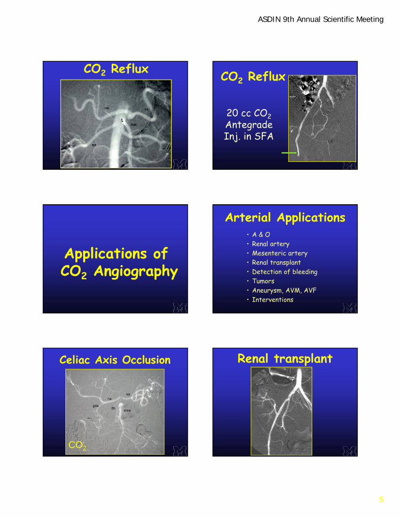
ASDIN 9th Annual Scientific Meeting
5
CO2 Reflux
20 cc CO2AntegradeInj. in SFA
CO2 Reflux
Applications ofCO2 Angiography
Arterial Applications• A & O • Renal artery• Mesenteric artery• Renal transplant• Detection of bleeding• Tumors• Aneurysm, AVM, AVF• Interventions
Celiac Axis Occlusion
CO2
Renal transplant

ASDIN 9th Annual Scientific Meeting
6
CO2 A & O Occlusion of SFA
EDS 1VRenal Artery Rupture
CO2
Coil Embolization
Renal Cell CarcinomaCO2
CO2-guided Renal Stenting

ASDIN 9th Annual Scientific Meeting
7
Post Renal Biopsy Hematuria
CO2
Common Iliac Artery Aneurysm
CO2
Venous Applications of
CO2 Angiography
40
Indications• Opacify central veins
• PTA and stent placement
• IVC filter placement
• TIPS
• Transjugular liver biopsy
• Splenoportography
• Portal vein access
Upper Limb Venograms
CO2
CO2 Central Venograms

ASDIN 9th Annual Scientific Meeting
8
CO2 Jugular Venogram CO2 Jugular Venogram
CO2 for PICC CO2 fistulogram
Occlusion of left subclavian vein
CO2
Contrast
Axillosubclavian Vein Thrombosis

ASDIN 9th Annual Scientific Meeting
9
L iliac Vein Stenting
CO2
CO2 Filter Placement
• Air contamination• Vapor lock:
– Pulmonary artery (hypotension)– Mesenteric artery (Intestinal ischemia)– Simultaneous nitrous oxide anesthesia
• Neurotoxicity (CO2 injection in carotid artery)• Paradoxical embolism• Hepatic capsule laceration (CO2 wedge injection)
Potential Complications ofCO2 Angiography
X-table LAT
Change position
CO2 DSA for AAA
Trapped CO2 in AAA.What should be done
and why?
CO2 guided Vascular Mapping for Hemodialysis Access Surgery in a Patient with Failing Renal Allograft
Case History • 51 y/o F w/ PCKD and ESRD• Failing renal txp• Failed dialysis fistula, bil UE• L basilic vein-radial artery fistula • Request for:
– L arm venogram– L arm arteriogram

ASDIN 9th Annual Scientific Meeting
10
L arm CO2venogram
Brachial Artery Puncture forCO2 DSA
Adverse reaction: lightheaded, bradycardic and hypotensive
L forearm DSA How to makeCO2 angiography work better?
• DSA• Endhole catheter • Reflux angiography• Selective injections• New mask (move mask)• Stacking• Elevate area of interest• Vasodilators
Monitoring CO2 Angiography
• Oxygenation = Pulse oximetry
• Perfusion = BP, HR, ECG
• Ventilation = Capnography
Capnography monitor

ASDIN 9th Annual Scientific Meeting
11
• CO2 is the only proven safe contrast agent in allergy and renal failure.
• CO2 should not be used as an arterial contrast agent above the diaphragm.
• Use a closed system for CO2 delivery to prevent air contamination.
• Do not use with nitrous oxide anesthesia.• The advantages of CO2 include use of unlimited
total volume and low viscosity for vascular diagnosis and endovascular intervention.
Conclusions
Gadolinium Angiography
Physical Properties of CO2, Gadolinium and Iodine
Atomic number
CO2 C = 6, O =8
Gd 64
Iodine 53
Fewer Gd atoms /cc of contrast: 1/3 attenuation values
Saline Omni 240 Gadolinium Omni 300
Radiopacity Comparison Gadolinium-assisted Renal Stenting

ASDIN 9th Annual Scientific Meeting
12
Gd-guided Renal Embolization
Hematuria after percutaneous renal biopsy. Gd renal arteriogram demonstrates active extravasation in lower pole of left kidney that was embolized with microcoils
• CO2 for arteriography and venography, and endovascular intervention
• Dilute iodinated contrast agent when needed.
Iodinated ContrastSaving Strategies
Thank you