
urgical - adiologi natomy · microfilm, in electronic data bases, video disks, etc., whithout first...
15
urgical - adiologi natomy
Transcript of urgical - adiologi natomy · microfilm, in electronic data bases, video disks, etc., whithout first...

urgical -adiologi natomy

and
Founded as Anatomia Clinica by P Rabischong
Journal of Clinical Anatomy Anatomy is a morphological sc ience which cannot fail to interest the cl inician. The practical appl icat ion of anatomical research to clinical Prob lems necessitates special adaptation and selectivity in choosing from numerous international works. Although there is a tendency to believe that meaningful advances in anatomy are unlikely, constant revision is necessary. Surgical and Radiologie Anatomy, the first international Journal of Clinical anatomy has been created in this spirit.
Its goal is to severe cl inicians, regardless of special i ty-physic ians, surgeons, radiologists or other specia l is ts-as an indispensable aid with which they can improve their knowledge of anatomy. Each issue includes: Original papers, review articles, articles on the anatomical bases of medica l , surgical and radiological techniques, brief reviews of anatomical publications of clinical interest.
Particular attention is given to high quality illustrations, which are indispensable for a better understanding of anatomical problems.
Surgical and Radiologie Anatomy is a Journal written by anatomists for cl inicians with a special interest in anatomy.
Copyright
Submiss ion of a manuscript implies: that the work descr ibed has not been publ ished before (except in the form of an abstract or as pari of a publ ished lecture, review of thesis); that it is not under considerat ion for publication elsewhere; that its publ icat ion has been approved by all coauthors, if any, as well as by the responsible authorities at the institute where the work has been carried out; that, if and when the manuscript is aeeepted for publication, the authors agree to automatic transfer of the Copyright to the (publisher, society); and that the manuscript will not be publ ished elsewhere in any language without the consent of the Copyright holders.
All articles publ ished in this Journal are protected by Copyright, which Covers the exclusive rights to reproduce and distribute the article (e.g., as offprints), as well as all translation rights. No material publ ished in this Journal may be reproduced photographically or stored on microfi lm, in electronic data bases , video d isks, etc., whithout first obtaining written permission from the publisher.
The use of general descript ive names, trade names, trademarks, etc., in this publication, even if not speeifieally identified, does not imply that these names are not protected by the relevant laws and regulations. While the advice and Information of this Journal is bel ieved to be true and accurate at the date of its going to press, neither the authors, the editors, nor the publisher can aeeept any legal responsabil i ty for any errors or omissions that may be made. The publ ished makes no warranty, express or implied, with respect to the material contained herein.
S p e c i a l r e g u l a t i o n s f o r p h o t o c o p i e s i n t h e U S A .
Photocopies may be made for personal or in-house use beyond the limitations stipulated under Sect ion 107 or 108 of U.S. Copyright Law, provided a fee is paid. This fee is U S $ 0.20 per page, or a minimum of U S $ 1.00 if an article contains fewer than five pages. All fees should be paid to the Copyright Clearance Center, Inc., 21 Congress Street Salem M A 01970, U S A stating the ISSN 0930-312X, the volume, and the first and last page numbers of each article cop ied. The Copyright owner 's consent does not include copying for general distribution, promotion, new works, or resale. In these cases , specif ic written permission must first be obtained from the publisher.
Subscription Information
Volume 15 {4 issues) appears in 1993.
F r a n c e a n d F r e n c h - s p e a k i n g c o u n t r i e s . Edition with complete French translation, Subscr ipt ion rate; FF 1.850.00, including postage and handling. Orders should be addressed to:
Dawson France/Service Diffusion Rue de la Prairie, B P 40 F-91121 Palaiseau Cedex Tel. (1) 69 09 01 22 Telex 600 394 F, FAX 1/64 54 83 26
Membership of the European Association of Clinical Anatomy, the "College Medical Francais des Professeurs d'Anatomie" and of the French Society of Radiology offers reduced subscription rates of the Journal.
N o r t h A m e r i c a . Annual subscript ion rate: approx. U S $ 388.00 (Single issue price: approx. U S $ 114.00) including carriage charges. Subscr ipt ions are entered with prepayment only.
Orders should be addressed to: Springer-Verlag New York Inc. Service Center Secaucus 44 Hartz Way Secaucus , NJ 07094, U S A Tel. (201) 348-40 33, Telex 0023 125 994 Fax (201)348-4505
A l l o t h e r c o u n t r i e s . Orders subsrieptions can either be placed via a bookdealer or sent directly to: Springer-Verlag Heidelberg Platz 3 D-14197 Berlin Tel. (030) 80 07-0, Telex 01-83319 FAX (0)30/821 40 91
C h a n g e s o f a d d r e s s : Al low six weeks for all changes to become effective. All Communicat ions should include both old and new addresses (with Postal Codes) and should be aecompanied by a mailing label from a recent issue.
B a c k v o l u m e s : Pr ices are available on request.
M i c r o f o r m : Microform editions are available from: University Microfi lms International 300 N. Zeeb Road Ann Arbor, MI 48106, U S A
P roduc t i on
Springer-Verlag France J . York 26, rue des Carmes F-75005 Paris Tel. (331)43 26 11 07 FAX (331) 43 54 49 08
Adve r t i s semen ts
Mlle Anne Auquier, Springer-Verlag France 26, rue des Carmes F-75005 Paris Tel. (331)44 41 15 89 FAX (331) 43 54 49 08
P roduc t i on and acqu is i t i on edi tor
D i rec t r ice de la pub l i ca t ion : J Tovar
Imprimerie Soul isse et Cassegrain Niort, France D .L 3073 © Springer-Verlag France Printed in France C . P . : 71753
0 Springer International A2 v

Journal of Clinical Anatomy
Editor-in-Chief
Associate Editor
Honorary Editor
Managing Editors
Assistant Editors
International Advisory Board
JP Chevrel, Paris, France
H Ellis, Cambridge, United Kingdom
J Pegington, London, United Kingdom
GT Ho S Nazarian TSato Chongquing, PR China Marseille, France Tokyo, Japan T Naidich Dl R Putz B Senecail Dl Miami, USA Munich, Germany Brest, France
F Anderhuber JP Francke Dl R Pabst SZ Zhong Graz, Austria Lille, France Hannover, Germany Guangzhou, PR China C Fontaine 0 Gagey E Sassoon Lille, France Paris, France London, UK
P Abrahams SA Georgescu J Koebke ME Müller London, UK Bucharest, Rumania Cologne, Germany Bern, Switzerland G Bastide G Godlewski Y Kuru Dl H Nahum Dl Toulouse, France Nimes, France Tokyo, Japan Paris, France J Best Dl A Gouaze K Lackner Dl CV Penteado Edinburgh, UK Tours, France Wurzburg, Germany Campinas, Brazil F Bonnel RS Harris Dl J Lang W Platzer Montpellier, France St John's, Canada Wurzburg, Germany Innsbruck, Austria B Brogdon Dl D Hartman Dl P Lasjaunias Dl U Schumacher Mobile, USA Washington, USA Paris, France Southampton, UK M Caix RE Heitzman Dl P Le Floch-Prigent E van der Zypen Limoges, France Syracuse, USA Paris, France Bern, Switzerland LJA DiDio IM Holland Dl R Louis G Vanneuville Toledo, USA Nottingham, UK Marseille, France Clermont-Ferrand, France
C Faure LG Kempe DA McGrouther M Viamonte Di Grenoble, France Charleston, USA London, UK Miami Beach, USA W Firbas B Kendall Dl H Meire Dl EA Zerhouni Dl Vienna, Austria London, UK London, UK Baltimore, USA JB Flament C Kenesi A Morin HZ Zhang Reims, France Creteil, France Lyon, France Wuhan, PR China P Franchebois J Kirklin Dl M Moscovici Montreal, Canada Birmingham, UK Rio de Janeiro, Brazil
* D I D i a g n o s t i c I m a g i n g
Springer International A3

Get a head start in the race for
Information
For a selection of Journals in the life sciences and radiology
we offer
tables of Contents and BiblioAbstracts via Internet from our mailserver
with
automatic delivery several weeks before the new issue is in your hands.
Tables of contents are free of Charge; BiblioAbstracts are available for a small annual fee. For details please send an e-mail messagecontaining the line
h e l p
to our mailserver [email protected] Please do not send regulär e-mail to this address.
Springer New Media Journals Preview Service via Internet

Journal of Clinical Anatomy
Table of contents Volume 15 Number4, 1993
E d i t o r i a l
A discourse on the usefulness of anatomy P Dahhan
251
A n a t o m i e b a s e s o f m e d i c a l , r a d i o l o g i c a l a n d s u r g i c a l t e c h n i q u e s
The cutaneous territory of the transverse tensor fascia lata flap: further anatomical considerations
M Medot, J Fissette
255
The sartorius myocutaneous island flap ML Tang, XY Liu, JW Ren, DC Zhang, RS Li, YM Wen, BF Ge
259
Anatomie basis of laparoscopic surgery in the male pelvis
0 Cussenot, F Desgrandchamps, S Bassi, P Teillac, JP Lassau, A Le Duc
265
Anatomie basis of lymphatic spread from Carcinoma of the lung to the mediastinum: surgical and prognostic implications
M Riquet
271
O r i g i n a l a r t i c l e s
Cartilage degeneration in the human patellae and its relationship to the mineralisation of
279
the underlying bone: a key to the understanding of chondromalacia patellae and femoropatellar arthrosis?
F Eckstein, R Putz, M Müller-Gerbl, M Steinlechner, KP Benedetto
The vertebral foramen: 287 a report concerning its contents
F de Peretti, I Hovorka, P Ganansia, J P Puch, A Bourgeon, C Argenson
Pedicles of lumbar vertebrae 295 C Maillot, R Wolfram-Gabel
A tridimensional study using cuts at a low 301 temperature of the infratemporal region
C Madrid, D Lefebvre, L Gineste, D Duran, R Combelles, with the collaboration of P Roux, R Joly, A Laveran
Patterns of the coronary artery irrigation 309 in the left ventricle
J Reig, A Jornet, M Petit Anatomy of the seated position: 315 methodologic approach and initial findings
S Ghannouchi, A Ghorbel, C Cavallero, J Bonnoit Interaction between the muscles of the neck 321 and the extraoeular muscles of the myopic eye. An electromyographic study
B Valentino, A Fabozzo Ref lections and suggestions on the 325 nomenclature of the prostate
G Benoit, A Jardin, C Gillot
R a d i o l o g i c a l a n a t o m y
Rotation of the cervical spinal column: 333 a computed tomography in vivo study
J-L Dumas, M Sainte Rose, P Dreyfus, D Goldlust, J-P Chevrel
(Contd. next page)
Indexed in Current Contents and Index Medicus (MEDLINE data base)
0 Springer International

Table of contents Volume 15 Number4, 1993 (continued)
Three-dimensional modeling of the anatomy 341 of heart and great vessels
D Chatel, Y Martin-Bouyer, C Acar, H Bouchoucha, JL Sableyrolles, V Jebara, JC Chachques, A Carpentier
Sonographie anatomy of the rectus sheath: 349 an indication for new terminology and implications for rectus flaps
QM Ali
A n a t o m i e v a r i a t i o n s
Variations in arterial blood supply and 355 the risk of hemorrhage during percutaneous treatment of lesions of the pelviureteral junetion obstruetion: report of a case of testicular artery arising from an inferior polar renal artery
V Ravery, O Cussenot, F Desgrandchamps, P Teillac, Y Martin-Bouyer, JP Lassau, A Le Duc
Middle mesenteric artery: an anomalous 361 origin of a middle colic artery
T Yoshida, S Suzuki, T Sato A case of intraduodenal diverticulum 365 imitating choledochocele
L Kovachev, T Deliisky, E Marinov
R e p o r t s of t h e S o c i e t e A n a t o m i q u e d e P a r i s
The embryonic and early fetal development 369 of the lymphatics of the heart and lungs in humans (25.6.93)
M Riquet, J Briere, P Dupont, G Pennhouat, G Hidden
E A C A c o n g r e s s r e p o r t , M u n i c h 1 9 9 3 I i Subject, a u t h o r , XIII r e f e r e e a n d t r a n s t a t o r i n d e x e s
C h i n e s e J o u r n a l 264, 270, 278, 308, 320 o f C l i n i c a l A n a t o m y
R e v i e w s i n b r i e f 360,364, 368
A n n o u n c e m e n t s im B o o k s r e e e i v e d 340, 354
Indexed in Current Contents and Index Medicus (MEDLINE data base)
A8 0 Springer International
Surg Radiol Anal (1993) 15 : I-VII SUrCHCQl" -
Radiologie College Medical Franc,ais des Professeurs d'Anatomie
Anatomy Journal of Clinical Anatomy
© Springer-Verlag 1993
This list of the members of the C M F P A will hereafter be updated every two years. The next revision, with the addresses of the members of the E A C A , will appear in 1994. La liste des membres du C M F P A sera desormais publiee tout les deux ans, avec la liste des membres de l ' A E A C . La pro-chaine mise-ä-jour aura lieu en 1994.
Aaron Claude (1971)*
i'* Alexandre Jean-Henri (1966)
Ameil Marc
Aprosio Norbert (1961)
Argeme Maxime (1970)
Argenson Claude (1972)
Armstrong Olivier
Autissier Jean (1971)
Avisse Claude
Bailleul Jean-Pierre (1985)
* Barbin Jean-Yves (1961)
UFR Cochin-Port-Royal Anatomie, 24, rue du Faubourg Saint-Jacques, 75674 Paris -1.43.20.12.40 Dom. : 131, rue de la Sante, 75013 Paris -1.45.89.22.06
Service de Chirurgie, Höpital Broussais, 96, rue Didot, 75014 Paris - 1.43.95.95.95 ou 1.45.42.22.06 Dom. : 3. avenue Robert Schumann, 92100 Boulogne - 1.46.04.43.70
Faculte de Medecine, 51, rue Cognacq-Jay. 51096 Reims Cedex; et Service de Chirurgie Orthopedique et Traumatologique, 45, rue Cognacq-Jay, 51082 Reims Cedex -26.78.76.71 Dom. : 56, avenue Paul-Marchandeau. 51000 Reims
Institut d'Anatomie Normale. Faculte de Medecine, 4, rue Kirschleger, 67085 Strasbourg Cedex - 88.35.87.90
Faculte de Medecine, Anatomie. Secteur Nord, 2, boulevard Pierre-Dramard, 13326 Marseille Cedex 3 -91.69.88.21 ou 91.69.88.23 Dom. : 14, boulevard Henri-Fabre. 13012 Marseille-91.34.29.97
Dom.: Moulin des Baux. 13260 Cassis
Laboratoire d'Anatomie, Faculte de Medecine, 1, rue Gaston-Veil. 44035 Nantes -40.41.28.10
Faculte de Medecine, 1, boulevard Jeanne-d'Arc, 21000 Dijon - 80.65.23.23 Dom. : 37, rue J.-B. Baudin. 21000 Dijon -80.66.25.65
Laboratoire d'Anatomie, Faculte de Medecine, 51, nie Cognacq-Jay, 51095 Reims Cedex; et Service de Chirurgie Generale, Höpital R.-Debre. rue Alexis-Carrel, 51092 Reims Cedex -26.78.71.20 Dom. : 1, rue de Savoye, 51100 Reims -26.88.64.33
Laboratoire d'Anatomie, Faculte de Medecine, 1. place de Verdun, 59045 Lille Cedex -20.62.69.41 - IUFP : 20.60.07.17 Dom. : 7, avenue Foch. 59130 Lambersat -20.92.20.01
Laboratoire d'Anatomie, Faculte de Medecine. 1, rue Gaston-Veil. 44035 Nantes -40.41.21.36 Dom. : La Turballiere, Suce sur Erdre, 44240 La Chapelle sur Erdre - 40.77.71.45
Bargy Frederic (1988)
Bastian Dominique
Bastide Guy (1961)
Baulac Michel (1988)
Becade Philippe
Becue Jean (1974)
Bejui Jacques (1986)
Benhamou-Mayoux Marie-Anne
Benoit Gerard (1986)
Berthelot Jean-Louis
Bonnel Francis (1974)
Bonnoit Jean (1976)
La date entre parentheses est celle de la nomination ä l'agregation. Membre honoraire
Faculte Cochin-Port-Royal, 24, rue du Faubourg Saint-Jacques, 75014 Paris -1.44.41.23.60 (Fax : 44.41.22.23) Dom.: 19, rue d'Arcole, 75004 Paris -1.43.26.27.80
Laboratoire d'Anatomie, Faculte de Medecine Saint-Louis-Lariboisiere, 45. rue des Saints-Peres, 75270 Paris Cedex 06 - 1.42.86.20.47 Dom. : 14. rue de Vouille, 75015 Paris -1.48.56.02.93
Laboratoire d'Anatomie, Faculte de Medecine, 133, mute de Narbonne, 31077 Toulouse Cedex - 61.53.23.13, P. 341 ou 61.55.03.68 -Hop: 61.77.20.59 Dom. : 10. rue des Chalets, 31000 Toulouse -61.62.83.48
Laboratoire d'Anatomie, Faculte de Medecine Pitie-Salpetriere, 105. boulevard de l'Höpital, 75013 Paris - 1.40.77.97.22 (direct) ou 1.45.70.27.37 (direct Neuro, ä l'Höpital)
Clinique du Pont de Chaume, 330, avenue Marcel Unal, 82017 Montauban Cedex -63.68.34.50
Laboratoire d'Anatomie, Faculte de Medecine de Toulouse, 133. route de Narbonne, 31062 Toulouse Cedex - 61.55.03.68: ou C H U Rangueil, 31054 Toulouse - 61.32.27.51 Dom. : 299, avenue J.-Rieux, 31500 Toulouse -61.20.48.64
Laboiratoire d'Anatomie, Faculte Alexis-Carrel, 8, rue Guillaume-Paradin, 69008 Lyon - 78.77.86.23 (Fax : 78.77.87.01) Dom. : 52, nie du Lieutenant-Colonel-Prevosl, 69006 Lyon - 78.89.76.44 (Fax : 72.12.00.04)
Faculte de Medecine Cochin-Port-Royal, 24, rue du Faubourg Saint-Jacques. 75014 Pairs -1.43.20.12.40
Service Urologie, Höpital Kremlin-Bicetre, 78. rue du General Ledere, 94270 Le Kremlin-Bicetre - 1.45.21.36.94; et UFR des Saints-Peres, 45, rue des Saints-Peres, 75720 Paris Cedex 0 6 - 1.42.86.20.47 Dom. : I, rue Barye, 75017 Paris -1.43.80.70.25
Faculte Xavier-Bichat. 16, rue Huchard. 75018 Paris - 1.42.63.84.20 Dom. : Parc de Maugarny, 95680 Montlignon - 1.34.16.63.00
Faculte de Medecine, 2, rue de l'Ecole de Medecine. 34060 Montpellier Cedex - CHU : 67.33.87.26 Dom. : 11/4, avenue de la Pompignage, 34000 Montpellier-67.79.06.19
Laboratoire de Biomecanique Appliquee, Faculte de Medecine, Secteur Nord, 2 boulevard P.-Dramard. 13326 Marseille Cedex 15 -91.51.09.67 (Fax:91.51.12.55) Dom. : 2, rue Saint-Bazile, 13001 Marseille -91.95.76.70

Surg Radiol Anat (1993) 15 : 279-286 S U F O i C O l a n d
Radiologie Anatomy Journal of Clinical Anatomy © Springer-Verlag 1993
Original articles
Cartilage degeneration in the human patellae and its relationship to the mineralisation of the underlying bone: a key to the understanding of chondromalacia patellae and femoropatellar arthrosis?
F Eckstein1, R Putz 1, M Müller-Gerbl 1, M Steinlechner2 and KP Benedetto3
1 Anatomische Anstalt, Pettenkoferstr. 11, D-80336 München, Germany 2 Institut für Anatomie, Müllerstr. 59, A-6010 Innsbruck, Austria
Universitätsklinik für Unfallchirurgie. Anichstr. 35, A-6020 Innsbruck. Austria
Summary. According to the litera-ture subchondral bone plays a signifi-cant role in the transmission of load through joints and in the pathogenesis of osteoarthrosis. Therefore the degeneration of the articular cartilage was investigated in the patellae from 30 dissecting-room speeimens and of 20 patients, previously submitted to arthroscopy, and subchondral mineralisation of their underlying bone was at the same time assessed by means of CT osteoabsorptiometry. Lateral cartilage lesions were localised over highly mineralised subchondral bone; these appear to be due to long-term stress. They were mainly found in the older speeimens and showed a high rate of progression with increasing age. Medially localised cartilage lesions, on the other hand, were situa-ted in a transitional region between moderate and slight subchondral mineralisation; they may be caused by infrequent stress peaks and by shear stress in the articular cartilage, the very medial part of the Joint being deprived of mechanical Stimulation for much of the time. These lesions
Correspondence to : F Eckstein
were to be found predominantly in the younger speeimens and showed little progress with advancing age. Patients with lateral cartilage degeneration exhibited higher, patients with medial chondromalacia patellae lower mineralisation than normals. Their density patterns therefore indi-cate a different mechanical pathogenesis of the cartilage lesions in the lateral and medial facet. It could be shown that CT osteoabsorptiometry allows an assessment of the mechanical Situation, present in individual femoro-patellar joints, and that this Situation is highly relevant for the pathogenesis of patellar cartilage degeneration.
Etüde de la relation entre la dege-nerescence cartilagineuse et la mineralisation de Tos sous-chon-dral de la patella. Est-ce une fa^on pour comprendre la pathogenie de la chondromalacie patellaire et de Parthrose femoro-patellaire ?
Resume. Selon la litterature, Tos sous-chondral joue un role important dans la transmission des forces ä tra-vers les articulations et dans la patho
genie de Tarthrose. C'est pourquoi la degenerescence du cartilage articiilai-re a ete etudiee sur les patellas de 30 sujets anatomiques et de 20 patients venant de subir une arthroscopie et dont la mineralisation sous-chondrale de Tos sous-jacent avait ete appreciee au meme moment par absorptiome-trie osseuse par Scanner. Les lesions cartilagineuses laterales etaient situees sur de Tos sous-chondral for-tement mineralise ; elles paraissent etres dues ä des contraintes ä long terme. Elles furent principalement trouvees sur les speeimens äges et montraient un fort taux de progression en fonetion du vieillisement. A l'oppose, les lesions cartilagineuses mediales etaient situees dans une region de transition entre des minera-lisations moderee et faible ; elles pourraient etre dues ä des pics de contrainte intermittents et ä des contraintes en cisaillement sur le cartilage articulaire, la partie la plus mediale de rarticulation etant privee de stimulations mecaniques la plupart du temps. Ces lesions furent trouvees essentiellement sur les sujets les plus jeunes et montraient peu de progression avec le vieillissement. Les patients presentant une degenerescen-

280 F Eckstein et al: Cartilage degeneration in human patellae; relationship to mineralisation of underlying bone
ce cartilagineuse laterale avaient une mineralisation superieure ä la normale, les patients presentant une chon-dromalacie mediale avaient une mineralisation plus faible que la normale. La distribution de la densite indique donc un mecanisme pathoge-nique different pour les lesions cartilagineuses sur les facettes laterale et mediale. L'absorptiometrie osseuse par Scanner permettrait donc d'appre-cier la Situation mecanique individuelle de chaque articulation femoro-patellaire. Cette Situation est forte -ment correlee ä la pathogenie de la degenerescence du cartilage patellaire.
Key words: Patella — Cartilage degeneration — Subchondral mineralisation — Chondromalacia patellae — Femoropatellar arthrosis
It is widely accepted that mechanical factors play an important part in the initiation and progression of osteoarthrosis in general and chondromalacia of the patella in particu-lar [11, 25]. Nevertheless, the exact nature of the mechanism leading to Joint damage, and the exact locali-sation of the initial changes in cartilage degeneration are still a matter of discussion and disagreement [17]. Radin and Paul [26] and Radin et al [28] were able to show that the ability of a Joint to attenuate "longi-tudinal impact loading", which is an important factor in the pathogenesis of osteoarthrosis [27], is principally determined by the stiffness of the subchondral bone. As against this, the articular cartilage is supposed to contribute to reducing friction during movements of the Joint. These authors even suggest that it is a change in the nature of the subchondral bone, i.e. an increase in its stiffness, which represents the first step in the development of osteoarthrosis [29]. Up to now it has not been possible to assess such
changes in the living satisfactorily. Subchondral sclerosis, although accepted as a criterion for the radiological diagnosis of osteoarthrosis, does not permit quantitative evalua-tion of the relative distribution of subchondral mineralisation in the Joint surface to be made. However, computed tomography osteoabsorptiometry ( C T - O A M ) [18, 19] has recently provided a method for assessing the surface distribution of subchondral mineralisation in the l i v ing subject in terms of the Hounsfield scale. We were able to establish in a previous investigation [5] that the pattern of mineralisation in the patella does in fact reflect the biomechanical Situation present in the femoropatellar-joint. According to Hvid et al [12], the mechanical properties (and particularly the stiffness) of the subchondral bone can, with certain reservations, be inferred from this mineralisation pattern. The aim of this investigation is therefore to compare the localisation of macroscopically visible cartilage degeneration with the radiological (CT osteoabsorptio-metric) density of the underlying bone in the human patella, which according to Ficat and co-workers [7, 8] öfters an "observatoire ideal" of arthrotic degeneration.
Material and methods
We first examined 30 formalin-fixed speeimens of the knee-joint taken from dissecting-room subjects, selecting the youngest subjects available. Those with systemic disease of the locomotor apparatus or severe arthrosis of the knee-joint were excluded from the investigation. The ages of the 7 female and 23 male subjects ranged from 47 to 90 years. We divided these into two groups: 15 speeimens of less than 60 years (mean age 53) constituted Group Y , and 15 of more than 60 years (mean age 74) Group O. Twenty patients were then selected from the Depart
ment of Surgery, on whom arthro-scopy had been carried out for various reasons (mostly because of suspected damage to the meniscus). In each case, a CT osteoabsorptio-metric examination was made within 48 h of arthroscopy. The criterion for inclusion was the presence of cir-cumscribed damage of grades 1 or 2 [21] to the articular cartilage of the medial or lateral facet, or of the pri-mary ridge. There were no gross changes in the AP, or lateral radio-graphs, nor any severe disease of the femoropatellar-joint. Nine women and eleven men between the ages of 16 and 61 were examined, the avera-ge age being 31 years.
I n v e s t i g a t i o n of t h e s u b c h o n d r a l m i n e r a l i s a t i o n
The knee-joint speeimens and the knees of the arthroscopy patients (30° of knee-flexion) were examined in a CT Scanner (Siemens Delta scan 50 FS 2). Sectional images between the upper pole of the patella and the tibial plateau were made at intervals of 4 mm. CT osteoabsorptiometry [18, 19] was used to obtain isodensi-ties (i.e., contour lines joining points of equal density). The protection of their subchondral extension was marked on the surface of each sec-tion, and these marks transferred into the corresponding position of the sec-tion in a template of the articular surface (Fig. la). Six Hounsfield ranges (<400 H U - >800 HU) were recons-trueted for the speeimens, and seven (<300 H U - >1050) for the patients. The distribution pattern was then digitalised (Hewlett Packard, Scan Jet Plus) and read into an Apple Macintosh Computer, where it was provided with standardised grey values (Fig. lb), using a grey scale of 256 steps. Image summation was finally carried out in the Computer in order to produce average pictures. By means of program Software, the original pictures were laid together successively in pairs, one upon the

F Eckstein et al: Cartilage degeneration in human patellae; relationship to mineralisation of underlying bone 281
> 800 Hounsfieldunits
701 - 800 Hounsfieldunits
601 - 700 Hounsfieldunits
501 - 600 Hounsfieldunits
401 - 500 Hounsfieldunits
<401 Hounsfieldunits
Figs. la-d, 2a, b, 3a, b la Outline template of the articular surface of the patella built up from measurements of 30 speeimens. b Grey values of the Hounsfield ranges examined in the speeimens. c Summation picture for Group Y (age < 60 yrs, n=l5). d Summation picture for Group O (age > 60 yrs, n=l5). 2 Surface distribution of subchondral mineralisation in the patella of a patient suf-fering from left-sided Poliomyeli t is , a Left patella. b Right patella. 3 Surface distribution of cartilage degeneration in the patellae of 30 anatomical speeimens. w h i t e . intact cartilage; g r e y , mild degeneration (degree l ) ; b l a c k , severe cartilage damage (degree > 1). a Summation picture for Group Y (age < 60 yrs, n = 15). b Summation picture for Group O (age > 60 yrs, n = 15)
la Trace du contour de la surface articulaire de la patella. bäti ä partir des mesures de 30 speeimens. b Echelle des gris des niveaux de densite Hounsfield trouves sur les
speeimens. c Shema recapitulatif pour le Groupe Y (age < 60 ans, 15 cas). d Shema recapitulatif pour le groupe O (age > 60 ans, 15 cas). 2 Distribution de la mineralisation sous-chondrale sur les patellas d'un patient atteint de poliomyelite lateralisee ä gauche. a Patella gauche. b Patella droite. 3 Distribution de la degenerescence cartilagineuse sur les patellas de 30 speeimens anatomiques. Blanc\ cartilage intact; g r i s . degenerescence legere (degre 1); n o i r , degäts cartilagineux severes (degres > 1). a Shema recapitulatif pour le groupe Y (age < 60 ans, 15 cas). b Shema recapitulatif pour le groupe O (age > 60 ans, 15 cas)
other, and the grey values of corres-ponding pixels were fused to a value lying exactly between them in the grey scale. Intermediate grey values obtained from the Single summation steps were redefined in a final step as the dosest neighbouring grey value in order to restore the original iso-densities.
E x a m i n a t i o n of t h e c a r t i l a g e d e g e n e r a t i o n
The grade [21] and extent of the cartilage degeneration in the patellae of the 30 speeimens were pictorially recorded, using the Joint template mentioned above. The distribution
pattern was digitalised, and distin-guishing grey values allotted to the different degrees of degeneration. In this case, the Computergraphic method for producing the summation pictures depended upon a program which at each step picked up 50% of the pixels from each of two Originals. In the summation pictures obtained in this way, the grey value of the pixels reveals, as in the single pictures, the degree of cartilage degeneration, and their density the frequen-cy of the cartilage damage in the cor-responding area of the Joint. The grade and localisation of the cartilage degeneration in the patients was recorded on an arthroscopy form.
C o m p a r i s o n of t h e s u b c h o n d r a l m i n e r a l i s a t i o n w i t h t h e c a r t i l a g e d e g e n e r a t i o n
After comparison of the distribution patterns in the Single pictures, the grades of cartilage degeneration and the locally corresponding Hounsfield ränge were read from 38 measuring points regularly distributed over the articular surface. The percentage dis-tributions of grades of cartilage degeneration in the Hounsfield ranges were calculated for all the speeimens added together, and the level of signi-ficance of the differences between the density ranges statistically tested. The percentage distribution of the grade of cartilage degeneration in the different Hounsfield ranges was also determi-ned separately for Croups Y and O. Differences in the percentage of severe cartilage damage present in the Hounsfield ranges of the two groups were tested for Statistical significance.
Results
S u b c h o n d r a l m i n e r a l i s a t i o n
Maximum subchondral mineralisation was found in the proximal part of the lateral facet in all the patellae examined. This amounted in the anatomical speeimens to 600-1000 HU, and in the patients to 450-1100 HU, and decreased more or less steeply towards the periphery. Whereas there was little difference in the principal distribution of the mineralisation (high density, lateral; low density, medial) in Groups Y (Fig. lc) and O (Fig. Id), the younger individuals dis-played a slightly greater mineralisation of the subchondral bone (shown by the somewhat wider extension at all density levels). In one 33 year-old patient, who had suffered from polio-myelitis from the age of four years, and in whom there was marked muscle wasting of the whole left lower limb, the right patella (Fig. 2) was more highly mineralised than the left by 300 H U

282 F Eckstein et al: Cartilage degeneration in human patellae; relationship to mineralisation of underlying bone
Figs. 4a, b, 5a, b 4 Single example of lateral cartilage damage over highly mineralised subchondral bone. a Surface distribution of subchondral mineralisation. b Surface distribution of cartilage degeneration. w h i t e , intact cartilage; g r e y , mild degeneration (degree 1); h l a c k , severe cartilage damage (degree > I). 5 Single example of medial cartilage damage over poorly mineralised subchondral bone. a. Surface distribution of subchondral mineralisation. b Surface distribution of cartilage dege
neration white, intact cartilage; grey, mild degeneration (degree 1); h l a c k , severe cartilage damage (degree > I)
4 Exemple de degäts cartilagineux lateraux sur de Tos sous-chondral fortement mineralise. a Distribution de la mineralisation sous-chondrale. b Distribution des lesions cartilagineuses degeneratives. B l a n c . cartilage intact; g r i s , degenerescence legere (degre 1); n o i r , degäts cartilagineux severes (degres > 1). 5 Exemple de degäts cartilagineux mediaux sur de Tos sous-chondral faiblement mineralise. a Distribution de la mineralisation sous-chondrale. b Distribution des lesions cartilagineuses degeneratives. B l a n c , cartilage intact; g r i s , degenerescence legere (degre 1); n o i r . degäts cartilagineux severes (degres > 1)
< 401- 501- 601- 701- > 400 500 600 700 800 800 HU HU HU HU HU HU
gro in
j p Y = act ca e f t / 6
rtilage = righ t colur nn
9 fr < 401- 501- 601- 701- >
400 500 600 700 800 800 HU HU HU HU HU HU
100
80
60
40
20
0
—I—I—I—I—I— degree > 1 cartilaginous damage
i i i group Y = left / O = rig
it colu Tin
Ii v i o r - o f r - ^ i LJ t 400 HU
401-500 HU
501-600 HU
601-700 HU
701-800 HU
800 HU
50
40
30
20
10
0
-I—I—I—I-degree > 1 cartilaginous damage
difference between Y / O
I x II s II x II s i m X <400 401- 501-
HU 500 600 HU HU
601-700 HU
701-800 HU
»800 HU
c Fig. 6a-d a Percentage distribution (% total articular surface) of intact cartilage, mild damage (degree I) and severe damage (degree > l) in the Hounsfield ranges (< 400 H U - > 800 HU) from all 30 anatomical speeimens. b Percentage distribution of intact cartilage in Group Y (age < 60 yrs/L cols) and Group O (age > 60 yrs / R cols) in the different Hounsfield ranges. c Percentage distribution of severe cartilage degeneration (degree > l) in Group Y (L cols) and Group O (R cols) in the different Hounsfield ranges. d Difference in the percentage of severe cartilage degeneration (degree > l) between Groups Y and O in the different Hounsfield ranges
a Distribution en pourcentage (rapporte* ä la surface articulaire totale) du cartilage intact, des lesions legeres (degre l) et des degäts severes (degres > 1) dans les niveaux de densite Hounsfield (< 400 HU - > 800 HU) de 30 speeimens anatomiques. b Distribution en pourcentage du cartilage intact dans le groupe Y (äge < 60 ans, colonnes de gauche) et du groupe O (äge > 60 ans, colonnes de droite) dans les differents niveaux de densite Hounsfield. c Distribution en pourcentage des lesions cartilagineuses severes (degres > 1) dans le groupe Y (colonnes de gauche) et dans le groupe O (colonnes de droite) dans les differents niveaux de densite Hounsfield. d Difference dans les pourcentages des degenerescences cartilagineuses severes (degres superieurs ä 1) entre les groupes Y et O dans les differents niveaux de densite Hounsfield
Ca r t i l a g e d e g e n e r a t i o n
Although the pattern of subchondral mineralisation is relatively constant, the localisation of the cartilage degeneration, both in individuals and on comparison between the age groups, is rather variable. In the younger group, degeneration predominates medially and peripherally in the Joint (Fig. 3a), whereas central lesions of the lateral facet and near the princi-pal ridge become more noticeable with advancing age (Fig. 3b).
C o m p a r i s o n of s u b c h o n d r a l m i n e r a l i s a t i o n w i t h c a r t i l a g e d e g e n e r a t i o n
It can be shown in individual cases that degeneration of the lateral facet (Fig. 4b) is localised over areas of greater subchondral mineralisation (Fig. 4a), whereas lesions in the medial compartment (Fig. 5b) are found together with a lower density (Fig. 5a). If one examines the distribution of the grades of cartilage degeneration throughout different Hounsfield ranges (Fig. 6a), it is obvious that the articular cartilage is less often intact, and more often severely damaged, over regions of both very high and very low density than in association with bone showing an average amount of subchondral mineralisation (p < O.Ol). If the distributions for Groups Y and O are examined separa-tely, differences in the amount of intact (Fig. 6b) and severely damaged cartilage present (Fig. 6c) can be observed. The difference in the percentage of severe cartilage degeneration (> grade 1) between the two age groups is less at a level below 800 HU (average difference = 9% of the surface area) than above this figure (difference between Groups Y and O = 29% J. This difference is significant al a level of 1%. In the group of arthroscopy patients without cartilage degeneration the summation picture for subchondral mineralisation displays a considerable island peak of bone den-

F Eckstein et al: Cartilage degeneration in human patellae; relationship to mineralisation of underlying bone 283
Fig. 7a-d Surface distribution of subchondral mineralisation in the patellae of patients examined arthroscopically. a Summation picture for group withoul cartilage degeneration (n = 6). b Summation picture for group with central cartilage degeneration (n = 8). e Single example of patient with medial cartilage degeneration. d Single example of patient with lateral cartilage degeneration
> 1050 Hounsfieldunits 901 -1050 Hounsfieldunits 751 - 900 Hounsfieldunits 601 - 750 Hounsfieldunits
451 - 600 Hounsfieldunits 301 - 450 Hounsfieldunits < 301 Hounsfieldunits (HU)
Distribution de la mineralisation sous-chondrale des patellas des patients examines par arthroscopie. a Shema recapi
tulatif pour le groupe sans lesion cartilagineuse (6 cas). b Shema recapitulatif pour le groupe presen-tant des lesions cartilagineuses centrales (8 cas). c Exemple d'un patient avec une degenerescence cartilagineuse mediale, d Exemple d'un patient avec une lesion cartilagineuse laterale
Fig. 8 Distribution of stress in the femoropatellar-joint based on the contact areas determined by Hehne (1983) and the Joint forces calculated by Maquet (1976)
Distribution des contraintes sur rarticulation femoro-patellaire, basee sur les aires de contact determinees par Hehne (1983) et les forces articulaires calculees par Maquet (1976)
sity at the secondary ridge (Fig. 7a). This is not seen in patients with central damage to the tissue (Fig. 7b). The Single example of a patient with medial degeneration reveals signifi-cantly reduced mineralisation of the Joint (Fig. 7c), whereas in a patient with lateral degeneration bone density is raised to a remarkable extent (Fig. 7d).
Discussion
S u b c h o n d r a I m i n e r a l i s a t i o n
CT osteoabsorptiometry gives a picture of the mineralisation in the arthroscopy patients which agrees well with information obtained from the anatomical speeimens [5], and
makes it possible to take similar measurements in the living subject. Taking into consideration the contact areas described in the literature [11] one can interpret the density maxima in the proximal part of the lateral facet as the expression of more fre-quent contact under intensive load-ing. Whereas the proximal lateral facet is in contact in all positions of the knee-joint greater than 60°, medially there is a changeover from the paramedian segment (90°) across the secondary ridge (120°) to the "odd facet" (140°) . Taking into aecount the significant increase in force exerted by the quadrieeps as flexion increases [16], the finding of higher mineralisation in the proximal part of the lateral facet and a lower density medially and distally becomes plausible (Fig. 8). In this way, the density distribution can be explained in terms of the principal stress [22] or the "loading history" of a Joint [3], which makes it possible to infer the long-term distribution of load from the pattern of mineralisation. The Situation in the patient with Poliomyelit is confirms that the degree of mineralisation is determined by the magnitude of the mechanical stress acting on the Joint, and that the bone reacts with atrophy or hypertrophy to changes in the mechanical environment, as was sug-gested by Kummer [15]. These Undings are also in agreement with the observations of Müller Gerbl et al [20], who were able to show that, fol-lowing a corrective osteotomy at the knee Joint, the density of the tibial plateau at first decreases, probably due to a decrease in activity of the patient. About one year later the distribution pattern has changed in comparison with the preoperative Situation, indicating a more central Position of the resultant Joint force. The slightly higher bone density found in the younger age group may be attri-buted to the greater physical activity of these people, which is also sugges-ted by the more striking extension of
284 F Eckstein et al: Cartilage degeneration in human patellae: relationship to mineralisation of underlying bone
the highly mineralised regions towards the secondary ridge in the younger subjects. These findings suggest that Information on the distribution of the subchondral mineralisation and the position of the maxima is obtainable with C T - O A M and that the method allows a quantitative assessment of the degree of mineralisation of the Joint to be made. It is also clear that, compared with the conventional AP, lateral or tangential radiograph, the method provides a great deal more Information on the overall biomechanical conditions of an individual Joint.
C a r t i l a g e d e g e n e r a t i o n
The localisation of the cartilage degeneration agrees with that repor-ted in the literature. The damage associated with the "odd facet", which is more common than lateral lesions [4, 9] is frequently found in young people [6]. Lesions of the lateral facet [8] predominantly appear, however, with advancing age [33].
C o m p a r i s o n of s u b c h o n d r a l m i n e r a l i s a t i o n w i t h c a r t i l a g e d e g e n e r a t i o n
The greater degree of mineralisation of the lateral facet suggests that the cartilage degeneration found there may be due to intense and prolong-ed stress acting on this region of the Joint. A similar conclusion followed the clinical and radiological investi-gations of Ficat et al [7] and Ficat and Hungerford [8], who described a "lateral hyperpression-syndrome" in the patella. They attributed this pathological entity to functional dominance of the lateral retinacu-lum, due to imbalance between the action of the vastus lateralis and medialis. It has also been demon-strated in animals that, following a proximal t ibial osteotomy, the mechanical overloading of a Single Joint compartment can induce degenerative changes [10], and that these
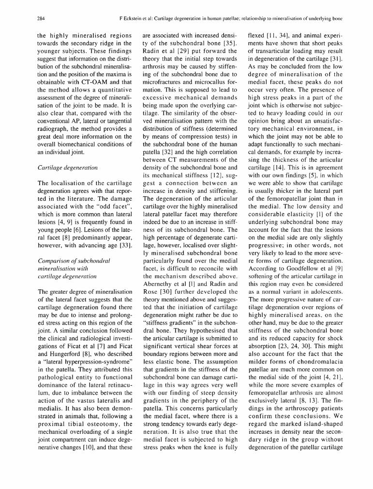
are associated with increased density of the subchondral bone [35]. Radin et al [29] put forward the theory that the initial step towards arthrosis may be caused by stiffen-ing of the subchondral bone due to microfractures and microcallus for-mation. This is supposed to lead to excessive mechanical demands being made upon the overlying cartilage. The similarity of the obser-ved mineralisation pattern with the distribution of stiffness (determined by means of compression tests) in the subchondral bone of the human patella [32] and the high correlation between CT measurements of the density of the subchondral bone and its mechanical stiffness [12], suggest a connection between an increase in density and stiffening. The degeneration of the articular cartilage over the highly mineralised lateral patellar facet may therefore indeed be due to an increase in stiffness of its subchondral bone. The high percentage of degenerate cartilage, however, localised over slight-ly mineralised subchondral bone particularly found over the medial facet, is difficult to reconcile with the mechanism described above. Abernethy et al [1] and Radin and Rose [30] further developed the theory mentioned above and sugges-ted that the initiation of cartilage degeneration might rather be due to "stiffness gradients" in the subchondral bone. They hypothesised that the articular cartilage is submitted to significant vertical shear forces at boundary regions between more and less elastic bone. The assumption that gradients in the stiffness of the subchondral bone can damage cartilage in this way agrees very well with our finding of steep density gradients in the periphery of the patella. This concerns particularly the medial facet, where there is a strong tendency towards early degeneration. It is also true that the medial facet is subjected to high stress peaks when the knee is fully
flexed [11, 34], and animal experi-ments have shown that short peaks of transarticular loading may result in degeneration of the cartilage [31]. As may be concluded from the low degree of mineralisation of the medial facet, these peaks do not occur very often. The presence of high stress peaks in a part of the Joint which is otherwise not subjected to heavy loading could in our opinion bring about an unsatisfac-tory mechanical environment, in which the Joint may not be able to adapt functionally to such mechanical demands, for example by increa-sing the thickness of the articular cartilage [14]. This is in agreement with our own findings [5], in which we were able to show that cartilage is usually thicker in the lateral part of the femoropatellar Joint than in the medial. The low density and considerable elasticity [1] of the underlying subchondral bone may account for the fact that the lesions on the medial side are only slightly progressive; in other words, not very likely to lead to the more severe forms of cartilage degeneration. According to Goodfellow et al [9] softening of the articular cartilage in this region may even be considered as a normal variant in adolescents. The more progressive nature of cartilage degeneration over regions of highly mineralised areas, on the other hand, may be due to the greater stiffness of the subchondral bone and its reduced capacity for shock absorption [23, 24, 30]. This might also account for the fact that the milder forms of chondromalacia patellae are much more common on the medial side of the Joint [4, 21], while the more severe examples of femoropatellar arthrosis are almost exclusively lateral [8, 13]. The findings in the arthroscopy patients confirm these conclusions. We regard the marked island-shaped increases in density near the secondary ridge in the group without degeneration of the patellar cartilage

F Eckstein et al: Cartilage degeneration in human patellae; relationship to mineralisation of underlying bone 285
as a result of the "point-like" Opposition of the patellae to the femur at 120° of flexion [11, 34]. The relati-vely high density here suggests that these people frequently bend their knees. Since this secondary maxi-mum is not found in patients with central cartilage degeneration, it is plausible that there exists a different mechanical enviroment in this group. It is possible that less fre-quent füll flexion contributes to degeneration of the articular cartilage due to a relative lack of intermit-tent pressure. This is in accordance with the finding of Alexander [2] that there is a positive correlation failure to take advantage of the füll ränge of physiological movement available at a Joint and its suscepti-bility to O s t e o a r t h r i t i s . Comparing the patients with severe medial and lateral cartilage degeneration suggests that the less than average mineralisation in the case of medial cartilage damage may be the result of too few general mechanical demands made on the Joint, and the very high degree of mineralisation in the case of lateral damage the result of excessive use.
We conclude from these findings that the subchondral mineralisation assessed by means of CT osteoabsorptiometry provides relevant data on the mechanics of a Joint, and that it may give some insight into the pathogenesis of cartilage degeneration which cannot be provided by the conventional radiological techniques. We believe that the gain in Information on the mechanical condition of an individual Joint justify applying this method now to the femoropatellar Joint in clinical practice.
A c k n o w l e d g e m e n t s . Our grateful thanks are due to Professor W. Platzer, Director of the Department of Anatomy, University of Innsbruck, for generally putting the speeimens at our disposal and for making the Computer tomographie exposures possible on the Scanner of the Institute. We thank Dr. F. Steel for the translation of the manuscript and H. Ruß for the drawing of Figure 8.
References
1. Abernethy PJ, Townsend PR, Rose R M , Radin E L (1978) Is chondromalacia patellae a separate c l inical entity? J Bone Joint Surg [Br] 6 : 205-210
2. Alexander C l (1989) Relationship between the utilisation profile of individual joints and their susceptibility to primary osteoarthrosis. Skeletal Radiol 18 : 199-205
3. Carter DR (1984) Mechanical loading histories and cortical bone remodelling. Calcif Tissue Res 36 : 519-524
4. Dashefsky JH (1987) Arthroscopic mea-surement of chondromalacia of patella cartilage using a microminiature pressure transducer. Arthroscopy 3 : 80-85
5. Eckstein F, Müller-Gerbl M , Putz R (1992) Distribution of subchondral bone density and cartilage thickness in the human patella. J Anat 180 : 425-433
6. Emery IH, Mechim G (1973) Surface morphology and topography of patello-femoral cartilage fibrillation in Liverpool necropsies. J Anat 116 : 103-120
7. Ficat P, Ficat C, Bailleux A (1975) Syndrome d'hyperpression externe de la rotule ( S H P E ) . Son interet pour la connaissance de l'arthrose. Rev Chir Orthop 61 : 39-59
8. Ficat P, Hungerford DS (1977) Disor-ders of the patellofemoral Joint. Mas-son, Paris
9. Goodfellow J, Hungerford DS, Woods C (1976) Patello-femoral Joint mechanics and pathology. Chondromalacia patellae. J Bone Joint Surg [Brj 58 : 291-299
10. Goodman SB, Lee J, Smith R L , Cson-gradi JC, Fomasier V L (1991) Mechanical overload of a Single compartment induces early degenerative changes in the rabbit knee: a preliminary study. J luvest Surg 4 : 161-170
11. Hehne HJ (1983) Das Patellofemoral gelenk. Enke, Stuttgart
12. Hvid I, Bentzen S M , Linde F, Mosekil-de L , Pongsoipetch B (1989) X - R a y quantitative computed tomography: the relations to physical properties of proximal tibial trabecular bone speeimens. J Biomech 22 : 837-844
13. Iwano T, Kurosawa H , Tokuyama H , Hoshikawa Y (1990) Roentgenographic and clinical findings of patellofemoral osteoarthrosis. With special reference to its relationship to femorotibial osteoarthrosis and e t io logic factors. C l i n Orthop 252: 190-197
14. Kiviranta I, Jurvelin J, Markku T, Sama-nen A M , Helminen HJ (1987) Weight bearing controls glycosaminoglycan concentration and articular cartilage thickness in knee joints of young beagle dogs. Arthritis Rheum 30 : 801-809
15. Kummer B (1962) Funktioneller Bau
und funktionelle Anpassung des Knochens. Anat Anz 110:261-293
16. Maquet PGJ (1976) Biomechanics of the knee. Springer, Berlin Heidelberg New York
17. Mohr W (1984) Gelenkkrankheiten. Diagnostik und Pathogenese makroskopischer und histologischer Strukturveränderungen. Thieme, Stuttgart New York
18. Müller-Gerbl M , Putz R, Hodapp N , Schulte E, Wimmer B (1989) Computed tomography osteoabsorptiometry for assessing the density distribution of subchondral bone as a measure of long term mechanical adaptation in individual joints. Skeletal Radiol 18 : 507-512
19. Müller-Gerbl M , Putz R, Kenn R (1992) Demonstration of subchondral bone density patterns by three dimensional C T osteoabsorptiometry as a noninvasive method for in vivo assessment of individual long-term stress in joints. J Bone M i n Res 7 [Suppl 2] : 411- 418
20. Müller-Gerbl M , Putz R, Kenn R, Beyer W, Hirschfelder H , Tager K H (1992) Reaction of the subchondral bone to the changes in mechanical stress in the knee Joint following osteotomy. Oral paper on the 8th meeting of the European Society of Biomechanics. June 21-24, Rome, Italy, Abstract, J Biomech (in press)
21. Outerbridge RE (1961) The etiology of chondromalacia patellae. J Bone Joint Surg [Br] 43 : 752-757
22. Pauwels F (1965) Gesammelte Abhandlungen zur funktionellen Anatomie des Bewegungsapparates. Springer, Berlin Heidelberg New York
23. Pedley R B , Meachim G (1979) Topo-graphical Variation in patellar subarticu-lar calcified tissue density. J Anat 128 : 737-745
24. Radin E L , Abernethy PJ, Townsend P M , Rose R M (1978) The role of bone changes in the degeneration of articular cartilage in osteoarthrosis. Acta Orthop Belg 44 : 55-63
25. Radin E L , Burr DB, Caterson B , Fyhrie D, Brown TD, Boyd RD (1991) Mechanical determinants of osteoarthrosis. Semin Arthritis Rheum 21 [Suppl 2] : 12-21
26. Radin E L , Paul IL (1970) Does cartilage compliance reduce skeletal impact loads? The relative force-attenuating properties of articular cartilage, synovial fluid, periarticular soft tissues and bone. Arthritis Rheum 13 : 139-144
27. Radin E L , Paul IL (1971) The response of joints to impact loading. In vitro wear. Arthritis Rheum 14 : 521-530
28. Radin E L , Paul IL, Lowy M (1970) A comparison of the dynamic force trans-mitting properties of subchondral bone and articular cartilage. J Bone Joint

286 F Eckstein et al: Cartilage degeneration in human patellae; relationship to mineralisation of underlying bone
Surg [AmJ 52 : 444-456 29. Radin E L , Paul IL, Rose R M (1972)
Role of mechanical faclors in the pathogenesis of primary osteoarthrosis. Lan-cet I : 519-522
30. Radin E L , Rose R M (1986) Role of subchondral bone in the initiation and Progression of cartilage damage. Cl in Orthop 213 : 34-40
31. Thompson RC, Oegema TR, Lewis JL, Wal lace L (1991) Osteoarthrose changes alter acute transarticular load.
An animal model. J Bone Joint Surg [Am] 73 : 990-1001
32. Townsend PR, Raux P, Rose R M , Mie-gel R E , Radin E L (1975) The distribution and anisotropy of the stiffness of cancellous bone in the human patella. J Biomech 8 : 363-367
33. Weh L , Luer C (1987) Plica medialis, Palellaform und Chondromalazie Z. Orthopade 125 : 54-62
34. Wiberg G (1941) Roentgenographic and anatomic studies on the femoropatellar
Joint. With special reference to chondromalacia patellae. Acta Orthop Scand 12 : 319-410
35. Wu DD, Buir DB, Boyd RD, Radin EL (1990) Bone and cartilage changes fol-lowing experimental varus or valgus tibial angulation. J Orthop Res 8 : 572-585
R e c e i v e d D e c e m b e r 2 1 , 1 9 9 2 / A c c e p t e d i n final f o r m A p r i l 3 0 , 1 9 9 3


