Sclerosing cholangitis andbiliary calculi -primary secondary?
Upload
radu-ionescuCategory
view
40download
2description

Review
Update on primary sclerosing cholangitis
Tom H. Karlsen1,2, Kirsten Muri Boberg1,3,4,⇑
1Norwegian PSC Research Center, Department of Transplantation Medicine, Division of Cancer Medicine, Surgery and Transplantation,Oslo University Hospital, Rikshospitalet, Oslo, Norway; 2Division of Gastroenterology, Institute of Medicine, University of Bergen,
Bergen, Norway; 3Institute of Clinical Medicine, University of Oslo, Oslo, Norway; 4Section of Gastroenterology,Department of Transplantation Medicine, Division of Cancer Medicine, Surgery and Transplantation, Oslo University Hospital,
Rikshospitalet, Oslo, Norway
Summary
Primary sclerosing cholangitis (PSC) remains one of the most chal-lenging conditions of clinical hepatology. There has been a steadygrowth in research to overcome this fact and the present reviewaims at summarizing the most recently published literature. Themain emphasis will be put on the link of recent pathogeneticinsights to clinical characteristics and patient management. Withregard to pathogenesis, there is no consensus yet as to whetherimmune mediated injury or factors related to bile acid physiologyare the most important. It also remains to be clarified whether PSCis a mixed bag of various secondary etiologies yet to be defined, or adisease entity predominantly represented by sclerosing cholangi-tis in the context of inflammatory bowel disease. Most important,there is no available medical therapy with proven influence onclinical end points, and timing of liver transplantation and patientfollow-up are challenging due to the unpredictable and high risk ofcholangiocarcinoma.� 2013 European Association for the Study of the Liver. Publishedby Elsevier B.V. All rights reserved.
Introduction
To our knowledge, sclerosing cholangitis was introduced as a med-ical term in 1867 by Hoffman [1]. In the mid 1960s several case ser-ies were reviewed, establishing the link to inflammatory boweldisease (IBD) and describing several other clinical characteristicsof a primary form of sclerosing cholangitis (PSC). The introductionof endoscopic retrograde cholangiography (ERC) throughout the1970s greatly facilitated diagnosis, and the clinical, radiologicaland histopathological criteria for PSC were stated by three publica-tions in 1980 from the US (Rochester), UK (London), and Norway
Journal of Hepatology 20
Keywords: Primary sclerosing cholangitis; Inflammatory bowel disease;Cholangiocarcinoma.Received 4 February 2013; received in revised form 12 March 2013; accepted 14March 2013⇑ Corresponding author. Address: Department of Transplantation Medicine,Division of Cancer Medicine, Surgery and Transplantation, Oslo UniversityHospital, Rikshospitalet, Postboks 4950 Nydalen, N-0424 Oslo, Norway. Tel.:+47 2307 2468; fax: +47 2307 3928.E-mail address: [email protected] (K.M. Boberg).
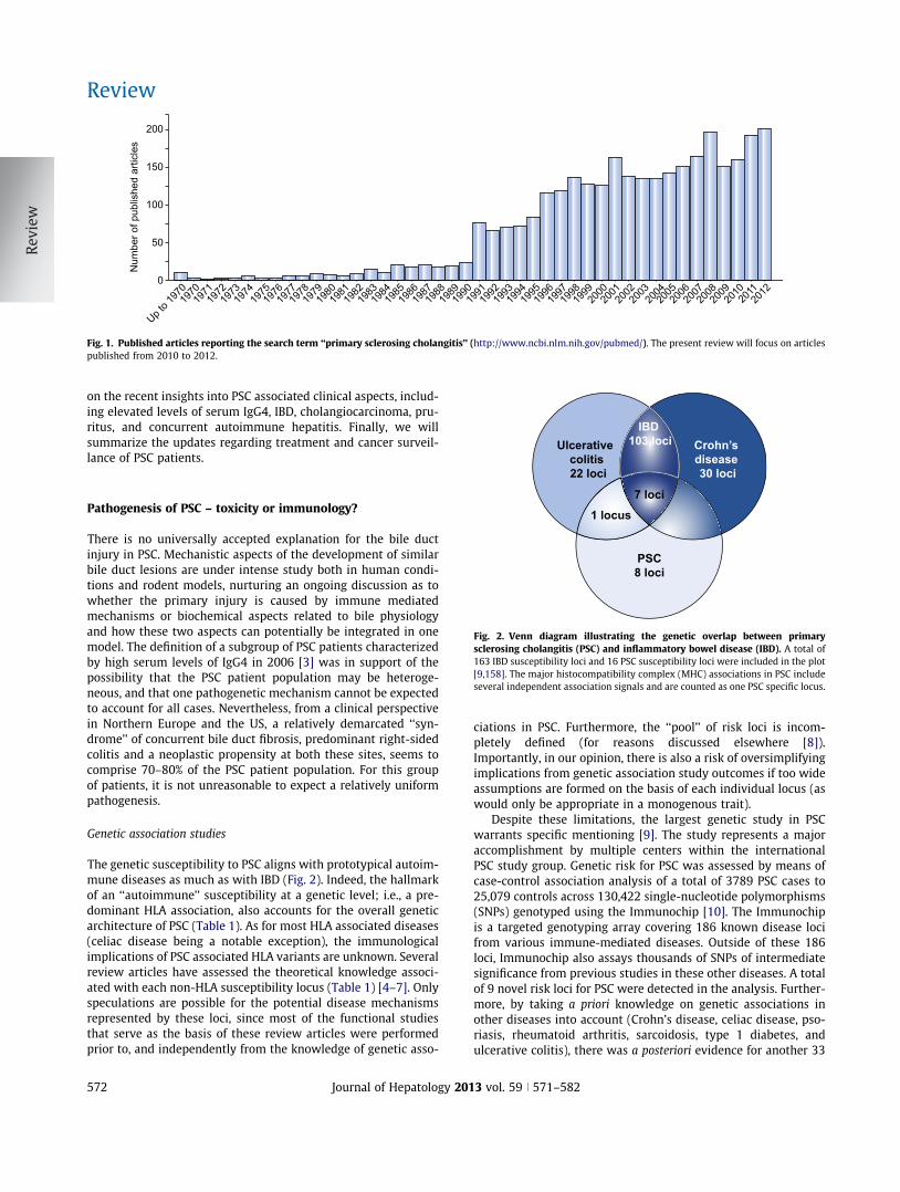
(Oslo). Later, magnetic resonance cholangiography (MRC) has beenrecommended as the primary diagnostic modality in suspectedcases of PSC (Clinical Points 1). There has been a steady growthin research activity around the many clinical challenges associatedwith PSC (Fig. 1), including the founding of an international PSCstudy group (www.ipscsg.org) in 2010. The aim of the present arti-cle is to summarize insights provided by the most recent research(published since our previous update [2]).
Clinical Points 1. Diagnosis of primary sclerosing cholangitis (PSC) [114, 117]
• In patients with a cholestatic biochemical profile not otherwise explained and where causes of secondary sclerosing cholangitis have been excluded, a diagnosis of PSC is made when magnetic resonance cholangiography (MRC) shows typical findings
• Endoscopic retrograde cholangiography (ERC) can be considered if high-quality MRC is uncertain and in patients with inflammatory bowel disease with normal high-quality MRC but high suspicion of PSC
• A liver biopsy is not necessary for the diagnosis of PSC in patients with typical cholangiographic findings
• A liver biopsy is recommended to diagnose small duct PSC if high-quality MRC (or ERC) is normal and in patients with disproportionally elevated aminotransferases to identify additional or alternative disease processes
The principle challenges in PSC all derive from the fact that
etiology and pathogenesis are still largely unknown. Since thedevelopment of sclerosing cholangitis represents a ‘‘final com-mon pathway’’ for multiple underlying mechanisms of bile ductinjury, in vivo data in patients with established PSC do not neces-sarily reflect etiology. The first part of this review elaborates onrecent insights into the pathogenesis of PSC, with a particularemphasis on research of relevance to novel treatment strategiescurrently in the testing phase. An update on aspects relevant todiagnosis of PSC will also be given, with a particular emphasis13 vol. 59 j 571–582
Ulcerative colitis
IBD 103 loci Crohn’s
disease
Num
ber o
f pub
lishe
d ar
ticle
s 200
150
100
50
0
Up to 1
970
1971
1970
197219
7319
7419
7519
7619
7719
7819
7919
8019
8119
8219
8319
8419
8519
8619
8719
8819
8919
9019
9119
9219
9319
9419
9519
9619
9719
9819
9920
0020
0120
0220
0320
0420
0520
0620
0720
0820
0920
1020
1120
12
Fig. 1. Published articles reporting the search term ‘‘primary sclerosing cholangitis’’ (http://www.ncbi.nlm.nih.gov/pubmed/). The present review will focus on articlespublished from 2010 to 2012.
Review
on the recent insights into PSC associated clinical aspects, includ-ing elevated levels of serum IgG4, IBD, cholangiocarcinoma, pru-ritus, and concurrent autoimmune hepatitis. Finally, we willsummarize the updates regarding treatment and cancer surveil-lance of PSC patients.
22 loci
PSC8 loci
7 loci
30 loci
1 locus
Fig. 2. Venn diagram illustrating the genetic overlap between primarysclerosing cholangitis (PSC) and inflammatory bowel disease (IBD). A total of163 IBD susceptibility loci and 16 PSC susceptibility loci were included in the plot[9,158]. The major histocompatibility complex (MHC) associations in PSC includeseveral independent association signals and are counted as one PSC specific locus.
Pathogenesis of PSC – toxicity or immunology?
There is no universally accepted explanation for the bile ductinjury in PSC. Mechanistic aspects of the development of similarbile duct lesions are under intense study both in human condi-tions and rodent models, nurturing an ongoing discussion as towhether the primary injury is caused by immune mediatedmechanisms or biochemical aspects related to bile physiologyand how these two aspects can potentially be integrated in onemodel. The definition of a subgroup of PSC patients characterizedby high serum levels of IgG4 in 2006 [3] was in support of thepossibility that the PSC patient population may be heteroge-neous, and that one pathogenetic mechanism cannot be expectedto account for all cases. Nevertheless, from a clinical perspectivein Northern Europe and the US, a relatively demarcated ‘‘syn-drome’’ of concurrent bile duct fibrosis, predominant right-sidedcolitis and a neoplastic propensity at both these sites, seems tocomprise 70–80% of the PSC patient population. For this groupof patients, it is not unreasonable to expect a relatively uniformpathogenesis.
Genetic association studies
The genetic susceptibility to PSC aligns with prototypical autoim-mune diseases as much as with IBD (Fig. 2). Indeed, the hallmarkof an ‘‘autoimmune’’ susceptibility at a genetic level; i.e., a pre-dominant HLA association, also accounts for the overall geneticarchitecture of PSC (Table 1). As for most HLA associated diseases(celiac disease being a notable exception), the immunologicalimplications of PSC associated HLA variants are unknown. Severalreview articles have assessed the theoretical knowledge associ-ated with each non-HLA susceptibility locus (Table 1) [4–7]. Onlyspeculations are possible for the potential disease mechanismsrepresented by these loci, since most of the functional studiesthat serve as the basis of these review articles were performedprior to, and independently from the knowledge of genetic asso-
572 Journal of Hepatology 201
ciations in PSC. Furthermore, the ‘‘pool’’ of risk loci is incom-pletely defined (for reasons discussed elsewhere [8]).Importantly, in our opinion, there is also a risk of oversimplifyingimplications from genetic association study outcomes if too wideassumptions are formed on the basis of each individual locus (aswould only be appropriate in a monogenous trait).
Despite these limitations, the largest genetic study in PSCwarrants specific mentioning [9]. The study represents a majoraccomplishment by multiple centers within the internationalPSC study group. Genetic risk for PSC was assessed by means ofcase-control association analysis of a total of 3789 PSC cases to25,079 controls across 130,422 single-nucleotide polymorphisms(SNPs) genotyped using the Immunochip [10]. The Immunochipis a targeted genotyping array covering 186 known disease locifrom various immune-mediated diseases. Outside of these 186loci, Immunochip also assays thousands of SNPs of intermediatesignificance from previous studies in these other diseases. A totalof 9 novel risk loci for PSC were detected in the analysis. Further-more, by taking a priori knowledge on genetic associations inother diseases into account (Crohn’s disease, celiac disease, pso-riasis, rheumatoid arthritis, sarcoidosis, type 1 diabetes, andulcerative colitis), there was a posteriori evidence for another 33
3 vol. 59 j 571–582
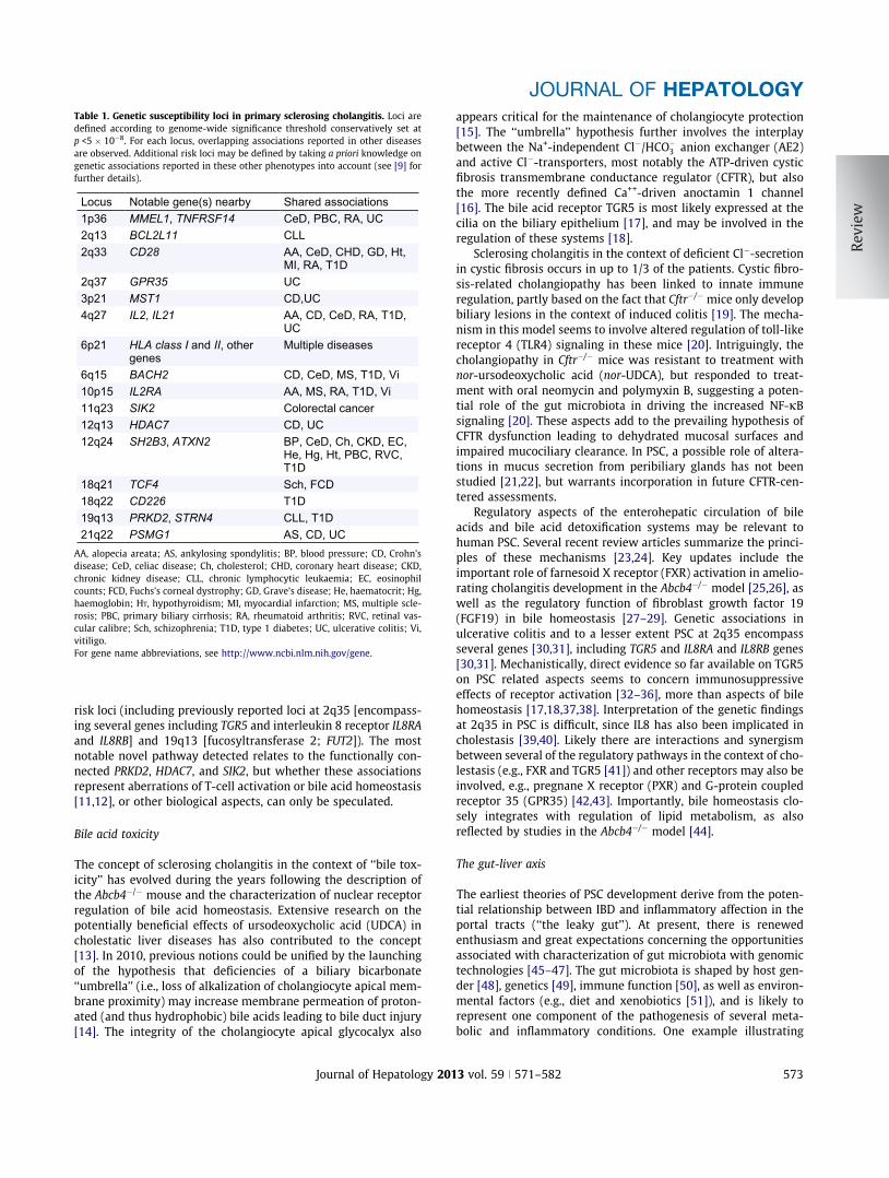
Table 1. Genetic susceptibility loci in primary sclerosing cholangitis. Loci aredefined according to genome-wide significance threshold conservatively set atp <5 � 10�8. For each locus, overlapping associations reported in other diseasesare observed. Additional risk loci may be defined by taking a priori knowledge ongenetic associations reported in these other phenotypes into account (see [9] forfurther details).
Locus Notable gene(s) nearby Shared associations1p36 MMEL1, TNFRSF14 CeD, PBC, RA, UC2q13 BCL2L11 CLL2q33 CD28 AA, CeD, CHD, GD, Ht,
MI, RA, T1D2q37 GPR35 UC3p21 MST1 CD,UC4q27 IL2, IL21 AA, CD, CeD, RA, T1D,
UC6p21 HLA class I and II, other
genesMultiple diseases
6q15 BACH2 CD, CeD, MS, T1D, Vi10p15 IL2RA AA, MS, RA, T1D, Vi11q23 SIK2 Colorectal cancer12q13 HDAC7 CD, UC12q24 SH2B3, ATXN2 BP, CeD, Ch, CKD, EC,
He, Hg, Ht, PBC, RVC, T1D
18q21 TCF4 Sch, FCD18q22 CD226 T1D19q13 PRKD2, STRN4 CLL, T1D21q22 PSMG1 AS, CD, UC
AA, alopecia areata; AS, ankylosing spondylitis; BP, blood pressure; CD, Crohn’sdisease; CeD, celiac disease; Ch, cholesterol; CHD, coronary heart disease; CKD,chronic kidney disease; CLL, chronic lymphocytic leukaemia; EC, eosinophilcounts; FCD, Fuchs’s corneal dystrophy; GD, Grave’s disease; He, haematocrit; Hg,haemoglobin; HT, hypothyroidism; MI, myocardial infarction; MS, multiple scle-rosis; PBC, primary biliary cirrhosis; RA, rheumatoid arthritis; RVC, retinal vas-cular calibre; Sch, schizophrenia; T1D, type 1 diabetes; UC, ulcerative colitis; Vi,vitiligo.For gene name abbreviations, see http://www.ncbi.nlm.nih.gov/gene.
JOURNAL OF HEPATOLOGY
risk loci (including previously reported loci at 2q35 [encompass-ing several genes including TGR5 and interleukin 8 receptor IL8RAand IL8RB] and 19q13 [fucosyltransferase 2; FUT2]). The mostnotable novel pathway detected relates to the functionally con-nected PRKD2, HDAC7, and SIK2, but whether these associationsrepresent aberrations of T-cell activation or bile acid homeostasis[11,12], or other biological aspects, can only be speculated.
Bile acid toxicity
The concept of sclerosing cholangitis in the context of ‘‘bile tox-icity’’ has evolved during the years following the description ofthe Abcb4�/� mouse and the characterization of nuclear receptorregulation of bile acid homeostasis. Extensive research on thepotentially beneficial effects of ursodeoxycholic acid (UDCA) incholestatic liver diseases has also contributed to the concept[13]. In 2010, previous notions could be unified by the launchingof the hypothesis that deficiencies of a biliary bicarbonate‘‘umbrella’’ (i.e., loss of alkalization of cholangiocyte apical mem-brane proximity) may increase membrane permeation of proton-ated (and thus hydrophobic) bile acids leading to bile duct injury[14]. The integrity of the cholangiocyte apical glycocalyx also
Journal of Hepatology 201
appears critical for the maintenance of cholangiocyte protection[15]. The ‘‘umbrella’’ hypothesis further involves the interplaybetween the Na+-independent Cl�/HCO�3 anion exchanger (AE2)and active Cl�-transporters, most notably the ATP-driven cysticfibrosis transmembrane conductance regulator (CFTR), but alsothe more recently defined Ca++-driven anoctamin 1 channel[16]. The bile acid receptor TGR5 is most likely expressed at thecilia on the biliary epithelium [17], and may be involved in theregulation of these systems [18].
Sclerosing cholangitis in the context of deficient Cl�-secretionin cystic fibrosis occurs in up to 1/3 of the patients. Cystic fibro-sis-related cholangiopathy has been linked to innate immuneregulation, partly based on the fact that Cftr�/� mice only developbiliary lesions in the context of induced colitis [19]. The mecha-nism in this model seems to involve altered regulation of toll-likereceptor 4 (TLR4) signaling in these mice [20]. Intriguingly, thecholangiopathy in Cftr�/� mice was resistant to treatment withnor-ursodeoxycholic acid (nor-UDCA), but responded to treat-ment with oral neomycin and polymyxin B, suggesting a poten-tial role of the gut microbiota in driving the increased NF-jBsignaling [20]. These aspects add to the prevailing hypothesis ofCFTR dysfunction leading to dehydrated mucosal surfaces andimpaired mucociliary clearance. In PSC, a possible role of altera-tions in mucus secretion from peribiliary glands has not beenstudied [21,22], but warrants incorporation in future CFTR-cen-tered assessments.
Regulatory aspects of the enterohepatic circulation of bileacids and bile acid detoxification systems may be relevant tohuman PSC. Several recent review articles summarize the princi-ples of these mechanisms [23,24]. Key updates include theimportant role of farnesoid X receptor (FXR) activation in amelio-rating cholangitis development in the Abcb4�/� model [25,26], aswell as the regulatory function of fibroblast growth factor 19(FGF19) in bile homeostasis [27–29]. Genetic associations inulcerative colitis and to a lesser extent PSC at 2q35 encompassseveral genes [30,31], including TGR5 and IL8RA and IL8RB genes[30,31]. Mechanistically, direct evidence so far available on TGR5on PSC related aspects seems to concern immunosuppressiveeffects of receptor activation [32–36], more than aspects of bilehomeostasis [17,18,37,38]. Interpretation of the genetic findingsat 2q35 in PSC is difficult, since IL8 has also been implicated incholestasis [39,40]. Likely there are interactions and synergismbetween several of the regulatory pathways in the context of cho-lestasis (e.g., FXR and TGR5 [41]) and other receptors may also beinvolved, e.g., pregnane X receptor (PXR) and G-protein coupledreceptor 35 (GPR35) [42,43]. Importantly, bile homeostasis clo-sely integrates with regulation of lipid metabolism, as alsoreflected by studies in the Abcb4�/� model [44].
The gut-liver axis
The earliest theories of PSC development derive from the poten-tial relationship between IBD and inflammatory affection in theportal tracts (‘‘the leaky gut’’). At present, there is renewedenthusiasm and great expectations concerning the opportunitiesassociated with characterization of gut microbiota with genomictechnologies [45–47]. The gut microbiota is shaped by host gen-der [48], genetics [49], immune function [50], as well as environ-mental factors (e.g., diet and xenobiotics [51]), and is likely torepresent one component of the pathogenesis of several meta-bolic and inflammatory conditions. One example illustrating
3 vol. 59 j 571–582 573
Pathogenesisof human
SSC
Gut microbiota
Lymphocytehoming
mechanismsIBD
Spontaneousrodent models
Inducedrodent models
Genetic association
studies
Pathogenesisof PSC
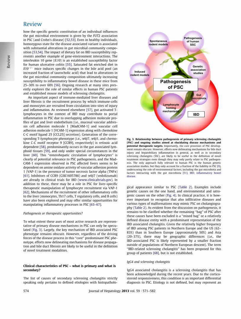
Fig. 3. Relationship between pathogenesis of primary sclerosing cholangitis(PSC) and ongoing studies aimed at elucidating disease mechanisms andpotential therapeutic targets. Importantly, the core processes of PSC develop-ment remain obscure. However, efforts now delineating mechanisms for bile ductinjury and hepatobiliary inflammation in general, as well as in secondarysclerosing cholangitis (SSC), are likely to be useful in the definition of noveltreatment strategies even though they may only partly relate to PSC pathogen-esis. The only approach fully relevant to human PSC is the human geneticassociation studies, but they only account for a fraction of the liability in PSC [9],underscoring the role of environmental factors, including the gut microbiota andfactors interacting with the gut microbiota [51]. IBD; inflammatory boweldisease.
Review
how the specific genetic constitution of an individual influencesthe gut microbial environment is given by the FUT2 associationin PSC (and Crohn’s disease) [52,53]. Even in healthy individuals,homozygous state for the disease associated variant is associatedwith substantial alterations in gut microbial community compo-sition [53,54]. The impact of dietary fat on IBD susceptibility rep-resents another example of gene-environment interactions. Theinterleukin 10 gene (IL10) is an established susceptibility factorfor human ulcerative colitis [55]. Saturated fat enriched diet inIl10�/� mice induces specific changes in the bile acid pool (anincreased fraction of taurocholic acid) that lead to alterations inthe gut microbial community composition ultimately increasingsusceptibility to inflammatory bowel disease in these mice from25–30% to over 60% [56]. Ongoing research at many sites pres-ently explores the role of similar effects in human PSC patientsand established mouse models of sclerosing cholangitis.An important aspect of immune-mediated liver diseases andliver fibrosis is the recruitment process by which immune-cellsand monocytes are recruited from circulation into sites of injuryand inflammation. As reviewed elsewhere [57], gut activated T-lymphocytes in the context of IBD may contribute to portalinflammation in PSC due to overlapping adhesion molecule pro-files of gut and liver endothelium (i.e., mucosal vascular addres-sin cell adhesion molecule 1 [MadCAM-1] and vascular celladhesion molecule 1 [VCAM-1] expression along with chemokineC-C motif ligand 25 [CCL25] secretion). Generation of the corre-sponding T-lymphocyte phenotype (i.e., a4b7, a4b1, and chemo-kine C-C motif receptor 9 [CCR9], respectively) is retinoic aciddependent [58], predominantly occurs in the gut associated lym-phoid tissues [59], and only under special circumstances in theliver [60]. This ‘‘enterohepatic circulation of lymphocytes’’ isclearly of potential relevance to PSC pathogenesis, and the Mad-CAM-1 expression observed in PSC affected livers seems to bedependent on amine oxidase activity of vascular adhesion protein1 (VAP-1) in the presence of tumor necrosis factor alpha (TNFa)[61]. Inhibitors of CCR9 (GSK1605786) and a4b7 (vedolizumab)are already in clinical trials for IBD (www.clinicaltrials.gov). Inaddition to these, there may be a role in PSC for liver-specifictherapeutic manipulation of lymphocyte recruitment via VAP-1[62]. Mechanisms of the recruitment of other inflammatory cellsto the liver (monocytes, Th17 cells, T regulatory cells, and B cells)have also been explored and may offer similar opportunities formanipulating inflammatory processes in PSC [63–67].
Pathogenesis or therapeutic opportunities?
To what extent these axes of most active research are represen-tative of primary disease mechanisms in PSC can only be specu-lated (Fig. 3). Largely, the key mechanism of IBD associated PSCphenotype remains obscure. However, regardless of the drivingforces of the disease process in this ‘‘core’’ predominant PSC phe-notype, efforts now delineating mechanisms for disease propaga-tion and bile duct fibrosis are likely to be useful in the definitionof novel treatment modalities.
Clinical characteristics of PSC – what is primary and what issecondary?
The list of causes of secondary sclerosing cholangitis strictlyspeaking only pertains to defined etiologies with histopatholo-
574 Journal of Hepatology 201
gical appearance similar to PSC (Table 2). Examples includegenetic causes on the one hand, and environmental and iatro-genic causes on the other (Fig. 4). In clinical practice, it is how-ever important to recognize that also infiltrative diseases andvarious types of malformations may mimic PSC on cholangiogra-phy (Table 2). As evident from the discussion on pathogenesis, itremains to be clarified whether the remaining ‘‘bag’’ of PSC afterthese causes have been excluded is a ‘‘mixed bag’’ or a relativelydefined disease entity with a predominant representation of theIBD associated cholangitis. Given the relatively higher frequencyof IBD among PSC patients in Northern Europe and the US (62–83%) than in Southern Europe (approximately 50%) and Asia(20–37%), there may be geographic differences (i.e., theIBD-associated PSC is likely represented by a smaller fractionoutside of populations of Northern European descent). The term‘‘IBD-related sclerosing cholangitis’’ has been proposed for thisgroup of patients [68], but is not established.
IgG4 and sclerosing cholangitis
IgG4 associated cholangitis is a sclerosing cholangitis that hasbeen acknowledged during the recent years. Due to the cortico-steroid responsiveness, this condition is an important differentialdiagnosis to PSC. Etiology is not defined, but may represent an
3 vol. 59 j 571–582
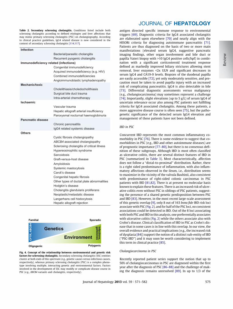
Table 2. Secondary sclerosing cholangitis. Conditions listed include bothsclerosing cholangitis according to defined etiologies and liver affections thatmay mimic primary sclerosing cholangitis (PSC) on cholangiography. Accordingto clinical practice guidelines, IgG4 related disease is now considered in thecontext of secondary sclerosing cholangitis [114,117].
Infection Bacterial/parasitic cholangitisRecurrent pyogenic cholangitis
Mechanic/toxicCholelithiasis/choledocholithiasisSurgical bile duct traumaIntra-arterial chemotherapy
IschaemicVascular trauma
Paroxysmal nocturnal haemoglobinuriaPancreatic disease
Chronic pancreatitisIgG4 related systemic disease
Others
ABCB4 associated cholangiopathySclerosing cholangitis of critical illnessHypereosinophilic syndromeSarcoidosisGraft-versus-host diseaseAmyloidosisSystemic mastocytosisCaroli’s disease
Other types of ductal plate abnormalitiesHodgkin’s diseaseCholangitis glandularis proliferansNeoplastic/metastatic diseaseLangerhans cell histiocytosisHepatic allograft rejection
Immunodeficiency related (infections)Congenital immunodeficiencyAcquired immunodeficiency (e.g. HIV)Combined immunodeficienciesAngioimmunoblastic lymphadenopathy
Hepatic allograft arterial insufficiency
Cystic fibrosis cholangiopathy
Congenital hepatic fibrosis
Familial Sporadic
Oligogenic Polygenic
CFT
R, A
BC
B4 Infections,
gallstones
Genetics
Environment
Fig. 4. Concept of the relationship between environmental and genetic riskfactors for sclerosing cholangitis. Secondary sclerosing cholangitis (SSC) entitiescluster at both ends of this spectrum (e.g., genetic causes versus infectious causes,respectively), whereas primary sclerosing cholangitis (PSC) is a complex pheno-type involving multiple, interacting genetic and environmental factors. Factorsinvolved in the development of SSC may modify or complicate disease course inPSC (e.g., ABCB4 variants and cholangitis, respectively).
JOURNAL OF HEPATOLOGY
Journal of Hepatology 201
antigen directed specific immune response to environmentaltriggers [69]. Diagnostic criteria for IgG4 associated cholangitisare elaborated upon elsewhere [70] and nearly align with theHISORt criteria for diagnosing autoimmune pancreatitis [71].Patients are thus diagnosed on the basis of two or more mainmanifestations (elevated serum IgG4, suggestive pancreaticimaging findings, other organ involvement and bile duct orpapilla Vateri biopsy with >10 IgG4 positive cells/hpf) in combi-nation with a significant corticosteroid treatment responsedefined as markedly improved biliary strictures allowing stentremoval, liver enzymes <2x ULN and significant decreases inserum IgG4 and CA19-9 levels. Biopsies of the duodenal papillaare easily accessible [72], yet only moderately sensitive, and pre-caution must be taken to avoid papilla injury with an increasedrisk of complicating pancreatitis. IgG4 is also detectable in bile[73]. Differential diagnostic assessments versus malignancy(e.g., cholangiocarcinoma) may sometimes represent a challenge[74]. Importantly, slight elevations (up to 5 g/L) of serum IgG4 ofuncertain relevance occur also among PSC patients not fulfillingcriteria for IgG4 associated cholangitis. Among these patients, amore aggressive disease course is often seen [75], but the patho-genetic significance of the detected serum IgG4 elevation andmanagement of these patients have not been defined.
IBD in PSC
Concurrent IBD represents the most common inflammatory co-morbidity in PSC [76]. There is some evidence to suggest that co-morbidities in PSC (e.g., IBD and other autoimmune diseases) areof prognostic importance [77–80], but there is no consensus defi-nition of these subgroups. Although IBD is most often classifiedas ulcerative colitis, there are several distinct features of IBD inPSC (summarized in Table 3). Most characteristically, affectiondoes not follow a ‘‘distal-to-proximal’’ distribution. Rather, thereis a right sided predominance of inflammation, with also inflam-matory affections observed in the ileum, i.e., distribution seemsto maximize in the vicinity of the valvula Bauhinii, also consistentwith predominance of right-sided colonic carcinomas in PSCpatients with IBD [81,82]. There is at present no molecular basisknown to explain these features. There is an increased risk of ulcer-ative colitis even without PSC in siblings of PSC patients, suggest-ing the presence of a shared genetic predisposition between PSCand IBD [83]. However, in the most recent large-scale assessmentof this genetic overlap [9], only 8 out of 163 bona fide IBD risk lociassociate with PSC (Fig. 2), and for half of the PSC loci, no consistentassociations could be detected in IBD. Out of the 8 loci associatingwith both PSC and IBD in this analysis, one preferentially associateswith ulcerative colitis (Fig. 2) while the others associate also withCrohn’s disease. Clinical classification of IBD in PSC as Crohn’s dis-ease that in some cases is in line with this overlap. In our view, theoverall evidence and practical implications (e.g., the increased riskof dysplasia [84]) support the notion of a distinct sub-entity of IBD(‘‘PSC-IBD’’) and it may soon be worth considering to implementthis term in clinical practice [85].
Cholangiocarcinoma in PSC
Recently reported patient series support the notion that up to50% of cholangiocarcinomas in PSC are diagnosed within the firstyear after the diagnosis of PSC [86–88] and the challenge of mak-ing the diagnosis remains unresolved [89]. In up to 1/3 of the
3 vol. 59 j 571–582 575
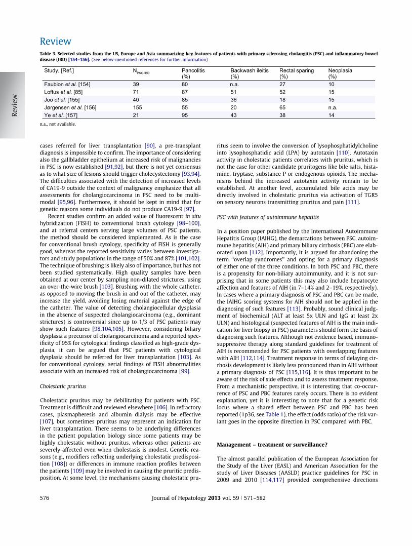
Table 3. Selected studies from the US, Europe and Asia summarizing key features of patients with primary sclerosing cholangitis (PSC) and inflammatory boweldisease (IBD) [154–156]. (See below-mentioned references for further information)
Study, [Ref.] NPSC-IBD Pancolitis (%)
Backwash ileitis (%)
Rectal sparing (%)
Neoplasia (%)
Faubion et al. [154] 39 80 n.a. 27 10Loftus et al. [85] 71 87 51 52 15Joo et al. [155] 40 85 36 18 15Jørgensen et al. [156] 155 55 20 65 n.a.Ye et al. [157] 21 95 43 38 14
n.a., not available.
Review
cases referred for liver transplantation [90], a pre-transplantdiagnosis is impossible to confirm. The importance of consideringalso the gallbladder epithelium at increased risk of malignanciesin PSC is now established [91,92], but there is not yet consensusas to what size of lesions should trigger cholecystectomy [93,94].The difficulties associated with the detection of increased levelsof CA19-9 outside the context of malignancy emphasize that allassessments for cholangiocarcinoma in PSC need to be multi-modal [95,96]. Furthermore, it should be kept in mind that forgenetic reasons some individuals do not produce CA19-9 [97].
Recent studies confirm an added value of fluorescent in situhybridization (FISH) to conventional brush cytology [98–100],and at referral centers serving large volumes of PSC patients,the method should be considered implemented. As is the casefor conventional brush cytology, specificity of FISH is generallygood, whereas the reported sensitivity varies between investiga-tors and study populations in the range of 50% and 87% [101,102].The technique of brushing is likely also of importance, but has notbeen studied systematically. High quality samples have beenobtained at our center by sampling non-dilated strictures, usingan over-the-wire brush [103]. Brushing with the whole catheter,as opposed to moving the brush in and out of the catheter, mayincrease the yield, avoiding losing material against the edge ofthe catheter. The value of detecting cholangiocellular dysplasiain the absence of suspected cholangiocarcinoma (e.g., dominantstrictures) is controversial since up to 1/3 of PSC patients mayshow such features [98,104,105]. However, considering biliarydysplasia a precursor of cholangiocarcinoma and a reported spec-ificity of 95% for cytological findings classified as high-grade dys-plasia, it can be argued that PSC patients with cytologicaldysplasia should be referred for liver transplantation [103]. Asfor conventional cytology, serial findings of FISH abnormalitiesassociate with an increased risk of cholangiocarcinoma [99].
Cholestatic pruritus
Cholestatic pruritus may be debilitating for patients with PSC.Treatment is difficult and reviewed elsewhere [106]. In refractorycases, plasmapheresis and albumin dialysis may be effective[107], but sometimes pruritus may represent an indication forliver transplantation. There seems to be underlying differencesin the patient population biology since some patients may behighly cholestatic without pruritus, whereas other patients areseverely affected even when cholestasis is modest. Genetic rea-sons (e.g., modifiers reflecting underlying cholestatic predisposi-tion [108]) or differences in immune reaction profiles betweenthe patients [109] may be involved in causing the pruritic predis-position. At some level, the mechanisms causing cholestatic pru-
576 Journal of Hepatology 201
ritus seem to involve the conversion of lysophosphatidylcholineinto lysophosphatidic acid (LPA) by autotaxin [110]. Autotaxinactivity in cholestatic patients correlates with pruritus, which isnot the case for other candidate pruritogens like bile salts, hista-mine, tryptase, substance P or endogenous opioids. The mecha-nisms behind the increased autotaxin activity remain to beestablished. At another level, accumulated bile acids may bedirectly involved in cholestatic pruritus via activation of TGR5on sensory neurons transmitting pruritus and pain [111].
PSC with features of autoimmune hepatitis
In a position paper published by the International AutoimmuneHepatitis Group (IAIHG), the demarcations between PSC, autoim-mune hepatitis (AIH) and primary biliary cirrhosis (PBC) are elab-orated upon [112]. Importantly, it is argued for abandoning theterm ‘‘overlap syndromes’’ and opting for a primary diagnosisof either one of the three conditions. In both PSC and PBC, thereis a propensity for non-biliary autoimmunity, and it is not sur-prising that in some patients this may also include hepatocyteaffection and features of AIH (in 7–14% and 2–19%, respectively).In cases where a primary diagnosis of PSC and PBC can be made,the IAIHG scoring systems for AIH should not be applied in thediagnosing of such features [113]. Probably, sound clinical judg-ment of biochemical (ALT at least 5x ULN and IgG at least 2xULN) and histological (suspected features of AIH is the main indi-cation for liver biopsy in PSC) parameters should form the basis ofdiagnosing such features. Although not evidence based, immuno-suppressive therapy along standard guidelines for treatment ofAIH is recommended for PSC patients with overlapping featureswith AIH [112,114]. Treatment response in terms of delaying cir-rhosis development is likely less pronounced than in AIH withouta primary diagnosis of PSC [115,116]. It is thus important to beaware of the risk of side effects and to assess treatment response.From a mechanistic perspective, it is interesting that co-occur-rence of PSC and PBC features rarely occurs. There is no evidentexplanation, yet it is interesting to note that for a genetic risklocus where a shared effect between PSC and PBC has beenreported (1p36, see Table 1), the effect (odds ratio) of the risk var-iant goes in the opposite direction in PSC compared with PBC.
Management – treatment or surveillance?
The almost parallel publication of the European Association forthe Study of the Liver (EASL) and American Association for thestudy of Liver Diseases (AASLD) practice guidelines for PSC in2009 and 2010 [114,117] provided comprehensive directions
3 vol. 59 j 571–582
JOURNAL OF HEPATOLOGY
for the diagnosis and management of PSC patients (Clinical Points1 and 2). In most aspects, the guidelines agree (reviewed else-where [2]), yet the discrepancy on principle directions for pre-scription of UDCA caused a still ongoing discussion on whichone of the conclusions was most appropriate. The EASL guidelinesconcluded that limitations in available data did not yet allow aspecific recommendation for the general use of UDCA in PSC.The same guidelines opened up to the use of moderate doseUDCA (15–20 mg/day) in individuals with strong family historyof colorectal cancer, previous colorectal neoplasia or longstand-ing extensive colitis, not for the improvement of liver disease,but to prevent neoplasia. The AASLD guidelines unambiguouslyrecommend against the use of UDCA in adult PSC patients.Clinical Points 2. Treatment of primary sclerosing cholangitis [114, 117]
• The role for ursodeoxycholic acid (UDCA) in PSC is currently debated. There is no definite evidence that UDCA improves survival or is efficacious in the prevention of colorectal or biliary neoplasia. Practice varies, however, between centers. It has been the tradition of the current authors to be reluctant to start PSC patients on UDCA
• Although not evidence-based, PSC patients with features overlapping with those of autoimmune hepatitis should be considered for therapy with corticosteroids and/or other immunosuppressive agents. Therapy should be individualized and adjusted according to response and the risk of side effects
• In PSC patients with dominant bile duct strictures causing biochemical and/or clinical signs of cholestasis, endoscopic dilatation with or without stenting is recommended. Short-term (2-3 weeks) stenting is favored by many, including the current authors. A multicenter, prospective, randomized intervention trial to compare the efficacy of single session balloon dilatation and short-term stenting is currently ongoing. Prophylactic antimicrobial therapy is recommended during the procedure
• Liver transplantation is a curative treatment modality in PSC patients with end-stage liver disease and in selected cases with severe symptoms of cholestasis. Transplantation may also be considered in patients with evidence of biliary epithelium dysplasia
Bile acid therapy
We will not re-review the literature that led to the slightly differ-ing conclusions on UDCA therapy in PSC [2], but rather mentionsome subsequent reports that have elaborated on the complexityof the problem. Serum alkaline phosphatase (ALP) level is a longestablished risk factor for disease progression in PSC [118]. Geneticrisk factors for PSC (i.e., FUT2) overlap with genetic factors influ-encing serum levels of ALP in healthy individuals [119,120], buthow these factors are involved in the prognostic importance ofALP has not been studied. In a retrospective analysis of 139 PSCpatients [121], those receiving UDCA and achieving an improve-ment of ALP to <1.5x ULN had significantly longer survival without
Journal of Hepatology 201
end points compared with patients without ALP normalization. Inseries involving patients from previous UDCA treatment trials, theassociation between ALP normalization and PSC behavior seemedto be independent of UDCA administration [122,123]. The inter-pretation of all these data requires some caution; in principle bothUDCA administration and PSC behavior seem to independentlyinfluence ALP levels. At present, it cannot be claimed that ALP nor-malization in the context of UDCA administration is representativeof drug efficacy. Accounting for FUT2 related biology, there is astrong basis for further studies to clarify these mechanisms thatseem to converge on a robust representative of disease intensityin PSC, potentially also amendable by means of bile acid therapy.
The effects of high-dose UDCA (28–30 mg/kg/day) treatmenton the bile acid pool warrant reflections [124]. In PSC patientsreceiving such treatment, there was both an expansion and com-positional changes of the total bile acid pool. While only theexpansion seemed to associate significantly with adverse out-comes (progression to cirrhosis, development of varices, cholan-giocarcinoma, liver transplantation, and death), the increasedproportion of lithocholic acid found in this trial is theoreticallyharmful. It needs to be noted that in another study of bile acidpool compositional effects from UDCA treatment, only UDCAitself was significantly enriched [125]. Nevertheless, given thedisease distribution in PSC, predominantly affecting the ‘‘proxim-ities of the enterohepatic circulation’’, the influence of bile acidtreatment, or treatment aiming at modifying the regulation ofbile acid homeostasis (e.g., FXR agonists), needs further consider-ation. Importantly, the bile acid pool serves not only as a poten-tial ‘‘toxic medium’’ to epithelial surfaces, but profoundlyinteracts with the metabolism and immune function via effectson the gut microbiota [56,126,127]. There is growing interest inthe influence of this interaction on IBD and liver diseases [128–130], and it may be wise to advice for future clinical trials inPSC to include assessments of bile acid composition and metage-nomic parameters in the basic study readout [51].
Antibiotics and beyond
Of the many approaches currently explored for treatment of PSC,antibiotics most clearly link up with the intersections betweeninflammation, bile acid homeostasis, and gut microbiota. Distin-guishing between non-absorbable (i.e., local effects on the gutmicrobiota only) and absorbable (i.e., including a portal and sys-temic anti-microbial effect) is reasonable. Non-absorbable antibi-otics like vancomycin exert effects on systemic immune function,e.g., by influencing regulatory T-cell subsets. In a trial of 14 pedi-atric patients with PSC [131], significant improvement in hepaticbiochemistries was observed, paralleling increased levels of reg-ulatory T-cells. Regarding absorbable antibiotics, improvementsin hepatic biochemistries have also been observed for metronida-zole [132], azithromycin [133] and minocycline [134], altogethersuggesting that liver affection in PSC may be influenced by anti-microbial therapies. We anticipate that upon the further charac-terization of metagenomic components in PSC pathogenesis(Fig. 3), specific attempts to manipulate these (by antibiotics, pro-biotics, prebiotics or dietary means) will be made.
The paradox of immunosuppression
The strong HLA association along with multiple shared risk locibetween PSC and prototypical autoimmune diseases (Table 1),
3 vol. 59 j 571–582 577

Review
more pronounced than for IBD, strongly suggests that PSC patho-genesis has an ‘‘autoimmune’’ component [2]. Antibodies againstbiliary as well as colonic epithelial cells have been reported, andthe preferential usage of particular T-cell receptor gene segmentsof hepatic T-cells also suggests the existence of tissue-specificantigens in PSC pathogenesis. In spite of this, no immunosuppres-sive drug tested to date has shown to significantly improve clin-ical outcomes. Of particular interest are cyclosporine A (CsA) andtacrolimus that exert immunosuppressive actions via inhibitionof calcineurin, which again is indispensable for cytokine induc-tion, interleukin 2 (IL2) in particular, upon engagement of theT-cell receptor. Genetic associations in PSC are reported both atthe IL2 and IL2 receptor alpha (IL2RA) loci (Table 1), directly sug-gesting an involvement of the IL2-signaling pathway in PSC path-ogenesis. Treatment trials show some influence from CsA on IBDactivity in PSC [135], and from tacrolimus on hepatic biochemist-ries [136], but no effects on clinical end points are evident andside effects preclude a broad application. Effects of CsA andtacrolimus may also differ, at least concerning IBD in the contextof PSC. This was recently underscored by the finding of enhancedIBD activity after liver transplantation for PSC in patients receiv-ing tacrolimus and mycophenolate compared with patients onCsA and azathioprine [137]. Mechanistic elucidation of theimmunological consequences of PSC susceptibility loci in vivoneeds to be performed before rational application of immunosup-pression can be drafted on the basis of genetics.Antifibrotic therapy
To some extent, it may be argued that present clinical trials in PSChave concerned patients in whom fibrotic lesions are already mani-fested and efficacy of immunosuppressive and antibiotic treatmentcannot be expected. Antifibrotic treatment strategies in preparationlargely build on studies on the Abcb4�/� mouse model. Some effectsconverge on nuclear receptor signaling pathways, including TGR5,FXR, and peroxisome proliferator activated receptor gamma (PPARc)[26,138]. So far, no nuclear receptor has been identified as a target ofnor-UDCA [139]. Other components of the Abcb4�/� fibrogenesis arecurrently being targeted in treatment trials, e.g., lysyl oxidase-like 2[140] by monoclonal antibody GS6624 (http://clinicaltrials.gov/ct2/show/NCT01672853). Up to the point where a diagnosis of PSC maybe made at a pre-clinical, pre-fibrotic stage, therapies derived fromthis model may hold promise.
Cholangiocarcinoma surveillance
The most challenging aspect of PSC follow-up remains the unpre-dictable occurrence of cholangiocarcinoma. Management andsurveillance strategies are reviewed elsewhere [89], and novelaspects predominantly derive from proteomic and epigeneticapproaches. In serum, a proteomic study report the potentialdiagnostic improvement achieved by including serum leucine-rich a-2-glycoprotein (LRG1) and IL6 in prediction models forcholangiocarcinoma [141]. In bile, a panel of 22 peptides detectedin a screening panel was shown to discriminate between cholan-giocarcinoma and PSC with an area under the receiver operatingcharacteristics curve of 87% [142], correctly detecting 8 out of 10cases of cholangiocarcinoma complicating PSC. In urine, a similarapproach led to comparable results [143], with correct classifica-tion of 10 out of 10 cases of cholangiocarcinoma arising in thecontext of PSC. Based on experience in other cancers and preli-
578 Journal of Hepatology 201
minary data [144,145], epigenetic markers are also potential bio-markers for early cholangiocarcinoma development. Thisaccounts for genes affected by CpG hypermethylation [146], aswell as cholangiocarcinoma-associated serum micro RNA pro-files. The prospective clinical utility of all these approaches ispresently under study at several sites and it is likely that someapproaches will be incorporated in patient management in thenear future. This will ease timing for liver transplantation andthus ultimately assist in reducing cholangiocarcinoma-relateddeaths in the PSC population.
Liver transplantation
Due to the lack of effective medical treatment, liver transplanta-tion remains the principle therapeutic option in PSC. The presentliterature on the high risk of acute cellular rejections in PSC follow-ing liver transplantation and PSC recurrence in the allograft wasrecently reviewed elsewhere [147]. One key ongoing discussionrelates to whether the presence of an intact colon in PSC patientswith IBD influences disease recurrence, but due to the lack ofreproduction so far [148], these data are only useful for pathophys-iological considerations and should not prompt pre-transplanta-tion colectomy on a general basis. In our center, biliaryepithelium dysplasia by brush cytology is considered an indicationfor liver transplantation in PSC, but this practice has not been pro-spectively evaluated. Also, in highly selected cases of hilar cholan-giocarcinoma, liver transplantation in conjunction with neo-adjuvant chemotherapy and radiation should be considered[149,150], in particular since patients with PSC may present withfavorable tumor characteristics compared with cholangiocarci-noma outside of the context of PSC [151]. Further studies areneeded to clarify the utility of liver transplantation for intrahepaticcholangiocarcinoma [152], which is presently advised against. Inareas where prioritizations for liver transplantations are made onthe basis of model of end-stage liver disease (MELD) assessments,issues related to malignancies in PSC add to an already ongoing dis-cussion on exception criteria for patients with PSC [153].
Conclusions
There is a growing interest in the hepatological community toresolve the main challenges associated with PSC. Ongoing effortsto delineate pathogenesis are of key importance, since they ulti-mately may guide the rational management of remaining clinicalchallenges, i.e., early diagnosis of PSC in patients with IBD, earlydiagnosis of cholangiocarcinoma in PSC, determining surrogatemarkers for disease progression and disease activity; and finally,treatment modalities that significantly influence clinical outcome.
Conflict of interest
The authors declared that they do not have anything to discloseregarding funding or conflict of interest with respect to thismanuscript.
References
[1] Hoffman CEE. Verschluss der gallenwege durch verdickung der wandungen.Arch Pathol Anat Physiol 1867;39:206–215.
3 vol. 59 j 571–582
JOURNAL OF HEPATOLOGY
[2] Karlsen TH, Schrumpf E, Boberg KM. Update on primary sclerosingcholangitis. Dig Liver Dis 2010;42:390–400.[3] Mendes FD, Jorgensen R, Keach J, Katzmann JA, Smyrk T, Donlinger J, et al.
Elevated serum IgG4 concentration in patients with primary sclerosingcholangitis. Am J Gastroenterol 2006;101:2070–2075.
[4] Karlsen TH. A lecture on the genetics of primary sclerosing cholangitis. DigDis 2012;30:32–38.
[5] Karlsen TH, Kaser A. Deciphering the genetic predisposition to primarysclerosing cholangitis. Semin Liver Dis 2011;31:188–207.
[6] Folseraas T, Melum E, Franke A, Karlsen TH. Genetics in primary sclerosingcholangitis. Best Pract Res Clin Gastroenterol 2011;25:713–726.
[7] Naess S, Shiryaev A, Hov JR, Franke A, Karlsen TH. Genetics in primarysclerosing cholangitis. Clin Res Hepatol Gastroenterol 2012;36:325–333.
[8] Manolio TA, Collins FS, Cox NJ, Goldstein DB, Hindorff LA, Hunter DJ, et al.Finding the missing heritability of complex diseases. Nature2009;461:747–753.
[9] Liu JZ, Hov JE, Folseraas T, Ellinghaus E, Rushbrook SM, Doncheva NT, et al.Dense genotyping of immune-related disease regions identifies nine newrisk loci for primary sclerosing cholangitis. Nature Genetics2013;45:670–675.
[10] Cortes A, Brown MA. Promise and pitfalls of the Immunochip. Arthritis ResTher 2011;13:101.
[11] Mitro N, Godio C, De Fabiani E, Scotti E, Galmozzi A, Gilardi F, et al. Insightsin the regulation of cholesterol 7 alpha-hydroxylase gene reveal a target formodulating bile acid synthesis. Hepatology 2007;46:885–897.
[12] Clark K, MacKenzie KF, Petkevicius K, Kristariyanto Y, Zhang J, Choi HG,et al. Phosphorylation of CRTC3 by the salt-inducible kinases controls theinterconversion of classically activated and regulatory macrophages. ProcNatl Acad Sci U S A 2012;109:16986–16991.
[13] Beuers U. Drug insight: mechanisms and sites of action of ursodeoxycholicacid in cholestasis. Nat Clin Pract Gastroenterol Hepatol 2006;3:318–328.
[14] Beuers U, Hohenester S, De Buy Wenniger LJ, Kremer AE, Jansen PL, ElferinkRP. The biliary HCO(3) (�) umbrella: a unifying hypothesis on pathogeneticand therapeutic aspects of fibrosing cholangiopathies. Hepatology2010;52:1489–1496.
[15] Hohenester S, Wenniger LM, Paulusma CC, van Vliet SJ, Jefferson DM,Elferink RP, et al. A biliary HCO3- umbrella constitutes a protectivemechanism against bile acid-induced injury in human cholangiocytes.Hepatology 2012;55:173–183.
[16] Dutta AK, Khimji AK, Kresge C, Bugde A, Dougherty M, Esser V, et al.Identification and functional characterization of TMEM16A, a Ca2+-acti-vated Cl� channel activated by extracellular nucleotides, in biliary epithe-lium. J Biol Chem 2011;286:766–776.
[17] Keitel V, Ullmer C, Haussinger D. The membrane-bound bile acid receptorTGR5 (Gpbar-1) is localized in the primary cilium of cholangiocytes. BiolChem 2010;391:785–789.
[18] Keitel V, Cupisti K, Ullmer C, Knoefel WT, Kubitz R, Haussinger D. Themembrane-bound bile acid receptor TGR5 is localized in the epithelium ofhuman gallbladders. Hepatology 2009;50:861–870.
[19] Blanco PG, Zaman MM, Junaidi O, Sheth S, Yantiss RK, Nasser IA, et al.Induction of colitis in Cftr�/� mice results in bile duct injury. Am J PhysiolGastrointest Liver Physiol 2004;287:G491–G496.
[20] Fiorotto R, Scirpo R, Trauner M, Fabris L, Hoque R, Spirli C, et al. Loss of CFTRaffects biliary epithelium innate immunity and causes TLR4-NF-kappaB-mediated inflammatory response in mice. Gastroenterology2011;141:1498–1508, e1491–1495.
[21] McMinn RM, Kugler JH. The glands of the bile and pancreatic ducts:autoradiographic and histochemical studies. J Anat 1961;95:1–11.
[22] Spitz L, Petropoulos A. The development of the glands of the common bileduct. J Pathol 1979;128:213–220.
[23] Halilbasic E, Claudel T, Trauner M. Bile acid transporters and regulatorynuclear receptors in the liver and beyond. J Hepatol 2013;58:155–168.
[24] Trauner M, Halilbasic E. Nuclear receptors as new perspective for themanagement of liver diseases. Gastroenterology 2011;140:1120–1125,e1121–1112.
[25] Modica S, Petruzzelli M, Bellafante E, Murzilli S, Salvatore L, Celli N, et al.Selective activation of nuclear bile acid receptor FXR in the intestineprotects mice against cholestasis. Gastroenterology 2012;142:355–365,e351–354.
[26] Baghdasaryan A, Claudel T, Gumhold J, Silbert D, Adorini L, Roda A, et al.Dual farnesoid X receptor/TGR5 agonist INT-767 reduces liver injury in theMdr2�/� (Abcb4�/�) mouse cholangiopathy model by promoting biliaryHCO output. Hepatology 2011;54:1303–1312.
Journal of Hepatology 201
[27] Schaap FG, van der Gaag NA, Gouma DJ, Jansen PL. High expression of thebile salt-homeostatic hormone fibroblast growth factor 19 in the liver ofpatients with extrahepatic cholestasis. Hepatology 2009;49:1228–1235.
[28] Triantis V, Saeland E, Bijl N, Oude-Elferink RP, Jansen PL. Glycosylation offibroblast growth factor receptor 4 is a key regulator of fibroblast growthfactor 19-mediated down-regulation of cytochrome P450 7A1. Hepatology2010;52:656–666.
[29] Zweers SJ, Booij KA, Komuta M, Roskams T, Gouma DJ, Jansen PL, et al. Thehuman gallbladder secretes fibroblast growth factor 19 into bile: towardsdefining the role of fibroblast growth factor 19 in the enterobiliary tract.Hepatology 2012;55:575–583.
[30] Anderson CA, Boucher G, Lees CW, Franke A, D’Amato M, Taylor KD, et al.Meta-analysis identifies 29 additional ulcerative colitis risk loci, increasingthe number of confirmed associations to 47. Nat Genet 2011;43:246–252.
[31] Hov JR, Keitel V, Laerdahl JK, Spomer L, Ellinghaus E, Elsharawy A, et al.Mutational characterization of the bile acid receptor TGR5 in primarysclerosing cholangitis. PLoS One 2010;5:e12403.
[32] Wang YD, Chen WD, Yu D, Forman BM, Huang W. The G-protein-coupledbile acid receptor, Gpbar1 (TGR5), negatively regulates hepatic inflamma-tory response through antagonizing nuclear factor kappa light-chainenhancer of activated B cells (NF-kappaB) in mice. Hepatology2011;54:1421–1432.
[33] Cipriani S, Mencarelli A, Chini MG, Distrutti E, Renga B, Bifulco G, et al. Thebile acid receptor GPBAR-1 (TGR5) modulates integrity of intestinal barrierand immune response to experimental colitis. PLoS One 2011;6:e25637.
[34] Keitel V, Donner M, Winandy S, Kubitz R, Haussinger D. Expression andfunction of the bile acid receptor TGR5 in Kupffer cells. Biochem BiophysRes Commun 2008;372:78–84.
[35] Kawamata Y, Fujii R, Hosoya M, Harada M, Yoshida H, Miwa M, et al. A Gprotein-coupled receptor responsive to bile acids. J Biol Chem2003;278:9435–9440.
[36] Chen WD, Yu D, Forman BM, Huang W, Wang YD. Deficiency of G-protein-coupled bile acid receptor Gpbar1 (TGR5) enhances chemically inducedliver carcinogenesis. Hepatology 2013;57:656–666.
[37] Li T, Holmstrom SR, Kir S, Umetani M, Schmidt DR, Kliewer SA, et al. The Gprotein-coupled bile acid receptor, TGR5, stimulates gallbladder filling. MolEndocrinol 2011;25:1066–1071.
[38] Keitel V, Haussinger D. TGR5 in the biliary tree. Dig Dis 2011;29:45–47.[39] Isse K, Harada K, Nakanuma Y. IL-8 expression by biliary epithelial cells is
associated with neutrophilic infiltration and reactive bile ductules. Liver Int2007;27:672–680.
[40] Bansal AS, Thomson A, Steadman C, Le Gros G, Hogan PG, Kerlin P, et al.Serum levels of interleukins 8 and 10, interferon gamma, granulocyte-macrophage colony stimulating factor and soluble CD23 in patients withprimary sclerosing cholangitis. Autoimmunity 1997;26:223–229.
[41] Baghdasaryan A, Claudel T, Gumhold J, Silbert D, Adorini L, Roda A, et al.Dual farnesoid X receptor/TGR5 agonist INT-767 reduces liver injury in theMdr2�/� (Abcb4�/�) mouse cholangiopathy model by promoting biliaryHCO(�)(3) output. Hepatology 2011;54:1303–1312.
[42] Karlsen TH, Lie BA, Frey Froslie K, Thorsby E, Broome U, Schrumpf E, et al.Polymorphisms in the steroid and xenobiotic receptor gene influencesurvival in primary sclerosing cholangitis. Gastroenterology2006;131:781–787.
[43] Ellinghaus D, Folseraas T, Holm K, Ellinghaus E, Melum E, Balschun T, et al.Genome-wide association analysis in primary sclerosing cholangitis andulcerative colitis identifies risk loci at GPR35 and TCF4. Hepatology 2013.http://dx.doi.org/10.1002/hep.25977, in press.
[44] Moustafa T, Fickert P, Magnes C, Guelly C, Thueringer A, Frank S, et al.Alterations in lipid metabolism mediate inflammation, fibrosis, and prolif-eration in a mouse model of chronic cholestatic liver injury. Gastroenter-ology 2012;142:e112.
[45] Weinstock GM. Genomic approaches to studying the human microbiota.Nature 2012;489:250–256.
[46] The Human Microbiome Project Consortium. Structure, function anddiversity of the healthy human microbiome. Nature 2012;486:207–214.
[47] Kuczynski J, Lauber CL, Walters WA, Parfrey LW, Clemente JC, Gevers D,et al. Experimental and analytical tools for studying the human microb-iome. Nat Rev Genet 2012;13:47–58.
[48] Markle JG, Frank DN, Mortin-Toth S, Robertson CE, Feazel LM, Rolle-Kampczyk U, et al. Sex differences in the gut microbiome drive hormone-dependent regulation of autoimmunity. Science 2013;339:1084–1088.
[49] Spor A, Koren O, Ley R. Unravelling the effects of the environment and hostgenotype on the gut microbiome. Nat Rev Microbiol 2011;9:279–290.
3 vol. 59 j 571–582 579
Review
[50] Garrett WS, Lord GM, Punit S, Lugo-Villarino G, Mazmanian SK, Ito S, et al.Communicable ulcerative colitis induced by T-bet deficiency in the innateimmune system. Cell 2007;131:33–45.
[51] Maurice CF, Haiser HJ, Turnbaugh PJ. Xenobiotics shape the physiology andgene expression of the active human gut microbiome. Cell 2013;15:39–50.
[52] McGovern DP, Jones MR, Taylor KD, Marciante K, Yan X, Dubinsky M, et al.Fucosyltransferase 2 (FUT2) non-secretor status is associated with Crohn’sdisease. Hum Mol Genet 2010;19:3468–3476.
[53] Rausch P, Rehman A, Kunzel S, Hasler R, Ott SJ, Schreiber S, et al. Colonicmucosa-associated microbiota is influenced by an interaction of Crohndisease and FUT2 (secretor) genotype. Proc Natl Acad Sci U S A2011;108:19030–19035.
[54] Wacklin P, Makivuokko H, Alakulppi N, Nikkila J, Tenkanen H, Rabina J,et al. Secretor genotype (FUT2 gene) is strongly associated with thecomposition of Bifidobacteria in the human intestine. PLoS One2011;6:e20113.
[55] Franke A, Balschun T, Karlsen TH, Sventoraityte J, Nikolaus S, Mayr G, et al.Sequence variants in IL10, ARPC2 and multiple other loci contribute toulcerative colitis susceptibility. Nat Genet 2008;40:1319–1323.
[56] Devkota S, Wang Y, Musch MW, Leone V, Fehlner-Peach H, Nadimpalli A,et al. Dietary-fat-induced taurocholic acid promotes pathobiont expansionand colitis in Il10�/� mice. Nature 2012;487:104–108.
[57] Adams DH, Eksteen B. Aberrant homing of mucosal T cells and extra-intestinal manifestations of inflammatory bowel disease. Nat Rev Immunol2006;6:244–251.
[58] Iwata M, Hirakiyama A, Eshima Y, Kagechika H, Kato C, Song SY. Retinoicacid imprints gut-homing specificity on T cells. Immunity2004;21:527–538.
[59] Eksteen B, Mora JR, Haughton EL, Henderson NC, Lee-Turner L, VillablancaEJ, et al. Gut homing receptors on CD8 T cells are retinoic acid dependentand not maintained by liver dendritic or stellate cells. Gastroenterology2009;137:320–329.
[60] Neumann K, Kruse N, Szilagyi B, Erben U, Rudolph C, Flach A, et al.Connecting liver and gut: murine liver sinusoidal endothelium induces guttropism of CD4+ T cells via retinoic acid. Hepatology 2012;55:1976–1984.
[61] Liaskou E, Karikoski M, Reynolds GM, Lalor PF, Weston CJ, Pullen N, et al.Regulation of mucosal addressin cell adhesion molecule 1 expression inhuman and mice by vascular adhesion protein 1 amine oxidase activity.Hepatology 2011;53:661–672.
[62] Lalor PF, Tuncer C, Weston C, Martin-Santos A, Smith DJ, Adams DH.Vascular adhesion protein-1 as a potential therapeutic target in liverdisease. Ann N Y Acad Sci 2007;1110:485–496.
[63] Shetty S, Bruns T, Weston CJ, Stamataki Z, Oo YH, Long HM, et al.Recruitment mechanisms of primary and malignant B cells to the humanliver. Hepatology 2012;56:1521–1531.
[64] Oo YH, Weston CJ, Lalor PF, Curbishley SM, Withers DR, Reynolds GM, et al.Distinct roles for CCR4 and CXCR3 in the recruitment and positioning ofregulatory T cells in the inflamed human liver. J Immunol2010;184:2886–2898.
[65] Aspinall AI, Curbishley SM, Lalor PF, Weston CJ, Liaskou E, Adams RM, et al.CX(3)CR1 and vascular adhesion protein-1-dependent recruitment ofCD16(+) monocytes across human liver sinusoidal endothelium. Hepatol-ogy 2010;51:2030–2039.
[66] Oo YH, Banz V, Kavanagh D, Liaskou E, Withers DR, Humphreys E, et al.CXCR3-dependent recruitment and CCR6-mediated positioning of Th-17cells in the inflamed liver. J Hepatol 2012;57:1044–1051.
[67] Barth MC, Ahluwalia N, Anderson TJ, Hardy GJ, Sinha S, Alvarez-Cardona JA,et al. Kynurenic acid triggers firm arrest of leukocytes tovascular endothelium under flow conditions. J Biol Chem2009;284:19189–19195.
[68] Ponsioen CY. Novel developments in IBD-related sclerosing cholangitis.Best Pract Res Clin Gastroenterol 2011;25:S15–S18.
[69] de Buy Wenniger LJ, Doorenspleet ME, Klarenbeek PL, Verheij J, Baas F,Elferink RP, et al. IgG4+ clones identified by next-generation sequencingdominate the b-cell receptor repertoire in IgG4-associated cholangitis.Hepatology 2013. http://dx.doi.org/10.1002/hep.26232, in press.
[70] Maillette de Buy Wenniger L, Rauws EA, Beuers U. What an endoscopistshould know about immunoglobulin-G4-associated disease of the pancreasand biliary tree. Endoscopy 2012;44:66–73.
[71] Dite P, Novotny I, Trna J, Sevcikova A. Autoimmune pancreatitis. Best PractRes Clin Gastroenterol 2008;22:131–143.
[72] Moon SH, Kim MH, Park do H, Song TJ, Eum J, Lee SS, et al. IgG4immunostaining of duodenal papillary biopsy specimens may be useful forsupporting a diagnosis of autoimmune pancreatitis. Gastrointest Endosc2010;71:960–966.
580 Journal of Hepatology 201
[73] Vosskuhl K, Negm AA, Framke T, Weismuller T, Manns MP, Wedemeyer H,et al. Measurement of IgG4 in bile: a new approach for the diagnosis ofIgG4-associated cholangiopathy. Endoscopy 2012;44:48–52.
[74] Oseini AM, Chaiteerakij R, Shire AM, Ghazale A, Kaiya J, Moser CD, et al.Utility of serum immunoglobulin G4 in distinguishing immunoglobulin G4-associated cholangitis from cholangiocarcinoma. Hepatology2011;54:940–948.
[75] Bjornsson E, Chari S, Silveira M, Gossard A, Takahashi N, Smyrk T, et al.Primary sclerosing cholangitis associated with elevated immunoglobul-inG4: clinical characteristics and response to therapy. Am J Ther2010;18:198–205.
[76] Lamberts LE, Janse M, Haagsma EB, van den Berg AP, Weersma RK.Immune-mediated diseases in primary sclerosing cholangitis. Dig Liver Dis2011;43:802–806.
[77] Halliday JS, Djordjevic J, Lust M, Culver EL, Braden B, Travis SP, et al. Aunique clinical phenotype of primary sclerosing cholangitis associated withCrohn’s disease. J Crohn’s Colitis 2012;6:174–181.
[78] Rupp C, Mummelthei A, Sauer P, Weiss KH, Schirmacher P, Stiehl A, et al.Non-IBD immunological diseases are a risk factor for reduced survival inPSC. Liver Int 2013;33:86–93.
[79] Ngu JH, Gearry RB, Wright AJ, Stedman CA. Inflammatory bowel disease isassociated with poor outcomes of patients with primary sclerosingcholangitis. Clin Gastroenterol Hepatol 2011;9:1092–1097.
[80] Rudolph G, Gotthardt D, Kloeters-Plachky P, Rost D, Kulaksiz H, Stiehl A. InPSC with dominant bile duct stenosis, IBD is associated with an increase ofcarcinomas and reduced survival. J Hepatol 2010;53:313–317.
[81] Broome U, Bergquist A. Primary sclerosing cholangitis, inflammatory boweldisease, and colon cancer. Semin Liver Dis 2006;26:31–41.
[82] Claessen MM, Lutgens MW, van Buuren HR, Oldenburg B, Stokkers PC, vander Woude CJ, et al. More right-sided IBD-associated colorectal cancer inpatients with primary sclerosing cholangitis. Inflamm Bowel Dis2009;15:1331–1336.
[83] Bergquist A, Montgomery SM, Bahmanyar S, Olsson R, Danielsson A,Lindgren S, et al. Increased risk of primary sclerosing cholangitis andulcerative colitis in first-degree relatives of patients with primary scleros-ing cholangitis. Clin Gastroenterol Hepatol 2008;6:939–943.
[84] Lindstrom L, Lapidus A, Ost A, Bergquist A. Increased risk of colorectalcancer and dysplasia in patients with Crohn’s colitis and primary sclerosingcholangitis. Dis Colon Rectum 2011;54:1392–1397.
[85] Loftus Jr EV, Harewood GC, Loftus CG, Tremaine WJ, Harmsen WS,Zinsmeister AR, et al. PSC-IBD: a unique form of inflammatory boweldisease associated with primary sclerosing cholangitis. Gut 2005;54:91–96.
[86] Chapman MH, Webster GJ, Bannoo S, Johnson GJ, Wittmann J, Pereira SP.Cholangiocarcinoma and dominant strictures in patients with primarysclerosing cholangitis: a 25-year single-centre experience. Eur J Gastroen-terol Hepatol 2012;24:1051–1058.
[87] Fevery J, Henckaerts L, Van Oirbeek R, Vermeire S, Rutgeerts P, Nevens F,et al. Malignancies and mortality in 200 patients with primary scleroseringcholangitis: a long-term single-centre study. Liver Int 2012;32:214–222.
[88] Boberg KM, Bergquist A, Mitchell S, Pares A, Rosina F, Broome U, et al.Cholangiocarcinoma in primary sclerosing cholangitis: risk factors andclinical presentation. Scand J Gastroenterol 2002;37:1205–1211.
[89] Boberg KM, Lind GE. Primary sclerosing cholangitis and malignancy. BestPract Res Clin Gastroenterol 2011;25:753–764.
[90] Chalasani N, Baluyut A, Ismail A, Zaman A, Sood G, Ghalib R, et al.Cholangiocarcinoma in patients with primary sclerosing cholangitis: amulticenter case-control study. Hepatology 2000;31:7–11.
[91] Said K, Glaumann H, Bergquist A. Gallbladder disease in patients withprimary sclerosing cholangitis. J Hepatol 2008;48:598–605.
[92] Karlsen TH, Schrumpf E, Boberg KM. Gallbladder polyps in primarysclerosing cholangitis: not so benign. Curr Opin Gastroenterol2008;24:395–399.
[93] Schramm C, Lohse AW. Gallbladder polyps in primary sclerosing cholan-gitis: indication for early intervention. Hepatology 2012;56:396.
[94] Eaton JE, Thackeray EW, Lindor KD. Likelihood of malignancy in gallbladderpolyps and outcomes following cholecystectomy in primary sclerosingcholangitis. Am J Gastroenterol 2012;107:431–439.
[95] Sinakos E, Saenger AK, Keach J, Kim WR, Lindor KD. Many patients withprimary sclerosing cholangitis and increased serum levels of carbohydrateantigen 19–9 do not have cholangiocarcinoma. Clin Gastroenterol Hepatol2011;9:e431.
[96] Razumilava N, Gores GJ. Classification, diagnosis, and management ofcholangiocarcinoma. Clin Gastroenterol Hepatol 2013;11:e11.
[97] Narimatsu H, Iwasaki H, Nakayama F, Ikehara Y, Kudo T, Nishihara S, et al.Lewis and secretor gene dosages affect CA19-9 and DU-PAN-2 serum levels
3 vol. 59 j 571–582
JOURNAL OF HEPATOLOGY
in normal individuals and colorectal cancer patients. Cancer Res1998;58:512–518.[98] Halme L, Arola J, Numminen K, Krogerus L, Makisalo H, Farkkila M. Biliarydysplasia in patients with primary sclerosing cholangitis: additional valueof DNA ploidity. Liver Int 2012;32:783–789.
[99] Barr Fritcher EG, Kipp BR, Voss JS, Clayton AC, Lindor KD, Halling KC, et al.Primary sclerosing cholangitis patients with serial polysomy fluorescencein situ hybridization results are at increased risk of cholangiocarcinoma.Am J Gastroenterol 2011;106:2023–2028.
[100] Barr Fritcher EG, Caudill JL, Blue JE, Djuric K, Feipel L, Maritim BK, et al.Identification of malignant cytologic criteria in pancreatobiliary brushingswith corresponding positive fluorescence in situ hybridization results. Am JClin Pathol 2011;136:442–449.
[101] Kipp BR, Stadheim LM, Halling SA, Pochron NL, Harmsen S, Nagorney DM,et al. A comparison of routine cytology and fluorescence in situ hybrid-ization for the detection of malignant bile duct strictures. Am J Gastroen-terol 2004;99:1675–1681.
[102] Charatcharoenwitthaya P, Enders FB, Halling KC, Lindor KD. Utility ofserum tumor markers, imaging, and biliary cytology for detecting cholan-giocarcinoma in primary sclerosing cholangitis. Hepatology2008;48:1106–1117.
[103] Boberg KM, Jebsen P, Clausen OP, Foss A, Aabakken L, Schrumpf E.Diagnostic benefit of biliary brush cytology in cholangiocarcinoma inprimary sclerosing cholangitis. J Hepatol 2006;45:568–574.
[104] Bangarulingam SY, Bjornsson E, Enders F, Barr Fritcher EG, Gores G,Halling KC, et al. Long-term outcomes of positive fluorescence in situhybridization tests in primary sclerosing cholangitis. Hepatology2010;51:174–180.
[105] Lewis JT, Talwalkar JA, Rosen CB, Smyrk TC, Abraham SC. Precancerous bileduct pathology in end-stage primary sclerosing cholangitis, with andwithout cholangiocarcinoma. Am J Surg Pathol 2010;34:27–34.
[106] Imam MH, Gossard AA, Sinakos E, Lindor KD. Pathogenesis and manage-ment of pruritus in cholestatic liver disease. J Gastroenterol Hepatol2012;27:1150–1158.
[107] Pares A, Herrera M, Aviles J, Sanz M, Mas A. Treatment of resistant pruritusfrom cholestasis with albumin dialysis: combined analysis of patients fromthree centers. J Hepatol 2010;53:307–312.
[108] Pauli-Magnus C, Meier PJ, Stieger B. Genetic determinants of drug-inducedcholestasis and intrahepatic cholestasis of pregnancy. Semin Liver Dis2010;30:147–159.
[109] Gay M, Pares A, Carrascal M, Bosch-i-Crespo P, Gorga M, Mas A, et al.Proteomic analysis of polypeptides captured from blood during extracor-poreal albumin dialysis in patients with cholestasis and resistant pruritus.PLoS One 2011;6:e21850.
[110] Kremer AE, Martens JJ, Kulik W, Rueff F, Kuiper EM, van Buuren HR, et al.Lysophosphatidic acid is a potential mediator of cholestatic pruritus.Gastroenterology 2010;139:1008–1018, e1001.
[111] Alemi F, Kwon E, Poole DP, Lieu T, Lyo V, Cattaruzza F, et al. The TGR5receptor mediates bile acid-induced itch and analgesia. J Clin Invest2013;123:1513–1530.
[112] Boberg KM, Chapman RW, Hirschfield GM, Lohse AW, Manns MP, SchrumpfE. Overlap syndromes: The International Autoimmune Hepatitis Group(IAIHG) position statement on a controversial issue. J Hepatol2010;54:374–385.
[113] Hennes EM, Zeniya M, Czaja AJ, Pares A, Dalekos GN, Krawitt EL, et al.Simplified criteria for the diagnosis of autoimmune hepatitis. Hepatology2008;48:169–176.
[114] European Association for the Study of the Liver. EASL clinical practiceguidelines: management of cholestatic liver diseases. J Hepatol2009;51:237–267.
[115] Luth S, Kanzler S, Frenzel C, Kasper HU, Dienes HP, Schramm C, et al.Characteristics and long-term prognosis of the autoimmune hepatitis/primary sclerosing cholangitis overlap syndrome. J Clin Gastroenterol2008;43:75–80.
[116] Al-Chalabi T, Portmann BC, Bernal W, McFarlane IG, Heneghan MA.Autoimmune hepatitis overlap syndromes: an evaluation of treatmentresponse, long-term outcome and survival. Aliment Pharmacol Ther2008;28:209–220.
[117] Chapman R, Fevery J, Kalloo A, Nagorney DM, Boberg KM, Shneider B, et al.Diagnosis and management of primary sclerosing cholangitis. Hepatology2010;51:660–678.
[118] Farrant JM, Hayllar KM, Wilkinson ML, Karani J, Portmann BC, Westaby D,et al. Natural history and prognostic variables in primary sclerosingcholangitis. Gastroenterology 1991;100:1710–1717.
Journal of Hepatology 201
[119] Chambers JC, Zhang W, Sehmi J, Li X, Wass MN, Van der Harst P, et al.Genome-wide association study identifies loci influencing concentrationsof liver enzymes in plasma. Nat Genet 2011;43:1131–1138.
[120] Folseraas T, Melum E, Rausch P, Juran BD, Ellinghaus E, Shiryaev A, et al.Extended analysis of a genome-wide association study in primary scleros-ing cholangitis detects multiple novel risk loci. J Hepatol 2012;57:366–375.
[121] Al Mamari S, Djordjevic J, Halliday JS, Chapman RW. Improvement of serumalkaline phosphatase to <1.5 upper limit of normal predicts better outcomeand reduced risk of cholangiocarcinoma in primary sclerosing cholangitis. JHepatol 2013;58:329–334.
[122] Lindstrom L, Hultcrantz R, Boberg KM, Friis-Liby I, Bergquist A. Associationbetween reduced levels of alkaline phosphatase and survival times ofpatients with primary sclerosing cholangitis. Clin Gastroenterol Hepatol2013. http://dx.doi.org/10.1016/j.cgh.2012.12.032, in press.
[123] Stanich PP, Bjornsson E, Gossard AA, Enders F, Jorgensen R, Lindor KD.Alkaline phosphatase normalization is associated with better prognosis inprimary sclerosing cholangitis. Dig Liver Dis 2011;43:309–313.
[124] Sinakos E, Marschall HU, Kowdley KV, Befeler A, Keach J, Lindor K. Bile acidchanges after high-dose ursodeoxycholic acid treatment in primarysclerosing cholangitis: Relation to disease progression. Hepatology2010;52:197–203.
[125] Rost D, Rudolph G, Kloeters-Plachky P, Stiehl A. Effect of high-doseursodeoxycholic acid on its biliary enrichment in primary sclerosingcholangitis. Hepatology 2004;40:693–698.
[126] Turnbaugh PJ. Microbiology: fat, bile and gut microbes. Nature2012;487:47–48.
[127] Tremaroli V, Backhed F. Functional interactions between the gut microbi-ota and host metabolism. Nature 2012;489:242–249.
[128] Duboc H, Rajca S, Rainteau D, Benarous D, Maubert MA, Quervain E, et al.Connecting dysbiosis, bile-acid dysmetabolism and gut inflammation ininflammatory bowel diseases. Gut 2013;62:531–539.
[129] Kakiyama G, Pandak WM, Gillevet PM, Hylemon PB, Heuman DM, Daita K,et al. Modulation of the Fecal Bile Acid Profile by Gut Microbiota inCirrhosis. J Hepatol 2013;58:949–955.
[130] Henao-Mejia J, Elinav E, Jin C, Hao L, Mehal WZ, Strowig T, et al.Inflammasome-mediated dysbiosis regulates progression of NAFLD andobesity. Nature 2012;482:179–185.
[131] Abarbanel DN, Seki SM, Davies Y, Marlen N, Benavides JA, Cox K, et al.Immunomodulatory effect of vancomycin on treg in pediatric inflamma-tory bowel disease and primary sclerosing cholangitis. J Clin Immunol2013;33:397–406.
[132] Farkkila M, Karvonen AL, Nurmi H, Nuutinen H, Taavitsainen M, Pikkarai-nen P, et al. Metronidazole and ursodeoxycholic acid for primary sclerosingcholangitis: a randomized placebo-controlled trial. Hepatology2004;40:1379–1386.
[133] Boner AL, Peroni D, Bodini A, Delaini G, Piacentini G. Azithromycin mayreduce cholestasis in primary sclerosing cholangitis: a case report andserendipitous observation. Int J Immunopathol Pharm 2007;20:847–849.
[134] Silveira MG, Torok NJ, Gossard AA, Keach JC, Jorgensen RA, Petz JL, et al.Minocycline in the treatment of patients with primary sclerosing cholan-gitis: results of a pilot study. Am J Gastroenterol 2009;104:83–88.
[135] Sandborn WJ, Wiesner RH, Tremaine WJ, Larusso NF. Ulcerative colitisdisease activity following treatment of associated primary sclerosingcholangitis with cyclosporin. Gut 1993;34:242–246.
[136] Talwalkar JA, Gossard AA, Keach JC, Jorgensen RA, Petz JL, Lindor RN.Tacrolimus for the treatment of primary sclerosing cholangitis. Liver Int2007;27:451–453.
[137] Jorgensen KK, Lindstrom L, Cvancarova M, Karlsen TH, Castedal M, FrimanS, et al. Immunosuppression after liver transplantation for primarysclerosing cholangitis influences activity of inflammatory bowel disease.Clin Gastroenterol Hepatol 2013;11:517–523.
[138] Baghdasaryan A, Claudel T, Kosters A, Gumhold J, Silbert D, Thuringer A,et al. Curcumin improves sclerosing cholangitis in Mdr2�/� mice byinhibition of cholangiocyte inflammatory response and portal myofibro-blast proliferation. Gut 2010;59:521–530.
[139] Trauner M, Baghdasaryan A, Claudel T, Fickert P, Halilbasic E, Moustafa T,et al. Targeting nuclear bile acid receptors for liver disease. Dig Dis2011;29:98–102.
[140] Nakken KE, Nygard S, Haaland T, Berge KE, Arnkvaern K, Odegaard A, et al.Multiple inflammatory-, tissue remodelling- and fibrosis genes are differ-entially transcribed in the livers of Abcb4 (�/�) mice harbouring chroniccholangitis. Scand J Gastroenterol 2007;42:1245–1255.
[141] Sandanayake NS, Sinclair J, Andreola F, Chapman MH, Xue A, Webster GJ,et al. A combination of serum leucine-rich alpha-2-glycoprotein 1, CA19-9
3 vol. 59 j 571–582 581
Review
and interleukin-6 differentiate biliary tract cancer from benign biliarystrictures. Br J Cancer 2011;105:1370–1378.[142] Lankisch TO, Metzger J, Negm AA, Vosskuhl K, Schiffer E, Siwy J, et al. Bileproteomic profiles differentiate cholangiocarcinoma from primary scleros-ing cholangitis and choledocholithiasis. Hepatology 2011;53:875–884.
[143] Metzger J, Negm AA, Plentz RR, Weismuller TJ, Wedemeyer J, Karlsen TH,et al. Urine proteomic analysis differentiates cholangiocarcinoma fromprimary sclerosing cholangitis and other benign biliary disorders. Gut2013;62:122–130.
[144] Kim BH, Cho NY, Choi M, Lee S, Jang JJ, Kang GH. Methylation profiles ofmultiple CpG island loci in extrahepatic cholangiocarcinoma versus thoseof intrahepatic cholangiocarcinomas. Arch Pathol Lab Med2007;131:923–930.
[145] Rawson JB, Bapat B. Epigenetic biomarkers in colorectal cancer diagnostics.Expert Rev Mol Diagn 2012;12:499–509.
[146] Andresen K, Boberg KM, Vedeld HM, Honne H, Hektoen M, Wadsworth CA,et al. Novel target genes and a valid biomarker panel identified forcholangiocarcinoma. Epigenetics 2012;7:1249–1257.
[147] Fosby B, Karlsen TH, Melum E. Recurrence and rejection in liver trans-plantation for primary sclerosing cholangitis. World J Gastroenterol2012;18:1–15.
[148] Alabraba E, Nightingale P, Gunson B, Hubscher S, Olliff S, Mirza D, et al. Are-evaluation of the risk factors for the recurrence of primary sclerosingcholangitis in liver allografts. Liver Transpl 2009;15:330–340.
[149] Darwish Murad S, Kim WR, Harnois DM, Douglas DD, Burton J, Kulik LM,et al. Efficacy of neoadjuvant chemoradiation, followed by liver transplan-tation, for perihilar cholangiocarcinoma at 12 US centers. Gastroenterology2012;143:88–98, e83; quiz e14.
[150] Rosen CB, Darwish Murad S, Heimbach JK, Nyberg SL, Nagorney DM, GoresGJ. Neoadjuvant therapy and liver transplantation for hilar cholangiocar-
582 Journal of Hepatology 201
cinoma: is pretreatment pathological confirmation of diagnosis necessary?J Am Coll Surg 2012;215:31–38.
[151] Darwish Murad S, Kim WR, Therneau T, Gores GJ, Rosen CB, Martenson JA,et al. Predictors of pretransplant dropout and posttransplant recurrence inpatients with perihilar cholangiocarcinoma. Hepatology 2012;56:972–981.
[152] Friman S, Foss A, Isoniemi H, Olausson M, Hockerstedt K, Yamamoto S, et al.Liver transplantation for cholangiocarcinoma: selection is essential foracceptable results. Scand J Gastroenterol 2011;46:370–375.
[153] Goldberg DS, Camp A, Martinez-Camacho A, Forman L, Fortune B, ReddyKR. Risk of waitlist mortality in patients with primary sclerosing cholan-gitis and bacterial cholangitis. Liver transplantation: official publication ofthe American Association for the Study of Liver Diseases and theInternational Liver Transplantation Society 2013;19:250–258.
[154] Faubion Jr WA, Loftus EV, Sandborn WJ, Freese DK, Perrault J. Pediatric‘‘PSC-IBD’’: a descriptive report of associated inflammatory bowel diseaseamong pediatric patients with PSC. J Pediatr Gastroenterol Nutr2001;33:296–300.
[155] Joo M, Abreu-e-Lima P, Farraye F, Smith T, Swaroop P, Gardner L, et al.Pathologic features of ulcerative colitis in patients with primary sclerosingcholangitis: a case-control study. Am J Surg Pathol 2009;33:854–862.
[156] Jorgensen KK, Grzyb K, Lundin KE, Clausen OP, Aamodt G, Schrumpf E, et al.Inflammatory bowel disease in patients with primary sclerosing cholan-gitis: Clinical characterization in liver transplanted and nontransplantedpatients. Inflamm Bowel Dis 2012;18:536–545.
[157] Ye BD, Yang SK, Boo SJ, Cho YK, Yang DH, Yoon SM, et al. Clinicalcharacteristics of ulcerative colitis associated with primary sclerosingcholangitis in Korea. Inflamm Bowel Dis 2011;17:1901–1906.
[158] Jostins L, Ripke S, Weersma RK, Duerr RH, McGovern DP, Hui KY, et al. Host-microbe interactions have shaped the genetic architecture of inflammatorybowel disease. Nature 2012;491:119–124.
3 vol. 59 j 571–582