Rheumatic manifestations in autoimmune liver disease · primary biliary cholangitis (PBC) and...
41
1 Rheumatic manifestations in autoimmune liver disease Carlo Selmi 1,2 , Elena Generali 1 , M. Eric Gershwin 3 1 Division of Rheumatology and Clinical Immunology, Humanitas Research Hospital, Rozzano, Milan, Italy; 2 BIOMETRA Department, University of Milan, Milan, Italy; 3 Division of Rheumatology, Allergy, and Clinical Immunology, University of California, Davis Corresponding author: Carlo Selmi MD PhD, Division of Rheumatology and Clinical Immunology, Humanitas Research Hospital, via A. Manzoni 56, 20089 Rozzano, Milan, Italy; tel +39-02-8224-5129, fax +39-02-8224-2298, email [email protected] Disclosure statement: the Authors have no conflicts of interest. Key words: immune tolerance; personalized medicine; autoimmune comorbidity; cholangitis; hepatitis; osteoporosis; methotrexate; biomarker; autoantibody Key points: - AIH is a rare disease, which is the result of an autoimmune destruction of the hepatocytes, manifesting with high liver aminotranferases and serum autoantibodies that may be specific for the disease. - PBC and PSC are chronic autoimmune cholestatic diseases that affect the biliary tree. PBC is characterized by AMA positivity in almost all cases, while conversely, PSC has no association with autoantibodies, suggesting a different pathogenesis. - Rheumatic diseases are found in nearly 20% of patients suffering from autoimmune liver diseases, and may be associated with different prognoses for the patients. For this reason, the identification of
Transcript of Rheumatic manifestations in autoimmune liver disease · primary biliary cholangitis (PBC) and...
1
Rheumatic manifestations in autoimmune liver disease
Carlo Selmi1,2, Elena Generali1, M. Eric Gershwin 3
1 Division of Rheumatology and Clinical Immunology, Humanitas Research Hospital, Rozzano,
Milan, Italy; 2 BIOMETRA Department, University of Milan, Milan, Italy; 3 Division of
Rheumatology, Allergy, and Clinical Immunology, University of California, Davis
Corresponding author: Carlo Selmi MD PhD, Division of Rheumatology and Clinical
Immunology, Humanitas Research Hospital, via A. Manzoni 56, 20089 Rozzano, Milan, Italy; tel
+39-02-8224-5129, fax +39-02-8224-2298, email [email protected]
Disclosure statement: the Authors have no conflicts of interest.
Key words: immune tolerance; personalized medicine; autoimmune comorbidity; cholangitis;
hepatitis; osteoporosis; methotrexate; biomarker; autoantibody
Key points:
- AIH is a rare disease, which is the result of an autoimmune destruction of the hepatocytes,
manifesting with high liver aminotranferases and serum autoantibodies that may be specific for the
disease.
- PBC and PSC are chronic autoimmune cholestatic diseases that affect the biliary tree. PBC is
characterized by AMA positivity in almost all cases, while conversely, PSC has no association with
autoantibodies, suggesting a different pathogenesis.
- Rheumatic diseases are found in nearly 20% of patients suffering from autoimmune liver diseases,
and may be associated with different prognoses for the patients. For this reason, the identification of

2
the co-occurrent disease at an early stage or even pre-clinically (using autoantibodies) is of pivotal
importance.
- Bone density is reduced in patients with AIH due to prolonged steroid use and in PBC / PSC due
to chronic cholestasis; therefore, osteoporosis management is an important issue in the care of these
patients.
- Treatment options should be personalized to address coexisting conditions, especially if
overlapping with specific rheumatic or autoimmune diseases.
3
Abstract.
Autoimmune liver diseases coexist with rheumatic disorders in approximately 30% of cases and
may also share pathogenetic mechanisms. Autoimmune liver diseases result from an immune-
mediated injury of different tissues, with autoimmune hepatitis (AIH) targeting hepatocytes,
primary biliary cholangitis (PBC) and primary sclerosing cholangitis (PSC) targeting
cholangiocytes. Sjögren’s syndrome is diagnosed in 7% of AIH cases and serological autoimmunity
profiles are a common laboratory abnormality, particularly in the case of serum anti-mitochondrial
(PBC) or anti-liver kidney microsomal antibodies (AIH). Therapeutic strategies may overlap
between rheumatic and autoimmune liver diseases and practitioners should be vigilant in managing
bone loss.
4
Introduction.
The link between autoimmune liver diseases and rheumatologic disease traces back to the first
report in the mid-1950s, when findings of active chronic hepatic disease were described in the
setting of systemic lupus erythematosous (SLE). This led to the concept of “lupoid hepatitis” with
positive LE cell tests and mild signs of rheumatic disease 1,2. When discussing autoimmune liver
disease, it is possible to distinguish autoimmune hepatitis (AIH, affecting hepatocytes) from
primary biliary cholangitis (PBC, until recently known as primary biliary cirrhosis), and primary
sclerosing cholangitis (PSC) based on the target tissue3,4. Cirrhosis and liver failure are potential
complication shared by inflammatory hepatobiliary diseases, regardless of the target tissue, while
the pathogenesis and therapeutics may vary within the clinical spectrum 5. The epidemiology of
autoimmune liver diseases is similar to other rare autoimmune or inflammatory disorders.6-8
Similarly, serum autoantibodies represent the hallmark for AIH and PBC, but not PSC, and are
usually positive years before the diagnosis (Table 1) 9-11.
Since the earliest reports, several others have shown the associations between PBC and systemic
sclerosis (SSc), 12,13 as well as Sjögren’s syndrome (SjS) 14. Moreover, the epidemiologic links
between these liver diseases and systemic rheumatic manifestations, are also reflected in shared
pathogenetic mechanisms. This is elegantly represented by the concept of “autoimmune
epithelitis,” coined as a descriptor for PBC and SjS 15. Serological profiles are also similar with
regard to antinuclear antibodies (ANA) positivity 16 and common laboratory abnormalities are
present, as is the case for hypergammaglobulinemia 17. Most importantly, therapeutic strategies may
also overlap, since steroids represent the first-line therapy in most cases 18, while new targeted
approaches are emerging 19,20. Non-classical associations have been also reported between
spondyloarthritis and PSC, with regard to inflammatory bowel diseases (IBD) 21. Finally, since
corticosteroids and chronic liver diseases are associated with bone density loss, osteoporosis and
bone fractures are conditions demanding the attention of the rheumatologist managing such patients
22-24.
5
The aims of the present review are 1) to provide an overview of the characteristics of the three
major autoimmune liver diseases, namely AIH, PBS and PSC; 2) elucidate the existing associations
between these conditions and rheumatic diseases. Particular attention is paid to both the shared and
unique epidemiology, serum autoantibodies, and treatments, as well as the approach to bone density
loss.
Autoimmune hepatitis.
AIH is a chronic inflammatory disease of unknown etiology resulting from the immune-mediated
destruction of hepatocytes with autoimmune features 25,26. AIH is characterized by the presence of
typical but non-specific findings on liver biopsy, serum autoantibodies, and elevated serum
aminotransferases and gamma-globulins 27. The incidence, though not precise, , is estimated at
approximately 1 per 100,000 person-years, with higher possible incidence in Scandinavia 28. AIH
most commonly affects women, with a male:female ratio of 1:4 28, and manifests a two-peak
incidence during adolescence and at 30–45 years of age 25,29. The onset of AIH is most frequently
insidious, with 20–30% of patients presenting with an acute icteric hepatitis, consistently associated
with hypergammablubulinemia. Clinical manifestations are non-specific and include
hepatosplenomegaly, jaundice, anorexia and fatigue 27,30. The most common extrahepatic
manifestations are arthralgia and rash.
Clinical features
Two types of AIH are distinguished, primarily based on autoantibody patterns; i.e. AIH type 1 with
ANA and/or anti-smooth muscle antibodies (anti-SMA), and AIH type 2 with anti-liver kidney
microsomal type 1 antibody (anti-LKM1) and/or anti-liver cytosol type 1 antibody (anti-LC1). Type
I AIH (AIH-1) can affect individuals of any age and sex. Patients with HLA DRB1*0301 AIH-1 are
more likely to be male, present with high IgG levels, be ANA/anti-SMA positive, deteriorate
despite glucocorticoid treatment, and progress more frequently to liver transplantation. Type II AIH
(AIH-2), primarily affects girls and young women, and has been linked to alleles encoding the DR3
6
(DRB1*0301) and DR7 (DRB1*0701) molecules 26. It also associates with anti-LKM antibodies
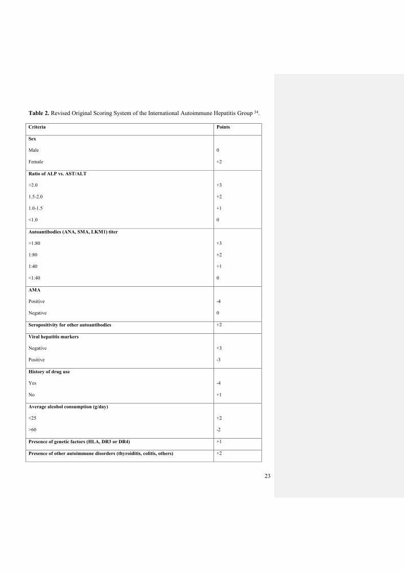
27,31,32. The diagnosis of AIH is defined as definite or probable, based on the Diagnostic Criteria of
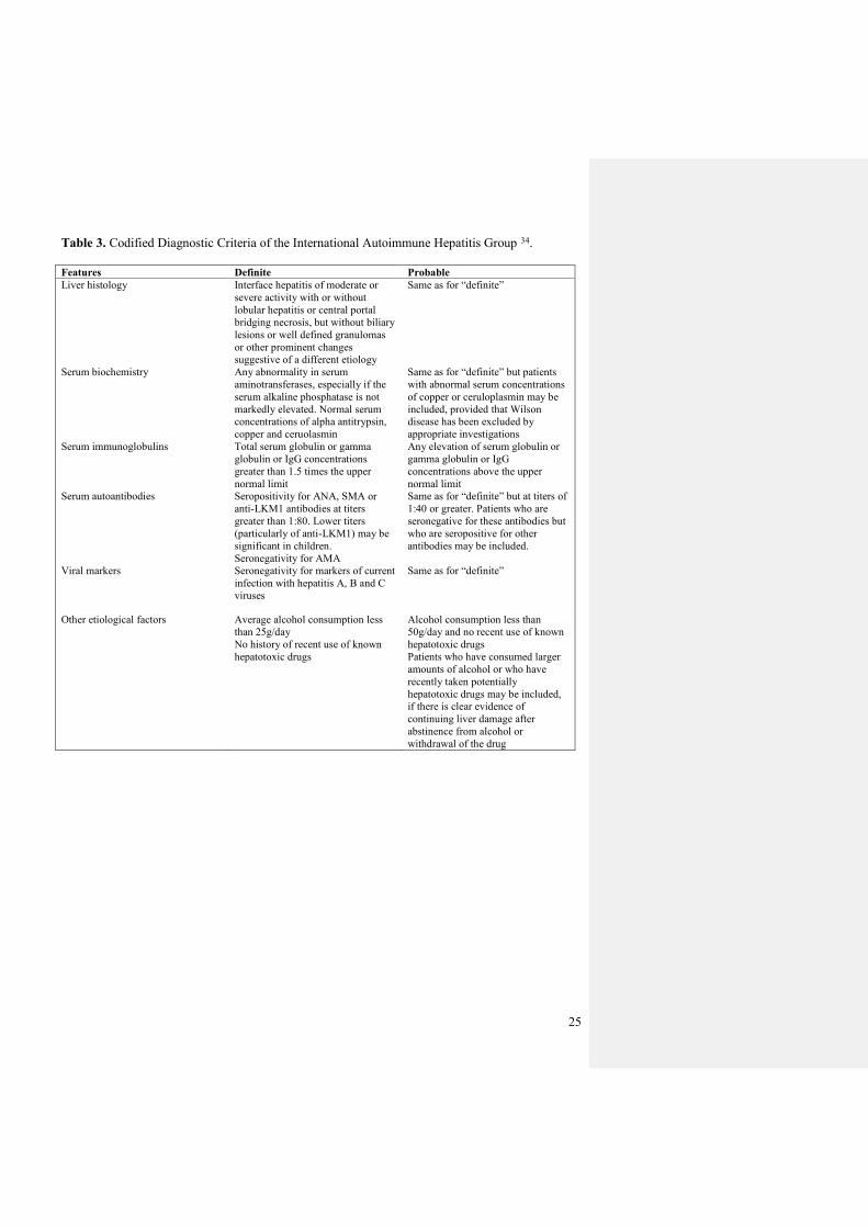
the International Autoimmune Hepatitis Group (IAIH-G, Table 2) 33,34. The clinical criteria for the
diagnosis are sufficient to establish or rule out a definite or probable AIH in the majority of
patients. The revised scoring system was developed as a research tool to ensure the comparability of
study populations in clinical trials, and can be used to assess treatment response (Table 3), similar
to classification criteria utilized in rheumatology 34. A pretreatment score of 10 points or higher, or
a posttreatment score of 12 points or higher, indicate ‘‘probable’’ AIH at presentation, with a
sensitivity of 100%, a specificity of 73%, and diagnostic accuracy of 67%. A pretreatment score of
15 points, indicative of ‘‘definite AIH’’ has a sensitivity of 95%, a specificity of 97%, and a
diagnostic accuracy of 94% 35.
The clinical course of untreated AIH results in significant mortality, with 5- and 10-year survival
rates of 50% and 10% respectively. The use of glucocorticoids has dramatically improved the
disease course with a 10-year survival rate now exceeding 90% 25. The complications associated
with AIH are similar to those of other progressive liver diseases, as chronic hepatitis can evolve to
cirrhosis and ultimately to hepatocellular carcinoma (HCC), despite the use of immunosuppressives.
At the time of diagnosis, approximately 30% of adults have histological evidence of cirrhosis; when
appropriately treated, however, only a small number develop cirrhosis during follow-up if
biochemical and histology inflammation resolves. The occurrence of HCC in patients with AIH is
rare and only develops in long-standing cirrhosis. In the absence of definitive data, primary liver
neoplasia incidence is assumed to be similar to other non-viral cases of cirrhosis 25.
Association with rheumatic diseases
AIH was originally described in association with SLE and currently extrahepatic autoimmune
manifestations are found in 20-50% of patients 36, with the most common being autoimmune
thyroiditis, diabetes, rheumatoid arthritis (RA) and ulcerative colitis (UC). Up to 43% of AIH cases
will have a family history of autoimmune diseases, in particular thyroid diseases and type 1 diabetes
7
37. The occurrence of other autoimmune diseases in AIH is included in the original and revised
International Autoimmune Hepatitis Scoring System (Table 3) 33. Concurrent autoimmune
disorders tend to cluster in women with AIH type 1, particularly if positive for the human leukocyte
antigen (HLA)-DR4 38. Moreover, elderly patients with AIH have higher frequency of concurrent
rheumatic conditions than young adults 39. SjS has been reported in up to 7% of AIH patients, while
RA in 2-4%. Though liver dysfunction has been reported in up to 60% of SLE patients, the
overlapping with AIH is rare 36.
An AIH-like entity linked to anti-TNF treatment has recently been described in case reports.40
Though these result in a significant liver injury, the pathogenesis remains clear 41. Liver biopsy
appears useful, however the differentiation between drug-induced liver injury is not an easy task.
Interestingly, most cases respond well to corticosteroids 42.
Autoantibodies
Autoantibodies represent a critical feature of AIH and may guide the diagnosis (Table 2). In 2004,
the IAIH-G established procedures and reference guidelines for more reliable serum autoantibody
testing to overcome the lack of standardization 43. In addition to serum ANA, anti-SM, and anti-
LKM, 44 other autoantibodies should also be sought in suspected cases, including anti-LC1,
perinuclear anti-neutrophil cytoplasmic antibodies (pANCA), SLA/LP, and the anti
asialoglycoprotein receptor antibodies 45. Finally, less specific autoantibodies may be detected in a
subset of patients, including anticardiolipin, anti-chromatin, anti-dsDNA, rheumatoid factor (RF),
anti-histones, anti-Ro/SSA, and anti-cyclic citrullinated peptides (anti-CCP) antibodies. Serum
ANA were the first autoantibodies observed in AIH sera over 50 years ago and remain the most
sensitive marker of AIH 46. These most frequently produce a homogeneous or speckled pattern.
However, the test is not specific for AIH, since ANA positivity is not uncommon in viral diseases,
other autoimmune liver diseases, as well as in up to 15% of healthy subjects, especially in older age
groups 47. Serum SMA are autoantibodies reacting with different proteins (actin, tubulin, vimentin,
desmin, cytokeratins) of the cytoskeletal components (microfilaments, microtubuli, intermediate

8
filaments). Their presence characterizes both autoimmune (AIH-1, coeliac disease) and viral
diseases (chronic hepatitis C, infectious mononucleosis). When detected at high titers (>1:80), they
are considered a sensitive marker for AIH-1, being found in up to 80% of cases. A recent study
showed that anti- SMA-T/G positive subject with normal liver function are at low risk of
progression to AIH, while positive SMA and raised ALT (>55IU/L) are at higher risk, though the
positive predictive value is only 22% 48. Serum autoantibodies against LKM-1 are the main
serological markers of AIH-2, recognizing the proximal renal tubule and hepatocellular cytoplasm.
Serum anti-SLA/LP antibodies are occasionally found in AIH patients who are negative for ANA,
SMA, or anti-LKM and are cumulatively detected in 10–30% of cases of AIH-1 and -2. Anti-
SLA/LP antibodies are detectable by radioimmunoassay and enzyme-linked immunosorbent assay
(ELISA) but not by immunofluorescence and are directed against different epitopes of a UGA
tRNA suppressor. Anti-LC1 antibodies are detected by indirect immunofluorescence in sera from
up to 50% of patients with type 2 AIH and less frequently in type 1 AIH or chronic hepatitis C.
Importantly, however, anti-LC1 are the only detectable markers in 10% of AIH cases. Interestingly,
serum anti-LC1 antibodies correlate with AIH severity and progression. Antibodies to the
asialoglycoprotein receptor are observed in up to 90% of patients with AIH and often coexist with
other autoantibodies, though they lack specificity for the disease. Similar to anti-LC1, however,
anti-asialoglycoprotein titers are associated with a more florid inflammatory disease activity and
may allow monitoring of treatment response.
With regard to non-specific antibodies, anti-CCP can be found in 9% of AIH sera, and their
detection is independent of concurrent RA but may distinguish early stage RA from nonspecific
arthralgia 39. Moreover, it has been reported than anti-CCP positive subjects are at higher risk of
cirrhosis at diagnosis and die more frequently from hepatic failure 21. Anti-cardiolipin antibodies
occur in nearly 40% of AIH, which is more frequently than hepatitis C (20%) and B (14%)
infections. The presence of anti-cardiolipin IgG/IgM is associated with cirrhosis and inflammatory
activity 49, with the IgM subtype being more frequent in AIH than PBC 50. Further, pANCA can be
9
detected by indirect immunofluorescence in sera from patients with AIH-1, but also in a subgroup
of patients with PSC or chronic viral hepatitis. Antibodies to histones are present in 35% of ANA-
positive patients with autoimmune hepatitis, while anti-dsDNA are detected in 23–34% cases,
depending on the nature of the assay and substrate used for their detection 51. Patients with anti-
histones are not distinguished by the severity of their disease 52, while anti-dsDNA positive subjects
do not respond or respond less to corticosteroid treatment 53.
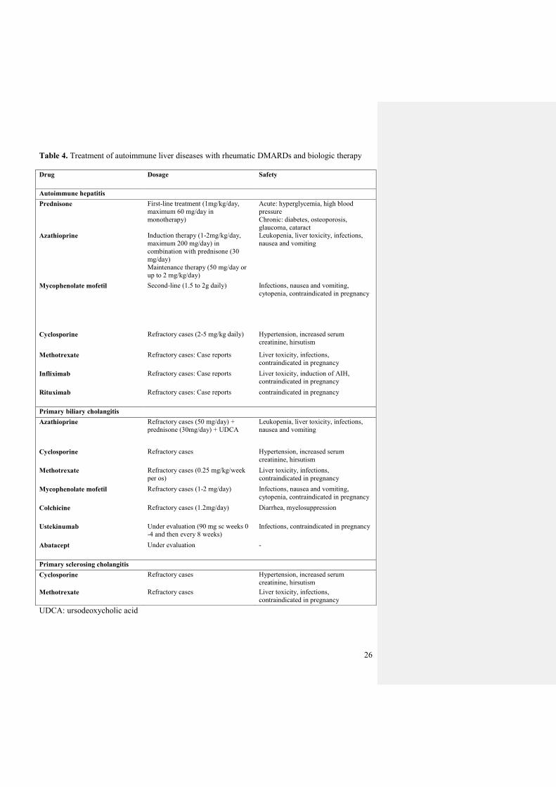
Therapy
In contrast to PBC and PSC, immunosuppressants represent the treatment of choice for AIH, based
on the good bio-chemical and histologic response, and survival (Table 4) 43,54. Glucocorticoids--in
particular prednisone--in monotherapy or in combination with azathioprine are the first-line
treatment. These induce remission (i.e. normal ALT and IgG) in over 80% of the patients,
regardless of the presence of cirrhosis 31. Once achieved, remission can be maintained with
azathioprine alone after steroid tapering. The dosage of azathioprine is typically low compared to
rheumatic diseases, usually requiring only 50 mg/day and never exceeding 150 mg/day 18. Relapses
following steroid discontinuation are common, since only 20% of patients remain in sustained
remission. It should be noted, however, that subgroups of patients manifest disease progression
(approximately 10%) or are intolerant to standard therapy (13%). In such patients, other drugs have
been anecdotally tried, including methotrexate 55, cyclophosphamide, tacrolimus, ursodeoxycholic
acid, cyclosporine and mycophenolate mofetil, the latter two constituting the most frequently
reported alternatives 18,56. Biologic therapies commonly used in rheumatology are of particular
interest, as pro-inflammatory cytokines, i.e. tumor necrosis factor (TNF)-alpha, are involved in AIH
pathogenesis 18. Infliximab has been used in refractory cases of AIH with reduction of
aminotransferases and IgG levels 57. Rituximab has been tried in a few refractory AIH cases,
resulting in improved liver enzymes and IgG, no significant side effects, and a reduction in
prednisolone dose for some patients 58. Future developments may include regulatory T cell therapy,
which could allow the avoidance of prolonged often life-long global immunosuppression in patients
10
with AIH 59. Liver transplantation is the most definitive treatment for AIH patients presenting with
acute liver failure or end-stage chronic liver disease and for those with HCC that meet the transplant
criteria. Although liver transplantation for these patients is very successful, AIH may recur after
transplant. AIH patients undergoing liver transplant have overall 5- and 10-year survival rates of
90% and 75%, respectively, although infectious complications and disease recurrence are common
60-63.
Primary biliary cholangitis
PBC is a chronic cholestatic disease characterized by high-titer serum anti-mitochondrial antibodies
(AMA) in nearly 100% of patients when sensitive techniques are used.64 It results in autoimmune-
mediated destruction of the small and medium-sized intrahepatic bile ducts 17,65. PBC prevalence
varies substantially according to geography; the highest rates appear in the northern US with a point
prevalence of 402 per million in Minnesota 66. Similar to other autoimmune diseases, PBC most
commonly affects women, with a 1:9 male:female ratio 17, and the average age at PBC diagnosis is
within the 5th and 6th decades of life 17.
Recently, PBC nomenclature has shifted from the term “cirrhosis” to “cholangitis”, which is more
precise and removes the stigma associated with cirrhosis. This change reflects the dramatically
improved PBC prognosis and treatment, since nowadays, two out of three patients diagnosed with
PBC and treated with ursodeoxycholic acid (UDCA) have an expected survival comparable to the
general population and only a minority will ever develop cirrhosis 67,68.
Clinical features
Early PBC symptoms are classically described as fatigue and pruritus while physical findings may
include skin hyperpigmentation, hepatosplenomegaly, and (rarely) xanthelasmas. Fatigue and
pruritus are nonspecific symptoms present in 70% of PBC patients. Conversely, end-stage
symptoms are secondary to the complications of liver cirrhosis, including ascites, jaundice, hepatic
encephalopathy, and upper digestive bleeding. Portal hypertension is frequently found in patients
11
with PBC and, importantly, does not imply the presence of liver cirrhosis. Metabolic bone disease is
elevated in PBC compared to sex- and age-matched healthy individuals (see below). Similar to
other types of cirrhosis, end-stage PBC can be complicated by the occurrence of HCC. The
progression of PBC varies widely, and the factors influencing the severity and progression of the
disease are largely unknown. The presence of symptoms at presentation, however, are a major
factor determining PBC survival rates; asymptomatic PBC produces 10-year survival rates shorter
than the general population, but symptomatic PBC produces even shorter survival rates 68.
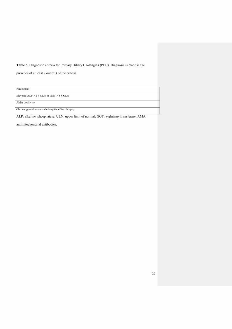
The diagnosis of PBC is generally based on the presence of two of the following three criteria: (1)
biochemical evidence of cholestasis with elevation of alkaline phosphatase activity over six months;
(2) presence of serum AMA at significant titers; and (3) histological non-suppurative cholangitis
and destruction of small or medium-sized bile ducts on biopsy specimen. The differential diagnosis
includes a cholestatic drug reaction, biliary obstruction, sarcoidosis, AIH and PSC (Table 4) 69.
Association with rheumatic diseases
PBC is commonly associated with a number of extrahepatic autoimmune conditions. A recent
monocentric study identified a co-occurrence in more than 60% of patients, with the most common
being SjS in 30% of patients, followed by Raynaud’s phenomenon in 18%, and Hashimoto’s
thyroiditis. PBC and SSc are associated in 6% of cases 70, while a higher frequency of RA (up to
10%) has been reported since the 1970s 71,72. PBC has also been reported in the presence of HLA-
B27 enthesopathy.73 Interestingly, 5% of PBC cases also suffer from autoimmune cutaneous
conditions 74, 75. Surprisingly, when extrahepatic autoimmune diseases co-occur with PBC, the
cases tend to be less severe; severe SjS occurs in 10.5% of PBC cases, and the PBC disease is
usually milder and at early stage (stage I-II at liver histology) in the presence of SjS 76,77. The same
observation has been made with PBC and SSc. PBC most commonly associates with limited
cutaneous SSc (lSSc), and patients with PBC/SSc overlap have a slower rate of liver-disease
progression compared to matched patients with PBC alone 78. Female sex is the only significant risk
12
factor for at having a second autoimmune condition 70, while neither autoantibodies nor liver
histology differ.
Autoantibodies
PBC is characterized serologically by the presence of AMA, which are highly specific for the
disease. These antibodies are found in 90-95% of PBC patients compared with less than 1% of
healthy subjects 79. Similar to other autoimmune diseases, AMA positivity arises years before the
development of PBC 9, and AMA are included in the internationally-accepted criteria for PBC
diagnosis (Table 5) 80. AMA are directed against components of the 2-oxoacid dehydrogenase (2-
OADC) family of enzymes within the mitochondrial respiratory chain, most frequently the E2 and
E3-binding protein (E3BP) components of the pyruvate dehydrogenase complex and the E2
components of the 2-oxo glutarate dehydrogenase and branched-chain 2-oxo acid dehydrogenase
complexes 81. All three antigen epitopes contain the motif DKA, with lipoic acid covalently bound
to the lysine (K) residue 78. ANA have been identified in 52% of patients, with the most specific
patterns being “nuclear-rim” and “multiple nuclear dots”, produced by antibodies directed against
the nuclear membrane gp120 and nucleoporin 62, and the nuclear body sp100, sp140 and
promyeolocytic leukaemia proteins, respectively (Table 1) 82-85. ANA-positive patients are more
frequently AMA-negative, possibly because of the lack of a masking effect of these latter antibodies
in such sera. While ACA are most specific for lSSc, found in up to 90% of cases, they are also
detectable in 9-30% of PBC patients. This prevalence exceeds that of the PBC/SSc overlap
syndrome 86,87. ACA recognize six centromere polypeptides belonging to the kinetochore proteins:
CENP-A, CENP-B, CENP-C, CENP-D, CENP-E, CENP-F, with the major autoantigen being
CENP-B. The clinical significance of ACA in PBC remains ill-defined as it is unclear whether
ACA represents a pre-clinical marker of lSSc or a subclinical form of the disease. Moreover, ACA
could simply represent an epiphenomenon of the immune dysregulation present in PBC 78. Some
have posited that since ACA can predict the development of SSc and since early SSc may be
frequent in PBC, these may facilitate timely detection of complications, preventing disability and
13
reducing the probability of liver transplantation 88,78. In any case, patients with PBC and positive
ACA with SSc-related symptoms should be assessed for organ involvement, and in particular,
assessment of pulmonary arterial hypertension by echocardiography should be considered in all
PBC/SSc patients. Unfortunately, currently utilized tools to predict this pulmonary arterial
hypertension (e.g. the DETECT score) have not been evaluated in PBC patients 89. Furthermore,
while PBC/SSc seems to have a milder disease course, ACA positive PBC patients have a more
severe bile duct injury and more frequently portal hypertension 78. With regard to other
autoantibodies 90, anti-ENA are positive in up to 40% of PBC cases, regardless of the extrahepatic
autoimmunity 70, with no effect on disease severity or progression. Anti-Ro/SSA are found in
PBC/SjS overlap in 10% of cases 90, anti-dsDNA in 22% of PBC patients 90 while anti-cardiolipin
IgM positive in 75% of PBC, advanced stage disease 91.
Therapy
PBC treatment is currently based on UDCA, which is the only approved drug. Its mechanism of
action is incompletely understood and possibly dependent on the various phases of the disease 5,92.
During the early disease, short-term glucocorticoids might be effective, however, prolonged use
raises safety concerns. Budesonide, due to its high first-pass metabolism, has minimum systemic
adverse effects, and, at 6–9 mg daily, has been demonstrated to be superior to UDCA both in terms
of histology and biochemical markers. Other immunosuppressants, such as methotrexate and
azathioprine, have also been suggested and there is evidence supporting the use of the latter in PBC
with autoimmune hepatitis overlap syndrome 93. The use of biologics targeting TNF-alpha has been
reported in few cases of overlap syndromes with rheumatic diseases 94,95. In the last years, improved
understanding of PBC pathogenesis has led to the testing of new targeted therapies, especially those
modulating the IL-17/23 axis. Ustekinumab, a monoclonal antibody against the p40 subunit,
however, demonstrated only a very modest decrease in ALP after 28 weeks of therapy, and was
otherwise deemed ineffective19. Other therapies targeting T cells 96, including those that bind
CTLA-4 (abatacept) or antagonize CD40 (FFP104) are under investigation 69,97,98. Of note, the use
14
CTLA-4 Ig in PBC murine model prevents cholangitis manifestations (AMA production,
intrahepatic T-cell infiltrates, and bile duct damage) and reduces disease severity in established
murine disease 99. When the disease has already progressed and bile is accumulated, obeticholic
acid (OCA)--an analogue of chenodeoxycholic acid with a much higher affinity to the farnesoid X
receptor (FXR)--has been shown to decrease bile synthesis, promote secretion, and induce liver
regeneration in animal models. Furthermore, a recent Phase III trial of OCA administered with
UDCA or as monotherapy for 12 months demonstrated decreases in alkaline phosphatase and total
bilirubin levels compared with placebo 97. Ultimately, UDCA represents the cornerstone therapy of
PBC and dosages ranging from 13 to 15 mg/kg lead to optimum bile enrichment, with 50% of
patients normalizing their alkaline phosphatase. Other immunosuppressive treatments should be
started only in combination with UDCA.
Liver transplantation may be necessary for end-stage PBC, with survival rates of 92% and 85% at 1
and 5 years after transplant, respectively. Recurrence is common seems to be influenced by
immunosuppressives, while the use of UDCA for recurrence is safe and recommended.
Primary sclerosing cholangitis
PSC is a progressive cholestatic liver disease of unknown etiology presenting with chronic
inflammatory features of the bile ducts of any size and associated with significant morbidity and
mortality 100,101. In contrast to PBC, PSC can affect all tracts of the biliary tree, including the
extrahepatic bile ducts visible with imaging modalities and the small bile ducts observed via liver
histology. The prevalence of PSC is approximately 10/100000 in Northern Europe 28 and in the US,
while it is far less common in Southern Europe and Asia; recent data from the Olmstead County,
Minnesota, report a prevalence of 20.9 per 100 000 men and 6.3 per 100 000 women 102. Different
from PBC and AIH, PSC affects more frequently men, with a 2:1 male:female ratio 102.
Clinical features
15
PSC symptoms are generally nonspecific and include abdominal pain, jaundice, and fever in the
case of bacterial cholangitis, while at more advanced stages, symptoms include those typical of
decompensated cirrhosis or neoplasia. Commonly, PSC is complicated by episodic bacterial
cholangitis precipitated by biliary strictures. Discrete subgroups of patients manifest the “small-
duct” or overlap syndrome variants. Due to the nonspecific symptoms, PSC is usually diagnosed
during routine blood tests in otherwise healthy individuals or patients with IBD 103. Testing
characteristically reveals a biochemical cholestatic pattern, as represented by elevated serum
alkaline phosphatase and γ-glutamyltransferase, though tests of liver function are normal until late
stages. Imaging (particularly bile duct MRI or endoscopy) represents a useful diagnostic tool, as it
may identify the classic strictured and dilated intrahepatic or extrahepatic bile ducts 102. Performing
a liver biopsy is generally not necessary for the diagnosis of PSC, except in the case of small-duct
PSC which requires histologic examination. The natural history of this form is relatively benign
and only a minority (12%) of patients progress to classical PSC. The median timespan from
diagnosis to liver-related death or liver transplantation is 18 years, and the prognosis is influenced
by the onset of cholangiocarcinoma (CCA). CCA is more common with chronic biliary
inflammation and difficult to distinguish from stricturing PSC 101.
Association with rheumatic diseases
The association of PSC with IBD is well established. Nearly 70% of PSC cases also demonstrate
findings of IBD 104, frequently in mild asymptomatic forms, while 7% of IBD patients have PSC
105. Liver abnormalities are more frequently found in psoriatic patients, and this is typically
attributed to non-alcoholic or alcoholic fatty liver 106. However, in generalized pustular psoriasis, a
less common form of psoriasis associated with extra-cutaneous manifestations, evidence for biliary
involvement has been suggested, and neutrophilic cholangitis has been observed on liver biopsy,
while magnetic resonance cholangiopancreatography showed features similar to those observed in
PSC 107.
Autoantibodies
16
In contrast to other autoimmune liver diseases, autoantibodies are of limited use in the diagnosis of
PSC due to low sensitivity and specificity; only a limited percentage of patients (33%) have positive
p-ANCA 108,109, for example. Only for PSC forms overlapping with AIH is serum ANA typically
detected.
Therapy
The treatment of PSC is largely an unmet need and currently includes medical and endoscopic
measures, short of liver transplantation 110,111. UDCA has been investigated in several clinical trials,
with conflicting results. Overall, the available evidence suggests that UDCA does not produce a
substantial change in the course of PSC, despite remaining the most prescribed drug. However, it
appears that high-dose UDCA (20 mg/kg/day) or norUDCA, a side chain-shortened homologue of
UDCA, may reduce reduced biochemical indices of cholestasis 112, the rate of progression, and
might prevent the development of colon cancer (particularly in patients with UC/PSC overlap).
Based on these inconclusive data, the use of UCDA in PSC varies widely, reflecting regional
practice trends rather than science. Endoscopic interventions are indicated to treat complicated PSC
through the dilation of short- and long-segment stenosis of the common bile duct and short-segment
stenosis of the hepatic ducts near to the bifurcation. The treatment can be repeated over time once
restenosis ensues and resulting survival rates are higher compared to patients not treated
endoscopically. Biologics, mainly anti-TNF, have been used in PSC with concomitant IBD or
rheumatic diseases with improvement in laboratory measurements 113,114 but patients with PSC are
generally excluded from IBD clinical trials, thus preventing firm conclusions regarding efficacy.
Finally, PSC represents an important indication for liver transplantation since patients are younger
than their counterparts with PBC. Recurrence of disease occurs in 20–40% of transplanted patients
during prolonged follow-up. The ability of UDCA to prolong survival after disease recurrence
remains a point on contention.
Overlap syndromes.
Commentato [L1]: Consider cutting...out of place in the treatment section.
Commentato [CS2]: I would maintain this as it is a less known secret for PSC people
17
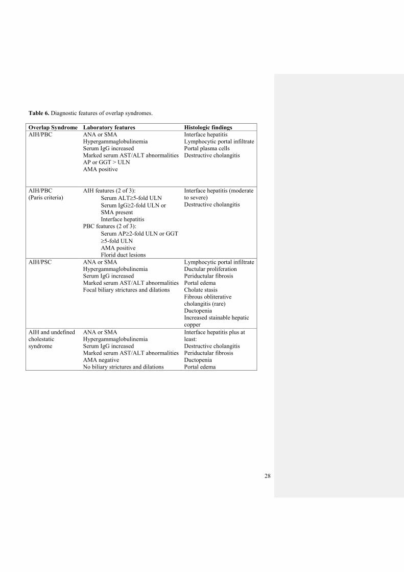
Autoimmune liver diseases, similarly to rheumatic disease, may overlap and present with both
hepatocellular and cholangiocellular patterns according to biochemical, histologic, and imaging-
based analysis. When left without treatment, these patients show a more progressive course toward
liver cirrhosis and failure. AIH–PBC overlap syndrome is found in 10% of adults with AIH whereas
AIH–PSC overlap syndromes affects 6–8% of children, adolescents, and young adults with AIH;
PBC-PSC overlap syndrome is exceptionally rare (Table 6). Besides overlaps, transitions are also
possible in rare cases from PBC to AIH, AIH to PBC, or AIH to PSC 115.
AIH may have an atypical presentation with serum alkaline phosphatase elevation, AMA positivity,
histologic features of bile duct injury/loss, or cholangiographic findings of focal biliary strictures
and dilations. These manifestations characterize the overlap syndromes. The clues to an overlap
syndrome consist of: (1) serum alkaline phosphatase >2-fold upper normal limit (ULN) at
presentation, which is present in only 20% of AIH patients; (2) serum GGT >ULN unimproved or
worsened during therapy; (3) AMA positivity; (4) histologic findings of bile duct injury or loss; (5)
concurrent IBD; (6) corticosteroid treatment failure or incomplete response 115.
Overlap features of PBC usually refers to simultaneous AIH in patients who have a diagnosis of
AMA-positive PBC and not to patients with AIH who have coincidental AMA. AMA occur in
about 5% of AIH patients in the absence of other biliary features (‘‘serological overlap’’), but may
disappear or persist for decades without an evolution into PBC. Approximately 4% of PBC cases
have simultaneous features of AIH. There are two different scoring systems that have been used to
evaluate patients with PBC for simultaneous evidence of overlapping AIH: (1) the IAIH-G score;
(2) looking for the presence of two of the three following features: (i) ALT activity 5 times upper
limits of normal; (ii) IgG 2 times upper limits of normal and/or positive anti–SMA antibody; and
overlap by (iii) liver biopsy with moderate or severe periportal or periseptal inflammation. A
PBC/AIH overlap syndrome may also refer to patients with sequential PBC followed by AIH, a
condition occurring in 2.4% of cases. In these cases, the diagnosis of PBC with positive AMA
occurs first and initially responds biochemically to UDCA therapy; subsequently, these patients
18
present with clinical features of AIH, lose their AMA seropositivity, exhibit liver histology more
typical of AIH, and respond to immunosuppressive therapy.
The term ‘autoimmune cholangitis’ was first coined to indicate AMA-negative PBC, possibly with
serum ANA. More recently, a broader concept has emerged that includes: (1) serum ANA and/or
SMA positivity and/or hyper-gammaglobulinemia; (2) serum AMA negativity by
immunofluorescence; (3) biochemical and/or histological features of cholestatic and hepatocellular
injury; and (4) exclusion of chronic viral, metabolic, or toxic liver disease. This definition possibly
subsumes PBC with atypical presentation, small-duct PSC, idiopathic adulthood ductopenia, AIH
with bile duct damage, concurrent AIH and small-duct PSC, and various transitional stages of the
classic diseases. Consensus is still wanting on this issue, and standardization of diagnostic criteria
for overlap syndromes is impeded by their uncommon occurrence in the setting of rare diseases.
Osteometabolic consequences of chronic autoimmune liver disease.
Advancements in the management of autoimmune liver diseases and cirrhosis complications have
increased survival rates. However, longer survival rates, compounded by an aging population, have
increased the risk of complications such as osteoporosis. Osteoporosis is associated with increased
risk of fracture, which is two-fold higher in cirrhotic patients regardless of the liver disease
etiology, and persists for years after liver transplantation 116-119. Moreover, patients receiving
glucocorticoids for AIH suffer an additional decrease in their bone mass.
According to the WHO definition, osteoporosis is diagnosed when bone density is less than 2.5
standard deviations below the peak value obtained from normal adults and adjusted for gender (T
score) 120. This definition is limited insofar as the threshold was established from studies of
postmenopausal Caucasian women, rather than for patients with liver diseases.117 Therefore, some
authors favor the term hepatic osteodystrophy, though this term also includes osteomalacia 121.
The mechanisms of cirrhosis-related osteoporosis are not fully understood, but it is generally
recognized that the association between liver and bone diseases occurs due to an imbalance of bone
19
turnover, which depends on the osteoblastic and osteoclastic activity 122. In PBC, while the exact
mechanism is also not completely understood, there is evidence that hormone balance, genetics, and
cholestasis may contribute to determine bone structure and density changes. There has been
conflicting evidence as to whether PBC-related osteoporosis results from diminished bone
formation, which is a low-turnover state, or from increased bone resorption, which is a high-
turnover state. However, recent data suggest that bone formation is the culprit. Cirrhosis is
associated with the reduction of specific growth factors, such as insulin-like growth factor 1, which
impairs osteoblast function and bone formation; severe cholestasis can allow build-up of lithocholic
acid, which inhibits osteoblast activity and can interfere with genetic regulation of bone formation
23.
The prevalence of osteoporosis in cirrhotic patients ranges from 12 to 70% according to the
diagnostic modality and the liver disease etiology, with cholestatic diseases having a higher
prevalence (20-44%, even without an established diagnosis of cirrhosis). Moreover, fracture rates
are increased in cholestatic diseases, varying from 13 to 22% according to the degree of liver
function 120.
Screening for osteoporosis is an important part of liver diseases management, and the guidelines
indicate that patients with cirrhosis and PBC should be screened by an initial dual-energy X-ray
absorptiometry (DXA) exam 121. If initial results are normal, the DXA should be repeated every 1 to
3 years to assess significant bone loss, depending on the presence of additional risk factors (BMI
less than 19 kg/m2, heavy alcohol use, tobacco use, early menopause [<age 45 years],
glucocorticoid use greater than 3 months, or family history of bone fragility fractures). 121 In
addition, BMD should be measured prior to liver transplantation.
Laboratory test are also helpful for evaluating bone metabolism, and include serum calcium, 25-
hydroxyvitamin D, phosphorus, osteocalcin, procollagen I carboxyterminal peptide, and parathyroid
hormone (PTH), as well as urinary amino telopeptides of collagen I and urinary calcium. Routine
20
monitoring of calcium, phosphorus, 25-hydroxyvitamin D, and PTH should be performed every 1 to
2 years 123.
The treatment of osteoporosis is based on results obtained from trials assessing postmenopausal
women, and few studies have included patients with liver diseases. Educational strategies include
elimination of modifiable risk factors, such as smoke and alcohol cessation. Calcium and vitamin D
supplementation is part of osteoporosis treatment. The total calcium intake should achieve a daily
ingestion of 1.0 to 1.5 grams, preferably from diet to facilitate patients compliance 120. Oral
cholecaciferol (vitamin D3) can be prescribed at 1,000-4,000 IU per day or ergocalciferol (vitamin
D2) at 50,000 IU per month. Given that calcitriol (1,25-dihydroxycholecalciferol or 1,25-
dihydroxyvitamin D3) is the final active vitamin D metabolite, it may represent a better treatment
for liver disease patients. Calcitriol is usually prescribed as a daily oral dose of 800 IU but can also
be taken at a weekly dose of 5000 IU 120. In PBC, calcium and vitamin D supplementation alone
was inferior to hormonal replacement therapy in improving BMD.124,125 However, testing for
vitamin D deficiency in cholestatic patients is useful to allow for appropriate supplementation,
particularly in those taking cholestyramine, since this impairs vitamin D absorption 116.
Bisphosphonates represent the treatment of choice for osteoporosis in cirrhotic patients, because
they attach to the bone surface and prevent resorption. 126. The threshold of intervention in patients
with liver disease, however, may be lower than in the general population, since in PBC T-scores
below -1.5 are associated with a significant risk for vertebral fractures 127. A recent randomized
controlled trial for osteoporosis therapy in PBC patients showed that both monthly ibandronic acid
and weekly alendronic acid improve bone mass and are comparable in safety, although adherence is
higher with the monthly regimen 128. Moreover, bisphosphonates and teriparatide reduce the risk of
vertebral fractures in chronically glucocorticoid-treated patients 129,130. Hormone replacement
therapy does not show any osteoporosis benefit for PBC patients, but its use is no longer
contraindicated in chronic cholestasis 131. Ultimately, treating PBC with UDCA may have
beneficial effects also on the bone, since UDCA may increase osteoblast differentiation and
21
mineralization, and neutralize the detrimental effects of lithocholic acid, bilirubin and sera from
jaundiced patients on osteoblastic cells 132.
Concluding remarks.
The coexistence of liver and rheumatic diseases represents an ideal example of the need for a
interdisciplinary approach to individualize treatments. Indeed, patients with autoimmune liver
diseases may be undertreated with anti-rheumatic drugs for liver safety concerns (as in the case of
methotrexate), while liver test changes may raise unnecessary concerns. Furthermore, new biologics
may be beneficial for more than one condition, though insufficient data exist from the hepatology
perspective. Lastly, the rapid deterioration of bone health associated with chronic liver diseases is
an obvious area of collaboration between gastroentology and rheumatology. With the current focus
on personalized medicine, the coexistence of liver autoimmunity and rheumatic disease is an ideal
area to develop and investigate the benefits of shared clinical practice.
22
Tables and figures.
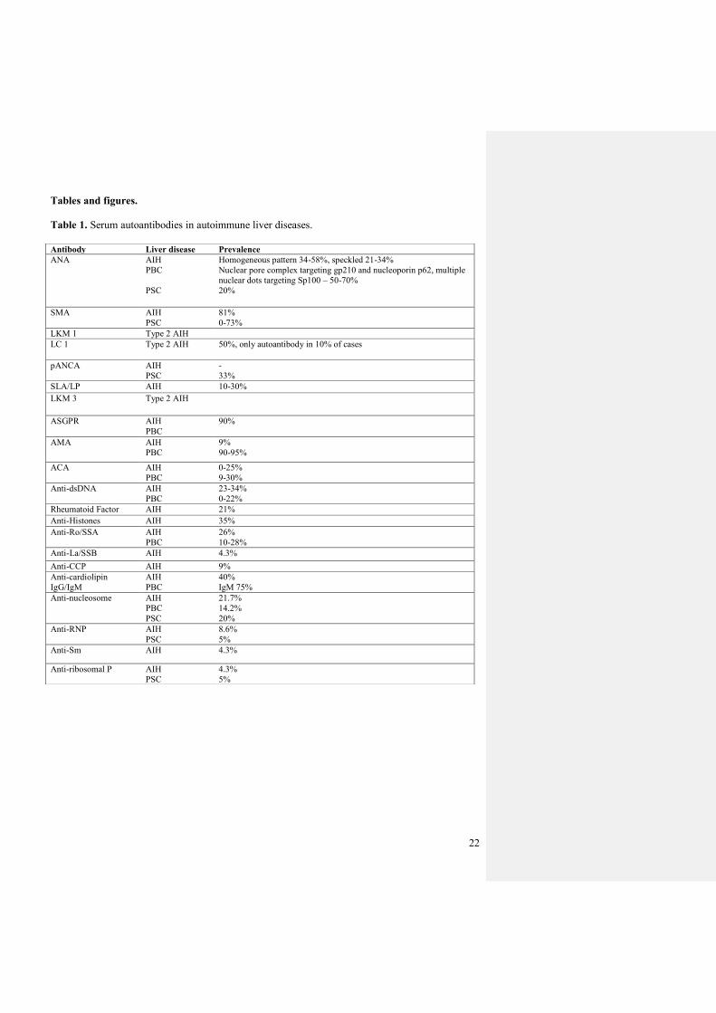
Table 1. Serum autoantibodies in autoimmune liver diseases.
Antibody Liver disease Prevalence
ANA AIH
PBC
PSC
Homogeneous pattern 34-58%, speckled 21-34%
Nuclear pore complex targeting gp210 and nucleoporin p62, multiple
nuclear dots targeting Sp100 – 50-70%
20%
SMA AIH
PSC
81%
0-73%
LKM 1 Type 2 AIH
LC 1 Type 2 AIH 50%, only autoantibody in 10% of cases
pANCA AIH
PSC
-
33%
SLA/LP AIH 10-30%
LKM 3 Type 2 AIH
ASGPR AIH
PBC
90%
AMA AIH
PBC
9%
90-95%
ACA AIH
PBC
0-25%
9-30%
Anti-dsDNA AIH
PBC
23-34%
0-22%
Rheumatoid Factor AIH 21%
Anti-Histones AIH 35%
Anti-Ro/SSA AIH
PBC
26%
10-28%
Anti-La/SSB AIH 4.3%
Anti-CCP AIH 9%
Anti-cardiolipin
IgG/IgM
AIH
PBC
40%
IgM 75%
Anti-nucleosome AIH
PBC
PSC
21.7%
14.2%
20%
Anti-RNP AIH
PSC
8.6%
5%
Anti-Sm AIH 4.3%
Anti-ribosomal P AIH
PSC
4.3%
5%
23
Table 2. Revised Original Scoring System of the International Autoimmune Hepatitis Group 34.
Criteria Points
Sex
Male
Female
0
+2
Ratio of ALP vs. AST/ALT
>2.0
1.5-2.0
1.0-1.5
<1.0
+3
+2
+1
0
Autoantibodies (ANA, SMA, LKM1) titer
>1:80
1:80
1:40
<1:40
+3
+2
+1
0
AMA
Positive
Negative
-4
0
Seropositivity for other autoantibodies +2
Viral hepatitis markers
Negative
Positive
+3
-3
History of drug use
Yes
No
-4
+1
Average alcohol consumption (g/day)
<25
>60
+2
-2
Presence of genetic factors (HLA, DR3 or DR4) +1
Presence of other autoimmune disorders (thyroiditis, colitis, others) +2
24
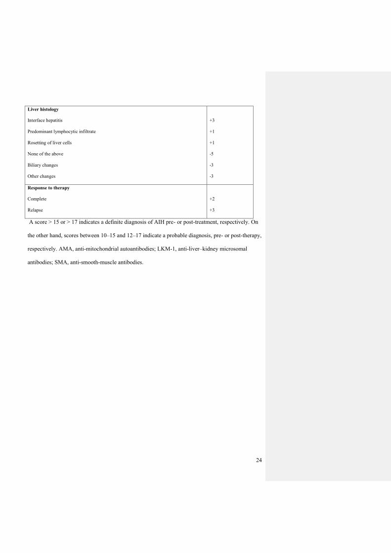
Liver histology
Interface hepatitis
Predominant lymphocytic infiltrate
Rosetting of liver cells
None of the above
Biliary changes
Other changes
+3
+1
+1
-5
-3
-3
Response to therapy
Complete
Relapse
+2
+3
A score > 15 or > 17 indicates a definite diagnosis of AIH pre- or post-treatment, respectively. On
the other hand, scores between 10–15 and 12–17 indicate a probable diagnosis, pre- or post-therapy,
respectively. AMA, anti-mitochondrial autoantibodies; LKM-1, anti-liver–kidney microsomal
antibodies; SMA, anti-smooth-muscle antibodies.
25
Table 3. Codified Diagnostic Criteria of the International Autoimmune Hepatitis Group 34.
Features Definite Probable
Liver histology Interface hepatitis of moderate or
severe activity with or without
lobular hepatitis or central portal
bridging necrosis, but without biliary
lesions or well defined granulomas
or other prominent changes
suggestive of a different etiology
Same as for “definite”
Serum biochemistry Any abnormality in serum
aminotransferases, especially if the
serum alkaline phosphatase is not
markedly elevated. Normal serum
concentrations of alpha antitrypsin,
copper and ceruolasmin
Same as for “definite” but patients
with abnormal serum concentrations
of copper or ceruloplasmin may be
included, provided that Wilson
disease has been excluded by
appropriate investigations
Serum immunoglobulins Total serum globulin or gamma
globulin or IgG concentrations
greater than 1.5 times the upper
normal limit
Any elevation of serum globulin or
gamma globulin or IgG
concentrations above the upper
normal limit
Serum autoantibodies Seropositivity for ANA, SMA or
anti-LKM1 antibodies at titers
greater than 1:80. Lower titers
(particularly of anti-LKM1) may be
significant in children.
Seronegativity for AMA
Same as for “definite” but at titers of
1:40 or greater. Patients who are
seronegative for these antibodies but
who are seropositive for other
antibodies may be included.
Viral markers Seronegativity for markers of current
infection with hepatitis A, B and C
viruses
Same as for “definite”
Other etiological factors Average alcohol consumption less
than 25g/day
No history of recent use of known
hepatotoxic drugs
Alcohol consumption less than
50g/day and no recent use of known
hepatotoxic drugs
Patients who have consumed larger
amounts of alcohol or who have
recently taken potentially
hepatotoxic drugs may be included,
if there is clear evidence of
continuing liver damage after
abstinence from alcohol or
withdrawal of the drug
26
Table 4. Treatment of autoimmune liver diseases with rheumatic DMARDs and biologic therapy
Drug Dosage Safety
Autoimmune hepatitis
Prednisone First-line treatment (1mg/kg/day,
maximum 60 mg/day in
monotherapy)
Acute: hyperglycemia, high blood
pressure
Chronic: diabetes, osteoporosis,
glaucoma, cataract
Azathioprine
Induction therapy (1-2mg/kg/day,
maximum 200 mg/day) in
combination with prednisone (30
mg/day)
Maintenance therapy (50 mg/day or
up to 2 mg/kg/day)
Leukopenia, liver toxicity, infections,
nausea and vomiting
Mycophenolate mofetil Second-line (1.5 to 2g daily) Infections, nausea and vomiting,
cytopenia, contraindicated in pregnancy
Cyclosporine Refractory cases (2-5 mg/kg daily) Hypertension, increased serum
creatinine, hirsutism
Methotrexate Refractory cases: Case reports Liver toxicity, infections,
contraindicated in pregnancy
Infliximab Refractory cases: Case reports Liver toxicity, induction of AIH,
contraindicated in pregnancy
Rituximab Refractory cases: Case reports contraindicated in pregnancy
Primary biliary cholangitis
Azathioprine
Refractory cases (50 mg/day) +
prednisone (30mg/day) + UDCA
Leukopenia, liver toxicity, infections,
nausea and vomiting
Cyclosporine Refractory cases Hypertension, increased serum
creatinine, hirsutism
Methotrexate Refractory cases (0.25 mg/kg/week
per os)
Liver toxicity, infections,
contraindicated in pregnancy
Mycophenolate mofetil Refractory cases (1-2 mg/day) Infections, nausea and vomiting,
cytopenia, contraindicated in pregnancy
Colchicine Refractory cases (1.2mg/day)
Diarrhea, myelosuppression
Ustekinumab Under evaluation (90 mg sc weeks 0
-4 and then every 8 weeks)
Infections, contraindicated in pregnancy
Abatacept Under evaluation -
Primary sclerosing cholangitis
Cyclosporine Refractory cases Hypertension, increased serum
creatinine, hirsutism
Methotrexate Refractory cases Liver toxicity, infections,
contraindicated in pregnancy
UDCA: ursodeoxycholic acid
27
Table 5. Diagnostic criteria for Primary Biliary Cholangitis (PBC). Diagnosis is made in the
presence of at least 2 out of 3 of the criteria.
Parameters
Elevated ALP > 2 x ULN or GGT > 5 x ULN
AMA positivity
Chronic granulomatous cholangitis at liver biopsy
ALP: alkaline phosphatase; ULN: upper limit of normal; GGT: γ-glutamyltransferase; AMA:
antimitochondrial antibodies.
28
Table 6. Diagnostic features of overlap syndromes.
Overlap Syndrome Laboratory features Histologic findings
AIH/PBC ANA or SMA
Hypergammaglobulinemia
Serum IgG increased
Marked serum AST/ALT abnormalities
AP or GGT > ULN
AMA positive
Interface hepatitis
Lymphocytic portal infiltrate
Portal plasma cells
Destructive cholangitis
AIH/PBC
(Paris criteria)
AIH features (2 of 3):
Serum ALT5-fold ULN
Serum IgG2-fold ULN or
SMA present
Interface hepatitis
PBC features (2 of 3):
Serum AP2-fold ULN or GGT
5-fold ULN
AMA positive
Florid duct lesions
Interface hepatitis (moderate
to severe)
Destructive cholangitis
AIH/PSC ANA or SMA
Hypergammaglobulinemia
Serum IgG increased
Marked serum AST/ALT abnormalities
Focal biliary strictures and dilations
Lymphocytic portal infiltrate
Ductular proliferation
Periductular fibrosis
Portal edema
Cholate stasis
Fibrous obliterative
cholangitis (rare)
Ductopenia
Increased stainable hepatic
copper
AIH and undefined
cholestatic
syndrome
ANA or SMA
Hypergammaglobulinemia
Serum IgG increased
Marked serum AST/ALT abnormalities
AMA negative
No biliary strictures and dilations
Interface hepatitis plus at
least:
Destructive cholangitis
Periductular fibrosis
Ductopenia
Portal edema
29
References.
1. Cowling DC, Mackay IR, Taft LI. Lupoid hepatitis. Lancet. 1956;271(6957):1323-1326.
2. Adiga A, Nugent K. Lupus Hepatitis and Autoimmune Hepatitis (Lupoid Hepatitis). Am J
Med Sci. 2017;353(4):329-335.
3. Doherty DG. Immunity, tolerance and autoimmunity in the liver: A comprehensive review.
J Autoimmun. 2016;66:60-75.
4. Liberal R, Selmi C, Gershwin ME. Diego and Giorgina Vergani: The two hearts of
translational autoimmunity. J Autoimmun. 2016;66:1-6.
5. Molinaro A, Marschall HU. Why Doesn't Primary Biliary Cholangitis Respond to
Immunosuppressive Medications? Curr Hepatol Rep. 2017;16(2):119-123.
6. Gatselis NK, Zachou K, Lygoura V, et al. Geoepidemiology, clinical manifestations and
outcome of primary biliary cholangitis in Greece. Eur J Intern Med. 2017;42:81-88.
7. Kim BH, Choi HY, Ki M, Kim KA, Jang ES, Jeong SH. Population-based prevalence,
incidence, and disease burden of autoimmune hepatitis in South Korea. PLoS One.
2017;12(8):e0182391.
8. Ji J, Sundquist J, Sundquist K. Gender-specific incidence of autoimmune diseases from
national registers. J Autoimmun. 2016;69:102-106.
9. Ma WT, Chang C, Gershwin ME, Lian ZX. Development of autoantibodies precedes
clinical manifestations of autoimmune diseases: A comprehensive review. J Autoimmun. 2017.
10. Toh BH. Diagnostic autoantibodies for autoimmune liver diseases. Clin Transl Immunology.
2017;6(5):e139.
11. Watad A, Azrielant S, Bragazzi NL, et al. Seasonality and autoimmune diseases: The
contribution of the four seasons to the mosaic of autoimmunity. J Autoimmun. 2017;82:13-30.
12. Murray-Lyon IM, Thompson RP, Ansell ID, Williams R. Scleroderma and primary biliary
cirrhosis. Br Med J. 1970;3(5717):258-259.
30
13. Clarke AK, Galbraith RM, Hamilton EB, Williams R. Rheumatic disorders in primary
biliary cirrhosis. Ann Rheum Dis. 1978;37(1):42-47.
14. Morgan MY. Primary biliary cirrhosis, scleroderma and keratoconjunctivitis sicca. Proc R
Soc Med. 1973;66(11):1112.
15. Selmi C, Meroni PL, Gershwin ME. Primary biliary cirrhosis and Sjogren's syndrome:
autoimmune epithelitis. J Autoimmun. 2012;39(1-2):34-42.
16. Sirotti S, Generali E, Ceribelli A, Isailovic N, De Santis M, Selmi C. Personalized medicine
in rheumatology: the paradigm of serum autoantibodies. Auto Immun Highlights. 2017;8(1):10.
17. Selmi C, Bowlus CL, Gershwin ME, Coppel RL. Primary biliary cirrhosis. Lancet.
2011;377(9777):1600-1609.
18. Cropley A, Weltman M. The use of immunosuppression in autoimmune hepatitis: A current
literature review. Clin Mol Hepatol. 2017;23(1):22-26.
19. Hirschfield GM, Gershwin ME, Strauss R, et al. Ustekinumab for patients with primary
biliary cholangitis who have an inadequate response to ursodeoxycholic acid: A proof-of-concept
study. Hepatology. 2016;64(1):189-199.
20. Chimenti MS, Talamonti M, Novelli L, et al. Long-term ustekinumab therapy of psoriasis in
patients with coexisting rheumatoid arthritis and Sjogren syndrome. Report of two cases and review
of literature. J Dermatol Case Rep. 2015;9(3):71-75.
21. Card TR, Langan SM, Chu TP. Extra-Gastrointestinal Manifestations of Inflammatory
Bowel Disease May Be Less Common Than Previously Reported. Dig Dis Sci. 2016;61(9):2619-
2626.
22. Whittier X, Saag KG. Glucocorticoid-induced Osteoporosis. Rheum Dis Clin North Am.
2016;42(1):177-189, x.
23. Glass LM, Su GL. Metabolic Bone Disease in Primary Biliary Cirrhosis. Gastroenterol Clin
North Am. 2016;45(2):333-343.
31
24. Imam MH, Talwalkar JA, Lindor KD. Clinical management of autoimmune biliary diseases.
J Autoimmun. 2013;46:88-96.
25. Liberal R, Krawitt EL, Vierling JM, Manns MP, Mieli-Vergani G, Vergani D. Cutting edge
issues in autoimmune hepatitis. J Autoimmun. 2016;75:6-19.
26. Hardtke-Wolenski M, Dywicki J, Fischer K, et al. The influence of genetic predisposition
and autoimmune hepatitis inducing antigens in disease development. J Autoimmun. 2017;78:39-45.
27. Krawitt EL. Autoimmune hepatitis. N Engl J Med. 2006;354(1):54-66.
28. Jepsen P, Gronbaek L, Vilstrup H. Worldwide Incidence of Autoimmune Liver Disease. Dig
Dis. 2015;33 Suppl 2:2-12.
29. Floreani A, Liberal R, Vergani D, Mieli-Vergani G. Autoimmune hepatitis: Contrasts and
comparisons in children and adults - a comprehensive review. J Autoimmun. 2013;46:7-16.
30. Wang Q, Yang F, Miao Q, Krawitt EL, Gershwin ME, Ma X. The clinical phenotypes of
autoimmune hepatitis: A comprehensive review. J Autoimmun. 2016;66:98-107.
31. Liberal R, Grant CR, Mieli-Vergani G, Vergani D. Autoimmune hepatitis: a comprehensive
review. J Autoimmun. 2013;41:126-139.
32. Webb GJ, Hirschfield GM. Using GWAS to identify genetic predisposition in hepatic
autoimmunity. J Autoimmun. 2016;66:25-39.
33. Alvarez F, Berg PA, Bianchi FB, et al. International Autoimmune Hepatitis Group Report:
review of criteria for diagnosis of autoimmune hepatitis. J Hepatol. 1999;31(5):929-938.
34. Manns MP, Czaja AJ, Gorham JD, et al. Diagnosis and management of autoimmune
hepatitis. Hepatology. 2010;51(6):2193-2213.
35. Czaja AJ. Performance parameters of the diagnostic scoring systems for autoimmune
hepatitis. Hepatology. 2008;48(5):1540-1548.
36. Wong GW, Heneghan MA. Association of Extrahepatic Manifestations with Autoimmune
Hepatitis. Dig Dis. 2015;33 Suppl 2:25-35.
32
37. van Gerven NM, Verwer BJ, Witte BI, et al. Epidemiology and clinical characteristics of
autoimmune hepatitis in the Netherlands. Scand J Gastroenterol. 2014;49(10):1245-1254.
38. Muratori P, Lenzi M, Cassani F, Lalanne C, Muratori L. Diagnostic approach to
autoimmune hepatitis. Expert Rev Clin Immunol. 2017;13(8):769-779.
39. Czaja AJ. Autoimmune liver disease and rheumatic manifestations. Curr Opin Rheumatol.
2007;19(1):74-80.
40. Borman MA, Urbanski S, Swain MG. Anti-TNF-induced autoimmune hepatitis. J Hepatol.
2014;61(1):169-170.
41. French JB, Bonacini M, Ghabril M, Foureau D, Bonkovsky HL. Hepatotoxicity Associated
with the Use of Anti-TNF-alpha Agents. Drug Saf. 2016;39(3):199-208.
42. Rodrigues S, Lopes S, Magro F, et al. Autoimmune hepatitis and anti-tumor necrosis factor
alpha therapy: A single center report of 8 cases. World J Gastroenterol. 2015;21(24):7584-7588.
43. Sebode M, Hartl J, Vergani D, Lohse AW, International Autoimmune Hepatitis G.
Autoimmune hepatitis: From current knowledge and clinical practice to future research agenda.
Liver Int. 2017.
44. Liberal R, Mieli-Vergani G, Vergani D. Clinical significance of autoantibodies in
autoimmune hepatitis. J Autoimmun. 2013;46:17-24.
45. Cancado EL, Abrantes-Lemos CP, Terrabuio DR. The importance of autoantibody detection
in autoimmune hepatitis. Front Immunol. 2015;6:222.
46. Muratori L, Deleonardi G, Lalanne C, et al. Autoantibodies in Autoimmune Hepatitis. Dig
Dis. 2015;33 Suppl 2:65-69.
47. Selmi C, Ceribelli A, Generali E, et al. Serum antinuclear and extractable nuclear antigen
antibody prevalence and associated morbidity and mortality in the general population over 15 years.
Autoimmun Rev. 2016;15(2):162-166.
33
48. Mullin S, Rabah R, Malas S, Bitar A. Autoimmune Hepatitis Type 2 Associated With
Positive Antimitochondrial Antibodies: An Overlap Syndrome? Clin Pediatr (Phila).
2016;55(5):479-482.
49. Liaskos C, Rigopoulou E, Zachou K, et al. Prevalence and clinical significance of
anticardiolipin antibodies in patients with type 1 autoimmune hepatitis. J Autoimmun.
2005;24(3):251-260.
50. Linares P, Vivas S, Olcoz JL. Autoimmune hepatitis associated with the antiphospholipid
syndrome and ulcerative colitis. Eur J Intern Med. 2005;16(5):376.
51. Czaja AJ. Autoantibodies as prognostic markers in autoimmune liver disease. Dig Dis Sci.
2010;55(8):2144-2161.
52. Czaja AJ, Ming C, Shirai M, Nishioka M. Frequency and significance of antibodies to
histones in autoimmune hepatitis. J Hepatol. 1995;23(1):32-38.
53. Czaja AJ, Morshed SA, Parveen S, Nishioka M. Antibodies to single-stranded and double-
stranded DNA in antinuclear antibody-positive type 1-autoimmune hepatitis. Hepatology.
1997;26(3):567-572.
54. Czaja AJ. Evolving paradigm of treatment for autoimmune hepatitis. Expert Rev Clin
Immunol. 2017;13(8):781-798.
55. Haridy J, Nicoll A, Sood S. Methotrexate Therapy for Autoimmune Hepatitis. Clin
Gastroenterol Hepatol. 2017.
56. Efe C, Hagstrom H, Ytting H, et al. Efficacy and Safety of Mycophenolate Mofetil and
Tacrolimus as Second-line Therapy for Patients with Autoimmune Hepatitis. Clin Gastroenterol
Hepatol. 2017.
57. Weiler-Normann C, Schramm C, Quaas A, et al. Infliximab as a rescue treatment in
difficult-to-treat autoimmune hepatitis. J Hepatol. 2013;58(3):529-534.

34
58. Burak KW, Swain MG, Santodomingo-Garzon T, et al. Rituximab for the treatment of
patients with autoimmune hepatitis who are refractory or intolerant to standard therapy. Can J
Gastroenterol. 2013;27(5):273-280.
59. Than NN, Jeffery HC, Oo YH. Autoimmune Hepatitis: Progress from Global
Immunosuppression to Personalised Regulatory T Cell Therapy. Can J Gastroenterol Hepatol.
2016;2016:7181685.
60. Schramm C, Bubenheim M, Adam R, et al. Primary liver transplantation for autoimmune
hepatitis: a comparative analysis of the European Liver Transplant Registry. Liver Transpl.
2010;16(4):461-469.
61. Cho CW, Kwon CHD, Kim JM, Choi GS, Joh JW, Lee SK. Comparative Analysis of the
Clinical Outcomes of Liver Transplantation for Probable and Definite Auto-immune Hepatitis by
International Diagnostic Scoring Criteria. Transplant Proc. 2017;49(5):1126-1128.
62. Neuberger J. An update on liver transplantation: A critical review. J Autoimmun.
2016;66:51-59.
63. Kerkar N, Yanni G. 'De novo' and 'recurrent' autoimmune hepatitis after liver
transplantation: A comprehensive review. J Autoimmun. 2016;66:17-24.
64. Shuai Z, Wang J, Badamagunta M, et al. The fingerprint of antimitochondrial antibodies and
the etiology of primary biliary cholangitis. Hepatology. 2017;65(5):1670-1682.
65. Webb GJ, Siminovitch KA, Hirschfield GM. The immunogenetics of primary biliary
cirrhosis: A comprehensive review. J Autoimmun. 2015;64:42-52.
66. Lleo A, Jepsen P, Morenghi E, et al. Evolving Trends in Female to Male Incidence and Male
Mortality of Primary Biliary Cholangitis. Sci Rep. 2016;6:25906.
67. Beuers U, Gershwin ME, Gish RG, et al. Changing Nomenclature for PBC: From 'Cirrhosis'
to 'Cholangitis'. Clin Gastroenterol Hepatol. 2015;13(11):1867-1869.
68. Floreani A, Tanaka A, Bowlus C, Gershwin ME. Geoepidemiology and changing mortality
in primary biliary cholangitis. J Gastroenterol. 2017;52(6):655-662.
35
69. Invernizzi P, Floreani A, Carbone M, et al. Primary Biliary Cholangitis: advances in
management and treatment of the disease. Dig Liver Dis. 2017;49(8):841-846.
70. Floreani A, Franceschet I, Cazzagon N, et al. Extrahepatic autoimmune conditions
associated with primary biliary cirrhosis. Clin Rev Allergy Immunol. 2015;48(2-3):192-197.
71. Mills P, MacSween RN, Watkinson G. Arthritis and primary biliary cirrhosis. Br Med J.
1977;2(6096):1224.
72. Parikh-Patel A, Gold E, Mackay IR, Gershwin ME. The geoepidemiology of primary biliary
cirrhosis: contrasts and comparisons with the spectrum of autoimmune diseases. Clin Immunol.
1999;91(2):206-218.
73. Kung YY, Tsai CY, Tsai YY, Tsai ST, Yu CL. Enthesopathy in a case of primary biliary
cirrhosis with positive HLA-B27. Clin Exp Rheumatol. 1997;15(6):708-709.
74. Philips C, Paramaguru R, Indiran DA, Augustine P. Dermatitis Herpetiformis as the Initial
Presentation of Primary Biliary Cholangitis in a Male with Gluten Sensitivity. Cureus.
2017;9(5):e1247.
75. Terziroli Beretta-Piccoli B, Guillod C, Marsteller I, et al. Primary Biliary Cholangitis
Associated with Skin Disorders: A Case Report and Review of the Literature. Arch Immunol Ther
Exp (Warsz). 2017;65(4):299-309.
76. Tsianos EV, Hoofnagle JH, Fox PC, et al. Sjogren's syndrome in patients with primary
biliary cirrhosis. Hepatology. 1990;11(5):730-734.
77. Uddenfeldt P, Danielsson A, Forssell A, Holm M, Ostberg Y. Features of Sjogren's
syndrome in patients with primary biliary cirrhosis. J Intern Med. 1991;230(5):443-448.
78. Liberal R, Grant CR, Sakkas L, Bizzaro N, Bogdanos DP. Diagnostic and clinical
significance of anti-centromere antibodies in primary biliary cirrhosis. Clin Res Hepatol
Gastroenterol. 2013;37(6):572-585.
36
79. Gershwin ME, Mackay IR, Sturgess A, Coppel RL. Identification and specificity of a cDNA
encoding the 70 kd mitochondrial antigen recognized in primary biliary cirrhosis. J Immunol.
1987;138(10):3525-3531.
80. Bowlus CL, Gershwin ME. The diagnosis of primary biliary cirrhosis. Autoimmun Rev.
2014;13(4-5):441-444.
81. Leung PS, Wang J, Naiyanetr P, et al. Environment and primary biliary cirrhosis:
electrophilic drugs and the induction of AMA. J Autoimmun. 2013;41:79-86.
82. Chantran Y, Ballot E, Johanet C. Autoantibodies in primary biliary cirrhosis:
antimitochondrial autoantibodies. Clin Res Hepatol Gastroenterol. 2013;37(4):431-433.
83. Liu H, Norman GL, Shums Z, et al. PBC screen: an IgG/IgA dual isotype ELISA detecting
multiple mitochondrial and nuclear autoantibodies specific for primary biliary cirrhosis. J
Autoimmun. 2010;35(4):436-442.
84. Rigopoulou EI, Davies ET, Bogdanos DP, et al. Antimitochondrial antibodies of
immunoglobulin G3 subclass are associated with a more severe disease course in primary biliary
cirrhosis. Liver Int. 2007;27(9):1226-1231.
85. Worman HJ, Courvalin JC. Antinuclear antibodies specific for primary biliary cirrhosis.
Autoimmun Rev. 2003;2(4):211-217.
86. Powell FC, Winkelmann RK, Venencie-Lemarchand F, Spurbeck JL, Schroeter AL. The
anticentromere antibody: disease specificity and clinical significance. Mayo Clin Proc.
1984;59(10):700-706.
87. Chan HL, Lee YS, Hong HS, Kuo TT. Anticentromere antibodies (ACA): clinical
distribution and disease specificity. Clin Exp Dermatol. 1994;19(4):298-302.
88. Tovoli F, Granito A, Giampaolo L, et al. Nailfold capillaroscopy in primary biliary
cirrhosis: a useful tool for the early diagnosis of scleroderma. J Gastrointestin Liver Dis.
2014;23(1):39-43.
37
89. Guillen-Del Castillo A, Callejas-Moraga EL, Garcia G, et al. High sensitivity and negative
predictive value of the DETECT algorithm for an early diagnosis of pulmonary arterial
hypertension in systemic sclerosis: application in a single center. Arthritis Res Ther.
2017;19(1):135.
90. Agmon-Levin N, Shapira Y, Selmi C, et al. A comprehensive evaluation of serum
autoantibodies in primary biliary cirrhosis. J Autoimmun. 2010;34(1):55-58.
91. von Landenberg P, Baumgartner M, Schoelmerich J, Lackner KJ, Klein R. Clinical
relevance of antiphospholipid antibodies in primary biliary cirrhosis. Ann N Y Acad Sci.
2005;1051:20-28.
92. Floreani A, Mangini C. Primary biliary cholangitis: Old and novel therapy. Eur J Intern
Med. 2017.
93. Bonis PA, Kaplan MM. Low-dose methotrexate in primary biliary cirrhosis.
Gastroenterology. 1999;117(6):1510-1513.
94. Selmi C, Generali E, Cantarini L. Tumor Necrosis Factor-Alpha at the Crossroad between
Rheumatoid Arthritis and Autoimmune Cholangitis. Isr Med Assoc J. 2015;17(2):112-113.
95. Del Ross T, Ruffatti A, Floreani A, Hoxha A, Punzi L. The efficacy of adalimumab in
psoriatic arthritis concomitant to overlapping primary biliary cholangitis and primary sclerosing
cholangitis: a case report. BMC Musculoskelet Disord. 2016;17(1):485.
96. Wang YH, Yang W, Yang JB, et al. Systems biologic analysis of T regulatory cells genetic
pathways in murine primary biliary cirrhosis. J Autoimmun. 2015;59:26-37.
97. Mousa HS, Carbone M, Malinverno F, Ronca V, Gershwin ME, Invernizzi P. Novel
therapeutics for primary biliary cholangitis: Toward a disease-stage-based approach. Autoimmun
Rev. 2016;15(9):870-876.
98. Yang XC, Fujino M, Cai SJ, Li SW, Liu C, Li XK. Genetic Polymorphisms of Cytotoxic T-
Lymphocyte Antigen 4 in Primary Biliary Cholangitis: A Meta-Analysis. J Immunol Res.
2017;2017:5295164.
38
99. Dhirapong A, Yang GX, Nadler S, et al. Therapeutic effect of cytotoxic T lymphocyte
antigen 4/immunoglobulin on a murine model of primary biliary cirrhosis. Hepatology.
2013;57(2):708-715.
100. Sarkar S, Bowlus CL. Primary Sclerosing Cholangitis: Multiple Phenotypes, Multiple
Approaches. Clin Liver Dis. 2016;20(1):67-77.
101. Gidwaney NG, Pawa S, Das KM. Pathogenesis and clinical spectrum of primary sclerosing
cholangitis. World J Gastroenterol. 2017;23(14):2459-2469.
102. Bowlus CL. Cutting edge issues in primary sclerosing cholangitis. Clin Rev Allergy
Immunol. 2011;41(2):139-150.
103. Yimam KK, Bowlus CL. Diagnosis and classification of primary sclerosing cholangitis.
Autoimmun Rev. 2014;13(4-5):445-450.
104. Palmela C, Peerani F, Castaneda D, Torres J, Itzkowitz SH. Inflammatory Bowel Disease
and Primary Sclerosing Cholangitis: A Review of the Phenotype and Associated Specific Features.
Gut Liver. 2017.
105. Vavricka SR, Schoepfer A, Scharl M, Lakatos PL, Navarini A, Rogler G. Extraintestinal
Manifestations of Inflammatory Bowel Disease. Inflamm Bowel Dis. 2015;21(8):1982-1992.
106. Xu X, Su L, Gao Y, Ding Y. The Prevalence of Nonalcoholic Fatty Liver Disease and
Related Metabolic Comorbidities Was Associated with Age at Onset of Moderate to Severe Plaque
Psoriasis: A Cross-Sectional Study. PLoS One. 2017;12(1):e0169952.
107. Viguier M, Allez M, Zagdanski AM, et al. High frequency of cholestasis in generalized
pustular psoriasis: Evidence for neutrophilic involvement of the biliary tract. Hepatology.
2004;40(2):452-458.
108. Seibold F, Slametschka D, Gregor M, Weber P. Neutrophil autoantibodies: a genetic marker
in primary sclerosing cholangitis and ulcerative colitis. Gastroenterology. 1994;107(2):532-536.
109. Metcalf JV, Mitchison HC, Palmer JM, Jones DE, Bassendine MF, James OF. Natural
history of early primary biliary cirrhosis. Lancet. 1996;348(9039):1399-1402.
39
110. Lazaridis KN, LaRusso NF. Primary Sclerosing Cholangitis. N Engl J Med.
2016;375(25):2501-2502.
111. Saffioti F, Gurusamy KS, Hawkins N, et al. Pharmacological interventions for primary
sclerosing cholangitis: an attempted network meta-analysis. Cochrane Database Syst Rev.
2017;3:CD011343.
112. Fickert P, Hirschfield GM, Denk G, et al. norUrsodeoxycholic acid improves cholestasis in
primary sclerosing cholangitis. J Hepatol. 2017.
113. Franceschet I, Cazzagon N, Del Ross T, D'Inca R, Buja A, Floreani A. Primary sclerosing
cholangitis associated with inflammatory bowel disease: an observational study in a Southern
Europe population focusing on new therapeutic options. Eur J Gastroenterol Hepatol.
2016;28(5):508-513.
114. Olmedo Martin RV, Amo Trillo V, Gonzalez Grande R, Jimenez Perez M. Efficacy and
safety of vedolizumab as a treatment option for moderate to severe refractory ulcerative colitis in
two patients after liver transplant due to primary sclerosing cholangitis. Rev Esp Enferm Dig.
2017;109.
115. Czaja AJ, Carpenter HA. Autoimmune Hepatitis Overlap Syndromes and Liver Pathology.
Gastroenterol Clin North Am. 2017;46(2):345-364.
116. Guanabens N, Pares A. Management of osteoporosis in liver disease. Clin Res Hepatol
Gastroenterol. 2011;35(6-7):438-445.
117. Luxon BA. Bone disorders in chronic liver diseases. Curr Gastroenterol Rep.
2011;13(1):40-48.
118. Giannini S, Nobile M, Ciuffreda M, et al. Long-term persistence of low bone density in
orthotopic liver transplantation. Osteoporos Int. 2000;11(5):417-424.
119. Zhao J, Li W, Cao J, Liang C, Yao DK. Association between primary biliary cholangitis and
fracture: A meta-analysis. Clin Res Hepatol Gastroenterol. 2017.

40
120. Santos LA, Romeiro FG. Diagnosis and Management of Cirrhosis-Related Osteoporosis.
Biomed Res Int. 2016;2016:1423462.
121. Leslie WD, Bernstein CN, Leboff MS, American Gastroenterological Association Clinical
Practice C. AGA technical review on osteoporosis in hepatic disorders. Gastroenterology.
2003;125(3):941-966.
122. Lopez-Larramona G, Lucendo AJ, Gonzalez-Castillo S, Tenias JM. Hepatic osteodystrophy:
An important matter for consideration in chronic liver disease. World J Hepatol. 2011;3(12):300-
307.
123. Pares A, Guanabens N. Treatment of bone disorders in liver disease. J Hepatol.
2006;45(3):445-453.
124. Pereira SP, O'Donohue J, Moniz C, et al. Transdermal hormone replacement therapy
improves vertebral bone density in primary biliary cirrhosis: results of a 1-year controlled trial.
Aliment Pharmacol Ther. 2004;19(5):563-570.
125. Crippin JS, Jorgensen RA, Dickson ER, Lindor KD. Hepatic osteodystrophy in primary
biliary cirrhosis: effects of medical treatment. Am J Gastroenterol. 1994;89(1):47-50.
126. Yurci A, Kalkan AO, Ozbakir O, et al. Efficacy of different therapeutic regimens on hepatic
osteodystrophy in chronic viral liver disease. Eur J Gastroenterol Hepatol. 2011;23(12):1206-1212.
127. Guanabens N, Cerda D, Monegal A, et al. Low bone mass and severity of cholestasis affect
fracture risk in patients with primary biliary cirrhosis. Gastroenterology. 2010;138(7):2348-2356.
128. Guanabens N, Monegal A, Cerda D, et al. Randomized trial comparing monthly ibandronate
and weekly alendronate for osteoporosis in patients with primary biliary cirrhosis. Hepatology.
2013;58(6):2070-2078.
129. Allen CS, Yeung JH, Vandermeer B, Homik J. Bisphosphonates for steroid-induced
osteoporosis. Cochrane Database Syst Rev. 2016;10:CD001347.
130. Kan SL, Yuan ZF, Li Y, et al. Alendronate prevents glucocorticoid-induced osteoporosis in
patients with rheumatic diseases: A meta-analysis. Medicine (Baltimore). 2016;95(25):e3990.
41
131. Rudic JS, Poropat G, Krstic MN, Bjelakovic G, Gluud C. Hormone replacement for
osteoporosis in women with primary biliary cirrhosis. Cochrane Database Syst Rev.
2011(12):CD009146.
132. Dubreuil M, Ruiz-Gaspa S, Guanabens N, et al. Ursodeoxycholic acid increases
differentiation and mineralization and neutralizes the damaging effects of bilirubin on osteoblastic
cells. Liver Int. 2013;33(7):1029-1038.