
Unravelling respiratory syncytial virus outbreaks in ... · Unravelling respiratory syncytial virus...
9
Contents lists available at ScienceDirect Virology journal homepage: www.elsevier.com/locate/yviro Unravelling respiratory syncytial virus outbreaks in Buenos Aires, Argentina: Molecular basis of the spatio-temporal transmission Gabriel Lihue Rojo a,c , Stephanie Goya a , Mariana Orellana b,c , Andrea Sancilio d , Alberto Rodriguez Perez e , César Montali f , Carolina García g , Lilian Sosa e , Alejandra Musto b,d , Daniela Alvarez b , Alejandro Castello b,c , Mariana Viegas a, ⁎ a Hospital de Niños “Dr. Ricardo Gutiérrez”, Ciudad Autónoma de Buenos Aires, Argentina b Universidad Nacional Arturo Jauretche, Florencio Varela, Buenos Aires, Argentina c Universidad Nacional de Quilmes, Bernal, Buenos Aires, Argentina d Hospital Interzonal General de Agudos “Evita”, Lanús, Buenos Aires, Argentina e Hospital Interzonal “Dr. Alberto Eurnekian”, Ezeiza, Buenos Aires, Argentina f Hospital El Cruce “Dr. Néstor Carlos Kirchner”, Florencio Varela, Buenos Aires, Argentina g Hospital Zonal General de Agudos “Evita Pueblo”, Berazategui, Buenos Aires, Argentina ARTICLE INFO Keywords: Respiratory syncytial virus Pediatric patients ALRTI Molecular analyses Phylogeographic analyses Viral transmission patterns High population density Overcrowding Vaccination targets ABSTRACT Respiratory syncytial virus (RSV) is the main viral cause of hospitalization due to acute lower respiratory tract infections in infants worldwide. Several vaccines against RSV are under research and development, which are about to be approved. We evaluated transmission patterns in different settings to determine age-specific vaccination targets from a viral perspective. We sequenced the G glycoprotein's ectodomain of a constant clinical sampling between two epidemic outbreaks in a limited geographical region and performed phylogeo- graphic analyses. We described a spatio-temporal transmission between local strains, which were originated in the center of the analyzed area and then spread to others. Interestingly, that central area reported the highest population density of the region and also showed overcrowding. This information should be considered by public health systems to evaluate vaccination at all ages in those areas to decrease viral transmission and in lower density populations only susceptible children should be vaccinated. 1. Introduction Respiratory syncytial virus (RSV) is known as the main viral cause of acute lower respiratory tract infections (ALRTI) in infants and children worldwide (Nair et al., 2010; Holberg et al., 1991). Viral transmission is established from one individual to another due to direct and indirect contact with nasal and oral secretions. Reinfections may occur throughout life (Glezen et al., 1986), being asymptomatic in adults but causing severe disease in the elderly population (Walsh and Falsey, 2012). In countries with temperate climates, as Argentina, RSV outbreaks occur mainly in the coldest months peaking annually from April to September, with a peak in June (Viegas et al., 2004). RSV is a negative-sense, single stranded RNA virus from the Pneumoviridae family (Afonso et al., 2016), with a genome of approximately 15,200 nt in length (Collins and Karron, 2013). RSV genome presents 10 genes that codify for 11 proteins, three of which are located on the viral surface: small hydrophobic protein (SH), fusion glycoprotein (F) and the attachment glycoprotein (G). There is a single serotype of RSV, but there are two different genetic groups that consist of the antigenic groups A and B (Anderson et al., 1985; Cristina et al., 1990) which differ mostly genetic and antigenically in the sequence of the G glycoprotein (Kim et al., 2007). The G gene presents two hyper- variable regions in its external domain, or ectodomain, which are the target to carry out molecular epidemiology and evolutionary studies of this virus. By those studies, it was recently described an unusual genetic event, a duplication of a segment located on the second hyper- variable region of the G gene's ectodomain in strains of both antigenic groups. The first event was a 60 nt duplication described on B antigenic group, found between 1997 and 1999 in Buenos Aires, Argentina, named as BA genotype (Trento et al., 2003, 2006). The second one was a 72 nt duplication on A antigenic group from the GA2 genotype that circulated for the first time during 2010–2011 winter season in Ontario, Canada, named as ON1 lineage (Eshaghi et al., 2012; Duvvuri et al., 2015). Since their discovery, those strains have evolved http://dx.doi.org/10.1016/j.virol.2017.04.030 Received 24 February 2017; Received in revised form 24 April 2017; Accepted 26 April 2017 ⁎ Correspondence to: Laboratorio de Virología, Hospital de Niños “Dr. Ricardo Gutiérrez”, Gallo 1330, 1425 Ciudad Autónoma de Buenos Aires, Argentina. E-mail address: [email protected] (M. Viegas). Virology 508 (2017) 118–126 0042-6822/ © 2017 Published by Elsevier Inc. MARK
Transcript of Unravelling respiratory syncytial virus outbreaks in ... · Unravelling respiratory syncytial virus...

Contents lists available at ScienceDirect
Virology
journal homepage: www.elsevier.com/locate/yviro
Unravelling respiratory syncytial virus outbreaks in Buenos Aires,Argentina: Molecular basis of the spatio-temporal transmission
Gabriel Lihue Rojoa,c, Stephanie Goyaa, Mariana Orellanab,c, Andrea Sanciliod,Alberto Rodriguez Pereze, César Montalif, Carolina Garcíag, Lilian Sosae, Alejandra Mustob,d,Daniela Alvarezb, Alejandro Castellob,c, Mariana Viegasa,⁎
a Hospital de Niños “Dr. Ricardo Gutiérrez”, Ciudad Autónoma de Buenos Aires, Argentinab Universidad Nacional Arturo Jauretche, Florencio Varela, Buenos Aires, Argentinac Universidad Nacional de Quilmes, Bernal, Buenos Aires, Argentinad Hospital Interzonal General de Agudos “Evita”, Lanús, Buenos Aires, Argentinae Hospital Interzonal “Dr. Alberto Eurnekian”, Ezeiza, Buenos Aires, Argentinaf Hospital El Cruce “Dr. Néstor Carlos Kirchner”, Florencio Varela, Buenos Aires, Argentinag Hospital Zonal General de Agudos “Evita Pueblo”, Berazategui, Buenos Aires, Argentina
A R T I C L E I N F O
Keywords:Respiratory syncytial virusPediatric patientsALRTIMolecular analysesPhylogeographic analysesViral transmission patternsHigh population densityOvercrowdingVaccination targets
A B S T R A C T
Respiratory syncytial virus (RSV) is the main viral cause of hospitalization due to acute lower respiratory tractinfections in infants worldwide. Several vaccines against RSV are under research and development, which areabout to be approved. We evaluated transmission patterns in different settings to determine age-specificvaccination targets from a viral perspective. We sequenced the G glycoprotein's ectodomain of a constantclinical sampling between two epidemic outbreaks in a limited geographical region and performed phylogeo-graphic analyses. We described a spatio-temporal transmission between local strains, which were originated inthe center of the analyzed area and then spread to others. Interestingly, that central area reported the highestpopulation density of the region and also showed overcrowding. This information should be considered bypublic health systems to evaluate vaccination at all ages in those areas to decrease viral transmission and inlower density populations only susceptible children should be vaccinated.
1. Introduction
Respiratory syncytial virus (RSV) is known as the main viral causeof acute lower respiratory tract infections (ALRTI) in infants andchildren worldwide (Nair et al., 2010; Holberg et al., 1991). Viraltransmission is established from one individual to another due to directand indirect contact with nasal and oral secretions. Reinfections mayoccur throughout life (Glezen et al., 1986), being asymptomatic inadults but causing severe disease in the elderly population (Walsh andFalsey, 2012). In countries with temperate climates, as Argentina, RSVoutbreaks occur mainly in the coldest months peaking annually fromApril to September, with a peak in June (Viegas et al., 2004).
RSV is a negative-sense, single stranded RNA virus from thePneumoviridae family (Afonso et al., 2016), with a genome ofapproximately 15,200 nt in length (Collins and Karron, 2013). RSVgenome presents 10 genes that codify for 11 proteins, three of whichare located on the viral surface: small hydrophobic protein (SH), fusion
glycoprotein (F) and the attachment glycoprotein (G). There is a singleserotype of RSV, but there are two different genetic groups that consistof the antigenic groups A and B (Anderson et al., 1985; Cristina et al.,1990) which differ mostly genetic and antigenically in the sequence ofthe G glycoprotein (Kim et al., 2007). The G gene presents two hyper-variable regions in its external domain, or ectodomain, which are thetarget to carry out molecular epidemiology and evolutionary studies ofthis virus. By those studies, it was recently described an unusualgenetic event, a duplication of a segment located on the second hyper-variable region of the G gene's ectodomain in strains of both antigenicgroups. The first event was a 60 nt duplication described on B antigenicgroup, found between 1997 and 1999 in Buenos Aires, Argentina,named as BA genotype (Trento et al., 2003, 2006). The second one wasa 72 nt duplication on A antigenic group from the GA2 genotype thatcirculated for the first time during 2010–2011 winter season inOntario, Canada, named as ON1 lineage (Eshaghi et al., 2012;Duvvuri et al., 2015). Since their discovery, those strains have evolved
http://dx.doi.org/10.1016/j.virol.2017.04.030Received 24 February 2017; Received in revised form 24 April 2017; Accepted 26 April 2017
⁎ Correspondence to: Laboratorio de Virología, Hospital de Niños “Dr. Ricardo Gutiérrez”, Gallo 1330, 1425 Ciudad Autónoma de Buenos Aires, Argentina.E-mail address: [email protected] (M. Viegas).
Virology 508 (2017) 118–126
0042-6822/ © 2017 Published by Elsevier Inc.
MARK

acquiring different mutations in both duplicated segments, leading tothe diversification of RSV. However, ON1 and BA have particularlyreplaced almost all genotypes and lineages from both antigenic groupsglobally (Trento et al., 2010). Some authors proposed the idea that theduplication brought them an evolutionary advantage over strainslacking it, giving them structural and antigenic changes, which alterimmunogenicity and pathogenicity (Hotard et al., 2015). In Argentina,the molecular epidemiological studies of RSV have been limited toBuenos Aires City, where the ON1 lineage and BA lineages have beencirculating during the last few years (Trento et al., 2006; Viegas et al.,2016).
There is only one prophylaxis method against RSV, namedPalivizumab. It consists of a humanized monoclonal antibody againstthe F glycoprotein, and can be used to prevent RSV severe disease inextremely premature infants or those with congenital heart disease orchronic obstructive pulmonary disease, but its high cost and the needof several administrations makes it difficult to use. There are non-approved vaccines to protect against RSV, but currently several vaccinecandidates are under research and development, and some of them arenext to be released (Anderson et al., 2013; Higgins et al., 2016).Different age groups are targeted, as young children, adults and olderadults. It has been proposed the vaccination of pregnant women todecrease transmissibility and to protect newborns by placental anti-body transfer or the vaccination of the elderly population which alsodevelops severe disease, but recent studies showed that vaccination ofchildren less than 5 years of age is the most effective strategy to avertRSV in children and elderly (Yamin et al., 2016). Children areresponsible for transmission because they present the highest infec-tious viral loads, which were found to increase disease severity as it wasreported in children and adult volunteers (DeVincenzo et al., 2010;Wathuo et al., 2016), and also have greater frequency and duration ofcontacts between children of their own age group, enabling theinfection with RSV within susceptible individuals (Hall et al., 1976).
Previously to a vaccine implementation, the epidemiological andmolecular characteristics of the circulating viruses should be consid-ered to know if the vaccine will be effective in a studied population. Inaddition, it is important to consider the transmission patterns of astudied virus in order to identify the most vulnerable population.Phylogeographic tools are useful for estimating these patterns, and atthe same time to predict the location of the most recent commonancestor that gave origin to that group of viruses.
It has been shown that areas characterized by critical socialdeterminants such as high overcrowding, stunting, and environmentalfactors such as in-house smokers, air humidity, temperature, airpollution might increase the RSV infection severity as well as favorthe viral transmission (Viegas et al., 2004; Okiro et al., 2008; Caballeroet al., 2015; Yamin et al., 2016). In recent years, there have been manystudies attempting to describe the chains of transmission of RSV fromthe individuals' perspective, describing contacts between the suscep-tible population and their cohabitants, contacts in schools, etc (Readet al., 2012; Munywoki et al., 2015). The aim of this work was todescribe the dynamic of RSV transmission from the viral perspective byperforming phylogeographic analyses taking advantage of having aconstant clinical sampling between two epidemic outbreaks and in alimited geographical region.
2. Materials and methods
2.1. Study area
For this study, the pediatric units from four public hospitalsparticipated. Those hospitals are part of a localized region of BuenosAires Province known as the VI Sanitary Region, which is located in thesouth of Buenos Aires City, Argentina. Those hospitals were: HospitalInterzonal General de Agudos “Evita” (Lanús district), HospitalInterzonal “Dr. Alberto Eurnekian” (Ezeiza district), Hospital Zonal
General de Agudos “Evita Pueblo” (Berazategui district) and HospitalEl Cruce “Dr. Néstor Carlos Kirchner” (Florencio Varela district),denoted with a white H in Fig. 1. The last hospital mentioned admitspatients with severe disease from other hospitals of the region that arepart of the VI Sanitary Region health service network: Hospital ZonalGeneral “Dr. Arturo Oñativia” (Rafael Calzada – Almirante Browndistrict); Hospital General de Agudos “Evita Pueblo” (Berazateguidistrict); Hospital Zonal de Agudos “Mi Pueblo” (Florencio Vareladistrict); Hospital Zonal General de Agudos “Dr. Isidoro Iriarte”(Quilmes district); Hospital Subzonal Especializado Materno Infantil“Dr. Oller” (San Francisco Solano, Quilmes district); Hospital ZonalGeneral de Agudos “Lucio Meléndez” (Adrogué district) (denoted witha green H in Fig. 1).
2.2. Ethics statement
A written informed consent to participate in this study was obtainedfrom parents or legal guardians of all patients, and the Medical Ethicsand Research Committees from the four participating hospitalsapproved the study protocol. Additionally, a survey form was alsoobtained from each patient, referring to their location and socio-sanitary information such as age, sex, exact location, inhabitants in ahousehold, housing characteristics, etc. To preserve the identity of thepatients, every sample was codified before being analyzed, according tothe Declaration of Helsinki and the Habeas Data law on protection ofpersonal data (Law no. 25326, Argentina). The sample codificationcontained an acronym corresponding to the hospital where eachsample was collected ("B" for the Berazategui district hospital, "C"for the El Cruce hospital, "E" for the Ezeiza district hospital and "L" forthe Lanús district hospital) followed by an internal number.
Fig. 1. Map of the studied area. The VI Sanitary Zone is delimited with red color and thedistricts names within are in bold. Population density of the Greater Buenos Aires from2010 census is shown (http://www.sig.indec.gov.ar/censo2010/). The H surrounded bycircles indicates the localization of hospitals: in white are shown the participatinghospitals and in green are those that derived patients to the Hospital “El Cruce” (Allhospitals names are between quotation marks).
G.L. Rojo et al. Virology 508 (2017) 118–126
119
2.3. Samples collection and handling
Clinical samples were nasopharyngeal aspirates (NPA) collectedfrom pediatric patients lower than 14 years of age who had beenhospitalized due to ALRTI. The sampling period was from June 2014 toJuly of 2015 and it included the final portion of the epidemiologicalpeak of 2014 and the beginning of the next one in 2015 to study therelation/connection between circulating strains from both years. Sincethere was a large number of patients per day attending each hospitalonly the first three samples from each day were considered andcollected. Immunofluorescence assay (IFA) was used for the detectionof respiratory viruses present in samples such as RSV, influenza virus Aand B, parainfluenza 1–3, metapneumovirus and adenovirus (LightDiagnostics, Chemicon Int. Inc., USA) (Gardner and McQuilin, 1968).RSV positive samples were cooled and transported to the VirologyLaboratory of the Ricardo Gutierrez Children's Hospital in BuenosAires city, where the molecular and bioinformatic analyses wereperformed.
2.4. Laboratory methods
Viral RNA was extracted from NPA by QIAamp® Viral RNAMini Kit(QIAGEN, Valencia, CA, USA) according to the manufacturer's instruc-tions (as well as all kits mentioned in this study), and stored at −70 °C.The G gene's ectodomain sequence was retrotranscribed and amplifiedby using the OneStep RT-PCR Kit (QIAGEN, GmbH, Hilden,Germany). The primer pairs were published before, but were optimizedto ensure the recognition of both antigenic groups. They wereRSVBG10f-TAG: 5′-CACGACGTTGTAAAACGACCGCAATGATAATCT-CAACCTC-3′(4825-4844)/RSVBF1r-TAG: 5′-CAGGAAACAGCTATGA-CCCAACTCCATKGTTATTTGCC-3′(5649-5668), with K=G or T. Theuniversal M13 phage tag are shown underlined for later sequencingsteps and the number following the primers refers to its position in thegenome of the reference strain Long/56 (GenBank accession number:AY911262). In some cases, an alternative forward primer was used,which was RSVAG267f-TAG: 5′-CACGACGTTGTAAAACGACGATGC-AACAAGCCAGATCAAG-3′(4918-4938). The retrotranscription andamplification cycles consisted of: 50 °C for 30 min; 95 °C for 15 min;94 °C for 0.5 min, 59 °C for 0.5 min, 72 °C for 1 min for 35 cycles and72 °C for 10 min. Amplified fragments were electrophoresed in a 1.5%w/v agarose gel with 1 μg/mL ethidium bromide and purified with theZymoclean™ Gel DNA Recovery Kit (Zymo Research Corporation,Irvine, CA, USA). The purified PCR products were labelled with M13universal primers by using the BigDye Terminator v3.1 cyclesequencing kit (Applied Biosystems, Foster City, CA, USA) andelectrophoresed in an ABI3500 Genetic Analyzer(AppliedBiosystems). Each sample's raw sequence was used todetermine its antigenic group by using the online Basic LocalAlignment Search Tool (BLAST). Then, consensus sequences wereobtained with the software SeqScape v2.7 (Applied Biosystems, FosterCity, CA, USA). The sequence nomenclature indicates the viral agent(RSV), the country of isolation (Argentina, ARG), the internallaboratory coded number for clinical samples (explained in EthicsStatement) and the date of sample collection. All RSV sequencesobtained in this study were submitted to GenBank (accessionnumbers KY634251 to KY634418).
2.5. Phylogenetic analyses
Sequences were aligned with G glycoprotein's ectodomain se-quences from different reported genotypes and lineages downloadedfrom GenBank by using the software MUSCLE (Edgar, 2004). The mostsuitable nucleotide substitution model for both alignments was selectedusing jModelTest 2.1.3 (Darriba et al., 2012) by using the AkaikeInformation Criteria. Phylogenetic inferences were obtained byNeighbor-Joining (Mega v.6.06) (Tamura et al., 2013) and Maximum
Likelihood (Mega v.6.06), for both inferences the branch support wasevaluated by bootstrapping with 1000 pseudo replica. In additionphylogenetic inference by Bayesian criteria (MrBayes) was performed(Ronquist and Huelsenbeck, 2003). The convergence of Monte CarloMarkov Chains (MCMC) was evaluated with split frequencies ≤0.01and an effective sample size (ESS) > 200, as well as all parameters wereevaluated with Tracer v.1.5 (http://tree.bio.ed.ac.uk/software/tracer/).Burn-in as the initial 10% of the run length was performed. Theconsensus trees were visualized with the software FigTree v.1.4.3(http://tree.bio.ed.ac.uk/software/figtree/).
2.6. Phylogeographic analyses
For both phylogeographic analyses (discrete and continuous),Bayesian coalescent-based methods were evaluated with the softwareBEAST v1.8.3 (Bayesian Evolutionary Analysis by Sampling Trees)(Drummond et al., 2012) provided by The CIPRES Science Gateway(https://www.phylo.org/). Tracer v1.5 was used for the selection of themost suitable molecular clock and demographic models available byBayes factor, and to analyze all BEAST run logs of the MCMC. TheMCMC convergence was evaluated with split frequencies ≤0.01 and anESS > 200 and a burn-in as the initial 10% of the run length. Tracerwas also used to determine the time of the most recent commonancestor (MRCA). The maximum clade credibility trees (MCCT) wereinferred by using the Tree-Annotator v1.8.3 and were visualized byFigTree v.1.4.3. Transmission pattern of local circulation clades wereanalyzed by the software SPREAD v1.0.6 (Spatial PhylogeneticReconstruction of Evolutionary Dynamics) (Bielejec et al., 2011) andvisualized with Google Earth v7.1.7.2606 (https://www.google.com/earth/).
2.7. Statistical analysis
The statistical analysis was performed using Chi-Square Test of thesoftware package InfoStat (v. 2012) (http://www.infostat.com.ar). Thestatistical significance was defined as p < 0.05.
3. Results
Over the entire study period (June 2014–July 2015), a total of 1256NPA samples were collected. The IFA diagnose confirmed 312 positivecases of respiratory virus, from which 263 (84.29%) resulted RSVpositive. Although this study included children up to 14 years of age,most cases belong to patients lower than 2 years of age with 92.82%and 93.85% for the 2014 and 2015 period respectively, and a 93.33%for both periods. Within them, children under 6 months of agerepresented the 59.11% and 67.21% of the 2014 and 2015 casesrespectively, and a 63.16% globally. ALRTI cases caused by RSV wererecorded from June to September of 2014 and from April to July of2015, and included the final portion of the installed outbreak of 2014and the starting of the 2015 one.
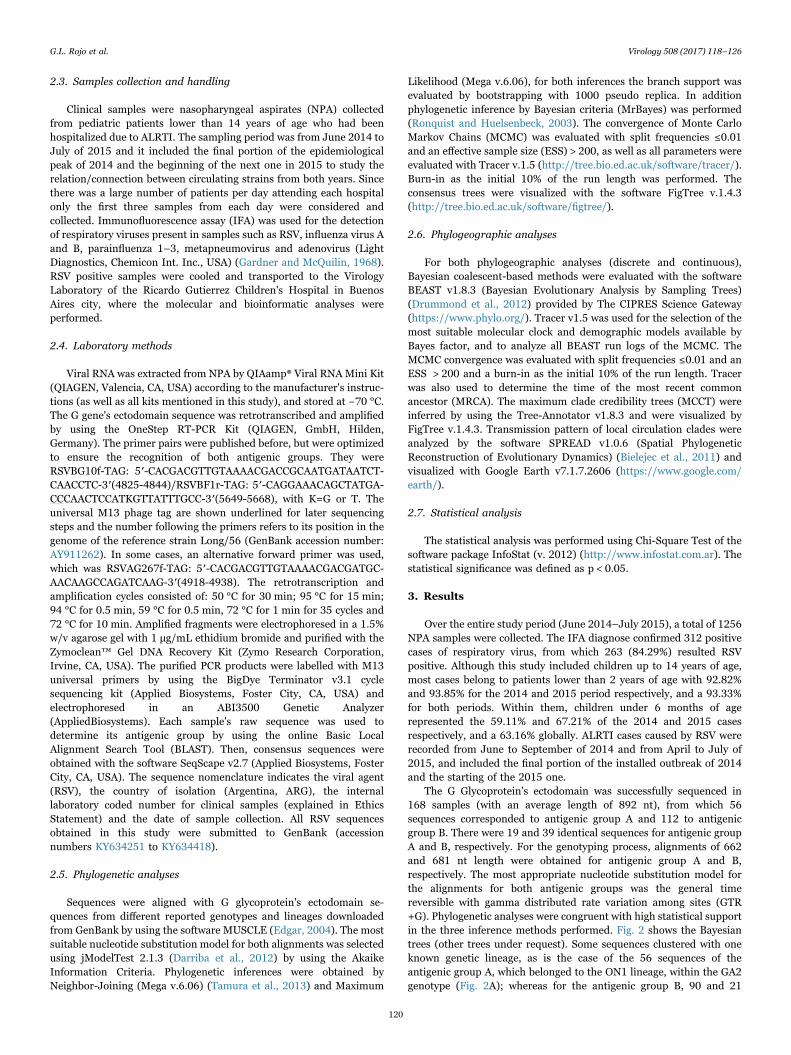
The G Glycoprotein's ectodomain was successfully sequenced in168 samples (with an average length of 892 nt), from which 56sequences corresponded to antigenic group A and 112 to antigenicgroup B. There were 19 and 39 identical sequences for antigenic groupA and B, respectively. For the genotyping process, alignments of 662and 681 nt length were obtained for antigenic group A and B,respectively. The most appropriate nucleotide substitution model forthe alignments for both antigenic groups was the general timereversible with gamma distributed rate variation among sites (GTR+G). Phylogenetic analyses were congruent with high statistical supportin the three inference methods performed. Fig. 2 shows the Bayesiantrees (other trees under request). Some sequences clustered with oneknown genetic lineage, as is the case of the 56 sequences of theantigenic group A, which belonged to the ON1 lineage, within the GA2genotype (Fig. 2A); whereas for the antigenic group B, 90 and 21
G.L. Rojo et al. Virology 508 (2017) 118–126
120
sequences clustered with the lineages BA9 and BA10, respectivelywithin the BA genotype. There was only one sequence(RSVARG_C017) from the B antigenic group which clustered withanother genetic lineage not mentioned before within the BA genotype(Fig. 2B). These results are shown on Table 1. The genotyping analysisshowed co-circulation of the ON1, BA9 and BA10 lineages throughoutthe analyzed period. The Bayesian analyses showed global, regional andlocal circulation patterns by their association with different referencesequences. Once the circulating lineages were known, it was interestingto determine if there was an exclusive set of sequences with ancestralorigin in Argentina. Thus, we decided to perform phylogeographic
analyses to detect the most probable location of the origin that couldallow the spread of RSV at the local level.
To determine the correlation between the sequences obtained inthis study and reference sequences reported worldwide during the lastyears (within the detected lineages, ON1, BA9 and BA10), discretephylogeographic analysis were estimated. Global spatio-temporal dif-fusion was achieved testing nucleotide substitution models, where themodel GTR+G was the most appropriate for both antigenic groups.Analyzing the obtained MCCTs, the MRCA for the ON1 lineage wasinferred in October 19th, 2010 located in Panama (Fig. 3A), and for theBA lineages was inferred in July 8th, 1998 in Argentina (Fig. 3B). The
Fig. 2. Bayesian phylogenetic trees. The evolutionary model GTR+G was used for Bayesian reconstructions of RSV antigenic groups A (A) and B (B). In both cases the Bayesian MarkovChain Monte Carlo (MCMC) chains were run for 5E+7 generations, a 5E+4 sample frequency to reach convergence and a 10% burn-in. Genotypes and genetic lineages described by thebibliography are indicated with vertical lines on the right. Node labels represent posterior probabilities (values higher than 0.6 are shown). The scale bar represents nucleotidesubstitutions per site. Sequences obtained in this study are denoted with a black circle.
G.L. Rojo et al. Virology 508 (2017) 118–126
121

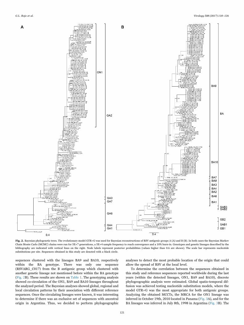
ON1 sequences obtained in this study gathered in two different cladeswith exclusive origin in Argentina and related to each other. A smallone originated in December 21st, 2013 that share a common ancestorin the past with reported sequences from Spain (ON1-2, blue clade inFig. 3A), and a large one originated in September 28th, 2011 (ON1-1,yellow clade in Fig. 3A). For the analyzed antigenic group B sequences,we also found two different clades with a common ancestor inArgentina. One clade of BA10 lineage (green in Fig. 3B) was originatedin April 14th, 2011 and shared a common ancestor in the past withreported sequences from Paraguay, and the other clade of BA9 lineagewas originated in March 08th, 2005 (red on Fig. 3B). For ON1-1 andBA9 clades, only sequences with exclusive local circulation wereconsidered in the subsequent analyses. In summary, on both MCCTs,we identified clades that were feasible for analyzing the viral transmis-sion patterns, hence they were considered for continuous phylogeo-graphic analyses. The results of these analyses are shown through thediffusion dynamic plots on Fig. 4 and the interactive animation inSupplementary files KML 1–4. They demonstrated that there was aspatio-temporal transmission between local strains. In the center of theanalyzed area (between Lomas de Zamora and Lanús districts), acommon hot spot for all the analyzed clades was found, where all theMRCAs were located. In all cases, the intermediate ancestors spreadfollowing a centrifugal dispersion pattern, moving north, southeast andsouthwest. Since it was interesting the finding that the MRCAs of allgenetic clades (independently analyzed) spread from the same place,we were interested in analyzing the beginning of each outbreak perclade, per year. Thus, we analyzed in the cases which were possible thetransmission pattern of each clade considering, on the one hand, onlythe sequences of 2014 and, on the other, those of 2015 (Fig. 5). For the2014 period, the location of the MRCA for the ON1-1, BA9 and BA10clades was not different from the one inferred in the analyses thatconsidered both years simultaneously (Fig. 5A and C and;Supplementary files KML 5–7), while in 2015 period, the inferredlocations of the MRCAs for the ON1-1 and BA9 clades were slightlyrelocated to the north, closer to Lanús district (Fig. 5B and D;Supplementary files KML 8 and 9). For ON1-2 (2014–2015) andBA10 (2015) lineages, the low number of sequences impeded theiranalysis.
Considering that the characteristic spreading inferred for RSVcould be attributed to some features in the studied population, weanalyzed the population density of the region and the survey formobtained from each patient. Initially, we found that the central andnorthern districts of the VI Sanitary Region (where the location for allMRCAs were estimated) have the highest population density as shownon Fig. 1, with Lanús district as the most populated.
Afterwards, we evaluated if there was a statistical correlationbetween the population density and the risk of overcrowding (accord-ing to the inhabitants in a household and number of rooms from eachpatient's house). All children that lived in houses with more than 3persons per room were classified as overcrowded as was determined in
Argentina in the document "Unsatisfied basic-needs methodology"reported by the Provincial Bureau of Statistics (Ministry of Economyof the Buenos Aires Province, 2016) and by UNICEF (https://www.unicef.org/argentina/spanish/monitoreo_Pobreza_Completo.pdf).The nine districts of the analyzed region were classified into three zoneswith low, medium or high population density (Fig. 1 and Table 2). Inorder to determine if there was a different amount of overcrowded ornot overcrowded children geolocalized in the three mentioned zones, astatistical analysis was performed. We found no significant differencesamong the different population densities (p=0.7815) (Table 2).
4. Discussion
The sequencing of the G gene's ectodomain allowed the subtypingand genotyping of the clinical samples collected in a population thathas not been studied before, and therefore it allowed the identificationof the circulating genotypes and lineages of RSV in the analyzed areaduring the studied period. Although the age of the analyzed patientswas extended to 14 years old, children under six months were found tobe the most susceptible to suffer severe infections, requiring hospita-lization. Therefore, this should be the target population to be protectedagainst severe infections. Nevertheless, it would be interesting toanalyze the RSV impact on ambulatory patients with ALRTI andwhether the most susceptible age group is different from the ones ofhospitalized patients. In addition, it would also be interesting todetermine if the onset of the outbreak in the ambulatory populationis earlier than that of the hospitalized patients which could thereforeexplain the transmission chains in the entire population.
We found a predominance of B antigenic group over A in bothstudied periods, which differs from what was reported in Buenos AiresCity in 2014 (Viegas et al., 2016). However, we cannot ruled out thatthe other antigenic group could have predominated during the first partof the outbreak (not analyzed in this study) and even could havechanged the prevailing antigenic group in the entire outbreak.
Discrete phylogeographic analyses showed that some strains wererelated to ancestors reported at neighboring or transatlantic countries,which were introduced to Argentina, and others samples related toancestors with local circulation. The continuous phylogeographicanalysis of the clades with exclusive origin in Argentina showed thatthe MRCA origin for all lineages was located in the center of theanalyzed area between Lomas de Zamora and Lanús districts.Interestingly, those districts show the highest population density. Theindividual analysis of the clades from the 2014 period resulted in non-significant variation of the inferred MRCAs location. The analyses ofthe ON1-1 and BA9 clades for the 2015 period showed a slight move tonorth, their MRCAs approached to the Lanús district, and since thesamples from 2015 belong to the onset of an outbreak, the reason forthat movement could support that the beginning of any new outbreakwould be linked to highly populated areas. Outbreaks of infections aremore frequent at high population densities given that the viral
Table 1Number of RSV positive samples related to their antigenic group and genotypic characteristics (genotypes and genetic lineages), year of collection and hospital's districts where thepatients were admitted.
Antigenic group A Antigenic group B Total
GA2 genotype BA genotype
ON1 BA9 BA10
2014 2015 2014 2015 2014 2015 2014 2015 2014–2015
Berazategui 4 4 11 7 1 0 16 11 27Florencio Varela 4 4 9 6 2 0 15 10 25Ezeiza 14 3 14 3 10 1 38 7 45Lanús 14 9 21 19 7 0 42 28 70Total 36 20 55 35 20 1 111 56 167
G.L. Rojo et al. Virology 508 (2017) 118–126
122

transmission is favored especially in the case of respiratory diseases. Itis important to mention that the sampling process conducted by allphysicians from different participating hospitals was continuous duringall the analyzed period and the number of samples collected in this
study did not present bias of collection. Therefore, the high number ofsamples from Lanús could support the phylogeographic results, indeedthe outbreak would begin there and expand to other districts as wasobserved in the continuous analyses, in which diverse intermediate
Fig. 3. Maximum Clade Credibility Tree (MCCT) discrete phylogeographic results. Trees for RSV antigenic group A ON1 lineage (A) and antigenic group B, BA9 and BA10 lineages (B)were obtained with the evolutionary model GTR+G. Lognormal relaxed clock (uncorrelated) model as the molecular clock. Bayesian Skyline was the most suitable demographic modelfor the antigenic group A, and the GMRF Bayesian Skyride for the B one. Branches are denoted in color by the location of the reported sequences, and the acronym indicates theircountry of origin. Node labels represent the probability support for that inference. Scale axis represents years. Local sequences names are highlighted: yellow for ON1-1; blue for ON1-2;red for BA9 clade and green for BA10 clade. Acronyms: ARG Argentina, BE Belgium, BR Brazil, CAN Canada, CB Cuba, CH China, HK Hong Kong, IND India, JP Japan, KEN Kenya, NLNederland, PAK Pakistan, PAN Panama, PY Paraguay, PER Peru, PHI Philippines, PY Paraguay, SAR South Africa, SK South Korea, SPA Spain, THA Thailand, USA United States, VNVietnam.
G.L. Rojo et al. Virology 508 (2017) 118–126
123
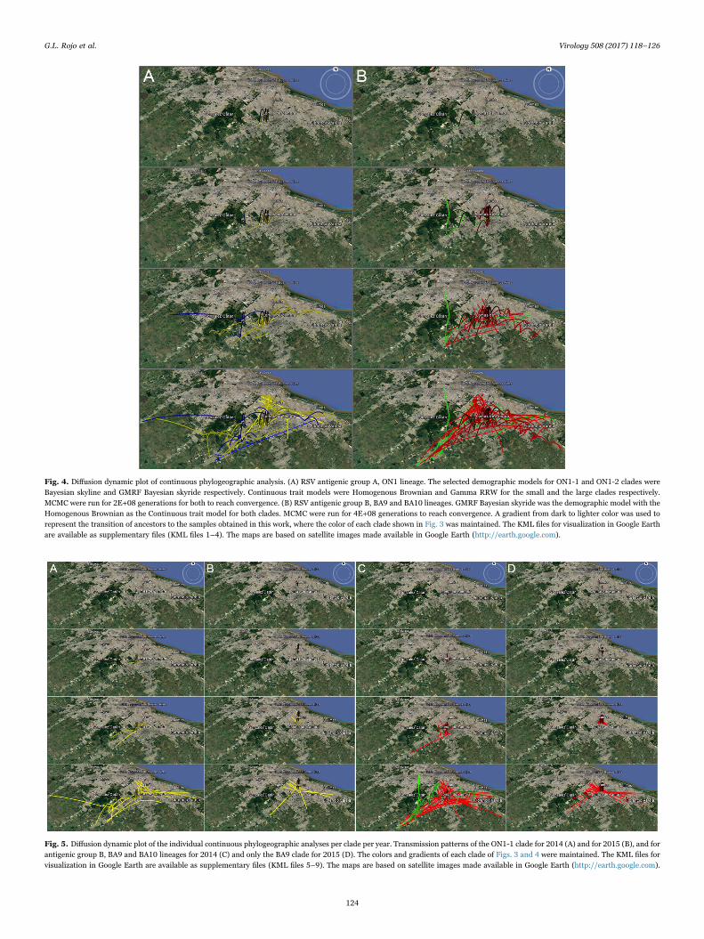
Fig. 4. Diffusion dynamic plot of continuous phylogeographic analysis. (A) RSV antigenic group A, ON1 lineage. The selected demographic models for ON1-1 and ON1-2 clades wereBayesian skyline and GMRF Bayesian skyride respectively. Continuous trait models were Homogenous Brownian and Gamma RRW for the small and the large clades respectively.MCMC were run for 2E+08 generations for both to reach convergence. (B) RSV antigenic group B, BA9 and BA10 lineages. GMRF Bayesian skyride was the demographic model with theHomogenous Brownian as the Continuous trait model for both clades. MCMC were run for 4E+08 generations to reach convergence. A gradient from dark to lighter color was used torepresent the transition of ancestors to the samples obtained in this work, where the color of each clade shown in Fig. 3 was maintained. The KML files for visualization in Google Earthare available as supplementary files (KML files 1–4). The maps are based on satellite images made available in Google Earth (http://earth.google.com).
Fig. 5. Diffusion dynamic plot of the individual continuous phylogeographic analyses per clade per year. Transmission patterns of the ON1-1 clade for 2014 (A) and for 2015 (B), and forantigenic group B, BA9 and BA10 lineages for 2014 (C) and only the BA9 clade for 2015 (D). The colors and gradients of each clade of Figs. 3 and 4 were maintained. The KML files forvisualization in Google Earth are available as supplementary files (KML files 5–9). The maps are based on satellite images made available in Google Earth (http://earth.google.com).
G.L. Rojo et al. Virology 508 (2017) 118–126
124

ancestors have spread to different directions: northbound, southeastand southwest. In this sense, the idea that the outbreak begins in themost populated area and then is distributed centrifugally to other areascould describe the social characteristics of a region, the connections ofpublic transport and relations among communities, as well as thearchitecture of the connection among the districts that comprise thesanitary region analyzed (Read et al., 2012). In fact, the division of theterritory into sanitary regions was carried out with the aim oforganizing health system and being able to make decisions in theprevention and treatment of the most important diseases that impact topublic health. The data provided in this work allowed us to trace a mapof vulnerability of ALRTI caused by RSV in the analyzed region, whichin the future could be implemented as a model for other health regionsor for other transmissible infections.
We supposed that the viral transmission pattern could be linked tocertain overcrowding characteristics of the analyzed population, but wedid not find any significant differences in the number of residents livingin overcrowded conditions among the three geographic areas definedby population density. However, as the overcrowding was similar ineach of the defined areas, the high population density should beconsidered as a risk factor for transmission. On the other hand, wecannot exclude that other social characteristics, such as number ofsmokers, type of home heating systems, structural characteristics of thedwellings could be related to the transmission pattern described.Further analyses considering those characteristics are needed tocorrelate socio-epidemiological features and RSV transmission pat-terns.
Since there are diverse vaccination strategies under study and manyof them are about to be approved, in the very near future, we will havethe possibility to prevent severe infections caused by this virus. In thissense, several studies have attempted to describe the patterns of RSVtransmission from the patient's point of view, with the objective ofdetermining the target population to be vaccinated. In the UnitedStates, Yamin et al. have shown that targeting of children younger thanfive years of age is highly efficient per dose, and is also the mosteffective strategy to reduce RSV in young children and older adultsacross all states and transmission settings. Nevertheless, they analyzedthe possibility to vaccinate the rest of the population to decrease thenumber of cases (although with substantially lower efficiency) and theyproposed a cost-effectiveness analysis to specific states and transmis-sion settings (Yamin et al., 2016). Considering the viral circulationpatterns described in this study -where each viral outbreak begins incertain areas with high population density- health systems shouldevaluate vaccination at all ages in those areas, while in lower densitypopulation areas only susceptible children should be vaccinated.
5. Conclusion
In this study, it was possible to identify viral transmission pathwaysthrough the analysis of RSV's genetic perspective from two consecutiveoutbreaks in a limited geographical region. Considering this study as aprecedent of the transmission dynamics of RSV in a population, it willbe useful to describe patterns of social mixing behavior at theindividual level and to contribute to the knowledge of the dynamics
of infection at the population level. These results suggest that futurecost-effectiveness analyses of RSV vaccination should be tailor-madefor specific population and to specific regions.
Funding information
This work was supported by the “Ramón Carrillo – Arturo Oñativia”grant 2014. Principal Investigator: Alejandro Castello. National HealthResearch Commission. Ministry of Health, Argentina.
Acknowledgements
We thank the laboratory staff from the participating hospitals forthe sample collection and the AVIRIM Team: Karina Dueñas, XimenaFlores, Yanina Díaz, Laura Aresca, Paula Riolfo.
Appendix A. Supporting information
Supplementary data associated with this article can be found in theonline version at doi:10.1016/j.virol.2017.04.030.
References
Afonso, C.L., Amarasinghe, G.K., Bányai, K., Bào, Y., Basler, C.F., Bavari, S., Bejerman,N., Blasdell, K.R., Briand, F.X., Briese, T., Bukreyev, A., Calisher, C.H., Chandran, K.,Chéng, J., Clawson, A.N., Collins, P.L., Dietzgen, R.G., Dolnik, O., Domier, L.L.,Dürrwald, R., Dye, J.M., Easton, A.J., Ebihara, H., Farkas, S.L., Freitas-Astúa, J.,Formenty, P., Fouchier, R.A., Fù, Y., Ghedin, E., Goodin, M.M., Hewson, R., Horie,M., Hyndman, T.H., Jiāng, D., Kitajima, E.W., Kobinger, G.P., Kondo, H., Kurath, G.,Lamb, R.A., Lenardon, S., Leroy, E.M., Li, C.X., Lin, X.D., Liú, L., Longdon, B.,Marton, S., Maisner, A., Mühlberger, E., Netesov, S.V., Nowotny, N., Patterson, J.L.,Payne, S.L., Paweska, J.T., Randall, R.E., Rima, B.K., Rota, P., Rubbenstroth, D.,Schwemmle, M., Shi, M., Smither, S.J., Stenglein, M.D., Stone, D.M., Takada, A.,Terregino, C., Tesh, R.B., Tian, J.H., Tomonaga, K., Tordo, N., Towner, J., Vasilakis,N., Verbeek, M., Volchkov, V., Wahl-Jensen, V., Walsh, J.A., Walker, P.J., Wang, D.,Wang, L.F., Wetzel, T., Whitfield, A.E., Xiè, J.T., Yuen, K.Y., Zhang, Y.Z., Kuhn, J.H.,2016. Taxonomy of the order mononegavirales: update 2016. Arch. Virol. 161 (8),2351–2360. http://dx.doi.org/10.1007/s00705-016-2880-1.
Anderson, L.J., Dormitzer, P.R., Nokes, D.J., Rappuoli, R., Roca, A., Graham, B.S., 2013.Strategic priorities for respiratory syncytial virus (RSV) vaccine development.Vaccine 31 (2), 209–215. http://dx.doi.org/10.1016/j.vaccine.2012.11.106.
Anderson, L.J., Hierholzer, J.C., Tsou, C., Hendry, R.M., Fernie, B.F., Stone, Y.,McIntosh, K., 1985. Antigenic characterization of respiratory syncytial virus strainswith monoclonal antibodies. J. Infect. Dis. 151 (4), 626–633.
Bielejec, F., Rambaut, A., Suchard, M.A., Lemey, P., 2011. SPREAD: spatial phylogeneticreconstruction of evolutionary dynamics. Bioinformatics 27 (20), 2910–2912.http://dx.doi.org/10.1093/bioinformatics/btr481.
Caballero, M.T., Serra, M.E., Acosta, P.L., Marzec, J., Gibbons, L., Salim, M., Rodriguez,A., Reynaldi, A., Garcia, A., Bado, D., Buchholz, U.J., Hijano, D.R., Coviello, S.,Newcomb, D., Bellabarba, M., Ferolla, F.M., Libster, R., Berenstein, A., Siniawaski,S., Blumetti, V., Echavarria, M., Pinto, L., Lawrence, A., Ossorio, M.F., Grosman, A.,Mateu, C.G., Bayle, C., Dericco, A., Pellegrini, M., Igarza, I., Repetto, H.A., Grimaldi,L.A., Gudapati, P., Polack, N.R., Althabe, F., Shi, M., Ferrero, F., Bergel, E., Stein,R.T., Peebles, R.S., Boothby, M., Kleeberger, S.R., Polack, F.P., 2015. TLR4 genotypeand environmental LPS mediate RSV bronchiolitis through Th2 polarization. J. Clin.Investig. 125 (2), 571–582. http://dx.doi.org/10.1172/JCI75183.
Collins, P.L., Karron, R., 2013. Respiratory syncytial virus and metapneumovirus. In:Knipe, D., Howley, P. (Eds.), Fields Virology. LWW, Philadelphia, USA, 1086–1123.
Cristina, J., López, J.A., Albó, C., García-Barreno, B., García, J., Melero, J.A., Portela, A.,1990. Analysis of genetic variability in human respiratory syncytial virus by theRNase A mismatch cleavage method: subtype divergence and heterogeneity. Virology174 (1), 126–134.
Darriba, D., Taboada, G.L., Doallo, R., Posada, D., 2012. jModelTest 2: more models, new
Table 2Number of children with positive RSV samples classified according to their overcrowding characteristics and to their location zone by population density.
Zone's population density (People/Km2) District Not overcrowded ( < 3 inhabitants/room)
Overcrowded ( > 3 inhabitants/room)
High (5000.1−15000.0) Lomas de Zamora, Avellaneda, Lanús 37 33Medium (1000.1−5000.0) Esteban Echeverría, Almirante Brown, Quilmes,
Florencio Varela, Berazategui46 40
Low (100.0−1000.0) Ezeiza 43 31
Statistical analysis: χ2 (0.493) and p-value (0.781).
G.L. Rojo et al. Virology 508 (2017) 118–126
125
heuristics and parallel computing. Nat. Methods 9 (8), 772. http://dx.doi.org/10.1038/nmeth.2109.
DeVincenzo, J.P., Wilkinson, T., Vaishnaw, A., Cehelsky, J., Meyers, R., Nochur, S.,Harrison, L., Meeking, P., Mann, A., Moane, E., Oxford, J., Pareek, R., Moore, R.,Walsh, E., Studholme, R., Dorsett, P., Alvarez, R., Lambkin-Williams, R., 2010. Viralload drives disease in humans experimentally infected with respiratory syncytialvirus. Am. J. Respir. Crit. Care Med. 182 (10), 1305–1314. http://dx.doi.org/10.1164/rccm.201002-0221OC.
Drummond, A.J., Suchard, M.A., Xie, D., Rambaut, A., 2012. Bayesian phylogeneticswith BEAUti and the BEAST 1.7. Mol. Biol. Evol. 29, 1969–1973. http://dx.doi.org/10.1093/molbev/mss075.
Duvvuri, V.R., Granados, A., Rosenfeld, P., Bahl, J., Eshaghi, A., Gubbay, J.B., 2015.Genetic diversity and evolutionary insights of respiratory syncytial virus A ON1genotype: global and local transmission dynamics. Sci. Rep. 30 (5), 14268. http://dx.doi.org/10.1038/srep14268.
Edgar, R.C., 2004. MUSCLE: multiple sequence alignment with high accuracy and highthroughput. Nucleic Acids Res. 32 (5), 1792–1797. http://dx.doi.org/10.1093/nar/gkh340.
Eshaghi, A., Duvvuri, V.R., Lai, R., Nadarajah, J.T., Li, A., Patel, S.N., Low, D.E., Gubbay,J.B., 2012. Genetic variability of human respiratory syncytial virus A strainscirculating in Ontario: a novel genotype with a 72 nucleotide G gene duplication.PLoS One 7 (3), e32807. http://dx.doi.org/10.1371/journal.pone.0032807.
Gardner, P.S., McQuilin, J., 1968. Viral diagnosis by immunofluorescence. Lancet 1,597–598.
Glezen, W.P., Taber, L.H., Frank, A.L., Kasel, J.A., 1986. Risk of primary infection andreinfection with respiratory syncytial virus. Am. J. Dis. Child. 140 (6), 543–546.
Hall, C.B., Douglas, R.G., Jr, Geiman, J.M., 1976. Respiratory syncytial virus infectionsin infants: quantitation and duration of shedding. J. Pediatr. 89 (1), 11–15.
Higgins, D., Trujillo, C., Keech, C., 2016. Advances in RSV vaccine research anddevelopment - A global agenda. Vaccine 34 (26), 2870–2875. http://dx.doi.org/10.1016/j.vaccine.2016.03.109.
Holberg, C.J., Wright, A.L., Martinez, F.D., Ray, C.G., Taussig, L.M., Lebowitz, M.D.,1991. Risk factors for respiratory syncytial virus-associated lower respiratoryillnesses in the first year of life. Am. J. Epidemiol. 133 (11), 1135–1151.
Hotard, A.L., Laikhter, E., Brooks, K., Hartert, T.V., Moore, M.L., 2015. Functionalanalysis of the 60-nucleotide duplication in the respiratory syncytial virus BuenosAires strain attachment glycoprotein. J. Virol. 89, 8258–8266. http://dx.doi.org/10.1128/JVI.01045-15.
Kim, Y.K., Choi, E.H., Lee, H.J., 2007. Genetic variability of the fusion protein andcirculation patterns of genotypes of the respiratory syncytial virus. J. Med Virol. 79(6), 820–828. http://dx.doi.org/10.1002/jmv.20891.
Ministry of Economy of the Buenos Aires Province, 2016. Argentina: ⟨http://www.estadistica.ec.gba.gov.ar/dpe/index.php/2016-05-30-15-56-27/2016-06-03-13-13-37/necesidades-basicas-insatisfechas/177-metodologia-necesidades-basicas-insatisfechas/230-metodologia-necesidades-basicas-insatisfechas⟩.
Munywoki, P.K., Koech, D.C., Agoti, C.N., Bett, A., Cane, P.A., Medley, G.F., Nokes, J.D.,2015. Frequent asymptomatic respiratory syncytial virus infections during anepidemic in a rural kenyan household cohort. J. Infect. Dis. 212 (11), 1711–1718.http://dx.doi.org/10.1093/infdis/jiv263.
Nair, H., Nokes, D.J., Gessner, B.D., Dherani, M., Madhi, S.A., Singleton, R.J., O'Brien,
K.L., Roca, A., Wright, P.F., Bruce, N., Chandran, A., Theodoratou, E., Sutanto, A.,Sedyaningsih, E.R., Ngama, M., Munywoki, P.K., Kartasasmita, C., Simões, E.A.,Rudan, I., Weber, M.W., Campbell, H., 2010. Global burden of acute lowerrespiratory infections due to respiratory syncytial virus in young children: asystematic review and meta-analysis. Lancet 375 (9725), 1545–1555. http://dx.doi.org/10.1016/S0140-6736(10)60206-1.
Okiro, E.A., Ngama, M., Bett, A., Cane, P.A., Medley, G.F., Nokes, J.D., 2008. Factorsassociated with increased risk of progression to respiratory syncytial virus-associatedpneumonia in young Kenyan children. Trop. Med. Int. Health 13 (7), 914–926.http://dx.doi.org/10.1111/j.1365-3156.2008.02092.x.
Read, J.M., Edmunds, W.J., Riley, S., Lessler, J., Cummings, D.A.T., 2012. Closeencounters of the infectious kind: methods to measure social mixing behavior.Epidemiol. Infect. 140 (12), 2117–2130. http://dx.doi.org/10.1017/S0950268812000842.
Ronquist, F., Huelsenbeck, J.P., 2003. MrBayes v.3.2.3: bayesian phylogenetic inferenceunder mixed models. Bioinforma. Oxf. Engl. 19 (12), 1572–1574.
Tamura, K., Stecher, G., Peterson, D., Filipski, A., Kumar, S., 2013. MEGA6: molecularevolutionary genetics analysis version 6.0. Mol. Biol. Evol. 30 (12), 2725–2729.http://dx.doi.org/10.1093/molbev/mst197.
Trento, A., Galiano, M., Videla, C., Carballal, G., García-Barreno, B., Melero, J.A.,Palomo, C., 2003. Major changes in the G protein of human respiratory syncytialvirus isolates introduced by a duplication of 60 nucleotides. J. Gen. Virol. 84 (11),3115–3120. http://dx.doi.org/10.1099/vir.0.19357-0.
Trento, A., Viegas, M., Galiano, M., Videla, C., Carballal, G., Mistchenko, A.S., Melero,J.A., 2006. Natural history of human respiratory syncytial virus inferred fromphylogenetic analysis of the attachment (G) glycoprotein with a 60-nucleotideduplication. J. Virol. 80 (2), 975–984. http://dx.doi.org/10.1128/JVI.80.2.975-984.2006.
Trento, A., Casas, I., Calderón, A., Garcia-Garcia, M.L., Calvo, C., Perez-Breña, P.,Melero, J.A., 2010. Ten years of global evolution of the human respiratory syncytialvirus BA genotype with a 60-nucleotide duplication in the G protein gene. J. Virol. 84(15), 7500–7512. http://dx.doi.org/10.1128/JVI.00345-10.
Viegas, M., Goya, S., Mistchenko, A.S., 2016. Sixteen years of evolution of humanrespiratory syncytial virus subgroup A in Buenos Aires, Argentina: GA2 the prevalentgenotype through the years. Infect. Genet. Evol. 43, 213–221. http://dx.doi.org/10.1016/j.meegid.2016.04.034.
Viegas, M., Barrero, P.R., Maffey, A.F., Mistchenko, A.S., 2004. Respiratory virusesseasonality in children under five years of age in Buenos Aires, Argentina: a five-yearanalysis. J. Infect. 49, 222–228. http://dx.doi.org/10.1016/j.jinf.2003.10.006.
Walsh, E.E., Falsey, A.R., 2012. Respiratory syncytial virus infection in adultpopulations. Infect. Disord. Drug Targets 12 (2), 98–102.
Wathuo, M., Medley, G.F., Nokes, D.J., Munywoki, P.K., 2016. Quantification anddeterminants of the amount of respiratory syncytial virus (RSV) shed using real timePCR data from a longitudinal household study. Wellcome Open Res. 1 (27). http://dx.doi.org/10.12688/wellcomeopenres.10284.1.
Yamin, D., Jones, F.K., DeVincenzo, J.P., Gertler, S., Kobiler, O., Townsend, J.P.,Galvani, A.P., 2016. Vaccination strategies against respiratory syncytial virus. Proc.Natl. Acad. Sci. USA 113 (46), 13239–13244. http://dx.doi.org/10.1073/pnas.1522597113.
G.L. Rojo et al. Virology 508 (2017) 118–126
126


![IMAGEN Respiratory Syncytial Virus (RSV) [PT]...IMAGEN Respiratory Syncytial Virus (RSV) 1. UTILIZAÇÃO PREVISTA O IMAGEN Respiratory Syncytial Virus (RSV) é um teste qualitativo](https://static.fdocuments.net/doc/165x107/609c20ca1e0ebf036346e66d/imagen-respiratory-syncytial-virus-rsv-pt-imagen-respiratory-syncytial-virus.jpg)