
UNIVERSITY OF CALGARY Association between - DSpace
239
University of Calgary PRISM: University of Calgary's Digital Repository Graduate Studies Legacy Theses 2011 Association between Antimicrobial Use and Antimicrobial Resistance in Bovine Mastitis Pathogens Saini, Vineet Saini, V. (2011). Association between Antimicrobial Use and Antimicrobial Resistance in Bovine Mastitis Pathogens (Unpublished doctoral thesis). University of Calgary, Calgary, AB. doi:10.11575/PRISM/15372 http://hdl.handle.net/1880/48858 doctoral thesis University of Calgary graduate students retain copyright ownership and moral rights for their thesis. You may use this material in any way that is permitted by the Copyright Act or through licensing that has been assigned to the document. For uses that are not allowable under copyright legislation or licensing, you are required to seek permission. Downloaded from PRISM: https://prism.ucalgary.ca
Transcript of UNIVERSITY OF CALGARY Association between - DSpace

University of Calgary
PRISM: University of Calgary's Digital Repository
Graduate Studies Legacy Theses
2011
Association between Antimicrobial Use and
Antimicrobial Resistance in Bovine Mastitis
Pathogens
Saini, Vineet
Saini, V. (2011). Association between Antimicrobial Use and Antimicrobial Resistance in Bovine
Mastitis Pathogens (Unpublished doctoral thesis). University of Calgary, Calgary, AB.
doi:10.11575/PRISM/15372
http://hdl.handle.net/1880/48858
doctoral thesis
University of Calgary graduate students retain copyright ownership and moral rights for their
thesis. You may use this material in any way that is permitted by the Copyright Act or through
licensing that has been assigned to the document. For uses that are not allowable under
copyright legislation or licensing, you are required to seek permission.
Downloaded from PRISM: https://prism.ucalgary.ca

UNIVERSITY OF CALGARY
Association between Antimicrobial Use and Antimicrobial Resistance
in Bovine Mastitis Pathogens
by
Vineet Saini
A THESIS
SUBMITTED TO THE FACULTY OF GRADUATE STUDIES
IN PARTIAL FULFILMENT OF THE REQUIREMENTS FOR THE
DEGREE OF DOCTOR OF PHILOSOPHY
DEPARTMENT OF MEDICAL SCIENCE
CALGARY, ALBERTA
NOVEMBER, 2011
Vineet Saini 2011

The author of this thesis has granted the University of Calgary a non-exclusive license to reproduce and distribute copies of this thesis to users of the University of Calgary Archives. Copyright remains with the author. Theses and dissertations available in the University of Calgary Institutional Repository are solely for the purpose of private study and research. They may not be copied or reproduced, except as permitted by copyright laws, without written authority of the copyright owner. Any commercial use or re-publication is strictly prohibited. The original Partial Copyright License attesting to these terms and signed by the author of this thesis may be found in the original print version of the thesis, held by the University of Calgary Archives. Please contact the University of Calgary Archives for further information: E-mail: [email protected]: (403) 220-7271 Website: http://archives.ucalgary.ca

ABSTRACT
Concurrent data on antimicrobial use (AMU) and resistance in food-animal production
systems are required to formulate policies for containing antimicrobial resistance (AMR)
in bacteria. The specific objectives of this PhD study were four-fold:
1) Determine AMU on Canadian dairy farms,
2) Determine AMR profiles of common bovine mastitis pathogens Staphylococcus
aureus, Escherichia coli, and Klebsiella species isolated from intramammary
infections and (sub) clinical mastitis cases,
3) Predict diagnostic accuracy and agreement of Sensititre® MIC susceptibility
system and agar disk diffusion test method employed for AMR profiling of udder
pathogens, and
4) Determine herd-level association between AMU and AMR in bovine mastitis
pathogens.
The data originated from 89 dairy farms located in four regions of Canada. Producers and
farm personnel were asked to deposit empty antimicrobial containers in specially
provided receptacles to quantify AMU. Three sets of milk samples were collected in the
winter and summer of 2007 and 2008. Minimum inhibitory concentrations were
determined using Sensititre® bovine mastitis plate and NARMS Gram-negative penal.
Sensititre® and the agar disk diffusion test methods had a moderate to high diagnostic
accuracy, and very good essential and categorical agreement for most udder pathogen-
antimicrobial combinations. ß-lactams, penicillin combinations, tetracyclines,
ii

trimethoprim-sulfonamide combinations and lincosamides were most commonly used.
Prevalence of AMR in S. aureus, E. coli and Klebsiella species isolates was 20.4, 17.7
and 36.6%, respectively. Resistance to penicillin and tetracycline was most common
among Gram-positive and Gram-negative isolates, respectively.
Use of penicillin-novobiocin combination administered for dry cow therapy, and
systemically administered penicillin was positively associated with penicillin resistance
in S. aureus isolates; use of pirlimycin for clinical mastitis treatment was associated with
pirlimycin resistance. Systemic administration of tetracycline and penicillin was
associated with tetracycline resistance in E. coli, while ampicillin resistance in E. coli
was associated with systemic administration of ceftiofur and penicillin. Tetracycline use
and resistance were associated in Klebsiella species isolates.
In conclusion, ß-lactams were most commonly used for mastitis treatment and control.
Prevalence of resistance in bovine mastitis pathogens was low. Herd-level use of certain
antimicrobials was associated with AMR in udder pathogens. Sensititre® and the agar
disk diffusion method can be readily employed in veterinary diagnostic laboratories.
iii

PREFACE
The following manuscripts have been published or accepted for publication. Vineet Saini
obtained and analyzed the data, interpreted the results, and wrote the papers with
guidance from his thesis committee (Drs. Herman Barkema, J McClure, David Léger,
Patrick Boerlin and Douglas Morck). All authors contributed important intellectual
content and provided critical review of the papers. Written permission for reproduction of
the article in its entirety has been obtained from the publisher.
Saini V., R. G. M. Olde Riekerink, J T. McClure, and H. W. Barkema. 2011. Diagnostic
accuracy assessment of Sensititre® and agar disk diffusion for determining antimicrobial
resistance profiles of bovine clinical mastitis pathogens. J. Clin. Microbiol. 49(4):1568-
1577.
Saini V., J T. McClure, David Léger, Simon Dufour, Aaron G. Sheldon, Daniel T. Scholl,
and H. W. Barkema. Antimicrobial use on Canadian dairy farms. J. Dairy Sci. Accepted
for publication: JDS-11-4527.
iv

ACKNOWLEDGEMENTS
First and foremost, I am extremely grateful to my advisor Dr. Herman Barkema for his
mentoring, guidance, and above all patience through out this process. His encouragement
and unwavering support made this journey a truly enjoyable and fulfilling experience.
Sincere thanks to my co-supervisor Dr. J T. McClure and committee, Dr. David Léger,
Dr. Patrick Boerlin, Dr. Douglas W. Morck, and Dr. Daniel T. Scholl for your guidance,
assistance and ultimately sticking and finishing the journey with me. Your intelligence,
guidance and patience through moments of panic is deeply appreciated and will never be
forgotten.
To my family who have always been proud of me and supported all that I have done.
They are the ones who navigated the choppy waters of graduate student life, as this
became a family endeavour as well. My brother, Puneet who supported me through thick
and thin – I thank you for sticking to me.
To my better half, Palvinder, who has always encouraged me forward – this would not
have been possible without your support. My sons, Timmy and Sunny, my world – thank
you for reminding me of what is really important in life.
This research was financed by the Natural Science and Engineering Research Council of
Canada, Alberta Milk, Dairy Farmers of New Brunswick, Nova Scotia, Ontario and
v

Prince Edward Island, Novalait Inc., Dairy Farmers of Canada, Canadian Dairy Network,
Agriculture and Agri-Food Canada, Public Health Agency of Canada, Technology PEI
Inc., Université de Montréal and University of Prince Edward Island, through the
Canadian Bovine Mastitis Research Network. The generous funding, educational
programs, and career development opportunities provided through the Canadian Bovine
Mastitis Research Network were valuable components of my training.
For those who supported, mentored, counseled, loved, sustained and just said what I
needed to hear in those critical moments – thank you.
vi

DEDICATION
This thesis is dedicated to my wife, Palvinder, whose unwavering support – emotional
and motivational – has led me to this day. The glowing spirits of my belated revered
parents, Piara Singh Saini and Krishna Saini helped foster my stalwart resolve to see my
PhD through to its fruition.
vii

TABLE OF CONTENTS
Abstract --------------------------------------------------------------------------------------------- ii Preface --------------------------------------------------------------------------------------------- iv Acknowledgements --------------------------------------------------------------------------------------------- v Dedication --------------------------------------------------------------------------------------------- vii Table of Contents --------------------------------------------------------------------------------------------- viii List of Tables --------------------------------------------------------------------------------------------- xi List of Figures --------------------------------------------------------------------------------------------- xv List of Symbols, Abbreviations, Nomenclature -------------------------------------------------------- xvi Epigraph --------------------------------------------------------------------------------------------- xviii CHAPTER ONE: INTRODUCTION ---------------------------------------------------------------------- 1
1.1 Mastitis in Dairy Cattle ----------------------------------------------------------- 2 1.2 Treatment of Bovine Mastitis ---------------------------------------------------- 3 1.3 Antimicrobial Resistance in Bovine Mastitis Pathogens ---------------------- 5 1.4 Association between Antimicrobial Use and Resistance ---------------------- 7 1.5 Antimicrobial Susceptibility Testing -------------------------------------------- 9 1.6 Objectives ------------------------------------------------------------------------------ 11 1.7 Thesis Organization ---------------------------------------------------------------- 11 1.8 References --------------------------------------------------------------------------- 13
CHAPTER TWO: VALIDATION OF SENSITITRE® AND AGAR DISK DIFFUSION ---------- 23
2.1 Abstract ------------------------------------------------------------------------------- 24 2.2 Introduction -------------------------------------------------------------------------- 26 2.3 Materials and Methods ------------------------------------------------------------- 28
2.3.1 Herd Selection, Sampling and Bacterial Culturing --------------------- 28 2.3.2 Antimicrobials -------------------------------------------------------------- 29 2.3.3 Agar Disk Diffusion Method --------------------------------------------- 30 2.3.4 Sensititre® System MIC Method ------------------------------------------ 30 2.3.5 Manual Broth Microdilution Test ---------------------------------------- 31 2.3.5.1 Culture and Inoculum Preparation ---------------------------- 31 2.3.5.2 Stock Solutions Preparation ---------------------------------- 31 2.3.5.3 Media Preparation ---------------------------------------------- 32 2.3.5.4 Microdilution Tray Preparation and Inoculation ---------- 32 2.3.6 Statistical Analyses -------------------------------------------------------- 33
2.4 Results -------------------------------------------------------------------------------- 35 2.4.1 Sensititre® Automatic MIC Readings and Broth Microdilution Comparison ---------------------------------------------------------------- 35 2.4.2 Sensititre® Manual and Automatic MIC Readings Comparison ----- 37 2.4.3 Agar Disk Diffusion Test Method ---------------------------------------- 38
2.5 Discussion ---------------------------------------------------------------------------- 39 2.5.1 Sensititre® Automatic MIC Readings ---------------------------------- 41 2.5.2 Sensititre® Automatic and Manual MIC Readings Comparison ----- 43 2.5.3 Agar Disk Diffusion and Broth Microdilution Comparison ---------- 43
viii

2.6 Conclusions -------------------------------------------------------------------------- 45 2.7 References ---------------------------------------------------------------------------- 46
CHAPTER THREE: ANTIMICROBIAL USE ON CANADIAN DAIRY FARMS ----------------- 61
3.1 Abstract ------------------------------------------------------------------------------- 62 3.2 Introduction -------------------------------------------------------------------------- 64 3.3 Materials and Methods ----------------------------------------------------------- 66
3.3.1 Dairy Cattle Herd Selection ---------------------------------------------- 66 3.3.2 Antimicrobial Use Data Collection Methodology --------------------- 67 3.3.3 Statistical Analyses --------------------------------------------------------- 68
3.4 Results -------------------------------------------------------------------------------- 71 3.4.1 Antimicrobial Drug Use Rates -------------------------------------------- 71
3.4.2 Region ------------------------------------------------------------------------ 72 3.4.3 Route of Administration --------------------------------------------------- 72 3.4.4 Intramammary -------------------------------------------------------------- 73 3.4.5 Systemic ---------------------------------------------------------------------- 73 3.4.6 Herd-level Factors --------------------------------------------------------- 73
3.5 Discussion ---------------------------------------------------------------------------- 74 3.6 Conclusions -------------------------------------------------------------------------- 81 3.7 References -------------------------------------------------------------------------- 83
CHAPTER FOUR: ANTIMICROBIAL RESISTANCE PROFILES ----------------------------------- 101
4.1 Abstract ------------------------------------------------------------------------------- 102 4.2 Introduction -------------------------------------------------------------------------- 104 4.3 Materials and Methods ------------------------------------------------------------- 107
4.3.1 Dairy Cattle Herd Selection ---------------------------------------------- 107 4.3.2 Sampling and Bacterial Culturing --------------------------------------- 108 4.3.3 Antimicrobials -------------------------------------------------------------- 109 4.3.4 Sensititre® System MIC Method ----------------------------------------- 110 4.3.5 Statistical Analyses --------------------------------------------------------- 111
4.4 Results -------------------------------------------------------------------------------- 112 4.4.1 Staphylococcus aureus ----------------------------------------------------- 112 4.4.2 Escherichia coli ------------------------------------------------------------- 114 4.4.3 Klebsiella Species --------------------------------------------------------- 115
4.5 Discussion ---------------------------------------------------------------------------- 115 4.6 Conclusions -------------------------------------------------------------------------- 126 4.7 References ---------------------------------------------------------------------------- 127
CHAPTER FIVE: ANTIMICROBIAL USE AND RESISTANCE ASSOCIATION IN STAPHYLOCOCCUS AUREUS ------------------------------------------------------------------------------ 148
5.1 Abstract ------------------------------------------------------------------------------- 149 5.2 Introduction -------------------------------------------------------------------------- 151 5.3 Materials and Methods ------------------------------------------------------------- 153
5.3.1 Herd Selection -------------------------------------------------------------- 153 5.3.2 Antimicrobial Use Data Collection Methodology --------------------- 154
ix

5.3.3 Sampling and Bacterial Culturing --------------------------------------- 155 5.3.4 MIC Determination --------------------------------------------------------- 156 5.3.5 Statistical Analyses --------------------------------------------------------- 156
5.4 Results -------------------------------------------------------------------------------- 158 5.4.1 Antimicrobial Resistance Proportions ----------------------------------- 158 5.4.2 Multivariable Analyses --------------------------------------------------- 158
5.5 Discussion ---------------------------------------------------------------------------- 159 5.6 Conclusions -------------------------------------------------------------------------- 164 5.7 References ---------------------------------------------------------------------------- 165
CHAPTER SIX: ANTIMICROBIAL USE AND RESISTANCE ASSOCIATION IN GRAM-NEGATIVE UDDER PATHOGENS -------------------------------------------------------------- 178
6.1 Abstract ------------------------------------------------------------------------------- 179 6.2 Introduction -------------------------------------------------------------------------- 181 6.3 Materials and Methods ------------------------------------------------------------- 183
6.3.1 Herd Selection -------------------------------------------------------------- 183 6.3.2 Antimicrobial Use Data Collection Methodology --------------------- 183 6.3.3 Sampling and Bacterial Culturing --------------------------------------- 184 6.3.4 MIC Determination --------------------------------------------------------- 185 6.3.5 Statistical Analyses --------------------------------------------------------- 186
6.4 Results -------------------------------------------------------------------------------- 188 6.4.1 Antimicrobial Resistance Proportions ----------------------------------- 188 6.4.2 Multivariable Analyses --------------------------------------------------- 189
6.5 Discussion ---------------------------------------------------------------------------- 191 6.6 Conclusions -------------------------------------------------------------------------- 196 6.7 References ---------------------------------------------------------------------------- 197
CHAPTER SEVEN: CONCLUSIONS AND FUTURE PERSPECTIVES ---------------------------- 213
7.1 References ---------------------------------------------------------------------------- 218
x

LIST OF TABLES
Table 2.1: Range of concentrations of antimicrobials used in the manual broth microdilution test method, Sensititre® bovine mastitis panel and commercial agar diffusion disks……………………………………………………………………………50 Table 2.2: Diagnostic accuracy estimates of Sensititre® automatic readings (off and on-scale MIC) and agar disk diffusion (ADD) test method with reference to manual broth microdilution test method for clinical bovine mastitis pathogens (n=119)……………...51 Table 2.3: Essential agreement (%) between Sensititre® MIC automatic readings (off and on-scale MIC) and manual broth microdilution MIC test method for clinical bovine mastitis pathogens (n=119)………………………………………………………………52 Table 2.4: Lower essential agreement (%) between Sensititre® MIC automatic readings (off and on-scale MIC) and manual broth microdilution MIC test method for Escherichia coli isolates in comparison to Gram-positive bovine clinical mastitis isolates (n=119)…55 Table 2.5: Categorical agreement (%) between Sensititre® automatic readings (off and on-scale MIC) and agar disk diffusion (ADD) test method with reference to manual broth microdilution test method for clinical bovine mastitis pathogens (n=119)……………...56 Table 2.6: Differences between Sensititre® automatic MIC readings (off and on-scale MIC) and Sensititre® manual MIC readings test method for clinical bovine mastitis pathogens (n=457)……………………………………………………………………….58 Table 3.1: National level estimate of antimicrobial drug use rate (ADUR; Animal defined-daily doses (ADD)/1000 cow-days) of various antimicrobial drug classes used across 89 Canadian dairy farms, 2007-2008……………………………………………..89 Table 3.2: Regional level estimate of antimicrobial drug use rate (ADUR; Animal Defined-Daily Doses/1000 cow-days) by drug class on 89 dairy farms in 4 regions of Canada, 2007-2008………………………………………………………………………91 Table 3.3: Regional level estimates of antimicrobial drug use rate (ADUR; Animal Defined-Daily Doses/1000 cow-days) by route of administration on 89 dairy farms in 4 regions of Canada, 2007-2008…………………………………………………………...93 Table 3.4: National level estimate of intramammary antimicrobial drug use rate (ADUR; Animal Defined-Daily Doses/1000 cow-days) by antimicrobial drug classes administered for dry cow therapy and clinical mastitis treatment on 89 Canadian dairy farms, 2007-2008………………………………………………………………………………………94
xi

Table 3.5: Regional level estimates of intramammary antimicrobial drug use rate (ADUR; Animal Defined-Daily Doses/1000 cow-days) of antimicrobial drug classes used for dry cow therapy and clinical mastitis treatment on 89 dairy farms in 4 regions of Canada, 2007-2008………………………………………………………………………………..95 . Table 3.6: Regional level estimates of systemic antimicrobial drug use rate (ADUR; Animal Defined-Daily Doses/1000 cow-days) of antimicrobial drug classes administered on 89 dairy farms in 4 regions of Canada, 2007-2008…………………………………..97 Table 3.7: Correlations between herd-level antimicrobial drug use rate (ADUR; Animal defined-daily doses (ADD)/1000 cow-days) and average herd milk production, average herd somatic cell count and average herd size on 89 Canadian dairy farms, 2007-2008...................................................................................................................................99 Table 4.1: Distribution of minimum inhibitory concentrations (MIC) of Staphylococcus aureus (n=562) isolated from udders of dairy cattle on 79 dairy farms across 6 provinces in Canada……………………………………………………………………………….136 Table 4.2: Minimum inhibitory concentrations (MIC, µg/mL) and resistance proportions (R) of Staphylococcus aureus (n=562) isolated from intramammary infections (IMI), subclinical and clinical mastitis cases in dairy cattle on 79 dairy farms across 6 provinces in Canada……………………………………………………………………………….137 Table 4.3: Distribution of minimum inhibitory concentrations (MIC) of Escherichia coli (n=394) isolated from udders of dairy cattle on 76 dairy farms across 6 provinces in Canada…………………………………………………………………………………..138 Table 4.4: Minimum inhibitory concentrations (MIC, µg/mL) and resistance proportions (R) of Escherichia coli isolated from intramammary infections (IMI), subclinical and clinical mastitis cases in dairy cattle on 76 dairy farms across 6 provinces in Canada…………………………………………………………………………………..140 Table 4.5: Distribution of minimum inhibitory concentrations (MIC) of Klebsiella species (n=139) isolated from udders of dairy cattle on 37 dairy farms across 6 provinces in Canada…………………………………………………………………………………..142 Table 4.6: Minimum inhibitory concentrations (MIC, µg/mL) and resistance proportions (R) of Klebsiella species isolated from intramammary infections (IMI), subclinical and clinical mastitis cases in dairy cattle on 37 dairy farms across 6 provinces in Canada…………………………………………………………………………………..144 Table 4.7: Multi-drug resistance patterns in Escherichia coli (n=394) and Klebsiella species (n=139) isolated from udders of dairy cows across 6 provinces in Canada…………………………………………………………………………………..146
xii

Table 5.1: Descriptive statistics of herd-level factors and antimicrobial drug use rate (Animal defined-daily doses (ADD)/1000 cow-days) of selected antimicrobial drug classes used across 89 Canadian dairy farms in 6 Canadian provinces………………...172 Table 5.2: Descriptive statistics of herd-level antimicrobial resistance outcomes in Staphylococcus aureus (n=562) isolated from intramammary infections (IMI), subclinical and clinical mastitis cases in dairy cattle on 79 dairy farms across 6 provinces in Canada…………………………………………………………………………………..174 Table 5.3: Final logistic regression models depicting herd-level association between antimicrobial use and antimicrobial resistance to penicillin and ampicillin in Staphylococcus aureus (n=562) isolated from bovine mastitis cases on 79 dairy farms in 6 Canadian provinces…………………………………………………………………...175 Table 5.4: Final logistic regression models depicting herd-level association between antimicrobial use and antimicrobial resistance to pirlimycin, penicillin-novobiocin combination and tetracycline in Staphylococcus aureus (n=562) isolated from bovine mastitis cases on 79 dairy farms in 6 Canadian provinces……………………………...176 Table 6.1: Descriptive statistics of herd-level factors and antimicrobial drug use rate (Animal defined-daily doses (ADD)/1000 cow-days) of various antimicrobial drug classes used across 89 dairy farms in 6 Canadian provinces…………………………...203 Table 6.2: Descriptive statistics of herd-level antimicrobial resistance outcomes in Escherichia coli (n=394) isolated from bovine mastitis cases on 76 dairy farms across 6 provinces in Canada…………………………………………………………………….205 Table 6.3: Descriptive statistics of herd-level antimicrobial resistance outcomes in
lebsiella species (n=139) isolated from bovine mastitis cases on 37 dairy farms across 6 rovinces in Canada…………………………………………………………………….206
Kp Table 6.4: Final logistic regression models depicting herd-level association between antimicrobial use and antimicrobial resistance to tetracycline and ampicillin in
scherichia coli (n=394) isolated from bovine mastitis cases on 76 dairy farms in 6 anadian provinces……………………………………………………………………..207
EC Table 6.5: Final logistic regression models depicting herd-level association between antimicrobial use and antimicrobial resistance to streptomycin and Trimethoprim-ulfamethoxazole (TMPS) combination in Escherichia coli (n=394) isolated from bovine astitis cases on 76 dairy farms in 6 Canadian provinces……………………………..209
sm Table 6.6: Final logistic regression models depicting herd-level association between antimicrobial use and antimicrobial resistance to ceftiofur and chloramphenicol in Escherichia coli (n=394) isolated from bovine mastitis cases on 76 dairy farms in 6 Canadian provinces……………………………………………………………………..211
xiii
Table 6.7: Final logistic regression model depicting herd-level association between tetracycline use and tetracycline resistance in Klebsiella species isolates (n=139) from bovine mastitis cases on 37 dairy farms in 6 Canadian provinces……………………..212
xiv

LIST OF FIGURES AND ILLUSTRATIONS
Figure 3.1: Relationship between antimicrobial drug use rate (ADUR: Animal Defined-Daily dose (ADD)/1000 cow-days) and herd average cow daily milk production (kg) on 89 Canadian dairy farms in 6 provinces, 2007-2008…………………………………...100
xv

LIST OF SYMBOLS, ABBREVIATIONS, NOMENCLATURE
Symbol Definition ADD Agar Disk Diffusion (Chapter1, 2) ADD Animal Defined-Daily Dose (Chapter 3) ADUR Antimicrobial Drug Use Rate AMR Antimicrobial Resistance AMU Antimicrobial Use AMXCLA Amoxycillin – Clavulanic acid Combination AUC Area under Curve BW Body Weight CBMRN Canadian Bovine Mastitis Research Network CIPARS Canadian Integrated Program for Antimicrobial Resistance Surveillance CLSI Clinical and Laboratory Standards Institute CNS Coagulase-Negative Staphylococci CVP Compendium of Veterinary Products DANMAP Danish Integrated Antimicrobial Resistance
Monitoring and Research Program DHI Dairy Herd Improvement ESBL Extended-Spectrum Beta-Lactamase FINRES-VET Finnish Veterinary Antimicrobial Resistance
Monitoring and Consumption of Antimicrobial Agents
IMI Intramammary Infection IU International Unit
xvi

LMHBCM Mueller-Hinton Broth with 5% Laked Horse Blood LOA Limits of Agreement MARAN Monitoring of Antimicrobial Resistance and
Antibiotic Usage in Animals in the Netherlands MDR Multi-Drug Resistance MHB Mueller-Hinton Broth MHBCM Mueller-Hinton Broth with Ca2+ and Mg2 + MIC Minimum Inhibitory Concentration MRSA Methicillin-Resistant Staphylococcus aureus NARMS National Antimicrobial Resistance Monitoring
System NMC National Mastitis Council OR Odds Ratio PCR Polymerase Chain Reaction QC Quality Control R Ratio ROC Receiver-Operating Characteristic SCC Somatic Cell Count TMP Trimethoprim TMPS Trimethoprim – Sulfamethoxazole Combination
xvii

xviii
EPIGRAPH If we knew what it was we were doing, it would not be called research, would it?
Albert Einstein

1
Chapter One: General Introduction

2
1.1 Mastitis in Dairy Cattle
Bovine mastitis is caused by entry of bacteria in the mammary gland leading to
inflammation (Bramley et al., 1996). This dynamic disease, in which “infection and
inflammation wax and wane” (Sandholm et al., 1990) is marked by physical and chemical
changes in the milk, and pathological changes in the glandular tissue (Radostits et al.,
2000). Bovine mastitis is generally classified into clinical and subclinical mastitis. Clinical
mastitis is characterized by local (e.g. swelling of the udder, heat and pain) or systemic (e.g.
fever, anorexia, depression) symptoms with milk abnormalities (e.g. milk clots, flakes,
watery secretions, blood), where as subclinical mastitis is marked by high SCC, milk
production losses and lowered milk quality (Gruet et al., 2001). Bovine mastitis remains the
most common, most frequently treated, and most costly infectious disease of dairy cattle
(Kossaibati, 1997). It is also the number one reason for use of antimicrobials on dairy farms
(Mitchell et al. 1998).
Of the 135 infectious agents associated with clinical mastitis episodes in dairy cattle, the
most commonly isolated are Staphylococcus aureus, Streptococcus uberis, Streptococcus
dysgalactiae, Streptococcus agalactiae, and Escherichia coli (Bramley et al., 1996; Watts,
1988). S. aureus and E. coli are the most frequent causes of contagious and environmental
clinical mastitis in dairy cattle, respectively (Barkema et al., 1998; Olde Riekerink et al.,
2008), while Klebsiella is the most frequently found clinical mastitis pathogen in free-stall
dairy cattle herds in Western Canada (Olde Riekerink et al., 2008). S. aureus remains one
of the most important causes of contagious clinical mastitis, and the most frequently

3
isolated pathogen in subclinical mastitis cases worldwide (Waage, 1997; Barkema et al.,
1998; Roberson et al., 1998; Sargeant et al., 1998; Waage et al., 1999; Olde Riekerink et
al., 2008; Sampimon et al., 2009). Its ubiquitous presence in dairy herds is potentially due
to its ability to cause chronically recurring infections, and to its resistance to antimicrobial
treatment (Wilson et al., 1999). Coliforms cause environmental mastitis in dairy cattle
mostly in early lactation with local and more often severe systemic signs than Gram-
positive mastitis (Barkema et al., 1998). The majority of these coliforms are E. coli that
originate from the cow’s environment and infect udder via the teat canal (Kaipainen et al.,
2002; Lehtolainen et al., 2003). These Gram-negative udder pathogens have been
implicated in as low as 20% to more than 60% of clinical mastitis cases in different
countries (Pyörälä and Honkanen-Buzalski, 1994; Shpigel et al., 1998). Despite the
advances in the control of bovine mastitis, current levels of disease caused by the udder
pathogens remain a persistent problem (Sargeant et al. 1998; Leigh, 1999; Waage et al.,
1999).
1.2 Treatment of Bovine Mastitis
Antimicrobial therapy is the preferred approach for treating bovine mastitis cases (Radostits
et al., 2000; Erskine et al., 2002). For an antimicrobial to be effective, it must reach and
persist at the site of infection in effective concentration. Antimicrobials are either
administered intramammarily or systemically for decreasing the incidence and duration of
udder infections in dairy cattle. Systemic antimicrobial therapy is preferred in cases of
bacteremia potentially due to coliform mastitis (Wagner and Erskine, 2006) or when the
udder is swollen thereby indicating that the milk duct system is swollen, compressed or
blocked by inflammatory debris and that the site of infection is inaccessible to an

4
antimicrobial agent (Gruet et al., 2001). However, the rate of passage of an antimicrobial
into milk after parentral administration depends upon degree of ionization, lipid solubility,
and extent of plasma-protein binding (Baggot, 2006). In general, only the lipid-soluble,
non-ionized and plasma protein unbound fraction of an antimicrobial can penetrate the
blood-milk barrier to enter into milk and diffuse into transcellullar fluid. On the contrary,
the intramammary route of administration has the potential to provide higher and persistent
drug concentration than systemic administration (Walker and Giguère, 2006), thereby
enabling smaller amounts of an antimicrobial to be used.
Antimicrobials are commonly administered for appropriate management of clinical mastitis
during lactation and effective dry cow management as a part of ten points recommended by
National Mastitis Council (NMC)’s mastitis control program. Dry cow therapy is intended
to cure existing infections and prevent new infections during the dry period (Gruet et al.,
2001; Aarestrup, 2004). Treatment of intramammary infection (IMI) at dry off has many
advantages over treatment during the lactation such as higher dosage of an antimicrobial
can be administered safely, a more uniform level of antimicrobial is maintained over longer
duration, higher cure rate, lower risk of contamination of milk with antibiotic residues, and
no discard of milk; incidence of new IMI during non-lactating period and clinical mastitis
at freshening is also reduced (Nickerson, 1991). However, the antimicrobials persist during
the early and mid dry period and not throughout the entire duration of the average 60 day
dry period (Gruet et al., 2001). The use of antimicrobials during lactation and non-lactating
period is hypothesized to select for antimicrobial resistance (AMR) in bovine mastitis
pathogens.

5
A limited number of antimicrobial drug classes are indicated for intramammary treatment
and prevention of bovine mastitis. Antimicrobial classes as cephalosporins (cephapirin
sodium, cephapirin benzathine, ceftiofur hydrochloride), penicillins (cloxacillin benzathine,
penicillin G procaine – novobiocin combination), penicillin combinations (penicillin G
procaine – dihydrostreptomycin sulfate – novobiocin sodium – polymyxin B sulfate
combination), macrolides (erythromycin), and lincosamides (pirlimycin) are most
commonly administered intramammarily either during the lactation or dry period for
treatment and (or) prevention of Gram-positive mastitis in dairy cattle (Pol and Ruegg,
2007). Antimicrobials such as oxytetracycline, sulfadimethoxine, ceftiofur, ampicillin and
amoxicillin have an appropriate spectrum of activity against E. coli and Klebsiella species
isolates (Wagner and Erskine, 2006). In general, broad-spectrum antimicrobials are
commonly used to treat coliform mastitis (Erskine et al., 2002); although there is no
convincing evidence of the efficacy (Erskine et al., 1992; Myllys et al., 1998; Pyörälä et al.,
1994). Similarly, despite best possible antimicrobial treatments, bacteriological cure
failures are common in S. aureus mastitis. Antimicrobial resistance is considered as one of
the reasons for low cure rates in bovine mastitis pathogens (Barkema et al., 2006).
1.3 Antimicrobial Resistance in Bovine Mastitis Pathogens
There are many definitions of AMR depending upon the criteria for classification such as
genetic, biochemical, microbiological and clinical (Guardabassi and Courvalin, 2006).
Microbiological and clinical AMR definitions are most commonly used. A strain is
classified as resistant according to microbiological definition, when it grows in the presence
of higher concentration of an antimicrobial than other strains of the same species. Clinical
definition of AMR is defined as the ability of a microorganism to withstand the effect of a

6
normally acquired concentration of an antimicrobial at the site of infection following
standard treatment procedures (Witte, 1998).
The emergence of AMR is not an unexpected phenomenon. In fact, antimicrobials and
AMR have closely followed each other since the origin of antimicrobials (Davies, 1997).
Even before the commercial production and use of antimicrobials in human and veterinary
medicine, antimicrobial resistant bacteria existed (McDermott et al., 2002). Presumably, the
emergence of AMR in the antibiotic-producing bacteria was a mechanism to protect them
from their own produce (Dancer, Shears, and Platt 1997). The commercial production and
use of antimicrobials after late 1940s in animal agriculture resulted in effective treatment of
infections, previously thought to be untreatable. Those developments were harbingers of
the “wonder drug era” (Prescott, 2006). However, soon after, AMR began emerging and
rising in the bacteria of human and animal origin at alarming rate (Kammer, 1982).
Concerns are also rising about the transfer of AMR determinants from animal to human
populations along the food chain (White and McDermott, 2001). Reduced efficacy of
treatment, increased morbidity, mortality, and health-care costs are considered as the
aftermath of AMR in bacterial pathogens (Travers and Barza, 2002; Witte, 1998). Various
national and international bodies have therefore recommended coordinated ongoing
surveillance of AMR in pathogens and potential pathogens in human and veterinary
medicine (Nicholls et al., 2001; WHO, 2001).
In Canada, Sabour et al. (2004) conducted a study to determine AMR in 288 S. aureus
isolates from clinical mastitis cases on 58 Eastern Canadian dairy farms in three provinces
(Ontario, Québec, Prince Edward Island). Twenty five percent of isolates were resistant to

7
one or more antimicrobials tested (penicillin, pirlimycin, tetracycline, ceftiofur, tilmicosin,
erythromycin, penicillin-novobiocin combination, cephalothin, oxacillin, and
sulfadimethoxine). Resistance to penicillin (10.0%) was most common followed by
resistance to sulfadimethoxine (8.0%). Multi-drug resistance was rare. Geographical
variation in the prevalence of AMR was observed; isolates from Ontario exhibited the
highest prevalence (30.0%), followed by Québec (20.0%) and Prince Edward Island
(19.0%). No isolate was found resistant to penicillin-novobiocin combination, and
cephalosporins (ceftiofur, cephalothin). In case of bovine mastitis coliforms, resistance
proportions in E. coli isolates ranged from 5.0 to 37.0% for tetracycline (FINRES-Vet,
2007; Makovec and Ruegg, 2003), 7.0 to 34.0% for sulfisoxazole (FINRES-Vet, 2007;
Srinivasan et al., 2007), 0 to 5.0% for ceftiofur (FINRES-Vet, 2007; Erskine et al., 2002),
and 7.0 to 21.0% for ampicillin (Lanz et al., 2003; Lehtolainen et al., 2003) across various
studies worldwide. Resistance proportions in Klebsiella species isolates also varied greatly
from 7.0 to 33.0% for tetracycline (Bengtsson et al., 2009; Erskine et al., 2002), 10.0 to
12.0% for sulfisoxazole / sulfamethoxazole (Bengtsson et al., 2009; Makovec and Ruegg,
2003) and 0 to 14.0% for ceftiofur (FINRES-Vet, 2007; Erskine et al., 2002) across studies.
Multi-drug resistance was common in bovine mastitis coliforms. In general, resistance to
various antimicrobials is frequently seen in bovine mastitis isolates (Güler et al., 2010;
Watts and Salmon, 1997; Makovec and Ruegg, 2003).
1.4 Association between Antimicrobial Use and Resistance in Bovine Mastitis
Pathogens
Increasing prevalence of AMR, and the associated negative health outcomes have lead to
intense scrutinization of the factors promoting the emergence and dissemination of

8
resistance among pathogens (and potential pathogens) in humans and animals (Bager et al.,
1999; Codex 2005; WHO 2000). Antimicrobial use (AMU) in human and veterinary
medicine is considered to be the main driver for emergence of resistance in bacteria (Levy
and Marshall, 2004). The increased prevalence and dissemination of AMR is in line with
the Darwinian principal of “survival of the fittest” (Boerlin and White, 2006).
Antimicrobial use over longer duration changes the microbial ecology in a given
environment such that resistant strains become dominant in the bacterial population (Levy,
1998).
Variation in antimicrobial susceptibility among bacteria of the same species from different
sites of infection in different animal species is observed. For example, Lanz et al. (2002)
observed differences in resistance frequency of clinical E. coli isolated from different
disease processes in pigs, dairy cattle, dogs and cats, and laying hens in Switzerland. It
would be preposterous to assume that factors impacting AMR frequency in bacteria
isolated from different sites of infection or from different animal species might be similar.
Various pharmacokinetic and pharmacodynamic factors influence the therapeutic effect of
an antimicrobial agent and potentially AMR in the invading pathogen. For example,
therapeutic effects of an antimicrobial agent depend upon the site and nature of the
infectious disease process (Martinez et al., 2006). Therefore, results from studies describing
risk factors of AMR in bacteria in one animal species or from one site of infection might
not be extrapolated to a different animal species or a different site of infection.
There is a lack of studies describing relationship between AMU and AMR in bovine
mastitis pathogens. Few studies have determined AMR in bovine mastitis pathogens
9
isolated from dairy farms with varying levels of AMU exposure (Pol and Ruegg, 2007;
Rajala-Schultz et al., 2004; Roesch et al., 2006; Tikofsky et al., 2003); however, the results
have been conflicting. None of these studies modeled AMR in bovine mastitis pathogens
by including AMU and non-AMU factors (e.g. managemental practices) that could
potentially impact AMU-AMR association. In case of coliforms, there are various studies
describing association between AMU and AMR in coliforms isolated from the feces of
young dairy calves, dairy cattle, beef cattle and swine (Akwar et al., 2008; Berge et al.,
2005; Berge et al., 2010; Checkley et al., 2008). However, studies describing the impact of
therapeutic and prophylactic AMU on AMR in bovine mastitis E. coli and Klebsiella
species isolates are rare (Srinivasan et al., 2007). Due to paucity of such studies, there is a
lack of convincing evidence that AMU for mastitis treatment and control is associated with
AMR in bovine mastitis pathogens in a dairy farm environment (Hillerton and Berry,
2005).
1.5 Antimicrobial Susceptibility Testing of Bovine Mastitis Pathogens
Antimicrobial susceptibility testing of udder pathogens is an important step in defining
appropriate farm-level treatment protocols. Determining accuracy and precision of a
measuring instrument is therefore of paramount importance in antimicrobial susceptibility
testing. Phenotypic and genotypic methods of antimicrobial susceptibility testing and
detection of resistance are being used commonly in veterinary diagnostic microbiology.
Among phenotypic methods of antimicrobial susceptibility testing, detection of resistance
in bacterial isolates is commonly done using agar disk diffusion (ADD) method of Bauer et
al. (1966). The ADD method has long been used in veterinary diagnostic microbiology due
to simplicity, low cost, and flexibility in type and number of drugs that can be tested

10
(Walker, 2000). The results are reported qualitatively as sensitive, intermediate or resistant
depending upon the zone of inhibition diameter cut-off. On the other hand, dilution
methods such as agar, and broth macro/micro dilution yield valuable quantitative
information about decreased susceptibility, or emerging resistance in bacterial pathogens in
terms of minimum inhibitory concentrations (MIC). Quantitative results in form of MIC
relate the qualitative results to time-varying concentrations of antimicrobials at the site of
injection (Wertz et al., 1978). In general, dilution methods are usually considered to be the
“gold standard” (Walker, 2000). However, methods of antimicrobial susceptibility testing
can yield erroneous results under non-standardized testing conditions. It is becoming
increasingly important to ascribe to standardized testing procedures such as those
advocated by regulatory agencies or professional organizations (e.g. Clinical and
Laboratory Standards Institute, CLSI). Therefore, whenever susceptibility testing is to be
performed, standardized testing procedures and conditions using accepted guidelines, and
appropriate quality assurance should be adhered to (White et al., 2001).
Automated susceptibility testing methods are being increasingly used in veterinary
diagnostic microbiology. Sensititre® (TREK Diagnostic Systems, Cleveland, Ohio) is one
commercial MIC susceptibility system that is a modification of broth microdilution test
method; it is referenced with CLSI standards (Gavan et al., 1980; Doern et al., 1985).
Various studies in human medicine have assessed diagnostic agreement of Sensititre® with
reference to manual broth microdilution test method for stock organisms and clinical
isolates (Gavan et al., 1980; Hansen and Freedy, 1983; Jones et al., 1980). In veterinary
medicine, although many diagnostic laboratories are using commercial antimicrobial
susceptibility systems, there is a dearth of validation studies data in this regard (Watts and

11
Yancey, 1994). To date, the Sensititre® has not been compared to a reference manual broth
microdilution MIC test method for bovine clinical mastitis pathogens.
1.6 Objectives
The objectives of this PhD study were a) to predict diagnostic accuracy of Sensititre® MIC
mastitis panel and the ADD method in reference to a manual broth microdilution test
method for determining antimicrobial susceptibility profiles of clinical bovine mastitis
pathogens, b) to quantify antimicrobial drug utilization on Canadian dairy farms, c) to
determine frequency of antimicrobial resistant bovine mastitis S. aureus, E. coli and
Klebsiella species pathogens, and d) to assess and evaluate if a herd-level association exists
between AMU and AMR in bovine mastitis pathogens.
1.7 Thesis Organization
This thesis examines relationship between antimicrobial use and antimicrobial resistance in
common bovine mastitis pathogens. Each chapter reports on a unique thesis component
formatted for independent publication as part of a paper-based thesis, however, all
components are linked by the common objective of improving knowledge of the
antimicrobial resistance outcomes in common mastitis pathogens associated with
antimicrobial use in dairy cattle.
Chapter one describes about determining diagnostic accuracy and agreement of Sensititre®
MIC bovine mastitis panel and the ADD method in reference to a manual broth
microdilution test method. Chapter two describes qualitative and quantitative aspects of
antimicrobial drug utilization on Canadian dairy farms. Antibiograms of S. aureus, E. coli

12
and Klebsiella species isolated from IMI, (sub) clinical bovine mastitis cases are described
in chapter three. Chapter four describes a herd-level association between AMU and
resistance in S. aureus isolates, where as chapter five describes a relationship between
AMU and AMR in bovine mastitis coliforms. Finally, overall conclusions and future
perspectives form chapter six.
13
1.8 References
Aarestrup, F. M. 2004. Monitoring of antimicrobial resistance among food animals:
Principles and limitations. J. Vet. Med. B. Infect. Dis. Vet. Public Health. 51:380-
388.
Akwar, H. T., C. Poppe, J. Wilson, R. J. Reid-Smith, M. Dyck, J. Waddington, D. Shang,
and S. A. McEwen. 2008. Associations of antimicrobial uses with antimicrobial
resistance of fecal Escherichia coli from pigs on 47 farrow-to-finish farms in Ontario
and British Columbia. Can. J. Vet. Res. 72:202-210.
Bager, F., F. Aarestrup, N. Jensen, M. Madsen, A. Meyling, and H. Wegener. 1999. Design
of a system for monitoring antimicrobial resistance in pathogenic, zoonotic and
indicator bacteria from food animals. Acta Vet. Scand. 92:77-86.
Baggot, J. D. 2006. Principles of antimicrobial drug bioavailability and disposition. Page 65
In Antimicrobial therapy in veterinary medicine. S. Giguère, ed. Blackwell
Publishing, Iowa, US.
Barkema, H., Y. Schukken, T. Lam, M. Beiboer, H. Wilmink, G. Benedictus, and A. Brand.
1998. Incidence of clinical mastitis in dairy herds grouped in three categories by bulk
milk somatic cell counts. J. Dairy Sci. 81:411-419.
Barkema, H. W., Y. H. Schukken, and R. N. Zadoks. 2006. Invited Review: The role of
cow, pathogen, and treatment regimen in the therapeutic success of bovine
Staphylococcus aureus mastitis. J. Dairy Sci. 89:1877-1895.
Bauer, A. W., W. M. Kirby, J. C. Sherris, and M. Turck. 1966. Antibiotic susceptibility
testing by a standardized single disk method. Am. J. Clin. Pathol. 45:493-496.

14
Bengtsson, B. O., H. E. Unnerstad, T. Ekman, K. Artursson, M. Nilsson-Ost, and K. P.
Waller. 2009. Antimicrobial susceptibility of udder pathogens from cases of acute
clinical mastitis in dairy cows. Vet. Microbiol. 136:142-149.
Berge, A., E. Atwill, and W. Sischo. 2005. Animal and farm influences on the dynamics of
antibiotic resistance in faecal Escherichia coli in young dairy calves. Prev. Vet. Med.
69:25-38.
Berge, A. C., D. D. Hancock, W. M. Sischo, and T. E. Besser. 2010. Geographic, farm, and
animal factors associated with multiple antimicrobial resistance in fecal Escherichia
coli isolates from cattle in the Western United States. J. Am. Vet. Med. Assoc.
236:1338-1344.
Boerlin, P., and D. G. White. 2006. Antimicrobial resistance and its epidemiology. Page 34
In Antimicrobial therapy in veterinary medicine. S. Giguère, ed. Blackwell
Publishing, Iowa, US.
Bramley, A. J., J. S. Cullor, R. J. Erskine, L. K. Fox, R. J. Harmon, J. S. Hogan, S. C.
Nickerson, S. P. Oliver, K. L. Smith, and L. M. Sordillo. 1996. Pages 1-3 In Current
concepts of bovine mastitis. 4th ed. National Mastitis Council. Madison, WI.
Checkley, S. L., J. R. Campbell, M. Chirino-Trejo, E. D. Janzen, and J. J. McKinnon. 2008.
Antimicrobial resistance in generic fecal Escherichia coli obtained from beef cattle
on arrival at the feedlot and prior to slaughter, and associations with volume of total
individual cattle antimicrobial treatments in one Western Canadian feedlot. Can. J.
Vet. Res. 72:101-108.
CODEX. 2005. Code of practice to minimize and contain antimicrobial resistance. Report
number CAC/RCP 61-2005, 2005.

15
www.codexalimentarius.net/download/standards/10213/CXP_061e.pdf. Last
accessed October 6, 2011.
Dancer, S. J., P. Shears, and D. J. Platt. 1997. Isolation and characterization of coliforms
from glacial ice and water in Canada's High Arctic. J. Appl. Microbiol. 82:597-609.
Davies, J. E. 1997. Origins, acquisition and dissemination of antibiotic resistance
determinants. Ciba Found. Symp. 207:15-35.
Doern, G. V., A. Dascal, and M. Keville. 1985. Susceptibility testing with the Sensititer
breakpoint broth microdilution system. Diagn. Microbiol. Infect. Dis. 3:185-191.
Erskine, R. J., R. C. Wilson, M. G. Riddell, Jr., J. W. Tyler, H. J. Spears, and B. S. Davis.
1992. Intramammary administration of gentamicin as treatment for experimentally
induced Escherichia coli mastitis in cows. Am. J. Vet. Res. 53:375-381.
Erskine, R. J., R. D. Walker, C. A. Bolin, P. C. Bartlett, and D. G. White. 2002. Trends in
antibacterial susceptibility of mastitis pathogens during a seven-year period. J. Dairy
Sci. 85:1111-1118.
FINRES-Vet, 2005 - 2006. 2007. Finnish Veterinary Antimicrobial Resistance Monitoring
and Consumption of Antimicrobial Agents. Finnish Food Safety Authority Evira,
Helsinki, Finland. http://www.evira.fi/uploads/WebShopFiles/1198141211941.pdf
Last accessed October 6, 2011.
Gavan, T. L., R. N. Jones, and A. L. Barry. 1980. Evaluation of the Sensititre system for
quantitative antimicrobial drug susceptibility testing: a collaborative study.
Antimicrob. Agents Chemother. 17:464-469.
Gruet, P., P. Maincent, X. Berthelot, and V. Kaltsatos. 2001. Bovine mastitis and
intramammary drug delivery: review and perspectives. Adv. Drug. Deliv. Rev.
50:245-249.

16
Guardabassi, L., and P. Couravalin. 2006. Modes of antimicrobial action and mechanisms
of bacterial resistance. Page 2 In Antimicrobial resistance in bacteria of animal origin.
F. M. Aarestrup, ed. American Society of Microbiology, Washington, DC.
Güler, L., Ü. Ok, K. Gündüz, Y. Gülcü, and H. H. Hadimli. 2010. Antimicrobial
susceptibility and coagulase gene typing of Staphylococcus aureus isolated from
bovine clinical mastitis cases in Turkey. J. Dairy Sci. 88:3149-3154.
Hansen, S. L., and P. K. Freedy. 1983. Concurrent comparability of automated systems and
commercially prepared microdilution trays for susceptibility testing. J. Clin.
Microbiol. 17:878-886.
Hillerton, J. E., and E. A. Berry. 2005. Treating mastitis in the cow--a tradition or an
archaism. J. Appl. Microbiol. 98:1250-1255.
Jones, R. N., T. L. Gavan, and A. L. Barry. 1980. Evaluation of the Sensititre microdilution
antibiotic susceptibility system against recent clinical isolates: three-laboratory
collaborative study. J. Clin. Microbiol. 11:426-429.
Kaipainen, T., T. Pohjanvirta, N. Y. Shpigel, A. Shwimmer, S. Pyorala, and S. Pelkonen.
2002. Virulence factors of Escherichia coli isolated from bovine clinical mastitis.
Vet. Microbiol. 85:37-46.
Kammer, R. B. 1982. Milestones in antimicrobial therapy. In Chemistry and biology of
Beta-lactam antibiotics. R. B. Morin, M. Gordon, eds. Academic Press, Orlando, FL.
Kossaibati, M. A., and R. J. Esselmont. 1997. The costs of production diseases in dairy
herds in England. Vet. J. 154:41-51.
Lanz, R., P. Kuhnert, and P. Boerlin. 2003. Antimicrobial resistance and resistance gene
determinants in clinical Escherichia coli from different animal species in
Switzerland. Vet. Microbiol. 91:73-84.

17
Lehtolainen, T., A. Shwimmer, N. Y. Shpigel, T. Honkanen-Buzalski, and S. Pyörälä.
2003. In vitro antimicrobial susceptibility of Escherichia coli isolates from clinical
bovine mastitis in Finland and Israel. J. Dairy Sci. 86:3927-3932.
Leigh, J. A. 1999. Streptococcus uberis: a permanent barrier to the control of bovine
mastitis? Vet. J. 157:225-38.
Levy, S. B. 1998. Multidrug resistance, a sign of the times. New Engl. J. Med. 338:1376-
1378.
Levy, S. B. and B. Marshall. 2004. Antibacterial resistance worldwide: causes, challenges
and responses. Nat. Med. 10:S122-S129.
Makovec, J. A. and P. L. Ruegg. 2003. Antimicrobial resistance of bacteria isolated from
dairy cow milk samples submitted for bacterial culture: 8,905 samples (1994-2001).
J. Am. Vet. Med. Assoc. 222:1582-1589.
Marilyn, M., P. -L. Toutain, and R. D. Walker. The pharmacokinetic-pharmacodynamic
relationship of antimicrobial agents. Pages 82 In Antimicrobial therapy in veterinary
medicine. S. Giguère, ed. Blackwell Publishing, Iowa, US.
McDermott, P. F., S. Zhao, D. D. Wagner, S. Simjee, R. D. Walker, and D. G. White. 2002.
The food safety perspective of antibiotic resistance. Anim. Biotechnol. 13: 71-84.
Mitchell, J. M., M. W. Griffiths, S. A. McEwen, W. B. McNab, and A. J. Yee. 1998.
Antimicrobial drug residues in milk and meat: causes, concerns, prevalence,
regulations, tests, and test performance. J. Food Prot. 61:742-56.
Myllys, V., K. Asplund, E. Brofeldt, V. Hirvelä-Koski, T. Honkanen-Buzalski, J. Junttila,
L. Kulkas, O. Myllykangas, M. Niskanen, H. Saloniemi, M. Sandholm, and T.
Saranpää. 1998. Bovine mastitis in Finland in 1988 and 1995--changes in prevalence
and antimicrobial resistance. Acta Vet. Scand. 39:119-126.

18
Nicholls, T., J. Acar, F. Anthony, A. Franklin, R. Gupta, Y. Tamura, S. Thompson, E. J.
Threlfall, D. Vose, M. van Vuren, D. G. White, H. C. Wegener, and M. L. Costarrica.
2001. Antimicrobial resistance: monitoring the quantities of antimicrobials used in
animal husbandry. Rev. Sci. Tech. 20:841-847.
Nickerson, S. C. 1991. Mastitis control in heifers and dry cows. Dairy Food Environ. Sanit.
11:438-443.
Olde Riekerink, R. G. M., H. W. Barkema, D. F. Kelton, and D. T. Scholl. 2008. Incidence
rate of clinical mastitis on Canadian dairy farms. J. Dairy Sci. 91:1366-1377.
Pol, M. and P. L. Ruegg. 2007. Relationship between antimicrobial drug usage and
antimicrobial susceptibility of Gram-positive mastitis pathogens. J. Dairy Sci.
90:262-273.
Prescott, F. J. 2006. History of antimicrobial usage in agriculture: an overview. Pages 19-
28 In Antimicrobial resistance in bacteria of animal origin. F. M. Aarestrup, ed.
American Society of Microbiology, Washington, DC.
Pyörälä, S., L. Kaartinen, H. Käck, and V. Rainio. 1994. Efficacy of two therapy regimens
for treatment of experimentally induced Escherichia coli mastitis in cows. J. Dairy
Sci. 77:453-461.
Pyörälä, S. and T. Honkanen-Buzalski. 1994. The status of mastitis in the Nordic countries.
Int. Dairy Fed. Mastitis News 19:14-15.
Radostits, O. M., C. C. Gay, D. C. Blood, and K. W. Hinchcliff. (eds). 2000. Pages 603-700
In Veterinary Medicine: A textbook of the diseases of cattle, sheep, pigs, goats and
horses. 9th ed., W. B. Saunders, London.
19
Rajala-Schultz, P. J., K. L. Smith, J. S. Hogan, and B. C. Love. 2004. Antimicrobial
susceptibility of mastitis pathogens from first lactation and older cows. Vet.
Microbiol. 102:33-42.
Roberson, J. R., L. K. Fox, D. D. Hancock, J. M. Gay, and T. E. Besser. 1998. Sources of
intramammary infections from Staphylococcus aureus in dairy heifers at first
parturition. J. Dairy Sci. 81:687-693.
Roesch, M., V. Perreten, M. G. Doherr, W. Schaeren, M. Schallibaum, and J. W. Blum.
2006. Comparison of antibiotic resistance of udder pathogens in dairy cows kept on
organic and on conventional farms. J. Dairy Sci. 89:989-997.
Sabour, P. M., J. J. Gill, D. Lepp, J. C. Pacan, R. Ahmed, R. Dingwell, and K. Leslie. 2004.
Molecular typing and distribution of Staphylococcus aureus isolates in Eastern
Canadian dairy herds. J. Clin. Microbiol. 42:3449-3455.
Sampimon, O., H. W. Barkema, I. Berends, J. Sol, and T. Lam. 2009. Prevalence of
intramammary infection in Dutch dairy herds. J. Dairy. Res. 76:129-136.
Sandholm, M., L. Kaartinen, and S. Pyörälä. 1990. Bovine mastitis – why does antibiotic
therapy not always work? An overview. J. Vet. Pharmacol. Therap. 13:248-260.
Sargeant, J. M., H. M. Scott, K. E. Leslie, M. J. Ireland, and A. Bashiri. 1998. Clinical
mastitis in dairy cattle in Ontario: frequency of occurrence and bacteriological
isolates. Can. Vet. J. 39:33-38.
Shpigel, N. Y., M. Winkler, G. Ziv, and A. Saran. 1998. Clinical, bacteriological and
epidemiological aspects of clinical mastitis in Israeli dairy herds. Prev. Vet. Med.
35:1-9.
Srinivasan, V., B. E. Gillespie, M. J. Lewis, L. T. Nguyen, S. I. Headrick, Y. H. Schukken,
and S. P. Oliver. 2007. Phenotypic and genotypic antimicrobial resistance patterns of

20
Escherichia coli isolated from dairy cows with mastitis. Vet. Microbiol., 124:319-
328.
Tikofsky, L. L., J. W. Barlow, C. Santisteban, and Y. H. Schukken. 2003. A comparison of
antimicrobial susceptibility patterns for Staphylococcus aureus in organic and
conventional dairy herds. Microb. Drug Resist. 9:S39-45.
Travers, K. and M. Barza. 2002. Morbidity of infections caused by antimicrobial-resistant
bacteria. Clin. Infect. Dis. 34:S131-134.
Trek Diagnostic Systems. http://www.trekds.com/products/Sensititre®/vet_ssmic.asp. Last
accessed October 6, 2011.
Waage, S. 1997. Comparison of two regimens for the treatment of clinical bovine mastitis
caused by bacteria sensitive to penicillin. Vet. Record. 141:616-620
Waage, S., T. Mørk, A. Røros, D. Aasland, A. Hunshamar, and S. A. Odegaard. 1999.
Bacteria associated with clinical mastitis in dairy heifers. J. Dairy Sci. 82:712-719.
Wagner, S., and R. Erskine. 2006. Antimicrobial drug use in bovine mastitis. Page 511 In
Antimicrobial therapy in veterinary medicine. S. Giguère, ed. Blackwell Publishing,
Iowa, US.
Walker, R. D. 2006. Antimicrobial susceptibility testing methods and interpretation of
results. Pages 11-25 In Antimicrobial therapy in veterinary medicine. S. Giguère, ed.
Blackwell Publishing, Iowa, US.
Walker, R. D., and S. Giguère. 2006. Principles of antimicrobial drug selection and use.
Page 114 In Antimicrobial therapy in veterinary medicine. S. Giguère, ed. Blackwell
Publishing, Iowa, US.

21
Watts, J. L. and S. A. Salmon. 1997. Activity of selected antimicrobial agents against
strains of Staphylococcus aureus isolated from bovine intramammary infections that
produce beta-lactamase. J. Dairy Sci. 80:788-791.
Watts, J. L. and R. J. Yancey Jr. 1994. Identification of veterinary pathogens by use of
commercial identification systems and new trends in antimicrobial susceptibility
testing of veterinary pathogens. Clin. Microbiol. Rev. 7:346-356.
Watts, J. L. 1988. Etiological agents of bovine mastitis. Vet. Microbiol. 16:41-66.
Wertz, R. K., J. C. Hathaway, B. Keine, and A. M. Cook. 1978. Automation of
antimicrobial susceptibility testing. Med. Instrum. 12:167-170.
White, D. and P. McDermott. 2001. Emergence and transfer of antibacterial resistance. J.
Dairy Sci. 84:E151-E155.
White, D. G., J. Acar, F. Anthony, A. Franklin, R. Gupta, T. Nicholls, Y. Tamura, et al.
2001. Antimicrobial resistance: standardisation and harmonisation of laboratory
methodologies for the detection and quantification of antimicrobial resistance. Rev.
Sci. Tech. 20:849-58.
Wilson, D. J., R. N. Gonzalez, K. L. Case, L. L. Garrison, and Y. T. Gröhn. 1999.
Comparison of seven antibiotic treatments with no treatment for bacteriological
efficacy against bovine mastitis pathogens. J. Dairy Sci. 82:1664-1670.
WHO. 2000. WHO global principles for the containment of antimicrobial resistance in
animals intended for food. WHO, Geneva, Switzerland.
http://whqlibdoc.who.int/hq/2000/who_cds_csr_aph_2000.4.pdf Last accessed
October 6, 2011.
WHO. 2001. WHO global strategy for the containment of antimicrobial resistance in
animals. Publication WHO/CDS/DRS/2001.2. WHO, Geneva, Switzerland.

22
Witte, W. 1998. Medical consequences of antibiotic use in agriculture. Science. 279:996-
997.

23
Chapter Two: Diagnostic accuracy assessment of Sensititre® and agar disk diffusion
for determining antimicrobial resistance profiles of bovine clinical mastitis pathogens

24
2.1 Abstract
Determining accuracy and precision of a measuring instrument is pertinent in antimicrobial
susceptibility testing. This study was conducted to predict diagnostic accuracy of
Sensititre® MIC mastitis panel (Sensititre®) and agar disk diffusion (ADD) method with
reference to manual broth microdilution test method for antimicrobial resistance profiling
of Escherichia coli (n=156), Staphylococcus aureus (n=154), streptococci (n=116) and
enterococci (n=31) bovine clinical mastitis isolates. Isolates were tested against ampicillin,
ceftiofur, cephalothin, erythromycin, oxacillin, penicillin, penicillin-novobiocin
combination, pirlimycin, and tetracycline. Diagnostic accuracy was determined by
estimating area under the receiver–operating characteristic curve; inter–test essential and
categorical agreement was determined as well.
Sensititre® and the ADD method demonstrated moderate to highly accurate (71–99%), and
moderate to perfect (71–100%) predictive accuracy in 74 and 76% of the isolate–
antimicrobial MIC combinations, respectively. However, the diagnostic accuracy was low
for S. aureus–ceftiofur / oxacillin combinations, and other streptococci–ampicillin
combinations by either testing method. Essential agreement between Sensititre® automatic
MIC readings and manual broth microdilution test method was 87%. Essential agreement
between Sensititre® automatic and manual MIC readings methods was 97%. Furthermore,
the ADD test method and Sensititre® MIC method exhibited 92 and 91% categorical
(sensitive, intermediate, resistant) agreement results respectively, when compared with the

25
reference method. However, both methods demonstrated lower agreement for E. coli –
ampicillin / cephalothin combinations than Gram–positive isolates.
In conclusion, Sensititre® and the ADD methods had a moderate to high diagnostic
accuracy, and very good essential and categorical agreement for most udder pathogen–
antimicrobial combinations and can be readily employed in veterinary diagnostic
laboratories.

26
2.2 Introduction
Antimicrobial therapy is generally the most common way of treating mastitis in dairy cattle
(Mitchell et al., 1998). Unfortunately, despite best possible antimicrobial treatments,
bacteriological cure rates (e.g. of Staphylococcus aureus mastitis) seldom exceed 50%.
Antimicrobial resistance (AMR) is potentially one of the reasons for treatment failures
(Barkema et al., 2006), hence antimicrobial susceptibility testing of udder pathogens is an
important step in defining appropriate farm–level treatment protocols.
The most common method used for AMR profiling of bacterial isolates is the agar disk
diffusion (ADD) method of Bauer et al. (1966). The ADD method has long been used in
veterinary diagnostic microbiology due to easy use, low cost, inter–laboratory repeatability,
and flexibility in type and number of drugs that can be tested (Walker, 2006). This test has
extensively been used for ascertaining antibiograms of bovine mastitis pathogens
(McDonald et al., 1977; Owens and Watts, 1988). However, the ADD method is sensitive
to changes in operator techniques, and zone of inhibition diameters interpretation, and only
qualitative results as sensitive, intermediate, and resistant are obtained. Therefore, to relate
these qualitative results to time–varying concentrations of antimicrobials at the site of
infection, quantitative results in form of minimum inhibitory concentrations (MIC) were
needed (Wertz, 1978). In order to speed up the process of MIC determination, various
commercial automated MIC susceptibility systems have been developed. One of the
commercial in vitro broth microdilution method used in veterinary microbiological
diagnostics for AMR profiling is the Sensititre® (TREK Diagnostic Systems, Cleveland,

27
Ohio). Results can be determined using either automated or manual reading system and is
referenced with the CLSI standards (Barry, 1976; Doern et al., 1985; Gavan et al., 1980).
The Sensititre® MIC testing system is of particular interest compared to other commercial
MIC systems because this system offers a MIC panel specifically for bovine mastitis
pathogens.
In human medicine, many studies have determined diagnostic agreement between
Sensititre® and manual broth microdilution test method with stock organisms and human
clinical isolates for assessing intra and inter–laboratory variations in antimicrobial
susceptibility testing (Gavan et al., 1980; Hansen and Freedy, 1983; Jones et al., 1980). In
veterinary medicine, although many diagnostic laboratories are using commercial
antimicrobial susceptibility systems, there is a dearth of validation studies data in this
regard (Watts and Yancey, 1994). Papp and Muckle (1991) compared a commercial
microdilution MIC system (Sceptor System) with an agar dilution method for veterinary
clinical isolates. Inter-test MIC comparisons were done for Gram-positive and Gram-
negative isolates. However, common Gram-negative bovine mastitis pathogens as
Escherichia coli were not tested, and the animal sources of these veterinary clinical isolates
were not described as well. Watson et al. (1991) compared a veterinary breakpoint MIC
system with ADD method for common veterinary pathogens. In this study, only a single
concentration of various antimicrobials was used, and isolates from bovine mastitis samples
were not included. Franklin and Wierup (1982) compared the Sensititre® MIC system to
agar dilution method for antimicrobial resistance profiling of veterinary pathogens isolated
from different animals, however the inter-test MIC comparisons were made on the genus
levels and the animal sources of isolates were not identified. To date, the Sensititre® has not

28
been compared to a reference broth microdilution MIC test method for bovine clinical
mastitis pathogens.
Similarly, other studies involving comparison between MIC susceptibility systems and
ADD methods for AMR profiling of veterinary pathogens are limited in scope to a few
udder pathogens, and a few antimicrobic drugs used for control and treatment of mastitis
(Hoblet et al., 1993; Klement et al., 2005; Schlegelova et al., 2001). These studies did not
use the commercial Sensititre® system. Furthermore, Sensititre® automatic readings method
has not been compared with manual readings method in the studies involving veterinary
pathogens.
The objectives of this study were therefore: 1) to predict diagnostic accuracy of Sensititre®
MIC mastitis panel and agar disk diffusion method using manual broth microdilution MIC
test method as the reference, 2) to assess diagnostic agreement between agar disk diffusion
and manual broth microdilution MIC test method, 3) to assess MIC diagnostic agreement
between Sensititre® system and manual broth microdilution MIC test method, and 4) to
assess agreement between Sensititre® automatic readings and manual readings test methods
in determining AMR profiles of clinical bovine mastitis pathogens.
2.3 Materials and Methods
2.3.1 Herd Selection, Sampling and Bacterial Culturing
Milk samples (n=3033) were obtained from quarters of dairy cows with clinical mastitis in
10 provinces across Canada (Olde Riekerink et al., 2008). In short, dairy farmers were
contacted through local veterinary practitioners or provincial Canadian Quality Milk
29
Program to submit producer–diagnosed clinical mastitis milk samples to the Atlantic
Veterinary College at Charlottetown, Canada. A total of 1,441 isolates were cultured from
these milk samples from 106 dairy farms. Keeping in mind that multiple isolates could be
coming from a single farm, and that antimicrobial resistance in isolates could potentially be
a herd-level factor, it was decided to keep number of isolates per farm as low as possible
for the purpose of statistical independence. Therefore, 457 isolates were selected for
comparing the Sensititre® system with the ADD method. These isolates were lyophilized
and stored afterwards. Two years later, out of these 457 isolates, a random subset (n=150,
@ 25 isolates per mastitis pathogen) was selected for validating Sensititre® system and the
ADD method using manual broth microdilution test method as the reference. However,
because not all lyophilized samples could be recultured, a total of 119 isolates were tested
finally with the manual broth microdilution test method. Bacterial culturing and
identification of the milk samples was done as per National Mastitis Council guidelines
(Hogan et al., 1999). The following reference strains were included in the study: S. aureus
ATCC 25923, S. aureus ATCC 29213, Enterococcus faecalis ATCC 29212, Streptococcus
pneumoniae ATCC 49619, and Escherichia coli ATCC 25922. Isolates of interest in this
study e.g. S. aureus, Streptococcus uberis, Streptococcus dysgalactiae, E. coli, other
streptococci and Enterococcus sp. were stored in skim milk in a commercial freezer at -
20°C.
2.3.2 Antimicrobials
Sensititre® Standard Susceptibility Mastitis Plate, CMV1AMAF, consisting of 10
antimicrobials in serial two–fold dilutions was used in the study (Trek Diagnostic Systems).
This bovine mastitis plate contains the following antimicrobials: ampicillin, ceftiofur,

30
cephalothin, erythromycin, oxacillin, penicillin, penicillin-novobiocin combination,
pirlimycin, sulfadimethoxine, and tetracycline (Table 2.1). Commercial antimicrobial disks
of ampicillin, ceftiofur, cephalothin, erythromycin, oxacillin, penicillin, novobiocin,
pirlimycin, and tetracycline were used for ADD method (Table 2.1). Since
sulfadimethoxine is hardly used for mastitis treatment and control, it was not used for ADD
and manual broth microdilution test method.
2.3.3 Agar Disk Diffusion Method
Bacteria were sub–cultured twice using a Columbia agar plate with 5% sheep blood (Oxoid
Canada, Nepean, Ontario, Canada). Thereafter, the inocula were prepared for Sensititre®
and ADD tests. The ADD test was carried out based on CLSI guidelines. In short, the
inoculum was prepared in sterile demineralized water to 0.5 McFarland turbidity standard
for estimating cell density. Seeding of the Mueller–Hinton (Oxoid Canada, Nepean,
Ontario) plate was done with the broth suspension using a cotton swab. Antimicrobial disks
were then placed on the agar plates. Plates were incubated overnight (18–24 h) at 37°C
(Bauer et al., 1966; NCCLS, 2003). Zone of inhibition diameters were measured in
millimeters.
2.3.4 Sensititre® System MIC Method
Pure culture, grown overnight on a Columbia agar plate with 5% sheep blood was used for
making a bacterial suspension in demineralized water for the Sensititre®. This suspension
was standardized to 0.5 McFarland turbidity standard and confirmed using the Sensititre®
Nephelometer. Subsequently, a 10μL aliquot was transferred using a calibrated loop into a
tube of Sensititre® Mueller–Hinton broth that was finally mixed on a vortex for

31
approximately 10 seconds. A Sensititre® single–use dose head was placed on the Mueller–
Hinton broth tube, and the tube was then placed in the Sensititre® AutoInoculater according
to manufacturer’s specifications. The AutoInoculater delivered 50μL into each well
containing serial two–fold dilutions of antimicrobials on the bovine mastitis plate. After
inoculation, the panel was covered with an adhesive seal, and incubated overnight. MIC of
different antimicrobial–bacterial combinations were determined manually. Afterwards, the
same person recorded the automatic readings by using the Sensititre® Auto Reader so as to
prevent bias.
2.3.5 Manual Broth Microdilution Test Method
2.3.5.1 Culture and Inoculum Preparation
A computer-driven method of drawing observations randomly without replacement was
used for selecting 119 isolates. These randomly selected isolates were streaked onto a
Columbia agar plate with 5% sheep blood. All isolates were incubated at 35°C without
CO2, except for streptococci, which were incubated in the presence of CO2 to obtain
sufficient growth. Well–isolated colonies of fresh isolates (18-24 h) were transferred from
the agar plate and diluted in 2mL of physiological saline to attain a 0.5 McFarland turbidity
standard.
2.3.5.2 Stock Solutions Preparation
Reference powders of ampicillin, cephalothin, erythromycin, oxacillin, penicillin,
novobiocin, and tetracycline were obtained commercially from Sigma-Aldrich® (Sigma–
Aldrich Canada Ltd., Oakville, Ontario, Canada) to prepare antimicrobial stock solutions.
Ceftiofur and pirlimycin powders were obtained directly from the manufacturer (Pfizer

32
Animal Health, Kirkland, Québec, Canada). All reference powders were stored as
recommended by the manufacturers. The stock solutions were prepared to contain drug
concentrations at four times the final concentrations of the highest concentration on the
MIC panel.
The stock solutions were sterilized by filtering through a membrane filter. All of the stock
solutions were dispensed in tubes and stored at -20°C, except for tetracycline which was
stored at 4°C. The tubes were removed as needed, and used on the same day. Any unused
solution was discarded at the end of day. Antimicrobial drug concentrations encompassed
both the QC range and the CLSI breakpoints.
2.3.5.3 Media Preparation
Mueller–Hinton broth (MHB) (Oxoid CM0405) was made following the manufacturer’s
instructions and was supplemented with Ca2+ and Mg2 + (MHBCM) after being autoclaved,
and chilled to 4C. For streptococcal isolates, 5% laked horse blood was added to the M–H
broth (LMHBCM).
2.3.5.4 Microdilution Tray Preparation and Inoculation
Using a single pipette, 0.1mL of double-strength MHBCM (42 g / L of distilled water) was
added to the first row followed by further additions of 0.1mL of single strength MHBCM
(21 g / L of distilled water) to the remaining wells in the microdilution tray. Thereafter,
0.1mL of the antimicrobial stock solution was added to the first row, and later transferred to
the remaining wells for serial dilutions, so that the final volume stayed at 0.1mL in the

33
wells. 0.1mL was discarded after last dilution.
After diluting the standardized inoculum to 1:100 ratio with single strength MHBCM, the
inoculum was dispensed in the wells within 15 minutes. Each plate included the CLSI
positive reference control well, as well as a series of inoculum free dilution wells serving as
a negative control. The plates were sealed and incubated for 18–24 h at 35C. The MICs
were determined based upon presence or absence of turbidity in the wells. Two trained
laboratory personnel recorded MICs afterwards.
To ascertain final inoculum density and purity, 0.1 mL of the standardized inoculum was
dispensed in 10 mL saline solution, and later 0.1 mL were streaked out on a Columbia agar
plate with 5% sheep blood. After incubation for 18–24 h, the plates were checked for purity
and the colonies were counted.
2.3.6 Statistical Analyses
Manual broth microdilution test method was used as the reference method. Minimum
inhibitory concentration values (as determined by Sensititre® automatic and manual
readings method, and reference test method) outside the antimicrobial dilution range were
defined as off–scale MICs, while the MICs within the dilution ranges were defined as on–
scale or finite MICs. The off–scale MIC pairs were assumed to be in agreement for the sake
of statistical analysis. Using CLSI guidelines, the isolates were classified as sensitive,
intermediate or resistant.

34
Receiver–operating characteristic (ROC) analysis methodology was used to assess
diagnostic accuracy of Sensititre® and ADD method with reference to manual broth
microdilution test method (Greiner et al., 2000). Area under the ROC curve (AUC) was
used as a summary statistic. Intermediate category isolates were merged with the resistant
category isolates to determine the AUC statistic. Based upon the AUC statistic, the
diagnostic test can either be non–informative (AUC=0.5), less accurate (0.5<AUC 0.7),
moderately accurate (0.7<AUC 0.9), highly accurate (0.9<AUC <1) and perfect test
(AUC=1) (Swets, 1988).
Quantitative agreement for MICs was measured in terms of absolute and essential
agreement between different test methods (Sensititre® automatic MIC method compared to
reference test method; Sensititre® manual readings method compared to automatic readings
method), and the test statistic was inter–test MIC ratio (R). Absolute agreement was
defined as percentage of inter–test MIC pairs with R=1. R=2 indicated one dilution
underestimation, whereas R=0.5 indicated one dilution overestimation by Sensititre® in
comparison to the manual broth microdilution test method (±1-log2). Since single two–fold
dilution was the inherent variability of the MIC dilution systems, inter–test MICs within
this tolerance range were considered to be in an essential agreement. In other words,
essential agreement was defined as the percentage of inter–test MIC pairs with values of
R=0.5, 1, and 2. Errors were defined as inter–test MIC pairs with values of R<0.5 or R >2.
Limits of agreement (LOA) analysis was used to assess agreement between Sensititre®
automatic MIC readings and manual MIC readings method as well (Bland and Altman,
35
1986). Limits of agreement values precisely quantified the differences between test
methods by comparing the differences in log MIC values determined from respective test
methods with the mean of the log MICs.
Proportion agreement analysis method was used to ascertain categorical agreement
(sensitive, intermediate and resistant) of Sensititre® automatic MIC and ADD method with
the reference manual broth microdilution method. Categorical agreement was defined as
percentage accordance between qualitative AMR profile results obtained using Sensititre®
automatic MIC or ADD method and the reference method. Very major error, major error,
and discrepancy percentages were calculated for different isolate–drug combinations. Very
major error was defined as an error in an AMR profile result if an isolate was categorized
as resistant by reference test method and sensitive by Sensititre® or ADD method (false–
sensitive). Major error was defined as an error in an AMR profile result if an isolate was
categorized as sensitive by reference test method and resistant by Sensititre® or ADD
method (false–resistant). Discrepancy was defined as an error in an AMR profile result if
an intermediate isolate was categorized as sensitive or resistant and vice–versa. Some of the
antimicrobial–isolate combinations were not analyzed for categorical agreement due to
clinical inappropriateness and /or intrinsic resistance (oxacillin / Penicillin / erythromycin /
Pirlimycin–E. coli), and lack of interpretive criteria (oxacillin–enterococci / streptococci,
tetracycline–enterococci, pirlimycin / ceftiofur–enterococci).
Data analyses were performed using Intercooled StataTM10.1 (Intercooled Stata for
Macintosh, version 10.1, Stata Corporation, College Station, TX).

36

37
2.4 Results
2.4.1 Sensititre® Automatic MIC Readings Method Compared with Manual Broth
Microdilution Test
The AUC estimates ranged from 0.27 to 1.00 (Table 2.2). Sensititre® exhibited the lowest
predictive accuracy for S. aureus–ceftiofur combination. The predictive accuracy was less
than 0.5 in 10% of the isolate–antimicrobial combinations. Overall, Sensititre® was non–
informative (AUC=0.5), less accurate (0.5<AUC≤0.7), moderately accurate
(0.7<AUC≤0.9), highly accurate (0.9<AUC<1) and perfect (AUC=1) in its predictive
accuracy in 7, 0, 53, 21, and 9% of the isolate–antimicrobial combinations compared to the
reference manual broth microdilution MIC method, respectively.
Absolute agreement (R=1) between respective MIC values (off and on–scale) was evident
in 12 to 100% of various isolate–antimicrobial combinations, the lowest in E. coli–
cephalothin combination (Table 2.3). Essential agreement between test methods was
evident in 16 to 100% of the isolate-antimicrobial combinations, the lowest for E. coli–
tetracycline combination. Among various isolate–antimicrobial combinations, the
underestimation bias was evident in 0 to 84% of isolate–antimicrobial combinations, the
highest in E. coli–tetracycline combination. Overestimation bias was evident in 0 to 15% of
different isolate–antimicrobial combinations. Overall, Sensititre® exhibited underestimation
and overestimation bias in 11 and 2% of the isolate–antimicrobial combinations; absolute
and essential agreement was evident in 75 and 87% of the isolate–antimicrobial
combinations, respectively.
38
Overall essential agreement values across Gram–positive isolates ranged from 79 to 95%,
the lowest value was evident for enterococcal isolates (Table 2.4). Overall essential
agreement value for all Gram–positive isolates was 89%. Among Gram–positive isolates,
essential agreement across different antimicrobials was the highest among other
streptococci (95%), followed by S. aureus (93%), Strep. uberis (89%), Strep. dysgalactiae
(88%), and enterococci (79%). Escherichia coli–different antimicrobials combinations had
a far lower overall essential agreement value (79%).
Overall categorical agreement was 91%; very major errors, major errors, and discrepancies
being 3, 1, and 5% respectively. Categorical agreement between test methods ranged from
32 to 100% for specific isolate–antimicrobial combinations (Table 2.5). Inter–test
categorical agreement was the lowest for E. coli–cephalothin combination with very major
errors and discrepancies occurring 20 and 48% of the time, respectively. Furthermore, E.
coli–ampicillin, and Strep. dysgalactiae–tetracycline combinations had low categorical
agreement values (72 and 50%). Escherichia coli–tetracycline, Strep. uberis–tetracycline /
pirlimycin, and Strep. dysgalactiae–tetracycline / pirlimycin combinations had notably
higher values of very major errors ( 10%).
2.4.2 Sensititre® Manual MIC Readings Method Compared with Automated Readings
Method
Sensititre® manual MIC readings method exhibited absolute and essential agreement with
automated readings method in 94 and 97% of the isolate–antimicrobial combinations,
respectively. Absolute and essential agreement between respective MIC values (off and on–

39
scale) was evident in 76 to 100%, and 83 to 100% of the various isolate–antimicrobial
combinations. Notably, 10% errors were evident in Strep. uberis–pirlimycin (17%), S.
aureus–penicillin / ampicillin (12 and 10%), and enterococci–ceftiofur / pirlimycin (10%
each) combinations, respectively.
Magnitude of the mean bias between Sensititre® automatic and manual MIC readings
methods ranged from an underestimation of 15% in S. aureus–penicillin combination to an
overestimation of 40% in enterococci–pirlimycin combination, respectively (Table 2.6).
Limits of agreement varied from an overestimation of 127% to an underestimation of 73%
in the former, and from an overestimation of 270% to an underestimation of 46% in the
latter.
2.4.3 Agar Disk Diffusion Test Method
The AUC estimates ranged from 0 to 1.00 (Table 2.2). Predictive accuracy of less than 0.5
was evident in 14% of the isolate–antimicrobial combinations. Overall, ADD test method
was non–informative, less accurate, moderately accurate, highly accurate and perfect in its
predictive accuracy in 3, 7, 36, 20, and 20% of the isolate–antimicrobial combinations,
respectively.
Overall categorical agreement was 92%; very major errors, major errors, and discrepancies
being 1, 1, and 6% respectively. Very major errors, major errors, and discrepancies ranged
from 0 to 20%, 0 to 12%, and 0 to 44% respectively. Enterococci–cephalothin, and E. coli–
ampicillin combination had the highest percentage of very major errors and discrepancies,

40
respectively (20 and 44%). Notably, lower categorical agreement values were evident in E.
coli–cephalothin / ampicillin (56% each), Strep. dysgalactiae–tetracycline (55%), and
enterococci–cephalothin combinations (47%) (Table 2.5).
2.5 Discussion
The primary objective of this study was to assess diagnostic accuracy of Sensititre® MIC
mastitis panel (Sensititre®) and agar disk diffusion method (ADD) of Bauer et al. (1966)
with reference to manual broth microdilution test method for antimicrobial resistance
profiling of udder pathogens. The study was designed to account for potential variation in
susceptibility prevalence due to geographical and epidemiological differences. To the best
of knowledge of the authors, Sensititre® and the ADD method have not been compared
with a reference manual broth microdilution test method for multiple species of the most
common clinical bovine mastitis pathogens.
Quantitative methods of antimicrobial susceptibility testing include agar dilution, broth
macrodilution and broth microdilution. Of these, standardized agar dilution test is
traditionally considered as the “gold standard” for antimicrobial susceptibility testing
(Walker, 2006). However, due to cumbersome nature and lower shelf life of the agar
(Schlegelova et al., 2001), and broth macrodilution testing method, broth microdilution is
commonly used as a reference method for antimicrobial susceptibility testing. It is an
efficient method, and decreased volumes of antimicrobials are used to attain equivalent
results with standardized macrodilution method (Gavan and Town, 1970).
In the present study, inter–test off–scale MIC pairs were assumed to be in an essential
41
agreement. However, the study results would not be valid if the data was not analyzed for
finite–scale MICs. Therefore, to avoid bias in the study, data was analyzed for finite–scale,
as well for off and on–scale MICs. Further, the data analysis was performed for individual
isolate–drug combinations; Hansen and Freedy (1983) made a similar recommendation in
their study as well.
Area under the ROC curve was used as a summary estimate of the diagnostic accuracy of
Sensititre® and the ADD method in reference to manual broth microdilution test method
(Greiner et al., 2000). This estimate indicates the ability of a diagnostic test
(Sensititre®/ADD) to discriminate between sensitive and resistant isolates (as determined
by reference method) over a range of values of a discriminatory variable (MIC / zone
diameter). The AUC estimate can be interpreted as the probability of a higher MIC / lower
zone diameter value for a randomly chosen resistant isolate over a sensitive isolate. AUC
estimates could not be determined for some isolate–antimicrobial combinations in the
present study, as the isolates were either all sensitive or all resistant as determined by the
reference method.
Limits of agreement analysis method was employed to quantify precisely, the differences in
MICs obtained using Sensititre® automatic and manual readings methods. This method
provides a finer approach to compare quantitative agreement between test methods by
determining the magnitude of inter–test bias (1 minus mean R value), and thereafter, the
limits up to which 95% of the inter–test differences could vary. In certain cases of specific
antimicrobial–isolate combinations where the inter–test MIC differences exceeded the
acceptable inherent variability range of the MIC dilution systems, the two methods should
42
not be used interchangeably. This methodology is appropriate to use than the product–
moment correlation coefficient (r) method because (r) measures the strength of an
association between two methods, and not the agreement (Bland and Altman, 1986).
Subsequently, if the LOA values for different isolate–antimicrobial MIC combinations are
with in the tolerance range of a single two–fold dilution, Sensititre® automatic and manual
reading methods can be used interchangeably.
2.5.1 Sensititre® Automatic MIC Readings Method
Overall, Sensititre® demonstrated a moderate to high predictive accuracy in majority of
different isolate–antimicrobial combinations when compared to manual broth microdilution
MIC method. There were no ROC AUC estimates for other mastitis pathogen–
antimicrobial combinations from previous studies available for comparison.
Sensititre® demonstrated a very high absolute and essential agreement with the reference
method for off and on–scale MICs. However, Sensititre® exhibited a profound inclination
towards underestimation across different isolate–antimicrobial combinations, for off and
on–scale MICs, and finite MICs as well. Even within the tolerance range of MICs,
Sensititre® demonstrated increased inclination towards underestimation. Essential
agreement between test methods for Gram–positive isolates–beta-lactams combinations
were similar to Gavan et al. (1980) (86.7% and 87.8%). However, Sensititre® demonstrated
underestimation in 10% of these isolate–antimicrobial combinations in the present study
unlike overestimation in the latter study. Lowest essential agreement percentage was
evident for E. coli–tetracycline combination in the present study (16%); it was far lower

43
than the lowest essential agreement of 86.7% for staphylococci–penicillin G combination in
the study by Jones et al. (1980).
When comparing essential agreement between all Gram–positive and E. coli isolates,
Sensititre® demonstrated an overall lower essential agreement for the latter. Sensititre®
demonstrated consistently lower essential agreement results for E. coli isolates as compared
to all Gram–positive isolates especially for ampicillin, and cephalothin. Notably, E. coli–
tetracycline combination had the lowest essential agreement (16%). Furthermore,
Sensititre® exhibited higher underestimation proportion in E. coli–antimicrobial
combinations as compared to all Gram–positive isolates (27% and 9%); on the contrary, no
overestimation was evident for the former. Therefore, this bias of Sensititre® with E. coli
isolates should be kept in mind while performing MIC testing. Even within all Gram–
positive isolates category, Sensititre® demonstrated higher essential agreement for S. aureus
and all streptococcal isolates in comparison to enterococci. Notably, the lowest essential
agreement within Gram–positive isolates was evident for enterococci–pirlimycin
combination.
Even though the categorical agreement between test methods was very high for most
pathogen–antimicrobial combinations, some of the isolate–antimicrobial combinations
exhibited a very high percentage of very major errors, notably E. coli–cephalothin and E.
coli–tetracycline (20% and 12%), Strep. uberis–pirlimycin (13%), and Strep. dysgalactiae–
tetracycline and pirlimycin (12% each). Gradus had arbitrated that percentage of very major
errors and major errors should be less than 1% and 5%, respectively (1985). Thus
susceptibility results from Sensititre® for these pathogen–antimicrobial combinations
should be interpreted with caution. Furthermore, Sensititre® exhibited a higher percentage

44
of categorical agreement results for ampicillin and cephalothin in all Gram–positive isolates
than E. coli, again demonstrating a bias against E. coli microorganisms for such
antimicrobial–isolate combinations.
2.5.2 Sensititre® Automatic MIC Readings Test Method in Comparison to Manual MIC
Readings Test Method
Except for sulfadimethoxine, overall absolute and essential agreement between test
methods for off and on–scale MICs was very high for different isolate–antimicrobial
combinations (94% and 97%). Sensititre® manufacturer’s instructions for interpretation of
sulfadimethoxine MIC states that they must be manually read, thus automatic MIC readings
for this antimicrobial are not valid. The lowest absolute agreement was evident in
enterococci–ceftiofur and enterococci–pirlimycin combinations. Magnitude of inter-test
MIC bias was measured, and the limits of agreement up to which 95% of inter–test MIC
differences (or ratio–R, Sensititre® automatic MIC / manual MIC) could vary were
determined as well. The acceptable limits of agreement between test methods are single-
two fold dilutions ( 1-log2 dilutions). In the present study, the wider limits of agreement
values exceeding the tolerance range of MICs between test methods for E. coli-tetracycline,
S. aureus–ampicillin / penicillin, Strep. uberis-pirlimycin / penicillin, Strep. dysgalactiae–
pirlimycin / erythromycin / tetracycline, and enterococci–ampicillin / oxacillin / ceftiofur /
pirlimycin combinations indicate caution while using manual MIC readings. Therefore,
manual MIC readings should be avoided, when possible for these isolate–antimicrobial
combinations.

45
2.5.3 Agar Disk Diffusion Method Compared to Manual Broth Microdilution Test
Method
Except for few isolate–antimicrobial combinations, the ADD demonstrated a moderate to
perfect predictive accuracy across majority of isolate–antimicrobial combinations.
However, ADD test method was non–informative for S. aureus–oxacillin / ceftiofur, and
other streptococci–ampicillin combinations. There were no ROC AUC estimates for other
mastitis pathogen–antimicrobial combinations from previous studies available for
comparison. Further, the ADD test method demonstrated lower categorical percentage in E.
coli–ampicillin and E. coli–cephalothin combinations in comparison to Gram–positive
isolates, a finding similar to Sensititre®, thereby, indicating a biased approach towards these
isolate–antimicrobial combinations. Interestingly, very major error percentage for
enterococci–various antimicrobials combinations was the highest; slow growth rate of
enterococci in MH–agar medium in the ADD test method could be a potential reason for
this observation (Hubert et al., 1998). Relatively higher proportions of errors were
encountered for various isolate-tetracycline combinations; variation in divalent cations as
calcium and magnesium in the MH–agar medium could be a potential reason (CLSI, 2008).
Overall categorical agreement percentage for S. aureus–ampicillin / penicillin combination
was higher in the present study in comparison to Schlegelova et al. (2001); categorical
agreement results for S. aureus–cephalothin combination were similar. Relatively higher
percentage of agreement for sensitive S. aureus–ampicillin / penicillin combination than in
for resistant ones was evident in the present study–a finding contrary to Schlegelova et al
(2001). Percentage of very major errors in these isolate– antimicrobial combinations was
lower in the present study in comparison to the latter.

46
2.6 Conclusions
Sensititre® demonstrated a range of predictive accuracy between 71 to 99% in 74% of
various isolate–antimicrobial combinations; agar disk diffusion method demonstrated a
range of predictive accuracy between 71 to 100% in 76% of the isolate–antimicrobial
combinations. However, both of these diagnostic tests demonstrated bias against E. coli
isolates in comparison to the Gram–positive isolates, notably for ampicillin, and
cephalothin antimicrobials. Even among Gram-positive isolates, Sensititre® demonstrated
higher essential agreement for S. aureus and all streptococcal isolates in comparison to
enterococci. Caution should therefore be employed while interpreting antimicrobial
susceptibility test results in such cases. While similar for most antimicrobial–isolate
combinations, Sensititre® automatic readings method is more accurate for most specific
isolate–antimicrobial combinations. Overall, both Sensititre® and the agar disk diffusion
test method demonstrated higher diagnostic agreement relative to diagnostic accuracy in
majority of isolate–antimicrobial combinations.

47
2.7 References
Barkema, H. W., Y. H. Schukken, and R. N. Zadoks. 2006. Invited Review: The role of
cow, pathogen, and treatment regimen in the therapeutic success of bovine
Staphylococcus aureus mastitis. J. Dairy Sci. 89:1877-1895.
Barry, A. L. 1976. The antimicrobic susceptibility test: Principles and practices. Lea and
Febiger, Philadelphia, USA.
Bauer A. W., W. M. Kirby, J. C. Sherris, and M. Turck.1966. Antibiotic susceptibility
testing by a standardized single disk method. Am. J. Clin. Pathol. 45:493-496.
Bland J. M., and D. G. Altman. 1986. Statistical methods for assessing agreement between
two methods of clinical measurement. Lancet. 1:307-310.
CLSI. 2008. Performance standards for antimicrobial disk and dilution susceptibility tests
for bacteria isolated from animals: Approved standard - Third edition. M31-A3:13-
23.
Doern G. V., A. Dascal, and M. Keville. 1985. Susceptibility testing with the Sensititre®
breakpoint broth microdilution system. Diagn. Microbiol. Infect. Dis. 3:185-191.
Franklin, A., and M. Wierup. 1982. Evaluation of the Sensititre® method adapted for
antimicrobial drug susceptibility testing in veterinary medicine. Vet. Microbiol.
7:447-454.
Gavan, T. L., and M. A. Town. 1970. A microdilution method for antibiotic susceptibility
testing: an evaluation. Am. J. Clin. Pathol. 53:880-885.
Gavan, T. L., R. N. Jones, and A. L. Barry. 1980. Evaluation of the Sensititre system for
quantitative antimicrobial drug susceptibility testing: a collaborative study.
Antimicrob. Agents Chemother. 17:464-469.

48
Gradus, M. S. 1985. Antimicrobial susceptibility testing systems: Past and Present, A
documented overview-Part II. In The Antimicrobial Newsletter. 2:73-82.
Greiner, M., D. Pfeiffer, and R. D. Smith. 2000. Principles and practical application of the
receiver–operating characteristic analysis for diagnostic tests. Prev. Vet. Med. 45:23-
41.
Hansen, L. S., and P. K. Freedy. 1983. Concurrent comparability of automated systems and
commercially prepared microdilution trays for susceptibility testing. J. Clin.
Microbiol. 17:878-886.
Hoblet, K. H., W. G. Queen, and N. D. Stewart. 1993. Antibiotic susceptibility testing in
mastitis: A comparison of a commercial test kit with Kirby-Bauer methods. Agri-
Pract. 14:45-51.
Hogan, J. S., R. N. González, R. J. Harmon, S. C. Nickerson, S. P. Oliver, J. W. Pankey,
and K. L. Smith. 1999. Laboratory handbook on bovine mastitis. Natl. Mastitis
Counc. Inc., Verona, WI.
Hubert, S. K., P. D. Nguyen, and R. D. Walker. 1998. Evaluation of a computerized
antimicrobial susceptibility system with bacteria isolated from animals. J. Vet. Diagn.
Invest. 10:164-168.
Trek Diagnostic Systems. http://www.trekds.com/products/Sensititre®/vet_ssmic.asp. Last
accessed October 6, 2011.
Jones, R. N., T. L. Gavan, and A. L. Barry. 1980. Evaluation of Sensititre® microdilution
antibiotic susceptibility system against recent clinical isolates: Three-laboratory
collaborative study. J. Clin. Microbiol. 11:426-429.
Klement E., M. Chaffer, G. Leitner, A. Shwimmer, S. Friedman, A. Saran, and N. Shpigel.
2005. Assessment of accuracy of disk diffusion tests for the determination of

49
antimicrobial susceptibility of common bovine mastitis pathogens: a novel approach.
Microb. Drug Resist. 11:342-350.
McDonald, J. S., T. J. McDonald, and A. J. Anderson. 1977. Antimicrobial sensitivity of
aerobic gram-negative rods isolated from bovine udder infections. Am. J. Vet. Res.
38:1503-1507.
Mitchell, J. M., M. W. Griffiths, S. A. McEwen, W. B. McNab, and A. J. Yee. 1998.
Antimicrobial drug residues in milk and meat: causes, concerns, prevalence,
regulations, tests, and test performance. J. Food Prot. 61:742-756.
NCCLS. 2003. Performance standards for antimicrobial disk and dilution susceptibility
tests for bacteria isolated from animals: Approved standard - Second edition. M31-
A2:55-59.
Olde Riekerink, R. G. M., H. W. Barkema, D. F. Kelton, and D. T. Scholl. 2008. Incidence
rate of clinical mastitis on Canadian dairy farms. J. Dairy Sci. 91:1366-1377.
Owens, W. E., and J. L. Watts. 1988. Antimicrobial susceptibility and beta-lactamase
testing of staphylococci isolated from dairy herds. J. Dairy Sci. 71:1934-1939.
Papp, J. R., and C. A. Muckle. 1991. Antimicrobial susceptibility testing of veterinary
clinical isolates with Sceptor system. J. Clin. Microbiol. 29:1249-1251.
Piddock, L. J. 1990. Techniques used for the determination of antimicrobial resistance and
sensitivity in bacteria. Antimicrobial Agents Research Group. J. Appl. Bacteriol.
68:307-318.
Schlegelova, J., D. Rysanek, I. Sediva, and V. Babak. 2001. Comparison of methods for the
determination of antimicrobial resistance in Staphylococcus aureus from bovine
mastitis. J. Vet. Med. B Infect. Dis. Vet. Public Health. 48:21-29.
Swets, J. A. 1988. Measuring the accuracy of diagnostic systems. Science. 240:1285-1293.

50
Walker, R. D. 2006. Antimicrobial susceptibility testing and interpretation of results. Pages
11-25 In Antimicrobial therapy in veterinary medicine. S. Giguere, J. F. Prescott, J.
D. Baggot, R. D. Walker, and P. M. Dowling, ed. Iowa State University Press, Ames,
IA.
Watson, C. K., J. R. Cole Jr, and A. R. Pursell. 1991. Comparison of a veterinary
breakpoint minimal inhibitory concentration system and a standardized disk agar
diffusion procedure for antimicrobic susceptibility testing. J. Vet. Diagn. Invest. 3:66-
71.
Watts, J. L., and R. J. Yancey Jr. 1994. Identification of veterinary pathogens by use of
commercial identification systems and new trends in antimicrobial susceptibility
testing of veterinary pathogens. Clin. Microbiol. Rev. 7:346-356.
Wertz, R. K., J. C. Hathaway, B. Keine, and A. M. Cook. 1978. Automation of
antimicrobial susceptibility testing. Med. Instrum. 12:167-170.

51
Table 2.1: Range of concentrations of antimicrobials used in the manual broth
microdilution test method, Sensititre® bovine mastitis panel and commercial agar diffusion
disks.
Antimicrobial Broth microdilution (g/mL)
Sensititre®
(g/mL) Agar Diffusion disk (g/mL)
Ampicillin1 0.0075 to 16 0.12 to 8 10
Ceftiofur2 0.00375 to 8 0.5 to 4 30
Cephalothin1 0.015 to 32 2 to 16 30
Erythromycin1 0.00375 to 8 0.25 to 4 15
Oxacillin1 0.00375 to 8 2 to 4 1
Penicillin1 0.0075 to 16 0.12 to 8 10 IU
Pirlimycin2 0.00375 to 8 0.5 to 4 2
Penicillin/Novobiocin2 0.00375 to 8 1/2 to 8/16 10 IU/30
Sulfadimethoxine3 - 32 to 256 -
Tetracycline1 0.0075 to 16 1 to 8 30 1Oxoid Canada, Nepean, Ontario.
2BD BBLTM, Oakville, Ontario.
3Rarely used in mastitis treatment and control.

52
Table 2.2: Diagnostic accuracy estimates of Sensititre® automatic readings (off and on-
scale MIC) and agar disk diffusion (ADD) test method with reference to manual broth
microdilution test method for clinical bovine mastitis pathogens (n=119).
Sensititre® ADD Isolate (n)1 AntimicrobialAUC2 (95% CI) 3 AUC2 (95% CI) 3
Escherichia coli (n = 25) Ampicillin 0.71 (0.50 to 0.87) 0.84 (0.63 to 0.95) Cephalothin 0.80 (0.59 to 0.93) 0.64 (0.42 to 0.82) Tetracycline 0.72 (0.50 to 0.87) 0.92 (0.73 to 0.99) Staphylococcus aureus (n = 24) Ampicillin 0.87 (0.67 to 0.97) 0.85 (0.62 to 0.95) Oxacillin 0.50 (0.29 to 0.70) 0.00 (0.00 to 0.14) 4 Penicillin 0.88 (0.67 to 0.97) 0.83 (0.62 to 0.95) Ceftiofur 0.27 (0.10 to 0.48) 0.05 (0.00 to 0.21) Streptococcus uberis (n = 20) Ampicillin 0.95 (0.75 to 0.99) 0.87 (0.62 to 0.96) Tetracycline 0.88 (0.68 to 0.98) 0.94 (0.75 to 0.99) Pirlimycin 1.00 (0.83 to 1.00) 4 1.00 (0.83 to 1.00) 4 Streptococcus dysgalactiae (n = 24) Tetracycline 0.90 (0.73 to 0.98) 0.95 (0.75 to 0.99) Pirlimycin 0.94 (0.77 to 0.99) 1.00 (0.85 to 1.00) 4 Other streptococci (n = 11) Ampicillin 0.44 (0.16 to 0.76) 0.50 (0.23 to 0.83) Tetracycline 1.00 (0.71 to 1.00) 4 1.00 (0.71 to 1.00) 4 Enterococci (n = 15) Cephalothin 0.98 (0.78 to 1.00) 0.89 (0.59 to 0.98) Penicillin 0.93 (0.68 to 0.99) 1.00 (0.78 to 1.00) 4 Erythromycin 0.90 (0.68 to 0.99) 0.73 (0.44 to 0.92)
1Isolates were either all sensitive or all resistant by manual broth microdilution test method,
and hence, no estimates for some antimicrobial-isolate combinations.
2AUC: Area under the receiver-operating characteristic (ROC) curve.
3CI: 95% binomial exact confidence interval; 4 one-sided, 97.5% confidence interval
53
Table 2.3: Essential agreement (%) between Sensititre® MIC automatic readings (off and on-scale MIC) and manual broth
microdilution MIC test method for clinical bovine mastitis pathogens (n=119).
Isolate (n) Antimicrobial R1<0.5 (%)
R1=0.5 (%)
R1=1 (%)
R1=2 (%)
R1>2 (%) EA2
(%) Errors3
(%) Escherichia coli (n=25) Ampicillin - 1 (4) 4 (16) 10 (40) 10 (40) 15 (60) 10 (40) Oxacillin - - 25 (100) - - 25 (100) - Cephalothin - - 3 (12) 13 (52) 9 (36) 16 (64) 9 (36) Penicillin / Novobiocin - - 10 (40) 14 (56) 1 (4) 24 (96) 1 (4) Erythromycin - - 25 (100) - - 25 (100) - Tetracycline - - 4 (16) - 21 (84) 4 (16) 21 (84) Penicillin - - 21 (84) - 4 (16) 21 (84) 4 (16) Ceftiofur - - 22 (92) - 2 (8) 22 (92) 2 (8) Pirlimycin - - 24 (100) - - 24 (100) - Staphylococcus aureus (n=24) Ampicillin 1 (4) 1 (4) 17 (71) - 5 (21) 18 (75) 6 (25) Oxacillin - - 23 (96) - 1 (4) 23 (96) 1 (4) Cephalothin - - 24 (100) - - 24 (100) - Penicillin / Novobiocin - - 24 (100) - - 24 (100) - Erythromycin 1 (4) 1 (4) 22 (92) - - 23 (96) 1 (4) Tetracycline - - 24 (100) - - 24 (100) - Penicillin 2 (8) - 17 (71) - 5 (21) 17 (71) 7 (29) Ceftiofur - 5 (22) 15 (61) 3 (13) 1 (4) 23 (96) 1 (4) Pirlimycin - - 24 (100) - - 24 (100) - Streptococcus uberis (n=20) Ampicillin - 2 (10) 17 (85) 1 (5) - 20 (100) - Oxacillin - - 17 (85) - 3 (15) 17 (85) 3 (15) Cephalothin 1 (5) - 18 (90) 1 (5) - 19 (95) 1 (5) Penicillin / Novobiocin - - 20 (100) - - 20 (100) - Erythromycin - - 20 (100) - - 20 (100) - Tetracycline 1 (5) - 16 (80) - 3 (15) 16 (80) 4 (20) Penicillin 3 (15) 2 (10) 15 (75) - - 17 (85) 3 (15)

54
Ceftiofur - - 15 (75) - 5 (25) 15 (75) 5 (25) Pirlimycin - - 16 (80) - 4 (20) 16 (81) 4 (20) Streptococcus dysgalactiae (n=24) Ampicillin 2 (8) - 22 (92) - - 22 (92) 2 (8) Oxacillin 1 (4) - 23 (96) - - 23 (96) 1 (4) Cephalothin 3 (12) - 21 (88) - - 21 (88) 3 (12) Penicillin / Novobiocin 1 (4) - 23 (96) - - 23 (96) 1 (4) Erythromycin 3 (12) - 21 (88) - - 21 (88) 3 (12) Tetracycline 1 (4) - 8 (33) 6 (25) 9 (38) 14 (58) 10 (42) Penicillin 2 (8) - 22 (92) - - 22 (92) 2 (8) Ceftiofur - - 24 (100) - - 24 (100) - Pirlimycin 1 (4) - 20 (84) - 3 (12) 20 (84) 4 (16) Other streptococci (n=11) Ampicillin 1 (9) - 8 (73) - 2 (18) 8 (73) 3 (27) Oxacillin - - 11 (100) - - 11 (100) - Cephalothin - 1 (9) 9 (82) - 1 (9) 10 (91) 1 (9) Penicillin / Novobiocin - - 11 (100) - - 11 (100) - Erythromycin - - 11 (100) - - 11 (100) - Tetracycline - - 10 (91) 1 (9) - 11 (100) - Penicillin - 1 (9) 10 (91) - - 11 (100) - Ceftiofur - - 10 (91) - 1 (9) 10 (91) 1 (9) Pirlimycin - - 11 (100) - - 11 (100) - Enterococci (n=15) Ampicillin - 1 (7) 7 (46) 3 (20) 4 (27) 11 (73) 4 (27) Oxacillin - - 2 (13) 8 (54) 5 (33) 10 (67) 5 (33) Cephalothin - 1 (7) 6 (40) 7 (46) 1 (7) 14 (93) 1 (7) Penicillin / Novobiocin - 6 (40) 3 (20) 5 (33) 1 (7) 14 (93) 1 (7) Erythromycin - - 6 (40) 7 (46) 2 (13) 13 (87) 2 (13) Tetracycline - 1 (7) 2 (13) 11 (73) 1 (7) 14 (93) 1 (7) Penicillin - 4 (27) 6 (40) 2 (13) 3 (20) 12 (80) 3 (20) Ceftiofur - - 10 (66) 1 (7) 4 (27) 11 (73) 4 (27) Pirlimycin 1 (7) - 7 (46) 1 (7) 6 (40) 8 (53) 7 (47) Overall 25 (2) 27 (3) 806 (75) 94 (9) 117 (11) 927 (87) 142 (13)

55
1R: Manual broth microdilution MIC/ Sensititre® automatic MIC ratio; R=1 indicates absolute inter-test MIC agreement (no difference
between Manual broth microdilution MIC and Sensititre® automatic MIC value); R=0.5 to 2 indicates acceptable inherent variability
(tolerance) range in MIC dilution systems; R<0.5 & R>2 indicates respective over and underestimation by Sensititre® in reference to
manual broth microdilution test method.
2EA: Essential agreement (Percentage of manual broth microdilution MIC and Sensititre® automatic MIC pairs within tolerance
range).
3Errors: Percentage of manual broth microdilution MIC and Sensititre® automatic MIC pairs out of tolerance range.

56
Table 2.4: Lower essential agreement (%) between Sensititre® MIC automatic readings (off and on-scale MIC) and manual broth
microdilution MIC test method for Escherichia coli isolates in comparison to Gram-positive bovine clinical mastitis isolates (n=119).
Antimicrobial Staphylococcus aureus
Streptococcus uberis
Streptococcus dysgalactiae
Other Streptococci
Enterococci All Streptococci1
Gram + Isolates2
Escherichia coli
Ampicillin 75 100 92 73 73 88 83 60 Oxacillin 96 85 96 100 67 94 89 100 Cephalothin 100 95 88 91 93 91 93 64 Penicillin / Novobiocin 100 100 96 100 93 99 98 96 Erythromycin 96 100 88 100 87 96 94 100 Tetracycline 100 80 58 100 93 79 86 16 Penicillin 71 85 92 100 80 92 86 84 Ceftiofur 96 75 100 91 73 89 87 92 Pirlimycin 100 81 84 100 53 88 84 100 Overall 93 89 88 95 79 91 89 79
1All Streptococcal isolates include Strep. uberis, Strep. dysgalactiae, and other Streptococci.
2All Gram-positive isolates include S. aureus, Strep. uberis, Strep. dysgalactiae, other Streptococci, and enterococci.

57
Table 2.5: Categorical agreement (%) between Sensititre® automatic readings (off and on-
scale MIC) and agar disk diffusion (ADD) test method with reference to manual broth
microdilution test method for clinical bovine mastitis pathogens (n=119).
Isolates (n) Sensititre® Agar Disk Diffusion
CA1 VME2 ME3 D4 CA1 VME2 ME3 D4
Escherichia coli (25) Ampicillin 18 (72) 2 (8) - 5 (20) 14 (56) - - 11 (44) Cephalothin 8 (32) 5 (20) - 12 (48) 14 (56) 2 (8) 1 (4) 8 (32) Tetracycline 20 (80) 3 (12) - 2 (8) 22 (88) 1 (4) - 2 (8) Ceftiofur 25 (100) - - - 25 (100) - - - Staphylococcus aureus (24) Ampicillin 22 (92) 2 (8) - - 22 (92) 1 (4) 1 (4) - Oxacillin 23 (96) 1 (4) - - 23 (96) 1 (4) - - Cephalothin 24 (100) - - - 24 (100) - - - Penicillin/Novobiocin 24 (100) - - - 24 (100) - - - Erythromycin 24 (100) - - - 20 (83) - - 4 (17) Tetracycline 24 (100) - - - 24 (100) - - - Penicillin 21 (88) 1 (4) 2 (8) 1 (4) 23 (96) - - 1 (4) Ceftiofur 23 (96) - - 1 (4) 23 (96) - - 1 (4) Pirlimycin 24 (100) - - - 24 (100) - - - Streptococcus uberis (20) Ampicillin 19 (95) - - 1 (5) 18 (90) - - 2 (10) Cephalothin 20 (100) - - - 20 (100) - - - Penicillin/Novobiocin 20 (100) - - - 20 (100) - - - Erythromycin 20 (100) - - - 20 (100) - - - Tetracycline 16 (80) 2 (10) 1 (5) 1 (5) 16 (80) - 1 (5) 3 (15) Penicillin 20 (100) - - - 20 (100) - - - Ceftiofur 20 (100) - - - 20 (100) - - - Pirlimycin 17 (87) 3 (13) - - 20 (100) - - - Streptococcus dysgalactiae (24) Ampicillin 22 (92) - 1 (4) 1 (4) 24 (100) - - - Cephalothin 23 (96) - 1 (4) - 24 (100) - - - Penicillin/Novobiocin 23 (96) - 1 (4) - 24 (100) - - - Erythromycin 21 (88) - 2 (8) 1 (4) 24 (100) - - - Tetracycline 12 (50) 3 (12) - 9 (37) 13 (55) - 3 (12) 8 (33) Penicillin 24 (100) - - - 24 (100) - - - Ceftiofur 24 (100) - - - 24 (100) - - - Pirlimycin 20 (84) 3 (12) 1 (4) - 24 (100) - - -

58
Isolates (n) Sensititre® Agar Disk Diffusion
CA1 VME2 ME3 D4 CA1 VME2 ME3 D4 Other streptococci (11) Ampicillin 9 (82) - - 2 (18) 9 (82) - - 2 (18) Cephalothin 11 (100) - - - 11 (100) - - - Penicillin/Novobiocin 11 (100) - - - 11 (100) - - - Erythromycin 11 (100) - - - 11 (100) - - - Tetracycline 10 (91) - - 1 (9) 10 (91) - - 1 (9) Penicillin 11 (100) - - - 11 (100) - - - Ceftiofur 11 (100) - - - 11 (100) - - - Pirlimycin 11 (100) - - - 11 (100) - - - Enterococci (15) Ampicillin 15 (100) - - - 14 (93) - - 1 (7) Cephalothin 13 (86) 1 (7) - 1 (7) 7 (47) 3 (20) - 5 (33) Erythromycin 13 (86) - - 2 (14) 11 (73) - - 4 (27) Penicillin 14 (93) 1 (7) - - 13 (86) 2 (14) - - Overall 740 (91) 27 (3) 9 (1) 40 (5) 747 (92) 11 (1) 6 (1) 52 (6)
1CA: Categorical agreement (Accordance for sensitive and resistant isolates between
Sensititre® automatic readings, agar disk diffusion test method with reference to manual
broth microdilution test method).
2VME: Very major error (An isolate categorized as resistant by reference method, but
classified as sensitive by Sensititre® automatic readings / agar disk diffusion test method
(false-sensitive)).
3ME: Major error (An isolate categorized as sensitive by reference method, but classified as
resistant by Sensititre® automatic readings / agar disk diffusion test method (false-
resistant)).
4D: Discrepancy (An intermediate isolate classified as sensitive or resistant and vice-verse).

59
Table 2.6: Differences between Sensititre® automatic MIC readings (off and on-scale MIC) and Sensititre® manual MIC readings
test method for clinical bovine mastitis pathogens (n=457).
Isolate (n) Antimicrobial Mean R1 (95% CI) Bias (%)2 Limits of R3 Limits of Agreement4
Escherichia coli (153) Ampicillin 1 (0.98 to 1.02) - 0.76 to 1.30 31 (+) to 24 (-) Oxacillin 1 (1 to 1) - 1 to 1 - Cephalothin 0.96 (0.93 to 0.99) 4 (+) 0.66 to 1.40 51 (+) to 28 (-) Penicillin/Novobiocin 0.94 (0.92 to 0.97) 6 (+) 0.65 to 1.37 53 (+) to 27 (-) Ceftiofur 0.99 (0.99 to 1.00) 1 (+) 0.89 to 1.10 12 (+) to 10 (-) Pirlimycin 1 (1 to 1) - 1 to 1 - Erythromycin 1 (1 to 1) - 1 to 1 - Tetracycline 0.91 (0.86 to 0.96) 9 (+) 0.45 to 1.80 122 (+) to 44 (-) Penicillin 0.98 (0.97 to 1.00) 2 (+) 0.81 to 1.19 23 (+) to 16 (-) Staphylococcus aureus (151) Ampicillin 1.13 (1.06 to 1.22) 11 (-) 0.47 to 2.74 112 (+) to 63 (-) Oxacillin 1 (1 to 1) - 1 to 1 - Cephalothin 1 (1 to 1) - 1 to 1 - Penicillin/Novobiocin 1 (1 to 1) - 1 to 1 - Ceftiofur 0.98 (0.96 to 1.00) 2 (+) 0.71 to 1.34 40 (+) to 26 (-) Pirlimycin 0.99 (0.98 to 1.00) 1 (+) 0.85 to 1.16 17 (+) to 14 (-) Erythromycin 1.04 (1.00 to 1.08) 3 (-) 0.63 to 1.71 58 (+) to 41 (-) Tetracycline 1.04 (1.00 to 1.08) 4 (-) 0.63 to 1.71 58 (+) to 41 (-) Penicillin 1.17 (1.08 to 1.27) 15 (-) 0.44 to 3.70 127 (+) to 73 (-) Streptococcus uberis (47) Ampicillin 1.04 (0.98 to 1.11) 4 (-) 0.66 to 1.64 51 (+) to 39 (-) Oxacillin 0.94 (0.86 to 1.02) 6 (+) 0.53 to 1.66 88 (+) to 39 (-) Cephalothin 1.10 (0.99 to 1.22) 10 (-) 0.54 to 2.24 85 (+) to 55 (-) Penicillin/Novobiocin 1 (1 to 1) - 1 to 1 - Ceftiofur 1 (1 to 1) - 1 to 1 - Pirlimycin 0.74 (0.63 to 0.88) 35 (+) 0.23 to 2.33 334 (+) to 58 (-)

60
60
Erythromycin 1.05 (0.98 to 1.15) 4 (-) 0.60 to 1.85 66 (+) to 45 (-) Tetracycline 0.98 (0.96 to 1.01) 2 (+) 0.81 to 1.19 23 (+) to 16 (-) Penicillin 1.16 (1.02 to 1.31) 14 (-) 0.50 to 2.66 100 (+) to 63 (-) Streptococcus dysgalactiae (49) Ampicillin 1.05 (0.98 to 1.15) 5 (-) 0.60 to 1.85 66 (+) to 45 (-) Oxacillin 1 (1 to 1) - 1 to 1 - Cephalothin 1.11 (1 to 1.24) 9 (-) 0.52 to 2.41 92 (+) to 58 (-) Penicillin/Novobiocin 1 (1 to 1) - 1 to 1 - Ceftiofur 0.98 (0.92 to 1.02) 2 (+) 0.65 to 1.43 53 (+) to 30 (-) Pirlimycin 0.89 (0.81 to 0.99) 12 (+) 0.44 to 1.82 127 (+) to 46 (-) Erythromycin 1.05 (0.88 to 1.25) 4 (-) 0.30 to 3.70 233 (+) to 72 (-) Tetracycline 0.98 (0.88 to 1.09) 2 (+) 0.45 to 2.13 122 (+) to 54 (-) Penicillin 1.10 (0.98 to 1.24) 10 (-) 0.48 to 2.50 108 (+) to 60 (-) Other streptococci (18) Ampicillin 1 (1 to 1) - 1 to 1 - Oxacillin 1 (1 to 1) - 1 to 1 - Cephalothin 1 (1 to 1) - 1 to 1 - Penicillin/Novobiocin 1 (1 to 1) - 1 to 1 - Ceftiofur 0.96 (0.88 to 1.04) 4 (+) 0.69 to 1.33 44 (+) to 25 (-) Pirlimycin 0.93 (0.78 to 1.08) 7 (+) 0.48 to 1.76 108 (+) to 44 (-) Erythromycin 1 (1 to 1) - 1 to 1 - Tetracycline 1 (1 to 1) - 1 to 1 - Penicillin 1 (1 to 1) - 1 to 1 - Enterococci (29) Ampicillin 0.86 (0.77 to 0.97) 16 (+) 0.49 to 1.52 104 (+) to 34 (-) Oxacillin 0.76 (0.61 to 0.95) 31 (+) 0.24 to 2.39 316 (+) to 58 (-) Cephalothin 0.93 (0.86 to 1.01) 7 (+) 0.60 to 1.41 66 (+) to 29 (-) Penicillin/Novobiocin 0.97 (0.92 to 1.02) 3 (+) 0.75 to 1.26 33 (+) to 20 (-) Ceftiofur 0.86 (0.72 to 1.03) 16 (+) 0.33 to 2.20 203 (+) to 55 (-) Pirlimycin 0.71 (0.60 to 0.86) 40 (+) 0.27 to 1.84 270 (+) to 46 (-) Erythromycin 0.93 (0.83 to 1.03) 7 (+) 0.53 to 1.63 88 (+) to 38 (-) Tetracycline 0.97 (0.92 to 1.02) 3 (+) 0.75 to 1.26 33 (+) to 20 (-) Penicillin 0.91 (0.83 to 0.99) 9 (+) 0.55 to 1.47 81 (+) to 32 (-)
1Mean R: Mean of Sensititre® automatic MIC / manual MIC ratios.

61
61
2Absolute Bias: Average over-estimation (+) or under-estimation (-) percentage by Sensititre® manual MIC readings method
when compared to Sensititre® automatic MIC readings method.
3Limits of R: Range of Sensititre® automatic MIC / manual MIC ratios.
4LOA: Limits of over-estimation (+) and / or under-estimation (-) percentage by Sensititre® manual MIC test method when
compared to Sensititre® automatic MIC readings method

62
Chapter Three: Antimicrobial use on Canadian dairy farms
63
3.1 Abstract
Antimicrobial use (AMU) data are critical for formulating policies for containing
antimicrobial resistance. The present study determined AMU on Canadian dairy farms and
characterized variation in AMU based on herd-level factors such as milk production,
somatic cell count, herd size, geographic region and housing type. Drug use data were
collected on 89 dairy herds in 4 regions of Canada (Alberta, Ontario, Québec and the
Maritime provinces [Prince Edward Island, New Brunswick, Nova Scotia]) for an average
of 540 days per herd. Dairy producers and farm personnel were asked to deposit empty
drug containers into specially provided receptacles. Antimicrobial use was measured as
antimicrobial drug use rate (ADUR) with the unit being number of animal defined-daily
doses (ADD)/1000 cow-days. Antimicrobial drug use rates were determined at farm, region
and national level.
Combined ADUR of all antimicrobial classes was 14.35 ADD/1000 cow-days nationally.
National level ADUR of the 6 most commonly used antimicrobial drug classes,
cephalosporins, penicillins, penicillin combinations, tetracyclines, trimethoprim-
sulfonamide combinations and lincosamides were 3.05, 2.56, 2.20, 1.83, 0.87 and 0.84
ADD/1000 cow-days, respectively. Dairy herds in Ontario were higher users of third-
generation cephalosporins (ceftiofur) than in Québec. Alberta dairy herds were higher users
of tetracyclines in comparison to Maritimes. ADUR was higher via systemic route as
compared to intramammary and other routes of administration (topical, oral, and
intrauterine). ADUR of antimicrobials used intramammarily was higher for clinical mastitis

64
treatment than dry cow therapy. For dry cow therapy, penicillin ADUR was greater than
ADUR of first-generation cephalosporins. For clinical mastitis treatment, ADUR of
intramammary penicillin combinations was greater than ADUR of cephapirin. Herd-level
milk production was positively associated with overall ADUR, ADUR of systemically
administered ceftiofur, cephapirin administered for dry cow therapy, and pirlimycin
administered for clinical mastitis treatment. Herd size and ADUR of systemically
administered ceftiofur were also positively associated.
In conclusion, ß-lactams were most commonly used on Canadian dairy farms. Among
antimicrobials of very high importance in human medicine, the use of fluoroquinolones was
rare, whereas third-generation cephalosporins and penicillin combinations containing
colistin were used very frequently.

65
3.2 Introduction
The importance of antimicrobials in human and veterinary medicine for disease prevention
and control cannot be understated (Morley et al., 2005). Not only are antimicrobials
indispensible tools for decreasing morbidity and mortality due to infectious diseases, their
use in veterinary medicine had a profound impact on animal health and productivity
(Johnston, 1998). The commercial production and use of antimicrobials after the late 1940s
in animal agriculture resulted in the effective treatment of infections, previously considered
being untreatable (Prescott, 2006). Unfortunately, emergence and dissemination of
antimicrobial resistance (AMR) has followed the introduction of antimicrobials.
Various national and international bodies are intensely scrutinizing the factors promoting
the emergence and dissemination of resistance among microbial pathogens in humans and
animals alike (CODEX, 2005; WHO, 2000). Decades of use and misuse of antimicrobials
in human and veterinary medicine is considered to be the primary factor responsible for
emergence of resistance in bacteria (Levy and Marshall, 2004). Although antimicrobial use
(AMU) and overuse is common in human medicine (Paskovaty et al., 2005), there is an
increased focus on veterinary AMU especially in food animals as a potential source and
disseminator of AMR in bacteria infecting humans (White and McDermott, 2001); the
concern being the use of relatively larger quantities, and the similarity to antimicrobial drug
classes used in human medicine (Silbergeld et al., 2008; Veterinary Drug Directorate,
2002). Despite the lack of scientifically sound information about adverse health aspects due
to AMU in food animals (Phillips et al., 2004), global pressure to protect the efficacy of

66
existing and new antimicrobials by reducing the selection pressure on bacterial populations
for emergence of resistant strains due to AMU is mounting. In order to understand the
public health risks associated with the use of antimicrobials in food animals, quantitative
assessment of AMU in food animals is imperative for determining AMR epidemiology.
The availability of AMU data can aid in interpreting patterns and trends of AMR, serve as a
basis of risk assessment of AMR, as a basis of decision-making for control measures, and
to evaluate the impact of interventions for controlling AMR (WHO, 2003). Data on AMU
in food animals are therefore becoming increasingly important for developing national and
international policies to contain AMR.
International bodies such as the Food and Agriculture Organization, the Office
International des Épizooties and the World Health Organization have recommended
implementing on-going and coordinated national surveillance programs for assessing AMU
in food animals (Nicholls et al., 2001; WHO, 2003). Various countries including Denmark,
The Netherlands, Norway, Sweden, and UK have therefore implemented AMU and AMR
monitoring programs. In North America, the United States has started collecting data on
AMU in dairy cattle through various surveys (Pol and Ruegg, 2007; Sawant et al., 2005;
Zwald et al., 2004). Data on AMU in food animals in Canada are limited mainly to swine
and beef herds (Carson et al., 2008; Dunlop et al., 1998; Gow and Waldner, 2009). Except
a study conducted by Meek et al. (1986) to assess AMU on dairy farms in Ontario, there is
a lack of information on AMU on Canadian dairy farms. Recently, the Canadian Integrated
Program for Antimicrobial Resistance Surveillance has started collaborating with various
universities to assess AMU and resistance in dairy cattle in Canada (CIPARS, 2007).

67
The objectives of the present study were to describe the qualitative and quantitative aspects
of AMU on Canadian dairy farms and to assess the association between AMU and herd-
level factors such as milk production, somatic cell count, herd size, geographic region and
housing type.
3.3 Materials and Methods
3.3.1 Dairy Cattle Herd Selection
Data for this study originated from the National Cohort of Dairy Farms of the Canadian
Bovine Mastitis Research Network, which consisted of 91 commercial dairy farms located
in 4 regions across Canada (Alberta, Québec, Ontario and the Maritime Provinces [Prince
Edward Island, Nova Scotia, and New Brunswick]). Herd selection criteria for the present
study have been described by Reyher et al. (2011). In short, dairy herds were selected to
replicate the provincial proportion of free-stall systems to within 15 percentage points and
to be uniformly distributed among three strata of the most recent 12-month bulk tank SCC
average (≤ 150,000 cells/mL, > 150,000 and ≤ 300,000 cells/mL, > 300,000 cells/mL);
herds with a 3X milking schedule and herds with less than 80% (or less than 15) Holstein
lactating and dry cows at the time of enrolment were excluded. Further, eligible dairy herds
must have been participating in a DHI data collection program. Eligible dairy farms were
identified and contacted by the regional center coordinators. Written consent to participate
in the research cohort was obtained. Two dairy herds dropped out of the study at the very
beginning. Average herd size of the 89 participating dairy herds was 84 cows (median: 66,
range: 33 – 297). Herd average daily milk production per cow ranged from 25 to 39kg
(mean and median: 32kg). Average herd SCC ranged from 91,000 – 500,000 cells/mL
(arithmetic mean: 230,000 cells/mL; median: 220,000 cells/mL). Sixty-one, 33, 5 and 1%

68
of these dairy herds housed lactating cows in tie-stalls, free-stalls, in a bedding packed
barn, and mixed barn type, respectively. On the regional level, dairy herds in Alberta had
the largest average herd size (mean: 110, median: 99, range: 42 – 297) followed by
Maritimes (mean: 87, median: 66, range: 52 – 234), Ontario (mean: 80, median: 67, range:
33 – 182), and Québec (mean: 68, median: 60, range: 33 – 184). Herd average daily milk
production per cow (kg/cow) was similar in dairy herds in Alberta (mean: 33, median: 33,
range: 27 – 38) and Ontario (mean: 33, median: 33, range: 26 – 38), followed by Maritimes
(mean: 32, median: 31, range: 28 – 39), and Québec (mean: 30, median: 31, range: 25 –
34). Average herd SCC was the highest in dairy herds in Ontario (arithmetic mean: 249,000
cells/mL, median: 244,000 cells/mL, range: 93,000 – 500,000 cells/mL), followed by
Québec (arithmetic mean: 245,000 cells/mL, median: 229,000 cells/mL, range: 91,000 –
467,000 cells/mL), Alberta (arithmetic mean: 218,000 cells/mL, median: 179,000 cells/mL,
range: 116,000 – 355,000 cells/mL), and Maritimes (arithmetic mean: 189,000 cells/mL,
median: 162,000 cells/mL, range: 95,000 – 416,000 cells/mL). An investigator and
technicians in each coordinating center were responsible for the data collection activities
related to farms located in that center’s area.
3.3.2 Antimicrobial Use Data Collection Methodology
Antimicrobial use data were collected from February 2007 until December 2008. Forty-liter
receptacles with round swing tops (Sterlite) were placed on participating farms for
collecting data for AMU. These receptacles were labeled as “ Drug containers only” and
were placed near the drug storage area, in the milking parlour or any place near where the
treatments were normally given (near the chute, for example). Producers, farm workers and
other farm personnel were instructed to deposit the empty containers of all drugs used by

69
him/her or the veterinarian for treatment in calves, heifers and adult cows (dry cows and
lactating cows) into these receptacles. These containers included all empty drug bottles,
drug containers, lactating cow or dry cow intramammary tubes, tablet and powder
containers, medicated milk replacer and feed tags and any other drug containers used on the
farms. Any remaining drug products were also placed in the receptacles. In case a drug
bottle was broken, producers were instructed to put the label into the receptacle instead.
Farms were visited at least once per month. The technician and students would empty the
receptacle, count the empty drug bottles and record the inventory in the drug tally sheets at
the dairy farm. The drug tally sheets contained information on the following: herd
identification number, start and end date of the current data collection period, date of
tallying drugs, product name, volume or weight of the product, and number of containers
deposited in the receptacle during current data collection period.
3.3.3 Statistical Analyses
Drug use data were entered into a customized database (Microsoft Office Access 2006,
Microsoft Corporation, Redmond, Washington, USA). A random sample of the drug tally
sheets (25%) was checked manually to detect errors in data entry. Data analyses were
performed using Intercooled Stata®11.1 (Intercooled Stata for Macintosh, version 11.1,
Stata Corporation, College Station, TX).
Antimicrobial use data were quantified in units of animal defined-daily doses (ADD). The
ADD (g/day) was defined as the average daily on-label dosage multiplied by the
approximate weight of an adult dairy cow (BW=600 kg) (Jensen et al., 2004) and was
based on the Canadian compendium of veterinary products (CVP). Animal defined-daily

70
doses for antimicrobials indicated for heifers and calves were calculated for body weight of
200 and 50 kg, respectively. In case of trimethoprim (TMP)-sulfonamide combinations, the
ADD was calculated on the basis of TMP (the constituent drug of interest) as per Grave et
al. (1999). For the remaining combination compounds, e.g. penicillin combinations
(intramammary preparations containing penicillin G procaine – dihydrostreptomycin
sulfate – novobiocin sodium – polymyxin B sulfate), the weights of active ingredients of
the constituents were summarized to determine total weight of active ingredients in the
combination compound. Amount of active ingredients given in IU were converted into mg
as follows: 1000 IU of penicillin G procaine = 0.6 mg, 1000 IU of polymyxin B = 0.1 mg
(Prescott and Dowling, 2006). Further, antimicrobial drug use rate (ADUR) – a herd-level
and time-sensitive parameter of AMU – was defined as number of ADD used on a farm per
1,000 cows per day:
Active ingredient used in the study period (g) x 1000
ADUR = -------------------------------------------------------------------------------
ADD x Number of adult cows x Number of days in the study period
Numbers of days of drug use on a dairy farm in a data collection period were calculated
from the starting and end date on a drug tally sheet. Total number of adult cattle (dry and
lactating) at the dairy farm in the study period was also determined concurrently. Because
total number of adult cattle varied very little from month to month (as determined by DHI
test day data at each farm), an average of total number of adult cattle per farm was
calculated and multiplied with total number of days of drug use at that farm to determine
farm level cow-days. Within a region, farm level cow-days of various farms were

71
aggregated to obtain region level cow-days, and later region level cow-days were
aggregated to obtain national level cow-days.
ADUR of various antimicrobial drug classes were determined at farm, region and national
level. Overall ADUR – the combined ADUR of all antimicrobial classes – was also
determined at these 3 levels. Overall ADUR at farm, region and national level were
estimated by dividing combined ADD of all antimicrobial drug classes used at a farm, in a
region and at national level by farm, region and national level cow-days, respectively.
Region level overall ADUR was the regional estimate of AMU, whereas national level
overall ADUR was the national estimate of AMU. Data from Maritime Provinces (Prince
Edward Island, Nova Scotia, and New Brunswick) were combined due to regional
homogeneity so as to facilitate statistical analysis.
At the national level, statistical significance of differences in the number of ADD among
various antimicrobial classes was evaluated by chi-square test. Thereafter, 95% confidence
intervals around ADD were calculated. Non-overlapping 95% confidence intervals
indicated statistically significant differences in ADUR among various antimicrobial classes
at the national level.
Statistical significance of variation in ADUR of various antimicrobial classes was
determined across 4 regions using Kruskal Wallis test (Null hypothesis: no differences in
ADUR across 4 regions; Alternative hypothesis: at least one of the regions is different). In
case of statistically significant result, pair-wise comparisons between regions were done
using Wilcoxon rank-sum test to determine pairs of regions that were statistically different.

72
Superscripts were placed on the herds within regions in the tables; pairs of regions with
common superscripts differed significantly in the distributions of ADUR from each other.
Lastly, variation in ADUR within 4 regions was determined by evaluating differences in
the number of ADD of various antimicrobial classes using chi-square test and 95%
confidence intervals. Again, non-overlapping 95% confidence intervals indicated
statistically significant differences in ADUR of various antimicrobial classes within 4
regions.
The differences between ADUR of antimicrobial drug classes administered by the
intramammary route for dry cow therapy and clinical mastitis treatment were determined
using the Wilcoxon matched-pairs signed-ranks test. Pair-wise comparisons of ADUR
between barn types (tie-stalls and free-stalls) were done using the Wilcoxon rank-sum test.
Correlation between herd-level ADUR and average herd-level milk production, average
herd SCC and herd size was estimated using Spearman’s rank correlation coefficient.
Bonferroni adjustments were done whenever multiple comparisons were made (Abdi,
2007). In all other instances, a P-value < 0.05 was considered statistically significant.
3.4 Results
3.4.1 Antimicrobial Drug Use Rates
Among the antimicrobial drug classes used, cephalosporins (cephapirin, ceftiofur),
penicillins (ampicillin, amoxicillin, penicillin G procaine, penicillin G procaine – penicillin
G benzathine combination, penicillin G procaine – novobiocin combination), penicillin
combinations (intramammary preparation containing penicillin G procaine –

73
dihydrostreptomycin sulfate – novobiocin sodium – polymyxin B sulfate combination),
TMP-sulfonamide combinations (TMP-sulfadoxine), tetracyclines (oxytetracycline and
tetracycline hydrochloride) and lincosamides (pirlimycin) were used on the majority of the
participating dairy farms (Table 3.1). At national level, cephalosporins were the
antimicrobials with the highest ADUR, followed by penicillins, penicillin combinations,
tetracyclines, TMP-sulfonamide combinations and lincosamides in declining order.
3.4.2 Region
ADUR of tetracyclines, third-generation cephalosporins (ceftiofur) and penicillins was the
highest among drug classes used in Alberta, Ontario, and Québec and Maritimes,
respectively (Table 3.2). Differences in the distribution of ADUR of third-generation
cephalosporins (ceftiofur) and tetracyclines between regions were observed. Dairy herds in
Ontario were significantly higher users of third-generation cephalosporins (ceftiofur) than
in Québec. Alberta dairy herds were significantly higher users of tetracyclines in
comparison to Maritimes.
3.4.3 Route of Administration
At the national level, systemically administered antimicrobials had the highest ADUR
followed by ADUR of antimicrobials administered via intramammary and, finally,
antimicrobials administered by other routes (topical, oral and intrauterine) (5.46, 5.09 and
3.79, respectively). Similar differences were evident in Alberta and Ontario as well, with
the exception of Québec where ADUR of intramammary antimicrobials was greater than
ADUR of systemically administered antimicrobials and antimicrobials administered by
other routes (Table 3.3). Further, differences in ADUR between systemic and

74
intramammary routes were not observed for Maritimes dairy herds.
3.4.4 Intramammary
At the national level, penicillin combinations had the highest intramammary ADUR
followed by penicillins, first-generation cephalosporins (cephapirin) and lincosamides
(2.20, 1.28, 0.83 and 0.66, respectively) (Table 3.4). Intramammary ADUR of penicillins
used for dry cow therapy was significantly higher in Québec than in Ontario (Table 3.5).
Maritimes dairy herds were significantly higher users of intramammary first-generation
cephalosporins (cephapirin) than Alberta.
3.4.5 Systemic
At national level, ß-lactams were the most common and fluoroquinolones were the least
commonly used systemic antimicrobial drug classes, being used on 88/89 and 4/89 dairy
farms, respectively. Third-generation cephalosporins (ceftiofur) had the highest ADUR
followed by penicillins, TMP-sulfonamide combinations and tetracyclines. Regional
differences for systemic antimicrobial drugs are presented in Table 3.6. Dairy herds in
Québec were significantly lower users of systemic ceftiofur than Ontario and Maritimes.
Further, dairy herds in Maritimes were significantly lower users of systemic tetracyclines
than in Alberta.
3.4.6 Herd-level Factors
Average herd milk production and herd-level overall ADUR were positively correlated (P
< 0.05, Table 3.7, Fig. 3.1). But, except for ADUR of systemic third-generation
cephalosporins (ceftiofur), intramammary first-generation cephalosporins (cephapirin)

75
administered for dry cow therapy and lincosamides administered for clinical mastitis
treatment, correlations between average herd milk production and herd-level ADUR of
specific antimicrobial drug classes were not significant and ranged from – 0.16 to 0.16 (P >
0.10 in all instances). Average herd SCC and herd-level overall ADUR were also not
significantly correlated. Average herd size and use of systemic third-generation
cephalosporins (ceftiofur) was positively correlated. Otherwise, average herd size and herd-
level overall ADUR were not significantly correlated.
Herd-level overall ADUR was not significantly different between farms using tie-stalls and
farms using free-stalls (11.03 and 16.66, respectively, P = 0.45). ADUR of systemically
administered penicillins was greater in tie-stalls than in free-stalls; however, the differences
were not significant (median: 1.34 and 0.61, respectively; P = 0.06). Similarly, ADUR of
intramammary penicillin combinations used for clinical mastitis treatment was also greater
in tie-stalls than in free-stalls; however differences were also not significant (median: 1.85
and 1.43, respectively, P = 0.06).
3.5 Discussion
The present study was conducted to determine AMU on Canadian dairy farms and to
identify herd-level factors that characterize variation in AMU. This was the first time that
such a Canada-wide prospective study was undertaken to quantify use of antimicrobials on
dairy farms. Antimicrobial drug use rate was an estimate of AMU. In order to compare
ADUR between different farms, it was assumed that antimicrobials were used as per
directions on the label, all replacement animals were born and raised on the farm (Pol and
Ruegg, 2007), and that herd size remained constant over the study period.

76
ADUR data were highly skewed, and therefore, non-parametric tests were used to
determine statistical significance of differences in the distribution of ADUR of various
antimicrobial classes. Kruskal Wallis and Wilcoxon rank-sum tests compared entire
distributions of ADUR across 4 regions and between pairs of regions, respectively, rather
than a particular test statistic as mean or median. Medians were reported to indicate the
direction of differences in the skewed distributions. Spearman’s rank correlation
coefficient, a non-parametric measure of statistical dependence between the ranked
variables, was estimated to determine correlations between herd-level ADUR and average
herd-level milk production, average herd SCC and herd size.
Various sources of determining information on AMU in food animal production systems
include wholesalers, pharmacists, veterinarians, feed companies and animal producers;
accessibility and accuracy of the information collected as such depends upon the objectives
of the study and data available (Chauvin et al., 2001). Methods used commonly for
measuring on-farm AMU include mailing out cross-sectional surveys to producers (Sawant
et al., 2005; Spicer et al., 1994), filling in of treatment diaries by dairy producers (Meek et
al., 1986), and inventory of empty antimicrobial containers (Carson et al., 2008). However,
inadequate response to the self-reported surveys, recall bias, producer non-compliance,
under recording/incomplete/inaccurate/unverifiable recording in the treatment diaries by
producers are some of the issues associated with methods of AMU measurement (Raymond
et al., 2006; Zwald et al., 2004; Sawant et al., 2005; Pol and Ruegg, 2007; Kaneene and
Ahl, 1987). Due to difficulty in collecting, validating and interpreting the recorded AMU
data, an audit system of the empty antimicrobial containers was preferred over treatment

77
diaries by Carson et al. (2008), and therefore this system was employed in the present study
for the same reasons as well. The prospective design of the study and placement of
receptacles for collecting antimicrobials prevented issues of recall bias, and of incomplete
and unverifiable records associated with self-reported surveys and treatment diaries. The
audit system was a producer and technician friendly system to determine herd-level AMU.
However, it is quite likely that producers might have forgotten to put all of the empty
antimicrobials in these receptacles. Still, the authors would recommend using such a system
for collecting herd-level information on AMU. However, electronic recorders for recording
animal level use of antimicrobials are also needed. At the animal level, electronic recording
systems would promote better accuracy and traceability of treatments (González et al.,
2010; Carson et al., 2008; Singer et al., 2006).
The veterinary analogue of defined-daily dose (animal defined-daily dose; ADD) holds a
specific relevance in pharmaco-epidemiologic studies of drug consumption. Animal
defined-daily dose corrects for differences in the therapeutic potency of active ingredients
and formulations of the antimicrobial drugs (Chauvin et al., 2001). Further, the ADD
considers pharmacological activity of an antimicrobial agent in exerting selection pressure
applied to a dairy farm environment. The ADD has been used to describe veterinary AMU
at the country level and the farm level as a unit for standardized drug utilization (Grave et
al., 1999; Grave et al., 2004; Jensen et al., 2004; Pol and Ruegg, 2007; Carson et al., 2008).
In the context of dairy farming in Canada, the use of ADD at farm level is a novel approach
for studying on-farm AMU. Nevertheless, ADD used in the present study cannot be
compared with similar units used in other studies due to variation in pharmacopoeia and
body weights of farm animals assumed in the calculation protocol. Further, ADD only

78
considers average on-label recommended dosage for the indicated conditions and fails to
consider extra-label drug use. Therefore, if the extra-label drug usage varies between
geographical regions, ADD would fail to reflect this difference. Further, ADD fails to
distinguish between the treatment protocol of 1-2 syringes per cow in 24 hours for clinical
mastitis treatment and the protocol of 4 tubes per cow for dry cow therapy. Even though the
number of syringes used per animal for dry cow therapy is much more than for clinical
mastitis treatment, both treatment protocols would still constitute a single ADD. In fact,
ADD is just a scaling factor and an index measure for comparing AMU between different
farms (Jensen et al., 2004).
Overall ADUR was found to be positively but weakly correlated with herd-level milk
production. Overall ADUR was not significantly associated with average herd SCC and
herd size, barn type or geographical regions. Similarly, Zwald et al. (2004) had found no
association between herd size and antimicrobial use in their study. However, the use of
some specific antimicrobial drug classes correlated with herd milk production level and
herd size. For example, positive correlation between herd-level milk production and ADUR
of systemic third-generation cephalosporins (ceftiofur), intramammary first-generation
cephalosporins (cephapirin) used for dry cow therapy, and intramammary lincosamides
administered for clinical mastitis treatment was evident in the present study; however, it is
highly likely that such an association is confounded by higher incidence of mastitis and (or)
differences in producers’ attitudes and preferences about mastitis treatment on these farms.
Similarly, ADUR of systemic third-generation cephalosporins (ceftiofur) and average herd
size was also correlated. Tie-stalls and free-stalls were the most common housing types on
Canadian dairy farms. Overall ADUR was not associated with barn type. However, ADUR

79
of penicillin combinations used for intramammary clinical mastitis treatment, and
systemically administered penicillins tended to be greater in tie-stall dairy herds than in
free-stalls even though such differences were not significant. Incidence of clinical mastitis
and distribution of mastitis causing pathogens varied by barn-types (Olde Riekerink et al.,
2008) and BMSCC (Barkema et al., 1998) thereby potentially explaining heterogeneity in
AMU between farms within and across regions. Interestingly, correlation between
intramammary ADUR and average herd SCC was not observed in the present study. It is
plausible that SCC being an intermediate variable on causal pathway between incidence of
mastitis (subclinical or clinical) and intramammary ADUR does not have an independent
impact on ADUR per se or the impact is negligible. Data on incidence of mastitis should
therefore be collected to evaluate relationship between SCC and ADUR in antimicrobial
drug utilization studies. In addition, information on herd-level management practices that
could potentially impact incidence of disease and therefore ADUR should be collected as
well.
Variation in the use of antimicrobials among dairy farms was evident in the present study
as also observed by Pol and Ruegg (2007) and González et al. (2010). Cephalosporins,
penicillins, penicillin combinations, and tetracyclines were the most commonly used
antimicrobial drug classes on Canadian dairy farms whereas macrolides and
fluoroquinolones were infrequently used; these observations are similar to what was found
on Dutch dairy farms (MARAN, 2008). Generally speaking, ß-lactams were used on all
dairy farms, and constituted the highest proportion of AMU - similar to what is found on
US dairy farms (Sawant et al., 2005; Raymond et al., 2006). Within ß-lactams,
cephalosporins had greater ADUR than penicillins. Within cephalosporins, third-generation

80
cephalosporins (ceftiofur) had greater ADUR than first-generation cephalosporins
(cephapirin); Pol and Ruegg (2007) and Zwald et al. (2004) had also reported the frequent
use of ceftiofur for treating various diseases on the majority of dairy farms. Ceftiofur, when
administered systemically as per indicated dose and duration, does not have a withdrawal
period for milk or meat (Erskine et al., 2002), and is therefore an attractive antimicrobial to
use on dairy farms. The use of third and fourth-generation cephalosporins (ceftiofur in
particular) tended to increase on Dutch dairy farms as well (MARAN, 2008).
Health Canada has categorized antimicrobial drug classes considering that some
antimicrobial classes are considered more important in treatment of serious bacterial
infections, and that development of resistance against these classes will have more serious
consequences for human health (Health Canada, 2009). These categories based on
importance in human medicine, are as follows: category I – very high importance (e.g.
third-generation cephalosporins, penicillin combinations containing polymyxin (colistin),
fluoroquinolones), category II – high importance (e.g. first-generation cephalosporins,
aminoglycosides, lincosamides, macrolides, penicillins, and TMP-sulfonamide
combinations), category III – medium importance (e.g. phenicols, tetracyclines,
sulfonamides), and category IV – low importance (e.g. ionophores). In the present study,
category II drug classes were used most on Canadian dairy farms followed by category I,
category IV and category III antimicrobials.
In case of antimicrobials administered via systemic route, significant differences in ADUR
of third-generation cephalosporins (ceftiofur) and tetracyclines were evident across the 4
studied regions. Dairy herds in Maritimes were lower users of systemic tetracyclines in

81
comparison to Alberta whereas dairy herds in Québec were lower users of third-generation
cephalosporins (ceftiofur) than Ontario and Maritimes. Penicillins, novobiocin and
cephalosporins (ceftiofur in particular) were antimicrobials of choice in Ontario dairy herds
consistent with what was previously observed by Léger et al (2003). Differences in
antimicrobial prescription policy between provinces can potentially explain geographical
variation in AMU. For example, antimicrobials cannot be purchased without veterinary
prescription in Québec unlike the rest of Canada where some antimicrobials can be
purchased. In addition, at least one visit to the dairy farm in the preceding 12 months by the
veterinarian is required in order to prescribe a veterinary compound in Québec. These
regulations might influence the type of antimicrobial use in this province.
Intramammary dry cow therapy was adopted on almost all (98%) the dairy herds in this
study, an observation similar to Zwald et al. (2004), Raymond et al. (2006), and Pol and
Ruegg (2007). Among intramammary drugs, antimicrobials were used in greater proportion
for clinical mastitis treatment as compared to dry cow therapy as also observed on US dairy
farms (Pol and Ruegg, 2007) and Finnish dairy farms (FINRES – Vet, 2005 – 2006). First-
generation cephalosporins (cephapirin) and penicillins (cloxacillin, and penicillin G
procaine-novobiocin combination) were most commonly used for dry cow therapy, an
observation similar to Raymond et al. (2006) and Pol and Ruegg (2007). Further, penicillin
combinations were used in higher proportion on the majority of dairy farms relative to first-
generation cephalosporins (cephapirin) and lincosamides (pirlimycin) for intramammary
clinical mastitis treatment unlike the study conducted by Pol and Ruegg (2007) where first-
generation cephalosporins (cephapirin) and lincosamides (pirlimycin) were the most
frequently used intramammary products for clinical mastitis treatment. However, penicillin

82
combinations are not available as intramammary mastitis treatment preparations in lactating
cows in the US. It can be concluded that ß-lactams are still the most commonly used
intramammary preparations for prevention and treatment of mastitis, as observed in other
studies as well (Zwald et al., 2004; Sato et al., 2005; Sawant et al., 2005; Pol and Ruegg,
2007).
Dairy herds in this study were not randomly selected. However, these herds were
representative of their respective dairy herd populations in some important parameters
(Reyher et al., 2011). Further, due to a lack of information on herd size in terms of calf and
heifer inventories on each farm, the antimicrobial drug use rates over estimate the actual
AMU on farms. Furthermore, inference from this herd-level study could not be applied at
the animal level due to ecological fallacy. Also, the study excluded 3times milking per day
herds that have higher milk production and potentially lower clinical mastitis incidence
(Smith et al., 2002) and therefore lower drug use. In general, the AMU data can be used as
a baseline to monitor temporal trends in antimicrobial drug utilization on Canadian dairy
farms, and also to evaluate the impact of interventions for promoting judicious use of
ntimicrobials in Canadian dairy farming. a
3.6 Conclusions
Variation in antimicrobial use between dairy farms within and across 4 regions was
evident. Overall ADUR increased with an increasing herd-level milk production, but was
not associated with average herd SCC, herd size, barn type, and geographical region. ß-
lactams were the most commonly used antimicrobials on Canadian dairy farms. Among
antimicrobials of very high importance in human medicine, the use of fluoroquinolones was

83
rare whereas third-generation cephalosporins and penicillin combinations containing
colistin were used very frequently. Coordinated ongoing surveillance of antimicrobial use
is needed to determine the impact of antimicrobial use on antimicrobial resistance.

84
3.7 References
Abdi, H. 2007. Bonferroni and Sidak corrections for multiple comparisons. Pages 103 –
106 In Encyclopedia of measurement and statistics. N. J. Salkind, ed. Sage,
Thousand Oaks, CA.
Barkema, H. W., Y. H. Schukken, T. J. G. M. Lam, M. L. Beiboer, H. Wilmink, G.
Benedictus, and A. Brand. 1998. Incidence of clinical mastitis in dairy herds grouped
in three categories by bulk milk somatic cell counts. J. Dairy Sci. 81:411-419.
Carson, C. A., R. Reid-Smith, R. J. Irwin, W. S. Martin, and S. A. McEwen. 2008.
Antimicrobial use on 24 beef farms in Ontario. Can. J. Vet. Res. 72:109-118.
Chauvin, C., F. Madec, D. Guillemot, and P. Sanders. 2001. The crucial question of
standardization when measuring drug consumption. Vet. Res. 32:533-543.
CIPARS. 2007. Canadian Integrated Program for Antimicrobial Resistance Surveillance.
Page 81, box-7. http://www.phac-aspc.gc.ca/cipars-picra/pdf/cipars-picra-2007-
eng.pdf Last accessed October 6, 2011.
CODEX. 2005. Code of practice to minimize and contain antimicrobial resistance. Report
number CAC/RCP 61-2005, 2005.
www.codexalimentarius.net/download/standards/10213/CXP_061e.pdf. Last
accessed October 6, 2011.
CVP. Canadian Compendium of Veterinary Products. 11th edition. North American
Compendiums.
http://ucalgary.naccvp.com.ezproxy.lib.ucalgary.ca/?u=countryandp=msds. Last
accessed October 6, 2011.

85
Dunlop, R. H., S. A. McEwen, A. H. Meek, W. D. Black, R. C. Clarke, and R. M.
Friendship. 1998. Individual and group antimicrobial usage rates on 34 farrow-to-
finish swine farms in Ontario, Canada. Prev. Vet. Med. 34:247-264.
Erskine, R. J., P. C. Bartlett, J. L. VanLente, and C. R. Phipps. 2002. Efficacy of systemic
ceftiofur as a therapy for severe clinical mastitis in dairy cattle. J. Dairy Sci. 85:2571-
2575.
FINRES – Vet, 2005 – 2006. 2007. Finnish Veterinary Antimicrobial Resistance
Monitoring and Consumption of Antimicrobial Agents. Finnish Food Safety
Authority Evira, Helsinki, Finland.
http://www.evira.fi/uploads/WebShopFiles/1198141211941.pdf. Last accessed
October 6, 2011.
González, S. M., A. Steiner, B. Gassner, and G. Regula. 2010. Antimicrobial use in Swiss
dairy farms: quantification and evaluation of data quality. Prev. Vet. Med. 95:50-63.
Gow, S. P., and C. L. Waldner. 2009. Antimicrobial drug use and reason for treatment in
203 Western Canadian cow-calf herds during calving season. Prev. Vet. Med. 90:55-
65.
Grave, K., C. Greko, L. Nilsson, K. Odensvik, T. Mørk, and M. Rønning. 1999. The usage
of veterinary antibacterial drugs for mastitis in cattle in Norway and Sweden during
1990-1997. Prev. Vet. Med. 42:45-55.
Health Canada. 2009. Categorization of antimicrobial drugs based on importance in human
medicine.
http://www.hc-sc.gc.ca/dhp-mps/consultation/vet/consultations/amr_ram_hum-med-
rev-eng.php Last assessed October 6, 2011.

86
Grave, K., M. C. Kaldhusdal, H. Kruse, L. M. Harr, and K. Flatlandsmo. 2004. What has
happened in Norway after the ban of avoparcin? Consumption of antimicrobials by
poultry. Prev. Vet. Med. 62:59-72.
Jensen, V. F., E. Jacobsen, and F. Bager. 2004. Veterinary antimicrobial-usage statistics
based on standardized measures of dosage. Prev. Vet. Med. 64:201-215.
Johnston, A. M. 1998. Use of antimicrobial drugs in veterinary practice. Br. Med. J.
317:665-667.
Kaneene, J. B., and A. S. Ahl. 1987. Drug residues in dairy cattle industry: epidemiological
evaluation of factors influencing their occurrence. J. Dairy Sci. 70:2176-2180.
Léger, D., D. Kelton, K. Lissemore, R. Reid-Smith, S. W. Martin, and N. Anderson. 2003.
Antimicrobial drug use by dairy veterinarians and free-stall producers in Ontario.
Page 318–319 In National Mastitis Council meeting proceedings. National Mastitis
Council, Inc., Madison, Wis.
Levy, S. B., and B. Marshall. 2004. Antibacterial resistance worldwide: causes, challenges
and responses. Nat. Med. 10:S122-129.
MARAN, 2008. MARAN – 2008 – Monitoring of Antimicrobial Resistance and Antibiotic
Usage in Animals in The Netherlands in 2008.
http://www.wur.nl/NR/rdonlyres/41DCFC7B-5033-4504-9495-
39C0973B3C7C/105836/MARAN_2008.pdf. Last accessed October 6, 2011.
Meek, A. H., S. W. Martin, J. B. Stone, I. McMillan, J. B. Britney, and D. G. Grieve. 1986.
The relationship among current management systems, production, disease and drug
usage on Ontario dairy farms. Can. J. Vet. Res. 50:7-14.

87
Morley, P. S., M. D. Apley, T. E. Besser, D. P. Burney, P. J. Fedorka-Cray, M. G. Papich,
J. L. Traub-Dargatz, and J. S. Weese. 2005. Antimicrobial drug use in veterinary
medicine. J. Vet. Intern. Med. 19:617-629.
Nicholls, T., J. Acar, F. Anthony, A. Franklin, R. Gupta, Y. Tamura, S. Thompson, E. J.
Threlfall, D. Vose, M. van Vuuren, D. G. White, H. C. Wegener, and M. L.
Costarrics. 2001. Antimicrobial resistance: monitoring the quantities of
antimicrobials used in animal husbandry. Rev. Sci. Tech. 20:841-847.
Olde Riekerink, R. G. M., D. T. Scholl, D. F. Kelton, and H. W. Barkema. 2008. Incidence
rate of clinical mastitis on Canadian dairy farms. J. Dairy Sci. 91:1366-1377.
Paskovaty, A., J. M. Pflomm, N. Myke, and S. K. Seo. 2005. A multidisciplinary approach
to antimicrobial stewardship: evolution into the 21st century. Int. J. Antimicrob.
Agents, 25:1-10.
Phillips, I., M. Casewell, T. Cox, B. De Groot, C. Friis, R. Jones, C. Nightingale, R.
Preston, and J. Waddell. 2004. Does the use of antibiotics in food animals pose a risk
to human health? A critical review of published data. J. Antimicrob. Chemother.
53:28-52.
Pol, M., and P. L. Ruegg. 2007. Treatment practices and quantification of antimicrobial
drug usage in conventional and organic dairy farms in Wisconsin. J. Dairy Sci.
90:249-261.
Prescott, F. J. 2006. History of antimicrobial usage in agriculture: an overview. Pages 19-
28 In Antimicrobial Resistance in bacteria of animal origin. F. M. Aarestrup, ed.
American Society of Microbiology, Washington, DC.
Prescott, F. J., and P. M. Dowling. 2006. Pages 128 and 171 In Antimicrobial therapy in
veterinary medicine. S. Giguère, ed. Blackwell Publishing, Iowa, US.

88
Raymond, M. J., R. D. Wohrle, and D. R. Call. 2006. Assessment and promotion of
judicious antibiotic use on dairy farms in Washington State. J. Dairy Sci. 89:3228-
3240.
Reyher, K. K., S. Dufour, H. W. Barkema, L. Des Côteaux, T. J. DeVries, I. R. Dohoo, G.
P. Keefe, J. P. Roy, and D. T. Scholl. 2011. The national cohort of dairy farms - A
data collection platform for mastitis research in Canada. J. Dairy Sci. 94:1616-1626.
Sato, K., P. C. Bartlett, R. Erskine, and J. Kaneene. 2005. A comparison of production and
management between Wisconsin organic and conventional dairy herds. Livest. Prod.
Sci. 93:105–115.
Sawant, A. A., L. M. Sordillo, and B. M. Jayarao. 2005. A survey on antibiotic usage in
dairy herds in Pennsylvania. J. Dairy Sci. 88:2991-2999.
Silbergeld, E. K., J. Graham, and L. B. Price. 2008. Industrial food animal production,
antimicrobial resistance, and human health. Annu. Rev. Public Health 29:151-169.
Singer, R. S., R. Reid-Smith, and W. M. Sischo. 2006. Stakeholder position paper:
Epidemiological perspectives on antibiotic use in animals. Prev. Vet. Med. 73:153-
161.
Smith, J. W., L. O. Ely, W. M. Graves, and W. D. Gilson. 2002. Effect of milking
frequency on DHI performance measures. J. Dairy Sci. 85:3526-3533.
Spicer, H. M., L. A. Goonewardene, A. O. McNeil, and W. L. Slack. 1994. Alberta dairy
farm survey response. J. Dairy Sci. 77:3460-3472.
Sterilite Corporation. P. O. Box 8001, Townsend, MA 01469-8001. http://www.Sterilite.com/SelectProduct.html?id=477&ProductCategory=206§ion=3.
Last assessed October 6, 2011.

89
Veterinary Drug Directorate, Health Canada. 2002. Use of antimicrobials in food animals
in Canada: Impact on resistance and Human health. http://www.hc-sc.gc.ca/dhp-
mps/pubs/vet/amr-ram_final_report-rapport_06-27_tc-tm-eng.php. Last accessed
October 6, 2011.
Walker, R. D., and P. M. Dowling. 2006. Page 280 In Antimicrobial Therapy in Veterinary
Medicine. S. Giguère, ed. Blackwell Publishing, Iowa, US.
White, D., and P. McDermott. 2001. Emergence and transfer of antibacterial resistance. J.
Dairy Sci. 84:E151-E155.
WHO. 2000. WHO global principles for the containment of antimicrobial resistance in
animals intended for food. WHO, Geneva, Switzerland.
http://whqlibdoc.who.int/hq/2000/WHO_CDS_CSR_APH_2000.4.pdf. Last accessed
October 6, 2011.
WHO. 2003. Joint FAO/OIE/WHO expert workshop on non-human antimicrobial usage
and antimicrobial resistance: Scientific assessment. Geneva, Switzerland, 1-5
December 2003. http://www.who.int/foodsafety/publications/micro/en/amr.pdf. Last
accessed October 6, 2011.
Zwald, A. G., P. L. Ruegg, J. B. Kaneene, L. D. Warnick, S. J. Wells, C. Fossler, L. W.
Halbert. 2004. Management practices and reported antimicrobial usage on
conventional and organic dairy farms. J. Dairy Sci. 87:191-201.

90
Table 3.1: National level estimate of antimicrobial drug use rate (ADUR; Animal defined-daily doses (ADD)/1000 cow-days) of
various antimicrobial drug classes used across 89 Canadian dairy farms, 2007-2008.
ADUR1 percentile
Drug Class Herds ADD2 (95% CI) ADUR Min. 5th 10th 25th 50th 75th 90th Max.
Cephalosporins – 1st 76 (87) 3,451 (3,320 to 3,559) 0.85 0 0 0 0.10 0.41 1.38 2.59 7.07
Cephalosporins – 3rd 80 (90) 8,949 (8,738 to 9,086) 2.20 0 0 0 0.55 1.24 2.94 5.29 7.34
Cephalosporins – All 87 (98) 12,400 (11,649 to 12,581) 3.05 a 0 0.27 0.45 0.99 2.70 3.74 6.06 8.94
Penicillins 85 (96) 10,421 (10,193 to 10,601) 2.56 a 0 0.08 0.50 1.63 2.37 3.50 4.59 7.20
All ß-lactams 89 (100) 22,821 (22,542 to 23,008) 5.62 0.45 1.41 2.26 3.36 5.01 6.63 9.33 12.87
Penicillin Combination3 84 (94) 8,942 (8,737 to 9,086) 2.20 a 0 0 0.05 0.53 1.65 2.99 3.93 19.68
Tetracyclines 57 (64) 7,445 (7,281 to 7,572) 1.83a 0 0 0 0 0.36 0.92 2.71 50.89
TMP-sulfa4 68 (76) 3,539 (3,378 to 3,611) 0.87a 0 0 0 0.07 0.52 1.34 2.33 3.96
Lincosamides 52 (58) 3,414 (3,261 to 3,494) 0.84 0 0 0 0 0.04 0.73 2.35 8.91
Macrolides 31 (35) 1,163 (1,048 to 1,223) 0.28 0 0 0 0 0 0.10 0.71 5.41
Phenicols 29 (33) 694 (640 to 699) 0.17 0 0 0 0 0 0.15 0.53 1.21
Aminoglycosides 10 (11) 429 (349 to 465) 0.10 0 0 0 0 0 0 0.09 1.28
Ionophores 4 (5) 318 (232 to 349) 0.07 0 0 0 0 0 0 0 3.79
Fluoroquinolones 4 (5) 11 (5 to 19) 0.003 0 0 0 0 0 0 0 0.15

91
Sulfonamides 2 (2) 9 (4 to 17) 0.002 0 0 0 0 0 0 0 0.08
Linco-Spectinomycin5 1 (1) 9,464 (9,261 to 9,611) 2.33 0 0 0 0 0 0 0 89.61
Overall 89 (100) 58,249 14.35 1.76 3.30 3.63 6.03 8.67 14.34 22.5 105.00 aAntimicrobial drug use rates of five most commonly used antimicrobial drug classes (P < 0.05).
1Differences in ADUR among antimicrobial drug classes at national level were determined using chi-square test (P < 0.05).
2 Number of animal defined-daily doses of an antimicrobial drug class. Non-overlapping 95% confidence intervals around ADD
indicate statistically significant differences in ADUR among various antimicrobial classes.
3Intramammary preparation containing Penicillin G Procaine/Dihydrostreptomycin sulfate/Novobiocin sodium/Polymyxin B
sulfate.
4Trimethoprim – sulfadoxine combination.
5Lincomycin – Spectinomycin combination.

92
Table 3.2: Regional level estimate of antimicrobial drug use rate (ADUR; Animal Defined-Daily Doses/1000 cow-days) by drug
class on 89 dairy farms in 4 regions of Canada, 2007-2008.
Alberta (n=17) Ontario (n=27) Québec (n=28) Maritimes (n=17)
Drug Class Herds (%)
ADUR Median Herds (%)
ADUR Median Herds (%)
ADUR Median Herds (%)
ADUR Median P- value1
Cephalosporins – 1st Gen. 15 (88) 0.32 0.30 21 (78) 0.93 0.42 24 (86) 0.89 0.42 16 (94) 1.30 0.82 0.14
Cephalosporins – 3rd Gen. 15 (88) 2.08 1.20 27 (100) a 2.97 1.94 22 (79) a 1.24 0.68 16 (94) 2.55 1.55 0.003
Penicillins 16 (94) 2.47 2.31 25 (93) 2.29 1.81 28 (100) 2.83 2.92 16 (94) 2.68 2.67 0.12
Penicillin Combination2 17 (100) 2.73 2.43 24 (89) 1.63 1.30 27 (96) 2.17 1.85 16 (94) 2.34 1.43 0.34
Tetracyclines 15 (88)a 3.68 1.25 15 (55) 0.68 0.36 20 (71) 0.68 0.31 7 (41)a 2.47 0 0.003
TMP-sulfa3 15 (88) 1.37 0.87 17 (63) 0.58 0.16 24 (86) 0.92 0.62 12 (71) 0.57 0.45 0.08
Lincosamides 12 (71) 1.33 0.40 16 (59) 1.19 0.07 17 (63) 0.63 0.12 7 (41) 0.08 0 0.08
Macrolides 5 (29) 0.44 0 11 (41) 0.31 0 10 (36) 0.31 0 5 (29) 0.03 0 0.72
Phenicols 9 (53) 0.22 0.10 9 (33) 0.11 0 7 (25) 0.21 0 4 (23) 0.13 0 0.22
Aminoglycosides 2 (12) 0.20 0 1 (4) 0.03 0 5 (18) 0.16 0 2 (12) 0.005 0 0.43
Ionophores 1 (6) 0.20 0 2 (8) 0.002 0 1 (4) 0.10 0 0 0 0 0.70
Fluoroquinolones 2 (12) 0.008 0 0 0 0 2 (8) 0.002 0 0 0 0 0.20
Sulfonamides 1 (6) 0.002 0 1 (4) 0.005 0 0 0 0 0 0 0 0.51

93
Alberta (n=17) Ontario (n=27) Québec (n=28) Maritimes (n=17)
Drug Class Herds (%)
ADUR Median Herds (%)
ADUR Median Herds (%)
ADUR Median Herds (%)
ADUR Median P- value1
Linco-Spectinomycin4 0 0 0 1 (4) 8.73 0 0 0 0 0 0 0 0.51
All ß-lactams 17 (100) 4.87 4.00 27 (100) 6.19 5.57 28 (100) 4.96 4.85 17 (100) 6.53 5.70 0.37
Overall 17 (100) 15.05 10.19 27 (100) 19.51 9.35 28 (100) 10.19 8.94 17 (100) 12.20 8.26 0.57 a Regions differed in the distribution of antimicrobial drug use rate from each other (Wilcoxon rank-sum test, P < 0.05).
1Statistical significance of variation in distribution of ADUR across 4 regions within a drug class was determined by Kruskal
Wallis test (P < 0.05).
2Intramammary preparation containing Penicillin G Procaine/Dihydrostreptomycin sulfate/Novobiocin sodium/Polymyxin B
sulfate.
3Trimethoprim – sulfadoxine combination.
4Lincomycin – Spectinomycin combination.

94
Table 3.3: Regional level estimates of antimicrobial drug use rate (ADUR; Animal Defined-Daily Doses/1000 cow-days) by
route of administration on 89 dairy farms in 4 regions of Canada, 2007-2008.
Alberta (n=17) Ontario (n=27) Québec (n=28) Maritimes (n=17)
Route Herds (%)
ADUR Median Herds (%)
ADUR Median Herds (%)
ADUR Median Herds (%)
ADUR Median P- value1
Intramammary 17 (100) a 5.73 4.41 25 (93) b 4.26 4.19 28 (100) c 5.38 4.46 17 (100) d 5.02 3.23 0.44
Systemic 17 (100) a 6.47 4.29 27 (100) b 5.79 5.00 27 (96) c 4.61 3.93 17 (100) e 4.89 3.16 0.29
Other2 8 (47) a 2.84 0 10 (37) b 9.46 0 5 (18) c 0.19 0 6 (35) d,e 2.28 0 0.23 a-eAntimicrobial drug use rates within a column with common superscripts differ (chi-square P < 0.05).
1Statistical significance of variation in ADUR among 4 regions within a route of administration was determined by Kruskal
Wallis test (P < 0.05).
2Other routes included topical, oral and intrauterine route of drug administration.

95
Table 3.4: National level estimate of intramammary antimicrobial drug use rate (ADUR; Animal Defined-Daily Doses/1000
cow-days) by antimicrobial drug classes administered for dry cow therapy and clinical mastitis treatment on 89 Canadian dairy
farms, 2007-2008.
Dry cow therapy Clinical mastitis therapy
Percentile Percentile Drug Class National Level
ADUR1
Herds (%)
ADUR 25th 50th 75th
Herds (%)
ADUR 25th 50th 75th
Cephalosporins – 1st Gen. 0.83 42 (47) 0.27 0 0a 0.27 64 (72) 0.56 0 0.26a 0.82
Cephalosporins – 3rd Gen. 0.09 - - - - - 28 (31) 0.09 0 0 0.07
Penicillins 1.28 83 (93) 1.28 0 1.34 1.84 - - - - -
Penicillin Combination2 2.20 - - - - - 84 (94) 2.20 0.53 1.65 2.99
Lincosamides 0.66 - - - - - 52 (58) 0.66 0 0.04 0.70
Macrolides 0.004 3 (3) 0.003 0 0 0 1 (1) 0.001 0 0 0
All ß-lactams 2.21 87 (98) 1.55 1.19 1.67a 1.94 71 (80) 0.66 0.06 0.39a 0.94
Overall 5.07 87(98) 1.55 1.19 1.67a 1.94 87 (98) 3.52 1.22 2.68a 4.58 a Antimicrobial drug use rates within a row differ from each other (Wilcoxon matched-pairs signed-ranks test, P < 0.05)
1Combined ADUR of intramammary antimicrobials used for dry cow therapy and clinical mastitis treatment.
2Intramammary preparation containing Penicillin G Procaine/Dihydrostreptomycin sulfate/Novobiocin sodium/Polymyxin B
sulfate.

96
Table 3.5: Regional level estimates of intramammary antimicrobial drug use rate (ADUR; Animal Defined-Daily Doses/1000
cow-days) of antimicrobial drug classes used for dry cow therapy and clinical mastitis treatment on 89 dairy farms in 4 regions
of Canada, 2007-2008.
Alberta (n=17) Ontario (n=27) Québec (n=28) Maritimes (n=17)
Drug Class1 Herds (%)
ADUR Median Herds (%)
ADUR Median Herds (%)
ADUR Median Herds (%)
ADUR Median P- value2
Dry cow therapy
Cephalosporins – 1st Gen. 5 (29) 0.08 0 13 (48) 0.48 0 13 (46) 0.16 0 11 (65) 0.34 0.07 0.15
Penicillins 16 (94) 1.32 1.34 23 (85) a 1.01 0.84 28 (100) a 1.52 1.64 16 (94) 1.27 1.59 0.03
All ß-lactams 17 (100) 1.40 1.48 25 (93) 1.49 1.68 28 (100) 1.68 1.74 17 (100) 1.61 1.75 0.23
Clinical mastitis therapy
Cephalosporins – 1st Gen. 12 (71) a 0.22 0.25 16 (59) 0.42 0.07 21 (75) 0.72 0.34 15 (88) a 0.95 1.12 0.04
Cephalosporins – 3rd Gen. 4 (23) 0.06 0 12 (44) 0.14 0 10 (36) 0.14 0 2 (12) 0.01 0 0.12
Lincosamides 12 (71) 1.33 0.40 16 (59) 0.53 0.04 17 (61) 0.63 0.12 7 (41) 0.08 0 0.08
Penicillin Combination3 17 (100) 2.70 2.43 24 (89) 1.63 1.30 27 (96) 2.17 1.85 16 (94) 2.34 1.43 0.34
All ß-lactams 12 (71) 0.28 0.32 20 (74) 0.56 0.24 23 (82) 0.86 0.47 16 (94) 0.96 0.56 0.07 a Regions differed in the distribution of antimicrobial drug use rate from each other (Wilcoxon rank-sum test, P < 0.05).
97
1Within all regions, ADUR differed among various classes used for dry cow and clinical mastitis treatment (chi-square test, P <
0.05). No differences between ADUR of first-generation cephalosporins and lincosamides used intramammary for clinical
mastitis treatment were observed in dairy herds in Québec.
2Statistical significance of variation in distribution of ADUR across 4 regions within a drug class was determined by Kruskal
Wallis test (P < 0.05).
3Intramammary preparation containing Penicillin G Procaine/Dihydrostreptomycin sulfate/Novobiocin sodium/Polymyxin B
sulfate.

98
Table 3.6: Regional level estimates of systemic antimicrobial drug use rate (ADUR; Animal Defined-Daily Doses/1000 cow-
days) of antimicrobial drug classes administered on 89 dairy farms in 4 regions of Canada, 2007-2008.
Alberta (n=17) Ontario (n=27) Québec (n=28) Maritimes (n=17)
Drug Class1 Herds (%)
ADUR Median Herds (%)
ADUR Median Herds (%)
ADUR Median Herds (%)
ADUR Median P- value2
Cephalosporins – 3rd Gen. 15 (88) 2.01 1.20 27 (100) a 2.83 1.93 19 (68) a,b 1.09 0.55 16 (94)
b2.54 1.55 0.001
Penicillins 12 (71) 1.15 0.63 22 (81) 1.28 0.80 25 (89) 1.30 1.39 16 (94) 1.41 0.89 0.68
Tetracyclines 15 (88) a 1.25 0.72 15 (55) 0.68 0.36 20 (71) 0.68 0.31 6 (35) a 0.19 0 0.006
TMP-sulfa3 15 (88) 1.37 0.87 17 (63) 0.58 0.16 24 (86) 0.92 0.62 12 (71) 0.57 0.45 0.08
Macrolides 5 (29) 0.44 0 9 (33) 0.30 0 10 (36) 0.31 0 3 (18) 0.03 0 0.43
Phenicols 9 (53) 0.22 0.10 9 (33) 0.11 0 7 (25) 0.21 0 4 (23) 0.13 0 0.22
Aminoglycosides 0 0 0 0 0 0 1 (4) 0.07 0 0 0 0 0.53
Fluoroquinolones 2 (12) 0.008 0 0 0 0 2 (7) 0.002 0 0 0 0 0.20
All ß-lactams 17 (100) 3.17 3.12 27 (100) 4.11 3.30 27 (96) 2.39 1.75 17 3.95 2.14 0.16 a,b Regions with common superscripts differed in the distribution of antimicrobial drug use rate from each other (Wilcoxon rank-
sum test, P < 0.05).

99
1Within all regions, ADUR differed among various antimicrobial classes (chi-square P < 0.05). However, no differences in
ADUR between penicillins and tetracyclines, and between tetracyclines and TMP-sulfadoxine combinations were observed in
dairy herds in Alberta.
2Statistical significance of variation in distribution of ADUR across 4 regions within a drug class was determined by Kruskal
Wallis test (P < 0.05).
3Trimethoprim – sulfadoxine combination.

100
Table 3.7: Correlations between herd-level antimicrobial drug use rate (ADUR; Animal defined-daily doses (ADD)/1000 cow-
days) and average herd milk production, average herd somatic cell count and average herd size on 89 Canadian dairy farms,
2007-2008.
Spearman’s rho (P-value)
Antimicrobial drug use rate (ADD/1000 cow-days) Milk production (kg) Somatic cell count (,000 cells/ml) Herd size
Overall ADUR 0.21 (P = 0.04) 0.07 (P = 0.49) 0.01 (P = 0.97)
Systemic cephalosporins – 3rd Gen. 0.27 (P = 0.01) - 0.01 (P = 0.95) 0.25 (P = 0.01)
Systemic penicillins 0.08 (P = 0.44) 0.01 (P = 0.90) - 0.19 (P = 0.07)
Systemic tetracyclines 0.09 (P = 0.37) 0.09 (P = 0.40) 0.05 (P = 0.59)
Systemic TMP-sulfonamide combinations 0.16 (P = 0.12) 0.14 (P = 0.18) - 0.07 (P = 0.47)
Intramammary cephalosporins – 1st Gen. (dry cow therapy) 0.22 (P = 0.03) - 0.03 (P = 0.71) - 0.06 (P = 0.53)
Intramammary penicillins (dry cow therapy) - 0.16 (P = 0.11) - 0.03 (P = 0.77) 0.05 (P = 0.60)
Intramammary cephalosporins – 1st Gen. (clinical mastitis therapy) 0.04 (P = 0.66) 0.001 (P = 0.98) - 0.09 (P = 0.35)
Intramammary penicillin combination (clinical mastitis therapy)1 0.14 (P = 0.16) 0.16 (P = 0.12) 0.05 (P = 0.61)
Intramammary lincosamides (clinical mastitis therapy) 0.27 (P = 0.01) - 0.08 (P = 0.42) - 0.01 (P = 0.88) 1Intramammary preparation containing Penicillin G Procaine/Dihydrostreptomycin sulfate/Novobiocin sodium/Polymyxin B
sulfate.

101
Figure 3.1: Relationship between antimicrobial drug use rate (ADUR: Animal Defined-
Daily dose (ADD)/1000 cow-days) and herd average cow daily milk production (kg) on 89
Canadian dairy farms in 6 provinces, 2007-2008

102
Chapter Four: Antimicrobial resistance profiles of common mastitis pathogens on
Canadian dairy farms

103
4.1 Abstract
Various national and international bodies are monitoring antimicrobial resistance (AMR) in
bacteria. The present study determined prevalence of AMR in common mastitis pathogens
Staphylococcus aureus, including methicillin-resistant S. aureus (MRSA) (n=1810),
Escherichia coli (n=394) and Klebsiella species (n=139), including extended-spectrum ß-
lactamase (ESBL) producing E. coli and Klebsiella species, isolated from milk samples on
89 dairy farms in six Canadian provinces. Minimum inhibitory concentrations were
determined using Sensititre® bovine mastitis plate and NARMS Gram-negative panel.
Denim blue chromogenic agar and real-time PCR was used to screen and confirm MRSA,
respectively.
Prevalence of AMR in S. aureus isolates was 20.4% (95% CI: 17.2 to 24.0%; range: 0% for
cephalothin and oxacillin – 8.8% for penicillin) and 15% of isolates were multi-drug
resistant (MDR). One MRSA isolate was confirmed (prevalence: 0.05%). Prevalence of
AMR in E. coli and Klebsiella species isolates was 17.7% (95% CI: 14.1 to 21.9%; range:
0% for ceftriaxone and ciprofloxacin – 14.8% for tetracycline) and 36.6% (95% CI: 28.6 to
45.2%; range: 0% for amikacin, ceftiofur, ciprofloxacin and nalidixic acid – 18.6% for
tetracycline), respectively. Further, 62.8 and 55% of the resistant E. coli and Klebsiella
species isolates were MDR, respectively. Resistance to > 5 and > 2 antimicrobials was most
common in E. coli and Klebsiella species isolates, respectively. No ESBL producers were
found.

104
In conclusion, prevalence of AMR in bovine mastitis pathogens was low. Most Gram-
negative udder pathogens were MDR. MRSA was rarely found and ESBL E. coli and
Klebsiella species isolates were absent in Canadian milk samples.

105
4.2 Introduction
In the context of food-animal production systems, mastitis is the leading cause of
antimicrobial use on dairy farms (Saini et al., 2011a). It is the most common and most
economically significant disease afflicting the dairy industry, which is ranked third in terms
of value after grains and red meat in Canadian agriculture economy (Agri-Food Canada,
2007). A variety of bacteria can be isolated from bovine mastitis cases. The most frequently
isolated major pathogens are Staphylococcus aureus, Streptococcus uberis, Streptococcus
dysgalactiae and Streptococcus agalactiae, and enteric bacteria such as Escherichia coli
and Klebsiella species. S. aureus and E. coli are the most common causes of contagious and
environmental clinical mastitis, respectively (Barkema et al., 1998; Olde Riekerink et al.,
2008), while Klebsiella is an emerging pathogen with rising incidence in North America
(Zadoks and Munoz, 2007). S. aureus, E. coli, S. uberis and coagulase-negative
staphylococi were the most commonly isolated bovine clinical mastitis pathogens in
Canada, while Klebsiella species was the most common udder pathogen in free-stall dairy
cattle herds in Western Canada (Olde Riekerink et al., 2008).
Antimicrobial therapy is the preferred way for mastitis prevention and control.
Unfortunately, despite best possible antimicrobial treatments, bacteriological cure failures
are common, especially for S. aureus mastitis, and antimicrobial resistance (AMR) is one
of the reasons for low cure rates (Barkema et al., 2006). Additionally, AMR in bacteria is a
public health hazard, and antimicrobial use is considered as a potentially important driver
of AMR (White and McDermott, 2001). For example, cloxacillin, an antimicrobial similar
106
to methicillin/oxacillin is extensively used for dry cow therapy (Saini et al., 2011a), and it
is hypothesized that cloxacillin use may potentially select for methicillin-resistance in S.
aureus. In fact, methicillin-resistant S. aureus (MRSA) have been isolated from mastitis
milk samples in dairy cattle (Huber et al., 2010; Vanderhaeghen et al., 2010) and have
shown genetic relatedness to human MRSA strains (Lee, 2003), thereby suggesting an
inter-species mode of transmission (Moon et al., 2007). Further, livestock can be reservoirs
of resistance genes such as those associated with the production of extended-spectrum ß-
lactamases (ESBL) in Enterobacteriaceae that could also be transferred to humans (Fey et
al., 2000; Schwarz et al., 2001). In addition to expanded-spectrum cephalosporins
(examples: ceftriaxone, ceftazidime, cefotaxime, and ceftiofur), ESBL producers frequently
carry resistance determinants that confer resistance to fluoroquinolones, aminoglycosides,
and trimethoprim-sulfamethoxazole combinations. Ceftiofur is commonly used to treat
dairy cattle including systemic treatment of coliform mastitis (Wagner and Erskine, 2006),
and ESBL E. coli and Klebsiella species isolates have been isolated from milk from dairy
cattle (MARAN, 2008; Hammad et al., 2008) and from milk filters on a dairy farm
(Dolejska et al., 2011). In general, emergence and transfer of AMR bacteria/genetic
determinants from animals to human populations via food chain is a growing concern
(Piddock, 1996; Welton et al., 1998), and therefore, assessing the prevalence status of these
multiple resistant pathogens such as MRSA, and ESBL E. coli and Klebsiella species in
foods and food animals has clinical and public health significance. In the context of dairy
farming, even though most of the milk produced is pasteurized, unpasteurized milk is still
consumed by dairy producers and their families, and raw milk cheese can be sold to
consumers; disease outbreaks have been linked to consumption of raw milk (Oliver et al.,
2004). Further, effluents from farms containing drug residues and resistant bacteria can

107
enter the aquatic and terrestrial ecosystems to create environmental reservoirs of drug
resistant bacteria (Chee-Sanford et al., 2001). It is therefore important to determine AMR in
food animal pathogens at the pre- and post-harvest stages of food production and monitor
AMR profile patterns in them over years for food safety, animal health and public health
aspect.
Many countries have implemented systems for surveillance of AMR in bacterial pathogens
(CIPARS, 2007; DANMAP, 2009; MARAN, 2008). There is a national recognition of the
value of this type of information in Canada (Health Canada, 1997). The leading body in
Canada for data collection on AMR in bacterial isolates from chicken, swine and beef cattle
meat is the Canadian Integrated Program for Antimicrobial Resistance Surveillance
(CIPARS) (2007). Unfortunately, it does not collect information on AMR profiles of
common mastitis pathogens at the retail or farm level. Hence, there is lack of information
on prevalence of AMR in mastitis pathogens in milk in Canada. Some of the previous
studies conducted to determine AMR profiles of bovine mastitis pathogens in Canada
included isolates from clinical mastitis cases submitted to diagnostic laboratories; disk
diffusion test method involving different antimicrobials than being used presently was
commonly employed in these studies (Hariharan et al., 1974; MacDonald et al., 1973;
Prescott, 2006). Sabour et al. (2004) assessed the antimicrobial susceptibility of S. aureus
isolates (n=212) collected from clinical mastitis cases in 58 dairy herds in provinces of
Ontario, Québec and Prince Edward Island. However, none of these studies collected
information on the prevalence of MRSA, ESBL E. coli and Klebsiella species in milk
samples and also did not collect isolates from subclinical mastitis cases or intramammary
infection (IMI). Therefore, epidemiological differences in the sources of isolates resulted in

108
a biased assessment of AMR situation in bovine mastitis pathogens in Canada.
Consequently, the importance of information on AMR profile patterns on Canadian dairy
farms had prompted CIPARS to collaborate with various universities so to assess
antimicrobial use and resistance in Canadian dairy cattle (CIPARS, 2007).
The objectives of the present study were to determine a) antimicrobial resistance profile
patterns of S. aureus, E. coli, and Klebsiella species pathogens isolated from subclinical
and clinical mastitis cases, and IMI and b) prevalence of MRSA, ESBL E. coli and
Klebsiella species in milk samples from Canadian dairy herds.
4.3 Materials and Methods
4.3.1 Dairy Cattle Herd Selection
Milk samples were collected from the National Cohort of Dairy Farms of the Canadian
Bovine Mastitis Research Network (CBMRN), which consisted of 91 commercial dairy
farms located in 4 regions across Canada (Alberta, Ontario , Québec and the Maritime
Provinces [Prince Edward Island, Nova Scotia, and New Brunswick]). Herd selection
criteria for the present study have been described by Reyher et al. (2011). In short, dairy
herds were selected to replicate the provincial proportion of free-stall systems to within 15
percentage points and to be uniformly distributed among three strata of the most recent 12-
month bulk tank somatic cell count (SCC) (≤ 150,000 cells/mL, > 150,000 and ≤ 300,000
cells/mL, > 300,000 cells/mL); herds with a 3 times daily milking schedule and herds with
less than 80% (or less than 15) Holstein-Friesian lactating and dry cows at the time of
enrolment were excluded. Further, eligible dairy herds must have been participating in
Dairy Herd Improvement data collection program. Eligible dairy farms were identified and
109
contacted by the regional center coordinators. Written consent to participate in the research
cohort was obtained. Two dairy herds dropped out of the study at the very beginning.
Average herd size of the 89 participating dairy herds was 84 cows (median: 66, range 33 –
297). Average herd milk production ranged from 25 to 39kg (mean and median: 32kg).
Average herd SCC ranged from 91,000 – 500,000 cells/mL (arithmetic mean: 230,000
cells/mL; median: 220,000 cells/mL). Sixty-one, 33, 5 and 1% of these dairy herds housed
lactating cows in tie-stalls, free-stalls, on a bedding pack in a free-stall and mixed barn
type, respectively. In each coordinating center, an investigator and technicians were
responsible for the data collection activities related to farms located in that center’s area.
4.3.2 Sampling and Bacterial Culturing
Three different sets of milk samples were collected (Reyher et al., 2011). The first set
included milk samples from clinical mastitis cases. All producer-diagnosed clinical mastitis
cases were sampled (M1) and then re-sampled twice at two-week intervals (M2 and M3).
These samples included only quarter milk samples.
The second set was from milk samples from nonclinical lactating cows; a sub-sample of 15
fresh and lactating cows was selected per farm. They were aseptically sampled and re-
sampled once every 3 weeks for a total of three samplings in the winter of 2007 (L1, L2
and L3); another sub-sample of 15 lactating cows was sampled once a week for 7 weeks in
the summer of 2007 (L1 – L7). Quarter and composite milk samples were collected. The
2008 data collection was similar to that of 2007 with the exception that both summer and
winter samplings were done 3 times at 3-week intervals and that fresh cows were not
included into the lactating cow sub-sample.

110
The third set of milk samples were collected from another selected group of 15 cows that
were expected to remain in the herd until at least 2 weeks after calving. A sub-sample was
aseptically sampled before dry-off (DC1 and DC2) and after calving (FC1 and FC2) in
2007 and this continued in 2008 as well. Quarter and composite milk samples were
collected. Samples were frozen at –200C and shipped to the regional CBMRN laboratory
where bacterial culturing and identification of the milk samples was done as per National
Mastitis Council guidelines (Hogan et al., 1999). Thereafter, samples were preserved with
bronopol and shipped to the University of Prince Edward Island for SCC determination.
Isolates of growth from all culture-positive quarters that were considered significant were
conserved (NCDF, 2009) (for details on conservation). Because multiple isolates could be
coming from a single cow, it was decided to include only one isolate per quarter. Clinical
mastitis was defined as an inflammation of the udder leading to occurrence of flakes, clots
or other gross alterations in milk. Subclinical mastitis was defined as SCC >200,000
cells/mL from a cow without clinical signs of mastitis, whereas IMI was defined as a
culture-positive sample (Reyher et al., 2011).
4.3.3 Antimicrobials
Minimum inhibitory concentrations (MIC) of these isolates were determined using the
Sensititre® microdilution system (TREK Diagnostic Systems Inc., Cleveland, Ohio).
Sensititre® Mastitis plate format CMV1AMAF, and Sensititre® NARMS Gram-negative
plate format CMV1AGNF were used for the Gram-positive and Gram-negative organisms,
respectively. Bovine mastitis plate contains antimicrobials that are commonly used for
111
mastitis prevention and control, including ampicillin, ceftiofur, cephalothin, erythromycin,
oxacillin, penicillin, penicillin-novobiocin combination, pirlimycin, sulfadimethoxine, and
tetracycline. NARMS Gram-negative plate includes the following antimicrobials: amikacin,
ampicillin, amoxicillin-clavulanic acid combination (amoxicillin-CLA combination),
ceftriaxone, chloramphenicol, ciprofloxacin, trimethoprim (TMP)-sulfamethoxazole
combination, cefoxitin, gentamicin, kanamycin, nalidixic acid, sulfisoxazole, streptomycin,
tetracycline, and ceftiofur.
4.3.4 Sensititre® System MIC Method
Antimicrobial susceptibility testing was done as per manufacturer’s instructions (TREK
Diagnostic Systems Inc., Cleveland, Ohio). Thereafter, 10µL of the Sensititre® Mueller-
Hinton broth inoculated with S. aureus suspension was pipetted onto denim blue agar plates
(Chromogenic MRSA Screening Agar, Oxoid, Canada), and streaked for individual
colonies for detection of methicillin-resistance. Concurrently, a plate of Sensititre®
Mueller-Hinton agar containing 0.25 µg/mL of penicillin G was also inoculated for
detecting penicillin resistance. Denim blue agar plates, and Mueller-Hinton agar plates
containing 0.25 µg/mL of penicillin G were incubated at 37± 20C for 18 and 48 hours,
respectively. Minimum inhibitory concentrations of various antimicrobial-isolate
combinations were determined using the Sensititre Auto Reader™. In case of
sulfadimethoxine, the MIC results were determined manually, as advised by Trek
diagnostics. Discrete denim blue colonies against white background were presumptive
positive for MRSA. An MRSA-positive isolate was confirmed to be S. aureus using a
Pastaurex-Plus latex agglutination test (Oxoid). Subsequently, genotypic confirmation of
the presence of mecA gene for methicillin-resistance was done using a real-time PCR assay

112
(Paula et al., 2005). Growth on Mueller-Hinton agar plates containing 0.25 µg/mL of
penicillin G indicated penicillin resistance in S. aureus. Further, E. coli and Klebsiella
species isolates were screened for ESBL production based on ceftriaxone MIC of ≥ 2
µg/mL as recommended by CLSI (2008). Later, a Sensititre® ESBL confirm plate was to be
used on suspect isolates for confirmation of ESBL production, and finally, all ESBL
isolates were to be confirmed as E. coli and/or Klebsiella species using a Sensititre® Gram-
negative identification plate. Following reference strains were included in the study: S.
aureus ATCC 25923, S. aureus ATCC 29213, E. coli ATCC 25922 and K. pneumoniae
ATCC 700603. Isolates of interest in this study i.e. S. aureus, E. coli, and Klebsiella
species were stored in skim milk in a commercial freezer at –200C.
4.3.5 Statistical Analyses
Data were checked for unlikely observations; however, no unlikely values were excluded.
Minimum inhibitory concentrations were determined; MIC was defined as the lowest
concentration of an antimicrobial that inhibited any visible growth of an isolate. In case of
antimicrobial combinations such as penicillin-novobiocin, amoxicillin-CLA and TMP-
sulfamethoxazole, the MIC of the first agent (penicillin, amoxicillin, TMP) was reported as
the MIC for the combination. MIC50 and MIC90 values were also determined for various
isolate-antimicrobial combinations. The MIC50 and MIC90 value represented the minimum
concentration of an antimicrobial, which inhibited the visible growth of ≥ 50% and ≥ 90%
of the isolates, respectively. Thereafter, the isolates were categorized as sensitive,
intermediate and resistant on the basis of CLSI based MIC breakpoints (2008). Intermediate
isolates were combined with resistant isolates for the sake of statistical analysis.
Proportions of isolates that were resistant to at least one antimicrobial were determined, and

113
95% exact binomial confidence intervals were calculated. Proportions of multi-drug
resistant (MDR) isolates were also determined. Multi-drug resistance was defined as
resistance to two or more antimicrobials. Prevalence of MRSA, ESBL E. coli and
lebsiella species isolates was determined as well. K
Univariate associations were determined between antimicrobial susceptibility results
(sensitive or resistant) and source of isolates (IMI, subclinical mastitis, clinical mastitis)
using chi-square tests, or Fisher’s exact test (in case of expected cell frequencies < 5). Data
analyses were performed using Intercooled Stata®11.1 (Intercooled Stata for Macintosh,
version 11.1, Stata Corporation, College Station, TX). In all instances, a P-value < 0.05 was
considered as statistically significant.
4.4 Results
4.4.1 Staphylococcus aureus
MIC values were determined for 562 isolates that came from 562 quarters of 462 cows on
79 dairy farms. Except for ceftiofur and sulfadimethoxine, the MIC90 values were less than
or equal to the lowest antimicrobial concentration tested. Notably, MIC90 value of
sulfadimethoxine was 3 dilutions higher than its MIC50 value, whereas no difference in
MIC50 and MIC90 value was evident for ceftiofur (Table 4.1). Overall, prevalence of AMR
was 20.4% (95% CI: 17.2 to 24.0%). Resistance proportions ranged from 0% for
cephalothin and oxacillin to 8.8% for penicillin (Table 4.2). There were no differences in
resistance proportions between isolates from IMI, subclinical and clinical mastitis cases.

114
Among the 114 resistant S. aureus isolates, 17 (15.0%) isolates were found to be MDR.
Resistance to 2 antimicrobials was most common among MDR isolates. Ampicillin–
penicillin, and penicillin–sulfadimethoxine resistance pattern was found in 9 and 1 isolate,
respectively. One isolate exhibited ampicillin–penicillin–sulfadimethoxine resistance
pattern whereas two isolates exhibited ampicillin–penicillin–ceftiofur resistance pattern.
Further, two isolates exhibited erythromycin–penicillin-novobiocin combination–
pirlimycin resistance pattern whereas one isolate exhibited erythromycin– penicillin-
novobiocin combination–pirlimycin–sulfadimethoxine–tetracycline resistance pattern.
Notably, one isolate also exhibited erythromycin–pirlimycin resistance pattern (macrolide–
lincosamide resistance).
A total of 1802 S. aureus isolates were screened for methicillin-resistance using
Chromogenic MRSA Screening Agar, and penicillin resistance using Mueller-Hinton agar
plates containing penicillin G. There was 1 confirmed case of MRSA (prevalence: 0.05%).
This isolate was not found among the original 562 isolates that were used for MIC testing,
but among the additional 1240 isolates that were screened for methicillin-resistance. The
MRSA isolate was sensitive to ceftiofur (MIC ≤ 2 µg/mL), cephalothin (MIC ≤ 2 µg/mL),
erythromycin (MIC ≤ 0.25 µg/mL), oxacillin (MIC ≤ 2 µg/mL), penicillin-novobiocin
(MIC ≤ 1/2 µg/mL), and pirlimycin (MIC ≤ 0.5 µg/mL), and resistant to ampicillin (MIC =
8 µg/mL), penicillin (MIC = 8 µg/mL), sulfadimethoxine (MIC > 256 µg/mL) and
tetracycline (MIC > 8 µg/mL). The prevalence of penicillin resistance determined using
Mueller-Hinton agar plates containing 0.25 µg/mL of penicillin G was 6.9% (125/1802
isolates).

115
e.
4.4.2 Escherichia coli
MIC values were determined for 394 isolates that came from 394 quarters of 353 cows on
76 dairy farms. Majority of MIC values were below the resistance breakpoints. Notably,
MIC90 value of tetracycline was 3 dilutions higher than its MIC50 value (Table 4.3).
Overall, prevalence of AMR was 17.7% (95% CI: 14.1 to 21.9%). Resistance proportions
ranged from 0% for ceftriaxone and ciprofloxacin to 14.8% for tetracycline (Table 4.4).
There were no differences in resistance proportions between isolates from IMI, subclinical
and clinical mastitis cases. Further, there were also no differences in MIC50 and MIC90
values in majority of antimicrobials. However, MIC90 value of tetracycline in subclinical
and clinical mastitis isolates was 3 dilutions higher than their MIC50 value. Furthermore,
MIC90 value of sulfisoxazole in subclinical mastitis isolates was more than 4 dilutions
higher than the MIC50 value, and MIC90 value of ampicillin in clinical mastitis isolates was
3 dilutions higher than the MIC50 value. Five isolates were flagged as ESBL suspects due
to reduced susceptibility to ceftriaxone (MIC ≥ 2 µg/mL). None of them was confirmed to
be an ESBL producer based on Sensititre® ESBL confirm plat
Of the 70 resistant E. coli isolates, 44 (62.8%) isolates were found to be MDR (Table 4.7).
Most commonly, 7 isolates were found resistant to 6 antimicrobials (ampicillin, kanamycin,
streptomycin, sulfisoxazole, TMP-sulfamethoxazole combination, and tetracycline), and
one isolate was found resistant to 10 antimicrobials (ampicillin, amoxicillin-CLA
combination, chloramphenicol, ceftiofur, cefoxitin, kanamycin, streptomycin,
sulfisoxazole, TMP-sulfamethoxazole combination, and tetracycline).

116
4.4.3 Klebsiella species
MIC values were determined for 139 isolates that came from 139 quarters of 114 cows on
37 dairy farms. Majority of MIC values were well below the resistance breakpoints. MIC90
values of sulfisoxazole and tetracycline were 4 and 3 dilutions higher than MIC50 values,
respectively (Table 4.5). Due to intrinsic resistance to ampicillin in Klebsiella species
isolates, ampicillin resistance was not accounted for while determining overall prevalence
of AMR. Fifty-one isolates or 36.6% (95% CI: 28.6 to 45.2%) were resistant to one or more
antimicrobials. Resistance proportions ranged from of 0% for amikacin, ceftiofur,
ceftriaxone, ciprofloxacin and nalidixic acid to 18.6% for tetracycline (Table 4.6). There
were no differences in resistance proportions between isolates from IMI, subclinical and
clinical mastitis cases. However, the MIC90 value of amoxicillin-CLA combination in
subclinical mastitis isolates was more than 4 dilutions higher than its MIC50 value. One
isolate was flagged as suspect ESBL due to reduced susceptibility to ceftriaxone (MIC ≥ 2
µg/mL). However, it was not confirmed to be an ESBL producer based on Sensititre®
ESBL confirm plate.
Of the 51 resistant Klebsiella isolates, 28 (55.0%) isolates were found to be MDR (Table
4.7). Two isolates were found resistant to 7 antimicrobials (chloramphenicol, gentamicin,
kanamycin, streptomycin, sulfisoxazole, TMP-sulfamethoxazole combination, and
tetracycline). Streptomycin–tetracycline, amoxicillin-CLA combination–cefoxitin, and
streptomycin–sulfisoxazole resistance patterns were found in 8, 7 and 6 isolates,
respectively.

117
4.5 Discussion
The study was designed to account for potential variation in AMR prevalence due to
geographical and epidemiological differences. This is the first time that a Canada-wide
study to determine AMR in common mastitis pathogens, and prevalence of MRSA, ESBL
E. coli and Klebsiella species isolated from milk samples in dairy cattle has been
conducted.
Antimicrobial susceptibility testing was done using the Sensititre® MIC system. Sensititre®
MIC and Kirby–Bauer disk diffusion test method have recently been validated for common
bovine mastitis pathogens and were found to have a moderate to high diagnostic accuracy,
and very good essential and categorical agreement for most udder pathogen-antimicrobial
combinations (Saini et al., 2011b). Sensititre® bovine mastitis MIC panel and NARMS
Gram-negative MIC panel included antimicrobials that are most frequently used against
Gram-positive and Gram-negative udder pathogens in a clinical setting or as representative
of their antimicrobial drug classes. Categorization of isolates (sensitive, intermediate,
resistant) was done mainly on the basis of interpretive criteria developed for human
medicine, because interpretive criteria for bovine mastitis pathogens have been developed
only for pirlimycin, ceftiofur and penicillin-novobiocin combination (Watts and Yancey,
1994). Further, the use of chromogenic agar is becoming common in diagnostic
microbiology for fast and accurate detection of methicillin-resistance in S. aureus. Denim
blue agar (Chromogenic MRSA Screening Agar) contains cefoxitin that renders it selective
to MRSA. In the present study, the MRSA isolate was confirmed for the presence of low-
affinity penicillin binding protein PBP2´ using latex agglutination test. Acquisition of

118
PBP2´ is encoded by mecA gene, which confers resistance to methicillin in S. aureus
(Cattoir and Leclercq, 2010). Further, this MRSA isolate was positive on two real-time
PCR assays for detecting femA gene (factor essential for methicillin-resistance) and mecA
gene in S. aureus (Paule et al., 2005).
Isolates were collected in a similar manner on the basis of one isolate per quarter so as to
ensure statistical independence. Collection of isolates from IMI, subclinical and clinical
mastitis cases also helped to test the hypothesis that AMR proportion estimates differed
between sources of isolates. However, such differences were not evident in the present
study; a similar observation was made by Botrel et al. (2010). Further, MIC50 and MIC90
values were determined to assess bimodal or trimodal MIC distributions. Differences of
several dilution steps between MIC50 and MIC90 values could point towards the presence
of two or more subpopulations of bacteria. Except for sulfadimethoxine, no such
differences were observed for S. aureus in the present study. However, in the case of E. coli
isolated from subclinical mastitis cases, MIC90 value of sulfisoxazole was more than 4
serial dilutions higher than the MIC50 value, thereby potentially indicating the presence of
different subpopulations. Similar differences were observed for Klebsiella species isolates
as well. Such differences were also observed in case of tetracycline in Gram-negative udder
pathogens. Further, MDR was defined as phenotypic resistance to two or more drugs either
belonging to the same or different antimicrobial drug classes. For certain drug classes such
as fluoroquinolones, resistance to different antimicrobials belonging to a drug class is
mediated by a common resistance mechanism. In such cases AMR is a class effect, and
resistance to a class representative can be reasonably generalized to remaining
antimicrobials in that drug class. However, in ß-lactams and aminoglycosides, single class

119
representatives cannot be defined due to the presence of potentially diverse resistance
mechanisms. AMR in such cases is not a class effect. Therefore, there is no universally
accepted definition of MDR (Schwarz et al., 2010).
While comparing AMR proportions and MIC90 values between studies, differences in
antimicrobial susceptibility testing methods, interpretive criteria, source of isolates,
sampling strategy, bacteriologic culturing and identification methods, and regional
differences in pathogen populations should be borne in mind (Aarestrup and Jensen, 1998;
Erskine et al., 2002). Most studies have determined MIC values and/or resistance
proportion estimates in (sub) clinical S. aureus isolates (n=76 to 2132), E. coli (n=93 to
1939), and Klebsiella species (n=215 to 637) using broth microdilution test method
(FINRES-Vet, 2007; Klement et al., 2005; MARAN, 2008; Oliveira et al., 2000; Sabour et
al., 2004; Salmon et al., 1998; Srinivasan et al., 2007; Welton et al., 1998), agar dilution
(Klement et al., 2005; MARAN, 2008), agar disk diffusion test method (Erskine et al.,
2002; Gentilini et al., 2000; Güler et al., 2005; Klement et al., 2005; Lanz et al., 2003;
Makovec and Ruegg, 2003; Owens and Watts, 1988; Pengov and Ceru, 2003) or E-test
(Gentilini et al., 2000; Pengov and Ceru, 2003). The majority of these studies used CLSI
based clinical breakpoints for AMR profiling. However, epidemiological cut-off values
were used to categorize isolates in a Finnish and Norwegian study (FINRES-Vet, 2007;
NORM/NORM-VET, 2009). The comparisons with the latter studies were made after
adjusting for differences in breakpoints.
Among various antimicrobials tested, resistance of S. aureus to penicillin was most
common, as also reported in other studies (Erskine et al., 2002; FINRES-Vet, 2007;

120
Gentilini et al., 2000; Güler et al., 2005; Klement et al., 2005; MARAN, 2008; Makovec
and Ruegg, 2003; Owens and Watts, 1988; Pengov and Ceru, 2003; Sabour et al., 2004;
Vintov et al., 2003). However, in our Canadian isolates, MIC90 values and resistance
proportions of most antimicrobials were lower than in most studies. For example,
ampicillin, penicillin, erythromycin and tetracycline resistance proportions ranged from
34.9 to 63.3% (Güler et al., 2005; Makovec and Ruegg, 2003), 9.9 to 63.3% (Güler et al.,
2005; Makovec and Ruegg, 2003), 0 to 11.6% (FINRES-Vet, 2007; Gentilini et al., 2000),
and 5.6 to 27.9% (Güler et al., 2005; Vintov et al., 2003) respectively, across studies and
they were higher than in our Canadian study. Resistance to oxacillin was absent as also
observed in many studies (Güler et al., 2005; Klement et al., 2005; Pengov and Ceru, 2003;
Watts and Salmon, 1997). However, some studies observed higher resistance to oxacillin
than our study (0.6% (Erskine et al., 2002), 0.9% (Sabour et al., 2004), 1.0% (MARAN,
2008), 1.8% (Makovec and Ruegg, 2003), 11.8% (FINRES-Vet, 2007)). Prevalence of
MRSA in our Canadian isolates was close to zero (0.05%) and lower than that reported
from European countries such as Hungary (7.2%), Belgium (9.3%) and Turkey (17.2%)
(Juhász-Kasazanyitzky et al., 2007; Türkyilmaz et al., 2009; Vanderhaeghen et al., 2010).
No S. aureus isolates were found resistant to cephalothin and similar results were observed
in various studies (FINRES-Vet, 2007; Klement et al., 2005; Sabour et al., 2004; Watts and
Salmon, 1997). Cephalothin is used as a class representative of first-generation
cephalosporins except cefazolin in antimicrobial susceptibility testing (CLSI, 2008).
Cephapirin, a first-generation cephalosporin is commonly used for dry cow therapy and
intramammary clinical mastitis treatment, whereas ceftiofur hydrochloride, a third-
generation cephalosporin is used for intramammary clinical mastitis treatment. Cephalothin
121
resistance (0%) in our Canadian isolates was lower than in many studies (0.1%, 0.2%,
1.0%) (Erskine et al., 2002; MARAN, 2008; Makovec and Ruegg, 2003). Ceftiofur
resistance (0.3%) in our study was similar to that in the US studies (0.1%, 0.2%) and higher
than in a Canadian and New Zealand study where no resistant isolates were observed
(Erskine et al., 2002; Makovec and Ruegg, 2003; Sabour et al., 2004; Salmon et al., 1998).
Generally speaking, resistance to ß-lactams (penicillins and cephalosporins) was low in S.
aureus, despite the fact that ß-lactams are most commonly used for mastitis treatment and
control in dairy cattle (Saini et al., 2011a). This suggests that most ß-lactams are still
effective for treatment and control of bovine mastitis despite being extensively used in the
past.
Resistance to pirlimycin and penicillin-novobiocin combination was rare as also reported in
many studies (Erskine et al., 2002; FINRES-Vet, 2007; Gentilini et al., 2000; McDonald et
al., 1973; Makovec and Ruegg, 2003; Sabour et al., 2004). Pirlimycin, a lincosamide
antimicrobial is indicated for treatment of clinical and subclinical mastitis in lactating dairy
cattle, whereas penicillin-novobiocin combination is indicated for treatment and prevention
of mastitis in dry cows. Intramammary pirlimycin and penicillin-novobiocin combination is
proven to be effective against Gram-positive bovine mastitis pathogens (Oliver et al.,
2004). Interestingly, high levels of resistance to sulfonamides in S. aureus were observed
(7.0%). Despite the rare use of sulfonamides for mastitis therapy in dairy cattle presently,
they were very commonly used in the past. Therefore, present levels of resistance might
potentially be attributed to higher usage in the past.
122
Multidrug resistance was uncommon in S. aureus isolates. Among MDR isolates, resistance
to 2 antimicrobials, i.e. ampicillin and penicillin, was most common. Production of ß-
lactamases is a primary reason for resistance to ß-lactams (Fluit and Visser, 2001).
Aminobenzyl penicillins such as ampicillin are also susceptible to S. aureus ß-lactamases
(Prescott et al., 1984). In comparison to the study by Güler et al. (2005), ampicillin-
penicillin resistance pattern was seen in fewer isolates in the present study. Penicillin-
sulfadimethoxine, and erythromycin-pirlimycin (ML) resistance pattern was observed in
one (0.2%) isolate each. Sabour et al. (2004) had also reported the similar resistance
patterns in 4 (1.9%) and 2 (0.9%) isolates, respectively in their Canadian study.
In case of E. coli, resistance to tetracyclines was most common followed by sulfonamides,
ampicillin, TMP-sulfonamide combination, and aminoglycosides in declining order. In
general, widespread acquired resistance to tetracyclines could be due to extensive use over
many years. For example, tetracyclines were the most commonly prescribed antimicrobials
administered for parenteral treatment of clinical mastitis in lactating cows (Sundolf and
Miller, 1995). Sulfonamides are also occasionally used to treat septicemia caused by
coliform mastitis in dairy cattle (Erskine et al., 2002). However, tetracycline (14.8%) and
sulfisoxazole (9.2%) resistance proportions in E. coli were lower than in most studies. For
example, tetracycline and sulfisoxazole resistance proportions ranged from 16.2 to 37.4%
(MARAN, 2008; Makovec and Ruegg, 2003) and 16.3 to 34.1% (Makovec and Ruegg,
2003; Srinivasan et al., 2007) respectively, across studies.
The resistance to TMP-sulfamethoxazole combination was low (5.8%), potentially due to
synergistic bactericidal activity of TMP and sulfonamides in a combination. MIC90 value of
123
TMP-sulfamethoxazole combination was lower (0.25 µg/mL) than in other studies where it
ranged from 2 to 16 µg/mL (Klement et al., 2005; Lehtolainen et al., 2003). However,
resistance proportion estimates were higher (5.8%) in comparison to many studies (2-4%)
(Botrel et al., 2010; Erskine et al., 2002; Lehtolainen et al., 2003; Makovec and Ruegg,
2003).
Among aminoglycosides, resistance to streptomycin and kanamycin was more common
than to amikacin and gentamicin. Amikacin, gentamicin, kanamycin and streptomycin have
declining order of potency, spectrum of activity and stability to enzymes from plasmid-
mediated resistance (Dolwing, 2006). Relatively low levels of aminoglycoside resistance in
coliforms could be due to low usage in food animals, because of persistence of drug
residues in meat. In general, resistance to amikacin (0.3%) and gentamicin (0.2%) in
coliforms was rare as also observed in most studies (gentamicin resistance prevalence: 2, 0,
2.6, 1.0, 0, 0, and 0%) (Erskine et al., 2002; FINRES-Vet, 2007; Klement et al., 2005; Lanz
et al., 2003; Lehtolainen et al., 2003; MARAN, 2008; NORM/NORM-VET, 2009).
In coliform mastitis cases with signs of systemic illness, ceftiofur, ampicillin and
amoxicillin can be used for systemic treatment in lactating dairy cattle (Wagner and
Erskine, 2006). Fewer E. coli isolates were resistant to ampicillin (8.2%) than in most
studies (9.7 to 98.4%) (Botrel et al., 2010; Srinivasan et al., 2007). Notably, higher MIC90
value (64 µg/mL) was reported in the Dutch and Norwegian AMR surveillance studies
(MARAN, 2008; NORM/NORM-VET, 2009) compared to this study (8.0 µg/ml). The
combination of amoxicillin and clavulanic acid, a ß-lactamase inhibitor, has increased
effectiveness against Enterobacteriaceae, especially E. coli. In this study, 1.7% of E. coli

124
isolates were resistant, which is similar to other studies 0 to 3% (Lanz et al., 2003;
MARAN, 2008). In general, resistance to amoxicillin-CLA combination is uncommon in E.
coli isolates but more common in Klebsiella sp. isolates (5.5%).
Chloramphenicol is banned from use in food animals in many parts of the world, however
florfenicol, a chloramphenicol analogue is used. Chloramphenicol is used as an indicator to
test for susceptibility to florfenicol. Therefore, chloramphenicol resistance in E. coli and
Klebsiella species may reflect the use of florfenicol as well as persistence of acquired
resistance when chloramphenicol was still used.
Cefoxitin, ceftriaxone and ceftiofur have high antibacterial activity against most
Enterobacteriaceae and are broadly resistant to ß-lactamases. Resistance to ceftiofur (0.8%)
and cefoxitin (2.5%) were uncommon and ceftriaxone was absent in our Canadian isolates.
Reduced susceptibility to ceftriaxone (MIC ≥ 2 µg/mL) is an indicator of ESBL production
in Enterobacteriaceae. However, no ESBL producing E. coli isolates were observed as also
reported in other studies (FINRES-Vet, 2007; NORM/NORM-VET, 2009). In general,
ESBL E. coli isolates from animals are uncommon but have been isolated from
rectal/cloacal swabs in pigs, chickens, and cattle in descending order (Dolejska et al., 2011;
Horton et al., 2011) whereas reports of ESBL E. coli and Klebsiella species isolated from
bovine mastitis cases are rare.
Ceftiofur resistance proportion (0.8%) and MIC90 value (0.5 µg/mL) in E. coli in our study
was similar to those in the Finnish and Norwegian study (0%, 0.5 µg/mL) (FINRES-Vet,
2007; NORM/NORM-VET, 2009). However, ceftiofur resistance proportion in E. coli

125
(0.8%) and Klebsiella (0%) species was far lower than in a US study by Erskine et al.
(2002) where 4.6% of E. coli and 14.1% of Klebsiella species isolates were resistant.
Fluoroquinolones are commonly used in companion animals. They have excellent activity
against Enterobacteriaceae. Ciprofloxacin is used an indicator to test susceptibility to
various fluoroquinolones. Ciprofloxacin is used extra-label in small animals whereas
enrofloxacin and danofloxacin are approved for use in both small animals and cattle,
respectively (Walker and Dowling, 2006). However, the extra-label use of fluoroquinolones
in food animals is strongly discouraged in North America. Resistance to nalidixic acid was
nearly absent in E. coli (0.2%) whereas all E. coli were sensitive to ciprofloxacin. Similar
results were observed in most studies (Botrel et al., 2010; FINRES-Vet, 2007; Lanz et al.,
2003; Lehtolainen et al., 2003; MARAN, 2008; NORM/NORM-VET, 2009; Srinivasan et
al., 2007). However, MIC90 values of ciprofloxacin (≤ 0.5 µg/mL) and nalidixic acid (2.0
µg/mL) were relatively lower in the present study. In general, resistance to nalidixic acid
and ciprofloxacin is nearly absent in E. coli. All Klebsiella species isolates were also
susceptible to nalidixic acid and ciprofloxacin. In general, resistance to antimicrobials of
very high importance to human medicine (as categorized by Health Canada, (2009)) such as
third-generation cephalosporins (ceftiofur, ceftriaxone), penicillin-ß-lactamase inhibitor
(amoxicillin-CLA combination) and fluoroquinolones (ciprofloxacin) is uncommon to rare
in bovine mastitis coliforms.
Multi-drug resistance was frequently found in resistant E. coli isolates. The vast majority of
MDR E. coli were resistant to kanamycin, streptomycin, ampicillin, sulfisoxazole, TMP-
sulfamethoxazole combination and tetracycline. Linkage between resistance determinants
126
of sulfonamides (sul1 and sul2 genes) and TMP (dfr1 gene) or streptomycin (aadA1) is
very common; co-location of resistance determinants on specific plasmids thereby leading
to linkage of resistance genes is a likely potential reason for such a large number of MDR
patterns in E. coli isolates (Lanz et al., 2003). Multi-drug resistance to ampicillin,
streptomycin, sulfisoxazole and tetracycline was also very commonly seen in E. coli
isolated from bovine mastitis cases in the study by Srinivasan et al. (2007). However,
proportion of MDR isolates was far higher in their study (90.7%). Lehtolainen et al. (2003)
had also reported tetracycline-dihydrostreptomycin-ampicillin as the most frequent AMR
pattern in MDR E. coli isolated from acute clinical bovine mastitis cases in Israel and
Finland in their study, thereby potentially indicating transferable resistance mechanisms
(Oppegard et al., 2001). In general, the occurrence of MDR E. coli isolated from bovine
mastitis isolates needs to be monitored in the coming years as advised by Dutch and
Norwegian AMR surveillance programs (MARAN, 2008; NORM/NORM-VET, 2009).
In contrast, the majority of MDR Klebsiella species isolates were resistant to two
antimicrobials. Streptomycin–tetracycline, streptomycin–sulfisoxazole, and cefoxitin–
amoxicillin-CLA combination resistance pattern was most commonly seen in Klebsiella
species. Resistance to 7 antimicrobials such as chloramphenicol, gentamicin, kanamycin,
streptomycin, sulfisoxazole, TMP-sulfamethoxazole combination and tetracycline was only
observed in 2 isolates. A single plasmid conferring resistance to multiple antimicrobial drug
classes could be the potential reason (Dowling, 2006).
Generally speaking, prevalence of AMR in udder pathogens is relatively low. Similar
observations have been made globally (Bengtsson et al., 2009; Erskine et al., 2002;
127
Makovec and Ruegg, 2003). Commonly employed “full-dose short-term” treatment
regimens are unlikely to promote dissemination of resistance determinants in an udder
environment (WHO, 1997) and could be a potential reason for lower prevalence of AMR in
bovine mastitis pathogens.
4.6 Conclusions
In general, AMR prevalence was uncommon in the bovine mastitis pathogens S. aureus, E.
coli, and Klebsiella species. Differences in resistance proportions between isolates from
IMI, subclinical and clinical mastitis cases were not evident. Multi-drug resistance was
more commonly seen in E. coli and Klebsiella species than in S. aureus. Resistance to
antimicrobials of very high importance to human medicine was rare in bovine mastitis
isolates. Only 1 out of 1802 S. aureus isolates screened for MRSA was positive (prevalence
0.05%), and no ESBL E. coli and Klebsiella species were found in Canadian milk samples.
The study results suggest a low risk of transmission of antimicrobial resistant bacteria from
milk or milk products to human populations in Canada.

128
4.7 References
Aarestrup F. M., and N. E. Jensen. 1998. Development of penicillin resistance among
Staphylococcus aureus isolated from bovine mastitis in Denmark and other countries.
Microb. Drug Resist. 4:247-256.
Agriculture and Agri-Food Canada. 2007. An overview of Canadian agriculture and agri-
food systems 2007. http://www.dairyinfo.gc.ca/index_e.php?s1=cdi-ilc Last accessed
October 6, 2011.
Barkema H. W., Y. H. Schukken, and R. N. Zadoks. 2006. Invited Review: The role of
cow, pathogen, and treatment regimen in the therapeutic success of bovine
Staphylococcus aureus mastitis. J. Dairy Sci. 89:1877-1895.
Barkema H. W., Y. H. Schukken, T. J. G. M. Lam, M. L. Beiboer, H. Wilmink, G.
Benedictus, and A. Brand. 1998. Incidence of clinical mastitis in dairy herds grouped
in three categories by bulk milk somatic cell counts. J. Dairy Sci. 81:411-419.
Bengtsson B., H. E. Unnerstad, T. Ekman, K. Artursson, M. Nilsson-Ost, and K. P. Waller.
2009. Antimicrobial susceptibility of udder pathogens from cases of acute clinical
mastitis in dairy cows. Vet. Microbiol. 136:142-149.
Botrel M.-A., M. Haenni, E. Morignat, P. Sulpice, J.-Y. Madec, and D. Calavas. 2010.
Distribution and antimicrobial resistance of clinical and subclinical mastitis
pathogens in dairy cows in Rhône-Alpes, France. Foodborne Pathog. Dis. 7:479-487.
CIPARS. 2007. Canadian Integrated Program for Antimicrobial Resistance Surveillance.
http://www.phac-aspc.gc.ca/cipars-picra/surv-eng.php Last accessed October 6, 2011.
CLSI. 2008. Performance standards for antimicrobial disk and dilution susceptibility tests
for bacteria isolated from animals. Approved standard. 3rd Edition. M31-A3: 65-72.

129
Cattoir V., and R. Leclercq. 2010. ß-lactams and Staphylococci. Page 102 In Antibiogram.
P. Courvalin, R. Leclercq, Louis B. Rice, eds. ESKA publishing, Oregon, USA.
Chee-Sanford J. C., R. I. Aminov, I. J. Krapac, N. Garrigues-Jeanjean, and R. I. Mackie.
2001. Occurrence and diversity of tetracycline resistance genes in lagoons and
groundwater underlying two swine production facilities. Appl. Environ. Microbiol.
67:1494-1502.
DANMAP. 2009. Use of antimicrobial agents and occurrence of antimicrobial resistance in
bacteria from food animals, foods and humans in Denmark.
http://www.danmap.org/pdfFiles/Danmap_2009.pdf. Last accessed October 6, 2011.
Dolejska M., Z. Jurcickova, I. Literak, L. Pokludova, J. Bures, A. Hera, L. Kohoutova, J.
Smola, and A. Cizek. 2011. IncN plasmids carrying blaCTX-M-1 in Escherichia coli
isolates on a dairy farm. Vet. Microbiol. 149:513-516.
Dowling P. M. 2006. Aminoglycosides. Pages 207-229 In Antimicrobial therapy in
veterinary medicine. S. Giguère, J. F. Prescott, J. D. Baggot, R. D. Walker, and P. M.
Dowling, ed. Iowa State University Press, Ames, IA.
Erskine R. J., R. D. Walker, C. A. Bolin, P. C. Bartlett, and D. G. White. 2002. Trends in
antibacterial susceptibility of mastitis pathogens during a seven-year period. J. Dairy
Sci. 85:1111-1118.
FINRES-Vet, 2005 - 2006. 2007. Finnish Veterinary Antimicrobial Resistance Monitoring
and Consumption of Antimicrobial Agents. Finnish Food Safety Authority Evira,
Helsinki, Finland.
http://www.evira.fi/uploads/WebShopFiles/1198141211941.pdf Last accessed April
8, 2011.
130
Fey P. D., T. J. Safranek, M. E. Rupp, E. F. Dunne, E. Ribot, P. C. Iwen, P. A. Bradford, F.
J. Angulo, and S. H. Hinrichs. 2000. Ceftriaxone-resistant salmonella infection
acquired by a child from cattle. N. Engl. J. Med. 342:1242-1249.
Fluit A., and M. Visser. 2001. Molecular detection of antimicrobial resistance. Clinical
Microbiol. Rev. 14:836-871.
Gentilini E., G. Denamiel, P. Llorente, S. Godaly, M. Rebuelto, and O. DeGregorio. 2000.
Antimicrobial susceptibility of Staphylococcus aureus isolated from bovine mastitis
in Argentina. J. Dairy Sci. 83:1224-1227.
Güler L., U. Ok, K. Gündüz, Y. Gülcü, and H. H. Hadimli. 2005. Antimicrobial
susceptibility and coagulase gene typing of Staphylococcus aureus isolated from
bovine clinical mastitis cases in Turkey. J. Dairy Sci. 88:3149-3154.
Hammad A. M., A. M. Ahmed, Y. Ishida, and T. Shimamoto. 2008. First characterization
and emergence of SHV-60 in raw milk of a healthy cow in Japan. J. Vet. Med. Sci.
70:1269-1272.
Hariharan H., D. A Barnum, and W. R. Mitchell. 1974. Drug resistance among pathogenic
bacteria from animals in Ontario. Can. J. Comp. Med. 38:213-221.
Health Canada. 2009. Categorization of antimicrobial drugs based on importance in human
medicine.
http://www.hc-sc.gc.ca/dhp-mps/consultation/vet/consultations/amr_ram_hum-med-rev-
eng.php Last assessed October 6, 2011.
Health Canada and Canadian Infectious Disease Society. 1997. Controlling antimicrobial
resistance. An integrated action plan for Canadians. Can. Commmun. Dis. Rep. 23
S7:1-32.

131
Hogan J. S., R. N. Gonzalez, R. Harmon, S. C. Nickerson, S. P. Oliver, J. W. Pankey, and
K. L. Smith. 1999. Laboratory handbook on bovine mastitis. National Mastitis
Council Inc., Verona, WI, USA.
Horton R. A., L. P. Randall, E. L. Snary, H. Cockrem, S. Lotz, H. Wearing, D. Duncan, A.
Rabie, I. McLaren, E. Watson, R. M. La Ragione, and N. G. Coldham. 2011. Faecal
carriage and shedding density of CTX-M ESBL Escherichia coli in cattle, chickens
and pigs: Implications for environmental contamination and food production. Appl.
Environ. Microbiol. 77:3715-3719.
Huber H., S. Koller, N. Giezendanner, R. Stephan, and C. Zweifel. 2010. Prevalence and
characteristics of meticillin-resistant Staphylococcus aureus in humans in contact
with farm animals, in livestock, and in food of animal origin, Switzerland, 2009.
Euro. Surveill. 15: pii 19542.
Juhász-Kaszanyitzky E., S. Jánosi, P. Somogyi, A. Dán, L. van der Graaf-van Bloois, E.
van Duijkeren, and J. A. Wagenaar. 2007. MRSA transmission between cows and
humans. Emerg. Infect. Dis. 13:630-632.
Klement E., M. Chaffer, G. Leitner, A. Shwimmer, S. Friedman, A. Saran, and N. Shpigel.
2005. Assessment of accuracy of disk diffusion tests for the determination of
antimicrobial susceptibility of common bovine mastitis pathogens: a novel approach.
Microb. Drug Resist. 11:342-350.
Lanz R., P. Kuhnert, and P. Boerlin. 2003. Antimicrobial resistance and resistance gene
determinants in clinical Escherichia coli from different animal species in
Switzerland. Vet. Microbiol. 91:73-84.

132
Lee J. H. 2003. Methicillin (Oxacillin)-resistant Staphylococcus aureus strains isolated
from major food animals and their potential transmission to humans. Appl. Environ.
Microbiol. 69:6489-6494.
Lehtolainen T., A. Shwimmer, N. Y. Shpigel, T. Honkanen-Buzalski, and S. Pyörälä. 2003.
In vitro antimicrobial susceptibility of Escherichia coli isolates from clinical bovine
mastitis in Finland and Israel. J. Dairy Sci. 86:3927-3932.
MARAN. 2008. MARAN – 2008 – Monitoring of antimicrobial resistance and antibiotic
usage in animals in the Netherlands in 2008.
http://www.cvi.wur.nl/NR/rdonlyres/DDA15856-1179-4CAB-BAC6-
28C4728ACA03/110563/MARAN__2008__definitief_corrected.pdf. Last accessed
October 6, 2011.
MacDonald K., J. Greenfield, and C. Andress. 1973. Current antibiograms for selected
bacterial pathogens isolated from animals and poultry in British Columbia. Can. Vet.
J. 14:291-292.
Makovec J. A., and P. L. Ruegg. 2003. Antimicrobial resistance of bacteria isolated from
dairy cow milk samples submitted for bacterial culture: 8,905 samples (1994-2001).
J. Am. Vet. Med. Assoc. 222:1582-1589.
Moon J.-S., A.-R. Lee, H.-M. Kang, E.-S. Lee, M.-N. Kim, Y. H. Paik, Y. H. Park, Y.-S.
Joo, and H. C. Koo. 2007. Phenotypic and genetic antibiogram of methicillin-resistant
staphylococci isolated from bovine mastitis in Korea. J. Dairy Sci. 90:1176-1185.
NORM/NORM-VET. 2009. Usage of antimicrobial agents and occurrence of antimicrobial
resistance in Norway. Tromsø/Oslo, Norway.
http://www.vetinst.no/eng/Research/Publications/Norm-Norm-Vet-Report/Norm-
Norm-Vet-report-2009 Last accessed October 6, 2011.

133
National Cohort of Dairy Farms (NCDF). 2009. National Cohort of Dairy Farms Reference
Manual. http://www.mastitisnetwork.org Last accessed October 6, 2011.
Olde Riekerink R. G. M., H. W. Barkema, D. F. Kelton, and D. T. Scholl. 2008. Incidence
rate of clinical mastitis on Canadian dairy farms. J. Dairy Sci. 91:1366-1377.
Oliveira P. De, J. L. Watts, S. A Salmon, and F. M. Aarestrup. 2000. Antimicrobial
susceptibility of Staphylococcus aureus isolated from bovine mastitis in Europe and
the United States. J. Dairy Sci. 83:855-862.
Oliver S. P., B. E. Gillespie, S. J. Ivey, M. J. Lewis, D. L. Johnson, K. C. Lamar, H.
Moorehead, H. H. Dowlen, S. T. Chester, and J. W. Hallberg. 2004. Influence of
prepartum pirlimycin hydrochloride or penicillin-novobiocin therapy on mastitis in
heifers during early lactation. J. Dairy Sci. 87:1727-1731.
Oliver S. P., B. M. Jayarao, and R. A. Almeida. 2005. Foodborne pathogens in milk and the
dairy farm environment: food safety and public health implications. Foodborne
Pathog. Dis. 2:115-129.
Oppegaard H., T. M. Steinum, and Y. Wasteson. 2001. Horizontal transfer of a multi-drug
resistance plasmid between coliform bacteria of human and bovine origin in a farm
environment. Appl. Environ. Microbiol. 67:3732-3734.
Owens W. E., and J. L. Watts. 1988. Antimicrobial susceptibility and beta-lactamase
testing of staphylococci isolated from dairy herds. J. Dairy Sci. 71:1934-1939.
Paule S. M., A. C. Pasquariello, R. B. Thomson, K. L. Kaul, and L. R. Peterson. 2005.
Real-time PCR can rapidly detect methicillin-susceptible and methicillin-resistant
Staphylococcus aureus directly from positive blood culture bottles. Am. J. Clin.
Pathol. 124:404-407.

134
Pengov A., and S. Ceru. 2003. Antimicrobial drug susceptibility of Staphylococcus aureus
strains isolated from bovine and ovine mammary glands. J. Dairy Sci. 86:3157-3163.
Piddock L. J. 1996. Does the use of antimicrobial agents in veterinary medicine and animal
husbandry select antibiotic-resistant bacteria that infect man and compromise
antimicrobial chemotherapy? J. Antimicrob. Chemother. 38:1-3.
Prescott J. F. 2006. Beta-lactam Antibiotics: Penam Penicillins. Page 133 In Antimicrobial
therapy in veterinary medicine. S. Giguère, J. F. Prescott, J. D. Baggot, R. D. Walker,
and P. M. Dowling, ed. Iowa State University Press, Ames, IA.
Prescott J. F., V. P. Gannon, G. Kittler, and G. Hlywka. 1984. Antimicrobial drug
susceptibility of bacteria isolated from disease processes in cattle, horses, dogs and
cats. Can. Vet. J. 25:289-292.
Reyher K. K., S. Dufour, H. W. Barkema, L. Des Côteaux, T. J. Devries, I. R. Dohoo, G. P.
Keefe, J.-P. Roy, and D. T. Scholl. 2011. The National Cohort of Dairy Farms - A
data collection platform for mastitis research in Canada. J. Dairy Sci. 94:1616-1626.
Sabour P. M., J. J. Gill, D. Lepp, J. C. Pacan, R. Ahmed, R. Dingwell, and K. Leslie. 2004.
Molecular typing and distribution of Staphylococcus aureus isolates in Eastern
Canadian dairy herds. J. Clin. Microbiol. 42:3449-3455.
Saini V., J. T. McClure, D. Léger, S. Dufour, A. G. Sheldon, D. T. Scholl, and H. W.
Barkema. 2011. Antimicrobial use on Canadian dairy farms. Accepted (JDS -11-
4527).
Saini V., R. G. M. Olde Riekerink, J. T. McClure, and H. W. Barkema. 2011. Diagnostic
accuracy assessment of Sensititre® and agar disk diffusion for determining
antimicrobial resistance profiles of bovine clinical mastitis pathogens. J. Clin.
Microbiol. 49: 1568-1577.

135
Salmon S. A., J. L. Watts, F. M. Aarestrup, J. W. Pankey, and R. J. Yancey. 1998.
Minimum inhibitory concentrations for selected antimicrobial agents against
organisms isolated from the mammary glands of dairy heifers in New Zealand and
Denmark. J. Dairy Sci. 81:570-578.
Schwarz S., C. Kehrenberg, and T. R. Walsh. 2001. Use of antimicrobial agents in
veterinary medicine and food animal production. Int. J. Antimicrob. Agents 17:431-
437.
Schwarz S., P. Silley, S. Simjee, N. Woodford, E. van Duijkeren, A. P. Johnson, and W.
Gaastra. 2010. Editorial: assessing the antimicrobial susceptibility of bacteria
obtained from animals. Vet. Microbiol. 65:601-604.
Srinivasan V., B. E. Gillespie, M. J. Lewis, L. T. Nguyen, S. I. Headrick, Y. H. Schukken,
and S. P. Oliver. 2007. Phenotypic and genotypic antimicrobial resistance patterns of
Escherichia coli isolated from dairy cows with mastitis. Vet. Microbiol. 124:319-328.
Sundlof S. F., J. B. Kaneene, and R. A. Miller. 1995. National survey on veterinarian-
initiated drug use in lactating dairy cows. J. Am. Vet. Med. Assoc. 207:347-352.
Türkyilmaz S., S. Tekbiyik, E. Oryasin, and B. Bozdogan. 2009. Molecular epidemiology
and antimicrobial resistance mechanisms of methicillin-resistant Staphylococcus
aureus isolated from bovine milk. Zoonoses Public Health 57:197-203.
Vanderhaeghen W., T. Cerpentier, C. Adriaensen, J. Vicca, K. Hermans, and P. Butaye.
2010. Methicillin-resistant Staphylococcus aureus (MRSA) ST398 associated with
clinical and subclinical mastitis in Belgian cows. Vet. Microbiol. 144:166-171.
Vintov J., F. M. Aarestrup, C. E. Zinn, and J. E. Olsen. 2003. Association between phage
types and antimicrobial resistance among bovine Staphylococcus aureus from 10
countries. Vet. Microbiol. 95:133-147.

136
Wagner S., and R. Erskine. 2006. Antimicrobial Drug Use in Bovine Mastitis. Page 511 In
Antimicrobial therapy in veterinary medicine. S. Giguère, J. F. Prescott, J. D. Baggot,
R. D. Walker, and P. M. Dowling, ed. Iowa State University Press, Ames, IA.
Walker R. D., and P. M. Dowling. 2006. Fluoroquinolones. Page 511 In Antimicrobial
therapy in veterinary medicine. S. Giguère, J. F. Prescott, J. D. Baggot, R. D. Walker,
and P. M. Dowling, ed. Iowa State University Press, Ames, IA.
Watts J. L., and S. A. Salmon. 1997. Activity of selected antimicrobial agents against
strains of Staphylococcus aureus isolated from bovine intramammary infections that
produce beta-lactamase. J. Dairy Sci. 80:788-791.
Watts J. L., and R. J. Yancey Jr. 1994. Identification of veterinary pathogens by use of
commercial identification systems and new trends in antimicrobial susceptibility
testing of veterinary pathogens. Clin. Microbiol. Rev. 7:346-356.
Welton L. A., L. A. Thal, M. B. Perri, S. Donabedian, J. McMahon, J. W. Chow, and M. J.
Zervos. 1998. Antimicrobial resistance in enterococci isolated from turkey flocks fed
virginiamycin. Antimicrob. Agents Chemother. 42:705-708.
White D., and P. McDermott. 2001. Emergence and transfer of antibacterial resistance. J
Dairy Sci 84:E151-E155.
World Health Organization (WHO). 1997. Pages 11-16 In Recommendations in
WHO Proceedings: The medical impact of antimicrobial use in food animals.
WHO/EMC/ZOO/97.4, 1997. Geneva, Switzerland: WHO, 1997.
Zadoks R. N., and M. A. Munoz. 2007. The emergence of Klebsiella as a major
mastitis organism. Pages 100-111 In Proc. NMC 46th Annual Meeting, San Antonio,
TX, National Mastitis Council Inc, Verona, WI, USA.

137
Table 4.1: Distribution of minimum inhibitory concentrations (MIC) of Staphylococcus aureus (n=562) isolated from udders of
dairy cattle on 79 dairy farms across 6 provinces in Canada.
MIC (µg/mL)
Antimicrobial 0.12 0.25 0.50 1 2 4 8 16 32 64 128 256 > 256 MIC50 MIC90
Ampicillin 92.4 5.0 2.2 0.2 0.2 0.12 0.12
Penicillin 91.2 6.8 1.0 1.0 0.12 0.12
Erythromycin 94.1 5.2 0.7 0.25 0.25
Oxacillin 100 2 2
Pirlimycin 94.8 2.3 0.5 1.2 1.2 0.50 0.50
Penicillin-novobiocin 98.9 0.5 0.2 0.4 1 1
Tetracycline 97.0 0.2 0.2 2.6 1 1
Cephalothin 99.8 0.2 2 2
Ceftiofur 45.1 50.3 4.3 0.3 1 1
Sulfadimethoxine 56.1 18.6 10.9 7.4 7.0 32 256
Note: Numbers indicate percentage of isolates. White areas indicate range of dilutions tested for each antimicrobial agent.
Numbers in bold indicate percentage of isolates with MIC values greater than the highest concentration in the dilution range.
Vertical lines indicate clinical breakpoints with values to the left of the line being sensitive or intermediate and those to the right
being resistant. MIC50 and MIC90 values are concentrations at which ≥ 50% and ≥ 90% of isolates are inhibited.

138
Table 4.2: Minimum inhibitory concentrations (MIC, µg/mL) and resistance proportions (R) of Staphylococcus aureus (n=562)
isolated from intramammary infections (IMI), subclinical and clinical mastitis cases in dairy cattle on 79 dairy farms across 6
provinces in Canada.
Overall (n=562)
IMI (n=118)
Subclinical mastitis (n=250)
Clinical mastitis (n=183)
Antimicrobial
MIC Range R (%) (95% CI) MIC50 MIC90 R (%)
MIC50 MIC90 R (%)
MIC50 MIC90 R (%)
P-value1
Ampicillin 0.12 to 1 2.6 (1.4 to 4.3) 0.12 0.12 2.5 0.12 0.12 3.6 0.12 0.12 1.1 0.23
Penicillin 0.12 to 1 8.8 (6.6 to 11.5) 0.12 0.12 11.2 0.12 0.12 8.9 0.12 0.12 6.7 0.40
Erythromycin 0.25 to > 4 0.7 (0.1 to 1.7) 0.25 0.25 0 0.25 0.25 0.4 0.25 0.25 1.6 0.27
Oxacillin - - 0 2 2 0 2 2 0 2 2 0 - -
Pirlimycin 0.50 to > 4 2.4 (1.3 to 4.1) 0.50 0.50 1.7 0.50 0.50 2.8 0.50 0.50 2.8 0.88
Penicillin-novobiocin 1 to > 8 0.6 (0.1 to 1.5) 1 1 0 1 1 0.4 1 1 1.1 0.59
Tetracycline 1 to > 8 2.6 (1.4 to 4.3) 1 1 1.7 1 1 2.0 1 1 3.9 0.49
Cephalothin 2 to 4 0 2 2 0 2 2 0 2 2 0 - -
Ceftiofur 0.50 to > 4 0.3 (0.04 to 1.2) 1 1 0 1 1 0 1 1 1.1 0.15
Sulfadimethoxine 32 to > 256 7.0 (5.0 to 9.4) 32 256 5.9 32 256 10.0 32 256 4.4 0.07 1P-value of an association between AMR profiles (Sensitive, Resistant) and source of isolates (IMI, subclinical mastitis, clinical
mastitis cases).

139
Table 4.3: Distribution of minimum inhibitory concentrations (MIC) of Escherichia coli (n=394) isolated from udders of dairy
cattle on 76 dairy farms across 6 provinces in Canada.
MIC (µg/mL)
Antimicrobial 0.015 0.03 0.06 0.12 0.25 0.5 1 2 4 8 16 32 64 128 256 >256 MIC50 MIC90
Amikacin 0.5 15 67.6 14.7 1.6 0.3 0.3 2 4
Ampicillin 15.5 47.3 27.0 0.5 1.5 0.7 7.5 2 8
Amoxi-CLA1 5.0 34.4 45.9 11.5 1.5 1.5 0.2 4 8
Cefoxitin 1.2 30.9 58.2 5.2 2.0 1.5 1.0 4 4
Ceftriaxone 98.0 0.2 0.5 0.5 0.8 0.25 0.25
Ceftiofur 4.0 37.1 55.3 1.8 0.5 0.5 0.5 0.3 0.5 0.5
Chloramphenicol 3.7 41.2 50.9 1.2 3.0 8 8
Gentamicin 8.3 62.0 27.6 1.7 0.2 0.2 0.5 1
Kanamycin 94.3 1.0 0.5 4.2 8 8
Streptomycin 91.2 3.5 5.2 32 32
Ciprofloxacin 99.0 0.6 0.2 0.2
Nalidixic acid 0.2 10.5 75.1 14.0 0.2 2 2
Sulfisoxazole 76.7 13.3 0.5 0.3 9.2 16 64
TMP-sulfa2 82.3 11.3 0.2 0.2 0.2 5.8 0.12 0.25
Tetracycline 84.2 1.0 5.8 9.0 4 32 1Amoxicillin–clavulanic acid combination.
2Trimethoprim–sulfamethoxazole combination.
Note: Numbers indicate percentage of isolates. White areas indicate range of dilutions tested for each antimicrobial agent.
Numbers in bold indicate percentage of isolates with MIC values greater than the highest concentration in the dilution range.

140
Vertical lines indicate clinical breakpoints with values to the left of the line being sensitive or intermediate and those to the right
being resistant. MIC50 and MIC90 values are concentrations at which ≥ 50% and ≥ 90% of isolates are inhibited.

141
Table 4.4: Minimum inhibitory concentrations (MIC, µg/mL) and resistance proportions (R) of Escherichia coli isolated from
intramammary infections (IMI), subclinical and clinical mastitis cases in dairy cattle on 76 dairy farms across 6 provinces in
Canada.
Overall (n=394)
IMI (n=13)
Subclinical mastitis (n=38)
Clinical mastitis (n=326)
Antimicrobial
MIC Range R (%) (95% CI) MIC50 MIC90 R (%)
MIC50 MIC90 R (%)
MIC50 MIC90 R (%)
P-value3
Amikacin ≤ 0.5 to > 64 0.3 (0.01 to 1.3) 2 4 0 2 4 2.6 2 4 0 0.13
Ampicillin ≤ 1 to > 32 8.2 (5.7 to 11.4) 2 4 0 2 16 5.2 2 8 8.2 0.72
Amoxi-CLA1 ≤ 1 to > 32 1.7 (0.7 to 3.5) 4 8 0 4 8 0 4 8 1.8 1.00
Cefoxitin 1 to > 32 2.5 (1.2 to 4.5) 4 4 0 4 8 2.6 4 4 2.1 0.69
Ceftriaxone ≤ 0.25 to 16 0 0.25 0.25 0 0.25 0.25 0 0.25 0.25 0 - -
Ceftiofur ≤ 0.12 to > 8 0.8 (0.15 to 2.1) 0.5 0.5 0 0.5 0.5 0 0.5 0.5 0.6 1.00
Chloramphenicol ≤ 2 to > 32 3.0 (1.5 to 5.1) 4 8 0 4 8 2.6 8 8 2.4 1.00
Gentamicin ≤ 0.25 to > 16 0.2 (0.01 to 1.3) 0.5 1 0 0.5 1 0 0.5 1 0.3 1.00
Kanamycin ≤ 8 to > 64 4.7 (2.8 to 7.3) 8 8 0 8 8 7.8 8 8 4.2 0.46
Streptomycin ≤ 32 to > 64 8.7 (6.1 to 11.9) 32 32 0 32 32 7.8 32 32 8.9 0.74
Ciprofloxacin ≤ 0.015 to 1 0 0.015 0.015 0 0.015 0.015 0 0.015 0.015 0 - -
Nalidixic Acid ≤ 0.5 to > 32 0.2 (0.01 to 1.3) 2 2 0 2 4 2.6 2 4 0 0.13
Sulfisoxazole ≤ 16 to > 256 9.2 (6.6 to 12.5) 16 32 0 16 > 256 10.5 16 32 8.9 0.68
TMP-sulfa2 ≤ 0.12 to > 4 5.8 (3.6 to 8.5) 0.12 0.25 0 0.12 0.25 7.8 0.12 0.25 5.2 0.52
Tetracycline ≤ 4 to > 32 14.8 (11.4 to 18.9) 4 4 0 4 32 11.1 4 32 14.9 0.29
1Amoxicillin–clavulanic acid combination.

142
2Trimethoprim–sulfamethoxazole combination.
3P-value of an association between AMR profiles (Sensitive, Resistant) and source of isolates (IMI, subclinical mastitis, clinical
mastitis cases).

143
Table 4.5: Distribution of minimum inhibitory concentrations (MIC) of Klebsiella species (n=139) isolated from udders of dairy
cattle on 37 dairy farms across 6 provinces in Canada.
MIC (µg/mL)
Antimicrobial 0.015 0.03 0.06 0.12 0.25 0.5 1 2 4 8 16 32 64 128 256 >256 MIC50 MIC90
Amikacin 1.4 85.5 12.4 0.7 1 2
Ampicillin 82.7 17.3 32 > 32
Amoxi-CLA1 13.1 66.9 9.7 3.4 1.4 2.1 3.4 2 8
Cefoxitin 6.2 65.5 19.3 1.4 1.4 6.2 2 4
Ceftriaxone 98.6 0.7 0.7 0.25 ≤ 0.25
Ceftiofur 0.7 10.3 79.3 8.3 0.7 0.7 0.5 0.5
Chloramphenicol 15.1 49.0 33.1 1.4 1.4 4 8
Gentamicin 78.7 17.2 2.7 1.4 0.25 0.5
Kanamycin 93.9 2.0 2.8 1.3 8 8
Streptomycin 85.5 11.0 3.5 32 64
Ciprofloxacin 63.4 35.2 0.7 0.7 0.015 0.03
Nalidixic acid 4.1 86.2 9.7 2 2
Sulfisoxazole 60.0 20.7 4.8 2.8 11.7 16 > 256
TMP-sulfa2 67.6 29.7 0.7 2.0 0.12 0.25
Tetracycline 80.7 0.7 4.1 14.5 4 > 32 1Amoxicillin–clavulanic acid combination.
2Trimethoprim–sulfamethoxazole combination.
Note: Numbers indicate percentage of isolates. White areas indicate range of dilutions tested for each antimicrobial agent.
Numbers in bold indicate percentage of isolates with MIC values greater than the highest concentration in the dilution range.

144
Vertical lines indicate clinical breakpoints with values to the left of the line being sensitive or intermediate and those to the right
being resistant. MIC and MIC values are concentrations at which ≥ 50% and ≥ 90% of isolates are inhibited. 50 90

145
Table 4.6: Minimum inhibitory concentrations (MIC, µg/mL) and resistance proportions (R) of Klebsiella species isolated from
intramammary infections (IMI), subclinical and clinical mastitis cases in dairy cattle on 37 dairy farms across 6 provinces in
Canada.
Overall (n=139)
IMI (n=9)
Subclinical mastitis (n=18)
Clinical mastitis (n=109)
Antimicrobial MIC Range R (%) (95% CI) MIC50 MIC90 R (%)
MIC50 MIC90 R (%)
MIC50 MIC90 R (%)
P-value3
Amikacin ≤ 0.5 to 4 0 1 1 0 1 1 0 1 2 0 - -
Ampicillin ≤ 32 to > 32 100 - - - - 100 32 32 100 32 > 32 100 - -
Amoxi-CLA1 ≤ 1 to > 32 5.5 (2.4 to 10.5) 2 2 0 2 > 32 16.6 2 8 3.6 0.12
Cefoxitin 1 to > 32 7.6 (3.8 to 13.1) 2 4 0 2 > 32 16.6 2 4 6.4 0.25
Ceftriaxone ≤ 0.25 to 8 0 0.25 8 0 0.25 0.25 0 0.25 0.25 0 - -
Ceftiofur ≤ 0.12 to 4 0 0.5 4 0 0.5 1 0 0.5 0.5 0 - -
Chloramphenicol ≤ 2 to > 32 1.4 (0.2 to 4.8) 4 8 0 4 8 0 4 8 1.8 1.00
Gentamicin ≤ 0.25 to > 16 1.4 (0.2 to 4.8) 0.25 0.25 0 0.25 0.25 0 0.25 0.5 1.8 1.00
Kanamycin ≤ 8 to > 64 4.1 (1.5 to 8.7) 8 8 0 8 32 5.5 8 8 4.5 1.00
Streptomycin ≤ 32 to > 64 14.5 (9.1 to 21.2) 32 32 0 32 64 11.1 32 64 17.4 0.48
Ciprofloxacin ≤ 0.015 to 0.12 0 0.015 0.12 0 0.015 0.03 0 0.015 0.03 0 - -
Nalidixic acid 1 to 4 0 2 4 0 2 2 0 2 4 0 - -
Sulfisoxazole ≤ 16 to > 256 11.7 (6.9 to 18.1) 16 128 0 16 > 256 11.1 16 > 256 12.8 0.77
TMP-sulfa2 ≤ 0.12 to > 4 2.0 (0.4 to 5.9) 0.12 0.12 0 0.12 0.25 0 0.12 0.25 2.7 1.00
Tetracycline ≤ 4 to > 32 18.6 (12.6 to 25.9) 8 32 44.4 4 > 32 16.6 4 > 32 18.3 0.11
1Amoxicillin–clavulanic acid combination.

146
2Trimethoprim–sulfamethoxazole combination.
3P-value of an association between AMR profiles (Sensitive, Resistant) and source of isolates (IMI, subclinical and clinical
mastitis cases).

147
Table 4.7: Multidrug resistance patterns in Escherichia coli (n=394) and Klebsiella species (n=139) isolated from udders of dairy
cows across 6 provinces in Canada.
Escherichia coli Klebsiella species a
Multidrug resistance pattern # isolates1 % isolates2 # isolates3 % isolates4
AMP–AMX–CHL–XNL–FOX–KAN–STR–FIS–SXT–TET 1 (0.2) 1.4 - - - -
AMP–AMX–CHL–XNL–FOX–STR–FIS–TET 1 (0.2) 1.4 - - - -
AMP–AMX–FOX–KAN–STR–FIS–TET 2 (0.5) 2.8 - - - -
AMP–CHL–KAN–STR–FIS–SXT–TET 2 (0.5) 2.8 - - - -
AMP–FOX–KAN–NAL–STR–FIS–TET 1 (0.2) 1.4 - - - -
AMP–AMX–XNL–KAN–STR–FIS–TET 1 (0.2) 1.4 - - - -
CHL–GEN–KAN–STR–FIS–SXT–TET - - - - 2 (1.4) 4.0
AMP–KAN–STR–FIS–SXT–TET 7 (1.7) 10.0 - - - -
AMP–AMX–KAN–STR–FIS–TET 2 (0.5) 2.8 - - - -
CHL–GEN–KAN–FIS–SXT–TET 1 (0.2) 1.4 - - - -
CHL–KAN–STR–FIS–SXT–TET 1 (0.2) 1.4 - - - -
AMP–CHL–STR–FIS–SXT–TET 1 (0.2) 1.4 - - - -
AMP–STR–FIS–SXT–TET 4 (1.0) 5.7 - - - -
AMP–CHL–FIS–SXT–TET 2 (0.5) 2.8 - - - -
AMP–CHL–FIS–TET 3 (0.8) 4.2 - - - -
AMP–KAN–STR–FIS 1 (0.2) 1.4 - - - -
KAN–FIS–SXT–TET - - - - 1 (0.7) 1.9
AMX–FOX–FIS - - - - 1 (0.7) 1.9
STR–FIS–TET 3 (0.8) 4.2 2 (1.4) 4.0
FIS–SXT–TET 1 (0.2) 1.4 - - - -
AMX–FIS - - - - 1 (0.7) 1.9

148
AMX–FOX - - - - 7 (5.0) 13.7
STR–TET 5 (1.3) 7.1 6 (4.3) 11.8
FOX–TET 1 (0.7) 1.9
FIS–TET 2 (0.5) 2.8 1 (0.7) 1.9
STR–FIS - - - - 6 (4.3) 11.8
FIS–SXT 1 (0.2) 1.4 - - - -
AMP–STR 1 (0.2) 1.4 - - - -
AMP–KAN 1 (0.2) 1.4 - - - -
AMK = amikacin; AMP = ampicillin; AMX = amoxicillin–clavulanic acid; FOX = cefoxitin; XNL = ceftiofur; AXO =
ceftriaxone; CHL = chloramphenicol; CIP = ciprofloxacin; GEN = gentamicin; KAN = kanamycin; NAL = nalidixic acid; STR =
streptomycin; FIS = Sulfisoxazole; TET = tetracycline; SXT = trimethoprim–sulfamethoxazole combination.
a Ampicillin resistance in Klebsiella species isolates was not accounted for due to intrinsic resistance.
1Denominator is 394 = total number of isolates tested.
2Denominator is 70 = total number of resistant isolates.
3Denominator is 139 = total number of isolates tested.
4Denominator is 51 = total number of resistant isolates.

149
Chapter Five: Herd-level association between antimicrobial use and antimicrobial
resistance in bovine mastitis Staphylococcus aureus isolates on Canadian dairy farms
150
5.1 Abstract
Surveillance of antimicrobial use and resistance is needed to manage antimicrobial
resistance in bacteria. The present study collected data on antimicrobial use and resistance
in Staphylococcus aureus (n=562) isolated from intramammary infections and (sub) clinical
bovine mastitis cases on 89 dairy farms in four regions of Canada. Dairy producers and
farm personnel were asked to deposit empty drug containers into specially provided
receptacles and antimicrobial drug use rate was calculated to quantify antimicrobial use.
MIC were determined using Sensititre® bovine mastitis plate containing antimicrobials
commonly used for mastitis treatment and control. Multivariable logistic regression models
were built to determine herd-level risk factors of penicillin, ampicillin, pirlimycin,
penicillin-novobiocin combination, tetracycline and sulfadimethoxine resistance in S.
aureus isolates.
Prevalence of antimicrobial resistance in S. aureus isolates was 20.4% (95% CI: 17.2 to
24.0%; range: 0% for cephalothin and oxacillin – 8.8% for penicillin). Intramammary
administration of penicillin-novobiocin combination for dry cow therapy was associated
with penicillin and ampicillin resistance (Odds Ratio [OR]: 2.17 and 3.10, respectively).
Systemic administration of penicillin was also associated with penicillin resistance (OR:
1.63). Intramammary administration of pirlimycin for lactating cow mastitis treatment was
associated with pirlimycin resistance as well (OR: 2.07). Average herd parity was
associated with ampicillin and tetracycline resistance (OR: 3.88 and 0.02, respectively).
Average herd size was also associated with tetracycline resistance (OR: 1.02). Dairy herds

151
in the Maritime region had higher odds of penicillin and lower odds of ampicillin resistance
than dairy herds in Québec (OR: 2.18 and 0.19, respectively). Alberta dairy herds had
lower odds of ampicillin and sulfadimethoxine resistance than dairy herds in Québec (OR:
0.04 and 0.08, respectively). Ontario dairy herds had lower odds of tetracycline and
sulfadimethoxine resistance than dairy herds in Québec (OR: 0.05 and 0.33, respectively).
In conclusion, herd-level use of certain antimicrobials administered for mastitis treatment
and control such as intramammary penicillin and pirlimycin as well as systemically
administered penicillin and florfenicol was positively associated with antimicrobial
resistance in bovine mastitis pathogens in the field conditions. Differences in antimicrobial
resistance outcomes across four regions of Canada were observed.

152
5.2 Introduction
Antimicrobial use in humans and animals is considered a primary cause of antimicrobial
resistance (AMR) in bacteria, which is a public health hazard (Levy and Marshall, 2004).
There are growing concerns about the use of antimicrobials in food-animal production
systems and its potential role in creating reservoirs of AMR determinants that can be
transferred from animal to human populations along the food chain (Tikofsky et al., 2003,
White and McDermott, 2001). Dairy farms form an ideal environment in which bacteria are
subjected to antimicrobial treatments, and the subsequent selection pressure might favor
selection and dissemination of resistant strains (Acar and Moulin, 2006, Silbergeld et al.,
2008). It is therefore important to identify and measure factors impacting antimicrobial use
(AMU) and AMR in a dairy farm environment for both clinical and public health reasons.
Mastitis is the primary reason for antimicrobial use in dairy cattle (Mitchell et al., 1998). A
variety of mastitis pathogens Staphylococcus aureus, Streptococcus species, Escherichia
coli, Klebsiella species, and coagulase-negative staphylococi are commonly isolated from
bovine intramammary infections (IMI) (Olde Riekerink et al., 2008; Piepers et al., 2007). S.
aureus remains one of the most important causes of clinical mastitis, and the most
frequently isolated pathogen in subclinical mastitis cases worldwide (Waage, 1997;
Barkema et al., 1998; Roberson et al., 1998; Olde Riekerink et al., 2008; Sampimon et al.,
2009). Antimicrobial therapy is the preferred approach for decreasing the incidence and
duration of mastitis infection on a dairy farm (Erskine et al., 2002). Cephalosporins,
penicillins, penicillin-combinations, lincosamides, and macrolides are most commonly

153
administered intramammarily either during the lactation or dry period for treatment and/or
prevention of mastitis in dairy cattle (Saini et al., 2011b). However, despite best possible
antimicrobial treatments, bacteriological cure failures are common in S. aureus mastitis and
AMR is considered to be one of the reasons for low cure rates (Barkema et al., 2006).
Resistance to various antimicrobials is commonly seen in bovine S. aureus mastitis isolates.
For example, prevalence of AMR has ranged from 7 to 63% for penicillin (Watts and
Salmon, 1997; Güler et al., 2010), 0 to 12% for oxacillin (Saini et al., 2011a; FINRES-Vet,
2007), 0 to 93% for erythromycin (FINRES-Vet, 2007; Wang et al., 2008), 0 to 28% for
tetracycline (Watts and Salmon, 1997; Güler et al., 2010) and 4.5 to 7.5% for
sulfadimethoxine (Makovec and Ruegg, 2003; Sabour et al., 2004) across various studies.
In general, ß-lactamase negative S. aureus strains have higher bacteriological cure rates
than ß-lactamase producing stains (Sol et al., 2000; Ziv and Storper, 1985).
A few studies have assessed an association between AMU and AMR in bovine mastitis
pathogens with conflicting results (Tikofsky et al., 2003; Rajala-Schultz et al., 2004;
Roesch et al., 2006; Pol and Ruegg, 2007). Tikofsky et al. (2003) compared antimicrobial
susceptibility patterns of S. aureus in organic and conventional dairy herds in the US, and
found that isolates from organic herds were significantly more susceptible. No such
differences were found in a Swiss study while comparing antimicrobial susceptibility of S.
aureus isolated from organic and conventional farms (Roesch et al., 2006). Pol and Ruegg
(2007) collected information on herd-level AMU and antimicrobial susceptibility profiles
of Gram-positive mastitis pathogens including S. aureus on organic and conventional dairy
farms; the researchers found association between AMU for mastitis treatment and control
and AMR for some antimicrobials. None of these studies modeled AMR in bovine mastitis

154
pathogens by including AMU (quantity, route of administration) and other herd-level
predictors (region, milk production, barn type, SCC, parity, source of isolates [subclinical,
clinical mastitis, IMI], severity of mastitis [mild, moderate, severe]) that could potentially
impact AMU-AMR association. In fact, there is a lack of convincing evidence that AMU
for mastitis treatment and control is associated with AMR in bovine mastitis pathogens in a
dairy farm environment (Hillerton and Berry, 2005).
The objective of the present study was to determine the herd-level association between
AMU and other herd-level predictors, and AMR in S. aureus isolated from IMI and (sub)
clinical bovine mastitis cases on Canadian dairy farms.
5.3 Materials and Methods
5.3.1 Herd Selection
Data for this study originated from the National Cohort of Dairy Farms of the Canadian
Bovine Mastitis Research Network (CBMRN), which consisted of 91 commercial dairy
farms located in four regions across Canada (Alberta, Ontario Québec, and the Maritime
Provinces [Prince Edward Island, Nova Scotia, and New Brunswick]). Herd selection
criteria for the present study have been described by (Reyher et al., 2011). In short, dairy
herds were selected to replicate the regional proportion of free-stall systems to within 15
percentage points and to be uniformly distributed among three strata of the most recent 12-
month bulk tank SCC average (≤ 150,000 cells/mL, > 150,000 and ≤ 300,000 cells/mL, >
300,000 cells/mL); herds with three times milking schedule per day and herds with less
than 80% (or less than 15) Holstein lactating and dry cows at the time of enrolment were
excluded. Further, eligible dairy herds must have been participating in a DHI data

155
collection program. Eligible dairy farms were identified and contacted by the regional
center coordinators. Written consent to participate in the research cohort was obtained. Two
dairy herds dropped out of the study at the very beginning leaving 89 herds in the study. An
investigator and technicians in each coordinating center were responsible for the data
collection activities related to remaining farms located in that center’s area.
5.3.2 Antimicrobial Use Data Collection Methodology
Antimicrobial use data collection has been described by Saini et al. (2011b). In short, AMU
data were collected from February 2007 until December 2008. Forty-liter receptacles were
placed on participating farms for collecting data for AMU. These receptacles were placed
near the drug storage area, in the milking parlour or any place near where the treatments
were normally given. Producers, farm workers and other farm personnel were instructed to
deposit the empty containers of all drugs used by them or the veterinarian for treatment in
calves, heifers and adult cows (dry and lactating) into these receptacles. Farms were visited
at least once per month. The research technicians emptied the receptacles, counted the
mpty drug bottles and recorded the inventory in the drug tally sheets at the dairy farm. e
Antimicrobial use data were quantified in units of animal defined-daily doses (ADD). The
ADD (g/day) was defined as the average daily on-label dosage multiplied by the
approximate weight of an adult dairy cow (BW=600 kg) (Jensen et al., 2004) and was
based on the Canadian compendium of veterinary products. Further, antimicrobial drug use
rate (ADUR) was defined as number of ADD used on a farm per 1,000 cows (milking and
dry) per day. ADUR was the herd-level estimate of AMU (Table 5.1).

156
5.3.3 Sampling and Bacterial Culturing
Sampling and bacterial culturing of isolates has been described elsewhere (Reyher et al.,
2011). In short, three different sets of milk samples were collected. The first set included
milk samples from clinical mastitis cases. All producer-diagnosed clinical mastitis cases
were sampled and then re-sampled twice at two-week interval. The second set was from
milk samples from nonclinical lactating cows; a sub-sample of 15 fresh and lactating cows
was selected per farm. They were aseptically sampled and re-sampled once every 3 weeks
for a total of three samplings in the winter of 2007; another sub-sample of 15 lactating
cows was sampled once a week for 7 weeks in the summer of 2007.
The third set of milk samples were collected from another selected group of 15 cows that
were expected to remain in the herd until at least 2 weeks after calving. A sub-sample was
aseptically sampled before dry-off and after calving in 2007 and this continued in 2008 as
well. Quarter and composite milk samples were collected. Samples were frozen at –200C
and shipped to the regional CBMRN laboratory where bacterial culturing and identification
of the milk samples was done as per National Mastitis Council guidelines (Hogan et al.,
999). 1
Because multiple isolates could be coming from a single cow, it was decided to include
only one isolate per quarter. Clinical mastitis was defined as an inflammation of the udder
leading to occurrence of flakes, clots or other gross alterations in milk. Subclinical mastitis
was defined as SCC >200,000 cells/mL from a cow without clinical signs of mastitis,
whereas IMI was defined as a culture-positive sample (Reyher et al., 2011).

157
5.3.4 MIC Determination
Antimicrobial susceptibility testing of these S. aureus isolates has been described elsewhere
(Saini et al., 2011a). Minimum inhibitory concentrations (MIC) of these isolates were
determined using the Sensititre® microdilution system (TREK Diagnostic Systems Inc.,
Cleveland, Ohio). Sensititre® Mastitis plate format CMV1AMAF containing ampicillin,
ceftiofur, cephalothin, erythromycin, oxacillin, penicillin, penicillin-novobiocin
combination, pirlimycin, sulfadimethoxine, and tetracycline was used.
5.3.5 Statistical Analyses
Antimicrobial use data were entered into a customized database (Microsoft Office Access
2006, Microsoft Corporation, Redmond, Washington, USA). A random sample of the drug
tally sheets (25%) was checked manually to detect errors in data entry. Data analyses were
performed using Intercooled Stata®11.1 (Intercooled Stata for Macintosh, version 11.1,
Stata Corporation, College Station, TX).
Minimum inhibitory concentration was defined as the lowest concentration of an
antimicrobial that inhibited any visible growth of an isolate. In case of an antimicrobial
combination such as penicillin-novobiocin, the MIC of the first agent (penicillin) was
reported as the MIC for the combination. The isolates were also categorized as sensitive,
intermediate and resistant on the basis of CLSI based MIC breakpoints (2008). Intermediate
isolates were combined with resistant category isolates for the sake of statistical analysis.
For analytical purposes, the unit of analysis was resistance at the herd-level (0: number of
resistant isolates at a farm = 0; and 1: number of resistant isolates at a farm ≥ 1). Herd-level

158
prevalence percentage of AMR was calculated and defined as percentage of total number of
herds with AMR isolates divided by total number of herds sampled.
All independent variables (antimicrobial use and other herd-level factors such as region,
herd average milk production, barn type, herd average SCC, herd average parity, average
herd size, number of isolates sampled at a farm) were screened based on descriptive
statistics (means, variances and percentiles for continuous variables, and frequency
tabulations for categorical variables) so as to exclude variables that had little variability
(Dohoo et al., 2009). Subsequently, these independent variables were screened for
univariate associations using the Likelihood Ratio Test Statistic (Hosmer and Lemeshow,
2000). Variables significant at P ≤ 0.25 were eligible for inclusion in the multivariable
logistic regression models. Using backward elimination, variables were retained in models
only if significant at P ≤ 0.05 unless exclusion resulted in significant change in deviance.
Thereafter, any variables not selected for the original multivariable model were added back
into the model in order to identify variables that were not significantly associated with the
herd-level outcome, but made an important contribution in the presence of other variables
(distorter variables). Barn type, region, average herd size, and average herd parity were
considered a priori as potential confounders. Variables as average herd SCC, herd average
milk production per cow, average herd size and average herd parity were centered at their
respective lowest values for sensible interpretation of the intercept value.
The assumption of linearity in the logit of herd-level AMR outcome for continuous
variables was evaluated graphically using lowess smoother scatter plots (Dohoo et al.,
2009) and then by using fractional polynomials command in Intercooled Stata®11.1. Two-

159
way interaction terms were added one at a time to the main effects model and retained at P
≤ 0.05 unless exclusion resulted in significant change in deviance. Robust standard errors
were used to control for clustering of farms within regions. In case of nominal variables
such as region, the baseline/referent level was selected as the one with sufficiently large
sample size (Dohoo et al., 2009).
Multivariable logistic regression models were built for the following herd-level AMR
outcomes: penicillin, ampicillin, pirlimycin, penicillin-novobiocin combination,
tetracycline and sulfadimethoxine. No models could be built for oxacillin and cephalothin
due to absence of resistance, and for ceftiofur and erythromycin due to low prevalence of
herd-level resistance.
5.4 Results
5.4.1 Antimicrobial Resistance Proportions
Minimum inhibitory concentration values were determined for 562 isolates that came from
562 quarters of 462 cows on 79 dairy farms. Total numbers of isolates resistant to one or
more antimicrobials were 114 or a prevalence of 20.4% (95% CI: 17.2 to 24.0%). Total
number of isolates resistant to an antimicrobial ranged from 0 to 9 per farm. Herd-level
AMR prevalence proportions ranged from 0% for cephalothin and oxacillin to 35.4% for
penicillin and sulfadimethoxine (Table 5.2).
5.4.2 Multivariable Analyses
The odds for herds having at least one penicillin-resistant S. aureus isolate increased with
an increasing use of penicillin-novobiocin combination administered for dry cow therapy

160
and systemically administered penicillin (Table 5.3). Dairy herds in the Maritime region
had significantly higher odds of having at least one penicillin-resistant S. aureus than dairy
herds in Québec.
The odds of ampicillin resistance at the herd-level increased significantly with increased
use of penicillin-novobiocin combination administered for dry cow therapy and
systemically administered florfenicol (Table 5.3). The odds of ampicillin resistance also
increased significantly with an increase in average herd parity. Dairy herds in Alberta and
Maritime region had significantly lower odds of ampicillin resistance than dairy herds in
Québec.
The odds of pirlimycin resistance at the herd-level also increased significantly with increase
in the use of pirlimycin administered for lactating cow mastitis treatment (Table 5.4). Odds
of tetracycline resistance significantly decreased with increase in herd average parity. Odds
of tetracycline resistance increased significantly with increase in average herd size. Dairy
herds in Ontario had significantly lower odds of tetracycline resistance than dairy herds in
Québec. Dairy herds in Ontario and Alberta had significantly lower odds of
sulfadimethoxine resistance than dairy herds in Québec. No statistically significant
interaction terms between explanatory variables were found.
5.5 Discussion
The present study has assessed and evaluated the impact of herd-level AMU on herds’
AMR profile patterns in bovine mastitis S. aureus isolates. The study accounted for
potential differences in antimicrobial use and susceptibility due to geographical and

161
epidemiological differences. This is the first time that a Canada-wide prospective study to
quantify on-farm AMU, AMR profile patterns and to determine an association between the
two for bovine mastitis S. aureus pathogens has been undertaken.
Antimicrobial susceptibility testing was done using Sensititre® MIC system. Sensititre®
MIC and Kirby-Bauer disk diffusion test method have recently been validated for common
bovine mastitis pathogens and were found to have a moderate to high diagnostic accuracy,
and very good essential and categorical agreement for most udder pathogen-antimicrobial
combinations (Saini et al., 2011c). In general, prevalence of AMR was low in our Canadian
bovine mastitis S. aureus isolates. Selection, emergence and propagation of resistant
bacterial population requires a) repeated exposure to an antimicrobial, b) access of
bacterium to a large resistance gene pool in a multi-bacterial environment, and c) presence
of mobile genetic elements (Schwarz and Chaslus-Dancla, 2001). The environment inside
the udder is virtually sterile as compared to gastro-intestinal tract where no indigenous
bacterial flora is present (Werckenthin et al., 2001), and most IMI usually contain a single
pathogen thus limiting its exposure to resistant gene pools. Further, commonly employed
“full-dose short-term” treatment regimens are unlikely to promote dissemination of
resistance determinants in an udder environment (WHO, 1997) and could be a potential
reason for lower prevalence of AMR in bovine mastitis pathogens.
Cross-resistance, potential co-selection and direct effects of AMU on AMR were observed
in the present study. Direct effect means that AMU selects for AMR to the same
antimicrobial e.g. systemically administered penicillin selected for penicillin resistance in
S. aureus. Cross-resistance is observed when AMU selects for resistance to all

162
antimicrobials in the same class (e.g. sulfonamides), some antimicrobials in a class (e.g.
aminoglycosides) or antimicrobials belonging to different classes (e.g. resistance to
macrolides, lincosamides and B streptogramins) due to a single biochemical mechanism
such as ß-lactamase production, target overlapping or unspecified drug efflux (Guardabassi
and Courvalin, 2006). In this study, systemically administered penicillin cross-selected for
ampicillin resistance. Potential co-selection is observed when AMU selects for resistance to
an antimicrobial of a different class due to coexistence of genes or mutations in the same
strain. Systemically administered florfenicol potentially co-selected for ampicillin
resistance in the present study. These results indicate that use of some antimicrobials is
positively associated with AMR in bovine mastitis pathogens in the field conditions.
Route of administration of an antimicrobial could potentially impact AMR in udder
pathogens. For an antimicrobial to be effective, it must reach the site of infection in
effective concentration. The bovine udder is rich in blood supply. However, the rate of
passage of an antimicrobial into milk after parentral administration depends upon the
degree of lipid solubility, and extent of plasma-protein binding (Baggot, 2006). In general,
only the lipid-soluble, non-ionized and plasma-protein unbound fraction of an antimicrobial
can penetrate blood-milk barrier to enter into milk and diffuse into transcellullar fluid.
Penicillins are predominantly ionized in plasma, are less lipid-soluble and cross biological
membranes poorly; the concentration in milk is about one-fifth of that in the serum
(Prescott, 2006). Florfenicol is not labeled for use in lactating dairy cattle. However, it can
be potentially useful in treating mastitis in lactating dairy cattle due to appropriate
pharmacokinetics (Soback et al., 1995). In case of lactating dairy cattle, systemically
administered florfenicol readily crosses into milk, however it has lower bioavailability than
163
when administered by intramammary route. It is therefore quite likely that sub-therapeutic
concentrations of penicillin and florfenicol are achieved in the udder upon systemic
administration that may cause selection of penicillin and ampicillin resistant S. aureus
strains.
It has long been speculated that administration of antimicrobials in dry cow therapy is
associated with emergence of resistance in bovine mastitis pathogens (Osterås et al., 1999).
Present study results provide support for that hypothesis. Pol and Ruegg (2007) also found
a positive association between AMU and AMR in S. aureus isolates in case of penicillin
and ampicillin, and a positive correlation between pirlimycin use and MIC in their study
(Pol and Ruegg, 2007). In the present study, intramammary administration of penicillin-
novobiocin combination for dry cow treatment was associated positively with penicillin and
ampicillin resistance. However, no associations were observed for cloxacillin, cephapirin,
ceftiofur, and pirlimycin administered intramammary for dry cow and lactating cow
therapy. Interestingly, systemic administration of florfenicol was positively associated with
ampicillin resistance. This is surprising because florfenicol is not known to induce ß-
lactamase production in S. aureus. Therefore, either this is a spurious association or
indicative of resistance mechanisms such as co-selection that are yet to be identified in S.
aureus.
In general, AMR tended to be associated with average herd parity and barn type. For
example, odds of penicillin and ampicillin resistance at a farm increased with an increase in
average herd parity. Similar observations were made for CNS isolated from milk samples
obtained from primiparous and multiparous cows (Rajala-Schultz et al., 2004; Sol et al.,
164
2000). Increase in parity indicates an increase in age and therefore increased exposure to
antimicrobials and mastitis pathogens. Age or body weight could also influence relative
systemic availability of an antimicrobial, and that could potentially impact AMR as well.
Similar observations have been made in calves (Marshall and Palmer, 1980). Further,
except for tetracycline, prevalence of AMR in S. aureus tended to be higher in tie-stall
barns than free-stall barns. It is quite likely that barn type is a surrogate for management
practices that potentially impact AMU-AMR association in bovine mastitis pathogens.
Incidence of IMI and/or (sub) clinical mastitis is the primary reason for AMU on a dairy
farm. The authors advise that information about managemental factors impacting incidence
of IMI and/or (sub) clinical mastitis on a dairy farm such as nutrition, milk production,
leaking of milk, breed of cows, post-milking teat disinfection, housing, hygienic condition
of cubicles and cows, and milking procedures (Barkema et al., 1999) should also be
collected in studies determining risk factors of AMR.
The unit of analysis and concern in this AMU-AMR association study was the farm.
Antimicrobial resistance is potentially a herd-level phenomenon as farm ecosystems
provide an ideal environment for emergence, amplification, and dissemination of resistant
bacteria/determinants (Acar and Moulin, 2006). Secondly, data were collected on herd-
level use of antimicrobials; individual cow-level AMU data being unavailable, it will be a
fallacy to make inferences from this study at individual cow-level. Dairy herds in this study
were not randomly selected. However, these herds were representative of their respective
dairy herd populations in Canada (Reyher et al., 2011). The results of this study can
therefore be generalized to dairy herd populations in Canada.

165
5.6 Conclusions
Herd-level use of penicillin-novobiocin combination administered intramammary for dry
cow therapy, and systemically administered penicillin was positively associated with
penicillin resistance in S. aureus isolated from IMI and (sub) clinical bovine mastitis cases
on Canadian dairy farms. Ampicillin resistance was associated with the use of systemically
administered florfenicol. Intramammary administration of pirlimycin for clinical mastitis
treatment was also associated with pirlimycin resistance. Average herd parity was
associated with AMR to ampicillin and tetracycline. There were differences in AMR to
certain antimicrobials across four regions that could not be explained by antimicrobial use
data solely.

166
5.7 References
Acar, J. F. and G. Moulin. 2006. Antimicrobial resistance at farm level. Rev. Sci. Tech.
25:775-792.
Akwar, H. T., C. Poppe, J. Wilson, R. J. Reid-Smith, M. Dyck, J. Waddington, D. Shang,
and S. A. McEwen. 2008. Associations of antimicrobial uses with antimicrobial
resistance of fecal Escherichia coli from pigs on 47 farrow-to-finish farms in Ontario
and British Columbia. Can. J. Vet. Res.72:202-210.
Baggot, J. D. 2006. Principles of antimicrobial drug bioavailability and disposition. Page 65
In Antimicrobial therapy in veterinary medicine. S. Giguère, ed. Blackwell
Publishing, Iowa, US.
Barkema, H., Y. Schukken, T. Lam, M. Beiboer, H. Wilmink, G. Benedictus, and A. Brand.
1998. Incidence of clinical mastitis in dairy herds grouped in three categories by bulk
milk somatic cell counts. J. Dairy Sci. 81:411-419.
Barkema, H. W., Y. H. Schukken, T. J. Lam, M. L. Beiboer, G. Benedictus, and A. Brand.
1999. Management practices associated with the incidence rate of clinical mastitis. J.
Dairy Sci. 82:1643-1654.
Barkema, H. W., Y. H. Schukken, and R. N. Zadoks. 2006. Invited Review: The role of
cow, pathogen, and treatment regimen in the therapeutic success of bovine
Staphylococcus aureus mastitis. J. Dairy Sci. 89:1877-1895.
CLSI. 2008. Performance standards for antimicrobial disk and dilution susceptibility tests
for bacteria isolated from animals. Approved standard. 3rd Edition. M31-A3: 65-72.
Dohoo, I. R., W. Martin, and H. Stryhn. 2009. Veterinary epidemiologic research. 2nd
edition. VER Inc., Charlottetown, PEI, Canada.

167
Erskine, R. J., R. D. Walker, C. A. Bolin, P. C. Bartlett, and D. G. White. 2002. Trends in
antibacterial susceptibility of mastitis pathogens during a seven-year period. J. Dairy
Sci. 85:1111-1118.
FINRES-Vet, 2005 - 2006. 2007. Finnish Veterinary Antimicrobial Resistance Monitoring
and Consumption of Antimicrobial Agents. Finnish Food Safety Authority Evira,
Helsinki, Finland. http://www.evira.fi/uploads/WebShopFiles/1198141211941.pdf
Last accessed October 6, 2011.
Guardabassi, L., and P. Couravalin. 2006. Modes of antimicrobial action and mechanisms
of bacterial resistance. Page 5 In Antimicrobial resistance in bacteria of animal origin.
F. M. Aarestrup, ed. American Society of Microbiology, Washington, DC.
Güler, L., Ü. Ok, K. Gündüz, Y. Gülcü, and H. H. Hadimli. 2010. Antimicrobial
susceptibility and coagulase gene typing of Staphylococcus aureus isolated from
bovine clinical mastitis cases in Turkey. J. Dairy Sci. 88:3149-3154.
Hillerton, J. E. and E. A. Berry. 2005. Treating mastitis in the cow--a tradition or an
archaism. J. Appl. Microbiol. 98:1250-1255.
Hogan, J. S., R. N. Gonzalez, R. Harmon, S. C. Nickerson, S. P. Oliver, J. W. Pankey, and
K. L. Smith. 1999. Laboratory handbook on bovine mastitis. National Mastitis
Council Inc., Verona, WI, USA.
Hosmer, D. W. and S. Lemeshow. 2000. Applied logistic regression. 2nd edition. John
Wiley & Sons, Inc. NJ 07030.
Jensen, V. F., E. Jacobsen, and F. Bager. 2004. Veterinary antimicrobial-usage statistics
based on standardized measures of dosage. Prev. Vet. Med. 64:201-215.
Levy, S. B. and B. Marshall. 2004. Antibacterial resistance worldwide: causes, challenges
and responses. Nat. Med. 10:S122-129.

168
Makovec, J. A. and P. L. Ruegg. 2003. Antimicrobial resistance of bacteria isolated from
dairy cow milk samples submitted for bacterial culture: 8,905 samples (1994 –2001).
J. Am. Vet. Med. Assoc. 222:1582-1589.
Marshall, A. B. and G. H. Palmer. 1980. Injection sites and drug bioavailability. Page 54 In
Trends in veterinary pharmacology and toxicology. A. S. J. P. A. M Van Miert (eds).
Elsevier publishing company, Amsterdam.
Mitchell, J. M., M. W. Griffiths, S. A. McEwen, W. B. McNab, and A. J. Yee. 1998.
Antimicrobial drug residues in milk and meat: causes, concerns, prevalence,
regulations, tests, and test performance. J. Food Prot. 61:742-56.
Olde Riekerink, R. G. M., H. W. Barkema, D. F. Kelton, and D. T. Scholl. 2008. Incidence
rate of clinical mastitis on Canadian dairy farms. J. Dairy Sci. 91:1366-1377.
Osterås, O., S. W. Martin, and V. L. Edge. 1999. Possible risk factors associated with
penicillin-resistant strains of Staphylococcus aureus from bovine subclinical mastitis
in early lactation. J. Dairy Sci. 82:927-938.
Piepers, S., L. De Meulemeester, A. de Kruif, G. Opsomer, H. W. Barkema, and S. De
Vliegher. 2007. Prevalence and distribution of mastitis pathogens in subclinically
infected dairy cows in Flanders, Belgium. J. Dairy Res. 74:478-483.
Pol, M. and P. L. Ruegg. 2007. Relationship between antimicrobial drug usage and
antimicrobial susceptibility of Gram-positive mastitis pathogens. J. Dairy Sci.
90:262-273.
Prescott, F. J. 2006. Beta-lactam Antibiotics-Penam Penicillins. Page 127 In Antimicrobial
therapy in veterinary medicine. S. Giguère, ed. Blackwell Publishing, Iowa, US.

169
Rajala-Schultz, P. J., K. L. Smith, J. S. Hogan, and B. C. Love. 2004. Antimicrobial
susceptibility of mastitis pathogens from first lactation and older cows. Vet.
Microbiol. 102:33-42.
Reyher, K. K., S. Dufour, H. W. Barkema, L. Des Côteaux, T. J. Devries, I. R. Dohoo, G.
P. Keefe, J.-P. Roy, and D. T. Scholl. 2011. The National Cohort of Dairy Farms-A
data collection platform for mastitis research in Canada. J. Dairy Sci. 94:1616-1626.
Roberson, J. R., L. K. Fox, D. D. Hancock, J. M. Gay, and T. E. Besser. 1998. Sources of
intramammary infections from Staphylococcus aureus in dairy heifers at first
parturition. J. Dairy Sci. 81:687-693.
Roesch, M., V. Perreten, M. G. Doherr, W. Schaeren, M. Schallibaum, and J. W. Blum.
2006. Comparison of antibiotic resistance of udder pathogens in dairy cows kept on
organic and on conventional farms. J. Dairy Sci. 89:989-997.
Sabour, P. M., J. J. Gill, D. Lepp, J. C. Pacan, R. Ahmed, R. Dingwell, and K. Leslie. 2004.
Molecular typing and distribution of Staphylococcus aureus isolates in Eastern
Canadian dairy herds. J. Clin. Microbiol. 42:3449-3455.
Saini, V., J. T. McClure, D. Léger, G. P. Keefe, D. T. Scholl, D. W. Morck, and H. W.
Barkema. 2011a. Antimicrobial resistance profiles of common mastitis pathogens and
prevalence of methicillin-resistant Staphylococcus aureus and extended-spectrum
beta-lactamase producing Escherichia coli and Klebsiella species isolates on
Canadian dairy farms. Antimicrob. Agents. Chemother. (Submitted, AAC05550-11).
Saini, V., J. T. McClure, D. Léger, S. Dufour, A. G. Sheldon, D. T. Scholl, and H. W.
Barkema. 2011b. Antimicrobial use on Canadian dairy farms. J. Dairy Sci. Accepted
( JDS -11-4527).

170
Saini, V., R. G. M. Olde Riekerink, J. T. McClure, and H. W. Barkema. 2011c. Diagnostic
accuracy assessment of Sensititre® and agar disk diffusion for determining
antimicrobial resistance profiles of bovine clinical mastitis pathogens. J. Clin.
Microbiol. 49:1568-1577.
Sampimon, O., H. W. Barkema, I. Berends, J. Sol, and T. Lam. 2009. Prevalence of
intramammary infection in Dutch dairy herds. J. Dairy Res. 76:129-136.
Schukken, Y. H., F. J. Grommers, D. van de Geer, and A. Brand. 1989. Incidence of
clinical mastitis on farms with low somatic cell counts in bulk milk. Vet. Rec.
125:60-63.
Schwarz, S. and E. Chaslus-Dancla. 2001. Use of antimicrobials in veterinary medicine and
mechanisms of resistance. Vet. Res. 32:201-225.
Silbergeld, E. K., J. Graham, and L. B. Price. 2008. Industrial food animal production,
antimicrobial resistance, and human health. Annu. Rev. Public Health 29:151-169.
Soback, S., M. J. Pappe, R. Filep, and K. J. Verma. 1995. Florfenicol pharmacokinetics in
lactating cows after intravenous, intramuscular and intramammary administration. J.
Vet. Pharmacol. Ther. 18:413-417.
Sol, J., O. C. Sampimon, J. J. Snoep, and Y. H. Schukken. 1994. Factors associated with
bacteriological cure after dry cow treatment of subclinical staphylococcal mastitis
with antibiotics. J. Dairy Sci. 77:75-79.
Sol, J., O. C. Sampimon, J. J. Snoep, and Y. H. Schukken. 1997. Factors associated with
bacteriological cure during lactation after therapy for subclinical mastitis caused by
Staphylococcus aureus. J. Dairy Sci. 80:2803-2808.

171
Sol, J., O. C. Sampimon, H. W. Barkema, and Y. H. Schukken. 2000. Factors associated
with cure after therapy of clinical mastitis caused by Staphylococcus aureus. J. Dairy
Sci. 83:278-284.
Tikofsky, L. L., J. W. Barlow, C. Santisteban, and Y. H. Schukken. 2003. A comparison of
antimicrobial susceptibility patterns for Staphylococcus aureus in organic and
conventional dairy herds. Microb. Drug Resist. 9:S39-45.
Waage, S. 1997. Comparison of two regimens for the treatment of clinical bovine mastitis
caused by bacteria sensitive to penicillin. Vet. Rec. 141:616-620.
Wang, Y., C.-M. Wu, L.-M. Lu, G.-W. N. Ren, X.-Y. Cao, and J.-Z. Shen. 2008.
Macrolide-lincosamide-resistant phenotypes and genotypes of Staphylococcus aureus
isolated from bovine clinical mastitis. Vet. Microbiol. 130:118-125.
Watts, J. L. and S. A. Salmon. 1997. Activity of selected antimicrobial agents against
strains of Staphylococcus aureus isolated from bovine intramammary infections that
produce beta-lactamase. J. Dairy Sci. 80:788-791.
Werckenthin, C., M. Cardoso, J.-L. Martel, and S. Schwarz. 2001. Antimicrobial resistance
in staphylococci from animals with particular reference to bovine Staphylococcus
aureus, porcine Staphylococcus hyicus, and canine Staphylococcus intermedius. Vet.
Res. 32:341-362.
White, D. and P. McDermott. 2001. Emergence and transfer of antibacterial resistance. J.
Dairy Sci. 84:E151-E155.
Wilson, C. D., N. Agger, G. A. Gilbert, C. A. Thomasson, and S. T. Tolling. 1986. Field
trials with cefoperazone in the treatment of bovine clinical mastitis. Vet. Rec. 118:17-
19.

172
World Health Organization (WHO). 1997. Pages 11-16 In Recommendations in WHO
Proceedings: The medical impact of antimicrobial use in food animals.
WHO/EMC/ZOO/97.4, 1997. Geneva, Switzerland: WHO, 1997.
Ziv, G. and M. Storper. 1985. Intramuscular treatment of subclinical staphylococcal
mastitis in lactating cows with penicillin G, methicillin and their esters. J. Vet.
Pharmacol. Ther. 8:276-283.

173
Table 5.1: Descriptive statistics of herd-level factors and antimicrobial drug use rate (Animal defined-daily doses (ADD)/1000
cow-days) of selected antimicrobial drug classes used across 89 Canadian dairy farms in 6 Canadian provinces.
Explanatory variable Description Herds (%) Mean SEM Min. Median Max.
Herd size Average herd size 89 (100) 84 4.96 33 66 297
Somatic cell count Geometric herd SCC mean (x 1000 cells/mL) 89 (100) 230 9.26 91 220 500
Milk production Herd average milk production / cow (kg) 89 (100) 32 0.31 25 32 39
Parity Average herd parity 89 (100) 2.5 0.03 1.5 2.5 3.2
Antimicrobial drug use rate
Cloxacillin Intramammary use (dry cow therapy) 51 (57) 0.44 0.06 0 0.09 2.00
Cephapirin Intramammary use (dry cow therapy) 42 (47) 0.29 0.05 0 0 1.92
Penicillin-novobiocin Intramammary use (dry cow therapy) 54 (61) 0.80 0.09 0 0.45 2.76
Cephapirin Intramammary use (lactating cow therapy) 64 (72) 0.64 0.11 0 0.29 6.66
Ceftiofur Intramammary use (lactating cow therapy) 28 (31) 0.11 0.02 0 0 1.19
Penicillin Combination1 Intramammary use (lactating cow therapy) 84 (94) 2.31 0.30 0 1.73 19.68
Pirlimycin Intramammary use (lactating cow therapy) 52 (58) 0.68 0.15 0 0.04 8.91
Ceftiofur Systemic use 77 (86) 1.80 0.19 0 1.19 7.09
Penicillin Systemic use 75 (84) 1.30 0.12 0 1.06 5.24
Tetracycline Systemic use 56 (63) 0.69 0.10 0 0.31 4.13

174
TMP-sulfadoxine Combination Systemic use 68 (76) 0.88 0.10 0 0.52 3.96
Florfenicol Systemic use 39 (44) 0.14 0.03 0 0 1.21 1Contains Penicillin G Procaine/Dihydrostreptomycin sulfate/Novobiocin sodium/Polymyxin B sulfate.

175
Table 5.2: Descriptive statistics of herd-level antimicrobial resistance outcomes in Staphylococcus aureus (n=562) isolated from
intramammary infections (IMI), subclinical and clinical mastitis cases in dairy cattle on 79 dairy farms across 6 provinces in
Canada.
Resistance Outcome Herds1 (%) Isolates2 Mean3 SEM Min. Median Max.
Penicillin 28 (35.4) 49 0.62 0.15 0 0 9
Ampicillin 12 (15.1) 15 0.18 0.05 0 0 2
Erythromycin 4 (5.0) 4 0.05 0.02 0 0 1
Oxacillin 0 0 -- -- -- -- --
Pirlimycin 6 (7.5) 14 0.17 0.11 0 0 9
Penicillin-novobiocin 6 (7.5) 6 0.07 0.02 0 0 1
Tetracycline 7 (8.8) 13 0.16 0.06 0 0 4
Cephalothin 0 0 -- -- -- -- --
Ceftiofur 2 (2.5) 2 0.02 0.01 0 0 1
Sulfadimethoxine 28 (35.4) 38 0.48 0.08 0 0 3
1Number of herds with antimicrobial resistant Staphylococcus aureus isolates.
2Number of antimicrobial resistant Staphylococcus aureus isolates.
3Average number of antimicrobial resistant Staphylococcus aureus isolates per farm.
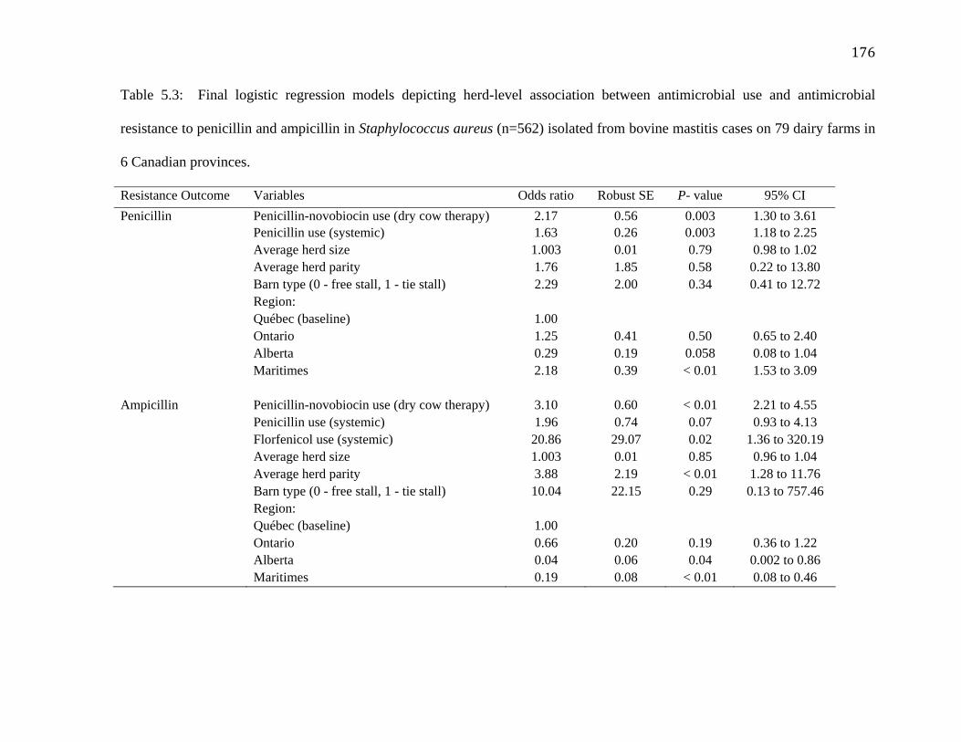
176
Table 5.3: Final logistic regression models depicting herd-level association between antimicrobial use and antimicrobial
resistance to penicillin and ampicillin in Staphylococcus aureus (n=562) isolated from bovine mastitis cases on 79 dairy farms in
6 Canadian provinces.
Resistance Outcome Variables Odds ratio Robust SE P- value 95% CI
Penicillin Penicillin-novobiocin use (dry cow therapy) 2.17 0.56 0.003 1.30 to 3.61 Penicillin use (systemic) 1.63 0.26 0.003 1.18 to 2.25 Average herd size 1.003 0.01 0.79 0.98 to 1.02 Average herd parity 1.76 1.85 0.58 0.22 to 13.80 Barn type (0 - free stall, 1 - tie stall) 2.29 2.00 0.34 0.41 to 12.72 Region: Québec (baseline) 1.00 Ontario 1.25 0.41 0.50 0.65 to 2.40 Alberta 0.29 0.19 0.058 0.08 to 1.04 Maritimes 2.18 0.39 < 0.01 1.53 to 3.09 Ampicillin Penicillin-novobiocin use (dry cow therapy) 3.10 0.60 < 0.01 2.21 to 4.55 Penicillin use (systemic) 1.96 0.74 0.07 0.93 to 4.13 Florfenicol use (systemic) 20.86 29.07 0.02 1.36 to 320.19 Average herd size 1.003 0.01 0.85 0.96 to 1.04 Average herd parity 3.88 2.19 < 0.01 1.28 to 11.76 Barn type (0 - free stall, 1 - tie stall) 10.04 22.15 0.29 0.13 to 757.46 Region: Québec (baseline) 1.00 Ontario 0.66 0.20 0.19 0.36 to 1.22 Alberta 0.04 0.06 0.04 0.002 to 0.86 Maritimes 0.19 0.08 < 0.01 0.08 to 0.46

177
Table 5.4: Final logistic regression models depicting herd-level association between antimicrobial use and antimicrobial
resistance to pirlimycin, penicillin-novobiocin combination and tetracycline in Staphylococcus aureus (n=562) isolated from
bovine mastitis cases on 79 dairy farms in 6 Canadian provinces.
Resistance Outcome Variables Odds ratio Robust SE P- value 95% CI
Pirlimycin Pirlimycin use (lactating cow therapy) 2.07 0.42 < 0.01 1.38 to 3.09
Average herd size 1.00 0.01 0.65 0.97 to 1.04
Average herd parity 0.34 0.57 0.52 0.01 to 9.03
Barn type (0-free stall, 1- tie stall) 4.15 9.72 0.54 0.04 to 407.87
Region:
Québec (baseline) 1.00
Ontario 0.91 0.43 0.85 0.36 to 2.31
Alberta Omitted due to absence of resistance
Maritimes 1.79 2.27 0.64 0.14 to 21.69
Penicillin-novobiocin Combination Penicillin-novobiocin use (dry cow therapy) 1.23 0.56 0.65 0.49 to 3.04
Florfenicol use (systemic) 34.56 92.95 0.18 0.17 to 6726
Average herd size 1.00 0.01 0.74 0.96 to 1.04
Average herd parity 12.63 28.56 0.26 0.15 to 1060
Barn type (0-free stall, 1- tie stall) 1.25 2.48 0.90 0.02 to 60.52
Region:
Québec (baseline) 1.00
Ontario 14.14 20.59 0.06 0.81 to 245.42
Alberta Omitted due to absence of resistance
Maritimes 6.85 9.09 0.14 0.50 to 92.21

178
Resistance Outcome Variables Odds ratio Robust SE P- value 95% CI
Tetracycline Tetracycline use (systemic) 1.13 1.11 0.89 0.16 to 7.73
Average herd size 1.02 0.008 0.005 1.007 to 1.03
Average herd parity 0.02 0.02 < 0.01 0.004 to 0.13
Barn type (0-free stall, 1- tie stall) 0.08 0.19 0.28 0.008 to 7.92
Region:
Québec (baseline) 1.00
Ontario 0.05 0.029 < 0.01 0.018 to 0.15
Alberta Omitted due to absence of resistance
Maritimes 0.37 0.41 0.37 0.04 to 3.27
Sulfadimethoxine TMPS1 use (systemic) 0.83 0.29 0.60 0.41 to 1.66
Average herd size 0.99 0.006 0.99 0.98 to 1.01
Average herd parity 1.27 1.48 0.83 0.12 to 12.55
Barn type (0-free stall, 1- tie stall) 1.05 1.35 0.96 0.08 to 13.02
Region:
Québec (baseline) 1.00
Ontario 0.33 0.07 < 0.01 0.22 to 0.51
Alberta 0.08 0.06 < 0.01 0.01 to 0.40
Maritimes 0.40 0.24 0.13 0.12 to 1.34 1Trimethoprim-sulfadoxine combination.

179
Chapter Six: Relationship between antimicrobial use and antimicrobial resistance in
Gram-negative bovine mastitis pathogens on Canadian dairy farms.

180
6.1 Abstract
Concurrent data on antimicrobial use and resistance are important for containing
antimicrobial resistance in bacteria. The present study determined a herd-level association
between antimicrobial use and resistance in Escherichia coli (n=394) and Klebsiella species
(n=139) isolated from intramammary infections and bovine mastitis cases on 89 dairy
farms in four regions of Canada. Antimicrobial use data were collected using inventory of
empty antimicrobial containers, and antimicrobial drug use rate was calculated to quantify
herd-level antimicrobial use. Minimum inhibitory concentrations were determined using
Sensititre® NARMS Gram-negative plate. Multivariable logistic regression models were
built to determine herd-level odds of resistance to tetracycline, ampicillin, ceftiofur,
chloramphenicol, trimethoprim-sulfamethoxazole (TMPS) combination and streptomycin
in coliforms.
Herd-level use of systemically administered tetracycline was associated with tetracycline
resistance in Klebsiella species isolates (Odds ratio [OR] = 2.59). The use of systemically
administered tetracycline was associated with tetracycline resistance in E. coli isolates on
dairy farms using tetracycline and penicillin (OR = 3.61). Ampicillin resistance in E. coli
isolates was associated with the use of systemically administered ceftiofur (OR = 21.33)
and penicillin (OR = 3.73). Use of systemically administered TMPS was also associated
with TMPS resistance in E. coli (OR = 1.68). The use of intramammary penicillin
combination containing dihydrostreptomycin administered for clinical mastitis treatment
and penicillin-novobiocin combination administered for dry cow therapy was associated

181
with streptomycin resistance as well (OR: 2.26, 1.75, respectively). Herds with tie-stall
barns had higher odds of ceftiofur and ampicillin resistance in E. coli isolates than herds
with free-stall barns (OR: 10.06, 14.15, respectively). Alberta dairy herds had lower odds
of ceftiofur-resistant E. coli isolates than dairy herds in Ontario (OR: 0.13); however, dairy
herds in Alberta had higher odds of TMPS, tetracycline, ampicillin, and chloramphenicol
resistance in E. coli isolates than dairy herds in Québec (OR: 1.89, 2.54, 5.48, 20.27,
respectively). Ontario dairy herds had lower odds of streptomycin resistance in E. coli
isolates than dairy herds in Québec (OR: 0.64).
In conclusion, association between herd-level antimicrobial use and resistance in bovine
mastitis coliforms was observed for certain antimicrobials. Barn type was positively
associated with ceftiofur and ampicillin resistance in E. coli. Geographical variation in
antimicrobial resistance was observed.

182
6.2 Introduction
Environmental mastitis in dairy cattle caused by coliforms occurs mostly in early lactation
with high producing cows overrepresented. Coliform mastitis can cause more often severe
systemic signs and occasionally death as compared to Gram-positive mastitis (Barkema et
al., 1998). The majority of coliform mastitis cases are caused by Escherichia coli
(Kaipainen et al., 2002; Lehtolainen et al., 2003). Klebsiella is another coliform organism
that is an emerging pathogen with rising incidence in North America (Zadoks and Munoz,
2007); Klebsiella was the most frequently found clinical mastitis pathogen in free-stall
dairy cattle herds in Western Canada (Olde Riekerink et al., 2008). These Gram-negative
udder pathogens have been implicated in as low as 20% to more than 60% of clinical
mastitis in different countries (Pyörälä and Honkanen-Buzalski, 1994; Shpigel et al., 1998).
Association between incidence of coliform clinical mastitis and bulk tank SCC has also
been observed. Incidence of coliform clinical mastitis and associated systemic signs was
higher in herds with low bulk tank SCC as compared to herds with high bulk tank SCC
(Barkema et al., 1998).
Broad-spectrum antimicrobials are commonly used to treat coliform mastitis (Erskine et al.,
2002b; Kaipainen et al., 2002), although there is no convincing evidence of the efficacy of
treatment (Sandholm et al., 1990; Erskine et al., 1992; Pyörälä et al., 1994); antimicrobial
resistance (AMR) could be one potential reason for the lack of efficacy. Antimicrobials
with an appropriate spectrum of activity against coliform bacteria that can be administered
systemically in lactating dairy cattle include oxytetracycline, sulfadimethoxine, ceftiofur,

183
ampicillin, and amoxicillin (Wagner and Erskine, 2006). However, AMR in bovine mastitis
coliforms is commonly observed. For example, resistance proportions in E. coli isolates
ranged from 5 to 37% for tetracycline (FINRES-Vet, 2007; Makovec and Ruegg, 2003), 7
to 34% for sulfisoxazole (FINRES-Vet, 2007; Srinivasan et al., 2007), 0 to 5% for ceftiofur
(FINRES-Vet, 2007; Erskine et al., 2002b), and 7 to 21% for ampicillin (Lanz et al., 2003;
Lehtolainen et al., 2003) across various studies. Resistance proportions in Klebsiella
species isolates also varied greatly from 7 to 33% for tetracycline (Bengtsson et al., 2009;
Erskine et al., 2002b), 9 to 12% for sulfisoxazole/sulfamethoxazole (Bengtsson et al., 2009;
Makovec and Ruegg, 2003) and 0 to 14% for ceftiofur (Saini et al., 2011a; Erskine et al.,
2002b) across studies.
Many studies have determined antimicrobial susceptibility of bovine mastitis coliforms
(Lanz et al., 2003; Lehtolainen et al., 2003; Makovec and Ruegg, 2003; Srinivasan et al.,
2007; Bengtsson et al., 2009), and some studies have evaluated the dynamics of AMR in
coliforms isolated from the feces of young dairy calves, dairy cattle, beef cattle and swine
(Berge et al., 2005; Akwar et al., 2008; Checkley et al., 2008; Berge et al., 2010).
Unfortunately, there is little information on the impact of therapeutic and prophylactic
AMU on AMR in bovine mastitis E. coli and Klebsiella species isolates (Srinivasan et al.,
2007). Therefore, it seems prudent to assess and evaluate relationship between AMU and
AMR in bovine mastitis coliforms.
The objective of the present study was to determine a herd-level association between AMU
and AMR in E. coli and Klebsiella species isolated from intramammary infections (IMI)
and (sub) clinical bovine mastitis cases on Canadian dairy farms.
184
6.3 Materials and Methods
6.3.1 Herd Selection
Data for this study originated from the National Cohort of Dairy Farms of the Canadian
Bovine Mastitis Research Network (CBMRN), which consisted of 91 commercial dairy
farms located in four regions across Canada (Alberta, Ontario Québec, and the Maritime
Provinces [Prince Edward Island, Nova Scotia, and New Brunswick]). Herd selection
criteria for the present study have been described by (Reyher et al., 2011). In short, dairy
herds were selected to replicate the regional proportion of free-stall systems to within 15
percentage points and to be uniformly distributed among three strata of the most recent 12-
month bulk tank SCC average (≤ 150,000 cells/mL, > 150,000 and ≤ 300,000 cells/mL, >
300,000 cells/mL); herds with three times milking schedule per day and herds with less
than 80% (or less than 15) Holstein lactating and dry cows at the time of enrolment were
excluded. Further, eligible dairy herds must have been participating in a DHI data
collection program. Eligible dairy farms were identified and contacted by the regional
center coordinators. Written consent to participate in the research cohort was obtained. Two
dairy herds dropped out of the study at the very beginning leaving 89 herds in the study. An
investigator and technicians in each coordinating center were responsible for the data
collection activities related to remaining farms located in that center’s area.
6.3.2 Antimicrobial Use Data Collection Methodology
Antimicrobial use data collection has been described by Saini et al. (2011b). In short, AMU
data were collected from February 2007 until December 2008. Forty-liter receptacles were
placed on participating farms for collecting data for AMU. These receptacles were placed
near the drug storage area, in the milking parlour or any place near where the treatments

185
were normally given. Producers, farm workers and other farm personnel were instructed to
deposit the empty containers of all drugs used by them or the veterinarian for treatment in
calves, heifers and adult cows (dry cows and lactating cows) into these receptacles. Farms
were visited at least once per month. The research technicians emptied the receptacles,
counted the empty drug bottles and recorded the inventory in the drug tally sheets at the
dairy farm.
Antimicrobial use data were quantified in units of animal defined-daily doses (ADD). The
ADD (g/day) was defined as the average daily on-label dosage multiplied by the
approximate weight of an adult dairy cow (BW=600 kg) (Jensen et al., 2004) and was
based on the Canadian compendium of veterinary products. Further, antimicrobial drug use
rate (ADUR) was defined as number of ADD used on a farm per 1,000 cows (milking and
dry) per day. ADUR was the herd-level estimate of AMU (Table 6.1).
6.3.3 Sampling and Bacterial Culturing
Sampling and bacterial culturing of isolates has been described elsewhere (Reyher et al.,
2011). In short, three different sets of milk samples were collected. The first set included
milk samples from clinical mastitis cases. All producer-diagnosed clinical mastitis cases
were sampled at the time of diagnosis and then re-sampled twice at two-week intervals. The
second set was from milk samples from nonclinical lactating cows; a sub-sample of 15
fresh and lactating cows was selected per farm. They were aseptically sampled and re-
sampled once every 3 weeks for a total of three samplings in the winter of 2007; another
sub-sample of 15 lactating cows was sampled once a week for 7 weeks in the summer of
2007.

186
The third set of milk samples were collected from another selected group of 15 cows that
were expected to remain in the herd until at least 2 weeks after calving. A sub-sample was
aseptically sampled before dry-off and after calving in 2007 and this continued in 2008 as
well. Quarter and composite milk samples were collected. Samples were frozen at –200C
and shipped to the regional CBMRN laboratory where bacterial culturing and identification
of the milk samples was done as per National Mastitis Council guidelines (Hogan et al.,
1999).
Because multiple isolates could be coming from a single cow, it was decided to include
only one isolate per quarter. Clinical mastitis was defined as an inflammation of the udder
leading to occurrence of flakes, clots or other gross alterations in milk. Subclinical mastitis
was defined as SCC >200,000 cells/mL from a cow without clinical signs of mastitis,
whereas IMI was defined as a culture-positive sample (Reyher et al., 2011).
6.3.4 MIC Determination
Antimicrobial susceptibility testing of bovine mastitis coliforms have been described
elsewhere (Saini et al., 2011a). Minimum inhibitory concentrations (MIC) of these isolates
were determined using the Sensititre® microdilution system (TREK Diagnostic Systems
Inc., Cleveland, Ohio). NARMS Gram-negative plate containing amikacin, gentamicin,
kanamycin, streptomycin, ampicillin, amoxicillin-clavulanic acid combination
(AMXCLA), cefoxitin, ceftriaxone, ceftiofur, chloramphenicol, ciprofloxacin, nalidixic
acid, sulfisoxazole, trimethoprim-sulfamethoxazole combination (TMPS) and tetracycline
was used (Varga et al., 2008).

187
6.3.5 Statistical Analyses
Antimicrobial use data were entered into a customized database (Microsoft Office Access
2006, Microsoft Corporation, Redmond, Washington, USA). A random sample of the drug
tally sheets (25%) was checked manually to detect errors in data entry. Data analyses were
performed using Intercooled Stata®11.1 (Intercooled Stata for Macintosh, version 11.1,
Stata Corporation, College Station, TX).
Minimum inhibitory concentration was defined as the lowest concentration of an
antimicrobial that inhibited any visible growth of an isolate. In case of an antimicrobial
combination such as TMPS, the MIC of the first agent (trimethoprim) was reported as the
MIC for the combination. The isolates were also categorized as sensitive, intermediate and
resistant on the basis of CLSI based MIC breakpoints (2008). Intermediate isolates were
combined with resistant isolates for the sake of statistical analysis. For analytical purposes,
the unit of analysis was resistance at the herd-level (0: number of resistant isolates at a farm
= 0; and 1: number of resistant isolates at a farm ≥ 1). Herd-level prevalence percentage of
AMR was calculated and defined as percentage of total number of herds with AMR isolates
divided by total number of herds sampled.
All independent variables (antimicrobial use and other herd-level factors such as region,
herd average milk production, barn type, herd average SCC, herd average parity, average
herd size, number of isolates sampled at a farm) were screened based on descriptive
statistics (means, variances and percentiles for continuous variables, and frequency
tabulations for categorical variables) so as to exclude variables that had little variability
(Dohoo et al., 2009). Subsequently, these independent variables were screened for

188
univariate associations using Likelihood Ratio Test Statistic (Hosmer and Lemeshow,
2000). Variables significant at P ≤ 0.25 were eligible for inclusion in the multivariable
logistic regression models. Using backward elimination, variables were retained in model
only if significant at P ≤ 0.05 unless exclusion resulted in significant change in deviance.
Thereafter, any variables not selected for the original multivariable model were added back
into the model in order to identify variables that were not significantly associated with the
herd-level outcome, but made an important contribution in the presence of other variables
(distorter variables). Barn type, region, average herd size, and average herd parity were
considered a priori as potential confounders. Variables as average herd SCC, herd average
milk production per cow, average herd size and average herd parity were centered at their
respective lowest values for sensible interpretation of the intercept value.
The assumption of linearity in the logit of herd-level AMR outcome for continuous
variables was evaluated graphically using lowess smoother scatter plots (Dohoo et al.,
2009) and then by using fractional polynomials command in Intercooled Stata®11.1. Two-
way interaction terms were added one at a time to the main effects model and retained at P
≤ 0.05 unless exclusion resulted in significant change in deviance. Robust standard errors
were used to control for clustering of farms within regions. In case of nominal variables
such as region, the baseline/referent level was selected as the one with sufficiently large
sample size (Dohoo et al., 2009).
Multivariable logistic regression models were built for the following AMR outcomes in E.
coli: tetracycline, ampicillin, ceftiofur, chloramphenicol, TMPS and streptomycin. In case
of Klebsiella species isolates, a logistic regressions model was only built for tetracycline.

189
No model could be built for ampicillin, as Klebsiella species are intrinsically resistant. Only
two and three farms had TMPS and chloramphenicol resistant Klebsiella species isolates,
respectively; low prevalence of herd-level resistance, therefore, prevented model building
for these antimicrobials. Further, variation in streptomycin resistance between barn types
within four regions was also very small to prevent model building for Klebsiella species
isolates.
No models could be built for cefoxitin, ceftriaxone, AMXCLA, amikacin, kanamycin and
nalidixic acid resistance outcomes in E. coli and Klebsiella species isolates, as these
antimicrobials were not used on Canadian dairy farms. Additionally, because gentamicin,
enrofloxacin and sulfonamides were rarely used on study farms, models could not be built
for gentamicin, ciprofloxacin and sulfisoxazole in E. coli and Klebsiella species isolates as
well.
6.4 Results
6.4.1 Antimicrobial Resistance Proportions
Minimum inhibitory concentration values were determined for 394 E. coli isolates that
came from 394 quarters of 353 cows on 76 dairy farms. Total number of isolates resistant
to one or more antimicrobials was 70 or a prevalence of 17.7% (95% CI: 14.1 to 21.9%).
Total number of isolates resistant to an antimicrobial ranged from 0 to six per farm. Herd-
level AMR prevalence ranged from 0% for ceftriaxone and ciprofloxacin to 38.1% for
tetracycline (Table 6.2).
In case of Klebsiella species, MIC values were determined for 139 isolates that came from

190
139 quarters of 114 cows on 37 dairy farms. Fifty-one isolates or 36.6% (95% CI: 28.6 to
45.2%) were resistant to one or more antimicrobials. Total number of isolates resistant to
an antimicrobial ranged from 0 to one per farm. Herd-level AMR prevalence ranged from
0% for amikacin, ceftiofur, ceftriaxone, ciprofloxacin and nalidixic acid to 24.3% for
tetracycline (Table 6.3).
6.4.2 Multivariable Analyses
The odds for herds having at least one tetracycline-resistant E. coli isolate increased with an
increasing use of systemically administered tetracycline on farms using tetracycline and
penicillin (Table 6.4). Herd-level odds of isolating at least one ampicillin-resistant E. coli
isolate increased with an increased use of systemically administered penicillin and
ceftiofur; the use of systemically administered tetracycline was, however, associated with
decreasing odds. Dairy herds in Alberta had significantly higher odds for having at least
one tetracycline and ampicillin resistant E. coli isolate than dairy herds in Québec. Herds
with tie-stall barns had significantly higher odds of having at least one ampicillin-resistant
E. coli isolate than herds with free-stall barns.
The use of penicillin combinations containing dihydrostreptomycin administered
intramammary for clinical mastitis treatment, penicillin-novobiocin combinations
administered for dry cow therapy and systemically administered penicillin was positively
associated with streptomycin resistance in E. coli isolates at the herd-level (Table 6.5). The
odds decreased with an increasing use of penicillin combinations administered
intramammary for clinical mastitis treatment on farms with larger herd size as compared to
farms with smaller herd size. Dairy herds in Ontario had significantly lower odds of having
191
at least one streptomycin-resistant E. coli isolate than dairy herds in Québec.
The odds for herds having at least one TMPS-resistant E. coli isolate at a farm increased
significantly with an increased use of systemically administered TMPS and ceftiofur, and
penicillin combinations administered intramammary for clinical mastitis treatment;
however, the use of pirlimycin administered intramammary for clinical mastitis treatment
was associated with decreasing odds of TMPS resistance in E. coli isolates (Table 6.5).
Dairy herds in Alberta had significantly higher odds of having at least one TMPS-resistant
E. coli isolate than dairy herds in Québec.
Herds with tie-stall barns had significantly higher odds of having at least one ceftiofur-
resistant E. coli isolate than herds with free-stall barns (Table 6.6). Dairy herds in Alberta
had significantly lower odds of having at least one ampicillin-resistant E. coli isolate than
dairy herds in Ontario.
The use of systemically administered florfenicol and tetracycline was significantly
associated with decreasing odds of having at least one chloramphenicol-resistant E. coli
isolate on a farm; however, the odds increased significantly with an increase in the use of
systemically administered penicillin (Table 6.7). The odds increased with an increase in
average herd size. Dairy herds in Alberta and Ontario had significantly higher odds of
having at least one chloramphenicol-resistant E. coli isolate than dairy herds in Québec.
The odds for herds having at least one tetracycline-resistant Klebsiella species isolate
increased with an increasing use of systemically administered tetracycline at a farm (Table

192
6.7).
6.5 Discussion
This is the first time that a Canada-wide prospective study was conducted to determine
herd-level risk factors of AMR in Gram-negative bovine mastitis pathogens. In addition to
AMU, data were collected on herd-level variables such as SCC, milk production, barn type,
herd size and geographical region that could potentially explain variation in AMR at the
herd-level.
Direct effects of AMU on AMR were observed for some antimicrobials e.g. the use of
systemically administered TMP-sulfadoxine combination was positively associated with
herd-level TMPS resistance. Intramuscular injection of TMP-sulfadoxine is commonly
recommended in cattle; however, it leads to poor udder penetration due to low
bioavailability (Prescott, 2006b), and hence could potentially select TMPS resistant strains.
Potential co-selection was observed as well. For example, the use of systemically
administered ceftiofur and intramammary administered penicillin combination containing
dihydrostreptomycin was positively associated with TMPS resistance. Multi-drug
resistance to ampicillin, ceftiofur, streptomycin and TMPS has been observed in bovine
mastitis E. coli isolates (Saini et al., 2011a). Class I integrons containing ß-lactam,
streptomycin and TMP resistance encoding genes have been isolated from multi-drug
resistant E. coli recovered from cattle and environment (including calves, milk filters,
overshoes, water sampled, swabs of the calf pen and feeding bucket) on 21 dairy farms in
Ireland (Karczmarczyk et al., 2011). The odds of streptomycin resistance in E. coli isolates
193
increased with an increase in the use of intramammary administered penicillin combination
containing dihydrostreptomycin, intramammary penicillin-novobiocin combination and
systemically administered penicillin. Intramammary penicillin combinations containing
dihydrostreptomycin were most commonly used for clinical mastitis treatment on Canadian
dairy farms (Saini et al., 2011b). In terms of potency, spectrum of activity and stability to
plasmid mediated enzymatic inactivation, streptomycin is least active among
aminoglycosides. Streptomycin / dihydrostreptomycin are synergistic with penicillin in
case of Gram-positive bacteria; however, synergism is usually absent in Gram-negative
bacteria (Dowling, 2006). Further, Enterobacteriaceae are intrinsically resistant to
penicillin, and the mechanisms of resistance to aminoglycosides are different from those of
penicillins, therefore these associations appear to be biologically implausible. However, it
is quite likely that when dihydrostreptomycin-containing penicillin combinations are used,
only the dihydrostreptomycin component is associated with streptomycin resistance, and
penicillin has no effect per se. Unfortunately, it is not possible to attribute streptomycin
resistance to the use of dihydrostreptomycin alone due to its presence with penicillin in a
combination product.
Cross-resistance to some antimicrobials was also observed. For example, resistance to
ampicillin in E. coli isolates was positively associated with the use of systemically
administered ceftiofur and penicillin. Resistance to aminopenicillins as ampicillin is usually
mediated through the production of ß-lactamases. Penicillins are predominantly ionized in
plasma, are less lipid soluble and cross biological membranes poorly; the concentration in
milk is about one-fifth of that in serum. Ceftiofur is also poorly distributed in udder due to
high plasma protein binding upon systemic administration (Prescott, 2006a). Therefore, it

194
seems unlikely, that sub-therapeutic concentrations of penicillin and ceftiofur are achieved
in the udder upon systemic administration that may cause selection of ampicillin resistant
E. coli strains in the udder. In fact, the majority of coliforms are from the gut that are shed
into the cow’s environment and infect udder via the teat canal (Kaipainen et al., 2002;
Lehtolainen et al., 2003). It is quite plausible, that selection of AMR occurred inside the gut
environment due to AMU in non-mastitis conditions.
Interestingly, the use of certain antimicrobials was negatively associated with AMR in E.
coli. For example, on dairy farms using only systemically administered tetracycline, the
odds for a herd having at least one tetracycline-resistant E. coli isolate decreased with an
increase in the use of systemically administered tetracycline. Tetracycline use is known to
induce the expression of tetracycline resistance via efflux mechanism encoded by tet gene
in Gram-negative bacteria (Poyart, 2010). Negative associations between AMU and AMR
seem to be implausible. These associations could be spurious or due to certain factors that
are yet to be identified (Akwar et al., 2008). However, on dairy farms using tetracycline
and penicillin, the odds of isolating at least one tetracycline resistant E. coli isolate
increased with an increase in the use of systemically administered tetracycline thereby
indicating an interaction between these two antimicrobials, although systemically
administered penicillin was not associated with tetracycline resistance.
In general, average herd parity and barn type tended to be associated with AMR in E. coli
isolates. Increase in parity indicates an increase in age and therefore increased exposure to
antimicrobials and udder pathogens. Subsequent selection pressure could potentially lead to
AMR in udder pathogens or changes in the distribution of resistant strains such that

195
resistant strains become more prevalent in older cows. Herds with tie-stall barns had higher
odds for having at least one resistant E. coli isolate than herds with free-stall barns. Barn
type potentially impacts managemental practices employed at a farm that might be
associated with AMR in bovine mastitis pathogens (Kirk et al., 2005). For example,
addition of organic or inorganic amendments in bedding may potentially cause an increase
or decrease in growth of microorganisms thereby leading to changes in bacterial
distributions to those more intrinsically resistant, and hence altering the AMR patterns
(Kirk et al., 2005). Increase in interval between grooming and replacement of bedding
material was positively associated with AMR. Wet udders had higher odds of AMR than
dry udders. In general, dairy practices could potentially be modifiable determinants of
AMR in environmental mastitis causing bacteria.
Differences in herd-level prevalence of AMR in bovine mastitis coliforms were observed
between regions. Different subpopulations of bacteria with varying antimicrobial
susceptibilities exist in different regions (Erskine et al., 2003; Kirk et al., 2005). A region
may be a surrogate for management-related differences due to environment, geography,
weather, and resources availability. For example, multidrug resistance in fecal E. coli
isolates collected from cattle (pre-weaned calves on calf ranches, steers on feedlots, dairy
and beef cows) were not directly associated with AMU but with geographic region, animal
age and purpose (beef versus dairy)(Berge et al., 2010).
Interestingly, some biologically unreasonable associations between AMU and AMR in
bovine mastitis coliforms have been observed in this study. However, it is noteworthy that
the unit of analysis was farm and not an individual cow. Secondly, data was collected on

196
herd-level use of antimicrobials; individual cow level AMU data being unavailable, it will
be a fallacy to make inferences from this study at individual cow level. Antimicrobial use
and resistance data was measured concurrently, and therefore is not reflective of the past
AMU that might have a bigger impact on AMR in these isolates. Such group level studies
are exploratory in nature as there is no direct measure of the exposure of interest due to
measurement constraints at the individual level (Dohoo et al., 2009). These studies are
primarily hypothesis generating in nature.
Antimicrobial resistance is potentially a herd-level phenomenon as farm ecosystems
provide an ideal environment for emergence, amplification, and dissemination of resistant
bacteria/determinants (Acar and Moulin, 2006). In case of bovine mastitis coliforms, it is
hypothesized that selection of AMR occurs outside bovine udder in the dairy farm soil due
to presence of antimicrobials or AMR bacteria in the feces and urine of animals that
accumulate in a dairy farm environment (Burgos et al., 2005; Hammad et al., 2008). In
general, pathogens and commensal bacteria are subjected to antimicrobial treatments in a
dairy farm environment, and the subsequent selection pressure might favor selection and
dissemination of resistant strains (Silbergeld et al., 2008; Tikofsky et al., 2003).
Interventions for managing AMR in bacteria should focus at the herd-level and not
individual cow level (Tragesser et al., 2006). Various studies have confirmed that AMR in
bacteria is not a simple outcome of AMU, and data should be collected on non-
antimicrobial use aspects impacting AMR (e.g. dairy practices related to hygiene, nutrition,
cow-stress) so as to understand the ecology of resistance in bacteria (Berge et al., 2005;
Berge et al., 2010; Gellin et al., 1989; Kirk et al., 2005; Langlois et al., 1988; Sol et al.,
2000).

197
6.6 Conclusions
Antimicrobial resistance in bovine mastitis coliforms was associated directly and indirectly
with the use of antimicrobials commonly administered on Canadian dairy farms. Herds
with tie-stall barns had higher odds for having at least one resistant coliform isolate than
herds with free-stall barns, and average herd parity was associated with herd-level AMR,
although statistically non-significant. Geographical variation in AMR was observed that
could not be explained by AMU. In addition to AMU data, information should also be
collected on dairy practices related to bedding and udder hygiene impacting AMR in
bovine mastitis pathogens.

198
6.7 References
Acar, J. F. and G. Moulin. 2006. Antimicrobial resistance at farm level. Rev. Sci. Tech.
25:775-792.
Akwar, H. T., C. Poppe, J. Wilson, R. J. Reid-Smith, M. Dyck, J. Waddington, D. Shang,
and S. A. McEwen. 2008. Associations of antimicrobial uses with antimicrobial
resistance of fecal Escherichia coli from pigs on 47 farrow-to-finish farms in Ontario
and British Columbia. Can. J. Vet. Res.72:202-210.
Barkema, H., Y. Schukken, T. Lam, M. Beiboer, H. Wilmink, G. Benedictus, and A. Brand.
1998. Incidence of clinical mastitis in dairy herds grouped in three categories by bulk
milk somatic cell counts. J. Dairy Sci. 81:411-419.
Bengtsson, B. o., H. E. Unnerstad, T. Ekman, K. Artursson, M. Nilsson-Ost, and K. P.
Waller. 2009. Antimicrobial susceptibility of udder pathogens from cases of acute
clinical mastitis in dairy cows. Vet. Microbiol. 136:142-149.
Berge, A., E. Atwill, and W. Sischo. 2005. Animal and farm influences on the dynamics of
antibiotic resistance in faecal Escherichia coli in young dairy calves. Prev. Vet. Med.
69:25-38.
Berge, A. C., D. D. Hancock, W. M. Sischo, and T. E. Besser. 2010. Geographic, farm, and
animal factors associated with multiple antimicrobial resistance in fecal Escherichia
coli isolates from cattle in the western United States. J. Am. Vet. Med. Assoc.
236:1338-1344.
Burgos, J. M., B. A. Ellington, and M. F. Varela. 2005. Presence of multidrug-resistant
enteric bacteria in dairy farm topsoil. J. Dairy Sci. 88:1391-1398.

199
Checkley, S. L., J. R. Campbell, M. Chirino-Trejo, E. D. Janzen, and J. J. McKinnon. 2008.
Antimicrobial resistance in generic fecal Escherichia coil obtained from beef cattle
on arrival at the feedlot and prior to slaughter, and associations with volume of total
individual cattle antimicrobial treatments in one western Canadian feedlot. Can. J.
Vet. Res. 72:101-108.
CLSI. 2008. Performance standards for antimicrobial disk and dilution susceptibility tests
for bacteria isolated from animals. Approved standard. 3rd Edition. M31-A3: 65-72.
Dohoo, I. R., W. Martin, and H. Stryhn. 2009. Veterinary epidemiologic research. 2nd
edition. VER Inc., Charlottetown, PEI, Canada.
Dowling, P. M. 2006. Aminoglycosides. Page 208 In Antimicrobial therapy in veterinary
medicine. S. Giguère, ed. Blackwell Publishing, Iowa, US.
Erskine, R. J., R. C. Wilson, M. G. Riddell, Jr., J. W. Tyler, H. J. Spears, and B. S. Davis.
1992. Intramammary administration of gentamicin as treatment for experimentally
induced Escherichia coli mastitis in cows. Am. J. Vet. Res. 53:375-381.
Erskine, R. J., P. C. Bartlett, J. L. VanLente, and C. R. Phipps. 2002a. Efficacy of systemic
ceftiofur as a therapy for severe clinical mastitis in dairy cattle. J. Dairy Sci. 85:2571-
2575.
Erskine, R. J., R. D. Walker, C. A. Bolin, P. C. Bartlett, and D. G. White. 2002b. Trends in
antibacterial susceptibility of mastitis pathogens during a seven-year period. J. Dairy
Sci. 85:1111-1118.
Erskine, R. J., S. Wagner, and F. J. Degraves. 2003. Mastitis therapy and pharmacology.
Vet. Clin. North Am. Food Anim. Pract. 19:109-138.
FINRES-Vet, 2005 - 2006. 2007. Finnish Veterinary Antimicrobial Resistance Monitoring
and Consumption of Antimicrobial Agents. Finnish Food Safety Authority Evira,
200
Helsinki, Finland. http://www.evira.fi/uploads/WebShopFiles/1198141211941.pdf
Last accessed October 6, 2011.
Gellin, G., B. E. Langlois, K. A. Dawson, and D. K. Aaron. 1989. Antibiotic resistance of
gram-negative enteric bacteria from pigs in three herds with different histories of
antibiotic exposure. Appl. Environ. Microbiol. 55:2287-2292.
Hammad A. M., A. M. Ahmed, Y. Ishida, and T. Shimamoto. 2008. First characterization
and emergence of SHV-60 in raw milk of a healthy cow in Japan. J. Vet. Med. Sci.
70:1269-1272.
Hogan, J. S., R. N. Gonzalez, R. Harmon, S. C. Nickerson, S. P. Oliver, J. W. Pankey, and
K. L. Smith. 1999. Laboratory handbook on bovine mastitis. National Mastitis
Council Inc., Verona, WI, USA.
Hosmer, D. W. and S. Lemeshow. 2000. Applied logistic regression. 2nd edition. John
Wiley & Sons, Inc. NJ 07030.
Jensen, V. F., E. Jacobsen, and F. Bager. 2004. Veterinary antimicrobial-usage statistics
based on standardized measures of dosage. Prev. Vet. Med. 64:201-215.
Kaipainen, T., T. Pohjanvirta, N. Y. Shpigel, A. Shwimmer, S. Pyorala, and S. Pelkonen.
2002. Virulence factors of Escherichia coli isolated from bovine clinical mastitis.
Vet. Microbiol. 85:37-46.
Karczmarczyk, M., Y. Abbott, C. Walsh, N. Leonard, and S. Fanning. 2011.
Characterisation of multidrug resistant Escherichia coli from animals presenting at a
University veterinary hospital. Appl. Environ. Microbiol. DOI:10.1128/AEM.00599-
11.
Kirk, J. H., B. McCowan, E. R. Atwill, K. S. Glenn, G. E. Higginbotham, C. A. Collar, A.
Castillo, B. A. Reed, N. G. Peterson, and J. S. Cullor. 2005. Association of minimum

201
inhibitory concentration cluster patterns with dairy management practices for
environmental bacteria isolated from bulk tank milk. J. Dairy Sci. 88:3710-3720.
Langlois, B. E., K. A. Dawson, I. Leak, and D. K. Aaron. 1988. Effect of age and housing
location on antibiotic resistance of fecal coliforms from pigs in a non-antibiotic-
exposed herd. Appl. Environ. Microbiol. 54:1341-1344.
Lanz, R., P. Kuhnert, and P. Boerlin. 2003. Antimicrobial resistance and resistance gene
determinants in clinical Escherichia coli from different animal species in
Switzerland. Vet. Microbiol. 91:73-84.
Lehtolainen T., A. Shwimmer, N. Y. Shpigel, T. Honkanen-Buzalski, and S. Pyörälä. 2003.
In vitro antimicrobial susceptibility of Escherichia coli isolates from clinical bovine
mastitis in Finland and Israel. J. Dairy Sci. 86:3927-3932.
Makovec, J. A. and P. L. Ruegg. 2003. Antimicrobial resistance of bacteria isolated from
dairy cow milk samples submitted for bacterial culture: 8,905 samples (1994-2001). J
Am. Vet. Med. Assoc. 222:1582-1589.
Olde Riekerink, R. G. M., H. W. Barkema, D. F. Kelton, and D. T. Scholl. 2008. Incidence
rate of clinical mastitis on Canadian dairy farms. J. Dairy Sci. 91:1366-1377.
Poyart, C. 2010. Tetracyclines. Page 331 In Antibiogram. P. Courvalin, R. Leclercq, Louis
B. Rice, eds. ESKA publishing, Oregon, USA.
Prescott, F. J. 2006a. Beta-lactam antibiotics: Cephalosporins. Page 151 In Antimicrobial
therapy in veterinary medicine. S. Giguère, ed. Blackwell Publishing, Iowa, US.
Prescott, F. J. 2006b. Sulfonamides, diaminopyrimidines, and their combinations. Page 260
In Antimicrobial therapy in veterinary medicine. S. Giguère, ed. Blackwell
Publishing, Iowa, US.

202
Pyörälä, S., L. Kaartinen, H. Käck, and V. Rainio. 1994. Efficacy of two therapy regimens
for treatment of experimentally induced Escherichia coli mastitis in cows. J. Dairy
Sci. 77:453-461.
Pyörälä, S. and T. Honkanen-Buzalski. 1994. The status of mastitis in the Nordic countries.
Int. Dairy Fed. Mastitis News 19:14-15.
Reyher, K. K., S. Dufour, H. W. Barkema, L. Des Côteaux, T. J. Devries, I. R. Dohoo, G.
P. Keefe, J.-P. Roy, and D. T. Scholl. 2011. The National Cohort of Dairy Farms-A
data collection platform for mastitis research in Canada. J. Dairy Sci. 94:1616-1626.
Saini, V., J. T. McClure, D. Léger, G. P. Keefe, D. T. Scholl, D. W. Morck, and H. W.
Barkema. 2011a. Antimicrobial resistance profiles of common mastitis pathogens and
prevalence of methicillin-resistant Staphylococcus aureus and extended-spectrum
beta-lactamase producing Escherichia coli and Klebsiella species isolates on
Canadian dairy farms. Antimicrob. Agents. Chemother. Submitted (AAC05550-11).
Saini, V., J. T. McClure, D. Léger, S. Dufour, A. G. Sheldon, D. T. Scholl, and H. W.
Barkema. 2011b. Antimicrobial use on Canadian dairy farms. J. Dairy Sci. Accepted
(JDS -11-4527).
Sandholm, M., L. Kaartinen, and S. Pyörälä. 1990. Bovine mastitis--why does antibiotic
therapy not always work? An overview. J. Vet. Pharmacol. Ther. 13:248-260.
Shpigel, N. Y., M. Winkler, G. Ziv, and A. Saran. 1998. Clinical, bacteriological and
epidemiological aspects of clinical mastitis in Israeli dairy herds. Prev. Vet. Med.
35:1-9.
Silbergeld, E. K., J. Graham, and L. B. Price. 2008. Industrial food animal production,
antimicrobial resistance, and human health. Annu. Rev. Public Health 29:151-169.

203
Sol, J., O. C. Sampimon, H. W. Barkema, and Y. H. Schukken. 2000. Factors associated
with cure after therapy of clinical mastitis caused by Staphylococcus aureus. J. Dairy
Sci. 83:278-284.
Srinivasan, V., B. E. Gillespie, M. J. Lewis, L. T. Nguyen, S. I. Headrick, Y. H. Schukken,
and S. P. Oliver. 2007. Phenotypic and genotypic antimicrobial resistance patterns of
Escherichia coli isolated from dairy cows with mastitis. Vet. Microbiol., 124:319-
328.
Tikofsky, L. L., J. W. Barlow, C. Santisteban, and Y. H. Schukken. 2003. A comparison of
antimicrobial susceptibility patterns for Staphylococcus aureus in organic and
conventional dairy herds. Microbiol. Drug Resist. 9 Suppl 1:S39-45.
Tragesser, L. A., T. E. Wittum, J. A. Funk, P. L. Winokur, and P. J. Rajala-Schultz. 2006.
Association between ceftiofur use and isolation of Escherichia coli with reduced
susceptibility to ceftriaxone from fecal samples of dairy cows. Am. J. Vet. Res.
67:1696-1700.
Varga, C., A. Rajic, M. E. McFall, B. P. Avery, R. J. Reid-Smith, A. Deckert, S. L.
Checkley, and S. A. McEwen. 2008. Antimicrobial resistance in generic Escherichia
coli isolated from swine fecal samples in 90 Alberta finishing farms. Can. J. Vet. Res.
72:175-180.
Wagner, S., and R. Erskine. 2006. Antimicrobial drug use in bovine mastitis. Page 511 In
Antimicrobial therapy in veterinary medicine. S. Giguère, ed. Blackwell Publishing,
Iowa, US.
Zadoks R. N., and M. A. Munoz. 2007. The emergence of Klebsiella as a major mastitis
organism. Pages 100-111 In Proc. NMC 46th Annual Meeting, San Antonio, TX,
National Mastitis Council Inc, Verona, WI, USA.
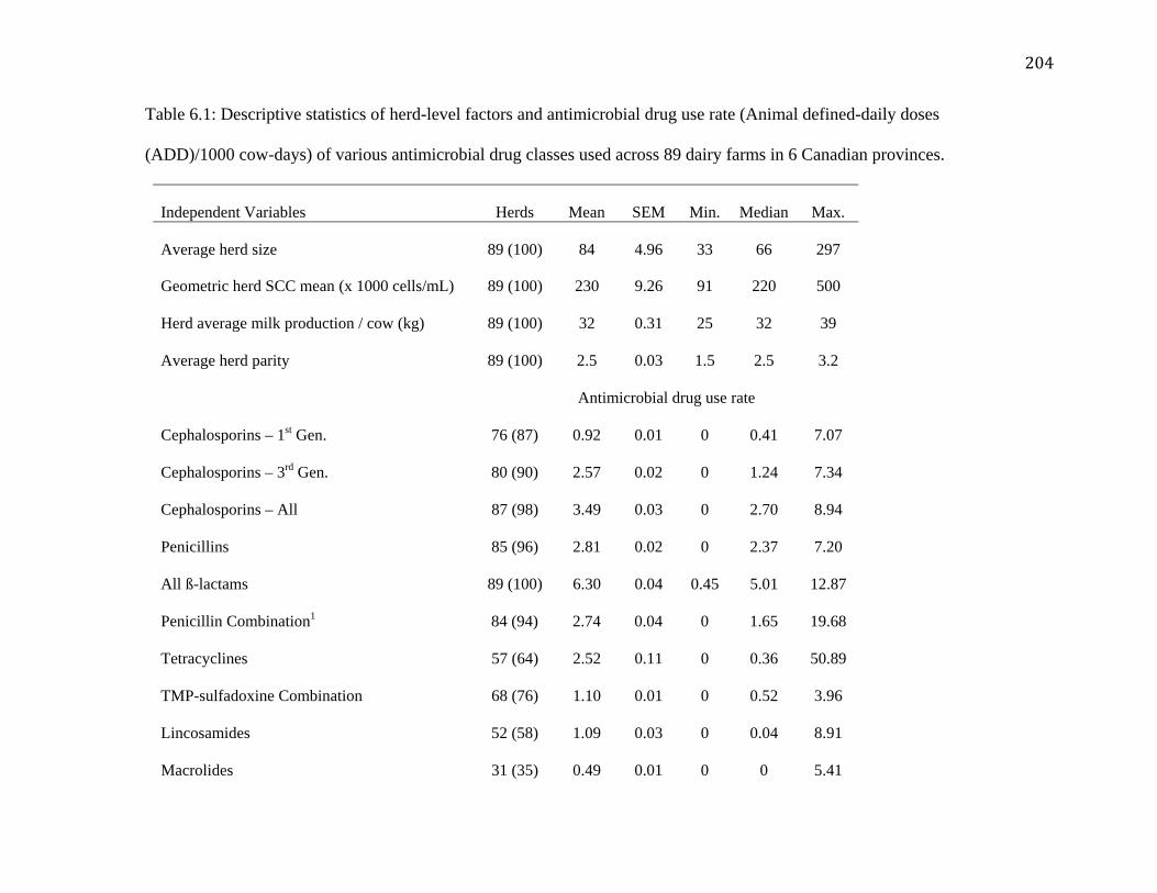
204
Table 6.1: Descriptive statistics of herd-level factors and antimicrobial drug use rate (Animal defined-daily doses
(ADD)/1000 cow-days) of various antimicrobial drug classes used across 89 dairy farms in 6 Canadian provinces.
Independent Variables Herds Mean SEM Min. Median Max.
Average herd size 89 (100) 84 4.96 33 66 297
Geometric herd SCC mean (x 1000 cells/mL) 89 (100) 230 9.26 91 220 500
Herd average milk production / cow (kg) 89 (100) 32 0.31 25 32 39
Average herd parity 89 (100) 2.5 0.03 1.5 2.5 3.2
Antimicrobial drug use rate
Cephalosporins – 1st Gen. 76 (87) 0.92 0.01 0 0.41 7.07
Cephalosporins – 3rd Gen. 80 (90) 2.57 0.02 0 1.24 7.34
Cephalosporins – All 87 (98) 3.49 0.03 0 2.70 8.94
Penicillins 85 (96) 2.81 0.02 0 2.37 7.20
All ß-lactams 89 (100) 6.30 0.04 0.45 5.01 12.87
Penicillin Combination1 84 (94) 2.74 0.04 0 1.65 19.68
Tetracyclines 57 (64) 2.52 0.11 0 0.36 50.89
TMP-sulfadoxine Combination 68 (76) 1.10 0.01 0 0.52 3.96
Lincosamides 52 (58) 1.09 0.03 0 0.04 8.91
Macrolides 31 (35) 0.49 0.01 0 0 5.41

205
Phenicols 29 (33) 0.20 0.004 0 0 1.21
Aminoglycosides 10 (11) 0.09 0.004 0 0 1.28
Ionophores 4 (5) 0.17 0.01 0 0 3.79
Fluoroquinolones 4 (5) 0.004 0.0003 0 0 0.15
Sulfonamides 2 (2) 0.003 0.0002 0 0 0.08
Lincomycin-Spectinomycin 1 (1) 1.10 0.14 0 0 89.61 1Intramammary preparation containing Penicillin G Procaine/Dihydrostreptomycin sulfate/Novobiocin sodium/Polymyxin B
sulfate.

206
Table 6.2: Descriptive statistics of herd-level antimicrobial resistance outcomes in Escherichia coli
(n=394) isolated from bovine mastitis cases on 76 dairy farms across 6 provinces in Canada.
Resistance Outcome Herds1 (%) Isolates2 Mean3 SEM Min. Median Max.
Amikacin 2 (2.6) 2 0.02 0.01 0 0 1
Ampicillin 24 (31.5) 38 0.50 0.09 0 0 3
Amoxi-CLA4 7 (9.2) 13 0.17 0.06 0 0 3
Cefoxitin 11 (14.4) 16 0.21 0.07 0 0 5
Ceftriaxone 0 0 -- -- -- -- --
Ceftiofur 4 (5.2) 5 0.06 0.03 0 0 2
Chloramphenicol 10 (13.1) 16 0.21 0.07 0 0 3
Gentamicin 2 (2.6) 2 0.02 0.01 0 0 1
Kanamycin 13 (17.1) 18 0.23 0.06 0 0 3
Streptomycin 24 (31.5) 34 0.44 0.09 0 0 4
Ciprofloxacin 0 0 -- -- -- -- --
Nalidixic acid 1 (1.3) 1 0.01 0.01 0 0 1
Sulfisoxazole 21 (27.6) 36 0.47 0.10 0 0 4
TMP-sulfa2 14 (18.4) 22 0.28 0.08 0 0 3
Tetracycline 29 (38.1) 57 0.75 0.15 0 0 6
1Number of herds with antimicrobial resistant Escherichia coli isolates. 2Number of antimicrobial resistant Escherichia coli isolates. 3Average number of antimicrobial resistant Escherichia coli isolates per farm. 4Amoxicillin – clavulanic acid combination.

207
Table 6.3: Descriptive statistics of herd-level antimicrobial resistance outcomes in Klebsiella species
(n=139) isolated from bovine mastitis cases on 37 dairy farms across 6 provinces in Canada.
Resistance Outcome Herds1 (%) Isolates2 Mean3 SEM Min. Median Max.
Amikacin 0 0 -- -- -- -- --
Ampicillin Intrinsic resistance
Amoxi-CLA4 4 (10.8) 9 0.10 0.05 0 0 1
Cefoxitin 5 (13.5) 10 0.13 0.05 0 0 1
Ceftriaxone 0 0 -- -- -- -- --
Ceftiofur 0 0 -- -- -- -- --
Chloramphenicol 3 (8.1) 4 0.08 0.04 0 0 1
Gentamicin 1 (2.7) 2 0.02 0.02 0 0 1
Kanamycin 6 (16.2) 9 0.16 0.06 0 0 1
Streptomycin 7 (18.9) 21 0.18 0.06 0 0 1
Ciprofloxacin 0 0 -- -- -- -- --
Nalidixic acid 0 0 -- -- -- -- --
Sulfisoxazole 8 (21.6) 17 0.21 0.06 0 0 1
TMP-sulfa2 2 (5.4) 3 0.05 0.03 0 0 1
Tetracycline 9 (24.3) 28 0.24 0.04 0 0 1
1Number of herds with antimicrobial resistant Klebsiella species isolates. 2Number of antimicrobial resistant Klebsiella species isolates. 3Average number of antimicrobial resistant Klebsiella species isolates per farm. 4Amoxicillin – clavulanic acid combination.
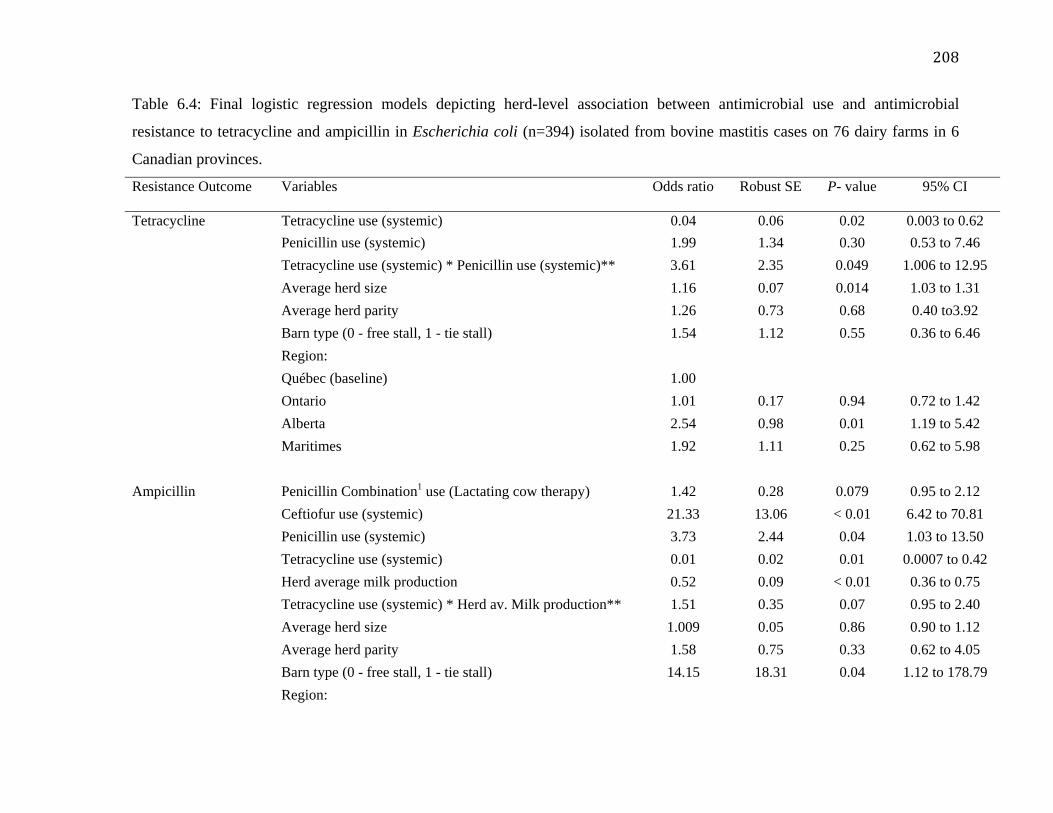
208
Table 6.4: Final logistic regression models depicting herd-level association between antimicrobial use and antimicrobial
resistance to tetracycline and ampicillin in Escherichia coli (n=394) isolated from bovine mastitis cases on 76 dairy farms in 6
Canadian provinces.
Resistance Outcome Variables Odds ratio Robust SE P- value 95% CI
Tetracycline Tetracycline use (systemic) 0.04 0.06 0.02 0.003 to 0.62
Penicillin use (systemic) 1.99 1.34 0.30 0.53 to 7.46
Tetracycline use (systemic) * Penicillin use (systemic)** 3.61 2.35 0.049 1.006 to 12.95
Average herd size 1.16 0.07 0.014 1.03 to 1.31
Average herd parity 1.26 0.73 0.68 0.40 to3.92
Barn type (0 - free stall, 1 - tie stall) 1.54 1.12 0.55 0.36 to 6.46
Region:
Québec (baseline) 1.00
Ontario 1.01 0.17 0.94 0.72 to 1.42
Alberta 2.54 0.98 0.01 1.19 to 5.42
Maritimes 1.92 1.11 0.25 0.62 to 5.98
Ampicillin Penicillin Combination1 use (Lactating cow therapy) 1.42 0.28 0.079 0.95 to 2.12
Ceftiofur use (systemic) 21.33 13.06 < 0.01 6.42 to 70.81
Penicillin use (systemic) 3.73 2.44 0.04 1.03 to 13.50
Tetracycline use (systemic) 0.01 0.02 0.01 0.0007 to 0.42
Herd average milk production 0.52 0.09 < 0.01 0.36 to 0.75
Tetracycline use (systemic) * Herd av. Milk production** 1.51 0.35 0.07 0.95 to 2.40
Average herd size 1.009 0.05 0.86 0.90 to 1.12
Average herd parity 1.58 0.75 0.33 0.62 to 4.05
Barn type (0 - free stall, 1 - tie stall) 14.15 18.31 0.04 1.12 to 178.79
Region:

209
Québec (baseline) 1.00
Ontario 0.76 0.34 0.55 0.31 to 1.86
Alberta 5.48 1.18 < 0.01 3.59 to 8.38
Maritimes 0.61 0.34 0.38 0.20 to 1.83
1Intramammary preparation containing Penicillin G Procaine/Dihydrostreptomycin sulfate/Novobiocin sodium/Polymyxin B
sulfate.
** Interaction between explanatory variables.

210
Table 6.5: Final logistic regression models depicting herd-level association between antimicrobial use and antimicrobial
resistance to streptomycin and Trimethoprim-sulfamethoxazole (TMPS) combination in Escherichia coli (n=394) isolated from
bovine mastitis cases on 76 dairy farms in 6 Canadian provinces.
Resistance Outcome Variables Odds ratio Robust SE P- value 95% CI
Streptomycin Penicillin Combination1 use (Lactating cow therapy) 2.26 0.80 0.02 1.13 to 4.54
Penicillin-novobiocin use (dry cow therapy) 1.75 0.37 < 0.01 1.14 to 2.66
Penicillin use (systemic) 3.31 0.62 < 0.01 2.29 to 4.79
Tetracycline use (systemic) 0.29 0.22 0.11 0.06 to 1.33
Average herd size 1.45 0.30 0.07 0.96 to 2.19
Penicillin combination use * average herd size** 0.87 0.03 < 0.01 0.80 to 0.95
Average herd parity 1.29 0.86 0.69 0.35 to 4.79
Barn type (0-free stall, 1- tie stall) 0.89 0.88 0.91 0.13 to 6.17
Region:
Québec (baseline) 1.00
Ontario 0.64 0.07 < 0.01 0.52 to 0.79
Alberta 2.71 2.09 0.19 0.59 to 12.32
Maritimes 1.88 1.72 0.48 0.31 to 11.32
TMPS TMPS use (systemic) 1.68 0.45 0.05 0.99 to 2.86
Ceftiofur use (systemic) 31.08 21.02 < 0.01 8.25 to 117.01
Penicillin Combination1 use (Lactating cow therapy) 1.53 0.16 < 0.01 1.24 to 1.89
Pirlimycin use (Lactating cow therapy) 0.44 0.05 < 0.01 0.35 to 0.55
Average herd size 0.96 0.08 0.72 0.81 to 1.14
Average herd parity 0.79 0.47 0.69 0.24 to 2.53
Barn type (0-free stall, 1- tie stall) 2.27 1.78 0.29 0.48 to 10.58

211
Region:
Québec (baseline) 1.00
Ontario 0.63 0.15 0.06 0.39 to 1.02
Alberta 1.89 0.39 < 0.01 1.26 to 2.85
Maritimes 2.00 0.78 0.07 0.92 to 4.32 1Intramammary preparation containing Penicillin G Procaine/Dihydrostreptomycin sulfate/Novobiocin sodium/Polymyxin B
sulfate.
** Interaction between explanatory variables.

212
Table 6.6: Final logistic regression models depicting herd-level association between antimicrobial use and antimicrobial
resistance to ceftiofur and chloramphenicol in Escherichia coli (n=394) isolated from bovine mastitis cases on 76 dairy farms
in 6 Canadian provinces.
Resistance Outcome Variables Odds ratio Robust SE P- value 95% CI
Ceftiofur Ceftiofur use (systemic) 0.96 0.11 0.76 0.77 to 1.20 Average herd size 0.95 0.07 0.57 0.82 to 1.11 Average herd parity 1.52 1.34 0.63 0.26 to 8.63 Barn type (0-free stall, 1- tie stall) 10.06 6.72 < 0.01 2.71 to 37.28 Region: Québec Omitted due to absence of resistance Ontario (baseline) 1.00 Alberta 0.13 0.007 < 0.01 0.12 to 0.15 Maritimes Omitted due to absence of resistance Chloramphenicol Florfenicol use (systemic) 0.08 0.04 < 0.01 0.02 to 0.26 Penicillin use (systemic) 3.04 1.21 0.005 1.39 to 6.66 Tetracycline use (systemic) 0.008 0.01 0.003 0.0003 to 0.19 Average herd size 1.21 0.07 0.001 1.08 to 1.36 Average herd parity 0.87 0.22 0.62 0.52 to 1.46 Barn type (0-free stall, 1- tie stall) 1.16 0.78 0.81 0.31 to 4.35 Region: Québec (baseline) 1.00 Ontario 2.77 0.39 < 0.01 2.09 to 3.67 Alberta 20.27 4.65 < 0.01 12.92 to 31.79 Maritimes 1.93 0.74 0.09 0.90 to 4.13

213
Table 6.7: Final logistic regression model depicting herd-level association between tetracycline use and tetracycline resistance in
Klebsiella species isolates (n=139) from bovine mastitis cases on 37 dairy farms in 6 Canadian provinces.
Resistance Outcome Variables Odds ratio Robust SE P- value 95% CI
Tetracycline Tetracycline use (systemic) 2.59 1.07 0.02 1.14 to 5.86
Average herd size 0.97 0.03 0.38 0.90 to 1.03
Average herd parity 4.20 3.82 0.11 0.70 to 24.90
Barn type (0 - free stall, 1 - tie stall) 0.20 0.45 0.47 0.002 to 15.89
Region:
Maritimes (baseline) 1.00
Ontario 2.37 1.19 0.08 0.88 to 6.37
Alberta 22.92 41.54 0.08 0.65 to 799.98
Québec Omitted due to absence of resistance

214
Chapter Seven: Conclusions and Future Perspectives

215
The general objectives of this thesis included: a) Validating methods commonly employed
for antimicrobial susceptibility testing of bovine clinical mastitis pathogens, b) Determining
qualitative and quantitative aspects of antimicrobial drug utilization on Canadian dairy
farms, c) Antimicrobial susceptibility testing of Staphylococcus aureus, Escherichia coli,
and Klebsiella species pathogens isolated from intramammary infections and (sub) clinical
mastitis cases, and d) Assessing and evaluating if a herd-level association between
antimicrobial use and resistance exists in common mastitis pathogens.
Bacteria isolated from different animal species or even different sites of infection might
vary in growth; such differences could impact antimicrobial susceptibility results.
Sensititre® MIC susceptibility system and the agar disk diffusion method was therefore
validated with manual broth microdilution test method for clinical mastitis pathogens
isolated from dairy cattle. The agar disk diffusion method and the Sensititre® MIC
susceptibility system had a moderate to high diagnostic accuracy for most antimicrobial-
isolate species combinations. As part of future research, it will be interesting to determine if
similar results are also observed for isolates from intramammary infections and subclinical
mastitis cases as well.
Even though the method of auditing empty antimicrobial containers was producer friendly
and circumvented issues of producer non-compliance observed in antimicrobial drug
utilization studies, it fails to account for actual usage including off-label use at the
individual cow-level. Data on dose and duration of actual treatment administered are vital

216
for determining selection pressure imposed by antimicrobial treatment on bacteria in an
animal. For example, under-dosing could facilitate acquiring of resistance mechanisms in
surviving mastitis pathogens whereas over-dosing could result in faster rate of resistance
development such as through the ‘Eagle-effect’ as observed for ß-lactams. The concept of
prescribed-daily dose is therefore superior to defined-daily dose. The prescribed-daily
doses may be calculated from prescriptions, information from veterinarian’s invoices or
producer-written treatment records. However, producer compliance in filling complete and
accurate information on health records is a potential challenge. In future, electronic means
of capturing antimicrobial use and health data at the individual cow-level such as hand-held
computers for producers should be relied upon to facilitate producer compliance.
Discordance between antimicrobial use data collected at an individual cow-level and herd-
level should also be determined for assessing measurement bias in such studies.
One isolate per quarter was selected for antimicrobial susceptibility testing. It was done to
ensure statistical independence between isolates presuming that emergence of antimicrobial
resistance in an isolate in a quarter might impact antimicrobial resistance in another isolate
in the same quarter. This seems unlikely because environment inside the udder is dynamic
compared to gastro-intestinal tract where reservoirs of resistance determinants do not exist.
In future studies, one isolate per cow should be selected for antimicrobial susceptibility
testing. The present study could not assess a temporal association between antimicrobial
use and resistance in bovine mastitis pathogens; such associations can only be assessed in a
case-control or cohort study. It is therefore recommended, that follow-up studies should be
carried out at the individual cow-level in the future.

217
Antimicrobial resistance prevalence was uncommon in S. aureus, E. coli, and Klebsiella
species isolates. Resistance to antimicrobials of very high importance to human medicine
was rare in bovine mastitis pathogens. The study results suggest a low risk of transmission
of antimicrobial resistant bacteria from milk or milk products to human populations in
Canada. However, monitoring of antimicrobial resistance in udder pathogens should remain
ongoing for collecting information concerning emergence and trends of antimicrobial
resistance in bacterial populations.
The use of antimicrobials in food-animal production systems remains a contentious issue.
Among antimicrobials of very high importance in human medicine, fluoroquinolones were
rarely used, whereas third-generation cephalosporins and penicillin combinations
containing colistin were used very frequently on Canadian dairy farms. The author believes
that there is room for conservative use of antimicrobials in dairy cattle. For example, most
of the commercial antimicrobials used for dry cow therapy are active against Gram-positive
udder pathogens, and have little or no activity against Gram-negative pathogens. Most
clinical mastitis cases caused by coliforms can resolve on their own without the use of
antimicrobials. In general, knowledge about the role of cow and pathogen factors in
therapeutic success of bovine mastitis is important while planning antimicrobial treatment
regimen (Barkema et al., 2006). The author believes that the use of antimicrobials will
remain an inalienable part of food-animal production system, but prudent (appropriate,
justifiable, judicious) use of antimicrobials is vital for containing antimicrobial resistance,
and that continuing education of producers is a key step towards conservative use of
antimicrobials.

218
Antimicrobial resistance in bacteria at a farm cannot be solely explained in terms of
antimicrobial use. Emergence and dissemination of antimicrobial resistance is a complex
phenomenon and depends upon the geographical location, hygiene levels, herd size, and the
type of integrated farming that takes place at a farm (Acar and Moulin, 2006). Use of
antimicrobials in animals, and antibiotic residues and resistant bacteria in the feces,
effluents, wastewater lagoons and soil create a reservoir of antimicrobial resistance
determinants that can transfer between different compartments / niches at a farm. Similar
observations have been made in this study e.g. barn type, herd size and region was
associated with herd-level antimicrobial resistance outcomes. In future, besides
antimicrobial use, information should be collected on such farm-level factors that impact
the ecology of antimicrobial resistance in bacteria.

219
7.1 References
Acar, J. F. and G. Moulin. 2006. Antimicrobial resistance at farm level. Rev. Sci. Tech.
25:775-792.
Barkema H. W., Y. H. Schukken, and R. N. Zadoks. 2006. Invited Review: The role of
cow, pathogen, and treatment regimen in the therapeutic success of bovine
Staphylococcus aureus mastitis. J. Dairy Sci. 89:1877-1895.

02
04
06
08
01
00
AD
UR
(#A
DD
/1000 c
ow
-day)
25 30 35 40
Herd average cow daily milk production (kg)


![3URYLQFLDO *OD]LHUV $VVRFLDWLRQ RI $OEHUWD %DODQFH … · Calgary Construction Association 2725 12 Street NE Calgary, AB T2E 7J2 Phone: 403–291–3350 mail@cca.cc Red Deer Construction](https://static.fdocuments.net/doc/165x107/60326380fd58df79834f472c/3urylqfldo-odlhuv-vvrfldwlrq-ri-oehuwd-dodqfh-calgary-construction-association.jpg)


