Unit 9 Lower Limb Muscles
13
1 BIOL 2210L Unit 9: Lower Limb Muscles Authors: Terri Koontz and Christine Woods, CNM Biology Department Creative Commons Attribution-NonCommercial 4.0 International License Terms to Know for Unit 9 Muscles of Anterior Thigh Muscles of Posterior Lower Leg Psoas major Triceps surae Iliacus Gastrocnemius Sartorius Soleus Adductor magnus Adductor brevis Muscles of Lateral and Anterior Lower Leg Gracilis Fibularis longus Quadriceps femoris Fibularis brevis Rectus femoris Tibialis anterior Vastus lateralis Extensor digitorum longus Vastus medialis Vastus intermedius Additional Instructor Terms Tensor fascia lata Muscles of Gluteal region and Posterior Thigh Gluteus maximus Gluteus medius Gluteus minimus Hamstrings Biceps femoris Semitendinosus Semimembranosus Learning Objectives (modified from HAPS learning outcomes) 1. Nomenclature of skeletal muscles a. Explain how the name of a muscle can help identify its action, appearance, or location. 2. Location & function of the major skeletal muscles in the terms list a. Identify the origin, insertion and action of the major skeletal muscles and demonstrate these muscle actions. 3. Group actions of skeletal muscles a. For a given movement, differentiate specific muscles that function as prime mover, antagonist, synergist or fixator.
Transcript of Unit 9 Lower Limb Muscles

1
BIOL 2210L Unit 9: Lower Limb Muscles
Authors: Terri Koontz and Christine Woods, CNM Biology Department
Creative Commons Attribution-NonCommercial 4.0 International License
Terms to Know for Unit 9 Muscles of Anterior Thigh Muscles of Posterior Lower Leg
Psoas major Triceps surae
Iliacus Gastrocnemius
Sartorius Soleus
Adductor magnus Adductor brevis Muscles of Lateral and Anterior Lower Leg
Gracilis Fibularis longus
Quadriceps femoris Fibularis brevis
Rectus femoris Tibialis anterior
Vastus lateralis Extensor digitorum longus
Vastus medialis Vastus intermedius Additional Instructor Terms
Tensor fascia lata
Muscles of Gluteal region and Posterior Thigh Gluteus maximus Gluteus medius Gluteus minimus Hamstrings Biceps femoris Semitendinosus Semimembranosus
Learning Objectives (modified from HAPS learning outcomes) 1. Nomenclature of skeletal muscles
a. Explain how the name of a muscle can help identify its action, appearance, or location. 2. Location & function of the major skeletal muscles in the terms list
a. Identify the origin, insertion and action of the major skeletal muscles and demonstrate these muscle actions.
3. Group actions of skeletal muscles a. For a given movement, differentiate specific muscles that function as prime mover,
antagonist, synergist or fixator.

2
Image 1: Muscles of anterior thigh
Cropped Creative Commons Attribution 4.0 International Openstax URL: Muscles of anterior thigh
Explanation of Anatomy In this last unit on muscles, we’ll learn muscles of the lower limb. First, we’ll study muscles of the
anterior thigh. Then, we’ll turn our gaze to the backside and learn the gluteal muscles and the posterior
thigh. We’ll finish the unit, studying muscles that make up the lower leg. With each muscle, we will look
at origin, insertion, and action.
Think about all the muscles that we use to get around in our environment. For example, imagine
throwing a baseball. At first you might only think about what your arms are doing during this act.
However, lower limb and torso muscles also are assisting in moving the skeleton. As you put one leg
forward and brace yourself with the other leg behind, you are slightly bending your lower body using
your lower limb muscles to cause flexion in your leg joints: hips, knees, and ankles. When you go to
throw the ball, your back leg extends at the hip and knee as your ankle plantarflexes. All the while, your
torso muscles are helping with balance, posture, and twisting of your upper body not to mention what is
a happening with the joints in your arm. In the last activity in this unit you’ll explore muscle actions
when throwing a baseball where you’ll identify which muscle is involved along each “step” of making
that pitch.
Muscles of Anterior Thigh There are two muscles located within each of the abdominopelvic iliac regions that move the thigh.
You’re probably noticing that muscles commonly act on the joint that is distal to them. The psoas major
and the iliacus, which are proximal to the hip, cause hip flexion (see Image 1). The psoas major in
quadrupeds is the tenderloin. Psoas means loin. The iliacus, which is a broader muscle than the psoas
major, directly attaches to the ilium. Both insert at the lesser trochanter of the femur.

3
The quadriceps femoris is a group of muscles that are named based on their location, femoral region.
Quadriceps refers to the fact that there are four muscles that belong to this group: rectus femoris,
vastus lateralis, vastus medialis, and vastus intermedius. The muscle that is superficial and is in the
middle of this group is named for its shape and location. The rectus femoris is straight down the middle
of the thigh region. The remaining three make up the bulk of the anterior thigh and the first word of
their names, vastus, means large. The relative location of these three muscles to one another makes up
the second word of their names. The most lateral of the three is named the vastus lateralis, the most
medial is the vastus medialis, and the one that is between and in the middle is the vastus intermedius.
The vastus intermedius is deep to the rectus femoris. All muscles of the quadriceps femoris share a
common insertion: the patella and tibial tuberosity. Image 1 depicts quadriceps femoral muscles.
Mini activity: Muscle movements of femoral muscles
Complete the table below. If your instructor wants you to know more than the muscles within the table,
add them.
Muscle Origin Insertion Primary Action
Psoas major
Iliacus
Rectus femoris
Vastus lateralis
Vastus medialis
Vastus intermedius
The anterior inner thigh is made up of four muscles: gracilis, adductor magnus, adductor longus, and
adductor brevis (see Images 1 and 2). The gracilis is named for its shape where the other three inner
thigh muscles are named based on their movement and relative sizes to one another. Gracilis means
slender; and this long, slender muscle travels the length of the femur. It is the most medial of all the

4
Image 2: Adductor thigh muscles
Cropped Creative Commons Attribution 4.0 International Openstax URL: Adductor thigh muscles
Image 3: Tensor fascia lata
Cropped Public Domain from 20th
edition of Gray’s Anatomy of the Human Body URL: Tensor fascia lata
inner thigh muscles. The gracilis along with the other three inner thigh muscles perform adduction of
the thigh. The biggest of all these muscles is called the adductor magnus. The adductor longus is a long
muscle that is superficial to the shorter muscle named the adductor brevis.
The sartorius and tensor fascia lata both abduct the thigh, so they are antagonistic to the thigh
adductors. Sartorius (see Image 1) means tailor and this muscle performs all the movements that a
tailor does when they cross one leg over the other when mending clothes. The sartorius muscle flexes
the hip, abducts and laterally rotates the thigh. While the tensor fascia lata (see Image 2) abducts the
thigh, it also tenses the aponeurosis called the iliotibial band that acts as a fascia by supporting thigh
muscles.

5
Image 4: Gluteal and posterior thigh muscles
Cropped Creative Commons Attribution 4.0 International Openstax URL: Gluteal and posterior thigh
muscles
Muscles of the Gluteal Region and Posterior Thigh Three muscles in the gluteal region are named based on their location and relative sizes to one another:
gluteal maximus, gluteus medius, and gluteus minimus. The largest of the three, gluteus maximus,
performs hip extension. The second largest, gluteus medius, is superficial to the smallest of the three,
gluteus minimus. The gluteus medius and gluteus minimus abduct and medially rotate the thigh.
The hamstrings are prime movers of hip extension and knee flexion. The hamstring muscles are a group
of muscles that are involved with movement of the joint both proximal and distal to themselves. Their
name refers to how old-world butchers would hang ham for smoking by this group’s tendons. The most
lateral of the three muscles has two origins so is named biceps femoris. The other two hamstring
muscles can be viewed as representing a half of one whole muscle. Semi- means half. The superficial of
these two medial hamstring muscles has a long thin tendon, semitendinosus. The deeper one is the
larger of the two; thus, it contains more of an outer membrane, semimembranosus. The hamstring
muscles are shown in Image 4 along with gluteal muscles.
Mini activity: Muscle etymology
By now you’ve learned a lot of muscles and learned how they got their names. The derivation of a word
is called etymology. Below write down at least three of your favorite muscles or muscle groups and next

6
Image 5: Posterior lower leg muscles
Cropped Creative Commons Attribution 4.0 International Openstax URL: Posterior lower leg muscles
to the muscle write how it got its name. If a muscle’s etymology wasn’t covered in this manual, you can
research how it got its name on your own. Either is acceptable for this mini activity.
Muscle 1 Etymology
Muscle 2 Etymology
Muscle 3 Etymology
Muscles of Posterior Lower Leg The muscles of the posterior leg are named based on their location, shape, and movement. Image 5
shows muscles of the posterior lower leg.
The triceps surae are two muscles that make up the calf: gastrocnemius and soleus. The gastrocnemius
has two heads that each can be interpreted as its own muscle, making this muscle a representation of
two muscles. This is why the gastrocnemius and soleus are collectively called the triceps surae. Gastro-
means belly and “kneme” means leg. If you use your imagination, you can view this muscle looking like
the belly of the lower leg, hence its name gastrocnemius. The soleus is the deeper of the two and is also
named based on its shape. The soleus looks like a fish and soleus means fish! The movement of the
triceps surae is plantar flexion.
7
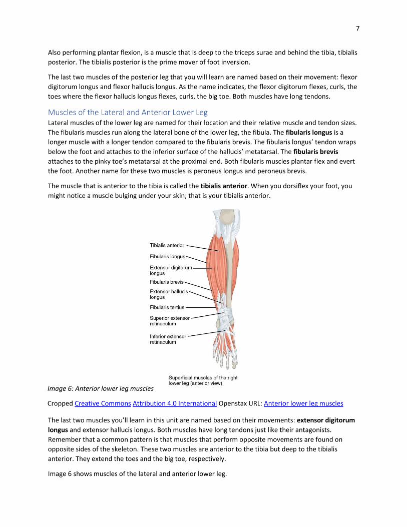
Image 6: Anterior lower leg muscles
Cropped Creative Commons Attribution 4.0 International Openstax URL: Anterior lower leg muscles
Also performing plantar flexion, is a muscle that is deep to the triceps surae and behind the tibia, tibialis
posterior. The tibialis posterior is the prime mover of foot inversion.
The last two muscles of the posterior leg that you will learn are named based on their movement: flexor
digitorum longus and flexor hallucis longus. As the name indicates, the flexor digitorum flexes, curls, the
toes where the flexor hallucis longus flexes, curls, the big toe. Both muscles have long tendons.
Muscles of the Lateral and Anterior Lower Leg Lateral muscles of the lower leg are named for their location and their relative muscle and tendon sizes.
The fibularis muscles run along the lateral bone of the lower leg, the fibula. The fibularis longus is a
longer muscle with a longer tendon compared to the fibularis brevis. The fibularis longus’ tendon wraps
below the foot and attaches to the inferior surface of the hallucis’ metatarsal. The fibularis brevis
attaches to the pinky toe’s metatarsal at the proximal end. Both fibularis muscles plantar flex and evert
the foot. Another name for these two muscles is peroneus longus and peroneus brevis.
The muscle that is anterior to the tibia is called the tibialis anterior. When you dorsiflex your foot, you
might notice a muscle bulging under your skin; that is your tibialis anterior.
The last two muscles you’ll learn in this unit are named based on their movements: extensor digitorum
longus and extensor hallucis longus. Both muscles have long tendons just like their antagonists.
Remember that a common pattern is that muscles that perform opposite movements are found on
opposite sides of the skeleton. These two muscles are anterior to the tibia but deep to the tibialis
anterior. They extend the toes and the big toe, respectively.
Image 6 shows muscles of the lateral and anterior lower leg.

8
Activity 1: Predicting Muscle Movement Your instructor has set up around the class skeletons and/or bones that have labels for origin and
insertion of selected muscles from this lab. Go around to each station and identify the following
information for each set of labeled bones. a. muscle name, b. origin, c. insertion, d. action, e. direction
of muscle fibers, and f. if and how the muscle crosses a joint.
If rubber bands or string are at the stations, use these tools to predict muscle movement (action). Or
visualize the muscle shortening as the insertion moves towards the origin in the direction of the muscle
fibers.
Station 1:
Station 2:
Station 3:
Station 4:

9
(Alternative) Activity 1: Learning Muscle Movement Make labels using the provided painter’s tape for each muscle your instructor wants you to learn origin,
insertion, and action for. Write the names of those muscles below. You might want to assign a number
for each muscle and make the labels using those assigned numbers.
Place the numbered pieces of tape on the correct muscles. Then, individually or as a group, take turns
identifying the following information for each number. Do not use your notes, tables, diagrams, or
textbook!
1. Muscle Name 2. Origin 3. Insertion 4. Action 5. Take note of direction of fibers, how it crosses a joint, layering of muscles, and things you find
important to help you learn the muscles and their actions.

10
Activity 2: Muscles and Their Actions Part 1 -List the leg muscles that have a common action in the table below. As you list the muscles for
each action, practice the action with your own body. Note any origins or insertions that may reoccur
within the common action group. Note the agonist muscle and antagonist muscle when you are listing.
Action Muscles That Perform This Action
Flexion of hip
Extension of hip
Adduction of hip
Abduction of hip
Flexion of knee
Extension of knee
Inversion
Eversion
Flex digits
Extend digits
Part 2 - Fill in muscle names to complete the charts.
Femoral Muscles: 1.
2.
3.
4.
1.
2.
3.
Quadriceps femoris
Hamstrings
11
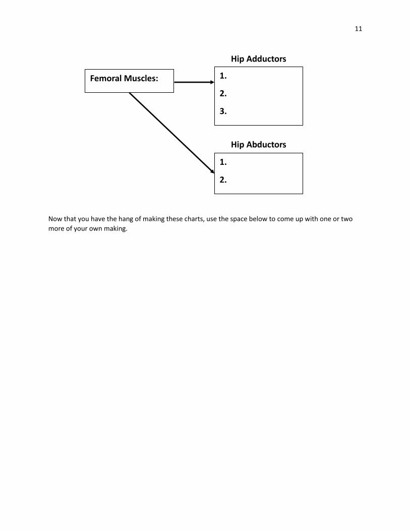
Now that you have the hang of making these charts, use the space below to come up with one or two
more of your own making.
Femoral Muscles: 1.
2.
3.
1.
2.
Hip Adductors
Hip Abductors

12
A.
Winding
Up
B. Early
Cocking
C. Acceleration
CC BY 2.0 by U.S. Embassy Jerusalem URL: Throwing A Baseball
Activity 3: Muscles Used When Throwing a Baseball We’ve already discussed at the beginning of the unit a little about what actions are happening to throw
a baseball. Now that we’ve learned a significant number of skeletal muscles, we can identify which ones
are used to throw a baseball.
Determine what muscle or group of muscles are participating in the various actions to throw a baseball.
Below are three images of how the body is positioned during different phases taken to make a pitch. For
each image there is a set of questions asking what muscles are involved to perform the different actions.
A. Winding Up: The front leg is flexing at the hip, knee, and the foot is dorsiflexed.
1. What muscle is performing hip flexion?
a. psoas major b. vastus lateralis c. gluteus maximus d. hamstrings
2. What muscle is performing knee flexion?
a. psoas major b. vastus lateralis c. gluteus maximus d. hamstrings
3. What muscle is performing dorsiflexion?
a. anterior tibialis b. soleus c. gastrocnemius d. fibularis brevis

13
B. Early
Cocking
C. Acceleration
B. Early Cocking: The back leg is extended at the hip, knee, and is plantarflexed.
4. What muscle is performing hip extension?
a. psoas major b. vastus lateralis c. gluteus maximus d. hamstrings
5. What muscle is performing knee extension?
a. psoas major b. vastus lateralis c. gluteus maximus d. hamstrings
6. What muscle is performing plantarflexion?
a. anterior tibialis b. soleus c. gastrocnemius d. fibularis brevis
What about the arms? For the arm throwing the baseball describe the
action at the shoulder, elbow, wrist, and fingers along with the muscles
performing those actions.
Shoulder
Elbow
Wrist
Fingers
C. Acceleration
Now you decide which leg you want to describe the movements you are
seeing at the hip, knee, and ankle joint. What muscles perform that
movement?
Hip
Knee
Ankle
Again using the throwing arm describe the action and muscles used to
perform each action at the shoulder, elbow, wrist, and fingers.
Shoulder
Elbow
Wrist
Fingers