Unit 6-Nucleus
71
Unit 6-Nucleus Dr. Pallee shree
Transcript of Unit 6-Nucleus

Unit 6-Nucleus
Dr. Pallee shree
Nucleus
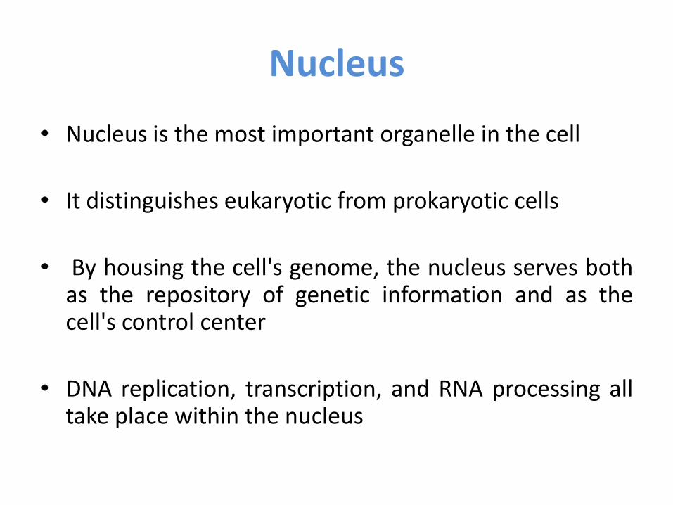
• Nucleus is the most important organelle in the cell
• It distinguishes eukaryotic from prokaryotic cells
• By housing the cell's genome, the nucleus serves both as the repository of genetic information and as the cell's control center
• DNA replication, transcription, and RNA processing all take place within the nucleus

Cont…
• A nucleus is a double-membraned eukaryotic cell organelle that contains the genetic material.
• It appears in an oval shape averages 5µm in width.
• It often lies in the centre of a cell
• The nucleus was the first organelle to be discovered
• Nuclei 1st discovered and named by Robert Brown
• Role of nucleus 1st demonestrated by Max Hammerling

Ultra structure of Nucleus
1. Nuclear envelope
2. nuclear pores
3. Nucleoplasm
4. Nucleolus
5. Chromosomes

1. Structure of Nuclear envelope
• The nuclear envelope has a complex structure consisting of
a) Two nuclear membranes separated by a perinuclear space measuring about 20–40 nm across
b) Underlying nuclear lamina • The nucleus is surrounded by a system
of two concentric membranes, called the inner and outer nuclear membranes
• The inner and outer nuclear membranes are joined at nuclear pore complexes

a. Nuclear membranes • The outer nuclear membrane is
continuous with the endoplasmic reticulum, so the space between the inner and outer nuclear membranes is directly connected with the lumen of the ER
• It is functionally similar to the membranes of the ER and has ribosomes bound to its cytoplasmic surface but protein composition differ slightly as they are enriched in proteins which binds to cytoskeleton
• The inner nuclear membrane carries proteins that are specific to the nucleus, such as those that bind the nuclear lamina
• The critical function of the nuclear membranes is to act as a barrier that separates the contents of the nucleus from the cytoplasm.
• Like other cell membranes, each nuclear membrane is a phospholipid bilayer permeable only to small nonpolar molecules
• Other molecules are unable to diffuse through the bilayer.

b. Nuclear lamina
• It is a fibrous meshwork that provides structural support to
the nucleus envelope
• Nuclear lamina is composed of fibrous proteins called lamins
• Lamins are a class of intermediate filament proteins
• Mammalian cells have three lamin genes, designated A, B,
and C, which code for at least seven distinct proteins.
• Like other intermediate filament proteins, the lamins
associate with each other to form higher order structures
although the extent and polarity of this association is
thought to differ from that of other intermediate filaments.
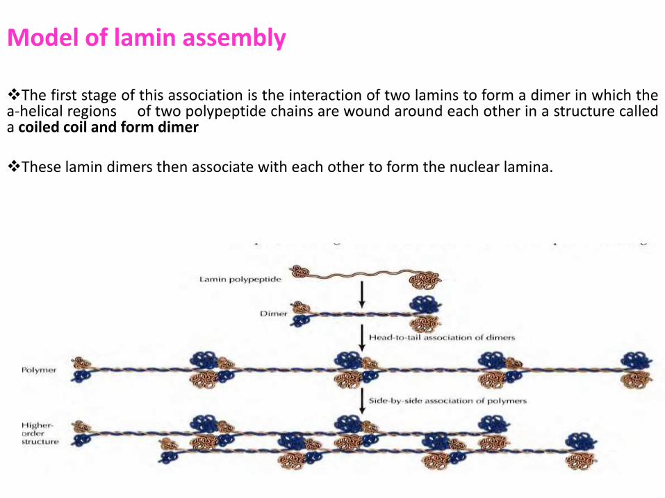
Model of lamin assembly The first stage of this association is the interaction of two lamins to form a dimer in which the a-helical regions of two polypeptide chains are wound around each other in a structure called a coiled coil and form dimer
These lamin dimers then associate with each other to form the nuclear lamina.

Nuclear lamina • The association of lamins with the inner nuclear membrane is facilitated by the
posttranslational addition of lipid- in particular, prenylation of C-terminal cysteine residues
• In addition, the Lamins bind to specific inner nuclear membrane proteins such as
emerin and the lamin B receptor, mediating their attachment to the nuclear envelope and localizing and organizing them within the nucleus
• The nuclear lamina also binds to chromatin through histones H2A and H2B as well as other chromatin proteins.
• Lamins also extend in a loose meshwork throughout the interior of the nucleus.
• Many nuclear proteins that function in DNA synthesis, transcription, or chromatin modification are known to bind lamins, although the significance of these interactions is only beginning to be understood.
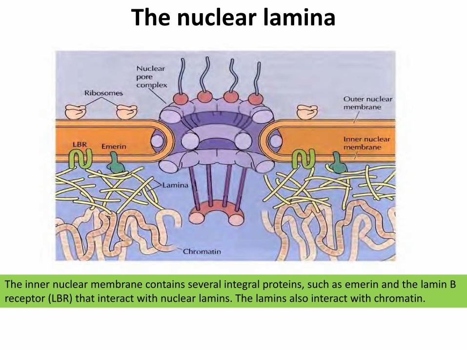
The nuclear lamina
The inner nuclear membrane contains several integral proteins, such as emerin and the lamin B receptor (LBR) that interact with nuclear lamins. The lamins also interact with chromatin.

2. Nuclear pore complexes (NPC)
• The nuclear pore complexes are the only channels through which small polar molecules, ions, and macromolecules (proteins and RNAs) can travel between the nucleus and the cytoplasm
• The nuclear pore responsible for the selective traffic of proteins and RNAs between the nucleus and the cytoplasm
• The nuclear pore complex is an extremely large structure about 30 times the size of a ribosome with a diameter of about 120 nm
• ln vertebrates, the nuclear pore complex is composed of 30-50 different pore proteins called nucleoporins, most of which are present in multiple copies.

Structure of nuclear pore complexes
• Visualization of nuclear pore complexes by electron microscopy reveals that the NPC appear to consist of eight structural subunits surrounding a central channel via which molecules pass
• Nuclear pore complex consists of an assembly of eight spokes arranged around a central channel therefore we can say it has its octagonal symmetry
• The spokes are connected to rings at the nuclear and cytoplasmic surfaces, and the spoke-ring assembly is anchored within the nuclear envelope at sites of fusion between the inner and outer nuclear membranes.
• Protein filaments extend from both the cytoplasmic and nuclear rings, forming a distinct basketlike structure on the nuclear side.
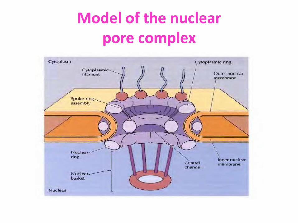
Model of the nuclear pore complex
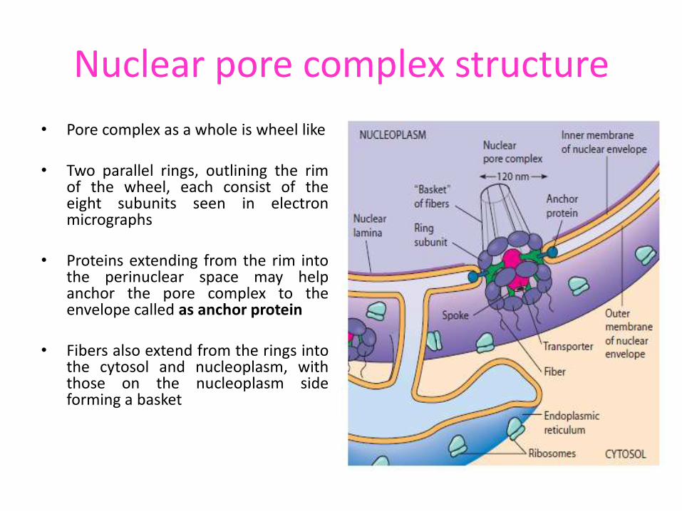
Nuclear pore complex structure
• Pore complex as a whole is wheel like
• Two parallel rings, outlining the rim of the wheel, each consist of the eight subunits seen in electron micrographs
• Proteins extending from the rim into the perinuclear space may help anchor the pore complex to the envelope called as anchor protein
• Fibers also extend from the rings into the cytosol and nucleoplasm, with those on the nucleoplasm side forming a basket

Functions of nuclear pore complex
• Controll the traffic of molecules between the nucleus and the cytoplasm, the nuclear pore complex plays a fundamental role in the physiology of all eukaryotic cells.
• RNAs synthesized in the nucleus are exported to the cytoplasm where they function in protein synthesis.
• Conversely, proteins required for nuclear functions e.g., transcription factors are transported to the nucleus from their sites of synthesis in the cytoplasm.
• Many proteins shuttle continuously between the nucleus and the cytoplasm.
• Depending on their size, molecules can travel through the nuclear pore complex by one of two different mechanisms
TRANSPORT OF MOLECULE ACROSS THE NPC

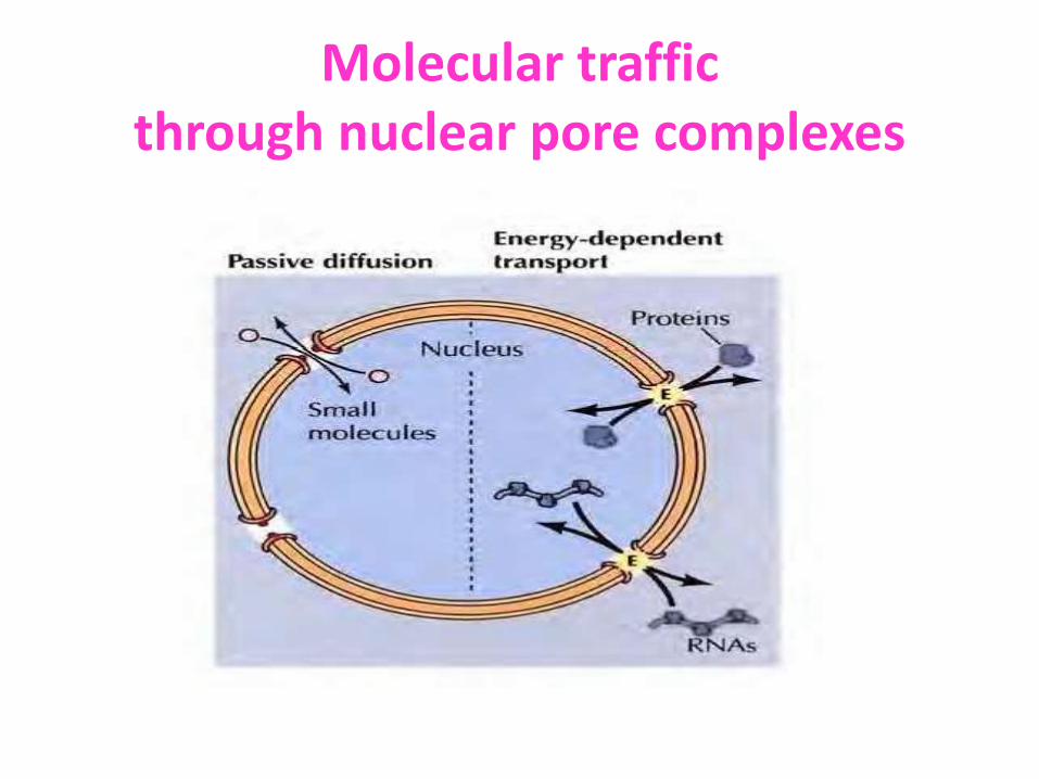
Molecular traffic
through nuclear pore complexes
Diffuse passively
Small molecules and some proteins of less molecular mass (20-40 kd) pass freely through the pore in either direction
Active transport
Most proteins and RNAs unable to
pass through open channels. So they
Pass via nuclear pore complex by an active process and selectively transported in a specific direction

Molecular traffic through nuclear pore complexes

Transport via nuclear pore complex
• The NPC forms the conduit for the exchange of information between the nucleus and cytoplasm
• Important molecules transported across the NPC are Proteins required for DNA replication and RNA

a. Transport of protein to and from the nucleus
• Transport of proteins are significant as they are responsible for all aspects of genome structure and function
• Important proteins transported are histones, DNA polymerases, RNA polymerases, transcription factors, splicing factors etc.
• Proteins are targeted to the nucleus by specific amino acid sequences called nuclear localization signals (NLS)
• The signal are recognized by transport receptors and they
direct the transport of the proteins through the nuclear pore complex.

Macromolecular Transport into and out of the Nucleus

Cont… 1. NLS was 1st time mapped in detail
and characterized by Alan Smith et al., 1984.
2. The viral Simian virus 40 (SV40) T antigen protein was used as a model for studies of nuclear localization in animal cells.
3. A virus-encoded protein T antigen is a 94-kd protein that is required for SV40 DNA replication and is normally localized to the nucleus of SV40-infected cells.
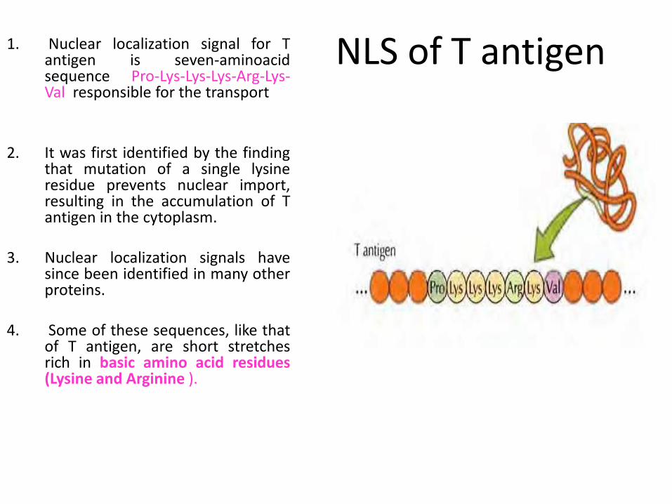
NLS of T antigen 1. Nuclear localization signal for T antigen is seven-aminoacid sequence Pro-Lys-Lys-Lys-Arg-Lys-Val responsible for the transport
2. It was first identified by the finding that mutation of a single lysine residue prevents nuclear import, resulting in the accumulation of T antigen in the cytoplasm.
3. Nuclear localization signals have since been identified in many other proteins.
4. Some of these sequences, like that of T antigen, are short stretches rich in basic amino acid residues (Lysine and Arginine ).

1. However, many times amino acids that form the nuclear localization signal are close together but not immediately adjacent to each other.
2. For example, the nuclear localization signal of nucleoplasrnin (a protein involved in chromatin assembly) consists of two parts: a Lys-Arg pair followed by four lysines located ten amino acids farther downstream
Nuclear localization signal of nucleoplasm in is bipartite, consisting of a Lys-Arg followed by a LysLys- Lys-Lys sequence located ten amino acids farther downstream sequence
Nuclear localization signal
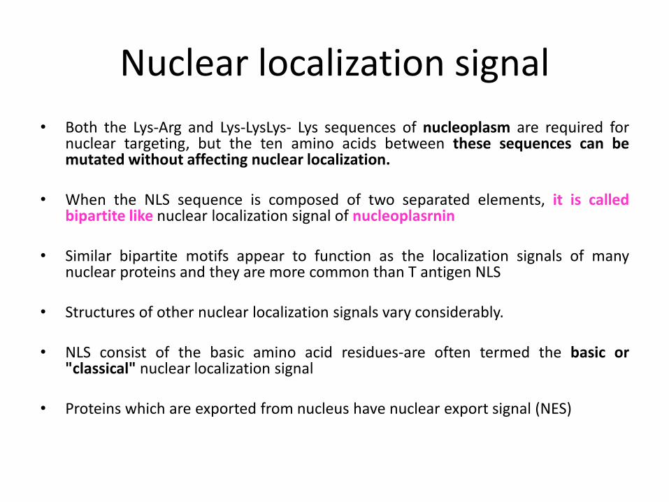
• Both the Lys-Arg and Lys-LysLys- Lys sequences of nucleoplasm are required for nuclear targeting, but the ten amino acids between these sequences can be mutated without affecting nuclear localization.
• When the NLS sequence is composed of two separated elements, it is called bipartite like nuclear localization signal of nucleoplasrnin
• Similar bipartite motifs appear to function as the localization signals of many nuclear proteins and they are more common than T antigen NLS
• Structures of other nuclear localization signals vary considerably.
• NLS consist of the basic amino acid residues-are often termed the basic or "classical" nuclear localization signal
• Proteins which are exported from nucleus have nuclear export signal (NES)

Recognition of nuclear localization signal
• NLS are recognized by proteins that function as nuclear transport receptors.
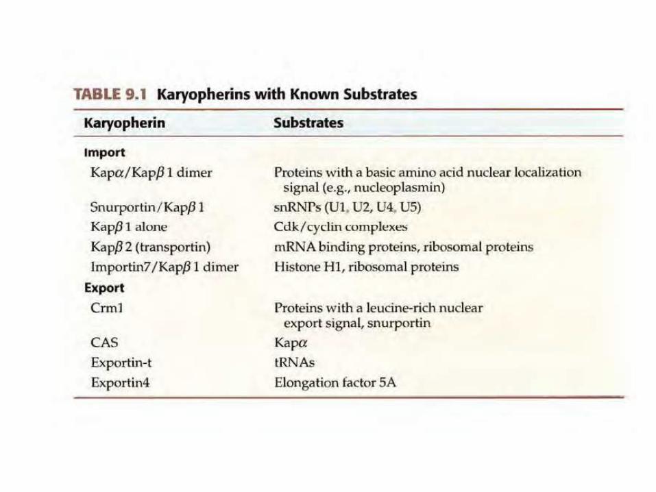
• Most of the Nuclear transport receptors are members of the karyopherin protein family
• Karyopherins are a group of proteins involved in transporting molecules between the cytoplasm and the nucleus of a eukaryotic cell
• They are of two types: importins and exportins
• Importins, transport macromolecules to the nucleus from the cytoplasm, exportins, which transport macromolecules from the nucleus to the cytoplasm
• Some importins (Kap, β l) act with an adapter karyopherin (Kapa) a heterodimer to import proteins containing the basic nuclear localization signal
• Some karyopherins act as importins for one protein and exportins for another.

Regulation of movement of macromolecules/proteins
• Movement of macromolecules through nuclear pore is regulated by a protein called Ran
• Ran also known as GTP-binding nuclear protein encoded by the RAN gene
• Activity of this Ran protein is regulated by GTP binding and hydrolysis.
• Ran is involved in transport into and out of the cell nucleus during interphase and also involved in mitosis
• It is a member of the Ras superfamily.

Ran GTP-binding nuclear protein
• Enzymes that stimulate GTP hydrolysis to GDP are localized to the cytoplasmic side of the nuclear envelope, whereas enzymes that stimulate the exchange of GDP for GTP are localized to the nuclear side
• Consequently, there is high concentration of Ran/GTP in the nucleus Ran GDP in cytoplasm
• This high concentration of Ran/GTP in the nucleus determines the directionality of nuclear transport of cargo proteins.
• The import and export cycles are both driven by a concentration gradient of Ran-GTP that is maintained by the presence of a GEF (guanine-nucleotide exchange factor) in the nucleus and a GAP (GTPase activating protein) in the cytosol. Distribution of Ran/ GTP across the nuclear
envelope

Protein import through the nuclear pore complex
• Ran regulates movement through the nuclear pore by controlling the activity of the nuclear transport receptors.
• Proteins are imported through the NPC when binds with importin via nuclear localization signal of a cargo protein in the cytoplasm
• This importin/ cargo complex then binds to proteins in the cytoplasmic filaments of the NPC, and transport proceeds by sequential binding to specific nuclear pore proteins located further
• At the nuclear side of the pore complex the importin/ cargo complex is disrupted by the binding of Ran/GTP
• This causes a change in the conformation of the importin, releases it into the nucleus.

Protein import through the nuclear pore complex
• The importin-Ran/GTP complex is
then exported back through the nuclear pore complex.
• In the cytoplasm the GTP hydrolyzed to GDP releases the importin in the cytoplasm for another round of transport.
• The Ran/GDP formed in the cytoplasm is then transported back to the nucleus by its own import receptor (a protein called NTF2}, where Ran/GTP is regenerated.
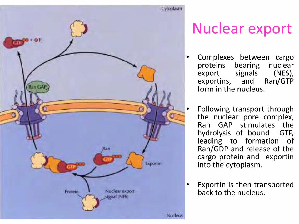
Nuclear export
• Complexes between cargo proteins bearing nuclear export signals (NES), exportins, and Ran/GTP form in the nucleus.
• Following transport through the nuclear pore complex, Ran GAP stimulates the hydrolysis of bound GTP, leading to formation of Ran/GDP and release of the cargo protein and exportin into the cytoplasm.
• Exportin is then transported back to the nucleus.

Overview of Transport Through the Nuclear Pore Complex
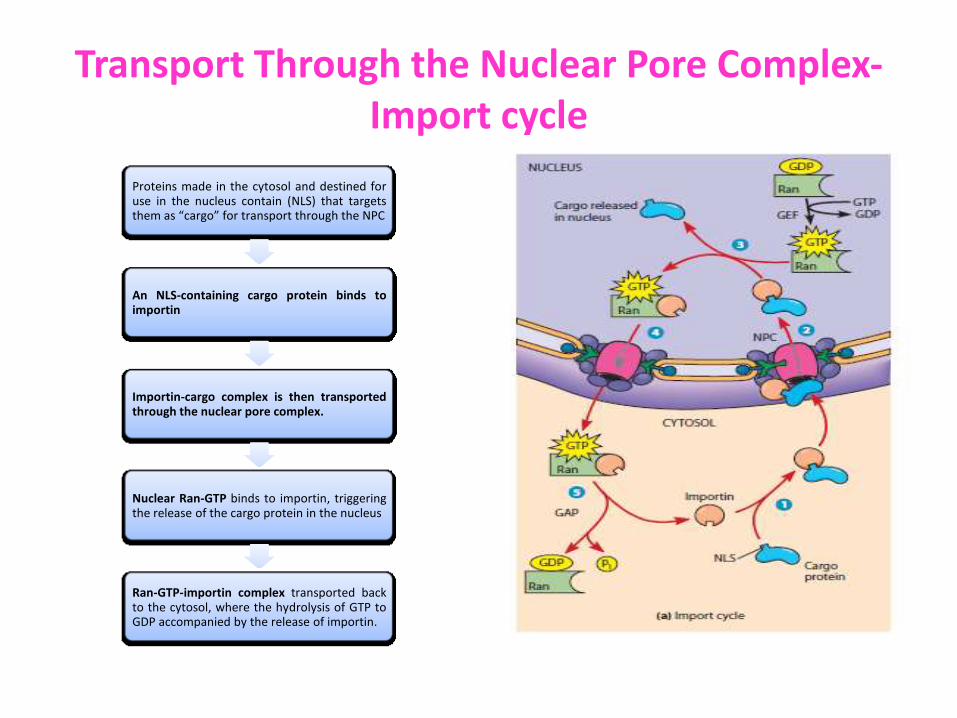
Transport Through the Nuclear Pore Complex-Import cycle
Proteins made in the cytosol and destined for use in the nucleus contain (NLS) that targets them as “cargo” for transport through the NPC
An NLS-containing cargo protein binds to importin
Importin-cargo complex is then transported through the nuclear pore complex.
Nuclear Ran-GTP binds to importin, triggering the release of the cargo protein in the nucleus
Ran-GTP-importin complex transported back to the cytosol, where the hydrolysis of GTP to GDP accompanied by the release of importin.
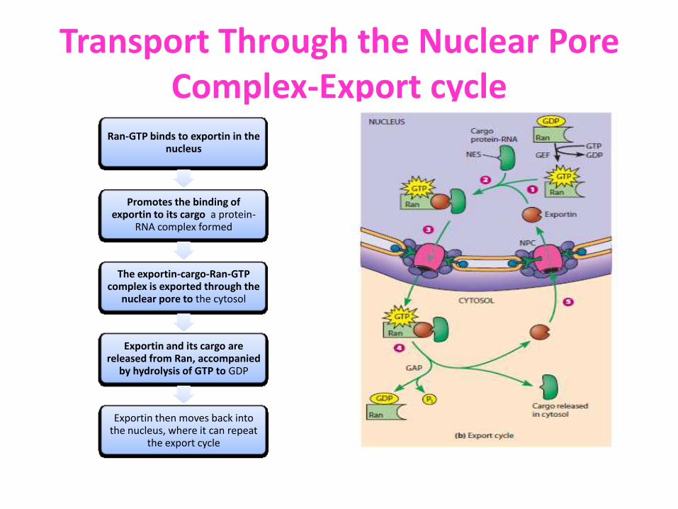
Transport Through the Nuclear Pore Complex-Export cycle
Ran-GTP binds to exportin in the nucleus
Promotes the binding of exportin to its cargo a protein-
RNA complex formed
The exportin-cargo-Ran-GTP complex is exported through the
nuclear pore to the cytosol
Exportin and its cargo are released from Ran, accompanied
by hydrolysis of GTP to GDP
Exportin then moves back into the nucleus, where it can repeat
the export cycle

Regulation of nuclear import of transcription factors
• The transcription factor NF-KB is maintained as an inactive complex with IKB, which masks its nuclear localization sequence (NLS) in the cytoplasm.
• In response to appropriate extracellular signals, IKB is phosphorylated and degraded by proteolysis, allowing the import of NF-KB to the nucleus.
• The yeast transcription factor Pho4 is maintained in the cytoplasm by phosphorylation in the vicinity of its nuclear localization sequence.
• Regulated dephosphorylation exposes the NLS and allows Pho4 to be transported to the nucleus.

Transport of RNAs
• Many proteins are selectively transported from the cytoplasm to the nucleus, most RNAs are exported from the nucleus to the cytoplasm.
• The export of mRNAs, rRNAs, and tRNAs is a critical step in gene expression in eukaryotic cells as protein are synthesized in cytoplasm
• The RNAs export through the NPC is an active, energy-dependent process requiring the transport receptors to interact with the nuclear pore complex
• Karyopherin importins and exportins transport most tRNAs, rRNAs, and small nuclear RNAs in a Ran/GTP-dependent manner
• mRNAs are exported by a complex of two proteins -the "mRNA exporter" one of which is related to the Ran/GDP transporter, NTF2
• Transport of mRNAs appears to be independent of Ran

RNAs transport
• RNAs are transported across the nuclear envelope as ribonucleoprotein complexes (RNPs)
• Ribosomal RNAs are first associated with both ribosomal proteins and specific RNA processing proteins in the nucleolus, and nascent 60S and 40S ribosomal subunits are then transported to the cytoplasm
• Their export from the nucleus is mediated by nuclear export signals present on proteins within the subunit complex
• Pre-mRNAs and mRNAs are associated with a set of at least 20 proteins throughout their processing in the nucleus and eventual transport to the cytoplasm, which is mediated by the mRNA exporter complex after its recruitment to the processed mRNA

Transport of RNAs
• tRNAs are exported from the nucleus by exportin-t, which binds directly to the tRNAs
• In contrast to mRNAs, tRNAs, and rRNAs, which function in the cytoplasm, many small RNAs (snRNAs and snoRNAs) function within the nucleus as components of the RNA processing machinery
• Perhaps surprisingly, snRNAs are initially transported from the nucleus to the cytoplasm, where they associate with proteins to form functional snRNPs and then return to the nucleus
• Transport receptor proteins that bind to the 5' caps of snRNAs appear to be involved in the export of the snRNAs to the cytoplasm.
• In contrast, sequences present on the snRNP proteins are responsible for the transport of snRNPs from the cytoplasm to the nucleus.

Transport of snRNAs between nucleus and cytoplasm
• Small nuclear RNAs (snRNAs) are initially exported from the nucleus to the cytoplasm where they associate with proteins to form snRNPs (Ribonucleoprotein complex).
• The assembled snRNPs are then transported back to the nucleus.
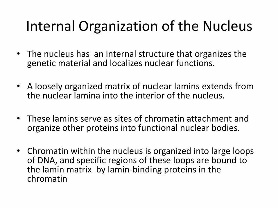
Internal Organization of the Nucleus
• The nucleus has an internal structure that organizes the genetic material and localizes nuclear functions.
• A loosely organized matrix of nuclear lamins extends from the nuclear lamina into the interior of the nucleus.
• These lamins serve as sites of chromatin attachment and organize other proteins into functional nuclear bodies.
• Chromatin within the nucleus is organized into large loops of DNA, and specific regions of these loops are bound to the lamin matrix by lamin-binding proteins in the chromatin
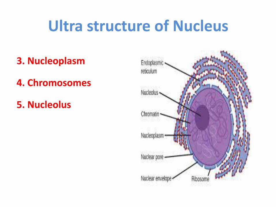
Ultra structure of Nucleus
3. Nucleoplasm
4. Chromosomes
5. Nucleolus
3. Nucleoplasm
• The space between the nuclear envelope and nucleolus is filled by transparent, semisolid granular and slightly acidophilic ground substance or matrix known as nucleoplasm or karyolymph
• Chromatin thread and nucleolus remain suspended in it
• It contains mainly the nucleoproteins but also co ntains nucleic acids, proteins enzymes and minerals

Components of nucleoplasm
Nucleoplasm
components
Nucleic acids
(DNA, RNA)
Proteins
Basic and acidic proteins
Enzymes- DNA polymerase RNA
polymerease, nucleoside phosphorylase
Lipids Minerals (P, K,Ca.Na,
Mg)
a.Basic proteins are nucleoprotamines and histones
b. Acidic proteins-like phosphoproteins

4. Chromosomes and Higher-Order Chromatin Structure
• Chromatin is the complex of the DNA, proteins, RNA which have provided valuable information about the structure of eukaryotic chromosomes.
• The proteins are of two major classes: 1st is basic (positively charged at neutral pH) proteins called histones and 2nd is heterogeneous, largely acidic (negatively charged at neutral pH) group of proteins collectively referred to as non-histone chromosomal proteins
• Before cell division cell’s Chromatin threads corresponding to each individual chromosome are extended dispersed within the nucleus.
• Chromatin of each chromosome has its own discrete location referred to as chromosome territories (Carl Rabl)

Chromosomes and Higher-Order Chromatin Structure
• The nuclear envelope helps organize chromatin by
binding parts of it to the inner nuclear envelope at sites that are closely associated with the nuclear pores
• Chromatin becomes highly condensed during mitosis to form the compact metaphase chromosomes that are distributed to daughter nuclei
• Actively transcribed genes are localized to the periphery of these territories, adjacent to channels separating the chromosomes

• Chromatin in interphase nuclei is organized into looped domains containing approximately 50 to 100 kb of DNA
• These chromatin domains appear to represent discrete functional units, which independently regulate gene expression
A model of chromosome organization-The chromosomes occupy discrete territories

THREE LEVELS OF DNA PACKAGING IN EUKARYOTIC CHROMOSOMES
• When isolated chromatin from interphase cells is examined by electron microscopy, it is found to consist of a series of ellipsoidal beads about 11 nm in diameter and 6.5 nm high) joined by thin threads callled linkers or interbead
• This “bead” or chromatin subunit is called the nucleosome
• The basic structural unit of chromatin, nucleosome the was described by Roger Kornberg in 1974
• This clearly involves an octamer of histone molecules, two each of histones H2a, H2b, H3, and H4.
• Binding of proteins to DNA in nucleosome protects regions of the DNA from nuclease digestion but linkers, or interbead threads of DNA, are susceptible to nuclease attack

1st level PACKAGING -Nucleosome

2nd level of PACKAGING
• As cells enter mitosis, the chromosomes become highly condensed so that they can be distributed to daughter cells
• The second level of condensation involves an additional folding or supercoiling of the 11-nm nucleosome fi ber, to produce the 30-nm chromatin fiber
• Histone H1 is involved in this supercoiling of the 11-nm nucleosome fiber to produce the 30-nm chromatin fiber.

2nd level of PACKAGING

3rd level PACKAGING
• Finally, nonhistone chromosomal proteins form a scaffold that is involved in condensing the 30-nm chromatin fiber into the tightly packed metaphase chromosomes
• This third level of condensation appears to involve the separation of segments of the giant DNA molecules present in eukaryotic chromosomes into independently supercoiled domains or loops

Three levels of condensation are required to package the 103 -105 m of DNA in a eukaryotic chromosome into a metaphase structure a few microns long

Euchromatin and Heterochromatin • The extent of chromatin condensation varies during the life cycle of the cell
• In interphase (nondividing) cells, most of the chromatin called euchromatin
which is relatively decondensed form and distributed throughout the nucleus
• During this period of the cell cycle, genes are transcribed and the DNA is replicated in preparation for cell division
• In contrast to euchromatin about 10% of interphase chromatin called heterochromatin) is in a very highly condensed state that resembles the chromatin of cells undergoing mitosis
• Heterochromatin is transcriptionally inactive and contains highly repeated DNA sequences, such as those present at centromeres and telomeres

Types of heterochromatin
Constitutive heterochromatin
Facultative heterochromatin

Heterochromatin
• These chromatin regions are highly compacted—that is, they are
heterochromatin.
• In electron micrographs, this material appears as a dark irregular layer around the nuclear periphery
• Constitutive heterochromatin, which contains DNA sequences that are generally not transcribed, such as the satellite sequences present at centromeres and the telomere (are attached to the nuclear envelope at times other than during cell division)
• Facultative heterochromatin, which contains sequences that are not transcribed in the cell being examined but are transcribed in other cell types
Cont…
• Most of it seems to be the type called constitutive heterochromatin, which exists
in a highly condensed form at virtually all times in all cells of the organism
• The amount of facultative heterochromatin varies depending on the transcriptional activity of the cell
• The DNA of constitutive heterochromatin consists of simple-sequence repeated DNA short sequences that repeat tandemly and are not transcribed
• Facultative heterochromatin varies with the particular activities carried out by the cell
• Thus, this type of chromatin differs from tissue to tissue and can even vary from time to time within a given cell

5. Nucleolus

Nucleolus
• The most prominent substructure within the nucleus is the nucleolus
• Frits Zernike in 1953 1st saw nucleolus
• It is the site of rRNA transcription and processing, and of ribosome assembly
• The nucleolus is a ribosome production factory, designed to fulfill the need for large-scale production of rRNAs and assembly of the ribosomal subunits
• Recent evidence suggests that nucleoli also have a more general role in RNA modification and that several types of RNA move in and out of the nucleolus at specific stages during their processing

Organization of the Nucleolus and the Ribosomal RNA Genes
• The nucleolus, which is not surrounded by a membrane, is organized around the chromosomal regions that contain the genes for the 5.8S, 18S, and 28S rRNAs
• Eukaryotic ribosome's contain four types of RNA, designated the 5S, 5.8S, 18S, and 28S rRNAs
• The 5.8S, 18S, and 28S rRNAs are transcribed as a single unit within the nucleolus by RNA polymerase I, yielding a 45S ribosomal precursor RNA
Ribosomal RNA genes. Each rRNA gene is a single transcription unit containing the 18S, 5.8S, and 28S rRNAs and transcribed spacer sequences. The rRNA genes are organized in tandem arrays, separated by non transcribed spacer DNA.
Cont… • The 45S pre-rRNA is processed to the 18S rRNA of the 40S (small)
ribosomal subunit and to the 5.8S and 28S rRNAs of the 60S (large) ribosomal subunit
• Transcription of the 5S rRNA, which is also found in the 60S ribosomal subunit, takes place outside of the nucleolus and is catalyzed by RNA polymerase III
• To meet the need for transcription of large numbers of rRNA molecules, all cells contain multiple copies of the rRNA genes.
• The human genome, for example, contains about 200 copies of the gene that encodes the 5.8S, 18S, and 28S rRNAs and approximately 2000 copies of the gene that encodes 5S rRNA.
• The genes for 5.8S, 18S, and 28S rRNAs are clustered in tandem arrays on five different human chromosomes (chromosomes 13, 14, 15, 21, and 22); the 5S rRNA genes are present in a single tandem array on chromosome 1
Cont….
• Morphologically, nucleoli consist of three distinguishable regions: 1. Fibrillar center, 2. Dense fibrillar component 3. Granular component
• These different regions are thought to represent the sites of progressive
stages of rRNA transcription, processing, and ribosome assembly
• The rRNA genes are located in the fibrillar centers, with transcription occurring primarily at the boundary of the fibrillar centers and dense fibrillar component
• Processing of the pre-rRNA is initiated in the dense fibrillar component and continues in the granular component, where the rRNA is assembled with ribosomal proteins to form nearly completed preribosomal subunits, ready for export to the cytoplasm

• Following each cell division, nucleoli form around the chromosomal regions that contain the 5.8S, 18S, and 28S rRNA genes, which are therefore called nucleolar organizing regions
• The formation of nucleoli requires the transcription of 45S pre-rRNA, which appears to lead to the fusion of small prenucleolar bodies that contain processing factors and other components of the nucleolus
• In most cells, the initially separate nucleoli then fuse to form a single nucleolus.
• The size of the nucleolus depends on the metabolic activity of the cell, with large nucleoli found in cells that are actively engaged in protein synthesis
Transcription and Processing of rRNA
• The processing of pre-rRNA requires the action of both proteins and RNAs that are localized to the nucleolus.
• Nucleoli contain a large number (about 200) of small nucleolar RNAs (snoRNAs) that function in pre-rRNA processing.
• The snoRNAs are complexed with proteins, forming snoRNPs.
• Individual snoRNPs consist of single snoRNAs associated with eight to ten proteins.
• The snoRNPs then assemble on the pre-rRNA to form processing complexes

Processing of pre-rRNA
• snoRNAs are responsible for the cleavages of pre-rRNA into 18S, 5.8S, and 28S products
• Most abundant nucleolar snoRNA is U3, is required for the initial cleavage of pre-rRNA within the 5′ external transcribed spacer sequences
• Similarly, U8 snoRNA is responsible for cleavage of pre-rRNA to 5.8S and 28S rRNAs, and U22 snoRNA is responsible for cleavage of pre-rRNA to 18S rRNA Processing of pre-rRNA. The 45S pre-rRNA
transcript contains external transcribed spacers (ETS) at both ends and internal transcribed spacers (ITS) between the sequences of 18S, 5.8S, and 28S rRNAs

Role of snoRNAs in base modification of pre-rRNA.
• Majority of snoRNAs, however, function to direct the specific base modifications of pre-rRNA, including the methylation of specific ribose residues and the formation of pseudouridines
• Most of the snoRNAs contain short sequences complementary to 18S or 28S rRNA
• These regions of complementarity include the sites of base modification in the rRNA.
• By base pairing with specific regions of the pre-rRNA, the snoRNAs act as guide RNAs that target the enzymes responsible for ribose methylation or pseudouridylation to the correct site on the pre-rRNA molecule
The snoRNAs contain short sequences complementary to rRNA. Base pairing between snoRNAs and pre-rRNA targets the enzymes that catalyze base modification (e.g., methylation) to the appropriate sites on pre-rRNA.

Ribosome Assembly
• The formation of ribosomes involves the assembly of the ribosomal precursor RNA with both ribosomal proteins and 5S rRNA
• The genes that encode ribosomal proteins are transcribed outside of the nucleolus by RNA polymerase II, yielding mRNAs that are translated on cytoplasmic ribosomes then transported f to the nucleolus, where they are assembled with rRNAs to form preribosomal particles
• Although the genes for 5S rRNA are also transcribed outside of the nucleolus, by RNA polymerase III, and are assembled into preribosomal particles
• The association of ribosomal proteins with rRNA begins while the pre-rRNA is still being synthesized, and more than half of the ribosomal proteins are complexed with the pre-rRNA prior to its cleavage
• The remaining ribosomal proteins and the 5S rRNA are incorporated into preribosomal particles as cleavage of the pre-rRNA proceeds
• Most of the preribosomal particles in the nucleolus represent precursors to the large subunit. The final stages of ribosome maturation follow the export of preribosomal particles to the cytoplasm, forming the active 40S and 60S subunits of eukaryotic ribosome
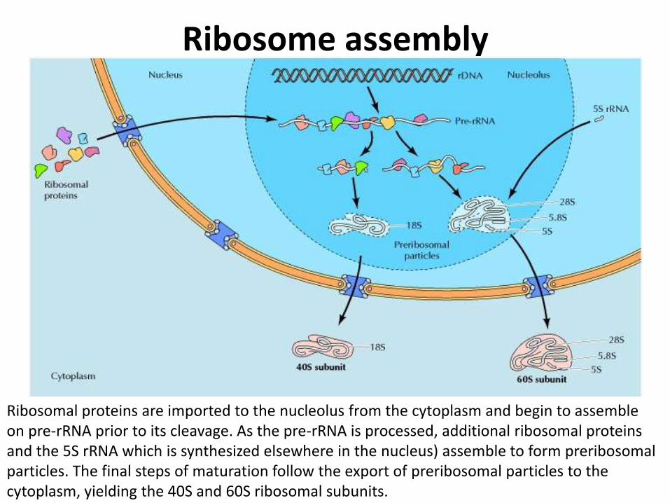
Ribosome assembly
Ribosomal proteins are imported to the nucleolus from the cytoplasm and begin to assemble on pre-rRNA prior to its cleavage. As the pre-rRNA is processed, additional ribosomal proteins and the 5S rRNA which is synthesized elsewhere in the nucleus) assemble to form preribosomal particles. The final steps of maturation follow the export of preribosomal particles to the cytoplasm, yielding the 40S and 60S ribosomal subunits.

Difference Between Nucleus and Nucleolus
Nucleus Nucleolus
1. Large in size. 1. Very small in size
2.Bounded by nuclear envelope 2. No limiting membrane
3. Contains chromosomes 3. Doesn’t hold chromosomes
4. Rich in DNA 4. It is rich in RNA

References
• Snustad, D. P., & Simmons, M. J. (2015). Principles of genetics. John Wiley & Sons.
• Cooper, G. M., & Hausman, R. E. (2004). The cell: Molecular approach. Medicinska naklada.